Article

Various Community Structures of Root-Associated Bacteria, Archaea, and Fungi of Spartina alterniflora


1
College of Humanities and Arts, Xi’an International University, Xi’an 710077, China
2
Marine Science and Technology College, Zhejiang Ocean University, Zhoushan 316022, China
*
Author to whom correspondence should be addressed.
Diversity 2026, 18(4), 211; https://doi.org/10.3390/d18040211
Submission received: 25 February 2026 / Revised: 26 March 2026 / Accepted: 27 March 2026 / Published: 5 April 2026

Abstract

Invasion by Spartina alterniflora has detrimental effects on existing ecosystems. Studies have shown that microorganisms can control plant growth and development. However, the root-associated community structures of bacteria, archaea, and fungi of S. alterniflora have rarely been investigated. Here, we applied metagenomics to reveal the bacterial, archaeal, and fungal communities across four root compartments, including the bulk soil, rhizosphere, rhizoplane, and endosphere. Our findings revealed the variation in different community structures. The bacterial and fungal communities exhibited greater potential environmental flexibility than the archaeal community. The endosphere environment had the simplest microbial networks and highest stability. Additionally, we identified root-exuded metabolites from S. alterniflora, which may influence microbial community assembly. Our results indicate that the rhizoplane plays a crucial role in controlling microbial entry into the root, selectively recruiting beneficial microbes for plant growth and colonization, thereby impacting nutrient cycling and plant health. This study provides insights into microbial diversity and function within the S. alterniflora root zone and suggests potential microbial-based strategies for managing this invasive species.

1. Introduction

Spartina alterniflora is a plant belonging to the family Poaceae and the genus Spartina [1]. Native to the Atlantic coast of North America, the species was introduced to China in the late 1970s for erosion control, subsequently becoming invasive and rapidly spreading along coastal areas [2]. Although S. alterniflora provides certain benefits such as soil stabilization and carbon sequestration, its uncontrolled rapid growth significantly impacts the existing ecosystem. The invasion of S. alterniflora can cause ecological shifts, including alterations in sedimentary environments, impacts on native species, and changes in nutrient cycling in the invaded ecosystems [3,4].
The ecological environment changes induced by the invasion of S. alterniflora are manifested in numerous aspects, including its occupation of native plant niches, a decline in biodiversity [1], alterations in the structure and nutrient content of sediments [5], and modifications to the elemental cycles of carbon, nitrogen, sulfur, and other elements in the soil [6]. It is noteworthy that S. alterniflora also exerts significant impacts on soil microbial diversity and community composition. In recent years, studies have indicated that the invasion of S. alterniflora can lead to a reduction in soil bacterial diversity, while having no significant effect on fungal diversity [2]. Invasion may also result in the simplification of soil microbial symbiotic networks [7].
The relationship between plants and microorganisms is one of interaction, rather than the unilateral influence of plants on the relevant microbial communities [8]. Root-associated microbial communities play a critical role in plant nutrition, growth, and stress tolerance. Recent studies have shed light on the dynamics and functions of these microbial communities in the root zone, providing insights into their ecological significance [9]. Durán et al.’s study [10] revealed that interactions between microbes and plants enhance the healthy growth of Arabidopsis. In contrast, Kong et al.’s research [11] indicates that the presence of pathogenic fungi within roots may suppress plant growth. Additionally, some bacteria and archaea can fix atmospheric nitrogen, aiding plants in the absorption of nutrients [12,13,14]. These studies illustrate that, to some extent, microorganisms can control plant growth and development.
These studies collectively demonstrate that microorganisms can directly or indirectly influence plant health through pathogenicity or by providing nutrients to plants. Manipulating these communities can affect the invasiveness of plants and reduce impacts on invaded ecosystems [9]. In the case of S. alterniflora, the root microbiome may represent a potential management strategy. To achieve this, a detailed understanding of the structure and function of microbial communities in different compartments is required, including the microcosms of the rhizosphere (RS), rhizoplane (RP), and endosphere (ES) of the S. alterniflora root zone. Current research on the root-associated microbiome of S. alterniflora primarily focuses on bacterial communities, emphasizing the relationship between microbes and whole plant roots [9,15,16]. Few studies have simultaneously examined the distribution and characteristics of archaeal, bacterial, and fungal communities in different compartments of the S. alterniflora root. In-depth research on these aspects will help us understand the specific roles of these three types of microorganisms in the roots of S. alterniflora, thereby laying a theoretical foundation for the use of microbial methods to prevent S. alterniflora invasion.
In this study, we aim to explore the diversity, function, and assembly of root-associated microbial communities in four continuous fine-scale root compartments (bulk soil, rhizosphere, rhizoplane, and endosphere) of S. alterniflora, including systematically characterizing the shifts in microbial community composition, diversity, and assembly processes across four root-associated compartments and comparing the responses of archaeal, bacterial, and fungal communities to this spatial gradient, by using 16S rRNA and internal transcribed spacer (ITS) gene amplicon and metagenomic sequencing. By integrating compartment-based and taxon-based comparisons, we sought to provide a comprehensive view of the root-associated microbiome structure in S. alterniflora, which may aid in identifying potential microbial-based strategies for the management of this invasive species.

2. Materials and Methods

2.1. Sampling Sites and Sample Collection

The sampling site was located at Xiaogan Island of Zhoushan City (29°58′14.75″ N, 122°90′50.72″ E), Zhejiang, China, where salt marsh plants are dominated by S. alterniflora. Five S. alterniflora plants with a distance of about 1 m were collected in March 2024 as five independent biological replicates. The root-associated sample of each plant was fractionated into 4 compartments: bulk soil (BS), RS, RP, and ES.
The method for compartment fractionation had been described by Edwards et al. [17]. Briefly, soil that could be shaken off by hand was collected accordingly as the BS sample. The remaining root samples were placed in a sterile flask with 50 mL of sterile phosphate-buffered saline solution at room temperature, and RS samples were separated from the roots via shaking by using a vortex oscillator (amplitude 5 mm, power 60 W; Hengao Technology, Tianjin, China) at 800 rpm for 3 min. The resulting suspensions were transferred into 50 mL sterile Falcon tubes and then centrifuged at 2000 rpm and 10 °C for 1 min. The remaining sediments after removing the supernatant were collected as the RS samples. This operation was repeated three times to fully remove RS from the roots.
The remaining root samples were used for the further separation of RP by ultrasonic oscillation for 1 min at 50–60 Hz (Branson Ultrasonics; Chicago, IN, USA) in a 50 mL Falcon tube with 30 mL PBS. The microorganisms that were removed from the roots by ultrasonic oscillation were collected by using the filtration method through 0.22 µm pore size membranes (Whatman; Maidstone, UK). Ultrasonic treatment and filtration were repeated three times to ensure that all microbes were removed from the root surface. The remaining roots were used for DNA extraction of ES samples.

2.2. Extraction of Root Exudates

After collecting S. alterniflora, the roots were rinsed with sterile water, and then placed in an aerated hydroponic solution under ample light to simulate natural conditions for three days. The roots were then immersed in 1.5 L sterile Milli-Q water and stirred at 150 rpm on a vortex oscillator at 18 °C in the dark for 4 h. The exudate was filtered through a 0.22 µm membrane. The samples were lyophilized and stored for subsequent non-targeted metabolomics analysis. The Vanquish UHPLC system (Thermo Fisher; Waltham, MA, USA) was used for UHPLC-MS/MS analyses, coupled with an Orbitrap Q ExactiveTMHF mass spectrometer (Thermo Fisher; Waltham, MA, USA). The samples were analyzed by using a Hypersil Goldcolumn (100 × 2.1 mm, 1.9 μm). The flow rate was set as 0.2 mL/min. The eluents for the positive and negative polarity modes were eluent A (0.1% FA in water) and eluent B (methanol) with a linear gradient as follows: 2% B, 1.5 min; 2–85% B, 3 min; 85–100% B, 10 min; 100–2% B, 10.1 min; 2% B, 12 min. Positive/negative polarity mode was used using a Q Exactive TMHF mass spectrometer (Thermo Fisher; Waltham, MA, USA) with a spray voltage of 3.5 kV, sheath gas flow rate of 35 psi, capillary temperature of 320 °C, S-lens RF level of 60, aux gas flow rate of 10 L/min, and aux gas heater temperature of 350 °C. The UHPLC-MS/MS data were processed by using Compound Discoverer 3.3 (Thermo Fisher; Waltham, MA, USA) for performing peak alignment, picking, and quantitation. The peak area was corrected with the first sample. The actual mass tolerance, signal intensity tolerance, and minimum intensity were set as 5 ppm, 30%, and 1% of the base peak, respectively. The peak intensities were normalized to the total spectral intensity, then normalized data were used to predict the molecular formula according to additive ions, fragment ions, and molecular ion peaks. All peaks were matched against mzCloud (https://www.mzcloud.org/ (accessed on 13 May 2024)), mzVault and MassList databases.

2.3. DNA Extraction, Amplification, and Sequencing

About 0.5 g of BS and RS soils was used for DNA extraction using the MoBio PowerSoil DNA Isolation Kit (MoBio; Carlsbad, CA, USA). The DNA from RP and ES was extracted using the MoBio Power Water DNA Isolation Kit (MoBio; Carlsbad, CA, USA) and the MoBio Power Plant DNA Isolation Kit (MoBio; Carlsbad, CA, USA), respectively. The DNA quality was assessed using an ND 2000 Spectrophotometer (Thermo Fisher; Waltham, MA, USA).
Primer pairs were utilized to amplify the V3−V4 region of 16S rRNA genes (5′-ACTCCTACGGGAGGCAGCA-3′; 5′-GGACTACHVGGGTWTCTAAT-3′) and the fungal ITS1 region (5′-CTTGGTCATTTAGAGGAAGTAA-3′; 5′-GCTGCGTTCTTCATCGATGC-3′). PCR reactions were performed in a mixture containing 4 μL of 5× FastPfu Buffer, 0.8 μL of each primer (5 μM), 2 μL of 2.5 mM dNTPs, 0.4 μL of FastPfu Polymerase, and 10 ng of DNA. PCR program was as follows: 1 cycle of denaturation at 95 °C for 5 min; 15 cycles of denaturation at 95 °C for 1 min, annealing at 50 °C for 1 min, and extension at 72 °C for 1 min; 1 cycle of a final extension at 72 °C for 7 min. The PCR products from the first step were purified using VAHTSTM DNA Clean Beads (Vazyme, Nanjing, China). Then, the second round of PCR was performed in a mixture containing 20 μL 2× Phusion HF MM, 10 μM of each primer, 8 μL ddH2O, and 10 μL PCR products from the first step. The second-step PCR conditions were as follows: 1 cycle of denaturation at 98 °C for 30 s; 10 cycles of denaturation at 98 °C for 10 s, annealing at 65 °C for 30 s, and extension at 72 °C for 30 s; 1 cycle of extension at 72 °C for 5 min. Quant-iT™ dsDNA HS Reagent (Thermo Fisher; Waltham, MA, USA) was used for quantifying all PCR products. High-throughput sequencing was performed by using the Illumina NovaSeq 6000 platform (2 × 250 paired ends; San Diego, CA, USA) at Biozeron Biotechnology Co., Ltd., Shanghai, China.

2.4. Sequence Analysis of 16S rRNA and ITS1 Gene Amplicons

Raw fastq files were demultiplexed using custom Perl scripts based on sample-specific barcode sequences, applying the following criteria: (i) Reads were trimmed using a 10 bp sliding window, with truncation occurring at positions where the average quality fell below 20; reads shorter than 50 bp after trimming were removed. (ii) Barcodes were required to match exactly, whereas reads containing ambiguous bases or exhibiting up to two mismatches in primer regions were discarded. (iii) Paired-end reads were merged only when the overlapping region was at least 10 bp; unassembled reads were excluded, and the DADA2 algorithm (http://benjjneb.github.io/dada2/, accessed on 5 January 2024) via QIIME 2 (https://qiime2.org/, accessed on 5 January 2024) was used to discern insertions, deletions, and substitutions of sequences. The paired-end reads were then trimmed and filtered, allowing for a maximum of two expected errors per read (maxEE = 2). After merging the sequences and filtering out chimeras, the phylogenetic affiliations of each 16S rRNA amplicon sequence variant (ASV) were scrutinized against the Silva (SSU132) 16S rRNA database (https://www.arb-silva.de/, accessed on 17 December 2023) by using the RDP Classifier (https://github.com/topics/rdpclassifier, accessed on 17 December 2023) with a confidence threshold of 70%. ITS sequences were analyzed with ITSx (https://github.com/USDA-ARS-GBRU/itsxpress, accessed on 28 December 2023), followed by clustering at 80% sequence identity via UPARSE61 (https://drive5.com/uparse/, accessed on 28 December 2023). Putative chimeras among fungal ASVs were identified using Uchime (http://drive5.com/uchime, accessed on 28 December 2023) with a dedicated reference database.

2.5. Shotgun Metagenome Sequencing Data Analysis

Metagenomic shotgun libraries were prepared and sequenced by Shanghai Biozeron Biological Technology Co., Ltd. (Shanghai, China). Briefly, the TruSeq DNA Library Preparation Kit (catalog no: FC-121-2001, Illumina, San Diego, CA, USA) was used for library construction per sample, and library concentrations were measured using the High-Sensitivity Double-Stranded DNA Kit on a Qubit Fluorometer (Thermo Fisher; Waltham, MA, USA). All samples were sequenced on an NGS platform in paired-end 150 bp (PE150) mode. Raw reads were quality-trimmed with Trimmomatic (http://www.usadellab.org/cms/index.php?page=trimmomatic, accessed on 30 July 2024) to eliminate adapter contamination and low-quality sequences. Reads meeting quality control standards were mapped against the S. alterniflora genome by the BWA mem algorithm with the parameters of -M -k 32 -t 16 (http://bio-bwa.sourceforge.net/bwa.shtml, accessed on 30 July 2024). Reads following removal of host-genome contamination and low-quality data were used for the further analysis as clean reads. The metagenomic assembly was performed using Megahit v1.2.9 (https://github.com/voutcn/megahit, accessed on 30 July 2024) in default mode. Open reading frames were predicted from assembled metagenomes using MetaGeneMark v.2.10 (http://topaz.gatech.edu/GeneMark, accessed on 3 August 2024). A non-redundant gene catalog (Unigenes) was generated with CD-HIT v.4.5.867 (https://www.bioinformatics.org/cd-hit/, accessed on 3 August 2024), and quality control was performed using SoapAligner v.2.21 (http://github.com/ShujiaHuang/SOAPaligner, accessed on 3 August 2024). Taxonomic and functional annotation was conducted with DIAMOND (https://github.com/bbuchfink/diamond, accessed on 23 August 2024; blastp, e-value ≤ 1 × 10⁻⁵) against the NR (https://ftp.ncbi.nlm.nih.gov/blast/db/FASTA/, accessed on 23 August 2024), KEGG68 (http://www.genome.jp/kegg/pathway.html, accessed on 23 August 2024), CAZy (http://www.cazy.org/, accessed on 23 August 2024), and EggNOG70 (http://eggnog.embl.de/version_4.0.beta/, accessed on 23 August 2024) databases. Gene abundances were normalized into transcripts per million (TPM) counts.

2.6. Statistical Analysis

The rarefaction analysis was performed based on Mothur (version 1.21.1) to calculate diversity indices. Bray–Curtis distances were calculated with normalized ASV tables. Alpha-diversity indices of Shannon, phylogenetic diversity (PD) faith, Pielou’s evenness (Pielou J), and Chao1 were calculated using the R package Vegan (version 2.7.3). Principal coordinate analysis (PCoA) for beta diversity analysis among compartments was performed using the R package Vegan (version 2.7.3). Additionally, permutational MANOVA was performed by using Vegan’s function adonis() to calculate effect sizes and significances of β-diversity. Student’s t-tests were conducted by using R 4.2.2 to test significant differences in relative abundance among compartments. Bonferroni correction was used to adjust the significance threshold for all pairwise t-tests (adjusted α = 0.05/number of comparisons). Venn diagrams were drawn using the VennDiagram package (version 1.8.2) in R 4.2.2 to analyze overlapped and unique ASVs. The R package psych (version 1.4.1) and Gephi (https://gephi.org, accessed on 5 September 2024) were applied to visualize the network diagrams. Network modularity analysis was conducted using the default parameters in Gephi, and network topology features, such as clustering coefficients, degree, and modularity index, were calculated accordingly. The average variation degree (AVD), an index that quantifies the community stability by calculating the degree of variation in species abundance based on high-throughput sequencing data, was calculated using R 4.2.2. Significant differences in alpha-diversity indices and AVD among compartments were tested using Kruskal–Wallis in R 4.2.2.
Niche breadth was analyzed by using the R package phyloseq (version 3.22). All statistical analyses were visualized using R packages, including psych (version 1.4.1), reshape2 (version 1.4.5), picante (version 1.8.2), ggplot2 (version 4.0.2), Vegan (version 2.7.3), ggsignif (version 0.6.4), and Cairo (version 1.6.2). We employed phylogenetic null models following established procedures to assess the relative importance of deterministic and stochastic processes in community assembly. Specifically, we calculated the β-nearest taxon index (βNTI) using a phylogeny shuffle null model with 999 randomizations (picante R package), which randomizes phylogenetic relationships while keeping species occurrence and richness unchanged. βNTI values < −2 or >+2 indicate significant deviations from the null expectation, reflecting homogeneous or variable selection (deterministic processes), respectively, whereas values between −2 and +2 suggest stochastic processes dominate. For communities with |βNTI| < 2, we further calculated the Raup–Crick metric based on Bray–Curtis dissimilarity (RCbray) using a taxon shuffle null model (999 randomizations) to distinguish homogenizing dispersal (RCbray < −0.95), dispersal limitation (RCbray > +0.95), and undominated processes (i.e., drift; |RCbray| ≤ 0.95). We implemented all null model analyses in R 4.2.2 using the picante (version 1.8.2) and iCAMP packages (version 1.2.8).

3. Results

3.1. Compositions of Microbial Communities and Exudates of the Root Compartments of S. alterniflora

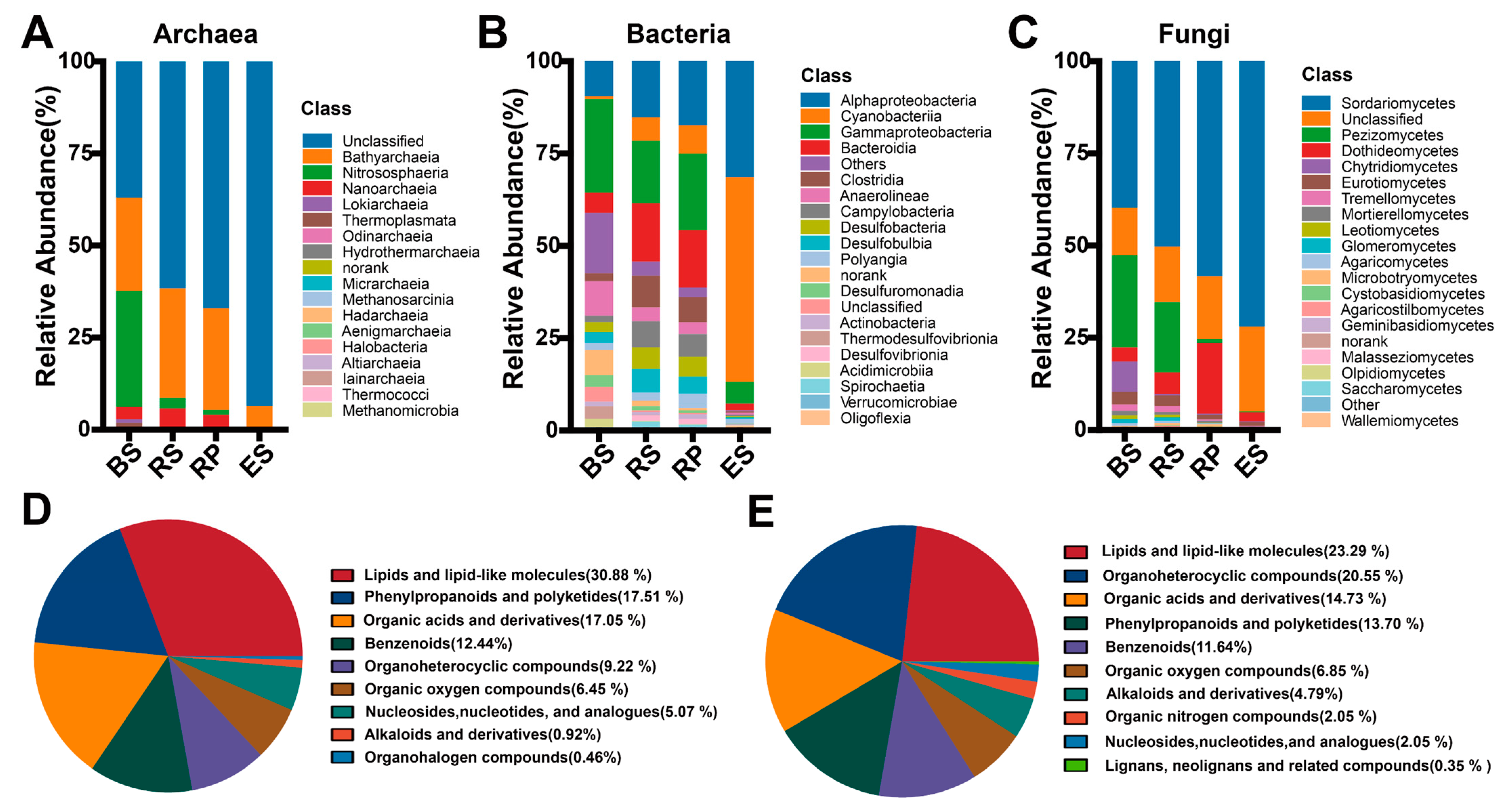
Except for the unidentified taxa, the components of bacterial, archaeal, and fungal communities varied across the root compartments of S. alterniflora (Figure 1A–C). Nitrososphaeria was the dominant archaeal phylum in the BS samples, whereas Bathyarchaeia was the dominant archaeal phylum in the RS, RP, and ES samples. Gammaproteobacteria was the dominant bacterial phylum in the BS and RP samples, Alphaproteobacteria was the dominant bacterial phylum in the RS samples, and Cyanobacteria was the dominant bacterial phylum in the ES samples. Sordariomycetes was the dominant fungal phylum in all samples.
Multiple metabolites were identified in S. alterniflora root exudates (Figure 1D,E). The most abundant metabolites identified with the negative module included lipids and lipid-like molecules (30.88%), phenylpropanoids and polyketides (17.51%), organic acids and derivatives (17.05%), benzenoids (12.44%) and organoheterocyclic compounds (9.22%), and those identified with the positive module were lipids and lipid-like molecules (23.29%), organoheterocyclic compounds (20.5%), organic acids and derivatives (14.73%), phenylpropanoids and polyketides (13.70%), and benzenoids (11.64%).

3.2. Analysis of Niche Breadth and Community Assembly Process of Archaeal, Bacterial and Fungal Communities

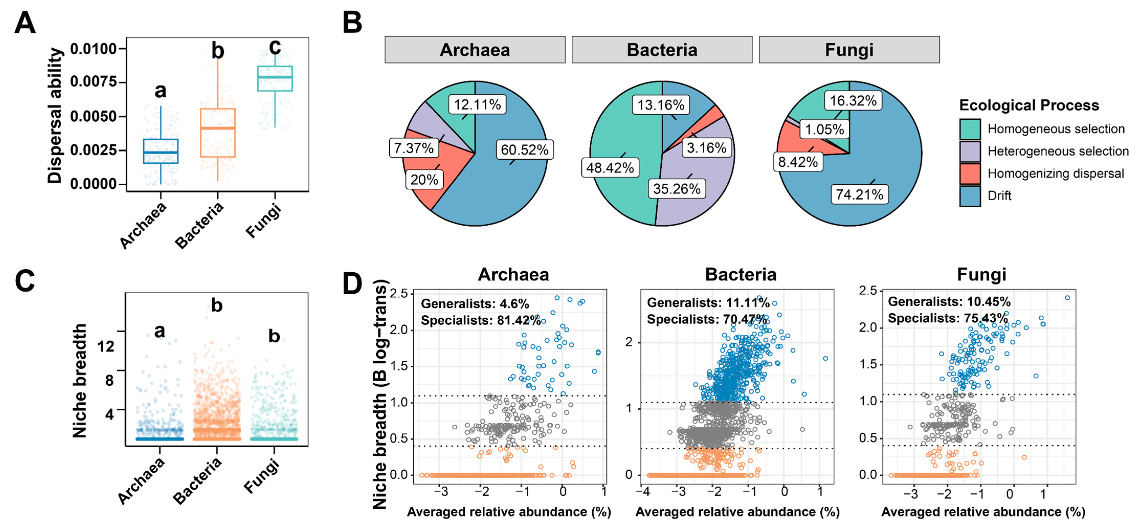
Significant differences were observed in the dispersal ability, niche width, and proportion of specialists among the communities of archaea, bacteria, and fungi. The archaeal communities had the lowest dispersal ability values, followed by bacteria and fungi (p < 0.05, Student’s t-test; Figure 2A). The niche widths of bacterial and fungal communities were higher than in the archaeal community (p < 0.05, Student’s t-test; Figure 2C). The proportion of specialists was highest in the archaeal community, followed by the fungal and bacterial communities (p < 0.05, Student’s t-test; Figure 2D). The ecological process analysis revealed that the homogeneous selection in deterministic processes contributed 48.42% to the community assembly for bacteria, but only contributed 12.11% and 16.32% to archaea and fungi, respectively. Drift was the ecological process with the highest contribution to archaea and fungi, accounting for 60.53% and 74.21%, respectively (Figure 2B).

3.3. Diversity of Archaea, Bacterial and Fungal Microbial Communities Among Four S. alterniflora Root Compartments

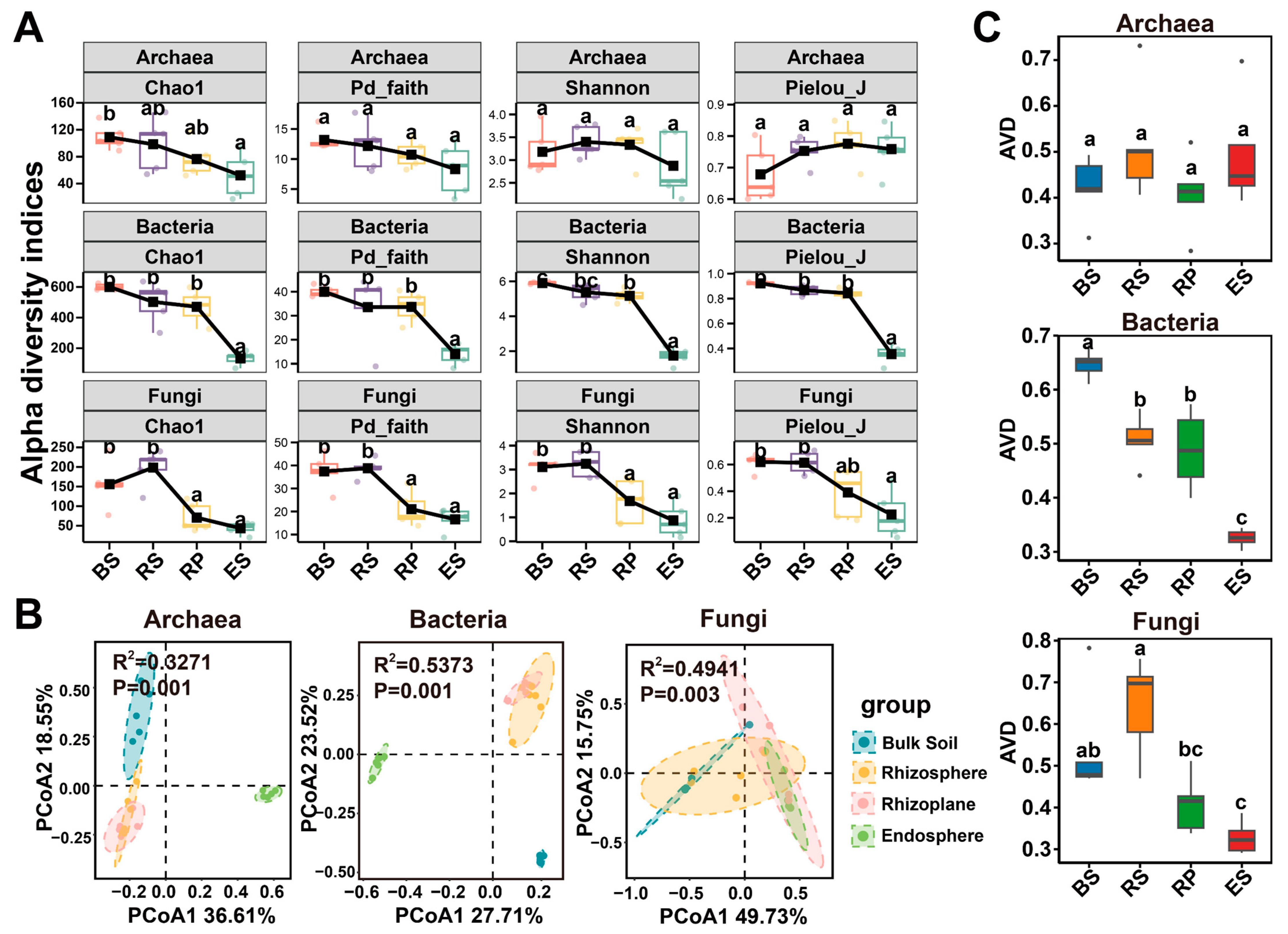
The lowest Chao1, Pd faith, Pielou_J and Shannon indices were observed in the ES for both bacterial and fungal communities, while those of archaeal community showed no significant differences among four root compartments (Figure 3A). The BS exhibited a higher bacterial diversity than the RS and RP, while fungal diversity indices were similar among the BS and RS (p < 0.05, Kruskal–Wallis test). PCoA showed that archaea, bacterial, and fungal communities in the four root compartments were well-separated (p < 0.05, MANOVA test; Figure 3B). No significant difference in AVD values was detected in archaeal community across four root compartments (p > 0.05, Kruskal–Wallis test). The AVD values of bacterial and fungal communities generally showed a decreasing trend from the BS to ES samples (p < 0.05, Kruskal–Wallis test). The network topology parameters showed that the number of nodes, average degrees, and average clustering coefficients of bacterial and fungal communities generally decrease from the BS to ES (Supplementary Figure S1).

3.4. Increases/Decreases in the Number of Microbial ASVs Among Four S. alterniflora Root Compartments

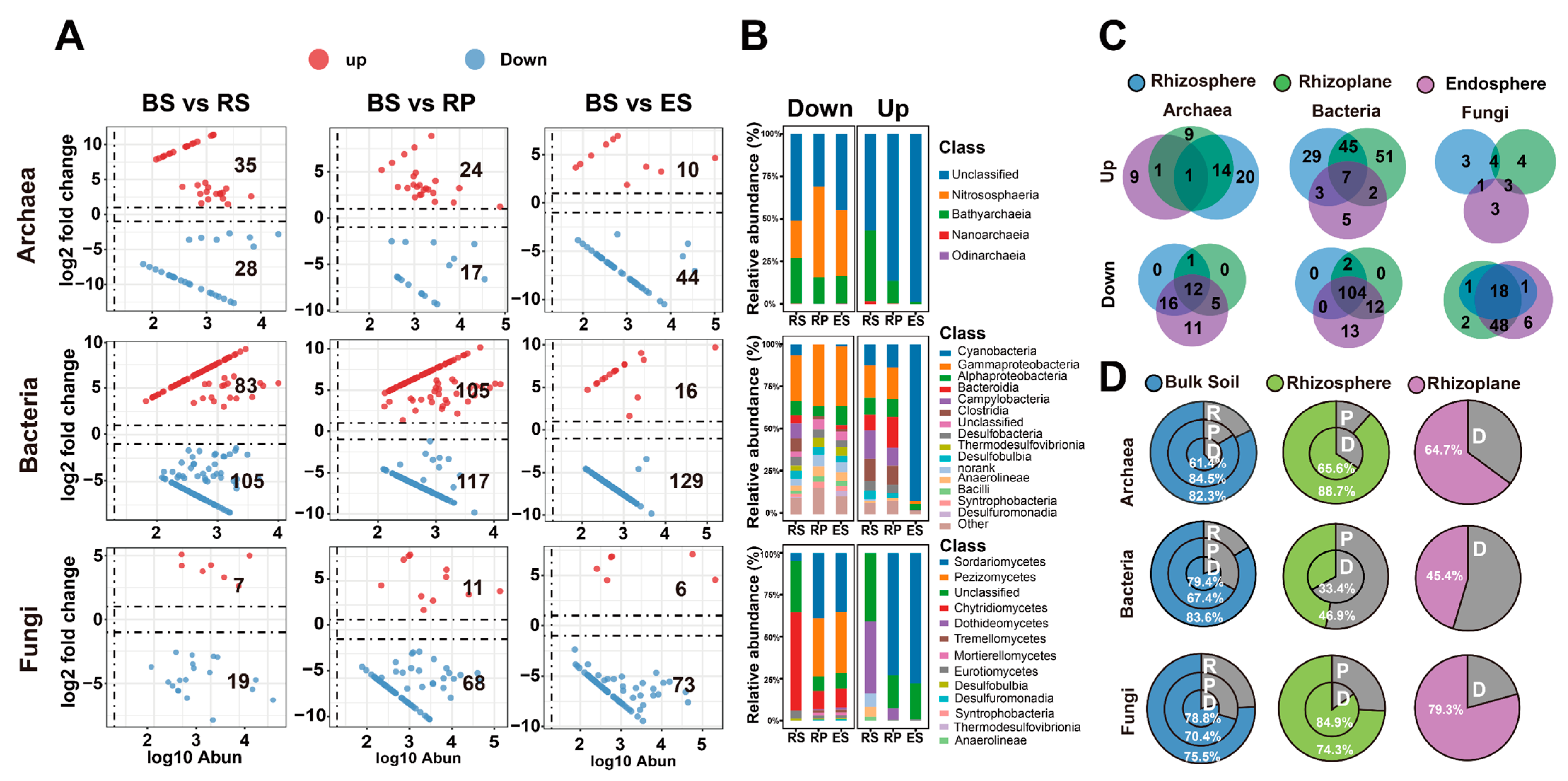
In the transition process from BS to RS, RP and ES, the number of ASVs in each compartment of bacterial and fungal communities underwent similar changes, with the abundance-increased ASVs being less than abundance-decreased ASVs. In the archaeal community, ASV changes from BS to ES were similar to those in bacterial and fungal communities, but during the transition from BS to RS and RP, the number of abundance-increased ASVs exceeded that of abundance-decreased ASVs. ES had the fewest abundance-increased ASVs and the most abundance-decreased ASVs across the three microbial communities (Figure 4A). Almost all archaeal ASVs enriched in the ES were from unclassified taxa, whereas the relative abundance of Nitrososphaeria consistently decreased from the outer root layers to ES. The relative abundance of Cyanobacteria increased upon entering the ES. For fungi, Chytridiomycetes showed a decreased in relative abundance during the transition from BS to ES (Figure 4B).
Notable overlaps were observed in the abundance-increased and -decreased ASVs of archaea, bacteria, and fungi across different compartments (Figure 4C). Among the 35 archaeal ASVs, 83 bacterial ASVs, and 7 fungal ASVs that increased in RS, 15, 55, and 5 ASVs, respectively, also exhibited an increase in either the RP or ES communities or both. The proportions of these ASVs were 42.9%, 66.3%, and 71.4%, respectively. In addition to these overlaps, all ASVs that decreased in RS also declined in RP and/or ES. The majority of bacterial ASVs present in ES were also found in BS (79.4%), with fewer being present in RP (45.5%). Furthermore, no difference was observed in the proportions of archaeal and fungal ASVs from ES that overlapped with those in BS and RP (archaea: BS—61.4%, RP—64.7%; fungi: BS—78.8%, RP—79.3%; Figure 4D).

3.5. The Relative Abundances of Function Genes of Root-Associated Microorganisms in the S. alterniflora Root Compartments

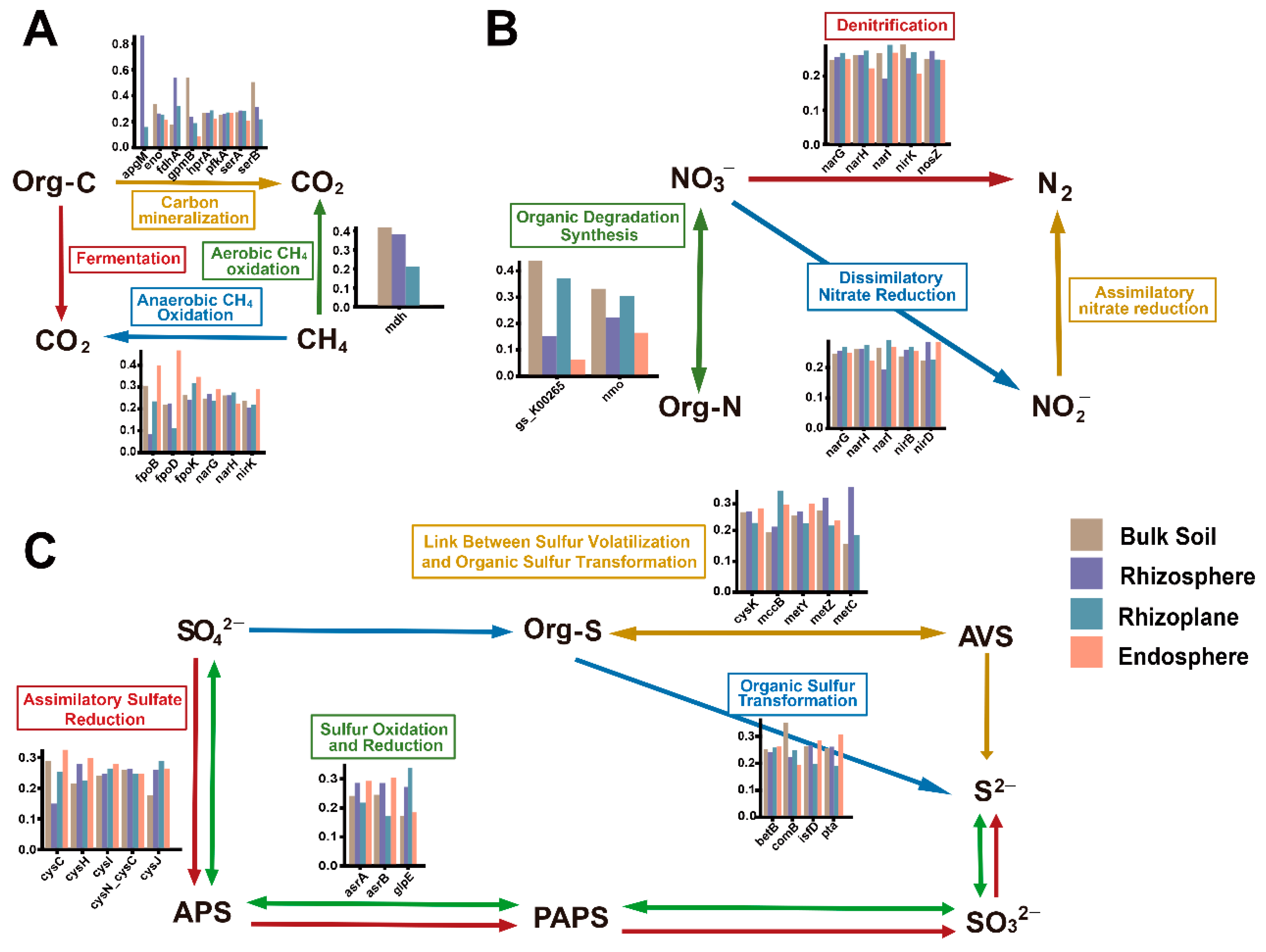
For the carbon cycle, the TPM abundances of anaerobic methane oxidation genes (fpoB/D/K, narG/H, and nirk) were highest in the ES, except for narH. Aerobic methane oxidation genes were absent in the ES (Figure 5A). Genes involved in organic nitrogen degradation and synthesis, denitrification, and NO3 reduction were detected in ES, BS, RS and RP compartments (Figure 5B). Additionally, for sulfur cycle genes, the TPM abundances of cysC/H/I genes were higher in the ES than BS, RS and RP (Figure 5C). In the ES, the asrA/B genes, which reduce sulfite to sulfide, were prevalent, while the glpE gene, implicated in thiosulfate oxidation, was less abundant. Organic sulfur transformation-related genes (betB, comB, isfD, and pta) exhibited inconsistent distribution across compartments. The microbiome from root-associated environments also harbored genes (metC/Y/Z, cysK, and mccB) that facilitated the conversion of organic sulfur compounds to sulfides.

4. Discussion

4.1. Environmental Flexibility of the Root-Associated Archaea, Bacteria, and Fungi of S. alterniflora

Diffusion capability is the capacity of a species to relocate in order to acquire resources necessary for reproduction and survival [18]. A higher diffusion capability indicates a species is more likely to occupy a greater number of ecological niches. Species are categorized into generalist or specialist based on their ecological niche breadth values, and a higher proportion of specialist species indicates a more fragile ecological environment [19]. The bacterial and fungal communities exhibited higher diffusion abilities, ecological niche breadth, and proportions of generalist species compared to the archaeal community in this work, probably indicating that bacteria and fungi possess stronger environmental flexibility and metabolic plasticity in the root-associated environments of S. alterniflora.
Results are consistent among other plant species; bacteria and fungi also dominate in the rhizosphere of rice or other plants [20,21,22]. Clearly, archaea may exert substantial influence within the microbial consortia of plant roots, yet bacteria and a minority of fungi generally prevail in these communities [23]. Bacteria associated with the root of S. alterniflora play a more critical role, such as inhibiting pathogen invasion and aiding the plant in acquiring nutrients from the soil, which benefits the stable growth of S. alterniflora [24].

4.2. The Diversity and Stability of Archaea, Bacteria, and Fungi in Four Root Compartments

This study analyzed the microbial diversity and potential stability of three types of microorganisms, including archaea, bacteria, and fungi, across four root compartments. The results generally support the core hypothesis that the community composition and functional characteristics of different microorganisms associated with the roots of S. alterniflora vary along the continuous fine-scale niches. Figure 1A–C illustrated that within the root of S. alterniflora, all three types of microorganisms showed a trend of a decrease in species richness during the transition from the BS to the ES. Correspondingly, the alpha diversity indices of the three microorganisms in the samples decreased gradually from the BS to the ES (Figure 3A), similar to the diversity of root microbial communities in mangroves [9], Arabidopsis, rice [17], and other plants. This indicates the selective role of the RP in controlling microbial entry into the root and reducing microbial diversity. Through this selective action, the RP contributes to the maintenance of stability within root-associated microbial communities, which is crucial for the growth of S. alterniflora and the health of the soil [25,26].
The variability in stability among the four compartments of the S. alterniflora root was noteworthy. The AVD of bacteria and fungi generally exhibited a decreasing trend from the BS to the ES, indicating a potential increase in community stability [27] (Figure 3C). Microbial networks can reveal complex interspecies interactions, demonstrating the complexity of microbial communities. The network topological values characterizing complexity were significantly lower in the ES, indicating simple interactions in the ES compared to the outer layers (Supplementary Figure S1). Furthermore, the AVD value in the ES was relatively low, probably indicating higher stability and lower complexity in the ES compared to the outer layers, contrasting with the results of numerous studies [27,28,29,30]. The anomalous stability in the ES of S. alterniflora may stem from the previously mentioned selection of bacterial microbes by the RP. The RP selected certain microbes to enter the ES, leading to reduced diversity in the ES, yet maintaining a stable state. This internal stability may be one of the reasons why S. alterniflora possesses strong environmental flexibility and can recover rapidly after disturbance. While network simplification has been associated with enhanced stability in some theoretical studies [20,21,22], this relationship is context-dependent and not directly demonstrated here. Further experiments, such as perturbation simulations or robustness analyses, are required to assess the stability of endosphere communities.

4.3. Increased/Decreased Microbial ASVs Among Four S. alterniflora Root Compartments

Volcano plots can more intuitively display the increase and decrease in microbial populations across the four root compartments. Archaea, bacteria, and fungi exhibited the lowest number of increased ASVs and the highest number of decreased ASVs in the ES (Figure 4A), suggesting that the ES is the most distinct compared to other compartments. Correspondingly, the ES exerted a stronger selective pressure on microbial colonization, resulting in a significantly lower number of microbes capable of colonizing in the ES compared to the BS. Classification of all ASVs across the four compartments revealed that the majority of bacterial ASVs in the ES were also present in the BS (79.4%), while only 45.5% of ASVs were found in the RP (Figure 4D). In comparison, the proportion of overlapping ASVs between fungi and archaea in the ES and those in the BS and RP was not as significantly different. This indicates that selective colonization occurs at the RP, and the RP may play a crucial role in restricting the entry of microorganisms—particularly bacteria—into the ES. Furthermore, a noteworthy overlap in the upward or downward movement of ASVs between different compartments was observed (Figure 4C), with the majority of ASVs located on the RS also present in the RP and/or ES of S. alterniflora roots. This suggests that some ASVs increased in the RS were capable of colonizing in the ES, with bacterial and fungal colonization accounting for more than half of the total and being more stable. The overlap in decreased ASVs was more pronounced. The selective action of the RP on microbes entering the ES is also present in mangrove plants, rice, and other plants [9,20]
To elucidate the ecological significance of RP selectivity, further study was conducted on microbial species showing variation across various root compartments (Figure 4B). In the archaeal community, Nitrososphaera was a key driver of nitrification, showing a decreasing trend in relative abundance from BS to the root interior. This decrease may represent a decline in the root’s nitrification potential [31]. Meanwhile, the low abundance of Nitrososphaeria in the ES may mitigate competition for nitrogen between Nitrososphaeria and plants. As Nitrososphaeria oxidizes ammonia to nitrate through nitrification, its high abundance in the ES could potentially compete with plants for nitrogen uptake [32,33]. By constraining the abundance of Nitrososphaeria within the root, plants can acquire nitrogen more efficiently, reducing the loss of this essential nutrient and benefiting plant growth and development. Among bacteria, Cyanobacteria exhibit a significant increase upon entering the root. Certain species within the Cyanobacteria are capable of biological nitrogen fixation, converting atmospheric nitrogen gas into ammonia or nitrate forms that plants can directly utilize. This process is crucial for plant nitrogen nutrition, particularly in nitrogen-limited environments. The nitrogen-fixing capability of Cyanobacteria can reduce the plant’s reliance on synthetic nitrogen fertilizers [34]. Cyanobacteria produce a variety of plant hormones and vitamins, such as auxins and cytokinins, which promote the growth and development of plant roots and enhance the plant’s ability to absorb nutrients [35]. When Cyanobacteria form symbiotic relationships with plants, they can assist in resisting adversities such as drought, salinity, and heavy metal contamination. By modulating the antioxidant systems within plants, Cyanobacteria enhance plant stress tolerance, which may be one of the reasons for the exceptional environmental flexibility of species like S. alterniflora in intertidal zones. In the fungal community, Chytridiomycetes showed a significant reduction upon entering the root from the BS, possibly due to the selective exclusion of plant pathogenic fungi by S. alterniflora [36,37]. Certain species within Chytridiomycetes exhibit pathogenicity, posing significant threats to the health of plants and animals [38]. The diminished presence of Chytridiomycetes in the ES further substantiates the selective exclusion of pathogenic microorganisms by the RP, a function that may aid in reducing the colonization of detrimental microbes and ensuring the healthy growth of S. alterniflora.
While this study provides a comprehensive characterization of microbial communities associated with S. alterniflora in a specific introduced habitat, several limitations should be considered when interpreting the findings. First, our sampling was restricted to a single small island near Zhoushan City, and the five plants collected may not capture the full spectrum of spatial heterogeneity present across broader geographic scales. In addition, as genotypic identification was not performed in this study, we cannot exclude the possibility that some of the observed variation among samples may be attributable to underlying genetic differences among individuals. Therefore, caution is warranted when extrapolating these results to other S. alterniflora marshes or even to different genotypes within the same marsh. Future studies integrating multi-site sampling with genotypic analysis of host plants will be necessary to disentangle the relative contributions of environmental factors, spatial distance, and plant genetics in structuring these microbial communities. Despite these limitations, our dataset provides a baseline for understanding the microbial ecology of S. alterniflora’s rhizosphere environment in its introduced range.

5. Conclusions

In this study, we demonstrated the effects of S. alterniflora root compartments on the diversity, environmental flexibility, and community function of root-associated microbial communities, as well as the increase and decrease in microbial communities between different compartments. Root-associated microbiomes could form four spatially separable compartments and exhibit divergent diversity and function patterns in the S. alterniflora root environment. Research indicates that, in comparison to archaea, bacteria and fungi possess greater environmental flexibility and are more likely to dominate the root-associated microbial communities of S. alterniflora. Additionally, the ES possesses a simpler microbial network and greater stability, which may be a consequence of the RP’s selective recruitment of microbes and could be a factor contributing to S. alterniflora’s strong environmental flexibility and resilience to disturbances. This study provides new insights into microbial diversity, function, and their assembly mechanisms in the S. alterniflora root environment, and also provides new perspectives for the management of S. alterniflora through microbial approaches. The specific functions of microbes in the elemental cycles within the four root compartments represent an area of knowledge that is yet to be fully explored and merits further research.

Supplementary Materials

The following supporting information can be downloaded at: https://www.mdpi.com/article/10.3390/d18040211/s1, Figure S1: Network topology parameters of archaea, bacteria, and fungi.

Author Contributions

S.Z.: Investigation, Methodology, Project administration, Supervision, Validation, Writing—original draft, Writing—review and editing. Y.Z.: Data curation, Investigation, Project administration, Resources, Supervision, Writing—review and editing. Y.Z.: Conceptualization, Data curation, Funding acquisition, Resources, Supervision, Validation. C.T.: Conceptualization, Data curation, Funding acquisition, Resources, Supervision, Validation. W.Q.: Funding acquisition, Investigation, Methodology, Project administration, Supervision, Validation, Writing—original draft, Writing—review and editing. All authors have read and agreed to the published version of the manuscript.

Funding

This study was supported by the Natural Science Foundation of Zhejiang (Nos. LQ24D060005 and LQ22D060004).

Institutional Review Board Statement

Not applicable.

Data Availability Statement

The sequencing data have been deposited in the NCBI Sequence Read Archive (SRA) database (https://www.ncbi.nlm.nih.gov/sra/ (accessed on 5 October 2024)). The nucleotide sequences for 16S rRNA amplicon sequencings data of bacteria and archaea were deposited in the SRA database under accession numbers PRJNA1173110 and PRJNA1173259. The nucleotide sequences for ITS amplicon sequencings data of fungi were deposited in the SRA database under accession numbers PRJNA1173313. Metagenomic data were deposited in the SRA database under accession number PRJNA1174881.

Conflicts of Interest

The authors declare no conflicts of interest.

References

  1. Zheng, X.; Javed, Z.; Liu, B.; Zhong, S.; Cheng, Z.; Rehman, A.; Du, D.; Li, J. Impact of Spartina alterniflora invasion in coastal wetlands of China: Boon or bane? Biology 2023, 12, 1057. [Google Scholar] [CrossRef] [Scilit]
  2. Zhang, T.; Song, B.; Wang, L.; Li, Y.; Wang, Y.; Yuan, M. Spartina alterniflora invasion reduces soil microbial diversity and weakens soil microbial inter-species relationships in coastal wetlands. Front. Microbiol. 2024, 15, 1422534. [Google Scholar] [CrossRef] [Scilit] [PubMed]
  3. Meng, W.; Feagin, R.A.; Innocenti, R.A.; Hu, B.; He, M.; Li, H. Invasion and ecological effects of exotic smooth cordgrass Spartina alterniflora in China. Ecol. Eng. 2020, 143, 105670. [Google Scholar] [CrossRef] [Scilit]
  4. Li, B.; Liao, C.-H.; Zhang, X.-D.; Chen, H.-L.; Wang, Q.; Chen, Z.-Y.; Gan, X.-J.; Wu, J.-H.; Zhao, B.; Ma, Z.-J. Spartina alterniflora invasions in the Yangtze River estuary, China: An overview of current status and ecosystem effects. Ecol. Eng. 2009, 35, 511–520. [Google Scholar] [CrossRef] [Scilit]
  5. Li, G.L.; Xu, S.X.; Tang, Y.; Wang, Y.J.; Lou, J.B.; Zhang, Q.Y.; Zheng, X.J.; Li, J.; Iqbal, B.; Cheng, P.F.; et al. Invasion altered soil greenhouse gas emissions via affecting labile organic carbon in a coastal wetland. Appl. Soil Ecol. 2024, 203, 105615. [Google Scholar] [CrossRef] [Scilit]
  6. Ren, G.; Zhao, Y.; Wang, J.; Wu, P.; Ma, Y. Ecological effects analysis of Spartina alterniflora invasion within Yellow River delta using long time series remote sensing imagery. Estuar. Coast. Shelf Sci. 2021, 249, 107111. [Google Scholar] [CrossRef] [Scilit]
  7. Wang, A.; Chen, J.; Li, D. Impact of Spartina alterniflora on sedimentary environment of coastal wetlands of the Quanzhou Bay. Ocean Eng. 2008, 26, 60–69. [Google Scholar]
  8. Williams, A.; Sinanaj, B.; Hoysted, G.A. Plant–microbe interactions through a lens: Tales from the mycorrhizosphere. Ann. Bot. 2024, 133, 399–412. [Google Scholar] [CrossRef] [Scilit]
  9. Zhuang, W.; Yu, X.; Hu, R.; Luo, Z.; Liu, X.; Zheng, X.; Xiao, F.; Peng, Y.; He, Q.; Tian, Y.; et al. Diversity, function and assembly of mangrove root-associated microbial communities at a continuous fine-scale. npj Biofilms Microbiomes 2020, 6, 52. [Google Scholar] [CrossRef] [Scilit]
  10. Durán, P.; Thiergart, T.; Garrido-Oter, R.; Agler, M.; Kemen, E.; Schulze-Lefert, P.; Hacquard, S. Microbial interkingdom interactions in roots promote Arabidopsis survival. J. Cell 2018, 175, 973–983.e914. [Google Scholar] [CrossRef] [Scilit]
  11. Mendes, R.; Garbeva, P.; Raaijmakers, J.M. The rhizosphere microbiome: Significance of plant beneficial, plant pathogenic, and human pathogenic microorganisms. FEMS Microbiol. Rev. 2013, 37, 634–663. [Google Scholar] [CrossRef] [Scilit] [PubMed]
  12. Bei, S.; Zhang, Y.; Li, T.; Christie, P.; Li, X.; Zhang, J.J. Response of the soil microbial community to different fertilizer inputs in a wheat-maize rotation on a calcareous soil. Agric. Ecosyst. Environ. 2018, 260, 58–69. [Google Scholar] [CrossRef] [Scilit]
  13. Hirsch, P.R.; Mauchline, T.H. The importance of the microbial N cycle in soil for crop plant nutrition. Adv. Appl. Microbiol. 2015, 93, 45–71. [Google Scholar] [PubMed]
  14. Igiehon, N.O.; Babalola, O.O. Rhizosphere microbiome modulators: Contributions of nitrogen fixing bacteria towards sustainable agriculture. Int. J. Environ. Res. 2018, 15, 574. [Google Scholar] [CrossRef] [Scilit]
  15. Rolando, J.L.; Kolton, M.; Song, T.; Kostka, J.E. The core root microbiome of Spartina alterniflora is predominated by sulfur-oxidizing and sulfate-reducing bacteria in Georgia salt marshes, USA. Microbiome 2022, 10, 37. [Google Scholar] [CrossRef] [Scilit]
  16. Liu, H.; Zhang, Y.; Xu, X.; Li, S.; Wu, J.; Li, B.; Nie, M. Root plasticity benefits a global invasive species in eutrophic coastal wetlands. Funct. Ecol. 2024, 38, 165–178. [Google Scholar] [CrossRef] [Scilit]
  17. Edwards, J.; Johnson, C.; Santos-Medellín, C.; Lurie, E.; Podishetty, N.K.; Bhatnagar, S.; Eisen, J.A.; Sundaresan, V. Structure, variation, and assembly of the root-associated microbiomes of rice. Proc. Natl. Acad. Sci. USA 2015, 112, E911–E920. [Google Scholar] [CrossRef] [Scilit]
  18. Chan, Y.H. Enhancing the diffusion capabilities of oxygen ions and hydrogen protons in protonic ceramic fuel cells at intermediate temperatures through doping material and ratio adjustments. Model. Simul. Mater. Sci. Eng. 2024, 32, 085021. [Google Scholar] [CrossRef] [Scilit]
  19. Chiminazzo, M.A.; Bombo, A.B.; Charles-Dominique, T.; Fidelis, A. Correction to: Bark production of generalist and specialist species across savannas and forests in the Cerrado. Ann. Bot. 2023, 131, 613–621. [Google Scholar] [CrossRef] [Scilit]
  20. Edwards, J.A.; Santos-Medellín, C.M.; Liechty, Z.S.; Nguyen, B.; Lurie, E.; Eason, S.; Phillips, G.; Sundaresan, V. Compositional shifts in root-associated bacterial and archaeal microbiota track the plant life cycle in field-grown rice. PLoS Biol. 2018, 16, e2003862. [Google Scholar] [CrossRef] [Scilit]
  21. Trivedi, P.; Leach, J.E.; Tringe, S.G.; Sa, T.; Singh, B.K. Plant–microbiome interactions: From community assembly to plant health. Nat. Rev. Microbiol. 2020, 18, 607–621. [Google Scholar] [CrossRef] [Scilit] [PubMed]
  22. Ling, N.; Wang, T.; Kuzyakov, Y. Rhizosphere bacteriome structure and functions. Nat. Commun. 2022, 13, 836. [Google Scholar] [CrossRef] [Scilit] [PubMed]
  23. Jung, J.; Kim, J.-S.; Taffner, J.; Berg, G.; Ryu, C.-M. Archaea, tiny helpers of land plants. Comput. Struct. Biotechnol. J. 2020, 18, 2494–2500. [Google Scholar] [CrossRef] [Scilit] [PubMed]
  24. Wang, K.; Wang, S.; Zhang, X.; Wang, W.; Wang, X.; Kong, F.; Xi, M. The amelioration and improvement effects of modified biochar derived from Spartina alterniflora on coastal wetland soil and Suaeda salsa growth. Environ. Res. 2023, 240, 117426. [Google Scholar] [CrossRef] [Scilit]
  25. van der Heijden, M.G.; Schlaeppi, K. Root surface as a frontier for plant microbiome research. Proc. Natl. Acad. Sci. USA 2015, 112, 2299–2300. [Google Scholar] [CrossRef] [Scilit]
  26. Zhang, Y.; Zhan, J.; Ma, C.; Liu, W.; Huang, H.; Yu, H.; Christie, P.; Li, T.; Wu, L. Root-associated bacterial microbiome shaped by root selective effects benefits phytostabilization by Athyrium wardii (Hook.). Ecotoxicol. Environ. Saf. 2024, 269, 115739. [Google Scholar] [CrossRef] [Scilit]
  27. Xun, W.; Liu, Y.; Li, W.; Ren, Y.; Xiong, W.; Xu, Z.; Zhang, N.; Miao, Y.; Shen, Q.; Zhang, R. Specialized metabolic functions of keystone taxa sustain soil microbiome stability. Microbiome 2021, 9, 35. [Google Scholar] [CrossRef] [Scilit]
  28. Aqeel, M.; Ran, J.; Hu, W.; Irshad, M.K.; Dong, L.; Akram, M.A.; Eldesoky, G.E.; Aljuwayid, A.M.; Chuah, L.F.; Deng, J.J.C. Plant-soil-microbe interactions in maintaining ecosystem stability and coordinated turnover under changing environmental conditions. Chemosphere 2023, 318, 137924. [Google Scholar] [CrossRef] [Scilit]
  29. Coyte, K.Z.; Schluter, J.; Foster, K.R.J.S. The ecology of the microbiome: Networks, competition, and stability. Science 2015, 350, 663–666. [Google Scholar] [CrossRef] [Scilit]
  30. Kuiper, J.J.; Van Altena, C.; De Ruiter, P.C.; Van Gerven, L.P.; Janse, J.H.; Mooij, W.M. Food-web stability signals critical transitions in temperate shallow lakes. Nat. Commun. 2015, 6, 7727. [Google Scholar] [CrossRef] [Scilit]
  31. Li, W.; Zheng, M.; Wang, C.; Shen, R. Nitrososphaera may be a major driver of nitrification in acidic soils. Soils 2021, 53, 13–20. [Google Scholar]
  32. Zhao, J.; Huang, L.; Chakrabarti, S.; Cooper, J.; Choi, E.; Ganan, C.; Tolchinsky, B.; Triplett, E.W.; Daroub, S.H.; Martens-Habbena, W. Nitrogen and phosphorous acquisition strategies drive coexistence patterns among archaeal lineages in soil. ISME J. 2023, 17, 1839–1850. [Google Scholar] [CrossRef] [Scilit]
  33. Dev, T.; Herbert, J. How Plant Root Exudates Shape the Nitrogen Cycle. Trends Plant Sci. 2017, 22, 661–673. [Google Scholar] [CrossRef] [Scilit]
  34. Nawaz, T.; Saud, S.; Gu, L.; Khan, I.; Fahad, S.; Zhou, R. Cyanobacteria: Harnessing the power of microorganisms for plant growth promotion, stress alleviation, and phytoremediation in the Era of sustainable agriculture. Plant Stress 2024, 11, 100399. [Google Scholar] [CrossRef] [Scilit]
  35. Cameron, E.S.; Sanchez, S.; Goldman, N.; Blaxter, M.L.; Finn, R.D. Diversity and specificity of molecular functions in cyanobacterial symbionts. Sci. Rep. 2024, 14, 18658. [Google Scholar] [CrossRef] [Scilit]
  36. Ekwomadu, T.I.; Mwanza, M. Fusarium fungi pathogens, identification, adverse effects, disease management, and global food security: A review of the latest research. Agriculture 2023, 13, 1810. [Google Scholar] [CrossRef] [Scilit]
  37. Yu, F.-M.; Jayawardena, R.S.; Luangharn, T.; Zeng, X.-Y.; Li, C.-J.-Y.; Bao, S.-X.; Ba, H.; Zhou, D.-Q.; Tang, S.-M.; Hyde, K.D. Species diversity of fungal pathogens on cultivated mushrooms: A case study on morels (Morchella, Pezizales). Fungal Divers. 2024, 125, 157–220. [Google Scholar] [CrossRef] [Scilit]
  38. van de Vossenberg, B.T.; Warris, S.; Nguyen, H.D.; van Gent-Pelzer, M.P.; Joly, D.L.; van de Geest, H.C.; Bonants, P.J.; Smith, D.S.; Lévesque, C.A.; van der Lee, T.A. Comparative genomics of chytrid fungi reveal insights into the obligate biotrophic and pathogenic lifestyle of Synchytrium endobioticum. Sci. Rep. 2019, 9, 8672. [Google Scholar] [CrossRef] [Scilit] [PubMed]
Figure 1. The composition of the microbial community in the root compartments of S. alterniflora and the main metabolites of root exudation. A bar chart shows the relative abundances of the top 20 classes of archaea (A), bacteria (B), and fungi (C) in different root compartments. Untargeted metabolomic analysis of root exudates with NEG (D) and POS (E) model. The pie chart shows the relative abundance of exudate compounds in S. alterniflora roots.
Diversity 18 00211 g001
Figure 2. Ecological niche width and community assembly process of archaea, bacteria, and fungi in the root compartments of S. alterniflora. (A) The dispersal ability of archaea, bacteria and fungi. (B) Ecological process of archaea, bacteria and fungi. (C) The niche breadth of archaea, bacteria and fungi. (D) The proportion of specialists in archaea, bacteria and fungi. The dashed lines in (D) represent the 95% confidence intervals derived from null model randomization. Specifically, taxa with niche breadth values above the upper dashed line (blue points) are classified as generalists, while those below the lower dashed line (orange points) are classified as specialists. Taxa falling within the dashed lines were considered intermediate and not different from the null expectation. Different lowercase letters (a, b, and c) in (A,C) indicate significant differences among groups, and the groups sharing the same letter are not significantly different.
Diversity 18 00211 g002
Figure 3. The diversity and network stability of archaeal, bacterial, and fungal communities in the four root compartments of S. alterniflora. (A) Alpha diversity of archaeal, bacterial and fungal communities across the four compartments. (B) Principal coordinates analysis (PCoA) of archaeal, bacterial and fungal communities, and the 95% confidence ellipses are shown around the samples and grouped based on four root-related compartments. (C) Average variation degree of archaeal, bacterial and fungal communities across the four compartments. Different lowercase letters (a, b, and c) in (A,C) indicate significant differences among groups, and the groups sharing the same letter are not significantly different.
Diversity 18 00211 g003
Figure 4. Increased/decreased ASVs among four S. alterniflora root compartments. (A) Increased and decreased archaeal, bacterial and fungal ASVs in RS, RP, and ES compartments compared to the BS control. Each point represents an individual ASV. Y-axis: log2 fold change (log2FC), calculated as log2 (relative abundance in target compartment/that in bulk soil). X-axis: log10-transformed average abundance of ASVs in the corresponding compartment. Dashed lines: log2FC = 1; red points: enriched ASVs in the target compartment (log2FC > 1); blue points: depleted ASVs (log2FC < −1). (B) Compared to BS, the community composition of microorganisms in the increased and decreased parts of the root compartment. (C) The number of increased and decreased ASVs in the RS, RP and ES compared to BS. (D) The proportion of ASVs in the ES that are shared with external compartments for archaea, bacteria, and fungi. Capital letters R, P, and D represent RS, RP, and ES compartments, respectively.
Diversity 18 00211 g004
Figure 5. Shotgun metagenome sequencing analysis of four S. alterniflora root compartment microbiota. Histograms depict the relative abundances of key genes involved in (A) carbon cycling, (B) nitrogen cycling and (C) sulfur cycling.
Diversity 18 00211 g005
Disclaimer/Publisher’s Note: The statements, opinions and data contained in all publications are solely those of the individual author(s) and contributor(s) and not of MDPI and/or the editor(s). MDPI and/or the editor(s) disclaim responsibility for any injury to people or property resulting from any ideas, methods, instructions or products referred to in the content.