Article

Karyological Study of Acanthocephalus lucii (Echinorhynchida): The Occurrence of B Chromosomes in Populations from PCB-Polluted Waters

1
Institute of Parasitology, Slovak Academy of Sciences, Hlinkova 3, 040 01 Košice, Slovakia
2
Department of Zoology, Faculty of Natural Sciences, Comenius University, Ilkovičova 6, 842 15 Bratislava, Slovakia
3
Biology Centre of the Czech Academy of Sciences, Institute of Entomology, Branišovská 31, 370 05 České Budějovice, Czech Republic
*
Author to whom correspondence should be addressed.
Diversity 2024, 16(3), 140; https://doi.org/10.3390/d16030140
Submission received: 22 December 2023 / Revised: 16 February 2024 / Accepted: 21 February 2024 / Published: 23 February 2024
(This article belongs to the Special Issue Diversity and Phylogenetics of Parasites in Aquatic Animals)

Abstract

:
In this study, we performed a cytogenetic analysis of Acanthocephalus lucii specimens from three sites with different levels of environmental pollution. Standard and fluorochrome staining (CMA3/DAPI), fluorescence in situ hybridization (FISH) with 18S rDNA and histone H3 probes, and silver impregnation were performed. Chromosome complements of 2n = 7/8 (male/female), n = 1m + 2sm + 1a (X), and CMA3-positive bands in all chromosomes were found in all three populations. FISH revealed one 18S rDNA locus on the X chromosome and one locus of H3 histone genes on the first chromosome pair. At the intraspecific level, the populations differed in the presence of supernumerary B chromosomes, which were found in all specimens from Zemplínska Šírava and in 89.4% of specimens from the Laborec River, but not at the reference site. The first two sites are considered to be water bodies with high toxin contamination. Based on this fact, we assume an increased frequency of chromosome breaks leading to the formation of DNA fragments that have the potential to form B chromosomes. The present results add to the very limited data on the organization of multigene families in the genome of Acanthocephala and suggest a possible causal link between water pollution and the occurrence of B chromosomes in fish parasites.

1. Introduction

Many things that make people’s lives easier cause environmental pollution, which has become one of the biggest problems in the world today. This also applies to our study site, Zemplínska Šírava, the largest Slovak reservoir, which is considered one of the basins most polluted with polychlorinated biphenyls (PCBs) in Europe and the world, due to their former production at the nearby chemical plant [1]. Aquatic and terrestrial organisms in this area are chronically exposed to a complex mixture of pollutants that can have a negative impact on their survival in general. Numerous ecotoxicological studies conducted over the past two decades have documented a link between environmental pollution and parasitism, particularly with regard to the use of parasites as indicators of environmental health [2]. A number of studies have demonstrated the ability of various endoparasite taxa to take up toxic heavy metals [3,4] and organic pollutants such as persistent PCBs [5,6,7] from their hosts. Cestodes and acanthocephalans in particular can accumulate pollutants at levels far above those found in the muscles of their hosts, thus greatly benefiting hosts by reducing chemical exposure and detoxification [8]. However, in the context of excessive bioconcentration of pollutants in the tissues of parasites, their genetic material can be affected, leading to genetic alterations at the cellular level and chromosomal abnormalities. The first evidence of this was provided by recent work on the tapeworm Caryophyllaeus laticeps, in which increased levels of chromosomal aberrations were observed in a highly polluted area compared with a reference area [9]. A possible link between the occurrence of supernumerary B chromosomes and pollution was first mentioned in the study of Acanthocephalus lucii [10], in which almost 85% of specimens from the polluted Ružín water basin [11] had small B chromosomes, and more recently in the congeneric species A. anguillae, in which 34% of specimens from Zemplínska Šírava had a B chromosome [12]. In aquatic ecotoxicology, free-living animals such as birds [13], molluscs [14], and especially fish [15,16,17,18] have been used as bioindicators for many years, and a variety of responses of organisms to toxic pollutants (i.e., molecular biomarkers) have been extensively documented (e.g., oxidative stress, telomere length shortening, DNA strand breaks, chromosomal abnormalities, and changes in DNA methylation). Fish are considered the most important bioindicators of the aquatic environment, but few studies have reported the negative effects of a polluted environment directly on their parasites. For example, morphological malformations, i.e., deviations from standard development, have been observed more frequently in parasites of fish caught in contaminated areas [19,20,21].
B chromosomes are additional chromosomes that occur in many organisms and have been described in about 15% of cytogenetically examined eukaryotes [22]. They were originally described as insignificant, parasitic chromosomes that occur in some populations, individuals in a population of a particular species, or only in some cells or tissues within an individual [23]. They do not pair, do not recombine with A chromosomes during meiosis, and vary in size in different species. They can be the largest, as large as the A chromosomes, or the smallest elements of the chromosome complement. Much research has been carried out on the biology of B chromosomes, but it is very difficult to uncover their exact ancestry. The most likely explanation is that the B chromosomes evolved from A chromosomes as a by-product of chromosomal rearrangements or unbalanced segregation, as similar repetitive DNA sequences have been identified on them, such as rDNA, centromeric DNA, telomeric DNA, histone genes, and mobile elements [24]. In addition, genes with important functions for their host have been found on B chromosomes in some species [25,26].
Acanthocephala is a small monophyletic group consisting exclusively of gonochoric endoparasites with complex life cycles. Despite their scientific importance, not only because of their intricate life cycles but also because of their potential insights into host–parasite interactions, evolution, and ecology, data are still limited to a few species, providing only fragmentary knowledge [27]. Cytogenetic data are no exception. The chromosomes of Acanthocephala species remain poorly studied. Karyological analyses have been performed on only 14 (see Table S1 in [12]) of a total of 1270 species [27]. In the present study, we conducted a cytogenetic investigation, using both conventional karyotyping and molecular cytogenetics, of Acanthocephalus lucii at three sites with different ecotoxicological impacts. Acanthocephalus lucii is a common and widespread parasite of freshwater fish in the Palaearctic regions, with the European perch (Perca fluviatilis) being the most common definitive host. This parasite species is tolerant of polluted environments and can accumulate much higher PCB concentrations in its tissues than its fish hosts [28]. The first study site, the Zemplínska Šírava reservoir, is an area heavily contaminated with PCBs, which led to strict regulations on fishing (“catch and release”) and a ban on fish consumption. Zemplínska Šírava is directly connected to the Laborec River, the second sampling site; therefore, it is very likely that the biota there are also negatively affected. Finally, the third site is a pond near Pozdišovce, which is considered a clean control area. Cytogenetics could provide a wealth of informative data that could help not only in deciphering complicated taxonomic relationships, but also in the search for chromosomal abnormalities. Multigene families, such as ribosomal RNA genes (rDNA) and H3 histone genes, are usually found in one or more chromosomal locations as separate clusters and have been shown to be important chromosomal markers (e.g., [29,30]). In Acanthocephala, the major rDNA has been mapped in three species, while the histone genes have been mapped in only one species [12,31]. Here, we present the standard karyotype and chromosomal organization of these multigene families. Furthermore, we analyzed the effects of environmental pollution on the possible occurrence of chromosomal aberrations and B chromosomes. Finally, we analyzed for the first time the course of oogenesis in this interesting species.

2. Material and Methods

2.1. Study Sites and Parasite Collection

The examined specimens of A. lucii came from perch (Perca fluviatilis) caught in three localities differing in their degree of environmental pollution. The first was the heavily PCB-polluted Zemplínska Šírava (ZŠ) reservoir (48°47′09.0″ N 21°57′20.5″ E), the second was the Laborec River (LB) (48°31′20.7″ N 21°54′17.5″ E), which is connected to the ZŠ reservoir (detailed information and a map of these two sites can be found in [7]), and the third, which was defined as an unpolluted reference site, was a small unpolluted pond near the eastern Slovakian village of Pozdišovce (PZ) (48°44′08.4″ N 21°50′29.3″ E). This pond covers an area of about 8 hectares and is fed by many small forest streams and rivulets. The fish were caught in September 2022 and in March and April 2023 with a permit from the Ministry of Environment of the Slovak Republic (No. 47/2022 and 48/2023) using electrofishing and fishing rods. The animal study was reviewed and approved by the Ethics Committee of the Institute of Parasitology of the Slovak Academy of Sciences (Hlinkova 3, Košice, Slovakia). All methods used in this study were conducted in accordance with the relevant guidelines and regulations (Decree of the Ministry of the Slovak Republic No. 381/2018 Coll. and Act No. 216/2018 Coll. on Fisheries).
A total of 91 worms were collected from three populations: 68 A. lucii individuals, including 33 males and 35 females, were collected from fish from ZŠ, 19 individuals, including 12 males and 7 females, from LB and 4 individuals, including 3 males and 1 female, from PZ. All individual parasites were rinsed in 0.9% saline solution immediately after isolation from the intestine of the fish host and identified microscopically by counting the hooks and hook rows and by the shape of the proboscis hook roots [32]. We found proboscises with 12 longitudinal rows of 6 to 8 hooks each. Then, the isolated worms were fixed in 100% ethanol for DNA extraction and the species identification was confirmed by sequencing the PCR-amplified 18S rDNA fragment (see below). The 18S rDNA sequence was deposited in GenBank under the accession number OR960499.

2.2. Chromosome Analysis

Whole live animals were treated with a 0.025% colchicine solution for 1 h at room temperature (RT). Hypotonization of parasite tissue was performed in a solution of 0.075 M KCl at RT. Female specimens were incubated whole for 4 to 5 h; from male individuals of A. lucii, the testes were dissected and only these were incubated for 20 min. Alternatively, some specimens were placed in 5 M sodium citrate overnight in the refrigerator to determine which hypotonic solution was better for acanthocephalans. However, no difference was found. The tissues used to obtain chromosomes were fixed in modified Carnoy’s fixative (methanol/acetic acid = 3:1) and stored at −20 °C until further use. Slides were prepared using the “hot plate” spreading technique [33]. Some slides were stained with 5% Giemsa in phosphate buffer (pH 6.8) to determine the karyotype and the presence and frequency of B chromosomes. Chromosome measurements included the lengths of the short and long arms of chromosomes in the 10 best mitotic spreads of at least five individuals of each population. The absolute length, relative length, and centromeric index were calculated (see in detail in [9]). The mean and standard deviation of the length of each chromosome pair and its arms were calculated using Microsoft Excel. Chromosomes were classified according to the four-type nomenclature system [34]. To identify G+C or A+T rich regions, two-color fluorochrome staining with CMA3/DAPI was performed according to the previously described protocol [12]. Silver nitrate (AgNO3) staining was performed according to the standard protocol used for Cestoda [35] and only on chromosome slides of ZŠ specimens.

2.3. DNA Extraction, PCR, Cloning and Preparation of 18S rDNA and H3 Histone Probes

Total genomic DNA (gDNA) was extracted using the cetyltrimethylammonium bromide (CTAB) method as previously described [36]. The extracted gDNA served as a template for generating an 18S rDNA probe by PCR with a pair of specific primers, Acant18SF (5′-AGATTAAGCCATGCATGCGTAAG-3′) and Acant18SR (5′-TGATCCTTCTGCAGGTTCACCTAC-3′) [37], under the following conditions: initial denaturation step at 95 °C for 3 min, followed by 35 cycles of denaturation at 94 °C for 30 s, annealing at 60 °C for 1 min and extension at 72 °C for 90 s, and a final extension at 72 °C for 10 min. The probes were labeled with biotin-16-dUTP (Roche Diagnostics, Mannheim, Germany) using the nick translation method as previously described [38]. The reaction time was 50 min at 15 °C. To obtain a specific H3 histone probe for A. lucii, fragments of the H3 histone genes were amplified by PCR from gDNA of A. lucii using the degenerate primers H3aF and H3aR [39], H3aF (5′-ATGGCTCGTACCAAGCAGAC(ACG)GC-3′), and H3aR (5′-ATATCCTT(AG)GGCAT(AG)AT(AG)GTG AC-3′). The resulting PCR product was cloned into the Promega pGEM®-T Easy vector (Promega, Madison, WI, USA) by ligation at 4 °C overnight, according to the manufacturer’s instructions. The plasmid DNA was isolated using the NucleoSpin Plasmid kit (Macherey-Nagel, Düren, Germany), sequenced by Sanger sequencing in SEQme (Dobříš, Czech Republic) and confirmed as the H3 histone gene by BLAST search. For the verified sequence, the new species-specific primers, ALH3F 5′-GGCTCGTACCAAGCAGACTG-3′ and ALH3R 5′-GGCATCCTCGAACAGTCCAA-3′, were designed using Geneious Prime version 2021.1.1 software. These primers were used in a standard 25 μL PCR with the following profile: initial denaturation step at 95 °C for 3 min; 35 cycles of 94 °C for 30 s, 59 °C for 30 s, and 72 °C for 90 s; final extension at 72 °C for 3 min. The amplified products were purified using ExoSAP-IT (ThermoFisher Scientific, Waltham, MA, USA) and the identity of the fragment was verified by Sanger sequencing in SEQme. The PCR product of the H3 histone gene was used as template DNA to produce labeled probes via PCR with biotin-16-dUTP. The labeled probes were checked on a 1% agarose gel in TAE buffer.

2.4. Fluorescence In Situ Hybridization (FISH)

These experiments were performed according to the published protocol for tapeworm species [40], with slight modifications as previously described [12]. The amount of biotinylated probes, either 18S rDNA or H3 histone, was ~50 ng per slide. The probes were denatured at 90 °C for 5 min and hybridized for ∼20 h at 37 °C in a humid chamber. After hybridization, the following stringency washes were performed: three times 2× SSC for 2 min at 46 °C, two times 0.1× SSC for 5 min at 62 °C, and the final wash was performed at RT in 4× SSC with 0.1% Tween 20. Hybridization signals were amplified via three-step detection with Cy3-conjugated streptavidin (Jackson ImmunoRes. Labs. Inc., West Grove, PA, USA), biotinylated anti-streptavidin (Vector Labs Inc., Burlingame, CA, USA), and Cy3-conjugated streptavidin. Finally, the preparations were counterstained with 0.5 μg/mL DAPI in a ProLong Antifade (Invitrogen, Carlsbad, CA, USA), sealed with nail polish, and stored in the dark at 4 °C prior to examination.

2.5. Microscopy and Image Processing

The stained slides were analyzed using a LEICA DM 4000 B (Leica Microsystems, Mannheim, Germany) combined light and fluorescence microscope equipped with a DFC 450 C digital camera (Leica Microsystems, Mannheim, Germany). Images were captured separately for each fluorescent dye, then pseudocolored and merged using Adobe Photoshop, version 7.0.

3. Results

3.1. Karyotype of Acanthocephalus lucii

All three populations of A. lucii examined showed the same modal diploid chromosome number and a karyotype composed of six autosomes and two sex chromosomes 2n = 8 (6 + XX) in females and six autosomes and one sex chromosome 2n = 7 (6 + X0) in males. Pair #1 is metacentric, pairs #2 and #3 are submetacentric, and the X chromosome is acrocentric; 2n = 7/8; n = 1m + 2sm + 1a (X) (Figure 1 and Figure S1F,G). Karyotype analysis was performed on well-spread oogonial and spermatogonial metaphases. The ten best metaphases from 10 male individuals of populations ZŠ and LB and from 3 male individuals of population PZ were used for morphometric analysis. A summary of the results obtained can be found in Table 1. The chromosomes were small with gradually decreasing length and ranged from 2.48 µm to 5.24 µm. Comparison of their lengths showed very similar values in the LB and PZ populations, and the chromosomes of the ZŠ population were slightly smaller. The gross chromosome morphology based on the centromeric indices was consistent. The total length of the haploid genome reached 14.36 μm, 16.89 μm, and 16.57 μm in the ZŠ, LB, and PZ populations, respectively. The chromosomes of the ZŠ population were more difficult to assess compared with the other two populations studied, as clearly shown by the representative mitotic metaphase images in Figure 1. Most of the examined mitotic chromosomes of the ZŠ population did not show clearly separated chromosome arms, whereas the chromosomes of the individuals from the LB and PZ populations were larger and showed a distinct morphology with a clearly recognizable centromere position.
Of the 68 individuals examined in the ZŠ population, each carried small supernumerary B chromosomes. Among the individuals in the LB population, 89.4% (12 males and 5 females) had these chromosomes; the frequency of B chromosomes was similar in males and females. B chromosomes showed intraindividual numerical variations from 0 to 3 (mostly 3) and from 0 to 1 in the cells of specimens from the ZŠ and LB populations, respectively. They were identified in 84% of the mitotic or meiotic nuclei of ZŠ and in 61.2% of LB. Detection of the supernumerary chromosomes was relatively easy due to their size in the chromosome complement, and they were identified as the smallest metacentric chromosomes (Figure 1 and Figure S1E,G,H,M–O). Finally, A. lucii from the unpolluted water body near Pozdišovce had the expected number of chromosomes and the same morphology as the other two populations. The only difference compared with the other two populations was the complete absence of B chromosomes (Figure 1C).

3.2. Distribution of Ribosomal RNA Genes (rDNA), H3 Histone Genes, and Heterochromatin Blocks

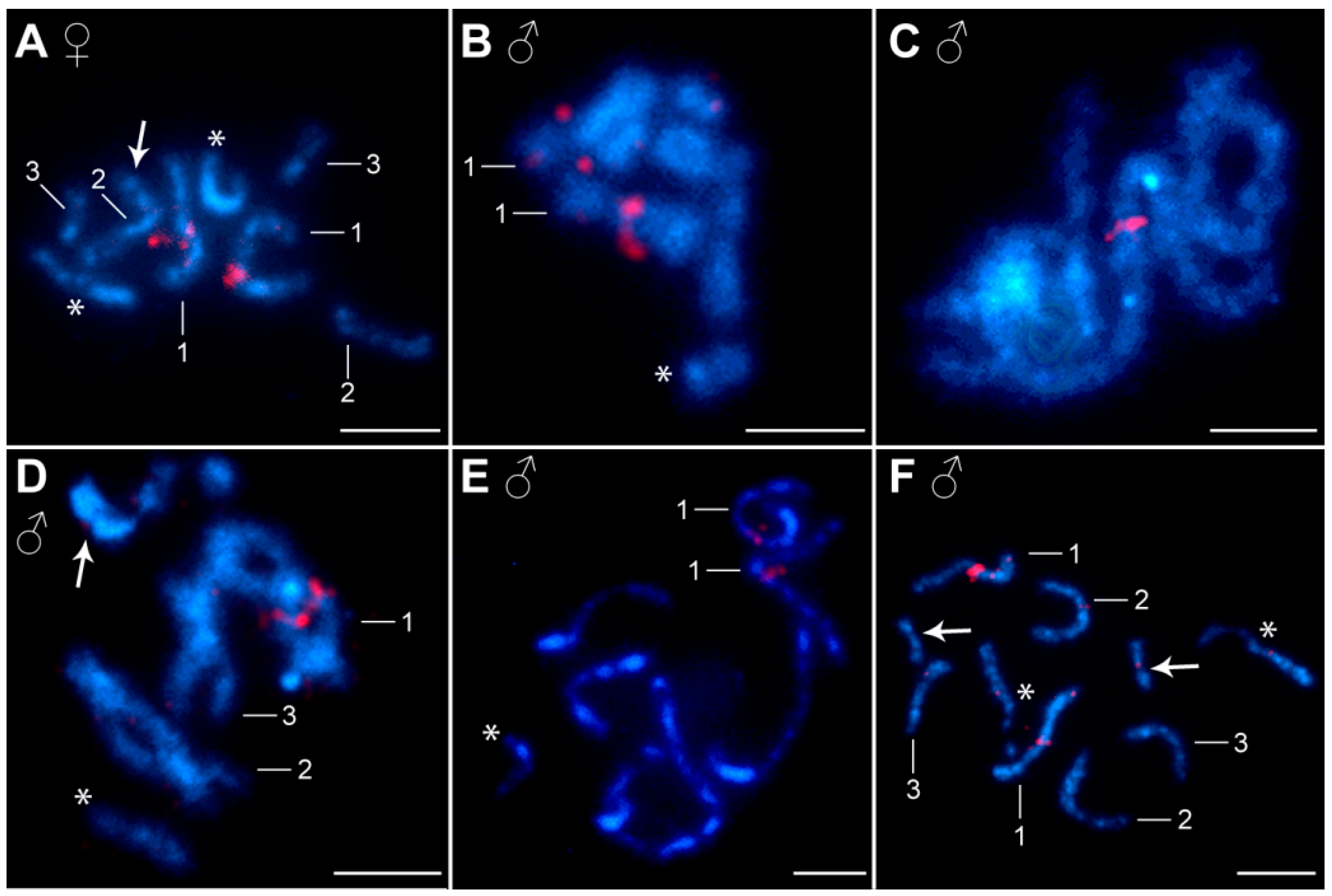
FISH experiments with an 18S rDNA probe showed one cluster of hybridization signals on the X sex chromosome in all three populations. The signals were about equally strong in both sexes. The rDNA locus (i.e., the nucleolar organizer region, NOR) was located on the short arms of the X chromosome in the pericentromeric region (Figure 2). The female chromosome spreads showed one or two clusters of hybridization signals, depending on the degree of chromosome pairing during the pachytene stage (Figure 2A). One cluster of hybridization signals was observed during meiotic prophase I in the male spreads (Figure 2B). At the diplotene stage of the males, the position of rDNA on the single X chromosome was clearly visible (Figure 2C). The activity of NOR was detected via AgNO3 staining. This method showed a compact, large nucleolus through the first meiotic division and a dark band at the sites previously identified using FISH with the 18S rDNA probe (Figure S1J–O). The distribution of H3 histone genes was also similar in all three populations examined. The H3 histone probe showed a single gene cluster on the first chromosome pair (Figure 3A,B). The array of the H3 genes was located on the long arms in the pericentromeric region. During meiotic division, FISH clearly showed a cluster of hybridization signals on a pachytene bivalent (Figure 3C) as well as on both homologous chromosomes #1 in the diplotene (Figure 3D) and in the anaphase I nuclei (Figure 3E,F).
The chromomycin A3 (CMA3) fluorochrome indicates the presence of GC-rich heterochromatin. CMA3-positive bands were observed at the ends of all chromosomes in all three populations (Figure 4). The fluorescent bands were more prominent in the metaphase and diplotene nuclei (Figure 4A,C,D) than in the pachytene nuclei (Figure 4B). In the ZŠ and LB populations, the signals were also present on the B chromosomes (Figure 4C). We did not observe pronounced DAPI-positive bands (AT-rich heterochromatin blocks) on either the A chromosomes or the B chromosomes; only weak centromeric heterochromatin bands were detected on the A chromosomes of all three populations (Figure 2 and Figure 3).

3.3. Meiotic Spermatogenesis and Oogenesis

The meiotic spermatogenesis of A. lucii followed the standard steps of eukaryotic cell division (Figure S1A–I). The process began with the arrangement of chromatin into thin and long strands (Figure S1A). At the pachytene and diplotene stages, we generally observed a clump of three bivalents (autosomes) with one univalent (X chromosome) (Figure S1B–E). One to four chiasmata (usually three) were observed on the larger bivalents #1 and #2, while only one was found on the bivalent #3 (Figure S1C). During meiosis, the B chromosomes did not pair with any chromosomes of the standard complement or with each other.
Oocyte differentiation followed the expected pattern—oogonia, primary oocyte, secondary oocyte, and mature ovum (Figure S2). The first stages of oogenesis included the formation of polar bodies (Figure S2A–C). Meiotic division began with the penetration of the sperm nucleus into the primary oocyte (Figure S2A,B). The first polar body formed after separation of the chromosomes from the bivalents in anaphase I. At this stage, the number of chromosomes was halved. One haploid nucleus then entered meiosis II. The second polar body and the egg pronucleus were formed by the separation of chromatids during anaphase II (Figure S2C). After the fusion of male and female nuclear material, the first mitotic division took place and cleavage of the zygote began. This process involved numerous mitotic divisions leading to the formation of macromeres and micromeres (Figure S2D–F). The final stage was the formation of a central mass of nuclei (Figure S2G), which further divided to form the so-called mature egg, an infectious larva (acanthor) enclosed in the egg envelope (Figure S2H).

4. Discussion

4.1. Standard Chromosome Complement

The karyotype of Acanthocephalus lucii has already been examined twice in addition to the present work. The first cytogenetic report on A. lucii was from the Iskar River in Bulgaria [41] and the second from the Ružín Reservoir (Hornád River, Slovakia) [10]. All studies, including our work on three geographically distant A. lucii populations, reported the same diploid chromosome number of 2n = 7♂/8♀. The morphology of the corresponding chromosomes is consistent; the main difference is the morphology of chromosome pair No. 3. Both in our study and in the Ružín population [10], the third pair was submetacentric, while in the Bulgarian population it was identified as acrocentric [41]. The basic characteristics of the karyotype (absolute and relative length) and TCL (Ružín—17.44; Bulgaria—18.66; LB—16.89; PZ—16.57; ZŠ—14.36) are relatively similar in all populations studied, except in the ZŠ population, which has the smallest chromosomes of all populations studied so far. To a certain extent, this difference in chromosome length may be due to the different degree of chromosome spiralization. However, the overall appearance of all studied chromosome complements from ZŠ was poor, with shapes and centromere positions that were difficult to recognize. Considering the heavy contamination of ZŠ, it is possible that the unfavorable environment played an important role in the shortening. The other two populations studied, LB with low contamination and PZ as a clean site, had larger chromosomes with a defined shape and an easily recognizable centromere position. The slight difference in the morphology of chromosome pair No. 3 between the above populations can be interpreted as the result of intrachromosomal rearrangements, pericentric inversions. Presumably, small intrachromosomal rearrangements have played an important role in the karyotype evolution within the genus Acanthocephalus. The available data, albeit only for two other species A. ranae and A. anguillae [12,42], showed a stable diploid chromosome number but a different chromosome morphology. Individual chromosome pairs in A. lucii and A. anguillae differ only slightly, namely in the morphology of the third chromosome pair, which is submetacentric in all populations of A. lucii from Slovakia, but acrocentric in A. anguillae. The chromosome morphology of A. ranae is completely different, as it has only metacentric chromosomes in its karyotype [42]. The evolution of the karyotype may play an important role in the speciation of the species [43]. The divergence of these Acanthocephalus species proceeded without changes in chromosome number and size, and the geographic interspecific variability in chromosome shape was probably caused by intrachromosomal structural rearrangements such as pericentric inversion. Similar mechanisms have also been found in some other parasite groups such as trematodes and cestodes [9,44].
The oogenesis of Acanthocephala has been studied in some species, especially in the 1960s and 1970s (e.g., [45,46]). The entire process of oogenesis has been described in Echinorhynchus truttae [47]. By using the fluorescent dye DAPI, it was possible to observe the division of oocytes and the first stages of embryogenesis in intact eggs of A. lucii. Our findings on the process of oogenesis are consistent with already known data for this group of parasites.
The rDNA loci are often located at different sites and in different numbers, which makes them important karyotypic and phylogenetic markers. This was also demonstrated in the first study on the location of ribosomal genes in the phylum Acanthocephala. The mapping of 18S rDNA by FISH supported the diversification of two closely related and morphologically very similar Acanthocephala, Pomphorhynchus laevis and P. tereticollis [31]. The authors observed different locations of the rRNA genes. In both, the 18S rDNA probe showed clusters of the major rDNA on the first and second largest chromosome pair, but the location was species-specific [31]. The distribution of ribosomal genes was also recently studied in the three populations of A. anguillae, where three clusters of major rDNA per haploid genome were detected [12]. One locus was located interstitially on the long arm near the centromere of chromosome pair No. 1, and two other interstitial clusters were observed near the centromere on the long and short arms of the second chromosome pair. In contrast to the congeneric A. anguillae, our study on A. lucii showed that the major rDNA is linked to the X sex chromosome, so the number of signals varied according to sex. In the male specimens, hybridization resulted in one signal, while the female specimens showed two signals. The presence of rDNA loci on sex chromosomes is not very common in animals. According to the animal rDNA loci database [48], rDNAs (45S and 5S) on sex chromosomes are found in only 11% of the karyotypes examined. There is very little information on Acanthocephala species, as only four species have been analyzed by FISH so far. Nevertheless, chromosomal mapping of 18S rDNA revealed a high variability in the numbers and location of this gene on the chromosomes, even in evolutionarily closely related species of the same genus. In the case of A. lucii and the evolutionarily related species A. anguillae, as well as within the genus Pomphorhynchus, the rDNA is variable in both number and chromosomal location. The most plausible mechanisms responsible for the variability of rDNA in Acanthocephala could be the above-mentioned structural rearrangements, unequal crossing-over, and transposition. The existence of a single cluster is probably the ancestral situation, although the existence of major rDNA on several chromosomes and its subsequent loss cannot be ruled out. It is known that rDNA clusters are capable of spreading throughout the genome and creating new rDNA loci [49,50]. Recent molecular phylogenetic data show that A. lucii is evolutionarily older than A. anguillae [51]. This would support the previously mentioned possibility of the existence of a single ancestral cluster. Given the lack of data, it is not possible to predict the possible evolutionary paths of the karyotype and the ancestral status of the order Echinorhynchida. Nevertheless, different patterns of ribosomal gene loci could indicate frequent changes in chromosomal rDNA distribution and the importance of rDNA in speciation.
The conspicuous genomic dispersion of H3 histone genes observed in A. anguillae, where the authors found multiple loci unevenly distributed on all (including Bs) chromosomes [12], was not observed in A. lucii. Similar to the major rDNA, the H3 histone genes in the karyotype of A. lucii were restricted to a single pair of chromosomes and clustered in an interstitial locus near the centromere on the long arms of the largest chromosome pair No. 1. Orosová et al. [12] suggested the presence of transposable elements in the H3 sequences of A. anguillae responsible for the spread of H3 histone repeats in the genome of the species, but a deeper analysis and reconstruction of the entire region is needed for confirmation. It will be very interesting to uncover the location of the H3 genes in other related species as well, to find out what the common location is in Acanthocephala.
The amount of constitutive heterochromatin detected with CMA3/DAPI staining showed that AT-rich heterochromatin is rare in acanthocephalans and most heterochromatin is GC-rich (in [present work] and [12,31]). The distribution of heterochromatin on the autosomes was uniform: (i) CMA3+ bands at the end of all chromosomes, (ii) faint DAPI+ bands in the centromeric region of all chromosomes, and (iii) in A. anguillae, DAPI+ bands next to the centromeric region in chromosome No. 2. The composition of telomeric repeats in Acanthocephala species is still unknown, and it remains unclear whether there is a novel telomeric motif or another mechanism that replaces telomerase in maintaining telomere length. Some organisms have lost the telomeric DNA repeats maintained by telomerase and have replaced them with tandem arrangements of satellite DNA sequences that can be extended by copying information from one chromosome to another [52]. Homologous recombination of long satellite sequences at telomeres has been described in representatives of lower Diptera [52]. Our results may suggest that an unknown telomere repeat motif of Acanthocephala species may be associated with GC bases. A more comprehensive analysis, including bioinformatic tools, is needed to identify undiscovered telomeres of the Acanthocephala phylum.

4.2. Supernumerary B Chromosomes and Environmental Pollution?

PCBs have been produced in large quantities for commercial purposes since the 1920s. Their key properties, such as thermal and chemical stability, persistence, low water solubility, and higher lipid solubility, led not only to their frequent use in industry, but also to their worldwide distribution in the environment [53]. Later, after mankind discovered the negative effects of PCBs on the environment and on the health and well-being of humans, animals, and plants, many countries banned or restricted the production of PCBs. Nevertheless, PCBs have become widespread environmental pollutants around the world [54]. One of the ways these hazardous substances can harm living organisms is by damaging them at the cellular level—the chromosomes. In general, chromosomes are delicate structures that are susceptible to defects caused by toxic environmental factors [55]. Our study provides further evidence for the occurrence of supernumerary B chromosomes in the chromosome complement of A. lucii from highly polluted environments. The first case dates back to 2002 and the study population originated from the Ružín water tank in the eastern part of Slovakia, which is heavily contaminated with various heavy metals [11]. The authors reported the presence of 1 to 5 small metacentric B chromosomes in 85% of the examined cells [10]. Only recently, B chromosomes were also discovered in the karyotype of the congeneric A. anguillae, and a possible connection with the long-term PCB contamination of the waters of Zemplínska Šírava was suggested [12]. In our study, B chromosomes were observed in the vast majority of cells (84%) in the ZŠ population and in 61.2% of cells in the LB population, while they were not found in the karyotypes of samples from the control region (PZ population). CMA3/DAPI staining showed that the B chromosomes are completely devoid of AT-rich heterochromatin, but the ends of the chromosomes are enriched with GC heterochromatin, similar to standard A chromosomes.
B chromosomes occur in 15% of species of all major animal and plant groups [23] and the question of their origin is still relevant. In several species, chromosome mapping of different classes of repetitive DNA (5S and 45S rDNA, histone genes, satellite DNA, snDNA, mobile elements) has provided valuable data on DNA sequences shared with standard A chromosomes (e.g., [24,56,57,58,59]) and in some cases has also shown which particular A chromosome is the ancestor of the B [60,61,62,63]. The answer to the question of where the B chromosomes in the populations of ZŠ and LB originated could probably lie in the poor ecological conditions of the sites studied. The ability of fish parasites to accumulate relatively high PCB concentrations in their tissues has been confirmed in several studies. The extremely high PCB contamination of ZŠ and the strict rules for fishing in this reservoir (“catch and release”) mean that the fish and their parasites are constantly exposed to the influence of these chemicals, which can affect the stability of the nuclear material by causing DNA damage. This also applies to the LB site, as the heavy pollution of the ZŠ reservoir means that the biota in the Laborec River are also negatively affected. The Laborec River is connected to the ZŠ reservoir by an inflow and outflow channel. Brázová et al. [28] showed that the PCB content in all organs and adipose tissue of perch from the ZŠ reservoir infected with A. lucii decreased compared with uninfected perch under natural conditions and the PCB content in the tissues of the parasites was relatively high. An increased incidence of chromosomal aberrations was reported for the tapeworm Caryophyllaeus laticeps from the same reservoir (ZŠ) as the parasites in this study [9]. DNA strand damage is defined as a permanent alteration of the nucleotide sequence during replication and can cause double-strand breaks (DSB), chromosome rearrangements, and gene mutations [64]. These perturbations lead to the activation of the DNA repair mechanism, which may contribute to the toxic response of PCBs. For example, when a DSB repair mechanism fails, it often leads to changes in chromosome structure (deletions, duplications, translocations, and others) [65]. One of the most important repair mechanisms is non-homologous end joining (NHEJ), which ensures rapid but error-prone repair [66]. Therefore, one of the possible causes of B chromosomes could be a failure to repair DNA damage. High environmental pollution could be the main factor that triggers chromosomal rearrangements and leads to the formation of B chromosomes through the amplification of DNA sequences in chromosomal fragments. This assumption is supported by the complete absence of B chromosomes in the population from the control area of PZ.
Several studies on other parasitic helminths have already shown a possible link between B chromosomes and unfavorable environmental conditions [67,68]. Study of the digenean flukes Notocotylus ephemera and Echinostoma revolutum from the heavily polluted cooling basin of a Lithuanian hydroelectric power plant revealed the presence of B chromosomes, probably due to the mutagenic effect of pollutants [67,68,69]. Some known cases in which B chromosomes have been associated with environmental pollution have been found in fish and mice. Supernumerary B chromosomes have been observed in several cichlid species from areas affected by pollution or construction of dams [70,71,72], as well as in South American catfish from waters downstream of sewage treatment plants with signs of domestic and industrial pollution [73]. An interesting case is the increase in B chromosomes in the population of the Korean field mouse Apodemus peninsulae from the shores of Lake Teletskoye, which is known to be polluted by remnants of rocket fuel components [74].

5. Conclusions

The data presented provide new information on the distribution of heterochromatin and the numbers and location of the major rDNA and H3 histone genes on the chromosomes of A. lucii. Our results contribute to the very sparse karyological knowledge about the phylum Acanthocephala. Based on the available karyotype data, chromosomal rearrangements in the process of diversification in this group of parasites are evident, and the application of different cytogenetic markers could help to clarify this process. Two cytogenetic markers are currently available, namely the 18S rDNA and the H3 histone genes, and their mapping has already shown that they will be helpful for that clarification. However, they have only been used for four species so far. Therefore, not only do new species-specific chromosomal markers need to be found but also a more comprehensive study of these species is urgently needed. In addition, we have found a third case of B chromosomes in representatives of Acanthocephala from a polluted environment. It is known that parasites, especially Acanthocephala, are able to detoxify their hosts in a polluted environment by excreting harmful substances from the infected host’s tissues [2] and accumulate these substances in significantly higher amounts compared with their hosts. Therefore, the appearance of B chromosomes in their karyotype may be caused by long-term exposure to water permanently contaminated with PCBs.

Supplementary Materials

The following supporting information can be downloaded at: https://www.mdpi.com/article/10.3390/d16030140/s1, Figure S1: Meiotic division of spermatocytes of Acanthocephalus lucii stained with Giemsa. (A–I) Chromosomes stained with AgNO3 (J–O). (A) Leptotene nucleus. (B) Pachytene. (C) Diplotene. (D) Diplotene nucleus with distinct secondary constriction on X chromosome. (E) Diplotene with one B chromosome. (F) Metaphase I nucleus without B chromosome. (G) Metaphase I nucleus with B chromosome. (H) Metaphase II nuclei with three B chromosomes. (I) Anaphase II. (J) Interphase nucleus. (K,L) Pachytene nuclei. (M) Pachytene (left) and diplotene (right) nuclei with three B chromosomes. (N) Diplotene with three B chromosomes. (O) Late anaphase II with separated bivalents and three B chromosomes. Asterisks indicate the X chromosome, arrow the B chromosome, and arrowheads indicate nucleolus residue (NOR). Bar = 10 µm. Figure S2: Oogenesis in Acanthocephalus lucii. (A) Primary oocyte penetrated by the sperm nucleus. (B) Meiotic division in oocyte resulting in formation of the first polar body, the sperm nucleus still present. (C) Formation of the second polar body after anaphase II. (D,E) Cleavage of the zygote, series of mitotic divisions. (F) Production of micro and macromeres. (G) Creation of central mass of nuclei. (H) Formed larva. Arrowheads indicate the polar bodies and arrows the sperm nucleus. The oocytes were stained with DAPI. Bar = 10 µm.

Author Contributions

M.O. (Martina Orosová) designed and supervised the study, co-organized the field trip, performed microscopical observations, A.M. performed experiments and data analysis, M.O. (Martina Orosová) and A.M. wrote the manuscript, and F.M. revised the manuscript. M.O. (Mikuláš Oros) and D.B. performed the field collection and examined the fish. All authors have read and agreed to the published version of the manuscript.

Funding

This work was supported by the Slovak Research and Development Agency (No. APVV 18-0467), Scientific Grant Agency of the Ministry of Education, Science, Research and Sport of the Slovak Republic and the Slovak Academy of Sciences (VEGA No. 2/0093/23).

Institutional Review Board Statement

For this study, ethical review and approval was waived as all fish caught were wild-caught with the permission of the Ministry of Environment of the Slovak Republic (No. 47/2022 and 48/2023).

Data Availability Statement

The raw data supporting the conclusions of this article will be made available by the authors on request.

Acknowledgments

The authors are deeply grateful to Olga Lisitsyna for her invaluable help in the morphological identification of the specimens.

Conflicts of Interest

The authors declare no conflicts of interest.

References

  1. Šalgovičová, D.; Zmetáková, Z. Polychorinated Biphenyls in Muscle Tissue of Freshwater Fish in East Slovakia. J. Food Nutr. Res. 2006, 45, 171–178. [Google Scholar]
  2. Sures, B.; Nachev, M.; Selbach, C.; Marcogliese, D.J. Parasite Responses to Pollution: What We Know and Where We Go in ‘Environmental Parasitology’. Parasite Vector 2017, 10, 65. [Google Scholar] [CrossRef]
  3. Duarte, G.S.C.; Lehun, A.L.; Leite, L.A.R.; Consolin-Filho, N.; Bellay, S.; Takemoto, R.M. Acanthocephalans Parasites of Two Characiformes Fishes as Bioindicators of Cadmium Contamination in Two Neotropical Rivers in Brazil. Sci. Total Environ. 2020, 738, 140339. [Google Scholar] [CrossRef]
  4. Nachev, M.; Rozdina, D.; Michler-Kozma, D.N.; Raikova, G.; Sures, B. Metal Accumulation in Ecto- and Endoparasites from the Anadromous Fish, the Pontic Shad (Alosa immaculata). Parasitology 2022, 149, 496–502. [Google Scholar] [CrossRef]
  5. Mille, T.; Soulier, L.; Caill-Milly, N.; Cresson, P.; Morandeau, G.; Monperrus, M. Differential Micropollutants Bioaccumulation in European Hake and Their Parasites Anisakis sp. Environ. Pollut. 2020, 265, 115021. [Google Scholar] [CrossRef]
  6. Brázová, T.; Miklisová, D.; Barčák, D.; Uhrovič, D.; Šalamún, P.; Orosová, M.; Oros, M. Hazardous Pollutants in the Environment: Fish Host-Parasite Interactions and Bioaccumulation of Polychlorinated Biphenyls. Environ. Pollut. 2021, 291, 118175. [Google Scholar] [CrossRef]
  7. Oros, M.; Barčák, D.; Miklisová, D.; Uhrovič, D.; Brázová, T. A Fish-Parasite Sentinel System in an Assessment of the Spatial Distribution of Polychlorinated Biphenyls. Sci. Rep. 2023, 13, 5164. [Google Scholar] [CrossRef]
  8. Goutte, A.; Molbert, N. Benefits of Parasitism in Polluted Environments: A Review and Perspectives. Front. Ecol. Evol. 2022, 10, 847869. [Google Scholar] [CrossRef]
  9. Orosová, M.; Marková, A.; Marec, F.; Barčák, D.; Brázová, T.; Oros, M. New Cytogenetic Data on Caryophyllaeus laticeps and Paracaryophyllaeus gotoi, Parasites of Evolutionary Interest. Parasitology 2022, 149, 1094–1105. [Google Scholar] [CrossRef] [PubMed]
  10. Špakulová, M.; Kráľová-Hromadová, I.; Dudiňák, V.; Reddy, P.V. Karyotype of Acanthocephalus lucii: The First Record of Supernumerary Chromosomes in Thorny-Headed Worms. Parasitol. Res. 2002, 8, 778–780. [Google Scholar] [CrossRef]
  11. Turčeková, L.; Hanzelová, V.; Špakulová, M. Concentration of Heavy Metals in Perch and Its Endoparasites in the Polluted Water Reservoir in Eastern Slovakia. Helminthologia 2002, 39, 76–80. [Google Scholar]
  12. Orosová, M.; Marková, A.; Zrzavá, M.; Marec, F.; Oros, M. Chromosome Analysis and the Occurrence of B Chromosomes in Fish Parasite Acanthocephalus anguillae (Palaeacanthocephala: Echinorhynchida). Parasite 2023, 30, 44. [Google Scholar] [CrossRef] [PubMed]
  13. Kushlan, J. Colonial Waterbirds as Bioindicators of Environmental Change. Colon. Waterbirds 1993, 16, 223–251. [Google Scholar] [CrossRef]
  14. Binelli, A.; Della Torre, C.; Magni, S.; Parolini, M. Does Zebra mussel (Dreissena polymorpha) Represent the Freshwater Counterpart of Mytilus in Ecotoxicological Studies? A Critical Review. Environ. Pollut. 2015, 196, 386–403. [Google Scholar] [CrossRef]
  15. Calapoğlu, M.; Sevinç, Z.; Toğay, A.; Kalyoncu, H. Evaluation of Oxidative Stress and Genotoxicity for Environmental Monitoring Using Farmed Rainbow Trout. Fresenius Environ. Bull. 2017, 26, 7105–7113. [Google Scholar]
  16. Kamollerd, C.; Senaphan, K.; Tengjaroenkul, B.; Monkheang, P.; Neeratanaphan, L. Oxidative Stress and Genetic Differentiation in Experimental Tilapia Fish Exposed to Heavy Metals in a Reservoir Near a Municipal Landfill. Appl. Ecol. Environ. Res. 2019, 17, 12893–12907. [Google Scholar] [CrossRef]
  17. Neeratanaphan, L.; Kanjanakunti, A.; Intamat, S.; Tengjaroenkul, B. Analysis of Chromosome Abnormalities in the Asian Swamp Eel (Monopterus albus) Affected by Arsenic Contamination Near a Gold Mine Area. Int. J. Environ. Stud. 2020, 77, 815–829. [Google Scholar] [CrossRef]
  18. Soulivongsa, L.; Tengjaroenkul, B.; Neeratanaphan, L. Effects of Contamination by Heavy Metals and Metalloids on Chromosomes, Serum Biochemistry and Histopathology of the Bonylip Barb Fish Near Sepon Gold-Copper Mine, Lao PDR. Int. J. Environ. Res. Public Health 2020, 17, 9492. [Google Scholar] [CrossRef]
  19. Šebelová, Š.; Kuperman, B.; Gelnar, M. Abnormalities of the Attachment Clamps of Representatives of the Family Diplozoidae. J. Helminthol. 2002, 76, 249–259. [Google Scholar] [CrossRef]
  20. Pečínková, M.; Vøllestad, L.A.; Koubková, B.; Huml, J.; Jurajda, P.; Gelnar, M. The Relationship Between Developmental Instability of Gudgeon Gobio gobio and Abundance or Morphology of Its Ectoparasite Paradiplozoon homoion (Monogenea). J. Fish Biol. 2007, 71, 1358–1370. [Google Scholar] [CrossRef]
  21. Brázová, T.; Orosová, M.; Šalamún, P.; Hanzelová, V. Morphological Abnormalities in Fish Parasites: A Potential Tool for Biomonitoring Natural Contaminants? Parasitol. Res. 2020, 119, 3297–3304. [Google Scholar] [CrossRef]
  22. Camacho, J.P. B Chromosomes. In The Evolution of the Genome; Gregory, T.R., Ed.; Academic Press: Cambridge, MA, USA, 2005. [Google Scholar] [CrossRef]
  23. Johnson Pokorná, M.; Reifová, R. Evolution of B Chromosomes: From Dispensable Parasitic Chromosomes to Essential Genomic Players. Front. Genet. 2021, 12, 727570. [Google Scholar] [CrossRef] [PubMed]
  24. Milani, D.; Ruiz-Ruano, F.J.; Camacho, J.P.M.; Cabral-de-Mello, D.C. Out of Patterns, the Euchromatic B Chromosome of the Grasshopper Abracris flavolineata is not Enriched in High-copy Repeats. Heredity 2021, 127, 475–483. [Google Scholar] [CrossRef]
  25. Houben, A.; Banaei-Moghaddam, A.M.; Klemme, S.; Timmis, J.N. Evolution and Biology of Supernumerary B Chromosomes. Cell. Mol. Life Sci. 2014, 71, 467–478. [Google Scholar] [CrossRef] [PubMed]
  26. Dalla Benneta, E.; Antoshechkin, I.; Yang, T.; Nguyen, H.Q.M.; Ferree, P.M.; Akbari, O.S. Genome Elimination Mediated by Gene Expression from a Selfish Chromosome. Sci. Adv. 2020, 6, eaaz980. [Google Scholar] [CrossRef] [PubMed]
  27. Perrot-Minnot, M.-J.; Cozzarolo, C.-S.; Amin, O.; Barčák, D.; Bauer, A.; García-Varela, M.; Hernández-Orts, J.S.; Le, T.T.Y.; Nachev, M.; Orosová, M.; et al. Hooking the Scientific Community on Thorny-Headed Worms: Interesting and Exciting Facts, Knowledge Gaps and Perspectives for Research Directions on Acanthocephala. Parasite 2023, 30, 23. [Google Scholar] [CrossRef]
  28. Brázová, T.; Hanzelová, V.; Miklisová, D. Bioaccumulation of Six PCB Indicator Congeners in a Heavily Polluted Water Reservoir in Eastern Slovakia: Tissue-Specific Distribution in Fish and their Parasites. Parasitol. Res. 2012, 111, 779–786. [Google Scholar] [CrossRef]
  29. Cabral-de-Mello, D.C.; Oliveira, S.G.; de Moura, R.C.; Martins, C. Chromosomal Organization of the 18S and 5S rRNAs and Histone H3 Genes in Scarabaeinae coleopterans: Insights into the Evolutionary Dynamics of Multigene Families and Heterochromatin. BMC Genet. 2011, 12, 88. [Google Scholar] [CrossRef]
  30. Provazníková, I.; Hejníčková, M.; Visser, S.; Dalíková, M.; Paladino, L.Z.C.; Zrzavá, M.; Voleníková, A.; Marec, F.; Nguyen, P. Large-Scale Comparative Analysis of Cytogenetic Markers Across Lepidoptera. Sci. Rep. 2021, 11, 12214. [Google Scholar] [CrossRef]
  31. Bombarová, M.; Marec, F.; Nguyen, P.; Špakulová, M. Divergent Location of Ribosomal Genes in Chromosomes of Fish Thorny-Headed Worms, Pomphorhynchus laevis and Pomphorhynchus tereticollis (Acanthocephala). Genetica 2007, 131, 141–149. [Google Scholar] [CrossRef]
  32. Lisitsyna, O. Fauna of Ukraine, Vol 31. Acanthocephala; Naukova Dumka: Kiev, Ukraine, 2019; p. 223. [Google Scholar]
  33. Orosová, M.; Špakulová, M. Tapeworms Chromosomes: Their Value in Systematics with Instructions for Cytogenetic Study. Folia Parasitol. 2018, 65, 001. [Google Scholar] [CrossRef]
  34. Dos Santos Guerra, M. Reviewing the Chromosome Nomenclature of Levan et al. Rev. Bras. Genet. 1986, 9, 741–743. [Google Scholar]
  35. Orosová, M.; Marková, A.; Provazníková, I.; Oros, M.; Radačovská, A.; Čadková, Z.; Marec, F. Molecular Cytogenetic Analysis of a Triploid Population of the Human Broad Tapeworm, Dibothriocephalus latus (Diphyllobothriidea). Parasitology 2021, 148, 787–797. [Google Scholar] [CrossRef]
  36. Cabral-de-Mello, D.C.; Zrzavá, M.; Kubíčková, S.; Rendón, P.; Marec, F. The Role of Satellite DNAs in Genome Architecture and Sex Chromosome Evolution in Crambidae Moths. Front. Genet. 2021, 12, 661417. [Google Scholar] [CrossRef]
  37. Garey, J.R.; Near, T.J.; Nonnemacher, M.R.; Nadler, S.A. Molecular Evidence for Acanthocephala as a Subtaxon of Rotifera. J. Mol. Evol. 1996, 43, 287–292. [Google Scholar] [CrossRef]
  38. Hejníčková, M.; Koutecký, P.; Potocký, P.; Provazníková, I.; Voleníková, A.; Dalíková, M.; Visser, S.; Marec, F.; Zrzavá, M. Absence of W Chromosome in Psychidae Moths and Implications for the Theory of Sex Chromosome Evolution in Lepidoptera. Genes 2019, 10, 1016. [Google Scholar] [CrossRef] [PubMed]
  39. Cabrero, J.; López-León, M.; Teruel, M.; Camacho, J.P.M. Chromosome Mapping of H3 and H4 Histone Gene Clusters in 35 Species of Acridid Grasshoppers. Chromosome Res. 2009, 17, 397–404. [Google Scholar] [CrossRef] [PubMed]
  40. Orosová, M.; Marec, F.; Oros, M.; Xi, B.W.; Scholz, T. A Chromosome Study and Localization of 18S rDNA in Khawia saurogobii (Cestoda: Caryophyllidea). Parasitol. Res. 2010, 106, 587–593. [Google Scholar] [CrossRef] [PubMed]
  41. Mutafova, T.; Nedeva, I.; Kanev, I. Chromosomes of Acanthocephalus lucii. J. Helminthol. 1997, 71, 261–262. [Google Scholar] [CrossRef]
  42. John, B. Chromosomes of Zooparasites I. Acanthocephalus ranae (Acanthocephala: Echinorhynchidae). Chromosoma 1957, 8, 730–738. [Google Scholar] [PubMed]
  43. Yoshida, K.; Kitano, J. Tempo and Mode in Karyotype Evolution Revealed by a Probabilistic Model Incorporating Both Chromosome Number and Morphology. PLoS Genet. 2021, 17, e1009502. [Google Scholar] [CrossRef]
  44. Hirai, H.; Taguchi, T.; Saitoh, M.; Kawanaka, M.; Sugiyama, H.; Habe, S. Chromosomal Differentiation of the Schistosoma japonicum Complex. Int. J. Parasitol. 2000, 30, 441–452. [Google Scholar] [CrossRef]
  45. Anantaraman, S.; Subramoniam, T. Oogenesis in Acanthosentis oligospinus nsp., an Aganthocephalan Parasite of the Fish, Macrones gulio. Proc. Indian Acad. Sci. 1975, 82B, 139–145. [Google Scholar] [CrossRef]
  46. Atkinson, K.H.; Byram, J.E. The Structure of the Ovarian Ball and Oogenesis in Moniliforrnis dubius (Acanthocephala). J. Morphol. 1976, 148, 391426. [Google Scholar]
  47. Parenti, U.; Antoniotti, M.L. Oogenesis in Echinorhynchus truttae (Schrank). Nature 1967, 58, 89–91. [Google Scholar]
  48. Sochorová, J.; Gálvez, F.; Matyášek, R.; Garcia, S.; Kovařík, A. Analyses of the Updated “Animal rDNA Loci Database” with an Emphasis on Its New Features. Int. J. Mol. Sci. 2021, 22, 11403. [Google Scholar] [CrossRef]
  49. Dubcovsky, J.; Dvořák, J. Ribosomal RNA Multigene Loci: Nomads of the Triticeae genomes. Genetics 1995, 140, 1367–1377. [Google Scholar] [CrossRef]
  50. Castro, J.; Rodríguez, S.; Pardo, B.G.; Sánchez, L.; Martínez, P. Population Analysis of an Unusual NOR-site Polymorphism in Brown Trout (Salmo trutta L.). Heredity 2001, 86, 291–302. [Google Scholar] [CrossRef]
  51. Lisitsyna, O.; Barčák, D.; Orosová, M.; Fan, C.K.; Oros, M. Acanthocephalans of Marine and Freshwater Fishes from Taiwan with Description of a New Species. Folia Parasitol. 2023, 70, 021. [Google Scholar] [CrossRef]
  52. Mason, J.M.; Reddy, H.M.; Capkova Frydrychova, R. Telomere Maintenance in Organisms without Telomerase. In DNA Replication-Current Advances; Seligmann, H., Ed.; InTech: Rijeka, Croatia, 2011. [Google Scholar] [CrossRef]
  53. Robertson, L.W.; Ludewig, G. Polychlorinated Biphenyl (PCB) Carcinogenicity with Special Emphasis on Airborne PCBs. Gefahrstoffe Reinhalt. Luft 2011, 71, 25–32. [Google Scholar]
  54. Yang, F.; Wilcox, B.; Jin, S.; Aguirre, A.A.; Rougée, L.; Xu, L.; Lu, Y. Detection and Quantitative Analysis of Polychlorinated Biphenyls in Tilapia from Hawaiian Waters. Chemosphere 2008, 73, 133–137. [Google Scholar] [CrossRef]
  55. Wójcik, E.; Smalec, E. The Effect of Environmental Factors on Sister Chromatid Exchange Incidence in Domestic Horse (Equus caballus) Chromosomes. Folia Biol. 2013, 61, 199–204. [Google Scholar] [CrossRef]
  56. Oliveira, N.L.; Cabral-de-Mello, D.C.; Rocha, M.F.; Loreto, V.; Martins, C.; Moura, R.C. Chromosomal Mapping of rDNAs and H3 Histone Sequences in the Grasshopper Rhammatocerus brasiliensis (Acrididae, Gomphocerinae): Extensive Chromosomal Dispersion and Co-localization of 5S rDNA/H3 Histone Clusters in the A Complement and B Chromosome. Mol. Cytogenet. 2011, 4, 24. [Google Scholar] [CrossRef] [PubMed]
  57. Silva, D.M.Z.d.A.; Pansonato-Alves, J.C.; Utsunomia, R.; Araya-Jaime, C.; Ruiz-Ruano, F.J.; Daniel, S.N.; Hashimoto, D.T.; Oliveira, C.; Camacho, J.P.M.; Porto-Foresti, F.; et al. Delimiting the Origin of a B Chromosome by FISH Mapping, Chromosome Painting and DNA Sequence Analysis in Astyanax paranae (Teleostei, Characiformes). PLoS ONE 2014, 9, e94896. [Google Scholar] [CrossRef] [PubMed]
  58. Felicetti, D.; Haerter, C.A.G.; Baumgärtner, L.; Paiz, L.M.; Takagui, F.H.; Margarido, V.P.; Blanco, D.R.; Feldberg, E.; da Silva, M.; Lui, R.L. A New Variant B Chromosome in Auchenipteridae: The Role of (GATA)n and (TTAGGG)n Sequences in Understanding the Evolution of Supernumeraries in Trachelyopterus. Cytogenet. Genome Res. 2021, 161, 70–81. [Google Scholar] [CrossRef] [PubMed]
  59. Stornioli, J.H.F.; Goes, C.A.G.; Calegari, R.M.; dos Santos, R.Z.; Giglio, L.M.; Foresti, F.; Oliveira, C.; Penitente, M.; Porto-Foresti, F.; Utsunomia, R. The B Chromosomes of Prochilodus lineatus (Teleostei, Characiformes) are Highly Enriched in Satellite DNAs. Cells 2021, 10, 1527. [Google Scholar] [CrossRef] [PubMed]
  60. Dhar, M.K.; Friebe, B.; Koul, A.K.; Gill, B.S. Origin of an Apparent B Chromosome by Mutation, Chromosome Fragmentation and Specific DNA Sequence Amplification. Chromosoma 2002, 111, 332–340. [Google Scholar] [CrossRef] [PubMed]
  61. Loreto, V.; Cabrero, J.; López-León, M.D.; Camacho, J.P.M.; Souza, M.J. Possible Autosomal Origin of Macro B Chromosomes in Two Grasshopper Species. Chromosome Res. 2008, 16, 233–241. [Google Scholar] [CrossRef] [PubMed]
  62. Teruel, M.; Cabrero, J.; Perfectti, F.; Camacho, J.P. B Chromosome Ancestry Revealed by Histone Genes in the Migratory Locust. Chromosoma 2010, 119, 217–225. [Google Scholar] [CrossRef] [PubMed]
  63. Bueno, D.; Palacios-Gimenez, O.M.; Cabral-de-Mello, D.C. Chromosomal Mapping of Repetitive DNAs in the Grasshopper Abracris flavolineata Reveal Possible Ancestry of the B Chromosome and H3 Histone Spreading. PLoS ONE 2013, 8, e66532. [Google Scholar] [CrossRef]
  64. Volkova, N.V.; Meier, B.; Gonzalez-Huici, V.; Bertolini, S.; Gonzalez, S.; Vohringer, H.; Abascal, F.; Martincorena, I.; Campbell, P.J.; Gartner, A.; et al. Mutational Signatures Are Jointly Shaped by DNA Damage and Repair. Nat. Commun. 2020, 11, 2169–2183. [Google Scholar] [CrossRef]
  65. So, C.C.; Martin, A. DSB Structure Impacts DNA Recombination Leading to Class Switching and Chromosomal Translocations in Human B cells. PLoS Genet. 2019, 15, e1008101. [Google Scholar] [CrossRef]
  66. Rassool, F.V.; Tomkinson, A.E. Targeting Abnormal DNA Double Strand Break Repair in Cancer. Cell. Mol. Life Sci. 2010, 67, 3699–3710. [Google Scholar] [CrossRef]
  67. Baršiené, J. The Chromosome Sets of Trematodes. Parazitologiya 1993, 27, 336–353. [Google Scholar]
  68. Petkevičiūtė, R.; Baršiené, J. The Comparative Karyological Analysis of Three Species of Trematodes of Genus Notocotylus. Parazitologiya 1988, 22, 21–28. [Google Scholar]
  69. Baršiené, J.; Kisilienė, V. Karyological Studies of Trematodes within the Genus Echinostoma. Acta Parasitol. 1991, 36, 23–29. [Google Scholar]
  70. Feldberg, E.; Porto, J.I.R.; Alves-Brinn, M.N.; Mendonça, M.N.C.; Benzaquem, D.C. B Chromosomes in Amazonian Cichlid Species. Genetics 2004, 106, 195–198. [Google Scholar] [CrossRef] [PubMed]
  71. Pires, L.B.; Sampaio, T.R.; Dias, A.L. Mitotic and Meiotic Behavior of B Chromosomes in Crenicichla lepidota: New Report in the Family Cichlidae. J. Hered. 2015, 106, 289–295. [Google Scholar] [CrossRef] [PubMed]
  72. Perazzo, G.X.; Noleto, R.B.; Vicari, M.R.; Gava, A.; Cestari, M.M. B Chromosome Polymorphism in South American Cichlid. Neotrop. Biodivers. 2018, 4, 3–9. [Google Scholar] [CrossRef]
  73. de Campos Júnior, E.O.; Pereira, B.B.; Morelli, S.; Pavanin, E.V.; Pavanin, L.A. Biological Monitoring and B Chromosome Frequency in Bagre (Rhamdia quelen) in Southeast Brazil. Environ. Toxicol. Phar. 2014, 38, 510–517. [Google Scholar] [CrossRef]
  74. Borisov, Y.M.; Abramov, S.A.; Borisova, M.Y.; Zhigarev, I.A. The Occurrence of Dot-Like Micro B Chromosomes in Korean Field Mice Apodemus peninsulae from the Shore of the Teletskoye Lake (Altai Mountains). Comp. Cytogenet. 2020, 14, 97–105. [Google Scholar] [CrossRef] [PubMed]
Figure 1. Mitotic metaphases (left panel) and derived karyotypes (right panel) of Acanthocephalus lucii males and females. (A) ZŠ population without and with B chromosomes (2n = 7/8 + 1–3B). (B) LB population without and with B chromosome (2n = 7+1B). (C) PZ population. Asterisks indicate X chromosomes and arrows indicate B chromosomes. Bar = 5 µm.
Diversity 16 00140 g001
Figure 2. FISH with the 18S rDNA probe (red) on chromosomes of Acanthocephalus lucii. (A) Female pachytene bivalents with one B chromosome at the periphery. (B) Male pachytene and early meiotic prophase I nucleus. (C) Male diplotene with three B chromosomes. (D) Male mitotic metaphase. (E) Female mitotic metaphase with two B chromosomes. Asterisks indicate X chromosomes and arrows indicate B chromosomes. Chromosomes were counterstained with DAPI. Bar = 10 µm.
Diversity 16 00140 g002
Figure 3. FISH with H3 histone probe (red) on chromosomes of Acanthocephalus lucii. (A) Female mitotic metaphase with one B chromosome. (B) Male mitotic metaphase. (C) Male pachytene. (D) Male diplotene with one B chromosome. (E,F) Male anaphase II nuclei, (E) without and (F) with two B chromosomes. Asterisks indicate X chromosomes and arrows indicate B chromosomes. Chromosomes were counterstained with DAPI. Bar = 5 µm.
Diversity 16 00140 g003
Figure 4. Distribution of GC-rich heterochromatin on chromosomes of Acanthocephalus lucii males after CMA3 staining. (A) Mitotic metaphase. (B) Pachytene. (C,D) Diplotene with (C) and without (D) B chromosome. Asterisks indicate X chromosomes and the arrow the B chromosome. Bar = 10 µm.
Diversity 16 00140 g004
Table 1. Measurements (mean ± SD) and classification of chromosomes of Acanthocephalus lucii.
Chromosome NumberStudy Site aAbsolute Length
(mean ± SD b)
(μm)
Relative Length
(mean ± SD)
(%)
Centromeric Index
(mean ± SD)
Classification c
14.62 ± 0.2932.19 ± 2.0245.79 ± 2.03m
LB5.21 ± 0.2730.87 ± 1.6046.48 ± 1.80m
PZ5.24 ± 0.2231.64 ± 1.3347.37 ± 1.70m
24.07 ± 0.3228.33 ± 2.2437.48 ± 1.94sm
LB4.43 ± 0.2226.20 ± 1.3138.09 ± 1.99sm
PZ4.61 ± 0.2427.82 ± 1.4738.36 ± 1.23sm
33.19 ± 0.3522.20 ± 2.4435.97 ± 3.08sm
LB3.80 ± 0.2622.50 ± 1.5334.81 ± 2.29sm
PZ3.61 ± 0.1721.78 ± 0.9128.86 ± 2.22sm
X2.48 ± 0.2417.27 ± 1.6720.72 ± 1.63a
LB3.45 ± 0.1320.43 ± 0.8023.89 ± 2.61a
PZ3.11 ± 0.1518.75 ± 0.9115.05 ± 1.68a
a Study site: ZŠ—Zemplínska Šírava reservoir, LB—Laborec River, PZ—Pozdišovce pond. b Mean absolute lengths were calculated from the 10 best mitotic metaphases for each A. lucii population. c Classification according to Dos Santos Guerra (1968) [34]: a—acrocentric; m—metacentric, sm—submetacentric chromosome pair.
Disclaimer/Publisher’s Note: The statements, opinions and data contained in all publications are solely those of the individual author(s) and contributor(s) and not of MDPI and/or the editor(s). MDPI and/or the editor(s) disclaim responsibility for any injury to people or property resulting from any ideas, methods, instructions or products referred to in the content.