Article

The RNA Viruses in Samples of Endemic Lake Baikal Sponges

1
Limnological Institute Siberian Branch of the Russian Academy of Sciences, 664033 Irkutsk, Russia
2
Faculty of Business Communication and Informatics, Irkutsk State University, 664003 Irkutsk, Russia
*
Author to whom correspondence should be addressed.
Diversity 2023, 15(7), 835; https://doi.org/10.3390/d15070835
Submission received: 21 April 2023 / Revised: 26 June 2023 / Accepted: 30 June 2023 / Published: 4 July 2023
(This article belongs to the Special Issue Viral Diversity in Marine and Freshwater)

Abstract

:
Sponges are unusual representatives of the animal kingdom; their viromes, as part of the associated community, began to be studied quite recently, and, accordingly, these studies are gaining momentum. The diversity of viruses in sponges is high, and they most likely play a significant role in the composition of the sponge holobiont, especially under stress conditions. The objects of our metagenomic study were RNA viruses of two common endemic species of Baikal sponges, Lubomirskia baikalensis and Baikalospongia bacillifera. As a result of viral RNA sequencing, we were able to identify fragments of viral genomes related to those from the RefSeq NCBI complete viral genome database. Most of the similar genomes belonged to viruses isolated from various invertebrates; some of the scaffolds were related to known plant viruses, and one of them was related to a vertebrate virus. The similarity of the putative proteins of viral scaffolds from the Baikal sponges with proteins of known viruses turned out to be low (20.7–67.3%), indicating the detection of novel viruses. The samples of diseased and visually healthy sponges were clustered separately, suggesting a shift in sponge virome composition during the course of the disease. In a comparative analysis, the viromes of the Baikal and marine sponges differed significantly, demonstrating the influence of the host species, habitat, and geographical location on virome composition in the sponge holobiont.

1. Introduction

Sponges are unique representatives of the animal kingdom. These ancient inhabitants (phylum Porifera) are complex symbiotic communities consisting of various microorganisms (bacteria, algae, fungi, protozoa, and others) [1]. Sponges are widespread in both freshwater and marine ecosystems and have important properties and functions in aquatic environments by passing large masses of water through themselves (known as filtering) and purifying them [2,3,4].
An inherent part of a sponge community (holobiont) is viruses, the diversity of which depends on the species of the sponges themselves, the composition of sponge-associated microorganisms [1], and the macroorganisms that inhabit sponges (gammarus, molluscs, etc.) [5]. Sponge viromes began to be studied relatively recently; the first works were mainly concerned with the study of DNA-containing viruses in marine sponges (most of which are bacteriophages) [6,7,8,9]. But some works [10,11,12,13] also included investigations of RNA-containing viruses in sponges, which revealed a high level of diversity of RNA viruses in sponge holobionts. Earlier, we carried out the first viromic studies (of DNA viruses) of Baikal sponges [14,15,16]. To the best of our knowledge, the viral communities of other freshwater sponges have not yet been studied.
As is known, during the past decade, sponges have been affected and died all over the world [17]; to date, the sponge populations of Lake Baikal have declined catastrophically. [18]. All of this happens against the backdrop of noticeable changes in the structure of coastal phyto- and zooplankton communities, indicating eutrophication of the coastal zone [19,20]. Hydrochemical and sanitary–microbiological analyses of the tributaries of rivers and the shallow zones in area of settlements indicate an increased anthropogenic impact [21,22,23,24]; however, mass death of sponges is observed throughout the Baikal water area, regardless of the level of anthropogenic load [25]. Sponge disease may be caused by various pathogenic agents of a bacterial, fungal [26,27], or viral nature. Numerous studies of the microbial communities of healthy and diseased Baikal sponges failed to reveal any specific pathogen [28], and the research of viral communities seems to be especially relevant for understanding the mechanisms of the onset and course of sponge disease (or damage).
At the beginning of this century, large-scale studies of RNA viruses of a wide range of aquatic invertebrates unfolded [10,29,30]. These RNA sequencing (viromic and meta-transcriptomic) studies uncovered a wide variety of new RNA viruses in natural ecosystems. The databases were replenished with numerous genomes of viruses assembled from the samples of various inhabitants of water bodies. Prior to the era of metagenomic research, only a few dozen invertebrate viruses were known, usually of commercial species [31,32]. It has become known that RNA viruses predominate DNA viruses in eukaryotic viromes, in contrast to bacterial and archaeal ones [33]. A huge level of genetic diversity of RNA viruses; shuffling of gene modules among different viral genomes; wide host range for many groups of viruses; and simultaneous monophyleticity of many other viruses with closely related hosts, indicating a long-term virus–host co-divergence, were revealed. Evolutionary concepts of RNA viruses have been revised, and it has been hypothesized that plant and vertebrate RNA viromes originated through multiple horizontal virus transfer (HVT) from marine invertebrate viromes [29,34,35].
The aim of this work was to study the viral RNA isolated from samples of the Baikal sponges Lubomirskia baikalensis and Baikalospongia bacillifera, members of the endemic family Lubomirskiidae (class Demospongiae, order Spongillida), and to obtain the first data regarding the diversity of RNA viruses circulating in Baikal sponges. In the present study, we used samples of both diseased and visually healthy sponges. This allowed us to trace changes in RNA viral communities over the course of the sponge diseases, as was previously shown for DNA viruses [16].

2. Materials and Methods

2.1. Sampling and Sample Processing

The Lubomirskia baikalensis and Baikalospongia bacillifera sponges were sampled in sterile tubes by divers using lightweight diving equipment. The L. baikalensis sponges (two specimens) were sampled in March 2015 in the Maloye More Strait, near the Malye Olkhonskiye Vorota Strait (the middle basin of Lake Baikal, 53°01′05.1″ N, 106°55′43.5″ E). Two branches of 2–3 cm3 in volume were collected from the same sponge: one branch looked healthy and another one had lesions (bleaching or brown spots) (Figure 1, Table 1). We also collected samples from L. baikalensis sponge individuals without visible damages; however, we failed to isolate viral RNA from these samples in an amount sufficient for the preparation of libraries. The B. bacillifera sponges were sampled in the southern basin of Lake Baikal, near the Bolshiye Koty settlement (51°54′07.5″ N, 105°06′12.0″ E), in May 2018. The four specimens of B. bacillifera, of 5–7 cm3 in volume, were collected and used in this study: two specimens looked healthy, and two others had necrosis and brown spot-like lesions (Figure 1, Table 1). Sponge samples were processed, and then concentrates of virus-like particles (VLPs) were obtained as described in [14,15]. Briefly, the sponge samples were washed in sterile Baikal water, thoroughly homogenized by blender, and centrifuged (3000 rpm, 30 min for L. baikalensis or 400 g for 15 min followed by 16,000× g for 30 min for B. bacillifera). The aqueous fraction was passed through a syringe filter with a pore size of 0.2 mm (Sartorius, Göttingen, Germany) and treated with DNase I (50 U/mL) and RNase A (100 mg/mL) enzymes (Thermo Fisher Scientific, Waltham, MA, USA) to remove contaminating nucleic acids. Viral RNA was extracted with TRI Reagent (Molecular Research Center, Cincinnati, OH, USA) [36] according to the manufacturer’s recommendations. Specifically, the samples were diluted 4-fold with TRI Reagent and incubated for 5 min at room temperature (RT); 0.2 mL of chloroform per 1 mL of TRI Reagent was added, and the mixture was shaken and incubated (RT, 15 min). Then, the samples were centrifuged (12,000× g, 15 min), and the collected upper phase was precipitated (12,000× g, 15 min, 4 °C) with isopropanol (0.5 mL per 1 mL TRI Reagent).

2.2. Library Preparation and Sequencing

The preparation and sequencing of DNA libraries were performed at the Genomics Core Facility of the Institute of Chemical Biology and Fundamental Medicine, Siberian Branch, of the Russian Academy of Sciences (ICBFM SB RAS, Novosibirsk, Russia), and at the Center of Shared Scientific Equipment “Persistence of microorganisms” of the Institute for Cellular and Intracellular Symbiosis, Ural Branch, of the Russian Academy of Sciences (ICIS UB RAS, Orenburg, Russia). Shotgun libraries were prepared by using and following the NEBNext Ultra II RNA Library Prep Kit (New England Biolabs, Ipswich, MA, USA) protocol. Sequencing of the libraries was conducted on the MiSeq platform (Illumina, San Diego, CA, USA) using a MiSeq Reagent Kit v3 (2 × 300 cycles).

2.3. Initial Shotgun Metagenomic Datasets on RNA Viruses in Marine and Freshwater Samples

For comparative analysis, we also used the available NCBI SRA datasets (Illumina platforms, paired-end reads) (Table 1) from the marine sponge (class Demospongiae) samples (and water samples, if available).

2.4. Primary Processing and Taxonomic Analysis of Initial Virome Reads

The quality visualization of the virome datasets (paired reads) was carried out using the FASTQC program. Trimming of the reads by quality was carried out using the Trimmomatic v.0.39 program (MAXINFO:40:0.05 AVGQUAL:15 MINLEN:100) [37].
Taxonomic classification was carried out with the Kaiju v.1.9.0 software [38] using the NCBI nr (non-redundant) protein database. To increase the sensitivity of the Kaiju software, the parameter length was specified as 6, and the mismatch parameters were specified as 30. To filter out false positive results, the e-value was chosen as ≤0.00001 and bit score ≥ 50. The results of the Kaiju analysis were grouped in a table (Kaiju taxon count table) where the elements contained information about the representation of taxa in the number of reads per sample. Based on taxonomic identifiers, information on RNA viruses was extracted from the “Kaiju taxon count table” in the form of a table of virotype representation. For further analysis, this table of virotype representation was used in two variants: absolute values (number of reads from a sample per RNA virotype) and relative values (percentage of sample reads per RNA virotype).

2.5. Assembly of Virome Reads

All RNA datasets from the Baikal sponges were aggregated into one array for de novo cross-assembly, as applied in our previous studies of viral communities [16]. For cross-assembly, the meta SPAdes assembler, optimized for metagenomics, was used with default parameters [39]. The Bowtie2 [40] and SAMtools [41] software were used to map paired-end reads on scaffolds and to calculate the total coverage of scaffolds in the assembly and the coverage of scaffolds by reads from each sample. The scaffolds with total coverage of more than five and a length of ≥500 bp were used for further analysis.

2.6. Viral Scaffolds Detection

We identified the RNA viral scaffolds and open reading frames (ORFs) within them using the VirSorter2 tool [42] with an integrated RNA viral database. The results of RNA-virus identification were checked by the CheckV program [43], according to the instructions (https://bitbucket.org/berkeleylab/checkv/src/master/, accessed on 23 December 2022).
The Bowtie2 [40] and SAMtools [41] results were used to determine the number of reads mapped on each predicted viral scaffold from each sample. Counts of the predicted viral proteins (ORFs) in samples were defined as the number of reads mapped on a scaffold containing a given protein. Consequently, the count table of viral scaffold representation in the analyzed samples was constructed. TPM (transcripts per million) normalization, recommended for metagenomics, was used to normalize the count table for scaffold length and number of reads per sample [44].

2.7. Taxonomic Assignment of Viral Scaffolds

Taxonomic identification of the viral scaffolds was carried out by comparisons of predicted viral proteins in scaffolds with the NCBI RefSeq complete viral proteome database [45]. The comparison was carried out using the DIAMOND algorithm [46] with a “more-sensitive” option, i.e., e-value ≤ 0.00001 and bit score ≥ 50. For each protein in the scaffold, the best match in terms of the bit score value was selected. If a single scaffold had multiple proteins that matched different taxa (NCBI RefSeq ID), the one with the largest number of matching proteins was chosen as the most closely related virus taxon (virotype) for this scaffold. If all identified proteins belonged to different taxa, the level of bit-score of the matched proteins was taken into account, and the NCBI RefSeq taxon (ID) with the highest bit-score was selected as the virotype.
Host prediction for the Baikal viral scaffolds was carried out based on the Virus–Host database [47].

2.8. Functional Assignment of Viral Scaffolds

The predicted viral proteins (ORFs) were matched with functional motifs of proteins in the KOfam database [48] using KofamScan software [49], and with functional motifs in the Pfam database using PfamScan software [50] with default options. For ORFs containing RNA-dependent RNA polymerase (RdRp) domains, i.e., most likely non-structural polyproteins, an additional analysis was performed to search for the closest analogues in the nr NCBI protein database using the online BLASTp application. The 50 top BLAST hits were chosen if the number of hits was more than 50, and all hits if their number was less than 50. For each ORF, a correlation analysis of the relationship between the similarities of BLASTp matches with the alignment length was performed (Spearman coefficients correlation with testing using Spearman statistics, with a p-value ≤ 0.05 indicating a significant correlation).

2.9. Statistical Analysis of Taxonomic and Functional Diversity

The potential underestimated number of RNA virus Kaiju virotypes (species richness) in the total communities was evaluated using Chao1 [51] and ACE [52] indices. The Shannon and Simpson indices [53] of viral diversity were also calculated.
For further analysis, the Kaiju proportion of reads for RNA virotypes and TPM per samples of the Baikal sponges were standardized into ranges from 0 to 1 by the “decostand” function of the “vegan” package for the R [54]. Standardized values of the Kaiju proportions of reads for RNA virotypes and TPM per sample were visualized using canonical correspondence analysis (CCA). Gradient vectors of the RNA viral family composition and scaffolds’ TPM per sample composition were fitted on a CCA scatter plot. The reliability of linear approximation for gradient vectors was assessed by multivariate linear regression analysis. Only vectors that showed reliable directionality were displayed. Biodiversity analysis and CCA ordination were carried out using the “vegan” package for the R [54], according to the instructions.
The Kaiju proportion of reads for the first 30 dominant RNA virotypes and TPM per samples of the Baikal sponges RNA viral scaffolds were visualized with a heat map using the “gplots” package [55] for the R with columns (samples) clustering, and were grouped in order of similarity (Euclidean distance with the complete-link clustering method).
The influence of different factors on scaffolds’ TPM per sample composition was assessed with PERMANOVA analysis (2000 permutations and Euclidean distance) [56] in the R package “vegan” [54]. The influence of the factor was considered significant at a p-value ≤ 0.05.

3. Results

3.1. Analysis of Virome Reads

Eight data sets were obtained from six individuals of the sponges L. baikalensis and B. bacillifera after separation of the viral fraction (<0.2 µm) from the samples, RNA isolation and sequencing, and primary processing of the reads (Table 1). The resulting metadata were analyzed using the Kaiju software, and the overall composition of the viromes (percentage of eukaryotes, bacteria, archaea, and viruses) was determined (Supplementary Table S1). Additionally, the available datasets from marine sponges were used for comparison (Table 1). The percentages of unidentified sequences in the Baikal samples were 22–46%; in other samples they reached up to 81–83% (in particular in the sets A and B of Halichondria panicea sponges). Large proportions consisted of bacterial and eukaryotic reads (respectively, 15–27% and 22–51% in the Baikal samples, 11–60% and 0.02–26% in the compared data) (Supplementary Table S1). It should be noted that these values were strongly influenced by the method of sample preparation; first of all, it mattered whether the viral fraction was enriched or not, as did what kinds of nucleic acids were isolated and sequenced (viral fraction, V, or total sample, T; RNA or both RNA and DNA, indicated in Table 1). Another important point is the depletion of ribosomal RNA during the preparation of libraries. In the case of the Baikal samples, we sequenced RNA isolated from viral fractions, and depletion was not performed; therefore, these datasets contained a large amount of rRNA, despite the fact that the rRNA during centrifugation at high speed (17,000× g, 3 min) in the samples was significantly reduced [57].
The percentage of RNA viruses in the samples was generally low: 0.001–0.12% in the Baikal sponges and 0.0004–2.4% in the marine sponges (Supplementary Table S1). The exception was the sample of Hymeniacidon sp. (Hym.sp_2, 27.3%), as well as the control seawater sample from the Great Barrier Reef (GBR.sw, 50.2%). A small portion of the sequences in the Baikal sets were affiliated with DNA-containing viruses (0.001–0.3%); in marine datasets, their proportion was generally higher (0.02–15.4%: minimum in H. panicea sponges, maximum in Mediterranean water samples, Med.sw.RM). In fact, in the case of the H. panicea sponges [10], enriched viral RNA was isolated, but in the study of the Mediterranean samples [13], the total viral RNA and DNA were used for analysis. Viral diversity indices for all datasets are shown in Supplementary Table S2; the number of virotypes in the Baikal samples ranged from 8 to 98, while in marine samples, it ranged from 13 to 466.
The difference between analyzed freshwater and marine sponges was noticeable at the levels of both families and virotypes (Figure 2, Supplementary Table S3). On the CCA plot (Figure 2A), apart from the Baikal ones, the most distant samples were also Rhopaloeides odorabile (from GBR) and Hymeniacidon sp. (Japan) sponges; the Mediterranean and Halichondria panicea (North Sea) sponges occupied an intermediate position (located approximately in the center of the graph). A distinctive feature of the Baikal sponges was the greater representation of the families Tombusviridae, Marnaviridae, and Luteoviridae. In Figure 2B, we were able to see the top virotypes for different sets. For almost all marine (with the exception of Hym.sp.), as well as for some Baikal, samples (B.b.Sv2480, L.b. Sv2.1d and Sv1.1h), the most numerous was a certain unclassified Picobirnavirus (Picobirnavirus sp. strain PBV/roe_deer/SLO/D38-14/2014, [58]). Moreover, Cylindrotheca closterium RNA virus 03 and Beihai sobemo-like virus 9 dominated in GBR sponge samples (up to 43% of viral reads); Sponge holobiont-associated RNA virus (up to 82.4%) in Hymeniacidon sp.; but Barns Ness breadcrumb sponge hepe-like virus 1 (28.3%) and Barns Ness breadcrumb sponge aquatic picorna-like virus 1 (11%) in H. panicea. Samples of Mediterranean sponges and H. panicea were highly represented in retrovirus sequences (Alpha-, Gammaretroviruses). Datasets from individual studies tended to be grouped separately, and their distribution was consistent with geographical distances (Figure 2A,B).
Read-based analysis was mainly used to compare datasets of the Baikal (freshwater) and other (marine) sponges. Further and more accurate analyses and reliable descriptions of the Baikal samples were carried out based on assembled scaffolds.

3.2. Assembly and Analysis of Viral Scaffolds

Assembly of reads from the Baikal sponges, further identification of viral sequences, and open reading frames (ORFs) using VirSorter 2 allowed 31 viral scaffolds, over 500 nucleotides in length (maximum length 9067 bp), to be revealed; in terms of the number of reads, this amounted to 0.03–0.5% of the total sequences. Some ORFs from 28 scaffolds were related to proteins from the NCBI RefSeq database; the similarity of hypothetical proteins with known viral proteins ranged from 20.7 to 67.3% (Supplementary Table S4). Taxonomic identification of the assembled scaffolds was carried out according to the previously published algorithm [16]. The identified virotypes (Table 2, Supplementary Table S5) belonged to the families Tombusviridae, Picobirnaviridae, and Marnaviridae, but the most were unclassified RNA-containing viruses (realm Riboviria).
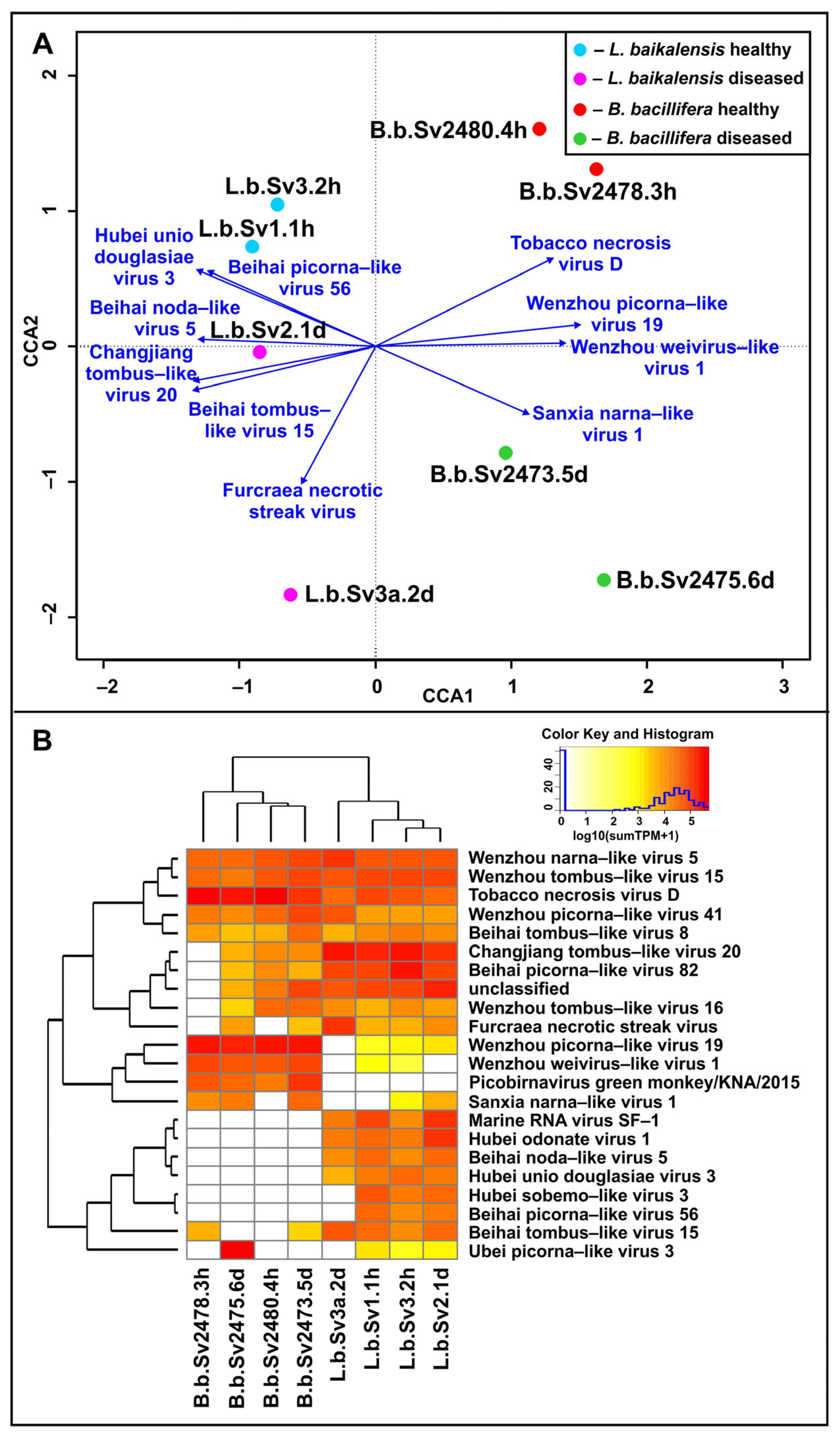
In the CCA analysis (Figure 3A), there was a trend of separation of diseased and healthy branches/individuals, and the influence of damage factors was confirmed statistically (p-value in PERMANOVA 0.042). Note that the distances between healthy individuals of L. baikalensis and B. bacillifera were much smaller than between diseased and healthy individuals and between all diseased specimens. In Figure 3A, one can also see a clear trend in the separation of the sponge species plots. Reliable virotype vectors were divided into two groups, mainly according to “sponge-species” affiliation; that is, one group was directed towards L. baikalensis and another towards B. bacillifera (Figure 3A). Depending on the quantitative representation of different virotypes, samples of the sponges L. baikalensis and B. bacillifera also formed two separate clusters (Figure 3B). However, despite the significance of the differences between the L. baikalensis and B. bacillifera samples (p-value in PERMANOVA 0.025), we cannot state the host–species- specificity of the identified viral communities since the samples of the two species were taken at different stations (at a distance of about 176 km, Figure 1) and during different seasons: in March 2015 (during the freeze-up period; the bottom water temperature at this time was about 0 °C with an average pH of 7.6 [59]) and May 2018 (after ice breaking; temperature 2–3 °C, average pH 8.2 [59]), respectively. The differences between the viromes of L. baikalensis and B. bacillifera most likely formed under the influence of all factors (sponge species, habitat, seasonal or longer-term changes in environmental parameters), but to a different extent.
Among the samples of L. baikalensis, the most numerous RNA-containing virotypes (by number of reads) were Changjiang tombus-like virus 20 and Beihai picorna-like virus 82 (Figure 3B, Supplementary Table S5). Tobacco necrosis virus D and Wenzhou picorna-like virus 19 dominated in B. bacillifera. Marine RNA virus SF-1 (Sanfarnavirus 1, according to ICTV) dominated in L. baikalensis #1 samples (L.b.Sv1.1h and L.b.Sv2.1d); this virotype, as well as Hubei odonate virus 1, quantitatively prevailed in the branch with a lesion (L.b.Sv2.1d). Viruses similar to Wenzhou narna-like virus 5, Furcraea necrotic streak virus, and Wenzhou picorna-like virus 41 were markedly more numerous in the affected branch of sponge L. baikalensis #2 (L.b.Sv3a.2d). For B. bacillifera sponges, some virotypes also predominated in the affected specimens (eg, Picobirnavirus green monkey/KNA/2015 in B.b.Sv2473.5d, or Ubei picorna-like virus 3 in B.b.Sv2475.6d), but others, on the contrary, dominated in apparently healthy individuals (eg, Tobacco necrosis virus D).

3.3. Potential Hosts for Identified Viruses

According to the Virus–Host database (https://www.genome.jp/virushostdb, accessed on 1 February 2023), only three viruses (Tobacco necrosis virus D, Furcraea necrotic streak virus, and Picobirnavirus green monkey/KNA/2015) from the list of virotypes were well-characterized and had known host ranges. The first two viruses infected various plant species, and the third virus infected vertebrates. The genomes of the remaining viruses were obtained from natural samples by assembling metagenomic (meta-transcriptomic) reads, so-called metagenome-assembled genomes (MAGs). As follows from Table 2, most of them (9 virotypes) were obtained from mollusk samples (two of them were also found in fish), five from crustaceans, and two from insects.

3.4. Diversity of RNA-Viral Proteins

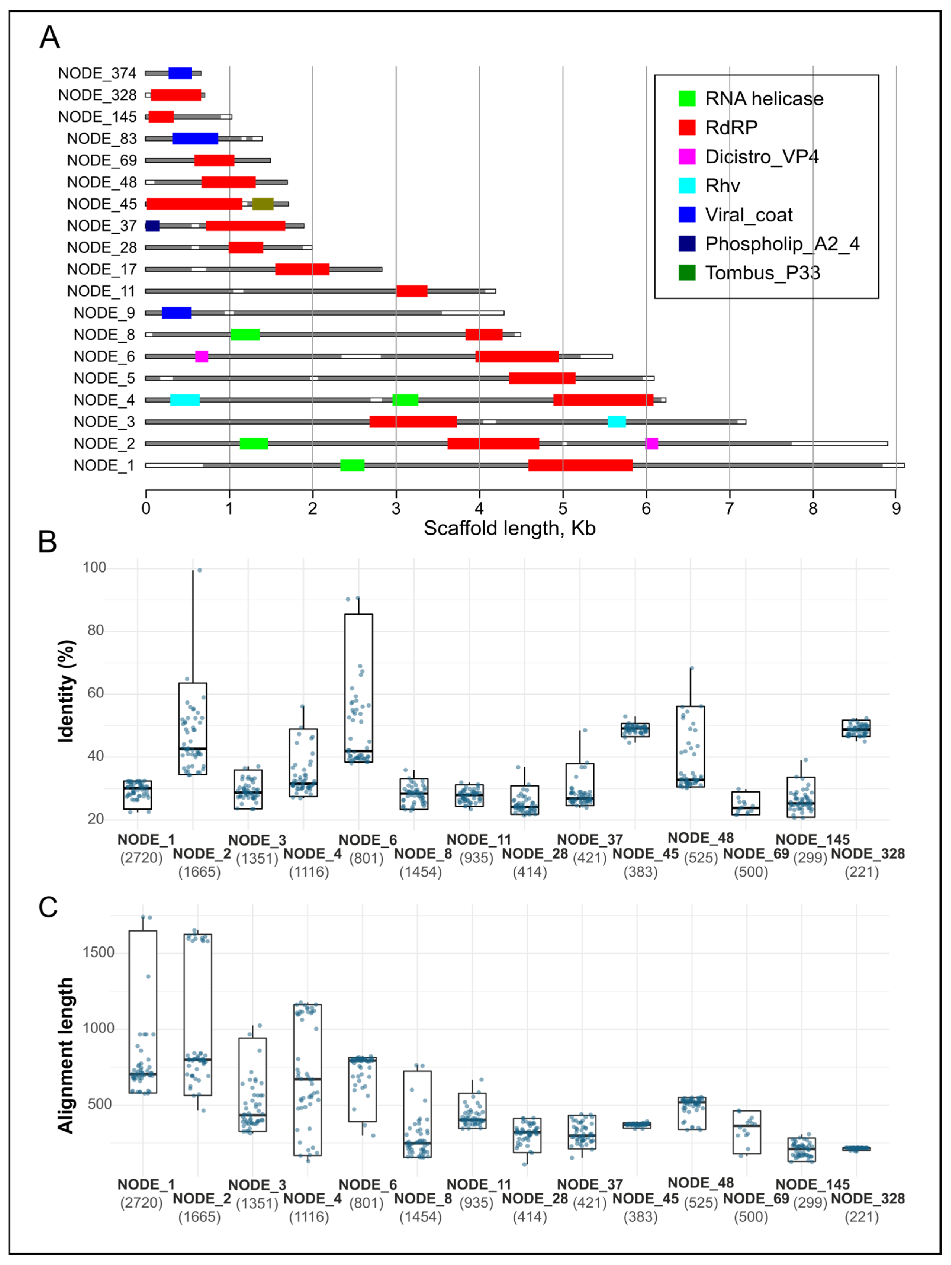
According to the Pfam database, a small portion of the scaffolds (7 scaffolds in total) contained the protein domains of Viral-coat, Rhv, DicistroVP4, and Phospholip_A2_4 as parts of structural polyproteins. Most of scaffolds contained the functional domains of enzymes involved in replication: RNA helicase (4 out of 19) and RNA-dependent RNA polymerase (RdRp and Tombus_P33, 16 out of 19) (Figure 4A, Supplementary Table S6). In addition to these proteins, using the KOfam database, some proteases (Enterovirus proteins 2C, 3C or 3CD), NTPase, kinases, etc. were also identified (Supplementary Table S7).
Our analysis showed a wide range of percent similarity and length alignment between putative functional polyproteins and BLAST hits from the nr NCBI database (Figure 4B,C). For the top 50 BLAST hits, the minimum similarity was 20.7% and the maximum was 99.5%; on average, from 23 to 42%. The largest spread in alignment length was observed for the longest scaffolds (NODE_1, NODE_2, NODE_3, NODE_4 and NODE_8) (Figure 4C). At the same time, the following trend could be traced: the longer the alignment, the lower the percentage of similarity, and vice versa. The significance of this negative correlation was confirmed for RdRps longer than 500 amino acid residues (p-value < 0.05), except for the scaffold NODE_11, where the length of the predicted ORF was 935 aa, but the correlation was absent. The observed results may be associated with a large variability in the sequences, lengths, and orders of different genes in the genomes of RNA viruses, and, accordingly, proteins in the structure of polyproteins. BLAST hits, identified in the nr NCBI database, are often “hypothetical proteins”, and can be either polyproteins or particular proteins (RdRp, Rhv, or others).

4. Discussion

In this study, we first evaluated the diversity of RNA-containing viruses in the Baikal sponges Lubomirskia baikalensis and Baikalospongia bacillifera, representatives of the endemic family Lubomirskiidae, by shotgun sequencing of RNA from an isolated fraction enriched with VLPs. First, we analyzed the composition of the sponge viromes and established the taxonomic affiliation of the reads, along with similar datasets of marine sponges from the SRA NCBI database (Table 1). Next, the reads from the Baikal samples were assembled, and the identified viral scaffolds and open reading frames of potential viral proteins in their structures were analyzed.
The proportion of RNA viral reads in the Baikal samples was small, but we were able to detect a variety of sequences belonging to RNA viruses. Several families were identified in the analysis of the scaffolds, namely, Tombusviridae, Picobirnaviridae, and Marnaviridae: these three families were also the most numerous in the direct analysis of the reads (Table S3). Tombusviridae is a family of ssRNA plant viruses [60]. The hosts for Picobirnaviruses (viruses with dsRNA genomes) were, until recently, thought to be mammals, but it is now known that these viruses can infect birds, reptiles, and invertebrates [29,61,62]. Marnaviridae (ssRNA viruses) is a family in the order Picornavirales that infects various photosynthetic marine protists [63]. In general, the picorna-like viruses are a unique group that infects a remarkable variety of organisms: humans, mammals, insects, plants, and even marine algae [47].
Most of the scaffolds in our study were similar to viruses with undetermined classifications (unclassified Riboviria) whose genomes were assembled in the metatranscriptomics studies of various invertebrates from aquatic biotopes [29] (Table 2); among them there were narna-like, picorna-like, noda-like, and sobemo-like viruses. Thus, the identified RNA viruses in sponge holobionts most likely infect nonbacterial hosts, namely, invertebrates and protists, in contrast to DNA viruses revealed previously in B. bacillifera sponges [16]. Our results also indicate a wide variety of poorly studied RNA viruses circulating among a wide range of aquatic organisms. Recent genomic and metagenomic studies have actively replenished the database with new genomes [10,29,30]; however, additional studies are required in order to establish the range of hosts and other properties of the identified viruses [64].
The similarity of proteins (structural and non-structural) identified in viral scaffolds from the Baikal sponges and those from the RefSeq NCBI database turned out to be low (20.7–67.3%, Supplementary Table S4). BLAST analysis with the nr NCBI database for ORFs with the RdRp and other function domains revealed a maximum level of similarity (99.5%) only between the NODE_2 scaffold and MAG from river sediment (URG14928.1, [65]), and between the NODE_6 scaffold and the nonstructural proteins of Flumine marna-like viruses 8 and 9 (UQB76253.1 and UQB76036.1, respectively) ([66]; in those case, the similarities were 90.6 and 90.3%, respectively. However, the alignment length in these cases was no more than 560 aa (<33% of the total length of the putative polyproteins). For the rest of the identified BLAST hits, the similarity did not exceed 68.3% (Figure 4). This indicates that we have identified previously unknown genomes of RNA viruses. It is interesting that the most similar BLAST hits were isolated from freshwater ecosystems (from sediment or shellfish in China and from water samples in New Zealand). Thus, the number of known viruses circulating in freshwater ecosystems is limited.
The samples of two species of Baikal sponges (L. baikalensis and B. bacillifera) differed in terms of the composition of the identified genotypes (scaffolds) and were clustered separately, which, first of all, would seem to indicate differences in the compositions of viruses of the two sponge species and to confirm the previously obtained host-specific patterns in the viral assemblages [7,8,11]. However, it should be noted once more that sampling of L. baikalensis and B. bacillifera was carried out at different stations and in different seasons (Table 1), and based on the results of this study, we cannot confirm the host–species-specificity of viral communities of these two sponge species. We are also unable to determine the influence of other factors, including habitat (“site-specificity”) [7,8,17], season (as shown for the sponge microbiomes [67]), or more long-term dynamics of environmental parameters.
Nevertheless, we were able to show a significant difference between damaged (or diseased) and visually healthy samples of the L. baikalensis and B. bacillifera sponges. Furthermore, the proportion of viral reads (Supplementary Tables S1 and S5) and the number of virotypes (α-diversity, Supplementary Table S2) in the affected sponges tended to be higher than in the visually healthy sponges, with the exception of L. baikalensis #2, where the number of viral reads and virotypes in the branch with damage (L.b.Sv3a.2d) was much lower than in the visible healthy branch (L.b.Sv3.2h). The distribution of CCA plots in Figure S3 indicates significant shifts and heterogeneity in the composition of viral communities of diseased samples relative to visually healthy ones. Taking into account the obtained results, and based on the available knowledge and many years of experience in monitoring of the states of the sponges (starting from 2015) [18,28], the following picture emerges. Under stress caused by unfavorable factors (climatic, anthropogenic and/or others), disturbances in metabolism, weakening of immunity, and gradual death of sponge cells and their symbionts (necrosis) occur. Following damage and loosening, the body of the sponge, starting from the surface, is colonized by atypical species of bacteria, algae, protozoa, or other organisms, for which a favorable environment rich in nutrients appears [68]. Various observed patterns or types of lesions (spots, necrosis, discoloration, etc. [28]) are result from colonization of sponges by various groups of invaders. Against the background of these stressful conditions, viral and other infections of the holobiont also appear; the hosts for viruses can be associated microorganisms as well as the sponges themselves. Active reproduction of some viruses that present in “healthy” holobionts may also occur. In general, in the event of damage (or disease), the diversity and quantity of viruses and various microorganisms can be replaced and enriched, or, conversely, can become poorer; thereby, the composition of the sponge holobiont varies greatly. This is recorded in the analyses of not only viral, but also microbial, communities of Baikal sponges [69].
Many researchers of marine sponges and coral reefs conclude that diseases of these aquatic organisms occur due to stress caused by changes in their environments. There is ample evidence that the rise in temperature and the influence of anthropogenic factors [17,70,71] preceded the marine sponge disease outbreak, exacerbating the course of the disease [72,73]. Sponges are unique holobionts, and it is the complex interaction of diverse microbial communities that ensures their stability and plasticity [74]. Stress causes changes in the physiological state of the host and leads to a loss of control over the sponge microbiome [17]. One of the most studied adverse factors for marine sponges is elevated temperatures. Under the influence of heat stress, there is a significant decrease in the expression of genes involved in cytoskeletal processes, signaling, and detoxification; an increase in the expression of heat shock proteins (Hsp70); and, at the same time, a change in the composition of the microbiome, which ultimately leads to cell apoptosis and sponge tissue necrosis [75]. The trend of warming of the atmosphere, as well as the surface of the water, in the Baikal region [76,77,78] correlates with the global trend [79]. Thus, the increase in the temperature of the surface water layer of Lake Baikal from 1890 to 2000 (from May to October) averaged about 1.0 degree [76], and during the periods of summer stratification (August–September) for 1977–2003, by 2.0 °C [77]. The increase in the atmospheric temperature over the past 65 years was 1.5 °C [78]. In addition, in the early 2000s (2000–2007), there was a change in the oxygen regime and a tendency for a decrease in oxygen concentration to occur in the summer and autumn months compared to the studied period of the previous century (1948–1951) [59]. Anthropogenic impact has also had a serious detrimental effect on aquatic organisms. For example, the influence of various chemical pollutants on the key stages of life and behavior of corals has been demonstrated [80]. Organic substances coming from wastewater settle on the surfaces of sponges, causing a decrease or blocking of filtration activity [81,82], and prolonged exposure can lead to the death of sponges [83]. An increase in the anthropogenic load on the coastal zone of Lake Baikal and the presence of organic layers on the surfaces of sponges (according to our observations) are adverse factors for sponges’ health and prerequisites for their disease and death.
The list of virotypes includes three well-studied viruses: Tobacco necrosis virus D, Furcraea necrotic streak virus, and Picobirnavirus green monkey/KNA/201. Tobacco necrosis virus D [84], together with Leek white stripe virus and Beet black scorch virus, form the genus Betanecrovirus within the family Tombusviridae. Furcraea necrotic streak virus [85] also belongs to the family Tombusviridae; it is the only member of the Macanavirus genus that causes the so-called “macana disease”. The symptoms of diseases caused by these and other tombusviruses are similar: mottling, wrinkling, and necrotic spots (rounded or in the form of stripes) appear on the surfaces of the leaves of plants. Among the BLAST hits for the proteins from viral scaffolds determined using the nr NCBI database, there were other plant viruses that caused necrosis in plants, such as the aforementioned Leek white stripe virus and Pelargonium ringspot virus (family Tombusviridae), as well as the Southern cowpea mosaic virus (Solemoviridae) (data not shown). Given the common symptomatology of sponge diseases, a reasonable assumption arises regarding the circulation of an unknown RNA virus (or viruses) in sponges which is closely related to a virus affecting plants. This is consistent with the data obtained by [29,34,35] on the close relationship between plant viruses and those of aquatic invertebrates.
The average values of the Shannon and Simpson indices (Supplementary Table S2) in freshwater sponges turned out to be higher than in marine ones (Shannon/Simpson—2.49/0.83 in the Baikal vs. 2.0/0.67 in the marine sponges); in our previous comparative study of DNA viruses [16], these values were almost the same (Shannon/Simpson—3.2/0.89 in B. bacillifera vs. 3.2/0.93 in Ianthella basta marine sponges [11]). The largest number of virotypes (α-diversity) among all datasets was found in the marine sponges Hymeniacidon sp. and Halichondria panacea (458–466 and 308–363, respectively). In the first case, a technique to enrich viral dsRNAs in samples was used [12]; in the second, pools of a large number of individuals (37 individuals in set A and 11 in set B) were analyzed [10]. The viral diversity between different individuals of the same species could vary greatly; for example, 26 to 199 virotypes were found in Aplysina aerophoba and 20 to 116 in Petrosia ficiformis in one study [13].
We were able to show significant differences in the composition of the viromes of the Baikal and marine sponges at the level of families and virotypes. For example, representatives of the families Tombusviridae and Marnaviridae, highly represented in the Baikal sponges, were absent or present in small numbers in the marine sponges. Dominant virotypes in the Baikal sponge samples were generally absent in marine sponges (such as Wenzhou picorna-like virus 19, Shahe picorna-like virus 6, Wenzhou tombus-like virus 1, Marine RNA virus SF-1, and Lactuca sativa marnavirus), and vice versa (Figure 2B, Supplementary Table S3). Among the dominant virotypes in distinct marine samples were sponge-associated viruses, whose genomes were identified only in relevant studies (namely, Sponge holobiont-associated RNA virus in Hymeniacidon sp. sponges [12], or Barns Ness breadcrumb sponge hepe-like virus 1 and Barns Ness breadcrumb sponge aquatic picorna-like virus 1 in the H. panacea [10]), demonstrating the “sponge-species” specificity and the uniqueness of some viruses in sponge holobionts. It is interesting that, in Hymeniacidon sp. sponges, despite having the largest number of virotypes, the Shannon/Simpson diversity indices were among the lowest (average: 1.50/0.53; Supplementary Table S2). This is associated with an uneven distribution of virotypes in normalized (TPM) data and a strong predominance of Sponge holobiont-associated RNA virus (up to 82.4%). The clustering (Figure 2A,B) also indicates the difference between the Baikal and other analyzed samples, which is not surprising given the remoteness of water bodies and the differences in their habitats (primarily the influence of the salinity, as well as the unique cold climate of Lake Baikal). Earlier, we obtained similar results when studying the diversity of DNA-containing viruses in samples of the Baikal sponge Baikalospongia bacillifera [16].

5. Conclusions

Sponges are unique animals, and an integral and ecologically significant component of many marine and freshwater ecosystems. As shown in a review [86], more than 70 studies have been published in which marine sponges were proposed as bioindicators of pollution of aquatic ecosystems with toxic elements (in particular, heavy metals), and the application of these organisms in bioremediation has been considered. Whether sponges can act as bioindicators of viral contamination of aquatic ecosystems due to anthropogenic pollution is still unknown.
Viruses (of sponges and symbionts) are a diverse and inherent part of the sponge holobiont and most likely play a significant role in the development of pathological processes; this is supported by a trend towards an increase in the proportion of viral reads and the number of virotypes in the affected samples. However, prerequisites or aggravating factors for the development of a viral disease, as in the case of any other organism, are still unfavorable conditions (i.e., stress). Sponges represent a complex symbiosis (holobiont), where each component plays an important role and the interactions between all components ensure the smooth and uninterrupted operation of the whole organism. Accordingly, dysbacteriosis or other imbalance in this biosystem, which can be a result of a viral infection, may lead to visible lesions and even death of the sponge. However, to confirm this assumption, additional studies are needed; first of all, more detailed studies of the virotypes we have identified, some of which may affect the sponges themselves, should be conducted.
New members of realm Riboviria have been identified in large numbers in recent studies of a wide range of aquatic organisms, including sponges [10,11,12,13,29,30,87,88]. However, these new data, including those from our research, are far from exhaustive, and an increasing variety of viral RNAs will be revealed in future studies.

Supplementary Materials

The following supporting information can be downloaded at: https://www.mdpi.com/article/10.3390/d15070835/s1, Table S1: General taxonomic composition of the datasets (in %) used in the study; Table S2: The viral diversity indices for the datasets used in the study; Table S3: The list of RNA-viruses (virotypes) revealed in datasets on the base of the reads; Table S4: The scaffolds assembled from the Baikal sponge datasets and their closest relatives from the RefSeq NCBI database (the number of identified proteins in scaffolds and their matches with those of virotypes are indicated); Table S5: The counting of reads from the Baikal samples per virotype; Table S6: Pfam domain, identified in the Baikal scaffolds; Table S7: Identification of proteins with the KEGG Orthology database.

Author Contributions

Conceptualization, methodology, data curation, T.V.B. and Y.S.B.; project administration, funding acquisition, T.V.B.; formal analysis, software, validation, visualization, Y.S.B., I.S.P. and A.N.B.; organization of the expedition and sampling, I.V.K.; writing—original draft preparation, T.V.B., Y.S.B. and O.O.M.; writing—review and editing, T.V.B., Y.S.B., I.S.P., A.N.B., O.O.M. and I.V.K. All authors have read and agreed to the published version of the manuscript.

Funding

This research was carried out within the basic budget funding (numbers 0279-2021-0005 and 0279-2021-0010) of Siberian Branch of the Russian Academy of Sciences and funded by the Russian Foundation for Basic Research (grant number 17-44-388080).

Institutional Review Board Statement

Not applicable.

Data Availability Statement

Raw virome reads for the samples of Lubomirskia baikalensis and Baikalospongia bacillifera were submitted to the National Center for Biotechnology Information (NCBI), Sequence Read Archive (SRA) database (BioProject PRJNA956241, BioSamples SAMN34206360-SAMN34206367). The direct URL to the data is as follows: https://www.ncbi.nlm.nih.gov/sra/PRJNA956241 (accessed on 31 May 2023).

Acknowledgments

We would like to thank The Center of Shared Scientific Equipment “Persistence of Microorganisms” of ICIS UB RAS (Orenburg, Russia) and Genomics Core Facility of ICBFM SB RAS (Novosibirsk, Russia) for carrying out the sequencing. The authors are grateful to Irkutsk Supercomputer Center SB RAS for providing the access to HPC-cluster “Akademik V.M. Matrosov”. The authors are grateful to the Bioinformatics Shared Access Center (Novosibirsk, Russia) for providing computational resources to perform calculations (assembly of paired reads).

Conflicts of Interest

The authors declare no conflict of interest.

References

  1. Webster, N.S.; Taylor, M.W. Marine sponges and their microbial symbionts: Love and other relationships. Environ. Microbiol. 2012, 14, 335–346. [Google Scholar] [CrossRef] [PubMed]
  2. Diaz, M.C.; Rützler, K. Sponges: An essential component of Caribbean coral reefs. Bull. Mar. Sci. 2001, 69, 535–546. [Google Scholar]
  3. Bell, J.J. The functional roles of marine sponges. Estuar. Coast. Shelf Sci. 2008, 79, 341–353. [Google Scholar] [CrossRef]
  4. Wulff, J. Ecological Interactions and the Distribution, Abundance, and Diversity of Sponges. Adv. Mar. Biol. 2012, 61, 273–344. [Google Scholar] [PubMed]
  5. Wulff, J.L. Ecological interactions of marine sponges. Can. J. Zool. 2006, 84, 146–166. [Google Scholar] [CrossRef]
  6. Laffy, P.W.; Wood-Charlson, E.M.; Turaev, D.; Weynberg, K.D.; Botté, E.S.; Van Oppen, M.J.H.; Webster, N.S.; Rattei, T. HoloVir: A workflow for investigating the diversity and function of viruses in invertebrate holobionts. Front. Microbiol. 2016, 7, 822. [Google Scholar] [CrossRef] [PubMed] [Green Version]
  7. Batista, D.; Costa, R.; Carvalho, A.P.; Batista, W.R.; Rua, C.P.J.; de Oliveira, L.; Leomil, L.; Fróes, A.M.; Thompson, F.L.; Coutinho, R.; et al. Environmental conditions affect activity and associated microorganisms of marine sponges. Mar. Environ. Res. 2018, 142, 59–68. [Google Scholar] [CrossRef]
  8. Pascelli, C.; Laffy, P.W.; Botté, E.; Kupresanin, M.; Rattei, T.; Lurgi, M.; Ravasi, T.; Webster, N.S. Viral ecogenomics across the Porifera. Microbiome 2020, 8, 144. [Google Scholar] [CrossRef]
  9. Nguyen, M.; Wemheuer, B.; Laffy, P.W.; Webster, N.S.; Thomas, T. Taxonomic, functional and expression analysis of viral communities associated with marine sponges. PeerJ 2021, 9, e10715. [Google Scholar] [CrossRef]
  10. Waldron, F.M.; Stone, G.N.; Obbard, D.J. Metagenomic sequencing suggests a diversity of RNA interference-like responses to viruses across multicellular eukaryotes. PLoS Genet. 2018, 14, e1007533. [Google Scholar] [CrossRef] [Green Version]
  11. Laffy, P.W.; Wood-Charlson, E.M.; Turaev, D.; Jutz, S.; Pascelli, C.; Botté, E.S.; Bell, S.C.; Peirce, T.E.; Weynberg, K.D.; van Oppen, M.J.H.; et al. Reef invertebrate viromics: Diversity, host specificity and functional capacity. Environ. Microbiol. 2018, 20, 2125–2141. [Google Scholar] [CrossRef] [PubMed]
  12. Urayama, S.I.; Takaki, Y.; Hagiwara, D.; Nunoura, T. DsRNA-seq reveals novel RNA virus and virus-like putative complete genome sequences from Hymeniacidon sp. Sponge. Microbes Environ. 2020, 35, ME19132. [Google Scholar] [CrossRef] [Green Version]
  13. Jahn, M.T.; Arkhipova, K.; Markert, S.M.; Stigloher, C.; Lachnit, T.; Pita, L.; Kupczok, A.; Ribes, M.; Stengel, S.T.; Rosenstiel, P.; et al. A Phage Protein Aids Bacterial Symbionts in Eukaryote Immune Evasion. Cell Host Microbe 2019, 26, 542–550.e5. [Google Scholar] [CrossRef]
  14. Butina, T.V.; Bukin, Y.S.; Khanaev, I.V.; Kravtsova, L.S.; Maikova, O.O.; Tupikin, A.E.; Kabilov, M.R.; Belikov, S.I. Metagenomic analysis of viral communities in diseased Baikal sponge Lubomirskia baikalensis. Limnol. Freshw. Biol. 2019, 1, 155–162. [Google Scholar] [CrossRef] [Green Version]
  15. Butina, T.V.; Khanaev, I.V.; Kravtsova, L.S.; Maikova, O.O.; Bukin, Y.S. Metavirome datasets from two endemic Baikal sponges Baikalospongia bacillifera. Data Br. 2020, 29, 105260. [Google Scholar] [CrossRef] [PubMed]
  16. Butina, T.V.; Petrushin, I.S.; Khanaev, I.V.; Bukin, Y.S. Metagenomic Assessment of DNA Viral Diversity in Freshwater Sponges, Baikalospongia bacillifera. Microorganisms 2022, 10, 480. [Google Scholar] [CrossRef]
  17. Pita, L.; Rix, L.; Slaby, B.M.; Franke, A.; Hentschel, U. The sponge holobiont in a changing ocean: From microbes to ecosystems. Microbiome 2018, 6, 46. [Google Scholar] [CrossRef]
  18. Khanaev, I.V.; Kravtsova, L.S.; Maikova, O.O.; Bukshuk, N.A.; Sakirko, M.V.; Kulakova, N.V.; Butina, T.V.; Nebesnykh, I.A.; Belikov, S.I. Current state of the sponge fauna (Porifera: Lubomirskiidae) of Lake Baikal: Sponge disease and the problem of conservation of diversity. J. Great Lakes Res. 2018, 44, 77–85. [Google Scholar] [CrossRef]
  19. Bondarenko, N.A.; Vorobyova, S.S.; Zhuchenko, N.A.; Golobokova, L.P. Current state of phytoplankton in the littoral area of Lake Baikal, spring. J. Great Lakes Res. 2020, 46, 17–28. [Google Scholar] [CrossRef]
  20. Kravtsova, L.; Vorobyeva, S.; Naumova, E.; Izhboldina, L.; Mincheva, E.; Potemkina, T.; Pomazkina, G.; Rodionova, E.; Onishchuk, N.; Sakirko, M.; et al. Response of aquatic organisms communities to global climate changes and anthropogenic impact: Evidence from Listvennichny bay of Lake Baikal. Biology 2021, 10, 904. [Google Scholar] [CrossRef] [PubMed]
  21. Khodzher, T.V.; Domysheva, V.M.; Sorokovikova, L.M.; Sakirko, M.V.; Tomberg, I.V. Current chemical composition of Lake Baikal water. Inland Waters 2017, 7, 250–258. [Google Scholar] [CrossRef]
  22. Onishchuk, N.A.; Netsvetaeva, O.G.; Tomberg, I.V.; Sakirko, M.V.; Domysheva, V.M.; Golobokova, L.P.; Khodzher, T.V. Seasonal dynamics of mineral forms of nitrogen in the river water, snow cover and precipitation near the Listvyanka settlement (west coast of the Southern Baikal). Limnol. Freshw. Biol. 2019, 3, 245–252. [Google Scholar] [CrossRef]
  23. Bukin, Y.S.; Bondarenko, N.A.; Rusanov, I.I.; Pimenov, N.V.; Bukin, S.V.; Pogodaeva, T.V.; Chernitsyna, S.M.; Shubenkova, O.V.; Ivanov, V.G.; Zakharenko, A.S.; et al. Interconnection of bacterial and phytoplanktonic communities with hydrochemical parameters from ice and under-ice water in coastal zone of Lake Baikal. Sci. Rep. 2020, 10, 11087. [Google Scholar] [CrossRef] [PubMed]
  24. Malnik, V.V.; Suturin, A.N.; Gorshkova, A.S.; Shtykova, Y.R.; Timoshkin, O.A. Water Quality in the Shallow Zone of Lake Baikal as Deduced from Sanitary and Microbiological Indicators. Geogr. Nat. Resour. 2022, 43, 141–148. [Google Scholar] [CrossRef]
  25. Maikova, O.; Bukshuk, N.; Kravtsova, L.; Nebesnyh, I.; Yakhnenko, A.; Butina, T.; Khanaev, I. Baikal endemic sponge disease and anthropogenic factor. IOP Conf. Ser. Earth Environ. Sci. 2021, 937, 22071. [Google Scholar] [CrossRef]
  26. Maldonado, M.; Sánchez-Tocino, L.; Navarro, C. Recurrent disease outbreaks in corneous demosponges of the genus Ircinia: Epidemic incidence and defense mechanisms. Mar. Biol. 2010, 157, 1577–1590. [Google Scholar] [CrossRef]
  27. Sweet, M.; Bulling, M.; Cerrano, C. A novel sponge disease caused by a consortium of micro-organisms. Coral Reefs 2015, 34, 871–883. [Google Scholar] [CrossRef]
  28. Maikova, O.O.; Bukshuk, N.A.; Kravtsova, L.S.; Onishchuk, N.A.; Sakirko, M.V.; Nebesnykh, I.A.; Lipko, I.A.; Khanaev, I.V. Sponge fauna of Lake Baikal in the monitoring system for six years of observations. Contemp. Probl. Ecol. 2023, 30, 11–24. [Google Scholar]
  29. Shi, M.; Lin, X.D.; Tian, J.H.; Chen, L.J.; Chen, X.; Li, C.X.; Qin, X.C.; Li, J.; Cao, J.P.; Eden, J.S.; et al. Redefining the invertebrate RNA virosphere. Nature 2016, 540, 539–543. [Google Scholar] [CrossRef]
  30. Zhang, Y.Y.; Chen, Y.; Wei, X.; Cui, J. Viromes in marine ecosystems reveal remarkable invertebrate RNA virus diversity. Sci. China Life Sci. 2022, 65, 426–437. [Google Scholar] [CrossRef]
  31. Johnson, P.T. Viral diseases of marine invertebrates. Helgoländer Meeresunters. 1984, 37, 65–98. [Google Scholar] [CrossRef] [Green Version]
  32. Ryabov, E.V. Invertebrate RNA virus diversity from a taxonomic point of view. J. Invertebr. Pathol. 2017, 147, 37–50. [Google Scholar] [CrossRef] [PubMed]
  33. Koonin, E.V.; Dolja, V.V.; Krupovic, M. Origins and evolution of viruses of eukaryotes: The ultimate modularity. Virology 2015, 479–480, 2–25. [Google Scholar] [CrossRef] [Green Version]
  34. Dolja, V.V.; Koonin, E.V. Metagenomics reshapes the concepts of RNA virus evolution by revealing extensive horizontal virus transfer. Virus Res. 2018, 244, 36–52. [Google Scholar] [CrossRef]
  35. Shi, M.; Zhang, Y.Z.; Holmes, E.C. Meta-transcriptomics and the evolutionary biology of RNA viruses. Virus Res. 2018, 243, 83–90. [Google Scholar] [CrossRef] [PubMed]
  36. Chomczynski, P. A reagent for the single-step simultaneous isolation of RNA, DNA and proteins from cell and tissue samples. Biotechniques 1993, 15, 532–537. [Google Scholar]
  37. Bolger, A.M.; Lohse, M.; Usadel, B. Trimmomatic: A flexible trimmer for Illumina sequence data. Bioinformatics 2014, 30, 2114–2120. [Google Scholar] [CrossRef] [PubMed] [Green Version]
  38. Menzel, P.; Ng, K.L.; Krogh, A. Fast and sensitive taxonomic classification for metagenomics with Kaiju. Nat. Commun. 2016, 7, 11257. [Google Scholar] [CrossRef] [Green Version]
  39. Nurk, S.; Meleshko, D.; Korobeynikov, A.; Pevzner, P.A. MetaSPAdes: A new versatile metagenomic assembler. Genome Res. 2017, 27, 824–834. [Google Scholar] [CrossRef] [Green Version]
  40. Langmead, B.; Salzberg, S.L. Fast gapped-read alignment with Bowtie. Nat. Methods 2012, 9, 357–359. [Google Scholar] [CrossRef] [Green Version]
  41. Danecek, P.; Bonfield, J.K.; Liddle, J.; Marshall, J.; Ohan, V.; Pollard, M.O.; Whitwham, A.; Keane, T.; McCarthy, S.A.; Davies, R.M. Twelve years of SAMtools and BCFtools. Gigascience 2021, 10, giab008. [Google Scholar] [CrossRef] [PubMed]
  42. Guo, J.; Bolduc, B.; Zayed, A.A.; Varsani, A.; Dominguez-Huerta, G.; Delmont, T.O.; Pratama, A.A.; Gazitúa, M.C.; Vik, D.; Sullivan, M.B.; et al. VirSorter2: A multi-classifier, expert-guided approach to detect diverse DNA and RNA viruses. Microbiome 2021, 9, 37. [Google Scholar] [CrossRef] [PubMed]
  43. Nayfach, S.; Camargo, A.P.; Schulz, F.; Eloe-Fadrosh, E.; Roux, S.; Kyrpides, N.C. CheckV assesses the quality and completeness of metagenome-assembled viral genomes. Nat. Biotechnol. 2021, 39, 578–585. [Google Scholar] [CrossRef]
  44. Zhao, S.; Ye, Z.; Stanton, R. Misuse of RPKM or TPM normalization when comparing across samples and sequencing protocols. RNA 2020, 26, 903–909. [Google Scholar] [CrossRef] [PubMed] [Green Version]
  45. O’Leary, N.A.; Wright, M.W.; Brister, J.R.; Ciufo, S.; Haddad, D.; McVeigh, R.; Rajput, B.; Robbertse, B.; Smith-White, B.; Ako-Adjei, D.; et al. Reference sequence (RefSeq) database at NCBI: Current status, taxonomic expansion, and functional annotation. Nucleic Acids Res. 2016, 44, D733–D745. [Google Scholar] [CrossRef] [PubMed] [Green Version]
  46. Buchfink, B.; Xie, C.; Huson, D.H. Fast and sensitive protein alignment using DIAMOND. Nat. Methods 2015, 12, 59–60. [Google Scholar] [CrossRef]
  47. Mihara, T.; Nishimura, Y.; Shimizu, Y.; Nishiyama, H.; Yoshikawa, G.; Uehara, H.; Hingamp, P.; Goto, S.; Ogata, H. Linking virus genomes with host taxonomy. Viruses 2016, 8, 66. [Google Scholar] [CrossRef] [Green Version]
  48. Kanehisa, M.; Sato, Y.; Kawashima, M.; Furumichi, M.; Tanabe, M. KEGG as a reference resource for gene and protein annotation. Nucleic Acids Res. 2016, 44, D457–D462. [Google Scholar] [CrossRef] [Green Version]
  49. Aramaki, T.; Blanc-Mathieu, R.; Endo, H.; Ohkubo, K.; Kanehisa, M.; Goto, S.; Ogata, H. KofamKOALA: KEGG Ortholog assignment based on profile HMM and adaptive score threshold. Bioinformatics 2020, 36, 2251–2252. [Google Scholar] [CrossRef] [Green Version]
  50. Mistry, J.; Chuguransky, S.; Williams, L.; Qureshi, M.; Salazar, G.A.; Sonnhammer, E.L.L.; Tosatto, S.C.E.; Paladin, L.; Raj, S.; Richardson, L.J.; et al. Pfam: The protein families database in 2021. Nucleic Acids Res. 2021, 49, D412–D419. [Google Scholar] [CrossRef]
  51. O’Hara, R.B. Species richness estimators: How many species can dance on the head of a pin? J. Anim. Ecol. 2005, 74, 375–386. [Google Scholar] [CrossRef]
  52. Colwell, R.K.; Coddington, J.A. Estimating terrestrial biodiversity through extrapolation. Biodivers. Meas. Estim. 1995, 345, 101–118. [Google Scholar]
  53. Hill, M.O. Diversity and Evenness: A Unifying Notation and Its Consequences. Ecology 1973, 54, 427–432. [Google Scholar] [CrossRef] [Green Version]
  54. Oksanen, J.; Simpson, G.L.; Blanchet, F.G.; Kindt, R.; Legendre, P.; Minchin, P.R.; O’Hara, R.B.; Solymos, P.; Stevens, M.H.H.; Szoecs, E.; et al. R Package, Version 2.6-4, Package ‘Vegan’: Community Ecology Package; 2022; pp. 1–297. Available online: https://cran.r-project.org/web/packages/vegan/vegan.pdf (accessed on 12 October 2022).
  55. Warnes, G.R.; Bolker, B.; Bonebakker, L.; Gentleman, R.; Liaw, W.H.A.; Lumley, T.; Maechler, M.; Magnusson, A.; Moeller, S.; Schwartz, M.; et al. R Package, Version 2.17.0; Package “gplots”: Various R Programming Tools for Plotting Data; ScienceOpen: Berlin, Germany, 2015; pp. 1–68. [Google Scholar]
  56. Anderson, M.J. A new method for non-parametric multivariate analysis of variance. Austral Ecol. 2001, 26, 32–46. [Google Scholar]
  57. Conceição-Neto, N.; Zeller, M.; Lefrère, H.; De Bruyn, P.; Beller, L.; Deboutte, W.; Yinda, C.K.; Lavigne, R.; Maes, P.; Ranst, M.V.; et al. Modular approach to customise sample preparation procedures for viral metagenomics: A reproducible protocol for virome analysis. Sci. Rep. 2015, 5, 16532. [Google Scholar] [CrossRef] [PubMed] [Green Version]
  58. Kuhar, U.; Vengust, G.; Jamnikar-Ciglenecki, U. Complete genome sequence of roe deer picobirnavirus strain PBV/roe_deer/SLO/D38-14/2014. Genome Announc. 2017, 5, e01329-17. [Google Scholar] [CrossRef] [Green Version]
  59. Golobokova, L.P.; Sakirko, M.V.; Onishchuk, N.A.; Pogodaeva, T.V.; Sez’ko, N.P.; Dolya, I.N. Hydrochemical characteristics of littoral waters of North-Western coast of Southern Baikal. In Index of Animal Species Inhabiting Lake Baikal and Its Catchment Area II (1); Timoshkin, O.A., Ed.; Nauka: Novosibirsk, Russia, 2009; pp. 760–784. (In Russian) [Google Scholar]
  60. King, A.M.Q.; Adams, M.J.; Carstens, E.B.; Lefkowitz, E. (Eds.) Family—Tombusviridae. In Ninth Report of the International Committee on Taxonomy of Viruses; Elsevier: Amsterdam, The Netherlands, 2012. [Google Scholar]
  61. Fregolente, M.C.D.; de Castro-Dias, E.; Martins, S.S.; Spilki, F.R.; Allegretti, S.M.; Gatti, M.S.V. Molecular characterization of picobirnaviruses from new hosts. Virus Res. 2009, 143, 134–136. [Google Scholar] [CrossRef]
  62. Delmas, B.; Attoui, H.; Ghosh, S.; Malik, Y.S.; Mundt, E.; Vakharia, V.N. ICTV virus taxonomy profile: Picobirnaviridae. J. Gen. Virol. 2019, 100, 133–134. [Google Scholar] [CrossRef]
  63. Lang, A.S.; Vlok, M.; Culley, A.I.; Suttle, C.A.; Takao, Y.; Tomaru, Y.; Siddell, S.G.; Lefkowitz, E.; Simmonds, P.; Zerbini, F.M.; et al. ICTV virus taxonomy profile: Marnaviridae. J. Gen. Virol. 2021, 102, 1633. [Google Scholar] [CrossRef]
  64. Obbard, D.J. Expansion of the metazoan virosphere: Progress, pitfalls, and prospects. Curr. Opin. Virol. 2018, 31, 17–23. [Google Scholar] [CrossRef] [Green Version]
  65. Chen, Y.M.; Sadiq, S.; Tian, J.H.; Chen, X.; Lin, X.D.; Shen, J.J.; Chen, H.; Hao, Z.Y.; Wille, M.; Zhou, Z.C.; et al. RNA viromes from terrestrial sites across China expand environmental viral diversity. Nat. Microbiol. 2022, 7, 1312–1323. [Google Scholar] [CrossRef]
  66. French, R.; Charon, J.; Lay, C.L.; Muller, C.; Holmes, E.C. Human land use impacts viral diversity and abundance in a New Zealand river. Virus Evol. 2022, 8, veac032. [Google Scholar] [CrossRef]
  67. Glasl, B.; Robbins, S.; Frade, P.R.; Marangon, E.; Laffy, P.W.; Bourne, D.G.; Webster, N.S. Comparative genome-centric analysis reveals seasonal variation in the function of coral reef microbiomes. ISME J. 2020, 14, 1435–1450. [Google Scholar] [CrossRef]
  68. Evseev, P.; Tikhonova, I.; Krasnopeev, A.; Sorokovikova, E.; Gladkikh, A.; Timoshkin, O.; Miroshnikov, K.; Belykh, O. Tychonema sp. BBK16 Characterisation: Lifestyle, Phylogeny and Related Phages. Viruses 2023, 15, 442. [Google Scholar] [CrossRef]
  69. Belikov, S.; Belkova, N.; Butina, T.; Chernogor, L.; Kley, A.M.-V.; Nalian, A.; Rorex, C.; Khanaev, I.; Maikova, O.; Feranchuk, S. Diversity and shifts of the bacterial community associated with Baikal sponge mass mortalities. PLoS ONE 2019, 14, e0213926. [Google Scholar] [CrossRef] [PubMed]
  70. Erwin, P.M.; Pita, L.; López-Legentil, S.; Turon, X. Stability of sponge-associated bacteria over large seasonal shifts in temperature and irradiance. Appl. Environ. Microbiol. 2012, 78, 7358–7368. [Google Scholar] [CrossRef] [PubMed] [Green Version]
  71. Slaby, B.M.; Franke, A.; Rix, L.; Pita, L.; Bayer, K.; Jahn, M.T.; Hentschel, U. Symbiotic Microbiomes of Coral Reefs Sponges and Corals; Springer: Dordrecht, The Netherlands, 2019; pp. 81–104. [Google Scholar]
  72. Webster, N.S. Sponge disease: A global threat? Environ. Microbiol. 2007, 9, 1363–1375. [Google Scholar] [CrossRef] [PubMed]
  73. Luter, H.M.; Webster, N.S. Sponge Disease and Climate Change. In Climate Change, Ocean Acidification and Sponges; Springer: Cham, Switzerland, 2017; pp. 411–428. [Google Scholar]
  74. Maldonado, M.; Aguilar, R.; Bannister, R.; Bell, J.; Conway, J.; Dayton, P.; Diaz, C.; Gutt, J.; Kelly, M.; Kenchington, E.; et al. Sponge Grounds as Key Marine Habitats: A Synthetic Review of Types, Structure, Functional Roles, and Conservation Concerns. In Marine Animal Forests: The Ecology of Benthic Biodiversity Hotspots; Springer International Publishing: Cham, Switzerland, 2017. [Google Scholar]
  75. Fan, L.; Liu, M.; Simister, R.; Webster, N.S.; Thomas, T. Marine microbial symbiosis heats up: The phylogenetic and functional response of a sponge holobiont to thermal stress. ISME J. 2013, 7, 991–1002. [Google Scholar] [CrossRef]
  76. Shimaraev, M.N.; Domysheva, V.M. Trends in Hydrological and hydrochemical Processes in Lake Baikal under Conditions of Modern Climate Change. In Climatic Change and Global Warming of Inland Waters: Impacts and Mitigation for Ecosystems and Societies; Goldman, C.R., Kumagai, M., Robarts, R.D., Eds.; John Wiley & Sons, Ltd.: Hoboken, NJ, USA, 2013; pp. 43–66. [Google Scholar]
  77. Izmest’eva, L.R.; Moore, M.V.; Hampton, S.E.; Ferwerda, C.J.; Gray, D.K.; Woo, K.H.; Pislegina, H.V.; Krashchuk, L.S.; Shimaraeva, S.V.; Silow, E.A. Lake-wide physical and biological trends associated with warming in Lake Baikal. J. Great Lakes Res. 2016, 42, 6–17. [Google Scholar] [CrossRef] [Green Version]
  78. Shimaraev, M.N.; Sinyukovich, V.N.; Sizova, L.N.; Troitskaya, E.S. Heat balance of Lake Baikal and the relationship of its ice-thermal and water regime with global atmospheric circulation in the Northern Hemisphere during the modern period. Limnol. Freshw. Biol. 2018, 2018, 10–14. [Google Scholar] [CrossRef]
  79. Valipour, M.; Bateni, S.M.; Jun, C. Global surface temperature: A new insight. Climate 2021, 9, 81. [Google Scholar] [CrossRef]
  80. Richmond, R.H.; Tisthammer, K.H.; Spies, N.P. The effects of anthropogenic stressors on reproduction and recruitment of corals and reef organisms. Front. Mar. Sci. 2018, 5, 226. [Google Scholar] [CrossRef] [Green Version]
  81. Tompkins-Macdonald, G.J.; Leys, S.P. Glass sponges arrest pumping in response to sediment: Implications for the physiology of the hexactinellid conduction system. Mar. Biol. 2008, 154, 973–984. [Google Scholar] [CrossRef]
  82. Bannister, R.J.; Battershill, C.N.; de Nys, R. Suspended sediment grain size and mineralogy across the continental shelf of the Great Barrier Reef: Impacts on the physiology of a coral reef sponge. Cont. Shelf Res. 2012, 32, 86–95. [Google Scholar] [CrossRef]
  83. Maldonado, M.; Giraud, K.; Carmona, C. Effects of sediment on the survival of asexually produced sponge recruits. Mar. Biol. 2008, 154, 631–641. [Google Scholar] [CrossRef]
  84. Smith, K.M.; Bald, J.G. A description of a necrotic virus disease affecting tobacco and other plants. Parasitology 1935, 27, 231–245. [Google Scholar] [CrossRef]
  85. Dabek, A.J.; Castano, J.J. The occurrence, symptomatology, transmission and virus aetiology of macana disease of fique (Furcraea spp.) in Colombia, South America. J. Phytopathol. 1978, 92, 57–69. [Google Scholar] [CrossRef]
  86. Roveta, C.; Annibaldi, A.; Afghan, A.; Calcinai, B.; Di Camillo, C.G.; Gregorin, C.; Illuminati, S.; Mantas, T.P.; Truzzi, C.; Puce, S. Biomonitoring of heavy metals: The unexplored role of marine sessile taxa. Appl. Sci. 2021, 11, 580. [Google Scholar] [CrossRef]
  87. Culley, A. New insight into the RNA aquatic virosphere via viromics. Virus Res. 2018, 244, 84–89. [Google Scholar] [CrossRef]
  88. Kolundžija, S.; Cheng, D.-Q.; Lauro, F.M. RNA Viruses in Aquatic Ecosystems through the Lens of Ecological Genomics and Transcriptomics. Viruses 2022, 14, 702. [Google Scholar] [CrossRef]
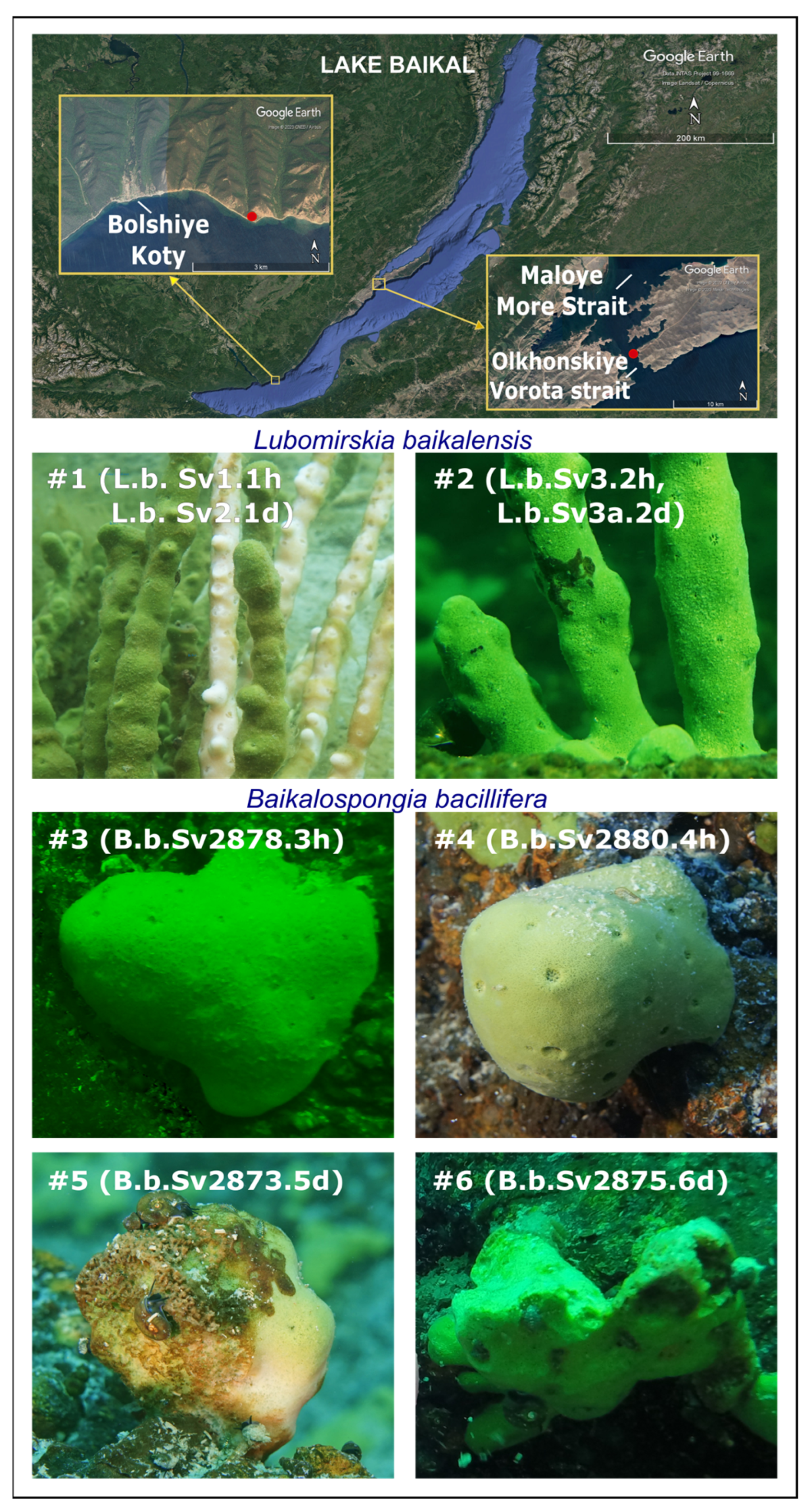
Figure 1. Sampling sites and Lubomirskia baikalensis and Baikalospongia bacillifera sponges used for the analysis of RNA viruses. Individuals of L. baikalensis collected in March 2015 from the Maloye More Strait showed signs of damage: discoloration (#1) and brown spots (#2). Four specimens of B. bacillifera were collected in May 2018 from the Bolshiye Koty region; two of them, #3–#4, were visually healthy, and two, #5–#6, showed necrosis and brown spot-like lesions. Numbers of samples for analysis are given in parentheses (with the letter “h” at the end—healthy, with “d”—diseased).
Diversity 15 00835 g001
Figure 2. Comparative analysis of the Baikal and marine samples based on the number of reads belonging to different RNA virotypes. (A)—CCA scatter plot with gradient vectors of RNA viral families; (B)—heatmap of top 30 dominant virotypes with clustering by Euclidean distances. Abbreviations: “Hepe-”—Hepeviridae, “Dicistro-”—Dicistroviridae, “vir”—virus.
Diversity 15 00835 g002
Figure 3. Representation of RNA virotypes in the samples of the Baikal sponges L. baikalensis and B. bacillifera: (A)—CCA analysis based on the revealed virotypes; (B)—heatmap and clustering (Euclidean distances) of the virotypes and samples.
Diversity 15 00835 g003
Figure 4. Layout of identified Pfam domains in viral scaffolds from the Baikal sponges ((A); the gray lines show the identified ORFs), percent similarity (B), and alignment length (C) of the ORFs, including non-structural domains, with the top 50 BLAST hits from the nr NCBI database. In brackets after the number of the scaffold (in (B,C)), the length of the ORF (aa) is indicated.
Diversity 15 00835 g004
Table 1. Description of sponge datasets used for analysis.
Dataset Name 1,2Sample DescriptionGeographic LocationLatitude and LongitudeDataNucleic AcidsExperimentsReference
L.b.Sv1.1hLubomirskia baikalensis, healthy branchRussia: Lake Baikal53.02 N 106.93 EMarch 2015RNA (V)SRX19982199This study
L.b.Sv2.1dLubomirskia baikalensis, branch with bleachingRussia: Lake Baikal53.02 N 106.93 EMarch 2015RNA (V)SRX19982200This study
L.b.Sv3.2hLubomirskia baikalensis, healthy branchRussia: Lake Baikal53.02 N 106.93 EMarch 2015RNA (V)SRX19982201This study
L.b.Sv3a.2dLubomirskia baikalensis, branch with brown spotsRussia: Lake Baikal53.02 N 106.93 EMarch 2015RNA (V)SRX19982202This study
B.b.Sv2478.3hBaikalospongia bacillifera, healthyRussia: Lake Baikal51.90 N 105.10 EJune 2018RNA (V)SRX19982203This study
B.b.Sv2480.4hBaikalospongia bacillifera, healthyRussia: Lake Baikal51.90 N 105.10 EJune 2018RNA (V)SRX19982204This study
B.b.Sv2473.5dBaikalospongia bacillifera, necrosis, brown spotsRussia: Lake Baikal51.90 N 105.10 EJune 2018RNA (V)SRX19982205This study
B.b.Sv2475.6dBaikalospongia bacillifera, necrosis, brown spotsRussia: Lake Baikal51.90 N 105.10 EJune 2018RNA (V)SRX19982206This study
Ch.reniformis_1Chondrosia reniformis, m/pSpain: Mediterranean Sea, RM42.08 N 3.20 EJuly 2016RNA/DNA (V)SRX5385349, SRX5385338[13]
Ch.reniformis_2Chondrosia reniformis, m/pSpain: Mediterranean Sea, RM42.08 N 3.20 EJuly 2016RNA/DNA (V)SRX5385350, SRX5385337[13]
Ch.reniformis_3Chondrosia reniformis, m/pSpain: Mediterranean Sea, RM42.08 N 3.20 EJuly 2016RNA/DNA (V)SRX5385353, SRX5385336[13]
Ap.aerophoba_1Aplysina aerophoba, m/pSpain: Mediterranean Sea, PL42.30 N 3.29 EJuly 2016RNA/DNA (V)SRX5385319, SRX5385345[13]
Ap.aerophoba_2Aplysina aerophoba, m/pSpain: Mediterranean Sea, PL42.30 N 3.29 EJuly 2016RNA/DNA (V)SRX5385320, SRX5385346[13]
Ap.aerophoba_3Aplysina aerophoba, m/pSpain: Mediterranean Sea, PL42.30 N 3.29 EJuly 2016RNA/DNA (V)SRX5385347, SRX5385351[13]
P.ficiformis_1Petrosia ficiformis, m/pSpain: Mediterranean Sea, RM42.08 N 3.20 EJuly 2016RNA/DNA (V)SRX5385334, SRX5385330[13]
P.ficiformis_2Petrosia ficiformis, m/pSpain: Mediterranean Sea, RM42.08 N 3.20 EJuly 2016RNA/DNA (V)SRX5385333, SRX5385329[13]
P.ficiformis_3Petrosia ficiformis, m/pSpain: Mediterranean Sea, RM42.08 N 3.20 EJuly 2016RNA/DNA (V)SRX5385332, SRX5385339[13]
Ag.oroides_1Agelas oroides, m/pSpain: Mediterranean Sea, RM42.08 N 3.20 EJuly 2016RNA/DNA (V)SRX5385321, SRX5385325[13]
Ag.oroides_2Agelas oroides, m/pSpain: Mediterranean Sea, RM42.08 N 3.20 EJuly 2016RNA/DNA (V)SRX5385322, SRX5385326[13]
Ag.oroides_3Agelas oroides, m/pSpain: Mediterranean Sea, RM42.08 N 3.20 EJuly 2016RNA/DNA (V)SRX5385323, SRX5385327[13]
Med.sw.PL (2)SeawaterSpain: Mediterranean Sea, PL42.30 N 3.29 EJuly 2016RNA/DNA (V)SRX5385341, SRX5385342[13]
Med.sw.RM (2)SeawaterSpain: Mediterranean Sea, RM42.30 N 3.29 EJuly 2016RNA/DNA (V)SRX5385343, SRX5385344[13]
Hym.sp_1Hymeniacidon sp.Japan: Tokyo bay35.34 N 139.64 EApril 2014RNA (T)DRX171015[12]
Hym.sp_2Hymeniacidon sp.Japan: Tokyo bay35.34 N 139.64 EApril 2015RNA (T)DRX171017[12]
GBR.sw (3)SeawaterAustralia: Great Barrier Reef18.83 S 147.63 EOctober 2014RNA (V)SRX2883311, SRX2883316, SRX2883317[11]
Rh.odorabile_1Rhopaloeides odorabileAustralia: Great Barrier Reef18.83 S 147.63 EOctober 2014RNA (V)SRX2883299[11]
Rh.odorabile_2Rhopaloeides odorabileAustralia: Great Barrier Reef18.83 S 147.63 EOctober 2014RNA (V)SRX2883312[11]
Rh.odorabile_3Rhopaloeides odorabileAustralia: Great Barrier Reef18.83 S 147.63 EOctober 2014RNA (V)SRX2883313[11]
Hal.panicea_A (37)Halichondria panaceaUnited Kingdom: North Sea, BN55.99 N–2.45 W2014RNA (T)SRX4378335[10]
Hal.panicea-B (11)Halichondria panaceaUnited Kingdom: North Sea, BN55.99 N–2.45 W2014RNA (T)SRX4378332[10]
1 The last two characters in the names of the Baikal samples indicate the number of the sponge and the state of the individual: “h”—visually healthy, “d”—diseased/ damaged. 2 The numbers in brackets indicate the number of samples or data sets (replicates). Abbreviations: PL—Portlligat; RM—Montgrí, Medes Islands and Baix Ter Natural Park; BN—Barns Ness; m/p—mesohyl and pinacoderm; V—viral; T—total.
Table 2. RNA virotypes revealed in the Baikal sponge datasets.
SpeciesFamily *Genus *Host/Isolation SourceNo. of Scaffolds
Tobacco necrosis virus DTombusviridaeBetanecrovirusPlants45
Changjiang tombus-like virus 20--/Crustaceans15, 17, 37
Wenzhou narna-like virus 5--/Mollusk24, 9, 532
Beihai picorna-like virus 82--/Crustaceans69
Wenzhou picorna-like virus 19--/Mollusk3
unclassified-- 5, 25, 374
Picobirnavirus green monkey/KNA/2015PicobirnaviridaePicobirnavirusVertebrates48
Wenzhou tombus-like virus 15--/Mollusk145, 173
Marine RNA virus SF-1MarnaviridaeLocarnavirus/Wasterwater6
Ubei picorna-like virus 3--/Insecta2
Hubei odonate virus 1--/Insecta1
Furcraea necrotic streak virusTombusviridaeMacanavirusPlants328
Wenzhou picorna-like virus 41--/Mollusk4
Wenzhou weivirus-like virus 1--/Mollusk10
Beihai tombus-like virus 15--/Crustaceans13
Hubei sobemo-like virus 3--/Mollusk167, 286
Beihai noda-like virus 5--/Crustaceans11
Beihai tombus-like virus 8--/Mollusk49
Hubei unio douglasiae virus 3--/Mollusk28
Wenzhou tombus-like virus 16--/Mollusk83
Beihai picorna-like virus 56--/Mollusk8
Sanxia narna-like virus 1--/Crustaceans168
* Unclassified viruses are marked with “-”.
Disclaimer/Publisher’s Note: The statements, opinions and data contained in all publications are solely those of the individual author(s) and contributor(s) and not of MDPI and/or the editor(s). MDPI and/or the editor(s) disclaim responsibility for any injury to people or property resulting from any ideas, methods, instructions or products referred to in the content.