Article

Delayed Signs of UV-C Damage to Chlorella sp. Observed through Fluorescent Staining

1
Department of Aquaculture, National Taiwan Ocean University, Keelung City 20224, Taiwan
2
Center of Excellence for Ocean Engineering, National Taiwan Ocean University, Keelung City 20224, Taiwan
3
Center of Excellence for the Oceans, National Taiwan Ocean University, Keelung City 20224, Taiwan
*
Author to whom correspondence should be addressed.
Diversity 2022, 14(5), 376; https://doi.org/10.3390/d14050376
Submission received: 12 April 2022 / Revised: 29 April 2022 / Accepted: 5 May 2022 / Published: 7 May 2022

Abstract

:
Ultraviolet (UV-C) irradiation is the most important part of water filtration, which has no side effects on the environment and has been used in water purification systems in the aquaculture and transistor industries. In this research, the effect of UV-C on Chlorella sp. was investigated. Chlorella sp. was irradiated 0, 1, 2 or 3 times at a fixed flow rate of 6.5 L min−1 and the effects of UV-C LED on the apoptosis rate and death rate of Chlorella sp. were analyzed by flow cytometry after staining cells with the nucleic acid dye SYTOX Green and the membrane-associated protein stain Annexin V-PE Reagent. As a result of UV-C irradiation, the Chlorella sp. cells underwent phosphatidylserine (PS) ectropion and plasma membrane damage, which resulted in death. The effect of UV-C was proportional to the number of times of irradiation. Three doses of UV-C LED irradiation resulted in a 91.76 ± 3.33% death rate, as observed through SYTOX Green staining, with no rebound within 72 h. This research is the first report to observe that delayed cellular apoptosis occurred in Chlorella sp., and we expect that our study can be used as a standard reference for future industrial applications.

1. Introduction

Chlorella sp. is a unicellular microalga that is classified as Chlorophyta, Trebouxiophyceae, Chlorellales and Chlorellaceae [1]. This microalga is spherical in shape, with an average diameter of 4~10 μm and high adaptability to variable environments, such as a tolerance to higher irradiance of 6000 to 12,000 Lux [2], and temperatures ranging from 3 to 30 °C [3]. It is found not only in fresh or salt water, but also in soil, ponds, hot springs and even in the Antarctic and the Arctic in planktonic form [3,4].
Chlorella sp. has a high photosynthetic efficiency and only requires carbon dioxide, water, light and a small amount of minerals to produce energy rapidly [5]. When Chlorella sp. is present in an optimal environment, it can increase asexually in large numbers; in the proliferation process, 2, 4, 8 or 16 autospores are produced in the mother cell of Chlorella sp. and released at maturity. After these autospores grow to maturity, they will reproduce asexually again in the same replication, which makes Chlorella sp. one of the microalgae that can increase rapidly in a short period [4]. In addition, Chlorella sp. is also rich in total lipid and fatty acid content, which makes it a food source for nourishing rotifers [6] and a protein source for Clarias gariepinus [7]. Because of this, Chlorella sp. is a very commercially important species in aquaculture and feed processing industries.
Many of the existing studies have focused on the commercial purposes of Chlorella sp., such as industrial application [8,9,10] or cultivation [11,12,13]. Although there are several kinds of research regarding the physiology of Chlorella sp. treated by industrial chemicals and residue [14,15,16], there is no specific report which defines the phenomenon of cell death in Chlorella sp.
Cell death has important ecological implications and it is divided into programmed cell death (PCD) and necrosis. PCD processes including chromatin condensation, DNA fragmentation and phosphatidylserine externalization [17] are essential for most organisms [18,19] and are also used to describe a form of death different from necrosis [20].
The redistribution of phosphatidylserine (PS) to the exterior of the cell is one of the defining features when PCD occurs in cells [21]. PS is located in the inner phospholipid bilayer of normal cell membranes. During PCD, PS is actively transferred to the outer layer of the membrane via the enzyme flippase [22].
In recent years, studies that attempt to determine changes in phytoplankton populations are focused on the processes of growth and grazing [23], but the models of phytoplankton have included a term for death, which has been poorly defined and often deemed negligible compared to other losses [24]. Moreover, the water used in industries needs to be clean and free of microorganisms. Out of all sterilization methods, UV treatment is considered both efficient in killing microorganisms and environmentally friendly.
In our commercial cultivation of Chlorella sp., we have observed that Chlorella sp. will not die immediately after UV-C irradiation, but there will be an intriguing physiological phenomenon, such as plasma membrane damage and PS redistribution, resulting in a delayed cell death. Therefore, to define and determine the delayed cellular apoptosis on Chlorella sp., the authors observed the cell death process in Chlorella sp. by flow cytometry with two stains, nucleic acid stain SYTOX Green and apoptosis detection kit Annexin V-PE Reagent, to increase knowledge of the physiology of Chlorella sp., a high-commercial-value microalgal species.

2. Materials and Methods

2.1. Algal Collection and Stocking

Chlorella sp. was sourced from the freshwater aquaculture waters in National Taiwan Ocean University and isolated by streak plating. The microalgae were cultivated with PG medium (Table 1), which was modified from Provasoli’s ES medium and Guillard’s f/2 medium [25,26,27] in 500 mL triangular conical bottles under an intermediate photoperiod (12 h light: 12 h dark), 100 μmol photons m−2 s−1 and 24 ± 0.1 °C in a plant incubator (SS-980, TOMINAGA, New Taipei City, Taiwan). About 1 mL of PG medium stock solution was added once per week. The microalgae were inoculated at about 1 × 10⁶ cells mL−1, cultured until the cell density reached 1 × 10⁷ cells mL−1, which took about 3 weeks, and then used in the experiments.

2.2. Induction of Cellular Apoptosis

A UV-C LED (280 nm) from a UV light purification module (DWM1-1, NIKKISO CO., Ltd., Taipei City, Taiwan) with an average irradiation of 330 mJ cm−2 was used in this research. A total of 187.5 mL of algal water with an initial concentration of 1 × 10⁷ was filled into a plastic container (container A; 75 × 54 × 40 cm) and 75 L of sterilized freshwater was added and stirred well to reach a concentration of 2.5 × 10⁴ of experimental algal water. A submerged motor (Model A-039-3000, UP AQUARIUM SUPPLY INDUSTRIES CO., LTD., Taoyuan City, Taiwan) was used to circulate the algal water, such that the algal water can flow through either the UV-C LED system (UV-C LED turned on) or the control system (UV-C LED turned off) at a flow rate of 6.5 L min−1 for a different number of times (1, 2 or 3 times). A complete circulation through the UV-C LED system was considered 1 flow time. Another plastic container (container B; receiving container) was used to hold the algal water that flowed out of the system after 1 flow time. About 500 mL of algal water was taken from the receiving container and set aside for analysis. For the 2 flow times group, the algal water in container B would flow through the system again and be filled into container A, which is now the receiving container. A total of 500 mL of algal water was taken from the receiving container and set aside. The process was repeated with the empty container as the receiving container for the 3 flow times group. Finally, 1 mL of the algal water samples set aside for 0, 24, 48 and 72 h was used to determine and define the cell death phenomenon induced by the inhibitory effect of UV-C on Chlorella sp., and the algal water samples without UV-C irradiation were the control treatments.

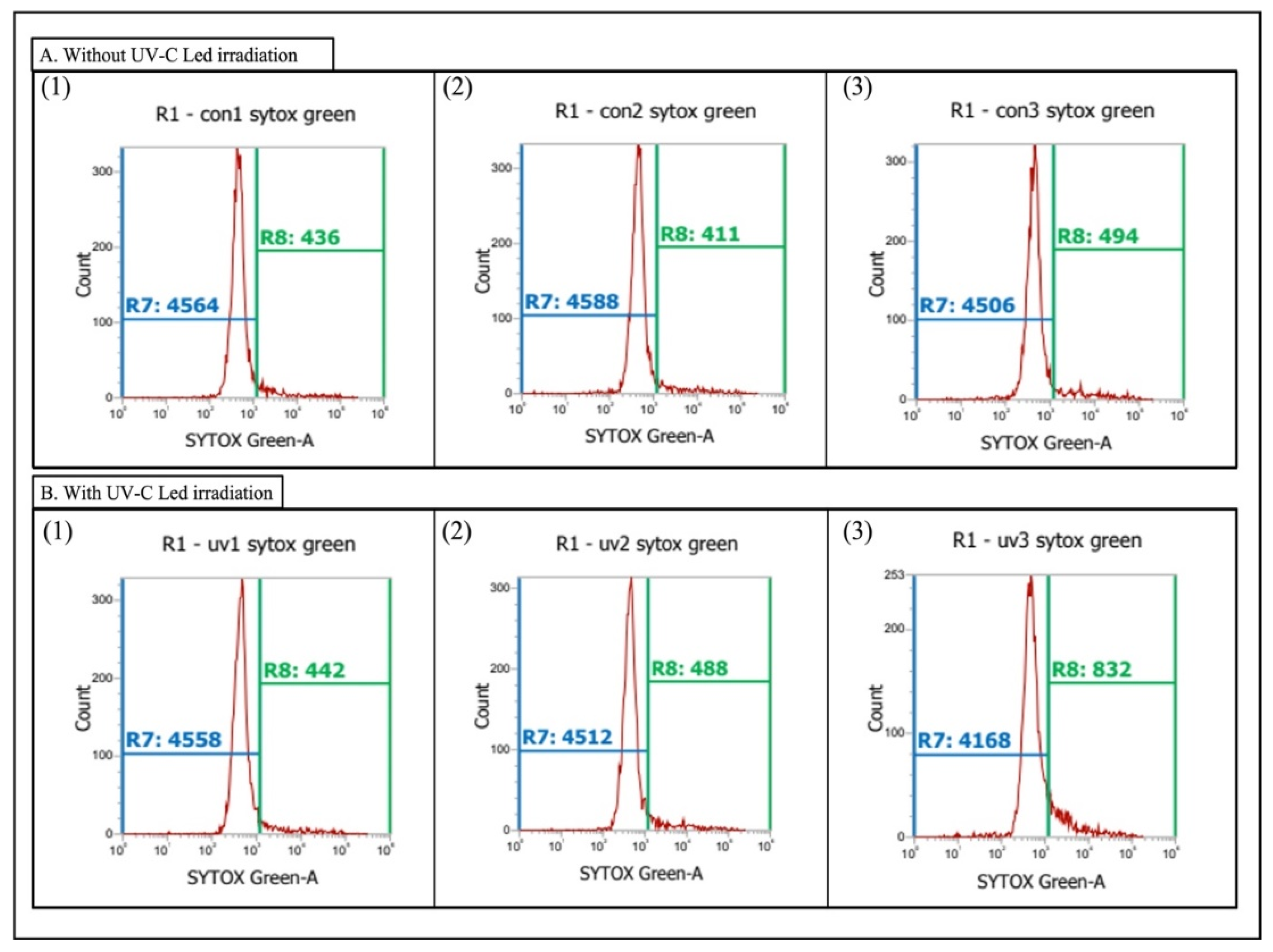
2.3. Plasma Membrane Damage on Chlorella sp.

In this experiment, SYTOX Green (S34860, Invitrogen Ltd., Massachusetts, United States) was used to stain the cells of Chlorella sp. in order to observe plasma membrane damage in the microalga. A total of 1 mL of algal water was mixed with 1 μL of SYTOX Green and left to react in the dark for about 30 min for subsequent analysis.
The analysis using flow cytometry was set to stop after detecting five thousand cells. The samples were analyzed by forward scatter (FSC) and side scatter (SSC). The Chlorella sp. stained by SYTOX Green was stimulated by the 488 nm, 50 mW laser. The BL1 (530/30 nm) filter, which can filter out unwanted wavelengths and collects light at specific wavelengths, was used to receive the fluorescence emitted from the Chlorella sp. stained by SYTOX Green.
To differentiate between normal and plasma membrane-damaged cells, a boundary line was set based on the control group. The recorded number of cells to the right of the boundary line was considered the positive cells number (PCN1), and to the left was negative. The plasma membrane damage rate (PMDR) of Chlorella sp. was calculated from Equation (1):
PMDR (%) = PCN1/5000 cells × 100

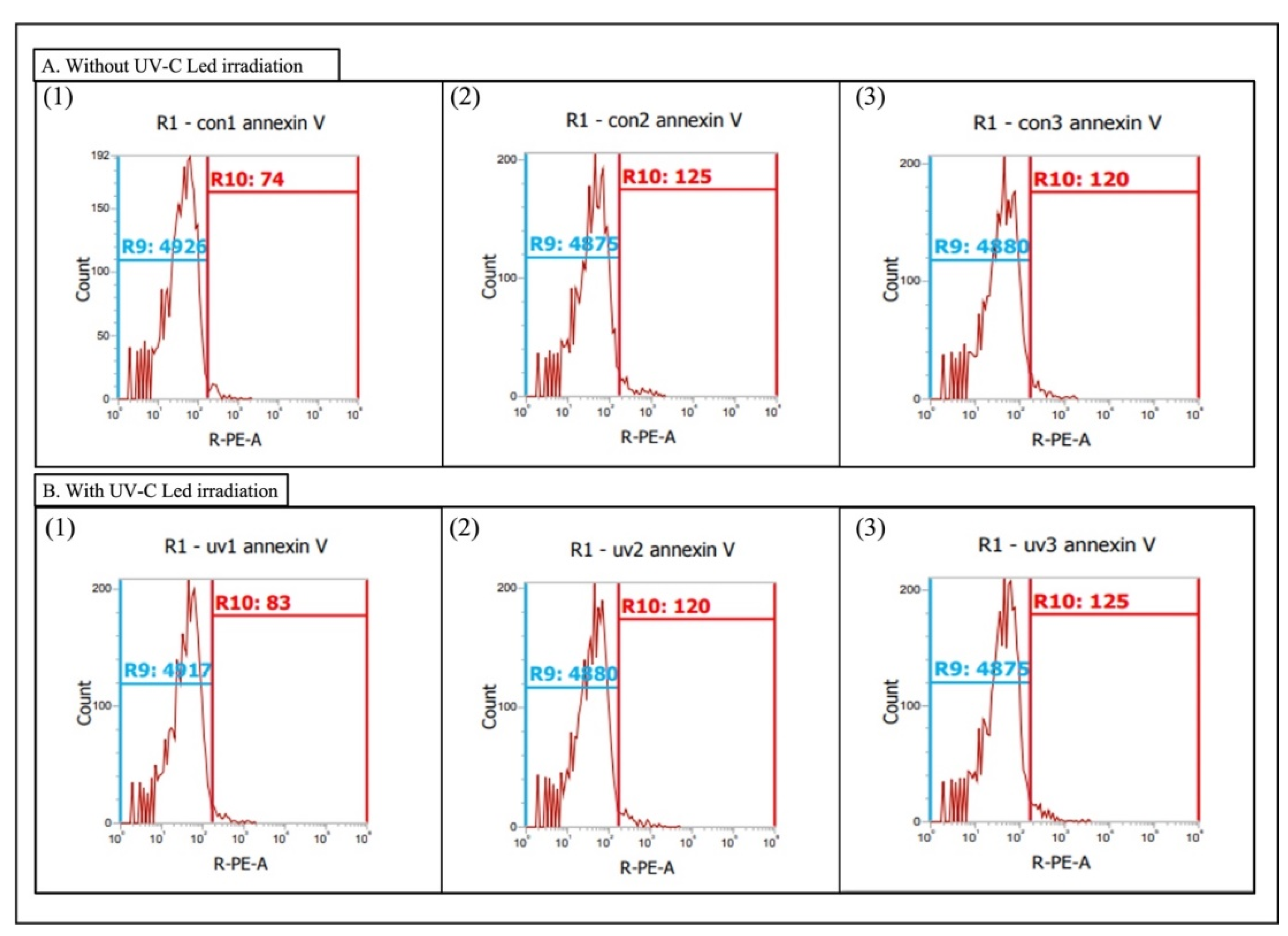
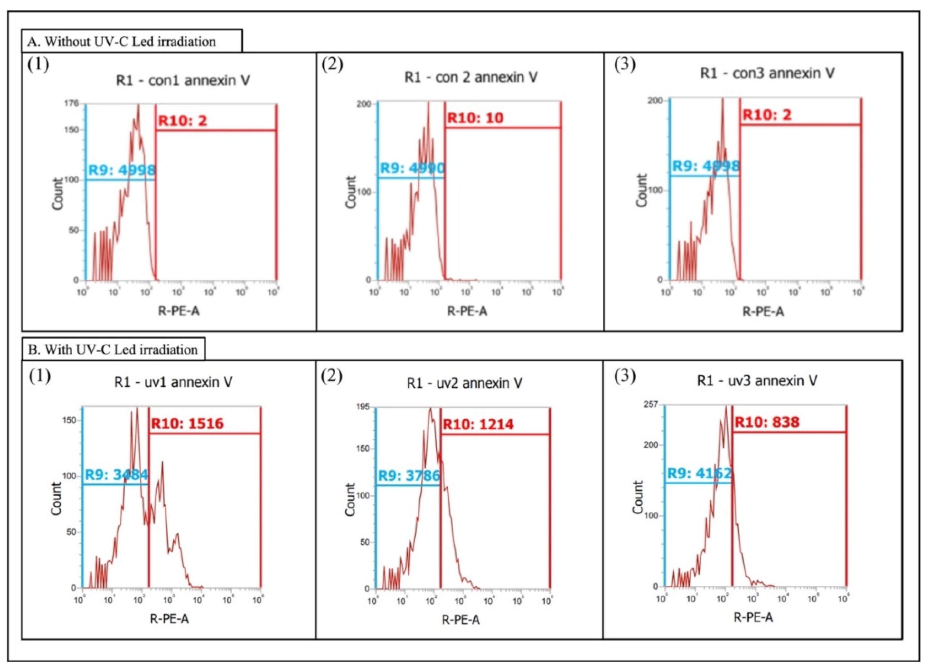
2.4. Phosphatidylserine Redistribution on Chlorella sp.

In this experiment, Annexin V-PE Reagent (#1014, BioVision, Massachusetts, United States) was used to quantify the apoptosis of Chlorella sp. with PS redistribution. In the study, a buffer (pH 7.4) consisting of 0.1 M HEPES, 1.4 M NaCl and 25 mM CaCl2 was prepared to bind Annexin V-PE Reagent to PS. For each group, 400 μL of algal water was mixed with 100 μL of buffer, and 1 μL of Annexin V-PE Reagent was added and left to react in the dark for 30 min.
Five thousand cells per sample was set as the condition for analysis by forward scatter (FSC) and side scatter (SSC) at a fixed voltage using flow cytometry, and the stained Chlorella sp. by Annexin V-PE Reagent was stimulated by the 488 nm, 50 mW laser. The BL2 (574/26 nm) filter that can filter out unwanted wavelengths and collects light at specific wavelengths was used to identify the Chlorella sp. stained by Annexin V-PE Reagent.
To define normal and PS redistribution (apoptotic) cells, a boundary line was set based on the control group. The recorded number of cells to the right of the boundary line was considered the positive cell number (PCN2), and to the left was negative. The phosphatidylserine redistribution rate (PSRR) of Chlorella sp. was calculated from the equation of PSRR as follows:
PSRR (%) = PCN2/5000 cells × 100

2.5. Statistical Analysis

The results were imported into Statistical Product and Service Solution (SPSS) (IBM, Taipei City, Taiwan) and analyzed by one-way ANOVA. If results exhibited significant differences, comparison of the mean values was conducted using the Tukey’s Honestly Significant Difference test (Tukey HSD). The significance level was set as α = 0.05 for all analyses.

3. Results

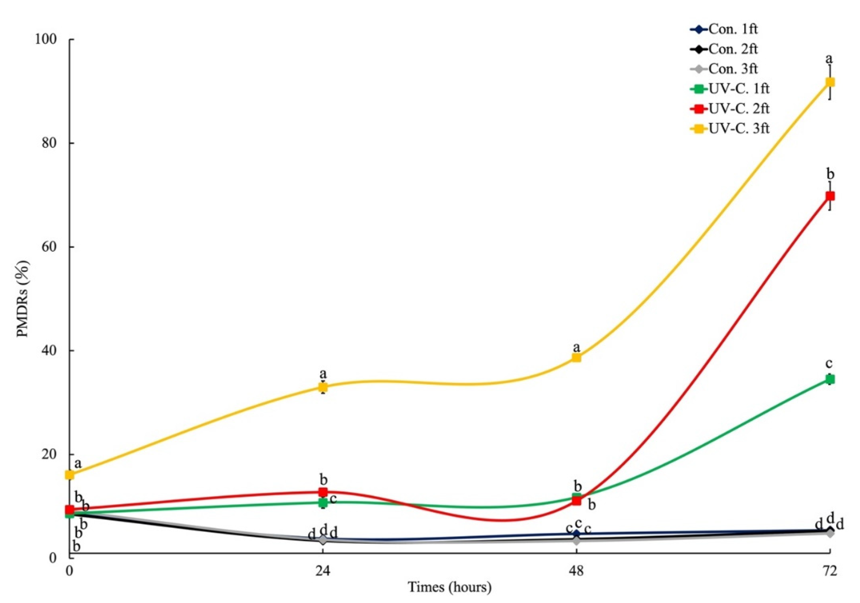
3.1. Plasma Membrane Damage on Chlorella sp.

As shown in Figure 1 and Figure 2, the PCNs after 72 h for treatment groups increased compared with the initial results, while the PCNs of control groups remained similar. As observed in Figure 3, the initial (0 h) PMDRs of Chlorella sp. in control groups were 8.68 ± 0.04% for one flow time, 8.52 ± 0.29% for two flow times and 9.12 ± 0.66% for three flow times through the UV-C LED system. On the other hand, the initial PMDRs after UV-C treatment for one flow time, two flow times and three flow times were 8.65 ± 0.20%, 9.43 ± 0.33% and 16.10 ± 0.90%, respectively. After 24 h, the PMDRs of control groups were 3.79 ± 0.23%, 3.37 ± 0.24% and 3.60 ± 0.20%, while the PMDRs of treatment groups were 10.76 ± 1.08%, 12.76 ± 0.46% and 33.00 ± 1.20% for one, two and three flow times, respectively. After 48 h, the PMDRs of control groups were 4.72 ± 0.42%, 3.70 ± 0.29% and 3.35 ± 0.06%, and the results of treatment groups were 11.76 ± 0.29%, 11.08 ± 0.18% and 38.71 ± 0.71%. After 72 h, the PMDRs of control groups were 5.42 ± 0.47%, 5.32 ± 0.44% and 4.79 ± 0.27%, and the results of treatment groups were 34.53 ± 0.99%, 69.85 ± 2.75% and 91.76 ± 3.33% for one, two and three flow times, respectively.
Statistical analysis showed that UV-C treatment for three flow times resulted in the highest (p < 0.05) PMDR right after UV-C treatment. Moreover, the highest PMDR at each subsequent time point, which was significantly higher than other groups (p < 0.05), was also observed in this group. The PMDRs of groups with UV-C treatment for one and two flow times showed statistical significance compared with control groups starting from the 24 h time point.

3.2. Phosphatidylserine Redistribution on Chlorella sp.

As shown in Figure 4 and Figure 5, the PCNs after 72 h for the treatment groups increased compared with the initial results, while the PCNs of control groups remained similar. As observed in Figure 6, the initial (0 h) PSRRs of Chlorella sp. without irradiation using UV-C were 1.29 ± 0.18% for one flow time, 2.40 ± 0.34% for two flow times and 2.26 ± 0.18% for three flow times through the UV-C LED system. On the other hand, the initial PSRRs of the UV-C irradiated Chlorella sp. were 1.93 ± 0.26%, 2.27 ± 0.19% and 2.78 ± 0.50% for one flow time, two flow times and three flow times, respectively. After 24 h, the PSRRs of control groups were 2.65 ± 3.30%, 1.22 ± 0.06% and 0.98 ± 0.00%, while the PSRRs of treatment groups were 2.85 ± 0.48%, 3.12 ± 0.56% and 4.21 ± 0.16% for one, two and three flow times, respectively. After 48 h, the PSRRs of control groups were 3.41 ± 0.27%, 3.98 ± 0.27% and 5.21 ± 0.72%, while the results of treatment groups were 11.02 ± 1.16%, 20.90 ± 1.64% and 15.32 ± 0.71% for one, two and three flow times, respectively. After 72 h, the PSRRs of control groups were 0.04 ± 0.00%, 0.18 ± 0.02% and 0.03 ± 0.01%, while the results of treatment groups were 31.24 ± 1.08%, 25.47 ± 1.66% and 16.74 ± 1.24% for one, two and three flow times, respectively.
Significant differences between the PSRRs of treatment groups and those of the control groups were present starting from the 48 h time point. After 48 h of incubation, the highest PSRR was observed in the group that had had UV-C treatment for two flow times (p < 0.05). After 72 h, the highest PSRR was observed in Chlorella sp. treated with UV-C for one flow time (p < 0.05).

4. Discussion

UV is known to be effective in suppressing the growth of cyanobacteria, such as Microcystis aeruginosa and Anabaena flos-aquae, and microalgae [28,29,30,31,32]. In addition, UV-C has been widely studied as a means of preventing harmful algal blooms [33]. In this study, we confirmed that UV-C LED lights can effectively inhibit the growth of Chlorella sp. and induce delayed cellular apoptosis in this species. We expect that this report will be useful for future research on the physiology of Chlorella sp. or other microalgae species.
Although there are many studies on the physiological effects of UV irradiation on Chlorella sp., such as reports about the production of polyphenols in Chlorella sp. [34], Chlorella-derived peptides against UV-C-induced cytotoxicity through inhibition of caspase-3 activity [35], the effects of UV-C on Chlorella vulgaris [36], and competitive alteration of Chlorella pyrenoidosa and two other microalgae species under UV-B radiation [37], our research is the first report that describes and quantifies the cell death process in Chlorella sp. after UV-C LED light exposure.
Previous reports have demonstrated the effectiveness of SYTOX Green, which has excitation/emission (Ex/Em) wavelengths at 504/523 nm, in tracing the plasma membrane integrity of microalgae that exhibit a red autofluorescence, such as Pseudokirchneriella subcapitata [38]. On the other hand, Annexin V-PE Reagent, which has Ex/Em wavelengths at 488/578 nm, was reported as a means of evaluating the loss of plasma membrane asymmetry during apoptosis. However, the use and interaction of SYTOX Green and Annexin V-PE Reagent with Chlorella sp. microalgal cells in flow cytometry in this report has never been conducted before. The dyes were chosen because both of them can be excited using a wavelength of 488 nm. Moreover, their emission wavelengths can be differentiated from the red autofluorescence emitted by chlorophyll-a that is detected through a separate channel (BL3; 695/40 nm) in flow cytometry. According to the results of our two staining treatments, the authors determined that UV-C can effectively induce apoptosis in Chlorella sp. cells and observed PCD of Chlorella sp. after UV-C irradiation using flow cytometry after staining the cells with SYTOX Green and Annexin V-PE Reagent. Therefore, this research contributes to the definition and determination of delayed cellular apoptosis in Chlorella sp., and we also contribute to the physiological knowledge of this microalga, which could even be applied to future physiological studies of other microalgae species.
In this research, we provide a detailed quantitative report of microalgal PCD expressed as apoptosis, which had not yet been reported. The cellular membrane is known as a critical target of the action of toxins, because it is one of the first structures with which they come into contact, and the integrity of membrane is also a criterion for defining the cell viability [38]. Thus, we expect that our research could be a useful reference for the quantitative evaluation of the disruption of cell membranes of the algae species (algicidal effect), by either physical or chemical treatments, in the future. Besides that, UV-C irradiation is found to induce delayed apoptosis, which leads to delayed cell death, in Chlorella sp.

Author Contributions

Conceptualization, F.-H.N., C.-Y.H. and M.-C.L.; methodology, F.-H.N. and C.-Y.H.; formal analysis, S.-J.Y.; resources, S.-J.Y. and H.-Y.Y.; writing—original draft preparation, S.-J.Y. and W.Q.C.L.; writing—review and editing, M.-C.L.; supervision, M.-C.L. All authors have read and agreed to the published version of the manuscript.

Funding

This research received no external funding.

Institutional Review Board Statement

Not applicable.

Informed Consent Statement

Not applicable.

Data Availability Statement

The data presented in this study are available in the article.

Conflicts of Interest

The authors declare no conflict of interest.

References

  1. Maruyama, K. Classification of Chlorella strains by cell appearance and group sera. Bot. Mag. Tokyo 1977, 90, 57–66. [Google Scholar] [CrossRef]
  2. Halldal, P.; French, C.S. Algal growth in crossed gradients of light intensity and temperature. Plant Physiol. 1958, 33, 249–252. [Google Scholar] [CrossRef] [PubMed] [Green Version]
  3. Teoh, M.L.; Chu, W.L.; Marchant, H.; Phang, S.M. Influence of culture temperature on the growth, biochemical composition and fatty acid profiles of six Antarctic microalgae. J. Appl. Phycol. 2004, 16, 421–430. [Google Scholar] [CrossRef]
  4. Kuhl, A.; Lorenzen, H. Chapter 10 Handling and Culturing of Chlorella. In Methods in Cell Biology; Prescott, D.M., Ed.; Academic Press: Cambridge, MA, USA, 1964; Volume 1, pp. 159–187. [Google Scholar]
  5. Scheffler, J. Underwater Habitats. Available online: https://illumin.usc.edu/underwater-habitats/ (accessed on 8 April 2022).
  6. Işik, O.; Sarihan, E.; Kuşvuran, E.; Gül, Ö.; Erbatur, O. Comparison of the fatty acid composition of the freshwater fish larvae Tilapia zillii, the rotifer Brachionus calyciflorus, and the microalgae Scenedesmus abundans, Monoraphidium minitum and Chlorella vulgaris in the algae-rotifer-fish larvae food chains. Aquaculture 1999, 174, 299–311. [Google Scholar] [CrossRef]
  7. Enyidi, U. Chlorella vulgaris as protein source in the diets of African catfish Clarias gariepinus. Fishes 2017, 2, 17. [Google Scholar] [CrossRef]
  8. Mujtaba, G.; Lee, K. Treatment of real wastewater using co-culture of immobilized Chlorella vulgaris and suspended activated sludge. Water Res. 2017, 120, 174–184. [Google Scholar] [CrossRef] [PubMed]
  9. Abdelnour, S.A.; El-Hack, M.E.A.; Arif, M.; Khafaga, A.F.; Taha, A.E. The application of the microalgae Chlorella spp. as a supplement in broiler feed. World’s Poult. Sci. J. 2019, 75, 305–318. [Google Scholar] [CrossRef]
  10. Cheng, P.; Chu, R.; Zhang, X.; Song, L.; Chen, D.; Zhou, C.; Yan, X.; Cheng, J.J.; Ruan, R. Screening of the dominant Chlorella pyrenoidosa for biofilm attached culture and feed production while treating swine wastewater. Bioresour. Technol. 2020, 318, 124054. [Google Scholar] [CrossRef]
  11. Doucha, J.; Lívanský, K. Production of high-density Chlorella culture grown in fermenters. J. Appl. Phycol. 2011, 24, 35–43. [Google Scholar] [CrossRef]
  12. González-Camejo, J.; Aparicio, S.; Ruano, M.V.; Borrás, L.; Barat, R.; Ferrer, J. Effect of ambient temperature variations on an indigenous microalgae-nitrifying bacteria culture dominated by Chlorella. Bioresour. Technol. 2019, 290, 121788. [Google Scholar] [CrossRef]
  13. Costa, S.S.; Peres, B.P.; Machado, B.R.; Costa, J.A.V.; Santos, L.O. Increased lipid synthesis in the culture of Chlorella homosphaera with magnetic fields application. Bioresour. Technol. 2020, 315, 123880. [Google Scholar] [CrossRef]
  14. Camargo, E.C.; Lonbardi, A.T. Effect of cement industry flue gas simulation on the physiology and photosynthetic performance of Chlorella sorokiniana. J. Appl. Phycol. 2017, 30, 861–871. [Google Scholar] [CrossRef]
  15. Candido, C.; Lombardi, A.T. The physiology of Chlorella vulgaris grown in conventional and biodigested treated vinasses. Algal Res. 2018, 30, 79–85. [Google Scholar] [CrossRef]
  16. Arora, N.; Philippidis, G.P. Insights into the physiology of Chlorella vulgaris cultivated in sweet sorghum bagasse hydrolysate for sustainable algal biomass and lipid production. Sci. Rep. 2021, 11, 6779. [Google Scholar] [CrossRef]
  17. Bai, M.D.; Hsu, H.J.; Wu, S.I.; Lu, W.C.; Wan, H.P.; Chen, J.C. Cell disruption of Chlorella vulgaris using active extracellular substances from Bacillus thuringiensis ITRI-G1 is a programmed cell death event. J. Appl. Phycol. 2017, 29, 1307–1315. [Google Scholar] [CrossRef]
  18. Leist, M.; Nicotera, P. The shape of cell death. Biochem. Biophys. Res. Commun. 1997, 236, 1–9. [Google Scholar] [CrossRef] [Green Version]
  19. Maghsoudi, N.; Zakeri, Z.; Lockshin, R.A. Programmed cell death and apoptosis—where it came from and where it is going: From Elie Metchnikoff to the control of caspases. Exp. Oncol. 2012, 34, 146–152. [Google Scholar]
  20. Kerr, J.F.; Wyllie, A.H.; Currie, A.R. Apoptosis: A basic biological phenomenon with wide-ranging implications in tissue kinetics. Br. J. Cancer 1972, 26, 239–257. [Google Scholar] [CrossRef] [Green Version]
  21. Bidle, K.D.; Falkowski, P.G. Cell death in planktonic photosynthetic microorganisms. Nat. Rev. Microbiol. 2004, 2, 643–655. [Google Scholar] [CrossRef]
  22. Bidle, K.D.; Bender, S.J. Iron starvation and culture age activate metacaspases and programmed cell death in the marine diatom Thalassiosira pseudonana. Eukaryot. Cell 2008, 7, 223–236. [Google Scholar] [CrossRef] [Green Version]
  23. Kozik, C.; Young, E.B.; Sandgren, C.D.; Berges, J.A. Cell death in individual freshwater phytoplankton species: Relationships with population dynamics and environmental factors. Eur. J. Phycol. 2019, 54, 369–379. [Google Scholar] [CrossRef]
  24. Reynolds, C.S. The Ecology of Freshwater Phytoplankton; Cambridge University Press: Cambridge, UK, 1984. [Google Scholar]
  25. Chen, Y.-C. The hormesis of the green macroalga Ulva fasciata with low-dose 60cobalt gamma radiation. J. Phycol. 2011, 47, 939–943. [Google Scholar] [CrossRef] [PubMed]
  26. Guillard, R.R.L. Culture of phytoplankton for feeding marine invertebrates. In Culture of Marine Invertebrate Animals; Smith, W.L., Chanley, M.H., Eds.; Plenum Press: New York, NY, USA, 1975; pp. 26–60. [Google Scholar]
  27. Provasoli, L. Media and products for the cultivation of marine algae. In Culture and Collections of Algae; Watanabe, A., Hattori, A., Eds.; Japanese Society of Plant Physiology: Tokyo, Japan, 1968; pp. 63–75. [Google Scholar]
  28. Bin Alam, M.D.Z.; Otaki, M.; Furumai, H.; Ohgaki, S. Direct and indirect inactivation of Microcystis aeruginosa by UV-radiation. Water Res. 2001, 35, 1008–1014. [Google Scholar] [CrossRef]
  29. Sakai, H.; Oguma, K.; Katayama, H.; Ohgaki, S. Effects of low-or medium-pressure ultraviolet lamp irradiation on Microcystis aeruginosa and Anabaena variabilis. Water Res. 2007, 41, 11–18. [Google Scholar] [CrossRef]
  30. Sakai, H.; Oguma, K.; Katayama, H.; Ohgaki, S. Effects of low or medium-pressure UV irradiation on the release of intracellular microcystin. Water Res. 2007, 41, 3458–3464. [Google Scholar] [CrossRef]
  31. Sakai, H.; Katayama, H.; Oguma, K.; Ohgaki, S. Kinetics of Microcystis aeruginosa growth and intracellular microcystins release after UV irradiation. Environ. Sci. Technol. 2009, 43, 896–901. [Google Scholar] [CrossRef]
  32. Tao, Y.; Zhang, X.; Au, D.W.T.; Mao, X.; Yuan, K. The effects of sub-lethal UV-C irradiation on growth and cell integrity of cyanobacteria and green algae. Chemosphere 2010, 78, 541–547. [Google Scholar] [CrossRef]
  33. Li, S.; Tao, Y.; Zhan, X.M.; Dao, G.H.; Hu, H.Y. UV-C irradiation for harmful algal blooms control: A literature review on effectiveness, mechanisms, influencing factors and facilities. Sci. Total Environ. 2020, 723, 137986. [Google Scholar] [CrossRef]
  34. Copia, J.; Gaete, H.; Zuniga, G.; Hidalgo, M.; Cabrera, E. Effect of ultraviolet B radiation on the production of polyphenols in the marine microalga Chlorella sp. Lat. Am. J. Aquat. Res. 2012, 40, 113–123. [Google Scholar] [CrossRef]
  35. Shih, M.F.; Cherng, J.Y. Protective effects of Chlorella-derived peptide against UVC-induced cytotoxicity through inhibition of caspase-3 activity and reduction of the expression of phosphorylated FADD and cleaved PARP-1 in skin fibroblasts. Molecules 2012, 17, 9116–9128. [Google Scholar] [CrossRef] [Green Version]
  36. Pfendler, S.; Alaoui-Sosse, B.; Alaoui-Sosse, L.; Bousta, F.; Aleya, L. Effects of UV-C radiation on Chlorella vulgaris, a biofilm-forming alga. J. Appl. Phycol. 2018, 30, 1607–1616. [Google Scholar] [CrossRef]
  37. Sun, Y.; Chen, Y.; Wei, J.; Zhang, X.; Zhang, L.; Yang, Z.; Huang, Y. Ultraviolet-B radiation stress alters the competitive outcome of algae: Based on analyzing population dynamics and photosynthesis. Chemosphere 2021, 272, 129645. [Google Scholar] [CrossRef]
  38. Machado, M.D.; Soares, E.V. Development of a short-term assay based on the evaluation of the plasma membrane integrity of the alga Pseudokirchneriella subcapitata. Appl. Microbiol. Biotechnol. 2012, 95, 1035–1042. [Google Scholar] [CrossRef] [Green Version]
Figure 1. The initial results of plasma membrane damage (A) without and (B) with UV-C irradiation on Chlorella sp. The Arabic numerals (1–3) represent the flow times of UV-C system. The range of R8 was used to represent cells of Chlorella sp. which were stained by Sytox green.
Diversity 14 00376 g001
Figure 2. After 72 h, the results of plasma membrane damage (A) without and (B) with UV-C irradiation on Chlorella sp. The Arabic numerals (1–3) represent thre flow times of UV-C system. The range of R8 was used to represent cells of Chlorella sp. which were stained by Sytox green.
Diversity 14 00376 g002
Figure 3. The plasma membrane damage rate of Chlorella sp. with and without UV-C LED. Data are presented as mean (±S. D.) of PMDRs in Chlorella sp. “ft” in graph legend means the flow times of Chlorella sp. through UV-C LED system. Different letters (a, b, c and d) represent significant differences (p < 0.05) among groups at the same sampling time (hours).
Diversity 14 00376 g003
Figure 4. The initial results of phosphatidylserine redistribution (A) without and (B) with UV-C irradiation on Chlorella sp. The Arabic numerals (1–3) represent the flow times of UV-C system. The range of R10 was used to represent cells of Chlorella sp. which were stained by Annexin V-Phycoerythrin.
Diversity 14 00376 g004
Figure 5. After 48 h, the results of phosphatidylserine redistribution (A) without and (B) with UV-C irradiation on Chlorella sp. The Arabic numerals (1–3) represent the flow times of UV-C system. The range of R10 was used to represent cells of Chlorella sp. which were stained by Annexin V-Phycoerythrin.
Diversity 14 00376 g005
Figure 6. The phosphatidylserine redistribution rate of Chlorella sp. with and without UV-C LED. Data are presented as mean (±S. D.) of PMDRs of Chlorella sp. “ft” in graph legend means the number of flow times of Chlorella sp. through UV-C LED system. Different letters (a, b, c and d) represent significant differences (p < 0.05) among groups at the same sampling time (hours).
Diversity 14 00376 g006
Table 1. The composition and usage of PG medium stocks.
StockCompositionConcentration (g L−1 ddH2O)Usage
Stock ANaNO375.01 mL L−1 sterilized water
NaH2PO4·2H2O5.6
NH4Cl26.8
Na2EDTA4.36
FeCl3·6H2O3.15
Stock BMnCl2·4H2O0.181 mL L−1 sterilized water
ZnSO4·7H2O0.023
CoCl2·6H2O0.01
CuSO4·5H2O0.01
Na2MoO4·2H2O0.006
Vitamin solution stockVitamin B1 (Thiamine)0.2 g0.5 mL L−1 sterilized water
Vitamin B7 (Biotin)0.001 g
Vitamin B12 (Cyanocobalamin)0.001 g
Publisher’s Note: MDPI stays neutral with regard to jurisdictional claims in published maps and institutional affiliations.

