Abstract
The cervicornis species group of Taumacera Thunberg, 1814, endemic to Sri Lanka, is revised. Seven species, including three species new to science, are recognized: Taumacera cervicornis (Baly, 1861), T. lewisi (Jacoby, 1887), T. mirabilis (Jacoby, 1887), T. unicolor (Jacoby, 1887), T. adamskii sp. nov., T. maskeliya sp. nov., and T. sigiriya sp. nov. The representatives of this group are characterized by pectinate antennomeres and by 12-segmented antennae. Antennomeres XI and XII are firmly fused in most species, separated by a distinct suture; however, in T. cervicornis, antennomere XII appears to be moveable. Color photographs of habitus, body details, and aedeagi as well as female genitalia are presented.
1. Introduction
The species of the Taumacera cervicornis species group were formerly classified in the genus Xenarthra Baly, 1861 (e.g., Weise 1924, Maulik 1936, Wilcox 1973) [1,2,3]. Except for the type species X. cervicornis Baly, 1861 [4], an additional three species were attributed to the genus by Jacoby (1887): X. lewisi Jacoby, 1887, X. mirabilis Jacoby, 1887 and X. unicolor Jacoby, 1887 [5]. All species are endemic to Sri Lanka. Bezděk (2019) redefined the genus Taumacera Thunberg, 1814 [6]. Based on the presence of the metasternal process in males, Xenarthra was synonymized with Taumacera, and the Xenarthra species were cumulated in the Taumacera cervicornis species group. The female of T. lewisi was described as Aenidea hirtipennis Jacoby, 1887 and synonymized by Bezděk (2019) [6]. The Himalayan species Xenarthra costata Allard, 1889 was transferred to the genus Japonitata Strand, 1935 by Bezděk (2012) [7].
The genus Taumacera comprises 97 species (Bezděk 2019, Nguyen and Bezděk 2021) [6,8]. However, as pointed out by Reid (1999) and Bezděk (2019), 26 African species very likely belong to other genera [6,9]. Nine African species were originally described in Xenarthra and four of them are still formally classified in Taumacera. The other five species are currently classified in Xenarthracella Laboissière, 1940; Palpoxena Baly, 1861; Spilocephalus Jacoby, 1888; Polexima Weise, 1903; and Kanahiiphaga Laboissière, 1931 [3]. Although the African species are in need of a comprehensive revision, their relationship to the Taumacera cervicornis species group can almost certainly be ruled out.
During the preparation of the Taumacera revision, about 20 species new to science were discovered and their future descriptions were announced (Bezděk 2019) [6]. The revision of the Vietnamese species of the Taumacera viridis species group with the description of one new species was already published by Nguyen and Bezděk (2021) [9]. In the present paper, the Taumacera cervicornis species group, endemic to Sri Lanka, is revised including the descriptions of three new species.
2. Material and Methods
Images of specimens were taken with Canon EOS 550D and Canon 800D digital cameras with a Canon MP-E 65 mm lens. Images of the same objects at different focal planes were combined using Helicon Focus 7 software.
Exact label data are cited for all type specimens; a double slash (//) divides the data on different labels, and a single slash (/) divides the data from different lines. Type localities are cited in the original spelling. Other comments and remarks are placed in square brackets: [p]—preceding data are printed, [h]—preceding data is handwritten, [w]—white label, [v]—violet label, [r]—red label, and [b]—blue label.
The examined material is housed in the following collections:
BMNH: The Natural History Museum, London, UK (Michael Geiser, Maxwell V. L. Barclay);
JBCB: Jan Bezděk collection, Brno, Czech Republic;
NHMB: Naturhistorisches Museum, Basel, Switzerland (Christoph Germann);
NMPC: National Museum, Prague, Czech Republic (Jiří Hájek, Lukáš Sekerka);
USNM: National Museum of Natural History, Smithsonian Institution, Washington DC, USA (Alexander S. Konstantinov).
3. Results
3.1. Genus Taumacera Thunberg, 1814
Taumacera Thunberg, 1814: 48 [10] (type species: Taumacera deusta Thunberg, 1814, by monotypy).
Diagnosis: Eyes are moderate. The penultimate maxillary palpomere is not greatly swollen. Antennae are 11-segmented (except for the T. cervicornis species group with 12 antennomeres), and often some antennomeres are strongly modified. Antennomeres are sometimes longitudinally ridged. Pronotum transverse, 1.2–1.6 times as wide as long, with a pair of discal depressions. An anterior pronotal border is absent. The elytral surface is usually almost glabrous or densely covered with longer setae (T. cervicornis species group). Procoxal cavities are closed. Metatibia is simple without a spur or with a subapical lobe sometimes with a short black spur. Tarsal claws are appendiculate. The metasternal process is present, usually with a thinly split apex. The last abdominal ventrite is apically trilobate. The aedeagus is flattened apically and with a split apex. Ventral side with longitudinal groove constricted subapically. Spermatheca has a C-shaped cornu and a well-developed bulbus. Gonocoxae with split apex, apical part with several long setae, base bifurcate.
3.2. Taumacera Cervicornis Species Group
Xenarthra Baly, 1861: 298 [4] (type species Xenarthra cervicornis Baly, 1861, by original designation); Chapuis (1875): 249 [11] (noted); Gemminger and Harold (1876): 3596 [12] (catalogue); Jacoby (1887): 108 [5] (comments); Allard (1889): cxiii [13] (noted); Maulik (1936): 481 [2] (redescription); Wilcox (1973): 629 [3] (catalogue); Seeno and Wilcox (1982): 114 [14] (catalogue); Bezděk (2019): 25 [6] (synonymized with Taumacera).
Taumacera cervicornis species group: Bezděk (2019): 29 [6] (definition).
Diagnosis: Antennae 12-segmented; male antennomeres are pectinate with long lateral branches. The anterior part of the head and labrum are unmodified. The elytra are either confusedly punctate and nonstriate, semistriate or slightly costate and always densely covered with setae, which are usually in longitudinal rows.
3.3. Key to the Males of Taumacera Cervicornis Species Group
- 1
- Elytra are metallic. ………………………………………………………………………… 2
- -
- Elytra are pale brown, sometimes with black spots. …………………………………………… 5
- 2
- Elytra are metallic green or blue-green, rarely with a brownish elytral disc. …….… 3
- -
- Elytra are metallic reddish brown. ……………………..… Taumacera adamskii sp. nov.
- 3
- -
- Metatarsus is modified, and metatarsomeres I and IV are distinctly enlarged. Mesotarsomere I is triangular. Antennomeres XI and XII are unmodified. ……………..… 4
- 4
- Antennomere III is gradually extended, IV–V are transverse with an inner apical margin produced in a short triangular process, and VI–IX are pectinate (Figure 14e). The mesofemora are unmodified. Metatarsomere I is axe-shaped, IV is large, subtriangular, flat, and with an emarginated apical margin, the ventral side is concave, and claws are inserted on the ventral side of the inner apical angle (Figure 15d,e). ……………………………………………………………..… Taumacera maskeliya sp. nov.
- -
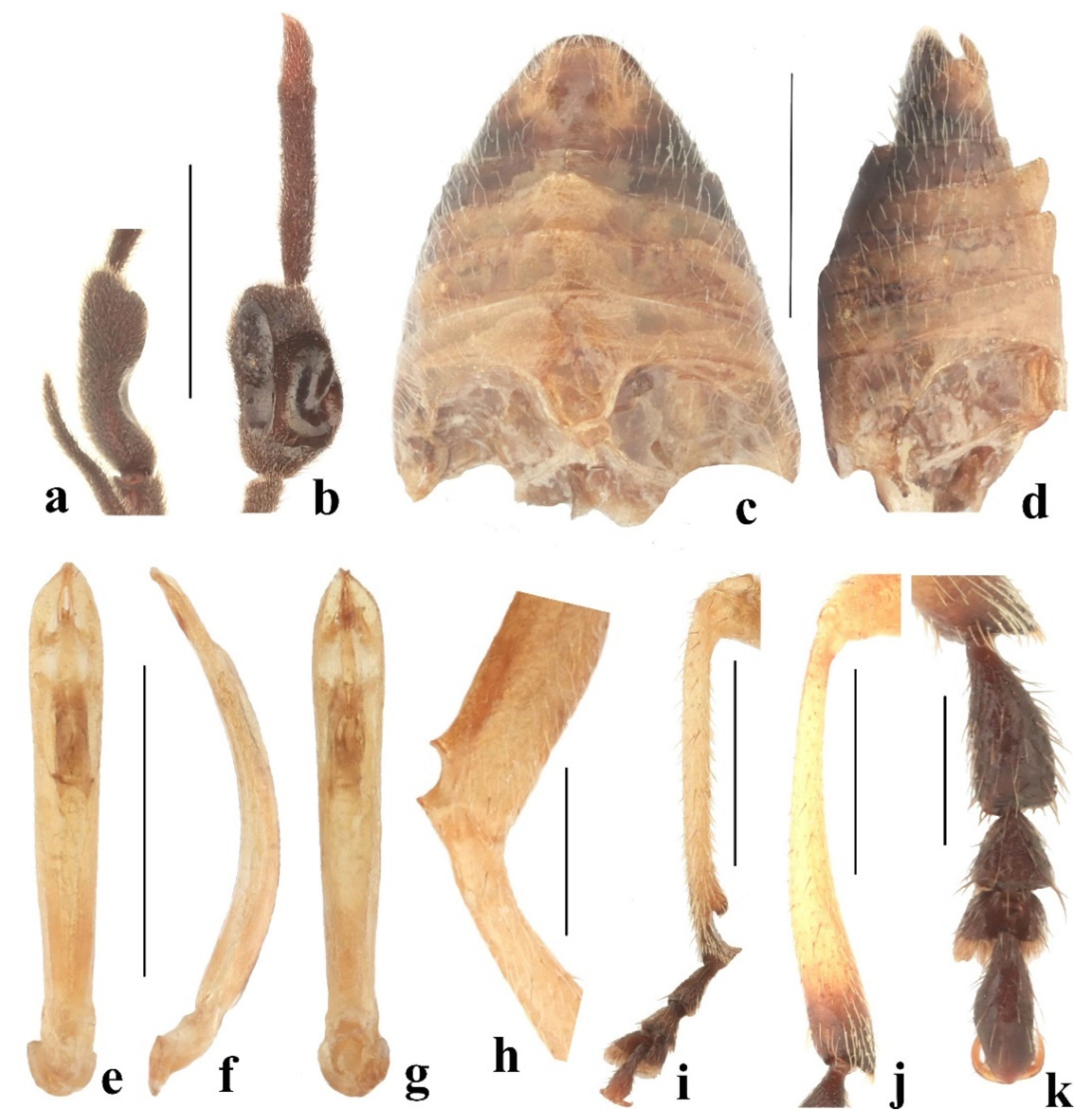
- Antennomeres III–IX are pectinate (Figure 16f). The mesofemora have a U-shaped emargination at the dorsal side near the apex (Figure 17h). Metatarsomere I is wide, and subclavate, the outer margin is nearly straight, the inner margin is widely rounded, IV is asymmetrical, enlarged, and flat, the outer margin with a rounded middle part, and the inner margin is sinuate (Figure 17k). ………………………………………………………………..… Taumacera sigiriya sp. nov.
- 5
- The dorsal side of the body is pale brown with black spots. …………… Taumacera mirabilis
- -
- The dorsal side of the body is uniformly pale brown. ………………………………… 6
- 6
- -
3.4. Taumacera cervicornis Species Group
3.4.1. Taumacera cervicornis (Baly, 1861)
Xenarthra cervicornis Baly, 1861: 299 [4] (original description); Gemminger and Harold (1876): 3596 [12] (catalogue); Allard (1889): cxiv [13] (key); Weise (1924): 152 [1] (catalogue); Maulik 1936: 482 [2] (redescription); Medvedev (1972): 182 [15] (faunistics); Wilcox (1973): 629 [3] (catalogue).
Xenorthra [sic!] cervicornis: Kimoto (2003): 31 [16] (faunistics).
Taumacera cervicornis: Bezděk (2019): 30 [6] (transferred to Taumacera).
Type locality: “Ceylon”.
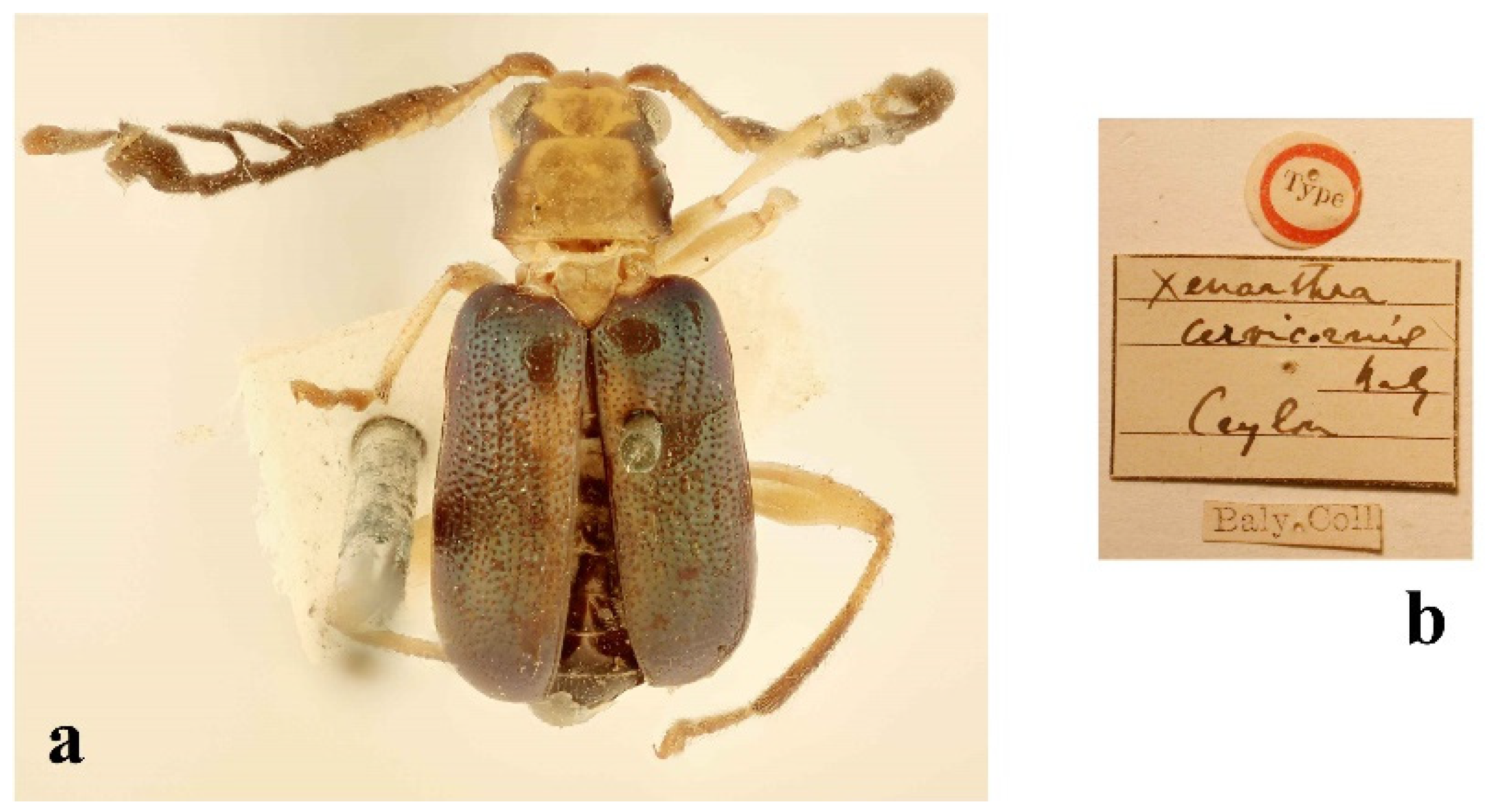
Type examined: Syntype: ♂ (BMNH, Figure 1a,b), “Type [white round label with red frame, p]//Xenarthra/cervicornis/Baly/Ceylon [grey, h]/ Baly Coll. [w, p]”.
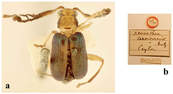
Figure 1.
Taumacera cervicornis (Baly, 1861): (a) syntype, male; (b) labels. (Not to scale).
Figure 1.
Taumacera cervicornis (Baly, 1861): (a) syntype, male; (b) labels. (Not to scale).
Additional material examined: SRI LANKA: Colombo District: Labugama Reservoir, jungle, 13.–14.x.1973, 1 ♀, K. V. Krombein, P. B. Karunaratne, P. Fernando, and J. Ferdinando leg. (USNM); Labugama, 24.viii.1973, 1 ♀, G. Ekis leg. (USNM). Galle District: Sinharaja Jungle, Kanneliya section, 13.–16.vii.1978, malaise trap, 1 ♀, K. V. Krombein, P. B. Karunaratne, T. Wijesinhe, V. Kulasekare, and L. Jayawickrema leg. (USNM). Kalutara District: Morapitiya, 27.–28.v.1975, 1 ♂, S. L. Wood, and J. L. Petty leg. (USNM). Kandy District: Kandy, iv.1907, 1 ♀ (BMNH); Kandy, 23.v.1910, 1 ♀ (BMNH); Kandy, vii.1910, 1 ♂ (BMNH); Kandy, vi.1908, 5 ♂♂ 8 ♀♀, G. E. Bryant leg. (BMNH); Kandy, iii.1910, 1 ♀, J. Uzel leg. (NMPC); Kandy, L. Horton’s Drive, 4.vi.1953, 1 ♂, F. Keiser leg. (NHMB); Kandy, 10.ix.1953, 1 ♂, F. Keiser leg. (NHMB); Kandy, Peak View Motel, 1.–14.i.1970, 1 ♀, Davis and Rowe leg. (USNM); Kandy, 1.–15.ii.1971, 1 ♀, Piyadasa and Somapala leg. (USNM); Kandy Reservoir, 4.vi.1976, 1 ♀, K. V. Krombein, P. B. Karunaratne, S. Karunaratne, and D. W. Balasooriya leg. (USNM); Kandy, 1.–18.iv.1991, ♂, J. Kolibáč leg. (NHMB); Udawattakele, 16.xi.1966, 5 ♂♂ 7 ♀♀, J. F. G. Clarke, and T. M. Clarke leg. (USNM); Udawattakele, 1.–3.x.1973, 1 ♀, K. V. Krombein, P. B. Karunaratne, and P. Fernando leg. (USNM); Udawattakele, 18.–21.i.1977, 1 ♂, K. V. Krombein, P. Fernando, D. W. Balasooriya, and V. Gunawardane leg. (USNM); Udawattakele Sanctuary, 6.–8.vi.1978, 2 ♀♀, K. V. Krombein, P. B. Karunaratne, T. Wijesinhe, V. Kulasekare, and L. Jayawickrema leg. (USNM); Udawattakele Sanctuary, 1.–3.ix.1980, 1 ♂, K. V. Krombein, P. B. Karunaratne, T. Wijesinhe, L. Jayawickrema, and V. Gunawardane leg. (USNM); Udawattakele Sanctuary, 12.–14.x.1980, 1 ♂, Malaise trap, K. V. Krombein, P. B. Karunaratne, T. Wijesinhe, L. Jayawickrema, and V. Gunawardane leg. (USNM); Peradeniya, 3.x.1913, 1 ♀, A. Rutheford leg. (BMNH). Unknown District: Ceylon, 1909, 3 ♂♂ 3 ♀♀, E. E. Green leg. (BMNH); Ceylon, 2 ♂♂ 1 ♀ (BMNH).
Redescription: Measurements. Males: 5.3–5.9 mm, females: 5.4–6.5 mm.
Male (Figure 2a). The body is an elongated oval, and flat. The head is pale brown, frontal tubercles are dark brown, and mandibles and lateral parts of the vertex behind the eyes are black. The pronotum is brown with lateral parts black, and the anterior part of the black pattern is wider. The scutellum is pale brown. The elytra are metallic green. The antennomeres I–VI are pale brown, I has a darker dorsal side, III has a dark anteroapical angle, IV–VI have darkened inner lateral parts, VII is black with a brownish base, VIII–XI are black, and XII is often brownish. The legs are pale brown with darkened apices of the tibiae, the protarsi is brownish, and the meso- and metatarsi are black. The underside with lateral parts of the prosternum, the entire metasternum, and the abdomen are black.
Head (Figure 2c,d). The labrum is transverse with a U-shaped incision in the middle of the anterior margin, the anterior angles are rounded, and the surface is slightly convex with several pores in a transverse row bearing long setae. The anterior part of the head is sparsely covered with long setae, with a straight anterior margin, and a surface with a keel forming a three-pointed star. The interantennal space is 0.80 times as wide as the transverse diameter of the antennal socket. The eyes are moderately large, the interocular space is 3.00 times as wide as the transverse diameter of the eye. The frontal tubercles are subquadrangular, with anterior angles projecting anteriorly, convex, semiopaque, covered with microsculpture, and separated by a thin furrow. The vertex is separated from the frontal tubercles by a shallow impressed line and a notch in the middle, the surface covered with microsculpture, and sparse punctures bearing short setae. The antennae (Figure 2e) are 1.10 times as long as the body, and the length ratio of the antennomeres equals 100-21-75-58-58-67-75-92-100-167-167-117 (100 = 0.6 mm). Antennomeres I and II are lustrous, covered with sparse setae, and III–XII are semiopaque, densely covered with short, dark setae. Antennomere I is clavate, II is very small and transverse, III is robust with distinct anterolateral angles, IV–VI are subrectangular and transverse, VII–IX are pectinate, VII has a large, rectangular transverse base with a wide hockey-stick process with a narrow U-shaped incision between the process and the apical part of the antennomere, VIII–IX are narrow with an extended apex, and long lateral branches starting at the base, X is strongly enlarged with a triangular basal one-third, parallel apical two-thirds, and a short, subtriangular, inner anterolateral process with a dorsal surface moderately convex, in lateral view with a small emargination on the outer side, in ventral view with an impression in the middle part surrounded by two lustrous oval elevations (one larger and longitudinal, one smaller and circular), XI enlarged, and elongate subtriangular, XII is paddle-shaped, bent, and probably movable (Figure 3a–c).
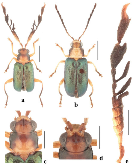
Figure 2.
Taumacera cervicornis (Baly, 1861): (a) male; (b) female; (c) head and pronotum, dorsal view; (d) head, frontal view; (e) left antenna. (Scale bars: (a,b) = 2 mm, (c–e) = 1 mm).
Figure 2.
Taumacera cervicornis (Baly, 1861): (a) male; (b) female; (c) head and pronotum, dorsal view; (d) head, frontal view; (e) left antenna. (Scale bars: (a,b) = 2 mm, (c–e) = 1 mm).
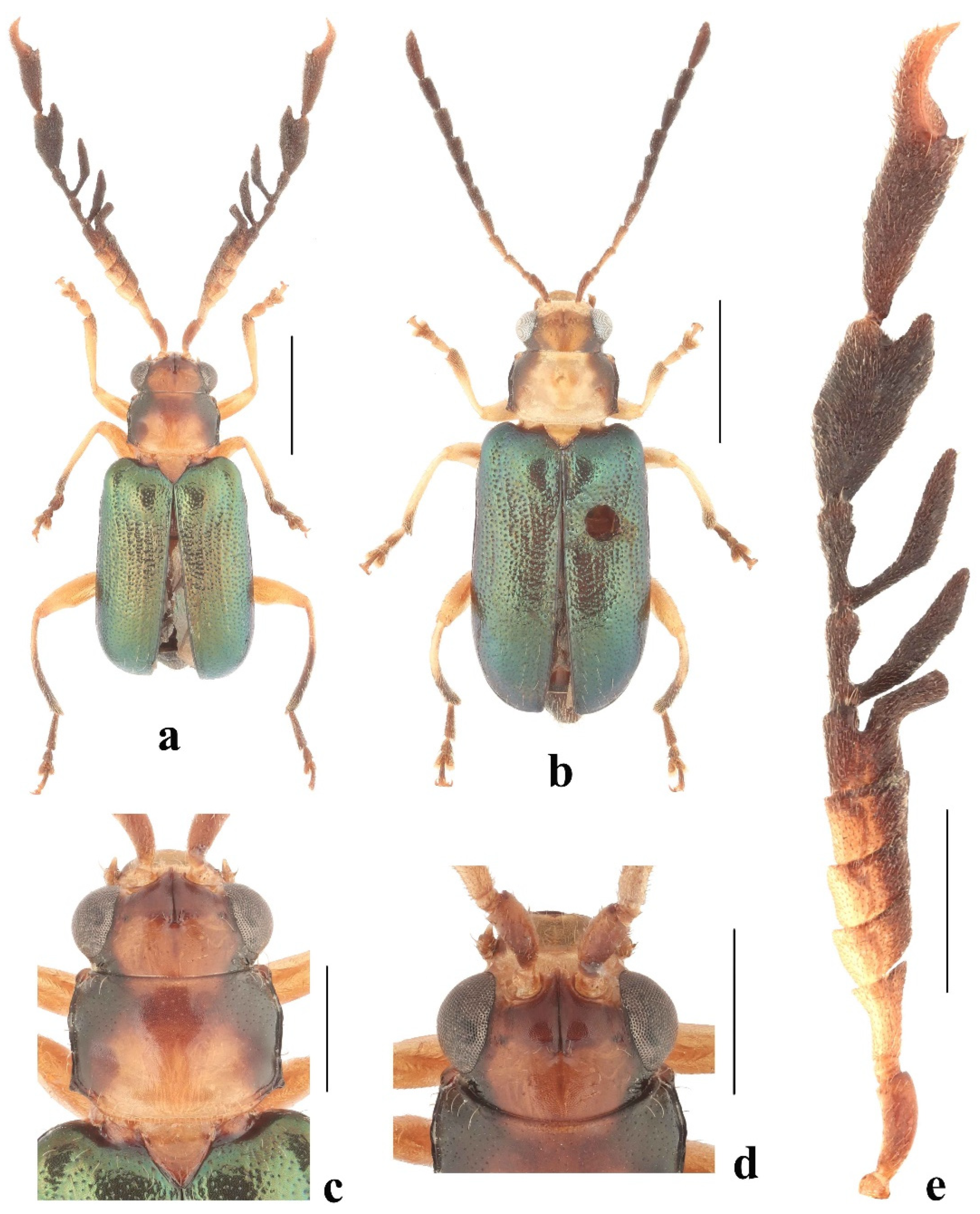
The pronotum (Figure 2c) is transverse, 1.45 times as wide as it is long, semiopaque, glabrous, widest at the anterior third, covered with microsculpture, and sparse fine punctures, with punctures more distinct in the anterolateral parts. The surface has two round impressions in the posterior half. The anterior margin is widely concave, the lateral parts are bordered and covered with several long setae directed posteriorly, the middle being unbordered, and without setae. The lateral margins are sinuate with distinct borders, and margins are sparse, and with long setae. The posterior margin is straight in the middle part, and without setae, the lateral parts are oblique, slightly concave, and with several long setae. Anterior angles are distinctly swollen, posterior angles are obtusangulate, and all angles have setigerous pores bearing very long, pale setae. The scutellum is triangular, covered with microsculpture, and short, pale setae.
The elytra are 1.48 times as long as they are wide (measured in the middle) and 0.63 times as long as the body, subparallel, and widest in middle. The surface is densely covered with small, confused punctures on the disc, with tendencies to form longitudinal double rows separated by indistinct ribs, and with short, erect, pale setae in longitudinal rows. The subscutellar area of each elytron is slightly convex. The epipleura are smooth, glabrous, wide at the basal half, then gradually narrowed towards the elytral apex. The humeral calli and hind wings are developed.
The metasternal process is 2.21 times as long as it is wide, widest at the base, slightly convergent to an apex in the middle with a long narrow incision, and both apices are moderately rounded (Figure 3k). The anterior process of abdominal ventrite I has a deep hole, the last abdominal ventrite is trilobed with two V-shaped incisions, and the median lobe has a straight posterior margin.
The legs are slender, and the mesotibiae are flattened (Figure 3d). Tarsi: protarsomere I is subtriangular, II is small and triangular; the length ratio of the protarsomeres equals 100-54-62-100 (100 = 0.30 mm); mesotarsomere I is dilated, the outer margin is straight, the inner margin is rounded (Figure 3f), II is small and triangular, and the length ratio of the mesotarsomeres equals 100-46-54-100 (100 = 0.30 mm); metatarsomere I is long, narrow, and parallel (Figure 3e), II is short, triangular, and slightly wider than I, and the length ratio of the metatarsomeres equals 100-32-33-58 (100 = 0.60 mm).
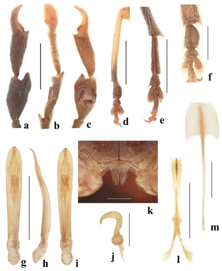
Figure 3.
Taumacera cervicornis (Baly, 1861): (a) antennomeres X–XII, dorsal view; (b) antennomeres X–XII, lateral view; (c) antennomeres X–XII, ventral view; (d) male mesotibia; (e) male metatarsus; (f) male mesotarsus; (g) aedeagus, dorsal view; (h) aedeagus, lateral view; (i) aedeagus, ventral view; (j) male metasternal process; (k) spermatheca; (l) gonocoxae; (m) sternite VIII. (Scale bars: (a–e,g–i) = 1 mm, (f,l,m) = 0.5 mm, (k) = 0.3 mm, (j) = 0.2 mm).
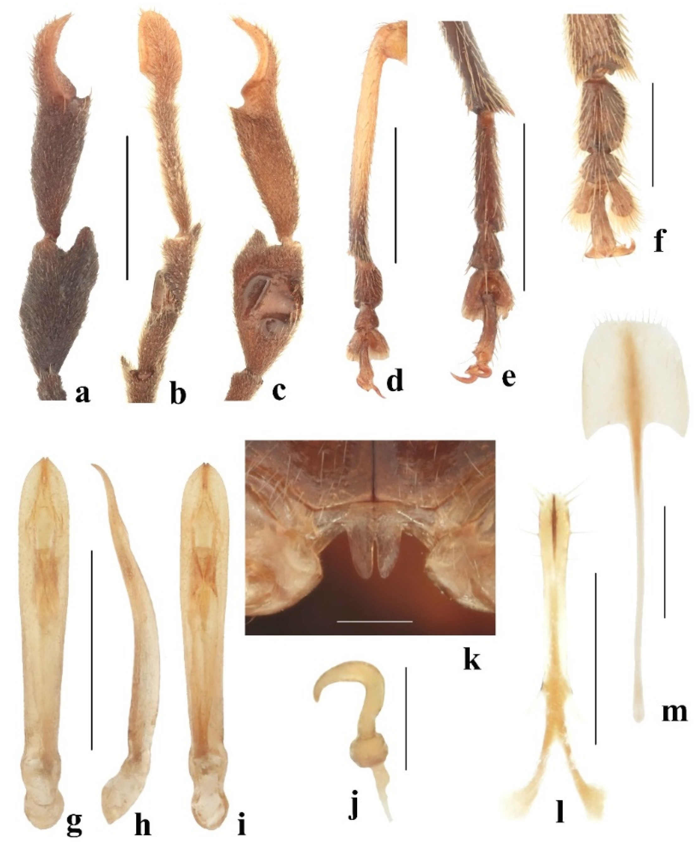
The aedeagus (Figure 3g–i) is long and narrow, and 7.30 times as long as it is wide; it is slightly wider and subparallel in the apical half, and the basal half is slightly convergent, the apex is rounded with tips pronounced to very short protrusions separated by a narrow slit; in the lateral view bisinuate (preapically and in the middle), the apical part gradually narrows; in the ventral view with long median furrows constricted by two small, triangular protuberances at the apical third of the aedeagus length.
Female (Figure 2b): The antennae are unmodified and filiform. The tarsomeres are unmodified. The sternite VIII is shovel-shaped, the posterior margin has short setae, several setae are also on the surface, and the tignum is slender, 2.8 times longer than the sternite VIII (Figure 3m). The spermatheca has a small spherical nodulus, cornu C-shaped, gradually narrowing towards the apex, the apex is moderately hooked, and the base of the cornu is slightly narrowed; the proximal part of the spermathecal duct is wider (Figure 3j). The apical fifth of the gonocoxae is slightly wider, each gonocoxa has six setae, the base is bifurcate, and each basal process is club-shaped (Figure 3l).
Variability: A black pattern on the pronotum is variable, rarely, and most pronota are black. The elytral disc is rarely brownish. One female from the Labugama Reservoir was probably immature with elytra completely brown with a slight metallic tint.
Differential diagnosis: Having a brown pronotum with black lateral margins and metallic green elytra, Taumacera cervicornis is very similar to T. maskeliya sp. nov. and T. sigiriya sp. nov. The males of all three species can be distinguished by the structures of the antennae, legs, aedeagi, and abdomina. In T. cervicornis, antennomeres IV–VI are subrectangular and transverse, VII–IX are pectinate, X is strongly enlarged, XI is enlarged, elongated, and subtriangular, and XII is paddle-shaped (Figure 2e and Figure 3a–c). The antennae of T. maskeliya sp. nov. have pectinate antennomeres VI–IX, and thin, unmodified antennomeres XI–XII (Figure 14e and Figure 15a–c). Taumacera sigiriya sp. nov. has pectinate antennomeres III–IX, and thin, unmodified antennomeres XI–XII (Figure 16f and Figure 17a,b). The mesotibiae are flattened but without preapical emargination in T. cervicornis (Figure 3d), while with distinct preapical emargination in T. maskeliya sp. nov. and T. sigiriya sp. nov. (Figure 15f and Figure 17i). The mesotarsomere I of T. cervicornis is dilated with a straight outer margin and a rounded inner margin (Figure 3f), while unmodified in T. maskeliya sp. nov. and T. sigiriya sp. Nov.; The metatarsus of T. cervicornis is unmodified while strongly modified in T. maskeliya sp. nov. and T. sigiriya sp. nov. (Figure 3e, Figure 15d,e and Figure 17k). The metasternal process of T. cervicornis is distinctly shorter than in T. maskeliya sp. nov. and T. sigiriya sp. nov. (Figure 3k, Figure 15j and Figure 16e). The aedeagus of T. cervicornis is bisinuate and less bent in lateral view (Figure 3h), while the aedeagus of T. sigiriya sp. nov. is not bisinuate and is more bent (Figure 17f). The abdomen of T. cervicornis is unmodified, but the middle parts of ventrites II and III are distinctly elevated in T. sigiriya sp. nov. (Figure 17c,d).
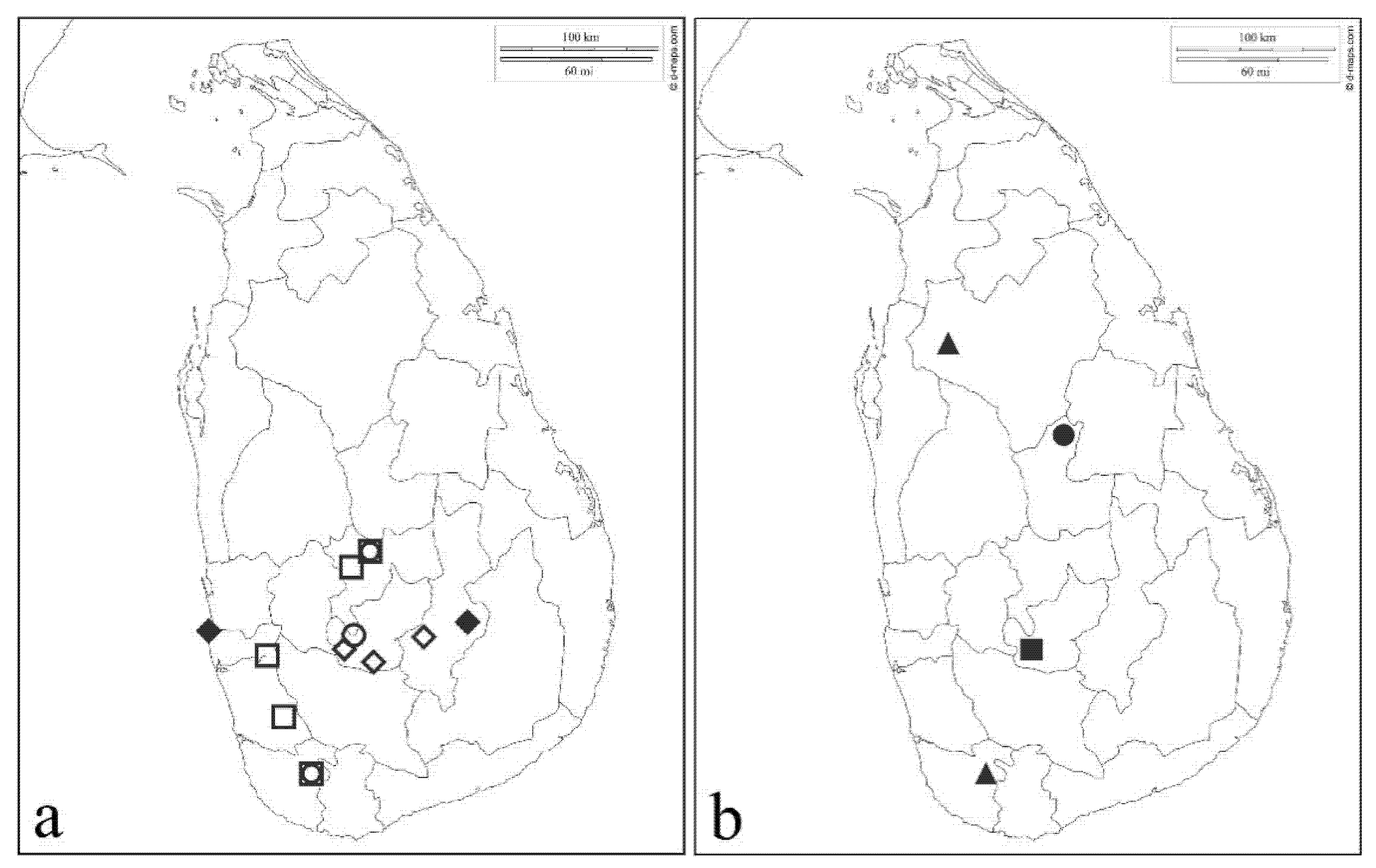
Distribution. Sri Lanka (Colombo, Galle, Kalutara, and Kandy districts) (Figure 18).
3.4.2. Taumacera lewisi (Jacoby, 1887)
Xenarthra lewisi Jacoby, 1887: 108 [5] (original description); Weise (1924): 152 [1] (catalogue); Maulik (1936): 485 [2] (redescription); Medvedev (1972): 183 [15] (faunistics); Wilcox (1973): 629 [3] (catalogue).
Xenorthra [sic!] lewisi: Kimoto (2003): 31 [16] (faunistics).
Taumacera lewisi: Bezděk (2019): 38 [6] (transferred to Taumacera).
Aenidea? hirtipennis Jacoby, 1887: 113 [5] (original description); Bezděk (2019): 38 [6] (synonymized with T. lewisi).
Palpoxena hirtipennis: Maulik (1936): 572 [2] (redescription); Wilcox (1973): 649 [3] (catalogue); Kimoto (2003): 34 [16] (faunistics).
Type localities. Xenarthra lewisi: “Dikoya”, Aenidea hirtipennis: “Dikoya”.
Type examined. Xenarthra lewisi: Syntypes: 1 ♂ (BMNH, Figure 4a,b), “Type/H. T. [white round label with red frame, p]//Ceylon./G. Lewis. / 1910–320. [w, p]/ Dikoya./3800–4200 ft./6.XII.81.–16.I.82. [w, p]//Xenarthra/lewisi Jac. [b, h]”; photograph of 1 ♂ (MCZ), “Xenarthra / lewisi Jac. [b, h]//1st Jacoby/Coll. [w, p]//Type [p]/18318 [r, h]”.
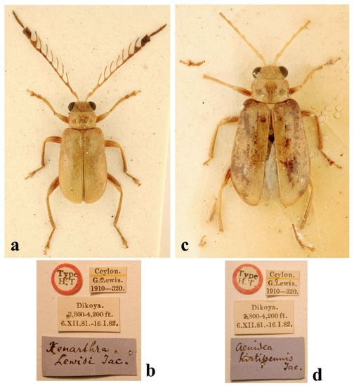
Figure 4.
Taumacera lewisi (Jacoby, 1887): (a) syntype, male; (b) labels; (c) holotype of Aenidea hirtipennis Jacoby, 1887, female; (d) labels. (Not to scale).
Figure 4.
Taumacera lewisi (Jacoby, 1887): (a) syntype, male; (b) labels; (c) holotype of Aenidea hirtipennis Jacoby, 1887, female; (d) labels. (Not to scale).
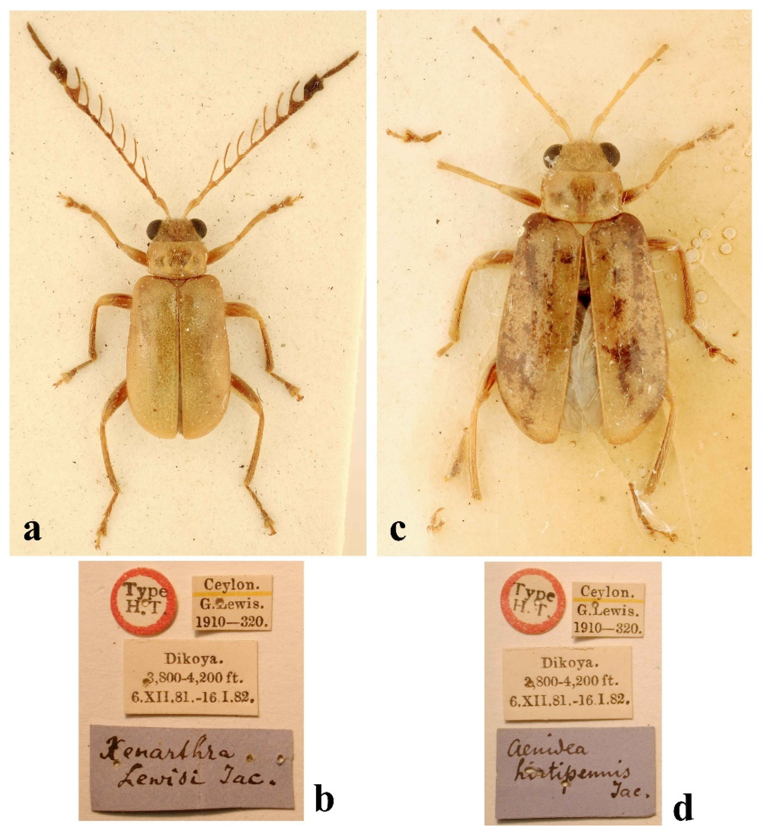
Aenidea hirtipennis: Holotype: ♀ (BMNH, Figure 4c,d), “Type/H. T. [white round label with red frame, p]//Ceylon./G. Lewis./1910–320. [w, p]//Dikoya./3800–4200 ft./6.XII.81.–16.I.82. [w, p]//Aenidea/hirtipennis/Jac. [b, h]”.
Additional material examined. SRI LANKA: Galle District: Kanneliya, 11.–16.i.1975, 1 ♂, K. V. Krombein, P. B. Karunaratne, P. Fernando, and N. V. T. A. Weragoda leg. (USNM). Kandy District: Kandy, vi.1908, 1 ♂ 1 ♀, G. E. Bryant leg. (BMNH).
Redescription. Measurements. Males: 5.0–5.6 mm, female: 5.3 mm.
Male (Figure 5a). The dorsal side is an elongated oval, and flat. The body is completely pale brown except for the apices of the mandibles, and the last three antennomeres are black.
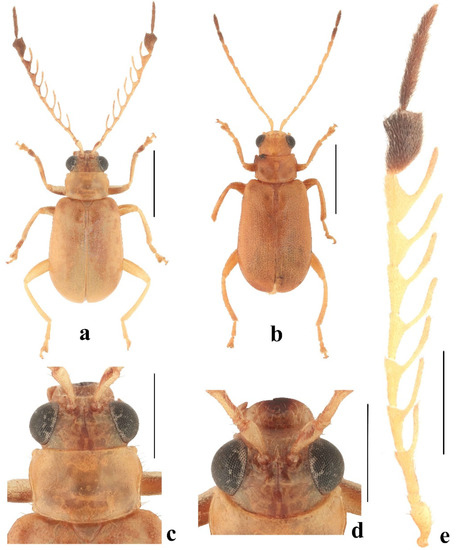
Figure 5.
Taumacera lewisi (Jacoby, 1887): (a) male; (b) female; (c) head and pronotum, dorsal view; (d) head, frontal view; (e) left antenna. (Scale bars: (a,b) = 2 mm, (c–e) = 1 mm).
Figure 5.
Taumacera lewisi (Jacoby, 1887): (a) male; (b) female; (c) head and pronotum, dorsal view; (d) head, frontal view; (e) left antenna. (Scale bars: (a,b) = 2 mm, (c–e) = 1 mm).
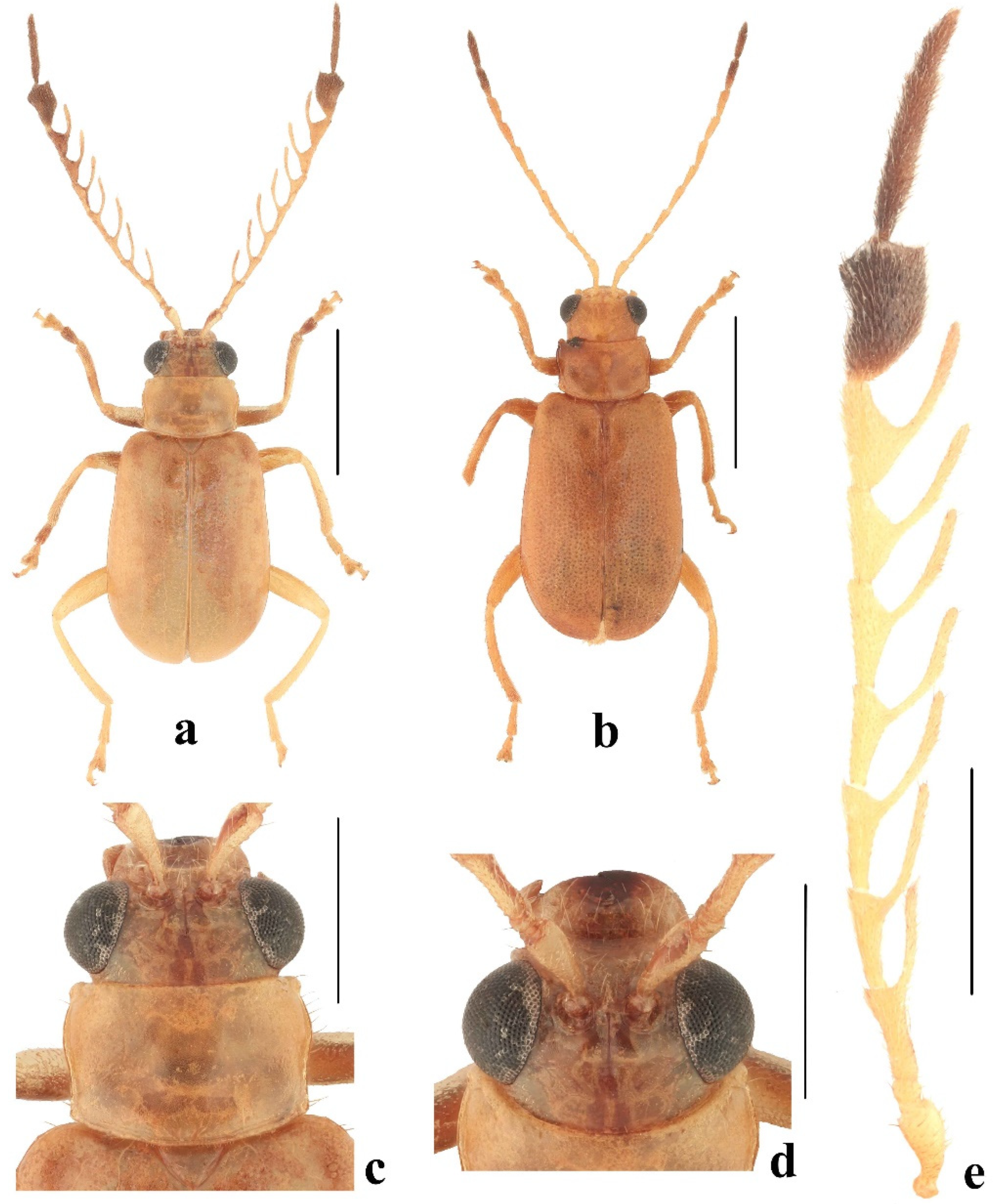
The head (Figure 5c,d). The labrum is transverse with a V-shaped incision in the middle of the anterior margin, the anterior angles are rounded, and the surface is slightly convex with a line of pores in the transverse row along the anterior margin bearing long setae. The anterior part of the head has a straight anterior margin, the surface has a keel forming a three-pointed star, and is sparsely covered with long, pale setae. The interantennal space is as wide as the transverse diameter of the antennal socket. The eyes are moderately large, with an interocular space 1.75 times as wide as the transverse diameter of the eye. The frontal tubercles are transverse with inner anterior angles projecting slightly anteriorly, moderately convex, semiopaque, covered with microsculpture, and separated by a thin furrow. The vertex is separated from the frontal tubercles by a shallow impressed line and a notch in the middle, the surface is covered with microsculpture, and fine punctures and the anterior part also has short setae. The antennae (Figure 5e) are 0.95 times as long as the body, and the length ratio of the antennomeres equals 100-25-90-90-95-90-90-80-95-130-155-70 (100 = 0.5 mm). Antennomeres I and II are covered with sparse, long setae, III–XII are semiopaque, and densely covered with short setae. Antennomere I is clavate, II is small, III–IX are pectinate, with lateral branches on antennomeres III–V starting at the inner apical angle, there is a lateral branch on antennomere VI starting at a triangularly extended apical third, lateral branches exist on antennomeres VII–IX starting at a triangularly extended middle part, antennomere X is strongly enlarged with a convex dorsal side, the outer margin is concave, the inner margin is widely rounded, and in lateral view, flat, the outer margin is widely concave, and in the ventral view, has a large impression covering two-thirds of the surface and with two distinct oblique keels, XI is slender and straight, 2.2 times as long as XII, antennomeres XI and XII are fused but with a distinct seam (Figure 6a,b).
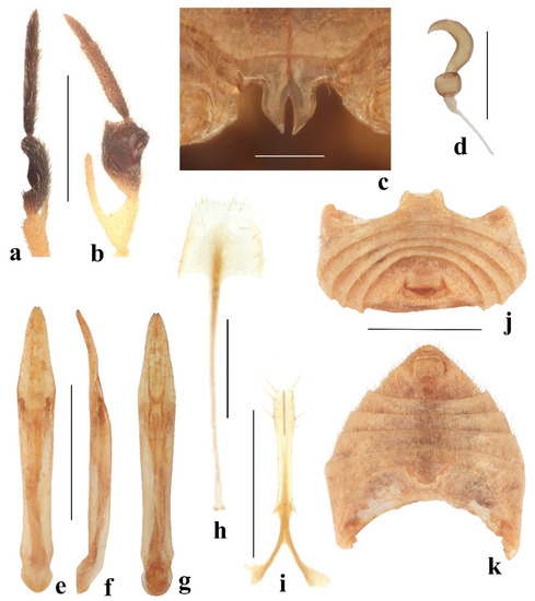
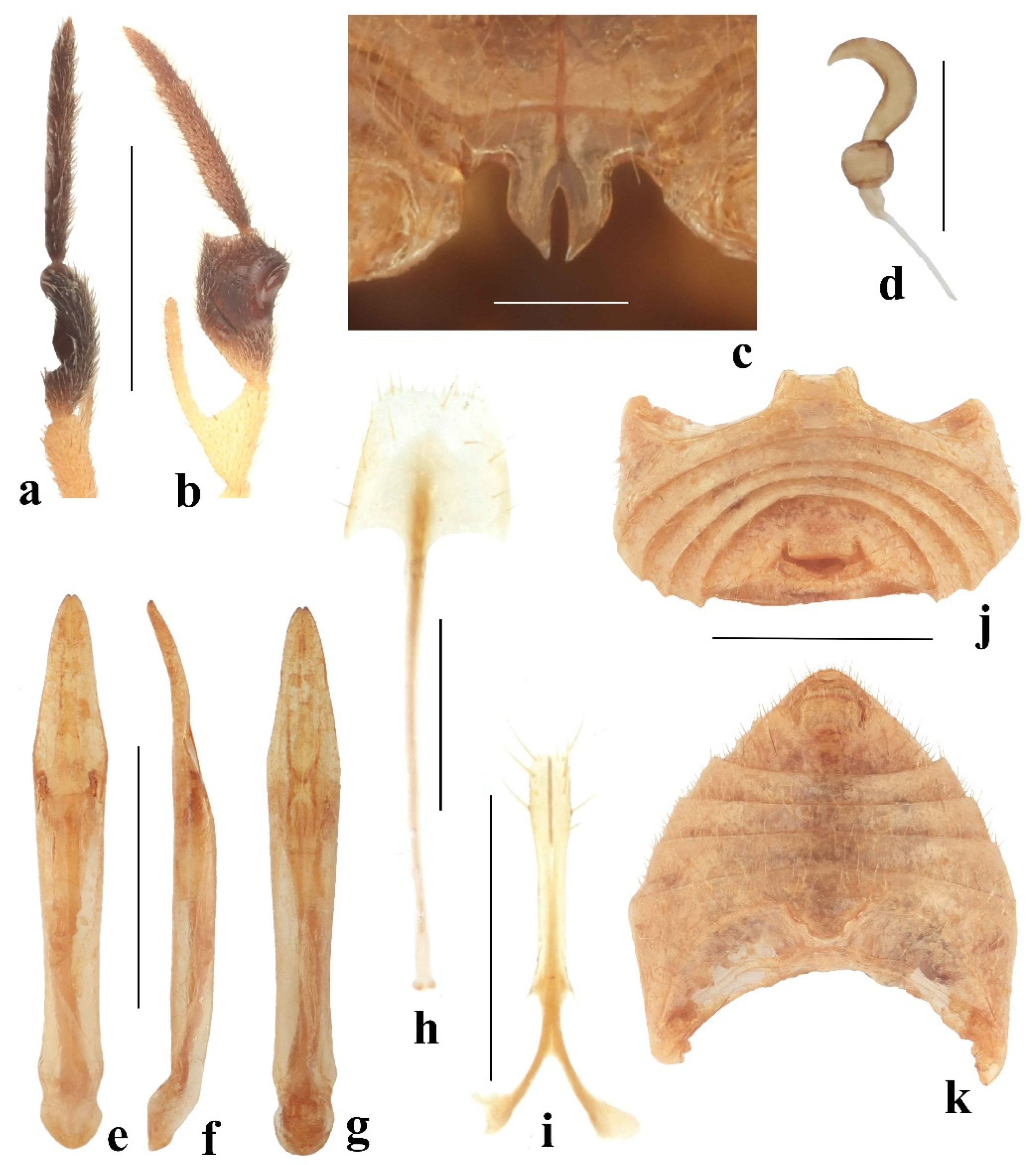
Figure 6.
Taumacera lewisi (Jacoby, 1887): (a) antennomeres X–XII, lateral view; (b) antennomeres X–XII, ventral view; (c) male metasternal process; (d) spermatheca; (e) aedeagus, dorsal view; (f) aedeagus, lateral view; (g) aedeagus, ventral view; (h) sternite VIII; (i) gonocoxae; (j) male abdomen, rear view; (k) male abdomen, ventral view. (Scale bars: (a,b,e–g,j,k) = 1 mm, (c,d) = 0.3 mm, (h,i) = 0.5 mm).
The pronotum (Figure 5c) is transverse, 1.63 times wide as it is long, semiopaque, widest at the anterior third, covered with microsculpture, and sparsely with fine punctures. The surface is glabrous, with a transverse impression in the posterior half, shallower in middle. The anterior margin is widely concave, bordered at lateral parts, unbordered in the middle, and lateral parts are covered with long setae. Lateral margins are sinuate with distinct borders, and margins have sparse, long setae. The posterior margin is rounded, narrowly bordered, and sparsely covered with short setae. Anterior angles are distinctly swollen, posterior angles are obtusangulate, and all angles have setigerous pores bearing very long, pale setae. The scutellum is triangular and covered with distinct punctures and pale setae.
The elytra are 1.48 times long as they are wide (measured at the widest place) and 0.66 times as long as the body, widest at the apical third. The surface is densely covered with small, confused punctures and microsculpture, locally with tendencies to form indistinct transverse wrinkles, and whole elytra are also covered with erect pale setae (often scattered on the disc). The epipleura are smooth, and wide at the basal half, then gradually narrowed towards the elytral apex, and the anterior half has dark setae along the elytral margin. Humeral calli and hind wings developed.
The metasternal process is 0.93 times long as it is wide, widest in middle, basal half nearly parallel, apical half triangular, and has a long narrow incision in the middle (Figure 6c). The anterior process of abdominal ventrite I is surrounded by thin lamella, the last abdominal ventrite is trilobed with two V-shaped incisions, and a median lobe with a slightly rounded posterior margin (Figure 6j,k).
The legs are moderately slender, covered with long, pale setae. Tarsi: protarsomere I is subtriangular, II is small and triangular, narrower than I, and the length ratio of protarsomeres equals 100-55-55-100 (100 = 0.30 mm); mesotarsomere I is subtriangular, II is small and triangular, and the length ratio of the mesotarsomeres equals 100-50-50-92 (100 = 0.30 mm); metatarsomere I is long, narrow, and parallel, II is short and triangular, and the length ratio of the metatarsomeres equals 100-41-32-64 (100 = 0.50 mm).
The aedeagus is lanceolate (Figure 6e–g) and is 7.30 times long as it is wide, widest at the anterior third, and the apex is subtriangular with a small incision in middle. In lateral view, the aedeagus is almost straight, only the apical part is slightly bent. In ventral view, the apical half of the aedeagus with a median furrow is constricted by two small, triangular protuberances at the apical third of the aedeagus.
Female (Figure 5b). The antennae are unmodified and filiform. Sternite VIII is shovel-shaped, the posterior margin has short setae, several setae also on the surface of sternite VIII, and the tignum is slender, 3.0 times longer than sternite VIII (Figure 6h). The spermatheca has a small spherical nodulus, cornu C-shaped, gradually narrowing towards the apex, the base of the cornu is slightly narrowed; the proximal part of the spermathecal duct is wider (Figure 6d). The apical fifth of the gonocoxae is slightly wider, each gonocoxa with six setae, base bifurcate, and each basal process is club-shaped (Figure 6i).
Differential diagnosis. Having a pale brown body without a black pattern on the pronotum and elytra Taumacera lewisi is similar to T. unicolor (Figure 5a and Figure 10a). Both species can be distinguished by the structure of the antennae (antennomere X is short and wide in T. lewisi, long and relatively narrow in T. unicolor) (Figure 5e and Figure 10f), the color of the antennae (antennomeres X–XII black in T. lewisi while antennae completely brown in T. unicolor) (Figure 5e and Figure 10f), the shape of pronotum (transverse, 1.63 times wide as it is long in T. lewisi, subquadrangular, 1.22 times wide as it is long in T. unicolor) (Figure 5c and Figure 10c), and body length (5.0–5.6 mm in T. lewisi, 8.2–8.4 mm in T. unicolor).
Distribution. Sri Lanka (Galle, Kandy, and Nuwara Eliya districts) (Figure 18).
Comments: Aenidea hirtipennis was described from one female collected of the same type and locality during the same expedition as the type specimens (males) of Taumacera lewisi. Bezděk (2019) examined the type material of both taxa and additional non-type material deposited in BMNH. Aenidea hirtipennis proved to be a female of T. lewisi and both taxa were synonymized.
3.4.3. Taumacera mirabilis (Jacoby, 1887)
Xenarthra mirabilis Jacoby, 1887: 107 [5] (original description); Weise (1924): 152 [1] (catalogue); Maulik (1936): 483 [2] (redescription); Wilcox (1973): 629 [3] (catalogue).
Taumacera mirabilis: Bezděk (2019): 29 [6] (transferred to Taumacera).
Type locality. “Bogawantalawa”.
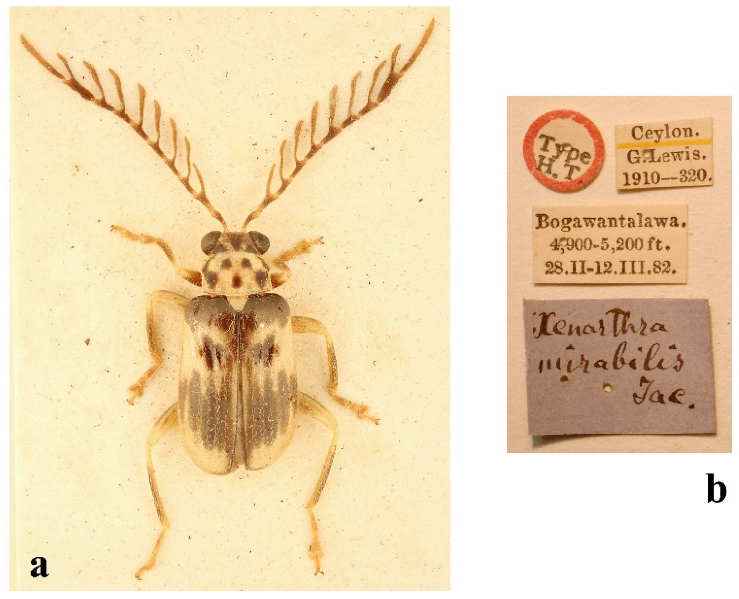
Type examined. Holotype: ♂ (BMNH, Figure 7a,b), “Type/H. T. [white round label with red frame, p]//Ceylon./G. Lewis./1910–320. [w, p]//Bogawantalawa./4900–5200 ft./28.II–12.III.82. [w, p]//Xenarthra / mirabilis/Jac. [b, h]’ (BMNH)”.
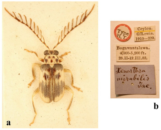
Figure 7.
Taumacera mirabilis (Jacoby, 1887): (a) holotype, male; (b) labels. (Not to scale).
Figure 7.
Taumacera mirabilis (Jacoby, 1887): (a) holotype, male; (b) labels. (Not to scale).
Additional material examined. SRI LANKA: Nuwara Eliya District: Maskeliya, ix.1905, 1 ♂ (BMNH); Badulla District: Hakgala Nature Reserve, 1675 m, 23.–25.ii.1977, 1 ♂, K. V. Krombein, P. B. Karunaratne, P. Fernando and D. W. Balasooriya leg. (USNM).
Redescription. Measurements. Male: 6.9–7.9 mm.
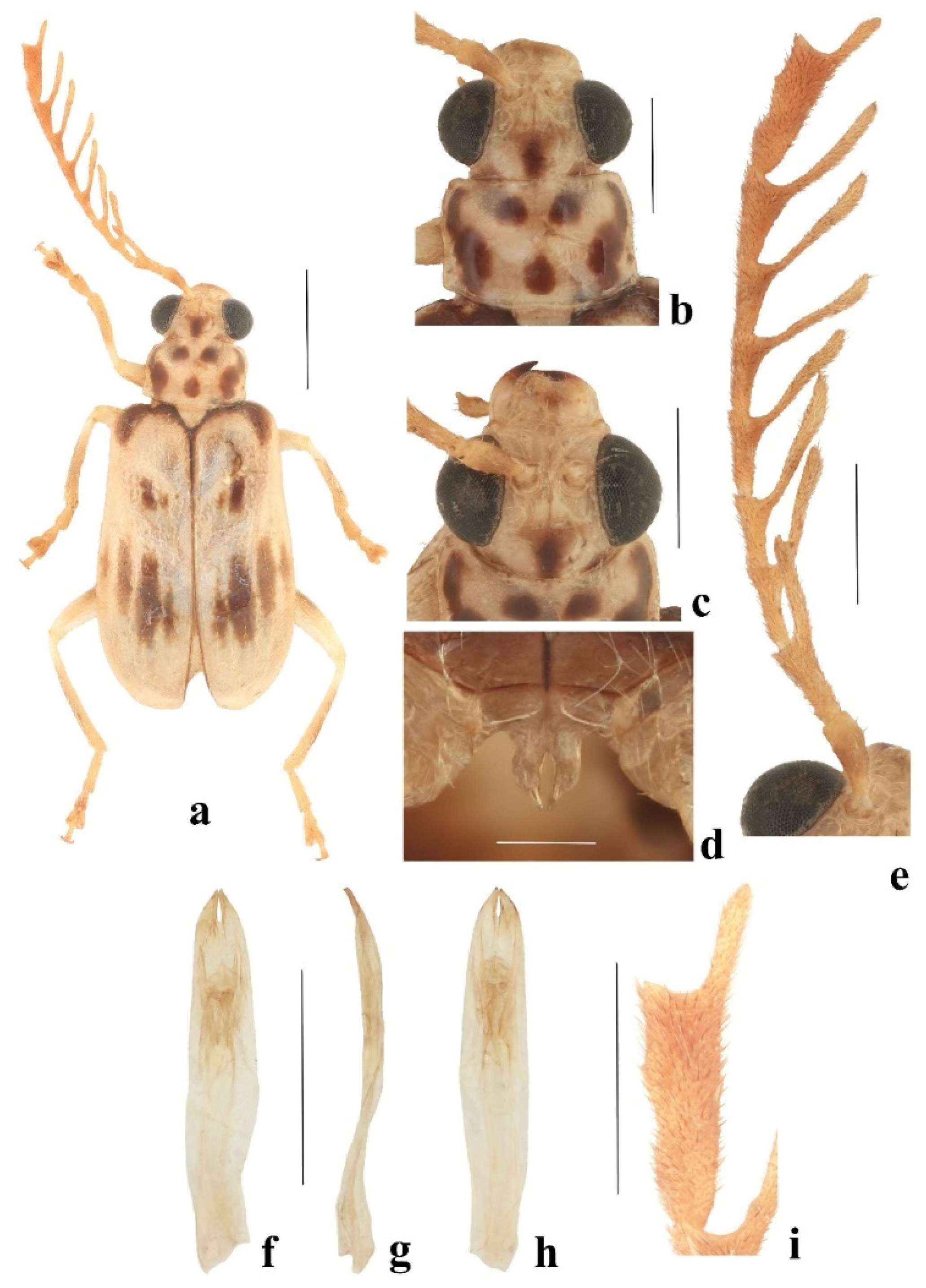
Male (Figure 8a). The dorsal side is an elongated oval, relatively flat. The body is pale brown except for the black apices of the mandibles, three black spots on the vertex, five black spots on the pronotum (two small round spots in the middle of the anterior part, one small round spot in the middle of the posterior part, and two irregular stripes along the lateral margins), black elytral markings (extreme suture, most of the epipleura, elytral base including humeral calli, three elongate spots in the middle of the anterior quarter and four large and partly connected longitudinal elongated spots on the posterior third), and metasternum. The legs are pale with the middle parts of the femora and tibiae darkened, and the tarsi infuscate. The antennae are dark brown with paler bases and apices of antennomeres I–X, and antennomeres XI–XII gradually paler towards the apex of XII.
Head (Figure 8b,c). The labrum is transverse, the anterior margin is emarginated in the middle, the anterior angles are rounded, and the surface is slightly convex with several pores in a transverse row bearing long setae. The anterior part of the head has a straight anterior margin, a surface with a keel forming a three-pointed star. The interantennal space is narrow, 0.66 times as wide as the transverse diameter of the antennal socket. The eyes are moderately large, with an interocular space 1.45 times as wide as the transverse diameter of the eye. The frontal tubercles are subquadrangular with inner anterior angles projecting anteriorly, convex, semiopaque, covered with microsculpture, and separated by a thin furrow. The vertex is separated from the frontal tubercles by a shallow impressed line and a notch in the middle, the surface is covered with microsculpture, and sparse punctures. The antennae are pectinate (Figure 8e), and the length ratio of the antennomeres I–XII equals 100-20-76-80-68-80-80-68-92-140-227-63 (100 = 0.7 mm). Antennomeres I and II are lustrous, and covered with sparse setae, III–XII are semiopaque, and densely covered with short brown setae. Antennomeres III–X are pectinate, III–IX with long lateral branches starting at an inner anterior angle, antennomere X enlarged, elongate, with two shallow emarginations on the outer side and a lateral branch shorter (Figure 8i), XI–XII are slender, straight, and fused but with a distinct seam.
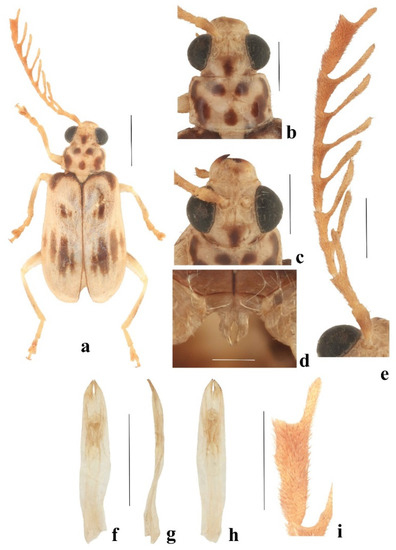
Figure 8.
Taumacera mirabilis (Jacoby, 1887): (a) male; (b) head and pronotum, dorsal view; (c) head, frontal view; (d) male metasternal process; (e) left antenna; (f) aedeagus, dorsal view; (g) aedeagus, lateral view; (h) aedeagus, ventral view; (i) antennomere X, detail. (Scale bars: (a) = 2 mm, (b,c,e–i) = 1 mm, (d) = 0.3 mm).
The pronotum (Figure 8b) is transverse, 1.45 times wide as it is long, semiopaque, glabrous, widest at the anterior quarter, covered with microsculpture, and sparse fine punctures. The surface has two round impressions in the posterior half. The anterior margin is widely concave, bordered at lateral parts close to the anterior angles, unbordered in the middle part, and without setae. The lateral margins are sinuate with distinct borders, and the margins have sparse, long setae. The posterior margin is nearly straight in the middle part, the lateral parts are oblique and straight. The anterior angles are slightly swollen, posterior angles obtusangulate, and all angles have setigerous pores bearing very long, pale setae. The scutellum is subtriangular with a rounded apex, covered with microsculpture, and short, pale setae.
The elytra are 1.50 times as long as they are wide (measured at the posterior third) and 0.69 times as long as the body, slightly divergent posteriorly, and widest at the posterior third. The surface is densely covered with small, confused punctures and with short, erect, pale setae in longitudinal rows. Each elytron is slightly convex in the subscutellar area and has an oblique bent impression in the anterior third. The Epipleura gradually narrowed towards the elytral apex, smooth, and lustrous, and have a row of setae along the elytral margin. The humeral calli and hind wings developed.
The metasternal process is 1.18 times as long as it is wide, parallel in the basal half, convergent in the apical third, and has a long lanceolate incision in the middle (Figure 8d). The last abdominal ventrite is trilobed with two V-shaped incisions, and a median lobe with a straight posterior margin.
The legs are moderately slender and covered with dense setae. Tarsi: protarsomere I is elongated, subtriangular, II is triangular, and the length ratio of the protarsomeres equals 100-71-53-94 (100 = 0.40 mm); mesotarsomere I is elongate and subtriangular, II is triangular, and the length ratio of the mesotarsomeres equals 100-65-47-100 (100 = 0.40 mm); metatarsomere I is long, narrow, and parallel, II is elongated and triangular, and the length ratio of the metatarsomeres equals 100-43-36-71 (100 = 0.70 mm).
The aedeagus (Figure 8f–h) of the examined male is poorly sclerotised, partly shrivelled, and with a missing basal part, the apical third is convergent with the apex gradually rounded towards the tip, and the apex has a narrow slit in middle. In ventral view, the apical part is impressed and has a median furrow constricted by two small, triangular protuberances in the apical third.
Variability. The male from the Hakgala Nature Reserve is generally paler with a reduced black elytral pattern, has legs with infuscate middle parts of femora and tibiae and tarsi are pale brown, and the antennae are completely pale brown.
Female unknown.
Differential diagnosis. Taumacera mirabilis differs from all congeners from Sri Lanka by a pale brown body with a characteristic black pattern on the pronotum and elytra (Figure 8a,b). Taumacera lewisi and T. unicolor are pale brown without a black pattern and their antennae have a different structure (Figure 5e and Figure 10f).
Distribution. Sri Lanka (Badulla and Nuwara Eliya districts) (Figure 18).
3.4.4. Taumacera unicolor (Jacoby, 1887)
Xenarthra unicolor Jacoby, 1887: 109 [5] (original description); Weise (1924): 152 [1] (catalogue); Maulik (1936): 485 [2] (redescription); Wilcox (1973): 629 [3] (catalogue).
Taumacera unicolor: Bezděk (2019): 45 [6] (transferred to Taumacera).
Type locality. “Colombo”.
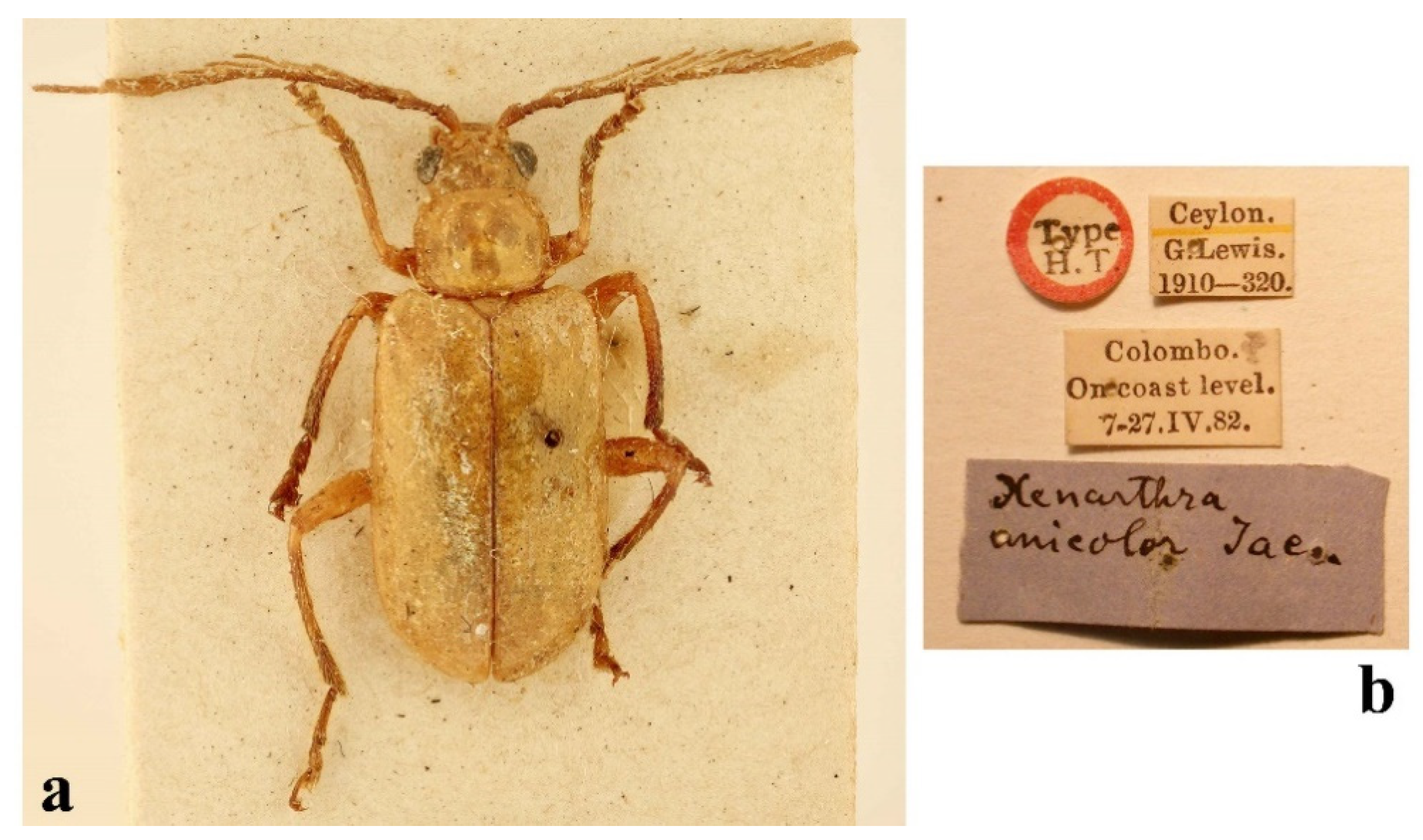
Type examined. Holotype: ♂ (BMNH, Figure 9a,b), “Type/H. T. [white round label with red frame, p]//Ceylon./G. Lewis./1910–320. [w, p]//Colombo./On coast level./7–27.IV.82. [w, p]//Xenarthra/unicolor Jac. [b, h]”.
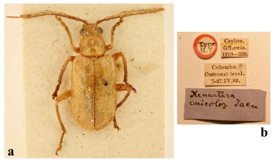
Figure 9.
Taumacera unicolor (Jacoby, 1887): (a) holotype, male; (b) labels. (Not to scale).
Figure 9.
Taumacera unicolor (Jacoby, 1887): (a) holotype, male; (b) labels. (Not to scale).
Additional material examined. SRI LANKA: Badulla District: Madulsima, 15.v.1908, 1 ♀, T. B. Fletcher leg. (BMNH). Not specified district: “Ceylon”, 1 ♂ (BMNH).
Redescription. Measurements. Male: 8.4 mm, female: 8.2 mm.
Male (Figure 10a). The dorsal side is an elongated oval, moderately convex. The body is completely brown, except for the apices of mandibles which are black.
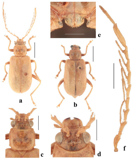
Figure 10.
Taumacera unicolor (Jacoby, 1887): (a) male; (b) female; (c) head and pronotum, dorsal view; (d) head, frontal view; (e) male metasternal process; (f) right antenna. (Scale bars: (a,b) = 2 mm, (c,d,f) = 1 mm, (e) = 0.3 mm).
Figure 10.
Taumacera unicolor (Jacoby, 1887): (a) male; (b) female; (c) head and pronotum, dorsal view; (d) head, frontal view; (e) male metasternal process; (f) right antenna. (Scale bars: (a,b) = 2 mm, (c,d,f) = 1 mm, (e) = 0.3 mm).
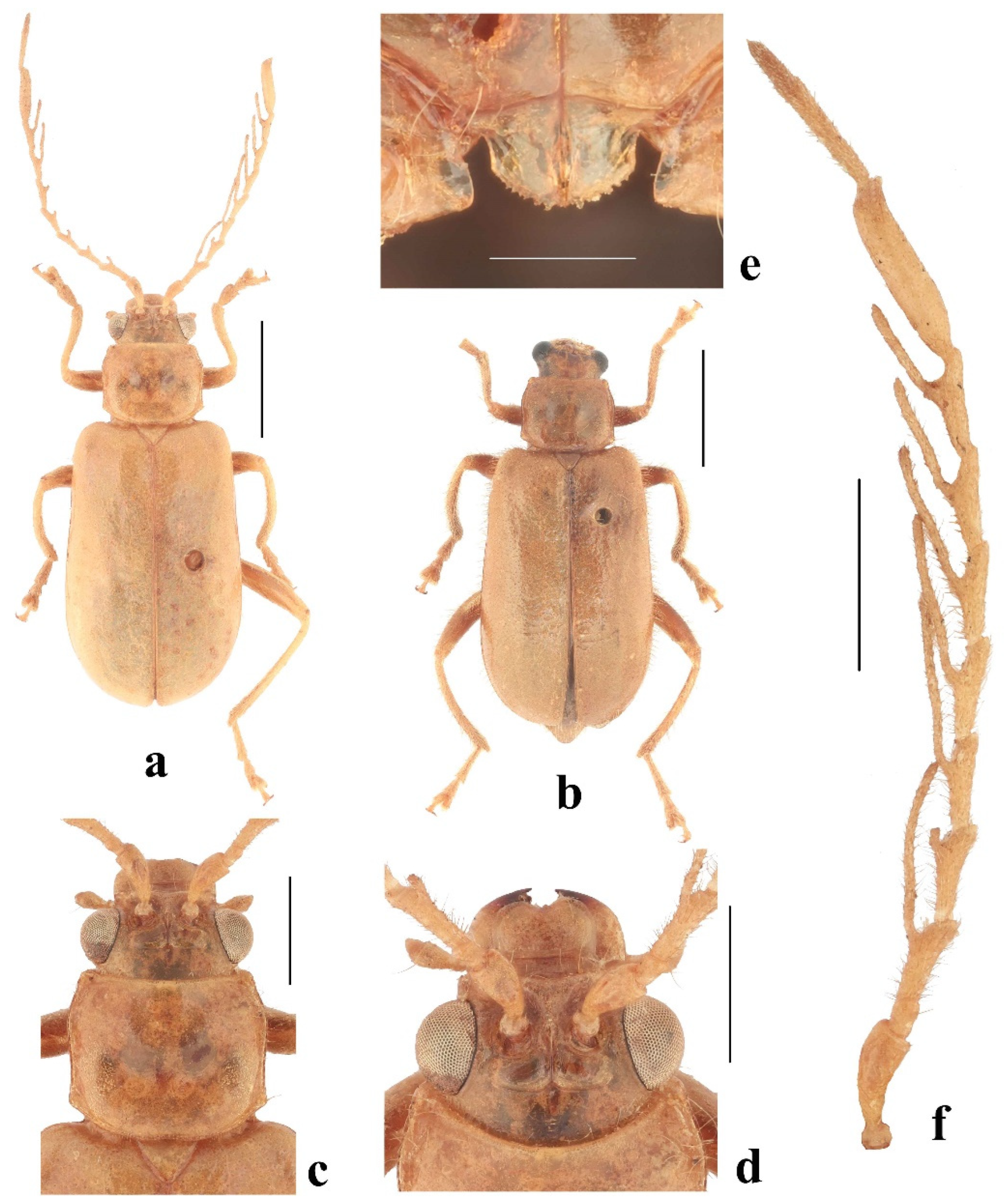
Head (Figure 10c,d). The labrum is transverse, the anterior margin is emarginated in the middle, the anterior angles are rounded, and the surface is slightly convex with several pores in a transverse row bearing long setae. The anterior part of the head has a straight anterior margin and a keel forming a three-pointed star, the surface is sparsely covered with long setae. The interantennal space is narrow, 0.72 times as wide as the transverse diameter of the antennal socket. The eyes are moderately large, the interocular space is 2.5 times as wide as the transverse diameter of the eye. The frontal tubercles are bean-shaped, slightly oblique, not reaching the inner margins of the eyes, with inner anterior angles projecting anteriorly, convex, semiopaque, covered with microsculpture, and separated by a thin furrow. The vertex is separated from the frontal tubercles by a shallow impressed line and a notch in the middle, the surface is sparsely covered with fine punctures and long setae. The antennae (Figure 10f) are 0.85 times the length of the body, and the length ratio of the antennomeres equals 100-25-69-75-75-69-69-69-69-138-94-44 (100 = 0.8 mm). Antennomeres I and II are lustrous, and covered with sparse setae, III–XII are semiopaque, and densely covered with short, dark setae. Antennomeres III–IX are pectinate, the lateral branches on antennomeres III–VI are very long, nearly twice as long as an appropriate antennomere, the lateral branches on antennomeres VII–IX are a little longer than an appropriate antennomere, the lateral branches on antennomeres III–V start subapically on the inner side of the antennomere, the lateral branches on antennomeres VI–IX start in the middle parts of the antennomeres, antennomere X is enlarged, is an elongated oval, with dorsal side convex, the outer apical angle is distinctly pronounced, XI–XII are slender, straight, and fused but with a distinct seam (Figure 11a).
The pronotum (Figure 10c) is subquadrangular, 1.22 times wide as it is long, widest at the anterior third, semiopaque, covered with very fine microsculpture, and sparse fine punctures. The surface has two oblique impressions in the posterior half, glabrous, except for the anterolateral parts with long setae. The anterior margin is widely concave, bordered at the lateral parts, and unbordered in the middle. Lateral margins are sinuate with a thin border, and the margins have sparse, long setae. The posterior margin is nearly straight, and lateral parts are oblique, and narrowly bordered. The anterior angles are distinctly swollen, the posterior angles are obtusangulate, and all angles have setigerous pores bearing very long, pale setae. The scutellum is triangular, impunctate, and glabrous.
The elytra are 1.61 times as long as they are wide (measured at the apical third) and 0.69 times as long as the body, widest at the apical third. The surface is glabrous (except for several setae on the lateral lobes), and densely covered with small, confused punctures. The subscutellar area of each elytron is very slightly convex. The epipleura is smooth, glabrous, wide at the basal half, and then gradually narrows towards the elytral apex. The humeral calli and hind wings are developed.
The metasternal process is wide, 1.90 times wide as it is long, widest at the base, the basal half is subparallel, the apical half widely rounded, the posterior margin has small denticles, and has a long narrow furrow in the middle (Figure 10e). The anterior process of abdominal ventrite I has a deep hole, the last abdominal ventrite is trilobed with two V-shaped incisions, and a median lobe which is distinctly impressed, with a rounded posterior margin (Figure 11c). The pygidium has a transverse, slightly concave apex.
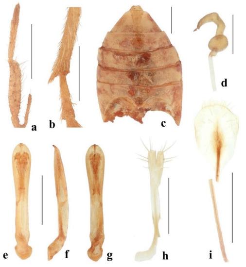
Figure 11.
Taumacera unicolor (Jacoby, 1887): (a) antennomeres X–XII, dorsal view; (b) apex of metatibia and metatarsomere I, male; (c) male abdomen, ventral view; (d) spermatheca; (e) aedeagus, dorsal view; (f) aedeagus, lateral view; (g) aedeagus, ventral view; (h) gonocoxae; (i) sternite VIII. (Scale bars: (a–c,e–g,i) = 1 mm, (d,h) = 0.5 mm).
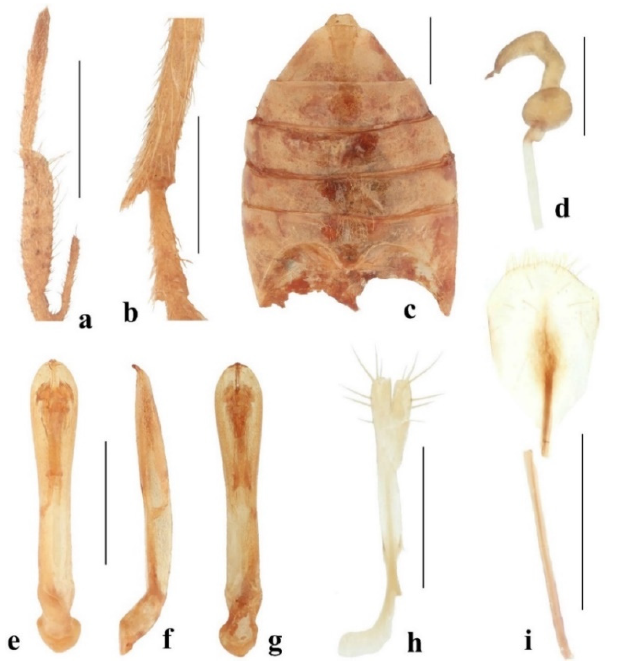
The legs are moderately slender, densely covered with short setae, and sparsely with very long setae. The apex of the metatibia is distinctly pronounced (Figure 11b). Tarsi: protarsomere I is elongated and subtriangular, II is small and triangular, and the length ratio of the protarsomeres equals 100-40-50-90 (100 = 0.50 mm); mesotarsomere I is elongated and subtriangular, II is small and triangular, and the length ratio of the mesotarsomeres equals 100-45-45-100 (100 = 0.60 mm); metatarsomere I is narrow and parallel, II is small and triangular, and the length ratio of the metatarsomeres equals 100-44-37-69 (100 = 0.80 mm).
The aedeagus (Figure 11e–g) is 6.10 times long as it is wide, the anterior third is extended, with widely rounded margins, widest at the preapical part, the basal two-thirds are narrower, subparallel, and the apex has two very small protuberances separated by a narrow slit. In lateral view, the aedeagus is nearly straight, and with a bent apex. In ventral view, the apical part is impressed with a long median furrow constricted by two small, triangular protuberances at the apical 2/5 of the aedeagus length.
Female (Figure 10b). the antennae are missing in the only available female but presumably filiform and unmodified. Sternite VIII is shovel-shaped, the posterior margin has short setae, several setae also on the surface of sternite VIII, and the tignum is slender, 1.7 times longer than sternite VIII (Figure 11i). The spermatheca has a small spherical nodulus, is cornu C-shaped, gradually narrowing towards the moderately hooked apex, the base of the cornu is slightly narrowed, and the proximal part of the spermathecal duct is wider (Figure 11d). The apical part of the gonocoxae is distinctly wider, each gonocoxa with six setae, base bifurcate, and each basal process is hockey-stick-shaped (Figure 11h).
Differential diagnosis. Having a pale brown body Taumacera unicolor is similar to T. lewisi. Both species differ in the following characteristics: antennomere X is long and relatively narrow in males of T. unicolor (short and wide in males of T. lewisi) (Figure 5e and Figure 10f), the antennae are completely brown in T. unicolor (antennomeres X–XII black in T. lewisi) (Figure 5e and Figure 10f), the pronotum is subquadrangular, 1.22 times as wide as long in T. unicolor (transverse, 1.63 times as wide as long in T. lewisi) (Figure 5c and Figure 10c), and body length is 8.2–8.4 mm in T. unicolor (5.0–5.6 mm in T. lewisi).
Distribution. Sri Lanka (Badulla and Colombo districts) (Figure 18).
3.4.5. Taumacera adamskii Bezděk, sp. nov.
Type locality. Sri Lanka, Anuradhapura District, Hunuwilagama [8°18′21.9″ N 80°8′55.3″ E].
Types. Holotype: ♂ (USNM), “SRI LANKA: Anu. Dist./Hunuwilagama, near/Wilpattu, 200 feet/black light/28 Oct–3 Nov 1976 [w, p]//Collected by:/G. F. Hevel/R. E. Dietz IV/S. Karunaratne/D. W. Balasooriya [w, p]//RESTRICTIONS APPLY/NMNH—Sri Lanka/Agreement #6 [v, p]”. Paratypes: 1 ♂ (USNM), “SRI LANKA: Gal. Dist./Kanneliya, 200 ft./black light/15–17 October 1976 [w, p]//Collected by:/G. F. Hevel/R. E. Dietz IV/S. Karunaratne/D. W. Balasooriya [w, p]//RESTRICTIONS APPLY/NMNH—Sri Lanka/Agreement #6 [v, p]”; 1 ♂ (JBCB), “CEYLON: Gal. Dist./Kanneliya Jungle/13–16 August 1972/K. V. Krombein &/P. B. Karunaratne [w, p]//RESTRICTIONS APPLY/NMNH—Sri Lanka/Agreement #6 [v, p]”. The type specimens are provided with one additional printed red label: “HOLOTYPUS [or PARATYPUS],/Taumacera/adamskii sp. nov.,/J. Bezděk det. 2022”.
Description. Male (holotype, Figure 12a,b). Body length 7.1 mm. The dorsal side is an elongated oval, moderately convex. The head, pronotum, scutellum, underside, antennae, and legs are pale brown, the apices of the mandibles are black, the last two antennomeres and penultimate tarsomeres are slightly darkened, the mesosternum and abdomen are orange, and the elytra are reddish brown with a slight metallic tint.
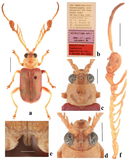
Figure 12.
Taumacera adamskii sp. nov., holotype: (a) holotype, male; (b) labels; (c) head and pronotum, dorsal view; (d) head, frontal view; (e) male metasternal process; (f) left antenna. (Scale bars: (a) = 2 mm, (c,d,f) = 1 mm, (e) = 0.3 mm, (b) not to scale).
Figure 12.
Taumacera adamskii sp. nov., holotype: (a) holotype, male; (b) labels; (c) head and pronotum, dorsal view; (d) head, frontal view; (e) male metasternal process; (f) left antenna. (Scale bars: (a) = 2 mm, (c,d,f) = 1 mm, (e) = 0.3 mm, (b) not to scale).
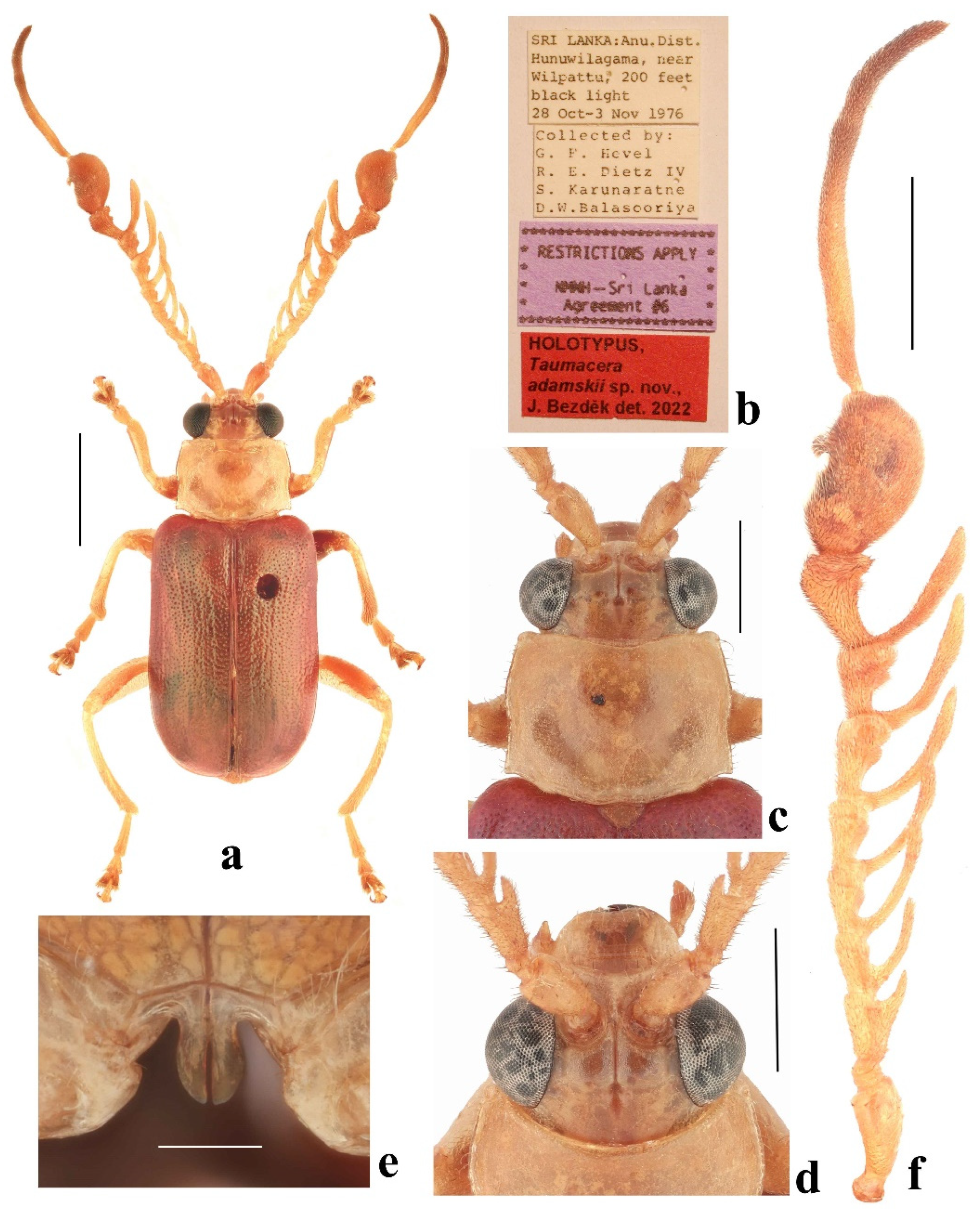
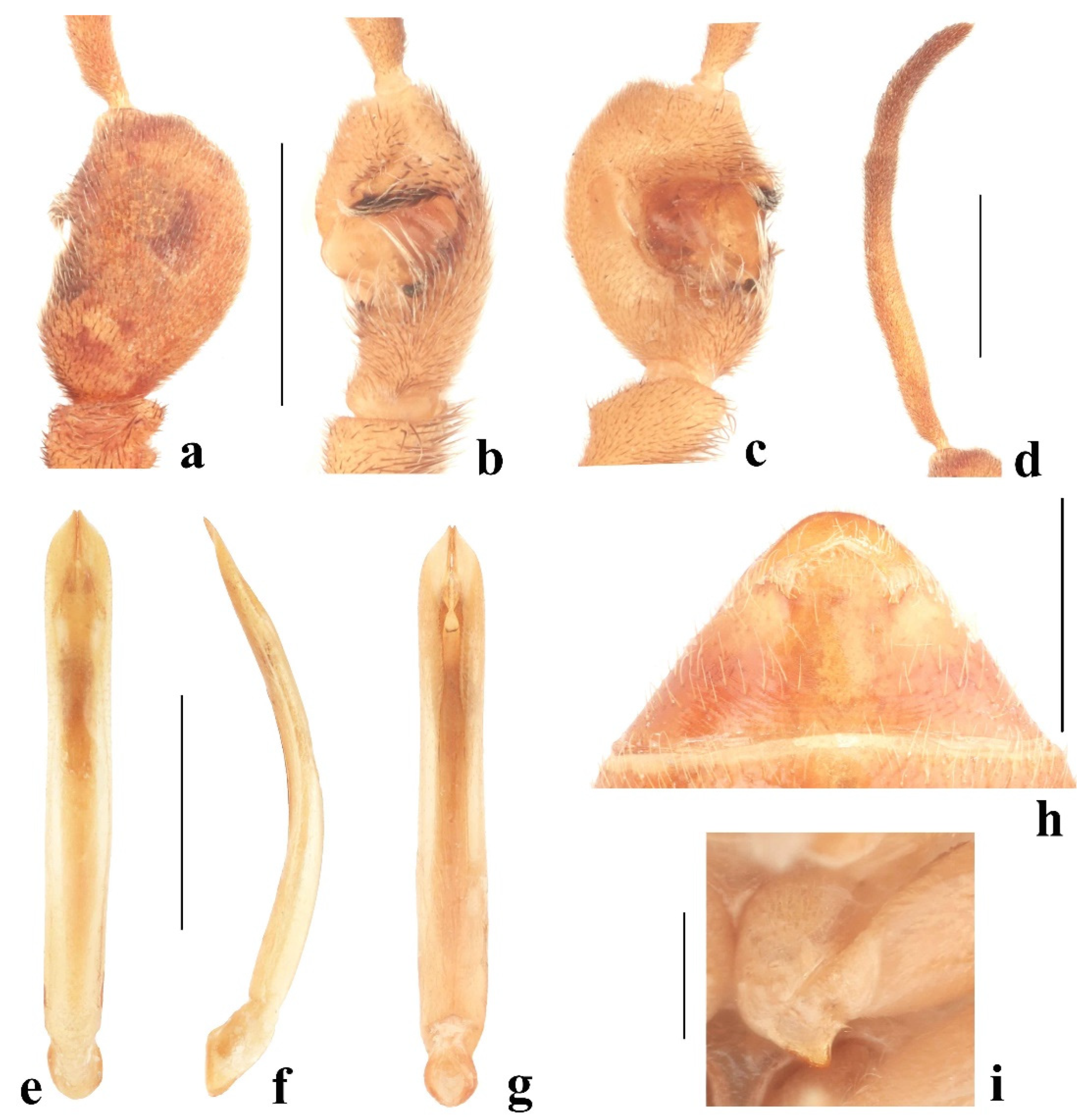
Head (Figure 12c,d). The labrum is transverse with an emarginated anterior margin and rounded anterior angles, the surface is slightly convex with four pores in a transverse row bearing long setae and several short setae on the anterior margin. The anterior part of the head has a straight anterior margin and a surface with a keel forming a three-pointed star. The interantennal space is narrow, 0.46 times as wide as the transverse diameter of the antennal socket. The eyes are large, and the interocular space is 1.7 times as wide as the transverse diameter of the eye. The frontal tubercles are subquadrangular with inner anterior angles projecting anteriorly, slightly convex, semiopaque, covered with microsculpture, and separated by thin furrows. The vertex is separated from the frontal tubercles by a shallow impressed line and deep notch in the middle, and the surface is impunctate and covered with microsculpture. The antennae (Figure 12f) are 1.10 times as long as the body, and the length ratio of the antennomeres equals 100-25-56-50-56-56-69-56-81-144-237-119 (100 = 0.8 mm). The antennal surface is covered with dense dark setae. Antennomeres III–IX are pectinate, the lateral branch on antennomere III is short and thorn-like, wide basally, placed in the middle of antennomere, the apical parts of antennomeres IV–VI are slightly enlarged; the apical parts of antennomeres VII–IX are strongly enlarged, clavate and distinctly impressed at the inner side; the lateral branches on antennomeres IV–IX are long, narrow, moderately bent, directed anteriorly, starting at the base of the antennomeres; antennomere X is strongly enlarged, suboval, the dorsal side moderately convex with a small lateral process on the external side, in lateral view with a deep U-shaped incision, in ventral view with a deep U-shaped cavity, cavity margins are covered with long, pale setae (Figure 13a–c); antennomere XI is slender, very long, slightly bent; antennomere XII is half as short as XI, moderately bent; and antennomeres XI and XII fused but with a distinct seam (Figure 13d).
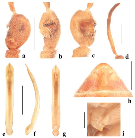
Figure 13.
Taumacera adamskii sp. nov., holotype: (a) antennomere X, dorsal view; (b) antennomere X, lateral view; (c) antennomere X, ventral view; (d) antennomeres XI–XII; (e) aedeagus, dorsal view; (f) aedeagus, lateral view; (g) aedeagus, ventral view; (h) last abdominal ventrite; (i) procoxa. (Scale bars: (a–h) = 1 mm, (i) = 0.5 mm).
The pronotum (Figure 12c) is transverse, 1.43 times as wide as long, semiopaque, glabrous, widest before the middle, covered with microsculpture, and fine punctures. The surface has a wide transverse impression in the posterior half, shallower in middle, and a subtriangular impression around the posterior angles. The anterior margin is widely concave, bordered at the lateral parts, unbordered in the middle, and without setae. The lateral margins are slightly convergent in the anterior half, then slightly constricted, subparallel in the posterior third, with distinct borders, and margins with sparse, long setae. The posterior margin is rounded, with indistinct angulation between the posterior angle and the midlength, and a small sinuation in the middle, narrowly bordered, the margin has several short setae near the posterior angles. The anterior angles are distinctly swollen, the posterior angles rectangular, and all angles have setigerous pores bearing very long, pale setae. The scutellum is small and subtriangular, with a widely rounded apex, and covered with distinct punctures and short, pale setae.
The elytra are 1.50 times as long as wide (measured at the humeral calli) and 0.63 times as long as the body, subparallel, and widest behind the middle. The surface is densely covered with small, confused punctures, on a disc with tendencies to form indistinct longitudinal rows, and with short, erect, pale setae. The subscutellar area of each elytron is slightly convex. The epipleura is smooth, glabrous, and wide at the basal half, then gradually narrows towards the elytral apex. The humeral calli and hind wings are developed.
The posterior margin of the procoxae have a large subtriangular protuberance (Figure 13i). The metasternal process is 1.30 times as long as wide, slightly constricted at the base, and apex widely rounded with a long narrow incision in the middle (Figure 12e). the last abdominal ventrite is trilobed with two V-shaped incisions and has a median lobe with a rounded posterior margin (Figure 13h).
The legs are moderately slender. The metatibia is slightly bent at the inner apical margin. Tarsi: protarsomere I is elongated and subtriangular, II is small and triangular, and the length ratio of the protarsomeres equals 100-53-59-112 (100 = 0.40 mm); mesotarsomere I is elongated and subtriangular, II is small and triangular, and the length ratio of the mesotarsomeres equals 100-55-55-100 (100 = 0.40 mm); metatarsomere I is long and subparallel, II is short and triangular, and the length ratio of the metatarsomeres equals 100-43-40-77 (100 = 0.75 mm).
The aedeagus (Figure 13e–g) is long and narrow, 8.90 times as long as wide, subparallel, with a subtriangular apex, and pronounced to a very short protrusion separated by a narrow slit. In lateral view, the aedeagus is widely bent. In ventral view, the apical part is impressed and has a long median furrow constricted by two small, triangular protuberances just behind the apical part, then it is slightly sinuate.
Female unknown.
Variability. The body length of males: is 6.7–7.1 mm.
Differential diagnosis. Taumacera adamskii sp. nov. can be distinguished from all other species of the T. cervicornis species group by the antennae which are completely pale brown with the antennomeres III–IX pectinate, X is strongly enlarged, XI–XII is slender (Figure 12f), and the elytra are reddish brown with a distinct metallic tint. Taumacera cervicornis, T. maskeliya sp. nov., and T. sigiriya sp. nov. have metallic green or bluish-green elytra and differently structured antennae (Figure 2a,e, Figure 14a,e and Figure 16a,f). Taumacera mirabilis is pale brown with the characteristic black pattern on the pronotum and elytra, and the antennae have different structures (Figure 8a,e). Taumacera lewisi and T. unicolor have completely pale brown dorsal sides and differently structured antennae (Figure 2a,e, and Figure 10a,f).
Distribution. Sri Lanka (Anuradhapura and Galle districts) (Figure 18).
Etymology. Dedicated to David Adamski (USNM), a specialist in Lepidoptera.
3.4.6. Taumacera maskeliya Bezděk, sp. nov.
Type locality. Sri Lanka, Kandy District, Maskeliya Oya, Norton Bridge [6°54′49.9″ N 80°31′17.0″ E].
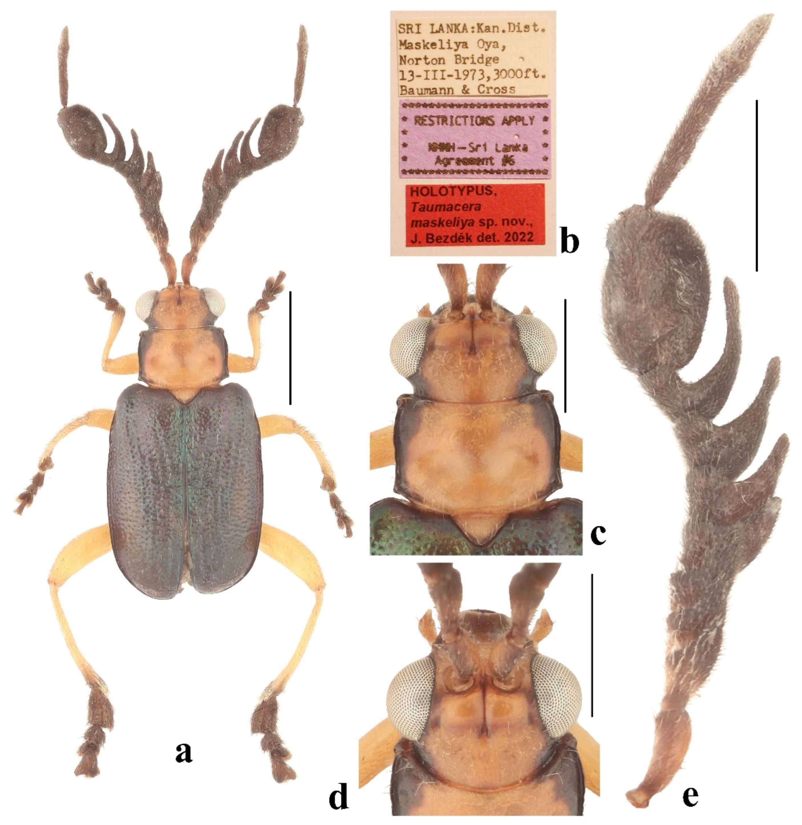
Types. Holotype: ♂ (USNM), “SRI LANKA: Kan. Dist. / Maskeliya Oya,/Norton Bridge / 13–III–1973, 3000ft./Baumann & Cross [w, p]//RESTRICTIONS APPLY/NMNH – Sri Lanka/Agreement #6 [v, p]”. Paratype: 1 ♂ (BMNH), “Ceylon [b, h]//53/88 [b, h, verso of the first label]”. The type specimens are provided with one additional printed red label: “HOLOTYPUS,/Taumacera / maskeliya sp. nov.,/J. Bezděk det. 2022”.
Description. Male (holotype, Figure 14a,b). Body length 5.2 mm. The dorsal side is an elongated oval, relatively flat. The head is orange-brown with black mandibles, labrum, and lateral parts of vertex behind eyes, and darkened inner margins of frontal tubercles. The pronotum is orange-brown, laterally black, with a black pattern wider in the anterior third and on the posterior margin. The scutellum is orange-brown. The antennae are black with dark brown antennomeres I–II and bases of antennomeres III–IV. The legs are pale brown with dark apices of tibiae and black tarsi. The elytra are metallic bluish-green. The ventral side is yellowish brown except for the lateral margins of the prosternum which are black.
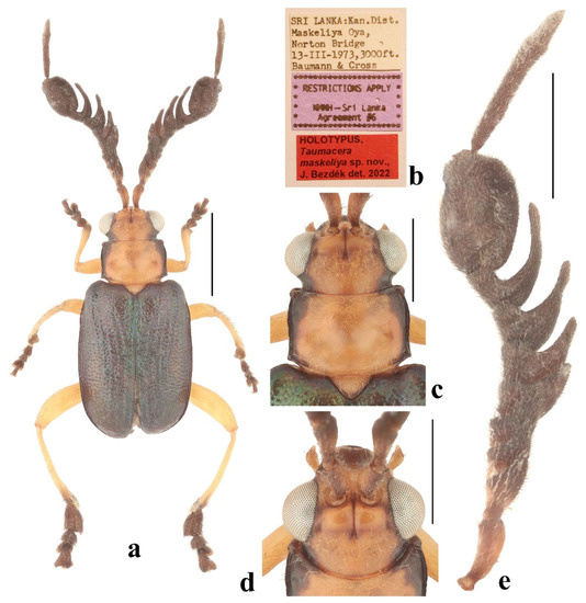
Figure 14.
Taumacera maskeliya sp. Nov., holotype: (a) holotype, male; (b) labels; (c) head and pronotum, dorsal view; (d) head, frontal view; (e) left antenna. (Scale bars: (a) = 2 mm, (c–e) = 1 mm, (b) not to scale).
Figure 14.
Taumacera maskeliya sp. Nov., holotype: (a) holotype, male; (b) labels; (c) head and pronotum, dorsal view; (d) head, frontal view; (e) left antenna. (Scale bars: (a) = 2 mm, (c–e) = 1 mm, (b) not to scale).
Head (Figure 14c,d). The labrum is transverse with a widely and shallowly emarginated anterior margin and rounded anterior angles, surface is convex with two pairs of pores close to the posterior angles bearing long setae. The anterior part of the head has a slightly concave anterior margin and a surface with a keel forming a three-pointed star. The interantennal space is 0.66 times as wide as the transverse diameter of the antennal socket. The eyes are moderately large, and the interocular space is 1.4 times as wide as the transverse diameter of the eye. The frontal tubercles are subquadrangular with inner anterior angles projecting anteriorly, convex, semiopaque, covered with microsculpture, and separated by a thin furrow. The vertex is separated from the frontal tubercles by a shallow impressed line and notch in the middle surrounded laterally with two short keels, the surface is covered with microsculpture, and in the anterior half also with fine punctures bearing setae. The antennae (Figure 14e) are 0.90 times as long as the body, and the length ratio of antennomeres equals 100-27-55-45-45-64-64-64-82-182-191-55 (100 = 0.6 mm). Antennomeres I and II are lustrous, and covered with sparse setae; III–XII are semiopaque, and densely covered with short, dark setae. Antennomere III is gradually extended with an inner apical margin pronounced to a short triangular process; IV–V are transverse with an inner apical margin pronounced to a short triangular process; VI–IX are pectinate with gradually longer lateral branches and a deeper emargination between the branch and the apex; antennomere X is strongly enlarged, suboval, with dorsal side cavities, in lateral view with U-shaped incision, in ventral view with transverse, lustrous and impunctate impression and longitudinal lustrous oval elevation; XI is very long, slender and straight; XII is short and as wide as XI; and XI and XII are fused but with a distinct seam (Figure 15a–c).
The pronotum (Figure 14c) is transverse, 1.50 times as wide as long, semiopaque, glabrous, widest at the anterior third, covered with microsculpture, and sparse fine punctures. The surface has two round impressions in the posterior half. The anterior margin is wide and shallowly concave, bordered at the lateral parts, unbordered in the middle, and lateral parts with several long setae directed posteriorly. Lateral margins are slightly sinuate with distinct borders, and margins with sparse setae. The posterior margin is rounded, with indistinct angulation between the posterior angle and the midlength, narrowly bordered, lateral parts with several long setae directed anteriorly. Anterior angles are distinctly swollen, posterior angles obtusangulate, and all angles have setigerous pores bearing very long, pale setae. The scutellum is subtriangular, smooth, and covered with short, pale setae.
The elytra are wide, 1.33 times as long as wide (measured in middle), and 0.65 times as long as the body, subparallel, and widest in middle. The surface is densely covered with small, confused punctures, on a disc with tendencies to form double longitudinal rows, and with short, erect, pale setae in longitudinal rows. The subscutellar area of each elytron is slightly convex. The epipleura are smooth, sparsely covered with setae, wide in the basal two-thirds, then gradually narrowed towards the elytral apex. The humeral calli and hind wings are developed.
The metasternal process is 1.40 times as long as wide, widest at the base, and slightly narrowed towards the apex, with a very long narrow incision in the middle (Figure 15j). The last abdominal ventrite is trilobed with two V-shaped incisions, a median lobe impressed, and a straight posterior margin.
The legs are moderately slender. The mesotibiae are flat, with a small preapical process at the inner side, and have a distinct emargination between the process and the apex (Figure 15f). Tarsi: protarsomere I is subtriangular, II is wide and triangular, and the length ratio of the protarsomeres equals 100-75-88-187 (100 = 0.20 mm); mesotarsomere I is subtriangular, II is wide and triangular, and the length ratio of the mesotarsomeres equals 100-50-80-150 (100 = 0.20 mm); the metatarsus is extremely enlarged (Figure 15d,e), metatarsomere I is axe-shaped, with the basal third convergent, outer side straight on the apical two-thirds, the inner side is extended and flattened, straight in the apical third, moderately rounded in the middle third; II is short, subtriangular, narrower than I; III is asymmetrical and heart-shaped with the outer lobe distinctly larger than the inner one, and as wide as II; IV is large, subtriangular, and flat, with an emarginated apical margin, ventral side is concave, claws inserted on the ventral side of the inner apical angle; and the length ratio of metatarsomeres equals 100-43-43-96 (100 = 0.50 mm).
The aedeagus (Figure 15g–i) is 7.30 times as long as wide, has an apical part gradually rounded, and a tip with a long narrow slit in the middle. In lateral view, the apical part is moderately bent, and the subapical part is straight (the rest of the aedeagus is poorly sclerotized, and shrivelled). In ventral view, the apical half has a median furrow constricted at the apical third by two small, triangular protuberances.
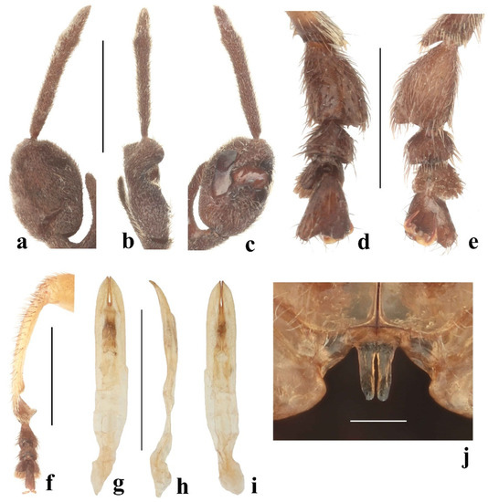
Figure 15.
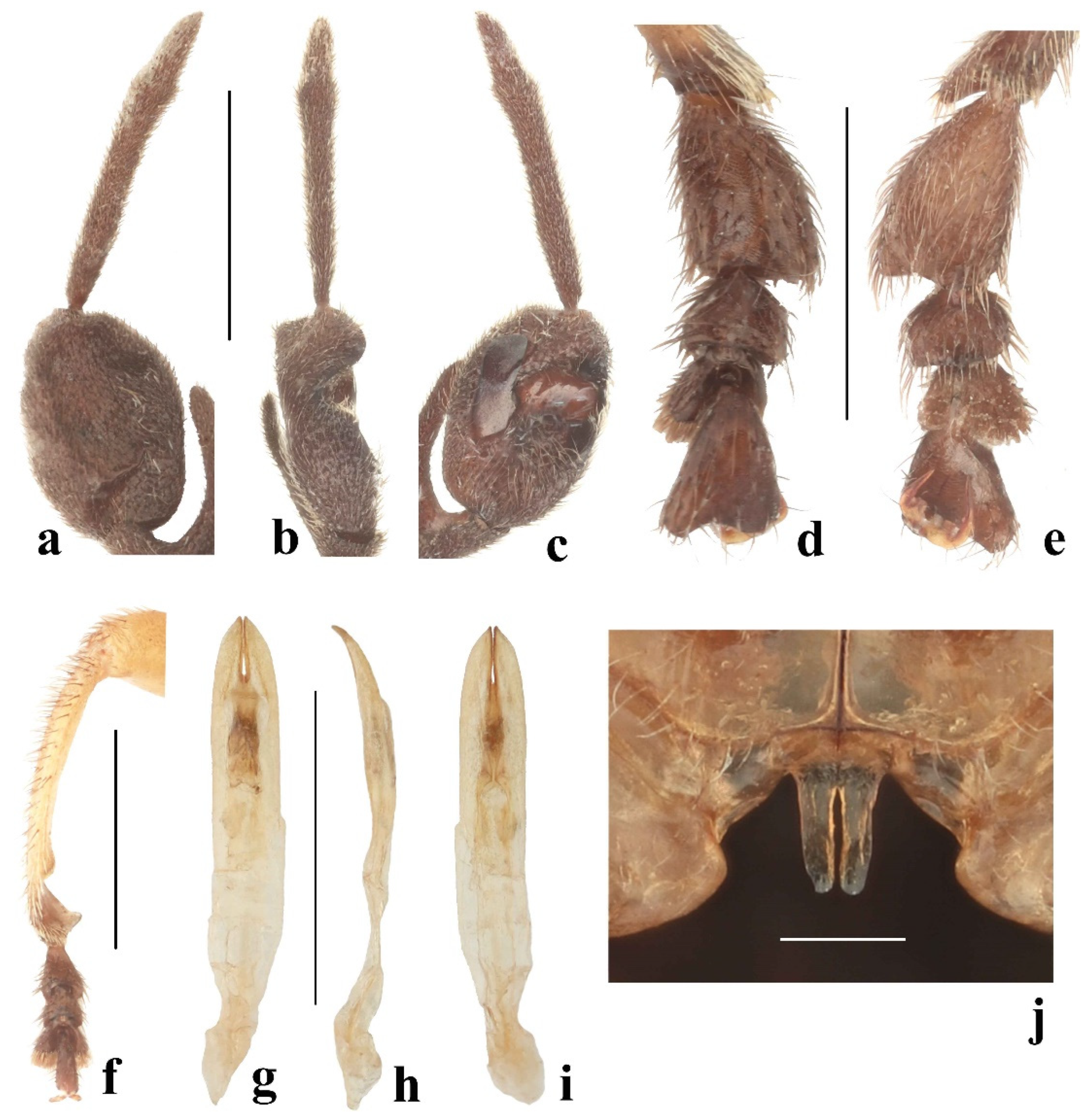
Taumacera maskeliya sp. nov.: (a) antennomeres X–XII, dorsal view; (b) antennomeres X–XII, lateral view; (c) antennomeres X–XII, ventral view; (d) metatarsus, dorsal view; (e) metatarsus, ventral view; (f) mesotibia; (g) aedeagus, dorsal view; (h) aedeagus, lateral view; (i) aedeagus, ventral view; (j) metasternal process. (Scale bars: (a–i) = 1 mm, (j) = 0.3 mm).
Female unknown.
Differential diagnosis. Taumacera maskeliya sp. nov. is very similar to T. cervicornis and T. sigiriya sp. nov. All three species share metallic green or blue-green elytra and a pale head and pronotum with darkened lateral parts. The male antennae of T. maskeliya sp. nov. have pectinate antennomeres VI–IX and thin, unmodified antennomeres XI–XII (Figure 14e), while the antennae of T. sigiriya sp. nov. have pectinate antennomeres III–IX and thin, unmodified antennomeres XI–XII (Figure 16f), and the antennae of T. cervicornis have antennomeres IV–VI subrectangular and transverse, VII–IX are pectinate, X is strongly enlarged, XI is enlarged, elongated subtriangular, and XII is paddle-shaped (Figure 2e). The mesotibiae of T. maskeliya sp. nov. and T. sigiriya sp. nov. have an oblique preapical process at the inner side and distinct emargination between processes but the mesotibia of T. maskeliya sp. nov. is moderately bent while almost straight in T. sigiriya sp. nov. (Figure 15f and Figure 17i). The metatarsus of T. maskeliya sp. nov. is extremely wide but the metatarsus of T. cervicornis is unmodified, and the metatarsus of T. sigiriya sp. nov. is less enlarged, with tarsomere IV asymmetrical (Figure 3e, Figure 15d,e and Figure 17k). The metasternal processes of T. maskeliya sp. nov. and T. sigiriya sp. nov. are very similar and distinctly longer than the metasternal process of T. cervicornis (Figure 3k, Figure 15j and Figure 16e).
Distribution. Sri Lanka (Kandy District) (Figure 18).
Etymology. Named after the Maskeliya Oya river where the holotype was collected. Noun in apposition.
3.4.7. Taumacera sigiriya Bezděk, sp. nov.
Type locality. Sri Lanka, Matale District, Sigiriya [7°56′57.5″ N 80°44′47.2″ E].
Types. Holotype: ♂ (USNM), “SRI LANKA: Mate. Dist./Sigirya, August 1974/S. Karunaratne [w, p]//RESTRICTIONS APPLY/NMNH—Sri Lanka/Agreement #6 [v, p]”. The holotype is provided with one additional printed red label: “HOLOTYPUS,/Taumacera/sigiriya sp. nov.,/J. Bezděk det. 2022”.
Description. Male (holotype, Figure 16a,b). Body length 5.1 mm. The dorsal side is an elongated oval, and relatively flat. The head is pale brown, the mandibles are dark brown with paler bases, and the outer margins before apices, frontal tubercles, and lateral parts of the vertex behind the eyes are dark brown. Antennomere I is brown with a darker dorsal side, II is brown, III and IV are brown with black lateral branches, V is black with a brownish base, VI–XII are black. The pronotum is brown with widely blackish lateral margins, and the anterior angles are brownish. The scutellum is pale brown. The elytra are metallic green with a brownish disc and outer margins of the epipleura. The ventral side is pale brown, the lateral margins of the prosternum are blackish, the abdomen with darkened ventrite IV and lateral parts of ventrite V, and the pygidium is black. The legs are pale brown with darkened apices of tibiae, and the tarsi are black.
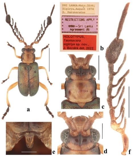
Figure 16.
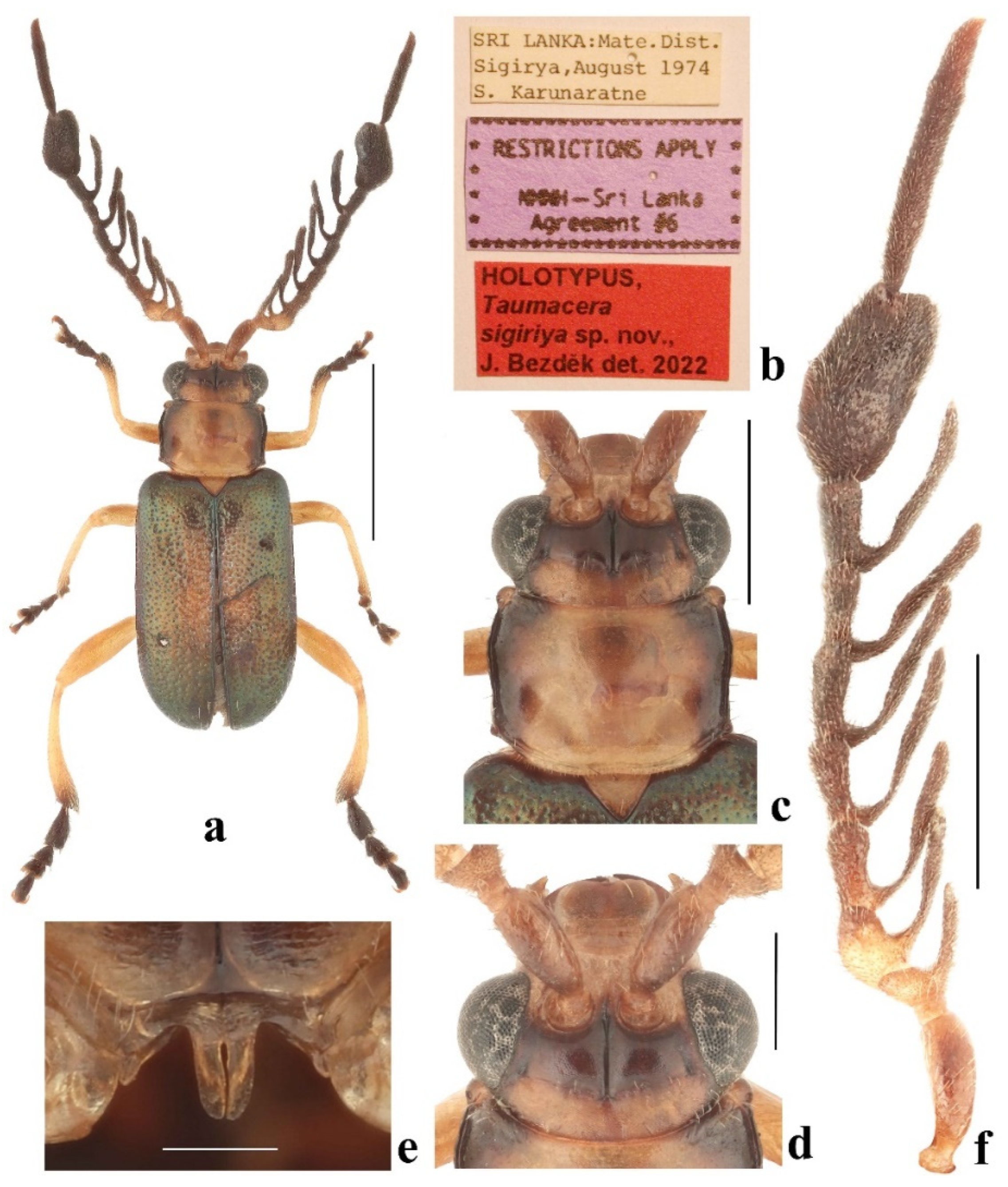
Taumacera sigiriya sp. nov., holotype: (a) holotype, male; (b) labels; (c) head and pronotum, dorsal view; (d) head, frontal view; (e) metasternal process; (f) left antenna. (Scale bars: (a) = 2 mm, (c,f) = 1 mm, (d) = 0.5 mm, (e) = 0.3 mm, (b) not to scale).
Figure 16.
Taumacera sigiriya sp. nov., holotype: (a) holotype, male; (b) labels; (c) head and pronotum, dorsal view; (d) head, frontal view; (e) metasternal process; (f) left antenna. (Scale bars: (a) = 2 mm, (c,f) = 1 mm, (d) = 0.5 mm, (e) = 0.3 mm, (b) not to scale).
Head (Figure 16c,d). the labrum is transverse with a U-shaped incision in the middle of the anterior margin, the anterior angles are rounded, and the surface is slightly convex with several pores in the transverse row bearing long setae. The anterior part of the head has a straight anterior margin and a surface with a keel forming a three-pointed star. The interantennal space is narrow, 0.50 times as wide as the transverse diameter of the antennal socket. The eyes are moderately large, and the interocular space is 2.5 times as wide as the transverse diameter of the eye. The frontal tubercles are transversely rectangular, slightly oblique, with inner anterior angles projecting anteriorly, convex, semiopaque, covered with microsculpture, and separated by a thin furrow. The vertex is separated from the frontal tubercles by a shallow impressed line and notch in the middle, surface covered with microsculpture, and sparse punctures. The antennae (Figure 16f) are as long as the body, and the length ratio of the antennomeres equals 100-17-21-43-50-50-57-43-50-121-136-50 (100 = 0.7 mm). Antennomeres I and II are lustrous, and covered with sparse setae; III–XII are semiopaque, and densely covered with short, dark setae. Antennomere I is clavate; II is very small and transverse; III–IX are pectinate, with the lateral branch on antennomere III shorter, and lateral branches on antennomeres IV–IX long, starting at the base of antennomeres; antennomere X is strongly enlarged, suboval, and has a convex dorsal side, in lateral view relatively flat and bent, in ventral view with elongated impression in the inner side and shorter and wider impression in outer side; antennomere XI is slender and straight; antennomere XII is nearly three times as short as XI; and antennomeres XI and XII are fused but with a distinct seam (Figure 17a,b).
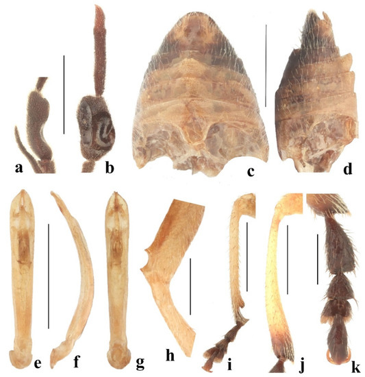
Figure 17.
Taumacera sigiriya sp. nov.: (a) antennomere X, lateral view; (b) antennomeres X–XII, ventral view; (c) abdomen, ventral view; (d) abdomen, lateral view; (e) aedeagus, dorsal view; (f) aedeagus, lateral view; (g) aedeagus, ventral view; (h) mesofemur; (i) mesotibia; (j) metatibia; (k) metatarsus. (Scale bars: (a–g,i,j) = 1 mm, (h,k) = 0.5 mm).
The pronotum (Figure 16c) is transverse, 1.48 times as wide as long, semiopaque, glabrous, covered with microsculpture and sparse fine punctures, and widest at the anterior third. The surface has two round impressions in the posterior half. The anterior margin is widely concave, bordered, and with long setae on lateral parts, unbordered in the middle, and without setae. The lateral margins are sinuate with distinct borders, and margins with sparse, long setae. The posterior margin is straight and oblique in lateral parts, nearly straight in the middle part, and narrowly bordered, and a margin which has several erected long setae near posterior angles. The anterior angles are distinctly swollen, posterior angles obtusangulate, and all angles with setigerous pores are bearing very long, pale setae. The scutellum is triangular, covered with fine microsculpture and indistinct punctures, and glabrous.
The elytra are 1.65 times as long as wide (measured at the humeral calli) and 0.65 times as long as the body, subparallel, widest in middle. The surface is densely covered with small, confused punctures; along the suture and lateral margins it is slightly finer, on a disc with tendencies to form indistinct longitudinal rows, and on lateral and apical slopes sparsely covered with erect setae forming longitudinal rows. The subscutellar area of each elytron is slightly convex. The epipleura are smooth, glabrous, wide at the basal half, then gradually narrowed towards the elytral apex. The humeral calli and hind wings are developed.
The metasternal process is 1.43 times as long as wide, widest at the base, slightly convergent to the apex, and preapical lateral parts are rounded, and with a long narrow incision in the middle (Figure 16e). The anterior process of abdominal ventrite I has a deep hole, and ventrites II and III are distinctly elevated in the middle parts. The last abdominal ventrite is trilobed with two V-shaped incisions, the median lobe being impressed, with a straight posterior margin (Figure 17c,d).
The legs are moderately slender, the mesofemora with a U-shaped emargination at the dorsal side near the apex (Figure 17h), mesotibiae with the large oblique preapical process at the inner side and with distinct emargination between the process and the apex (Figure 17i), metatibiae gradually extending towards the apex (Figure 17j). Tarsi: protarsomere I is subtriangular; II is small and triangular, and the length ratio of the protarsomeres equals 100-60-60-80 (100 = 0.30 mm); mesotarsomere I is subtriangular; II is small and triangular, and the length ratio of the mesotarsomeres equals 100-55-45-118 (100 = 0.30 mm); the metatarsus is enlarged (Figure 17k), metatarsomere I is wide, subclavate, and the outer margin is nearly straight, inner margin widely rounded; II is as wide as I, short and triangular; IV is asymmetrical, enlarged, flat, and the outer margin has a rounded middle part, the inner margin is sinuate, and the length ratio of the metatarsomeres equals 100-43-30-100 (100 = 0.60 mm).
The aedeagus (Figure 17e–g) is long and narrow, 7.50 times as long as wide, subparallel, widest at the preapical part, gradually slightly convergent towards the base, and the apex is subtriangular with a narrow slit in middle. In lateral view, the aedeagus is regularly widely bent, and the apical part is slightly narrower. In ventral view, the apical half has a median furrow and is constricted at the apical quarter by two small, triangular protuberances.
Female unknown.
Differential diagnosis. In appearance Taumacera sigiriya sp. nov. is very similar to T. cervicornis and T. maskeliya sp. nov. with which it shares a similar coloration. The males of all three species can be distinguished by the structures of the antennae, legs, aedeagi, and abdomina. The antennae of T. sigiriya sp. nov. have pectinate antennomeres III–IX and thin, unmodified antennomeres XI–XII (Figure 16f). In T. cervicornis antennomeres IV–VI are subrectangular and transverse, VII–IX are pectinate, X is strongly enlarged, XI is enlarged, elongate subtriangular, and XII is paddle-shaped (Figure 2e). The antennae of T. maskeliya sp. nov. have pectinate antennomeres VI–IX, and thin, unmodified antennomeres XI–XII (Figure 14e). Taumacera sigiriya sp. nov. has mesofemora with U-shaped emarginations on the dorsal side near the apex (Figure 17h), mesotibiae with the large oblique preapical process at the inner side, and with distinct emargination between process and apex (Figure 17i). The U-shaped emargination on the mesofemora is missing in T. cervicornis and T. maskeliya sp. nov. The mesotibiae are unmodified in T. cervicornis but the structure of the mesotibia of T. maskeliya sp. nov. is similar to that of T. sigiriya sp. nov. (Figure 15f and Figure 17i). The metatibiae of T. sigiriya sp. nov. are gradually extended towards the apex (Figure 17j) while unmodified in the other two species. The metatarsus of T. sigiriya sp. nov. is distinctly enlarged, with tarsomere IV being asymmetrical (Figure 17k). The metatarsus of T. cervicornis is unmodified, but the metatarsus of T. maskeliya sp. nov. is extremely wide (Figure 3e and Figure 15d,e). The metasternal processes of T. sigiriya sp. nov. and T. maskeliya sp. nov. are very similar and distinctly longer than the metasternal process of T. cervicornis (Figure 3k, Figure 15j and Figure 16e). The aedeagus of T. sigiriya sp. nov. is widely bent in lateral view (Figure 17f) while less bent and bisinuate in T. cervicornis (Figure 3h). The abdominal ventrites II and III are distinctly elevated in T. sigiriya sp. nov. (Figure 17c,d) while the abdomen is unmodified in T. maskeliya sp. nov. and T. cervicornis.
Distribution. Sri Lanka (Matale District) (Figure 18).
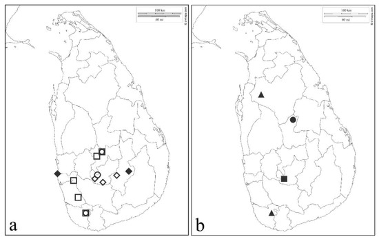
Figure 18.
Distributional maps of Taumacera from Sri Lanka: (a) □—T. cervicornis (Baly, 1861), ○—T. lewisi (Jacoby, 1887), ◊—T. mirabilis (Jacoby, 1887), ♦—T. unicolor (Jacoby, 1887); (b) ▲—T. adamskii sp. nov., ■—T. maskeliya sp. nov., ●—T. sigiriya sp. nov.
Figure 18.
Distributional maps of Taumacera from Sri Lanka: (a) □—T. cervicornis (Baly, 1861), ○—T. lewisi (Jacoby, 1887), ◊—T. mirabilis (Jacoby, 1887), ♦—T. unicolor (Jacoby, 1887); (b) ▲—T. adamskii sp. nov., ■—T. maskeliya sp. nov., ●—T. sigiriya sp. nov.
Etymology. Named after the type locality Sigiriya village in Matale District. Noun in apposition.
4. Discussion
Currently, 71 Asian species of the genus Taumacera are classified into seven species groups with 19 species not provisionally assigned to any group. The fauna of the Indian subcontinent is relatively poor, as the vast majority of these species occur in Southeast Asia. Only four species are known from mainland India and one more from the Andaman Islands. Until now, only four species of the T. cervicornis species group and T. facialis (Baly, 1886) from the T. nasuta species group, have been known from Sri Lanka (Bezděk 2019) [6]. However, looking at the maps with the indicated findings (Figure 18), it is evident that Sri Lanka is undercollected and large areas are still not sufficiently entomologically explored (at least for Chrysomelidae).
Bezděk (2019) [6] divided Taumacera species into seven more or less defined species groups and 19 species were left unassigned to any of the species groups. It cannot be excluded that the species groups or at least some of them will be classified as subgenera in the future. At the moment, due to the absence of molecular data which would support (or not) the monophyly of the species–groups, I prefer to cumulate the species in species groups.
The subfamily Galerucinae, like the entire family Chrysomelidae, is characterized by 11-segmented antennae (Nadein and Bezděk 2014) [17]. However, reductions in the number of antennal segments are rarely known in the tribe Alticini, e.g., 9-segmented antennae in Nonarthra Baly, 1862 and Kiskeya Konstantinov and Chamorro-Lacayo, 2006, or 10-segmented antennae in Psylliodes Latreille, 1829 and Monotalla Bechyné, 1956 (see Scherer 1961, Konstantinov and Chamorro-Lacayo 2006, Lee 2014) [18,19,20]. Reduced antennal segments are known also in some hispine genera (Gressitt and Kimoto 1963, Sekerka 2014) [21,22]. The representatives of the Taumacera cervicornis species group have 12 antennal segments. Antennomeres XI and XII are firmly fused in most species, separated by a distinct suture; however, in T. cervicornis antennomere XII appears to be moveable. Within Chrysomelidae, an increased number of antennal segments is known only in the genus Clytrasoma Jacoby, 1908, where some species also only have an indicated appendix on segment XI, but several species have a movable segment XII (Medvedev 1999, Schöller and Witte 2007) [23,24].
Funding
This research was supported by the Synthesys Project GB-TAF-6425 (http://www.synthesys.info/ (accessed on 27 November 2022)) financed by the European Community—Research Infrastructure Action under the Seventh Framework Programme.
Institutional Review Board Statement
Not applicable.
Data Availability Statement
Not applicable.
Acknowledgments
I would like to thank all curators and colleagues listed in the Material and Methods chapter for giving me the opportunity to study their collections.
Conflicts of Interest
The author declares no conflict of interest. The funders had no role in the design of the study; in the collection, analyses, or interpretation of data; in the writing of the manuscript; or in the decision to publish the results.
References
- Weise, J. Chrysomelidae: 13. Galerucinae. In Coleopterorum Catalogus. Pars 78; Schenkling, S., Ed.; W. Junk: Berlin, Germany, 1924; pp. 1–225. [Google Scholar]
- Maulik, S. Coleoptera, Chrysomelidae (Galerucinae). The Fauna of British India Including Ceylon and Burma; Taylor and Francis: London, UK, 1936; p. 648. [Google Scholar]
- Wilcox, J.A. Chrysomelidae: Galerucinae: Luperini: Luperina. In Coleopterorum Catalogus Supplementa. Pars 78(3), 2nd ed.; Wilcox, J.A., Ed.; W. Junk: Gravenhage, The Netherlands, 1973; pp. 433–664. [Google Scholar]
- Baly, J.S. Descriptions of new genera and species of Phytophaga. J. Entomol. 1861, 1, 275–302. [Google Scholar] [CrossRef]
- Jacoby, M. Descriptions of the phytophagous Coleoptera of Ceylon, obtained by Mr. George Lewis during the years 1881–1882. Proc. Zool. Soc. Lond. 1887, 55, 65–119. [Google Scholar] [CrossRef]
- Bezděk, J. Taumacera revisited, with new synonyms, new combinations and a revised catalogue of the species (Coleoptera: Chrysomelidae: Galerucinae). Acta Entomol. Mus. Nat. Pragae 2019, 59, 23–52. [Google Scholar] [CrossRef]
- Bezděk, J. Taxonomic and faunistic notes on Oriental and Palaearctic Galerucinae and Cryptocephalinae (Coleoptera: Chrysomelidae). Genus 2012, 23, 375–418. [Google Scholar]
- Nguyen, D.T.; Bezděk, J. Vietnamese species of Taumacera viridis-group with a description of new species (Coleoptera: Chrysomelidae: Galerucinae). J. Asia-Pacif. Entomol. 2021, 24, 536–543. [Google Scholar] [CrossRef]
- Reid, C.A.M. Reappraisal of the genus Taumacera Thunberg with descriptions of two new species from South-East Asia (Coleoptera: Chrysomelidae: Galerucinae). Austr. J. Entomol. 1999, 38, 1–9. [Google Scholar] [CrossRef]
- Thunberg, C.P. Beskrifning på tvänne nya insect-slägten, Gnatocerus och Taumacera från Goda-Hopps Udden. Kongl. Vetens. Acad. Handl. 1814, 499, 46–50. [Google Scholar]
- Chapuis, F. Famille des Phytophages. Histoire Naturelle des Insectes. Genera des Coléoptères ou Exposé Méthodique et Critique de Tous les Genres Proposés Jusqu’ici dans Cet Ordre d’Insectes. Tome Onzième; Roret: Paris, France, 1875; p. 420. [Google Scholar]
- Gemminger, M.; Harold, E. Catalogus Coleopterorum. Vol. XII: Chrysomelidae (Pars II), Languriidae, Erotylidae, Endomychidae, Coccinellidae, Corylophidae, Platypsyllidae; Theodor Ackermann: Monachii, Germany, 1876; pp. 3479–3676. [Google Scholar]
- Allard, E. Nouvelle note sur les Phytophages à la suite d’un examen des Galérucides appartenant au Musée royal de Belgique. Bull. Séanc. Soc. Entomol. Belg. 1889, 102–117. [Google Scholar]
- Seeno, T.N.; Wilcox, J.A. Leaf beetles genera (Coleoptera: Chrysomelidae). Entomography 1982, 1, 1–221. [Google Scholar]
- Medvedev, L.N. Chrysomelidae from Ceylon (Col.). Entomol. Arb. Mus. Frey 1972, 23, 178–185. [Google Scholar]
- Kimoto, S. The Chrysomelidae (Insecta: Coleoptera) collected by Dr. Akio Otake, on the occasion of his entomological survey in Sri Lanka from 1973 to 1975. Bull. Kitakyushu Mus. Natur. Hist. Hum. Hist. Ser. A 2003, 1, 23–43. [Google Scholar]
- Nadein, K.; Bezděk, J. Galerucinae Latreille, 1802. In Handbook of Zoology. Coleoptera, Beetles. Morphology and Systematics. Volume 3; Leschen, R.A.B., Beutel, R.G., Eds.; Walter de Gruyter: Berlin, Germany; Boston, MA, USA, 2014; pp. 251–259. [Google Scholar]
- Scherer, G. Bestimmungsschlüssel der Alticinen-Genera Afrikas (Col. Phytoph.). Entomol. Arb. Mus. Frey 1961, 12, 251–289. [Google Scholar]
- Konstantinov, A.S.; Chamorro-Lacayo, M.L. A new genus of moss-inhabiting flea beetles (Coleoptera: Chrysomelidae) from the Dominican Republic. Col. Bull. 2006, 60, 275–290. [Google Scholar] [CrossRef]
- Lee, C.-F. Review of the genus Nonarthra Baly (Coleoptera: Chrysomelidae: Galerucinae: Alticini) from Taiwan and Japan, with descriptions of two new species. Jap. J. Syst. Ent. 2014, 20, 251–263. [Google Scholar]
- Gressitt, J.L.; Kimoto, S. The Chrysomelidae (Coleopt.) of China and Korea, part 2. Pacif. Ins. Monogr. 1963, 1B, 301–1026. [Google Scholar]
- Sekerka, L. Review of Imatidiini genera (Coleoptera: Chrysomelidae: Cassidinae). Acta Entomol. Mus. Nat. Pragae 2014, 54, 257–314. [Google Scholar]
- Medvedev, L.N. Clytrinae (Coleoptera, Chrysomelidae) of Malayan region. Serangga 1999, 4, 53–74. [Google Scholar]
- Schöller, M.; Witte, V. A review of the genus Clytrasoma Jacoby, 1908, with description of a new species collected within a Camponotus sp. ant nest (Insecta: Coleoptera, Chrysomelidae; Hymenoptera: Formicidae). Senckenberg. Biol. 2007, 87, 51–61. [Google Scholar]
Publisher’s Note: MDPI stays neutral with regard to jurisdictional claims in published maps and institutional affiliations. |
© 2022 by the author. Licensee MDPI, Basel, Switzerland. This article is an open access article distributed under the terms and conditions of the Creative Commons Attribution (CC BY) license (https://creativecommons.org/licenses/by/4.0/).
