Article

The Morphology, Ultrastructure and Molecular Phylogeny of a New Freshwater Heterolobose Amoeba Parafumarolamoeba stagnalis n. sp. (Vahlkampfiidae; Heterolobosea)

by
Anastasia S. Borodina
1,2,
Alexander P. Mylnikov
1,†,
Jan Janouškovec
3,
Patrick J. Keeling
4 and
Denis V. Tikhonenkov
1,5,*
1
Papanin Institute for Biology of Inland Waters, Russian Academy of Sciences, 152742 Borok, Russia
2
Department of Zoology and Parasitology, Voronezh State University, Universitetskaya Ploshad 1, 394036 Voronezh, Russia
3
Centre Algatech, Laboratory of Photosynthesis, Institute of Microbiology, Czech Academy of Sciences, Opatovický Mlýn, 37981 Třeboň, Czech Republic
4
Department of Botany, University of British Columbia, 6270 University Boulevard, Vancouver, BC V6T1Z4, Canada
5
AquaBioSafe Laboratory, University of Tyumen, 625003 Tyumen, Russia
*
Author to whom correspondence should be addressed.
Alexander P. Mylnikov is deceased.
http://zoobank.org/References/e543a49a-16c1-4b7c-afdb-0bc56b632ef0
Diversity 2021, 13(9), 433; https://doi.org/10.3390/d13090433
Submission received: 20 August 2021 / Revised: 3 September 2021 / Accepted: 5 September 2021 / Published: 8 September 2021
(This article belongs to the Special Issue Aquatic Biodiversity: Evolution, Taxonomy and Conservation)

Abstract

:
Heterolobose amoebae are important members of marine, freshwater, and soil microbial communities, but their diversity remains under-explored. We studied the diversity of Vahlkampfiidae to improve our understanding of heterolobosean relationships and their representation in aquatic benthos. Using light and electron microscopy, and molecular phylogenies based on the SSU rRNA and ITS loci, we describe the fine morphology and evolutionary relationships of a new heterolobosean Parafumarolamoeba stagnalis n. sp. from a small pond in European Russia. Cells of P. stagnalis possess a clearly distinguishable anterior hyaline pseudopodium, eruptive movement, several thin and sometimes branched uroidal filaments, spherical cysts without pores and plugs, and mitochondria that have discoid cristae and are surrounded by cisternae of the endoplasmic reticulum. The genus Parafumarolamoeba has so far included a single species, Parafumarolamoeba alta from high-altitude soil in Tibet, which is morphologically distinct from P. stagnalis. Taxonomic description for a new Parafumarolamoeba species is therefore provided.

Graphical Abstract

1. Introduction

Heterolobosea Page and Blanton 1985 is a relatively small group of protists belonging to Discoba and combining heterotrophic amoeboflagellates, amoebae, flagellates and some slime molds (Acrasidae). Heteroloboseans are commonly amoeboflagellates, which alternate amoeboid and flagellar stages during their life cycle. The genus Naegleria is the most well studied of them: N. fowleri is a deadly human parasite [1] and N. gruberi is a model for the development of the flagellar apparatus [2]. However, other heteroloboseans have been studied to a much lesser extent, despite their wide environmental presence and morphological diversity.
Members of the family Vahlkampfiidae contribute significantly to the diversity and abundance of amoebas in a wide variety of aquatic and terrestrial habitats [3,4,5]. Vahlkampfia was initially distinguished by the lack of a flagellate stage and pores in the cyst wall [6]. Phylogenetic studies based on the small subunit ribosomal RNA (SSU rRNA) sequence, a universal marker for the taxonomy of heteroloboseans and many other protists, changed the systematics of the Vahlkampfiidae [7,8], including the splitting of Vahlkampfia into four genera: Tetramitus, Vahlkampfia, Neovahlkampfia and Paravahlkampfia [8]. Additional genera (e.g., Fumarolamoeba, Parafumarolamoeba) and species belonging to Vahlkampfiidae have since been described. It was established that species of Naegleria and other vahlkampfiids are difficult to distinguish by morphological characteristics [9], making molecular techniques pivotal in uncovering the vahlkampfiid phylogeny and diversity.
The less conservative ITS region has also been used to differentiate vahlkampfiid species and gives phylogenetic signal that is largely congruent with that of SSU rRNA trees. Different species within heterolobosean genera (e.g., Paravahlkampfia, Tetramitus, Naegleria,) can be up to 99.81% similar in the ITS region (Tetramitus thorntoni AJ698843 and T. jugosus AJ698845) and 99.76% by SSU rRNA (Paravahlkampfia ustiana KX068999 and P. francinae FJ169185). An integrated approach to conduct phylogenetic analysis of both genes is preferred over using a single marker [10,11].
Here we describe a new species of Parafumarolamoeba isolated from a freshwater pond and provide its morphological and ultrastructural characteristics, and phylogeny based on the SSU rRNA gene and ITS region.

2. Materials and Methods

2.1. Clone Isolation, Microscopy, and Laboratory Experiments

The clone Va-1 was derived from a single cell isolated by a micropipette from a sediment sample taken from a small pond near the Borok settlement, Russia (58°03′39.1” N 38°14′48.0” E) on 28 February 2012.
The culture was maintained in Petri dishes filled with Pratt medium (KNO3–100 mg L−1; K2HPO4–10 mg L−1; MgSO4·7H2O–10 mg L−1; FeCl3·6H2O–1 mg L−1) with Pseudomonas fluorescens Migula bacteria added as food. Clone Va-1 is currently being stored in a collection of live protozoan cultures at the Institute for Biology of Inland Waters, Russian Academy of Sciences.
Light microscopy observations were made by using the Zeiss AxioScope A.1 equipped with a DIC and phase contrast water immersion objective (63×). The images were taken with the AVT HORN MC-1009/S analog video camera and directly digitized using the Behold TV 409 FM tuner.
For scanning electron microscopy (SEM), cells in culture medium from exponential growth phase were fixed with 2% glutaraldehyde (final concentration) for 10 min at 22 °C and gently drawn onto a polycarbonate filter (diameter 24 mm, pores 0.8 μm). Following the filtration, the specimens were taken through a graded ethanol dehydration and acetone and finally put into a chamber of a critical point device for drying. Dry filters with fixed specimens were then mounted on aluminum stubs, coated with gold, and observed with the JSM-6510LV scanning electron microscope (JEOL, Japan).
For transmission electron microscopy (TEM), cells were centrifuged and fixed in a cocktail of 0.6% glutaraldehyde and 2% OsO4 (final concentration) prepared on 0.05 M cacodylate buffer for 30–60 min. Fixed cells were dehydrated in alcohol and acetone series (30, 50, 70, 96, and 100%, 20 min in each step). Afterwards, the cells were embedded in a mixture of Araldite and Epon. Ultrathin sections (50 nm) were prepared with the Leica EM UC6 ultramicrotome (Leica Microsystems, Germany) and observed by using the JEM 1011 transmission electron microscope (JEOL, Japan).
Experiments were carried out to check the ability of species to produce flagellar zoospores. Three different approaches were used. (A) The Petri dishes with amoeboid cells and cysts were incubated at different temperatures from 4° to 40 °C (the temperature was increased sequentially by 1 °C daily); (B) the cell suspension was shaken up for 3 h using a magnetic stirrer; (C) the media was replaced in the Petri dishes with amoeboid cells and cysts several times. We used the three approaches to induce zoospores alone and in combination (A, B and C).

2.2. DNA Sequencing and Phylogenetic Analysis

Cells of the strain Va-1 were grown in a clonal culture and collected by centrifugation (1000 g, room temperature) onto a 0.8 mm-pore membrane of the Vivaclear mini column (Sartorius Stedim Biotech, VK01P042). Genomic DNA was isolated using the MasterPure™ Complete DNA and RNA Purification Kit (Epicentre, Cat. No. MC85200). The SSU rRNA gene of strain Va-1 were amplified using the universal eukaryotic primers PF1-FAD4 [12]. The internal transcribed spacer (ITS) region, including the 5.8S rDNA was amplified using JITS-F and JITS-R primers [10]. EconoTaq PLUS GREEN 2X Master Mix (Lucigen, Cat. No 30033-1) was used in the following PCR amplification program: initial denaturation at 95 °C for 3 min, 35 cycles of 95 °C for 30 s, 52 °C for 30 s, 72 °C for 1.5 min, and a final extension at 72 °C for 5 min. The amplified DNA fragments were purified with QIAquick PCR Purification Kit (Quagen, Cat. No. 433160764). The PCR products were subsequently cloned (ITS) using StrataClone PCR Cloning Kit (Agilent, Part Number 240205) or sequenced directly (SSU rRNA) by Sanger dideoxy sequencing. Two additional internal primers 18SintF (5′- GGTAATTCCAGCTCCAATAGCGTA-3′) and 18SintR (5′- GTTTCAGCCTTGCGACCATACT-3′) were used for SSU rRNA sequencing. The final sequences were assembled from four overlapping reads using the Phred-Phrap-Consed package [13].
Seventy-three SSU rRNA sequences of Heterolobosea and twenty-five ITS sequences were aligned by the L-INS-i algorithm in MAFFT v7 [14] and trimmed by using the Gappyout method in TrimAl (v. 1.3) [15]. MrBayes v3.2.6 [16] was run with four categories of Gamma-distributed among site rate variation and calculation of the proportion of invariable sites (GTR+I+GAMMA4 substitution model). Four independent Bayesian runs, with four Metropolis-coupled Markov chains each, were sampled across 20 million generations and summarized at a 50% burn-in. The maximum likelihood phylogeny was computed in IQ-TREE v1.5.4 [17] by using the best fit model (as determined by the in-built ModelFinder), TIM2+F+R4 for the SSU rDNA and TN+F+G4 for the ITS region dataset, and 1000 non-parametric bootstraps.

3. Results

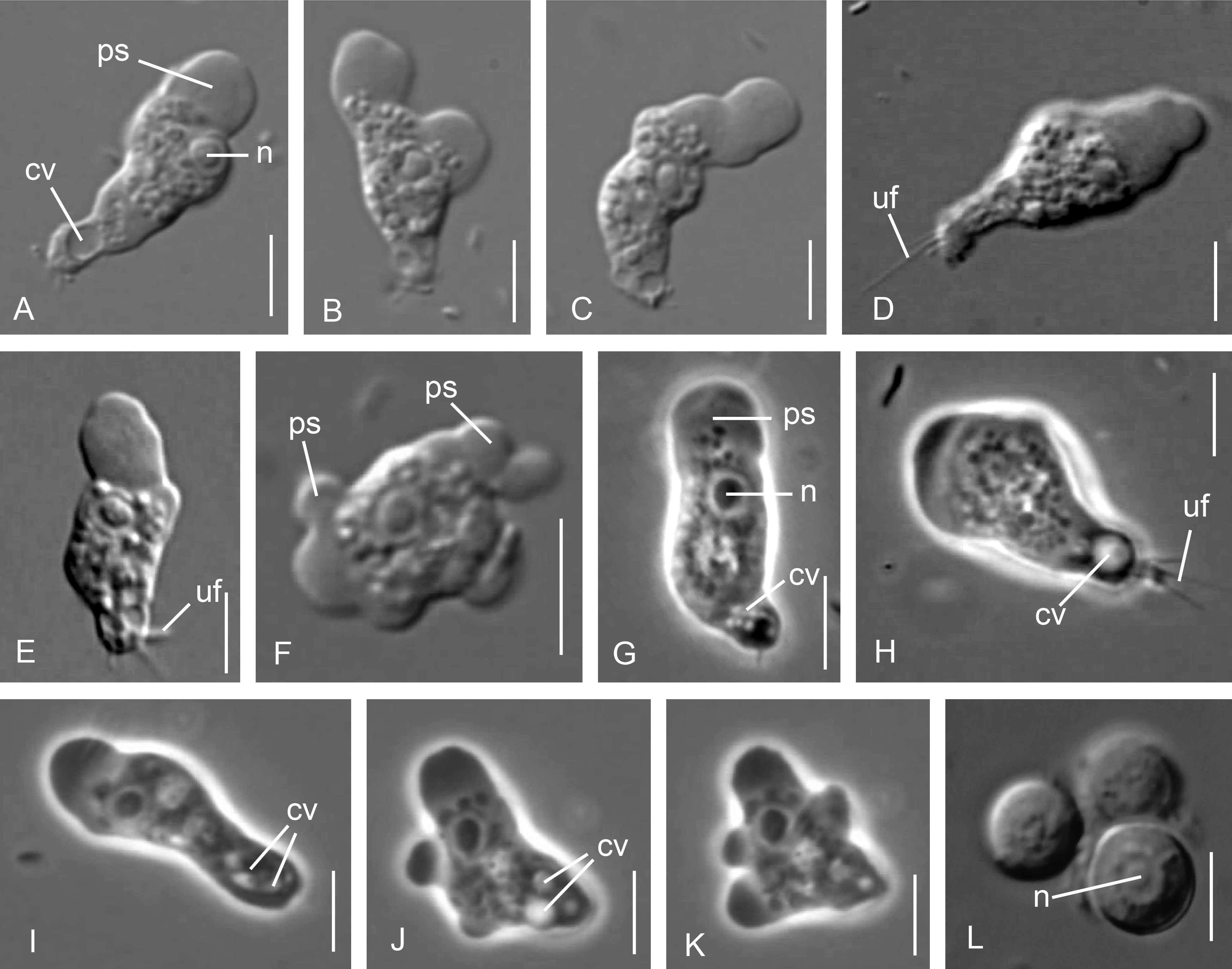
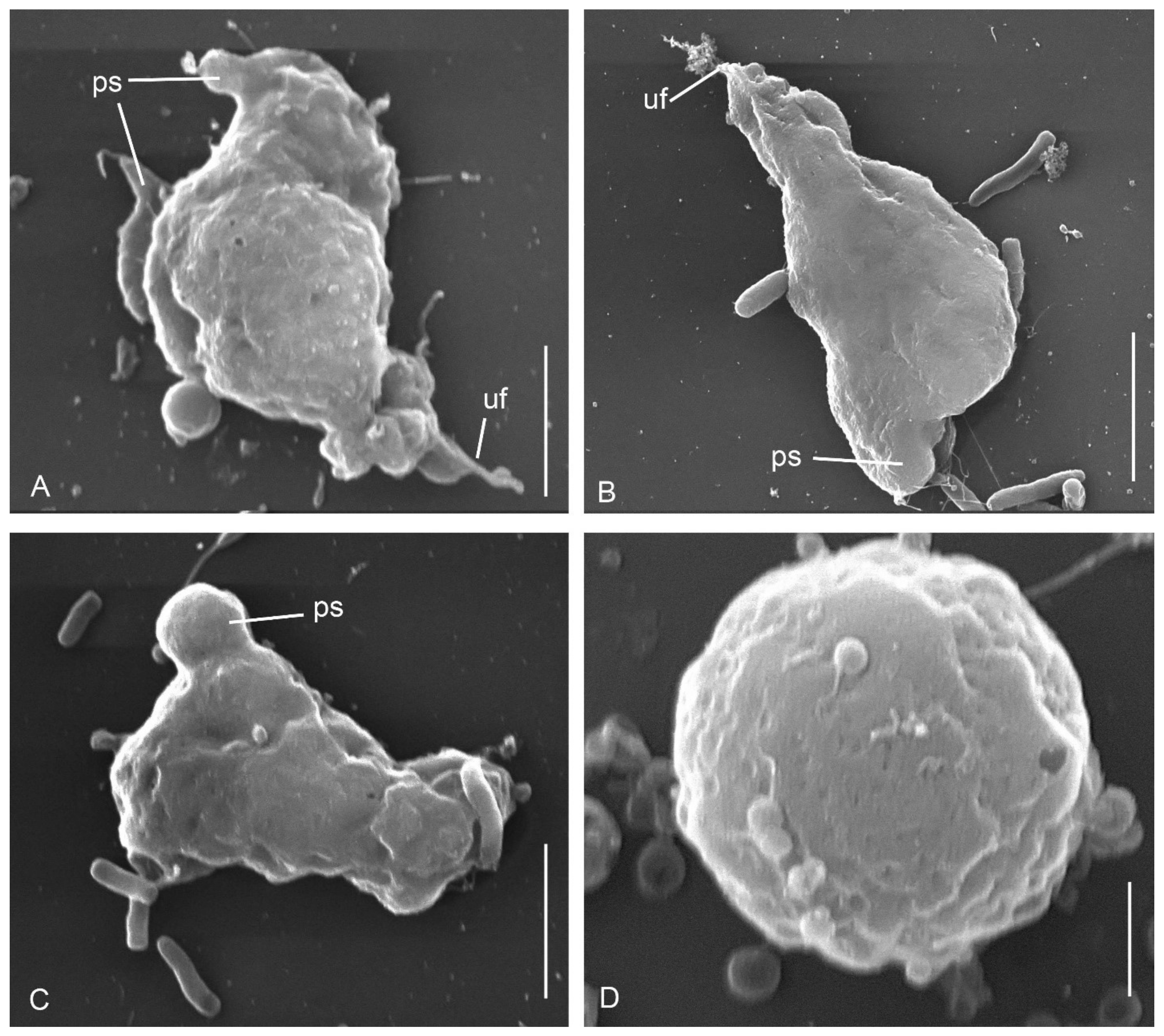
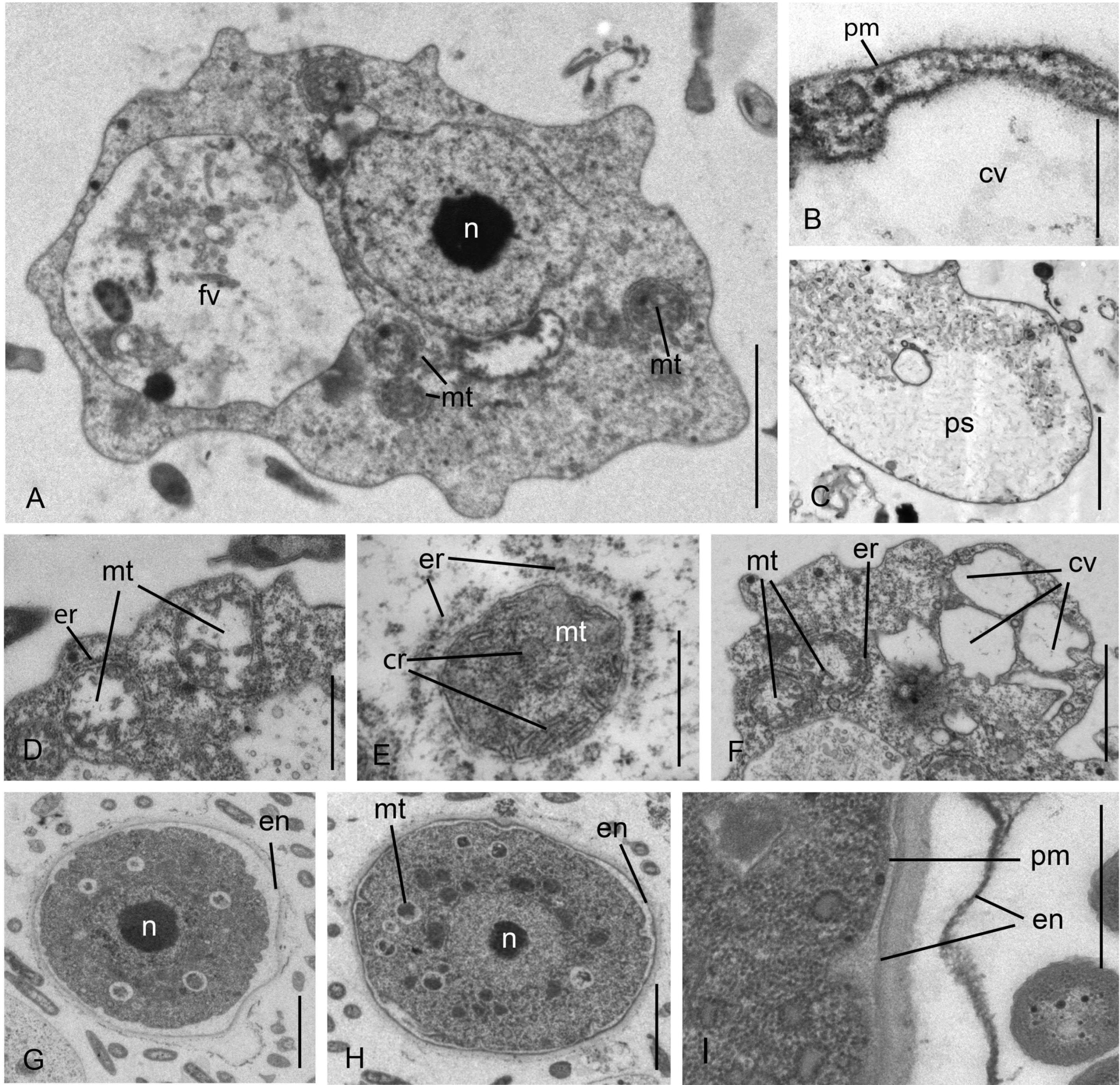
3.1. Cell Morphology

Parafumarolamoeba stagnalis n. sp.

The cells possess a distinct anterior hyaline pseudopodium, which is usually solitary but can also occur as a pair of competing pseudopodia formed by eruptive movement (Figure 1A–C; Video S1). These pseudopodia usually account for 15–25% of the cell length (Figure 1A–K). The length of motile cell is 13–28 μm (mean 17.9 ± 0.5 μm, n = 50), width is 9–12 μm (mean 10.4 ± 0.5 μm, n = 50). The length: width ratio is 4.3 in active cells and 1.7 in slowly moving cells. Some specimen possessed several thin and sometimes branched uroidal filaments (Figure 1D,E,H and Figure 2A,B). The cell contains small cytoplasmic granules, food vacuoles and a large posterior contractile vacuole (Figure 1A,H,J), which is created by the fusion of 5 to 7 smaller vacuoles (Figure 1G,I). The nucleus, 1.5–2.0 µm in diameter, lies just behind the hyaline pseudopodium (Figure 1A,G). The nucleus is rounded but could undergo shape change during cell movement. A caudal bulb is rarely present. Eruptive pseudopodia can form in the direction of cell movement or subapically (Figure 1I,J and Figure 2A–C). Slow moving cells can form pseudopodia along the cell perimeter (Figure 1K). Cells can quickly change the direction of movement at a 90° angle. Floating cells are irregular with short obtuse pseudopodia (not shown). Spherical cysts are 5–7 μm in diameter (mean 6.3 ± 0.2 μm, n = 30) with a conspicuously thick wall (Figure 1L). The cysts possess one central nucleus but no apparent pores or plugs (Figure 2D).
Cysts can form aggregations of 2 to 7 units. Flagellate stages were not observed despite attempts to induce them (see Section 2). The maximum temperature in which cells survived was 36 °C.
The cell is covered with a typical plasmalemma and weakly developed outer fibrous layer of the glycocalyx (Figure 3A,B). Glycostyles, scales, and granules are absent on the cell surface. Cytoplasm in the hyaline pseudopodium is less electron-dense and less osmiophilic than other cytoplasm (Figure 3C). The mitochondria are surrounded by cisternae of the endoplasmic reticulum and possess discoidal cristae (Figure 3D–F). The nucleus has a central nucleolus (Figure 3A). The food vacuole contains engulfed bacteria (Figure 3A). Several contractile vacuoles lie close to each other at the posterior side of the cell (Figure 3B,F). Cysts are spherical and covered with an envelope (Figure 3G–I), whose thickness positively correlates with the cyst maturation stage. In mature cysts, the envelope is three-layered, 100–200 nm thick (Figure 3I).

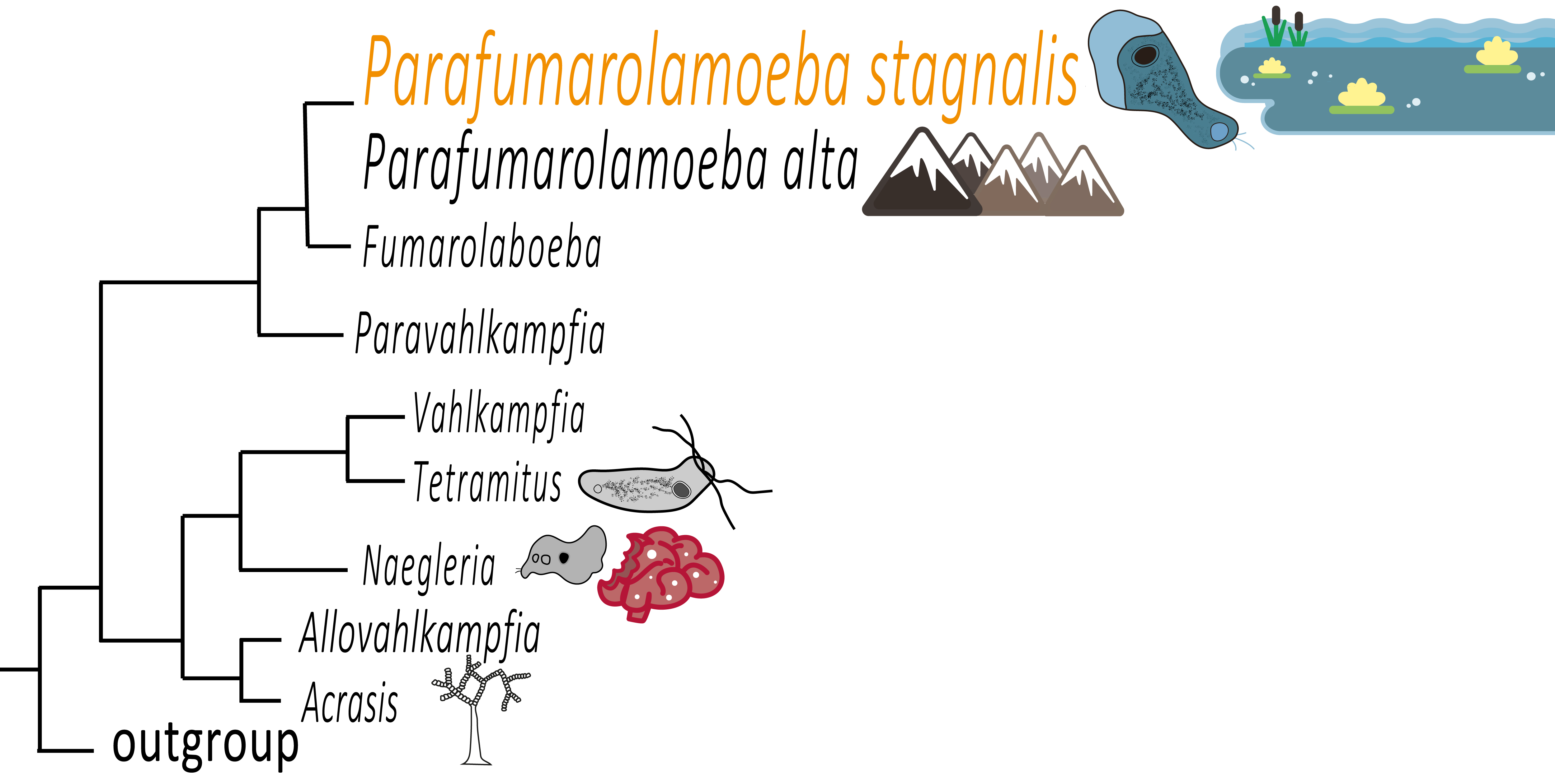
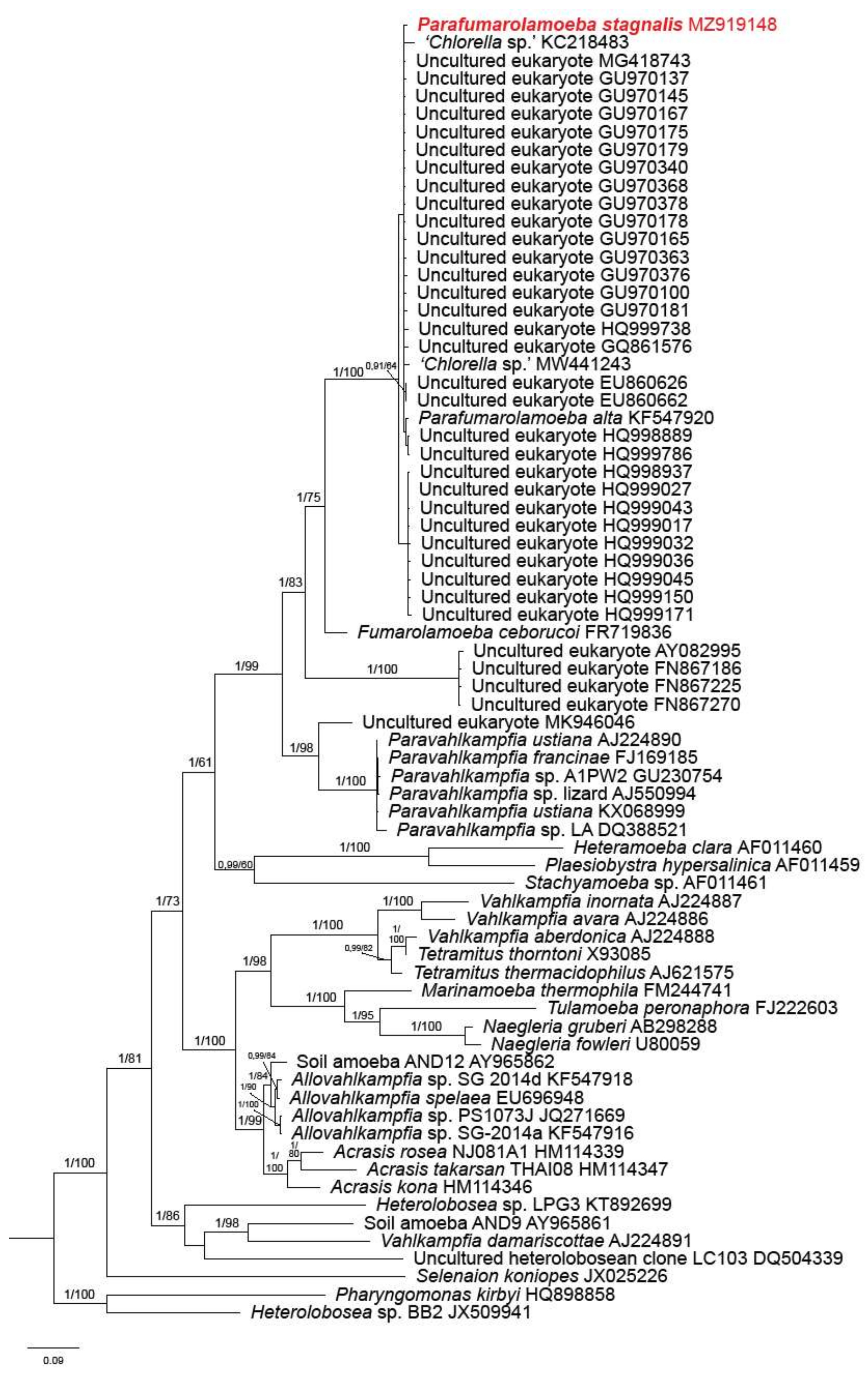
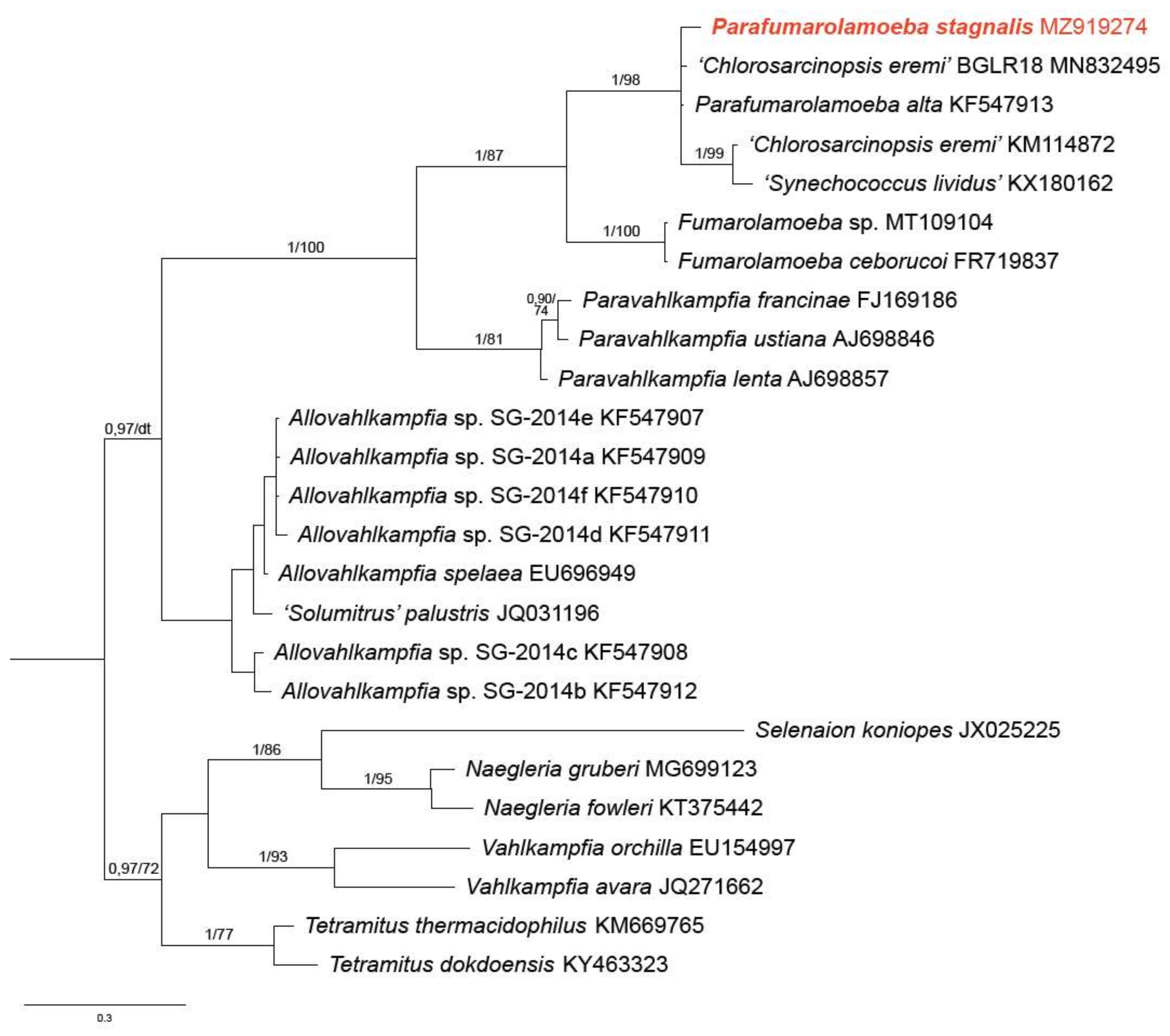
3.2. Phylogenetic Analysis

The phylogenetic position of the novel vahlkampfiid species was inferred from Bayesian and Maximum likelihood trees, which had nearly identical topologies. Phylogenetic analyses based on the SSU rRNA gene (Figure 4) placed Parafumarolamoeba stagnalis within the fully supported clade comprising Parafumarolamoeba alta [11], several environmental sequences, and two sequences apparently misannotated as ‘Chlorella sp.’ (MW441243 and KC218483). The closest sister group to Parafumarolamoeba is Fumarolamoeba.
The ITS phylogeny (Figure 5) grouped Parafumarolamoeba stagnalis (1/98 support) with Parafumarolamoeba alta (KF547913) and three sequences misannotated as ‘Chlorosarcinopsis eremi’ (Chlorophyta; MN832495 and KM114872) and ‘Synechococcus lividus’ (KX180162). Their closest sister groups (1/87 support) are the clade uniting Fumarolamoeba sp. (MT109104) and Fumarolamoeba ceborucoi (FR719837) and, more distantly, Paravahlkampfia (1/100 support). The SSU rDNA and ITS tree topologies are more different in their deeper splits, but this is perhaps not surprising because the ITS sequences are fast-evolving and little informative at above-genus level.
The SSU rRNA sequences MW441243 and KC218483 as well as ITS sequences MN832495, KM114872, and KX180162 are misannotated in the NCBI GenBank database as green algae and cyanobacteria and most likely represent unknown vahlkamphiids contaminating algal cultures that were studied in these unpublished surveys (see the corresponding NCBI GenBank records).

4. Discussion

The clone Va-1 clearly belongs to the Vahlkampfiidae because of its limax morphology and eruptive movement, as well as its mitochondrial ultrastructure and general cyst morphology. The absence of the flagellated stage and of pores in the cyst wall unites Va-1 with representatives of Fumarolamoeba, Paravahlkampfia, Allowahlkampfia, Vahlkampfia, Acrasis [18,19,20].
The shape of locomotive cells of Parafumarolamoeba stagnalis resembles Vahlkampfia anaerobica but differs from it by the presence of contractile vacuoles, cysts and conventional mitochondria [21]. P. stagnalis differs from V. ovis by the structure and size of its cysts and by smaller cell length. P. stagnalis lacks a prominent bulbous uroid, which is common in other vahlkampfiids, and possess very small cysts, probably the smallest in this family.
P. stagnalis resembles V. magna, V. debilis, V. avara by cyst wall structure [22], however, the latter cysts are larger, with a diameter of 10 μm or more. In addition, the amoeboid cells in V. debilis are smaller (15–20 μm) than in P. stagnalis, and they are capable of rapid gliding movement not observed in P. stagnalis.
P. stagnalis is smaller than V. caledonica (length 47.4 ± 16.0 μm, width 12.1 ± 3.2 μm) [23] and Paravahlkampfia lenta (length 37–80 μm, width 11–24 μm) [9]. The cells of parasitic Paravahlkampfia francinae are similar in size to P. stagnalis but are able to grow at higher temperature of up to 42 °C with a growth optimum at 37 °C [24].
The ultrastructure of the vesicular nucleus, mitochondria with discoid cristae, and mitochondria-adjacent cisternae of the endoplasmic reticulum unite P. stagnalis with P. francinae and other vahlkampfiids [25,26].
In comparison with the closest described relative Parafumarolamoeba alta, which forms dimorphic cysts (wrinkled and round), only round cysts were found in P. stagnalis. The cysts of P. stagnalis and Fumarolamoeba ceborucoi sometimes form aggregations. Mature cysts of P. stagnalis have three-layered envelope unlike F. ceborucoi, which forms cysts with a double wall [11,27].
Representatives of Parafumarolamoeba have a single nucleus, while trophozoites of F. ceborucoi can have from two to six nuclei (most commonly two). The size of the nucleolus can serve as one of the most distinctive features. The nucleolus of P. stagnalis is characterized by a large size and reaches about 2/3 of the diameter of the nucleus. P. alta and F. ceborucoi have smaller nucleoli, which reach about 1/3 and 1/2 of the diameter of the nucleus respectively [11,27].
Two closely related genera Fumarolamoeba and Parafumarolamoeba are also distinguishable by limax locomotion, which is typical for P. stagnalis and P. alta. However, the trophozoites of F. ceborucoi are rarely found. In addition, P. stagnalis and P. alta have protruding uroidal filaments, which were not detected in F. ceborucoi [11,27] (Table 1).
The presented morphological evidence leads us to classify the clone Va-1 to the genus Parafumarolamoeba as a new species. The genus Parafumarolamoeba has contained a single species, P. alta, isolated from high-altitude soil in Tibet. Closely related environmental sequences belonging to the Parafumarolamoeba clade are derived from three drinking water supplies in the Caribbean Leeward Antilles (HQ999738, HQ998889, HQ999786) [28], engineered water systems (GU970137, GU970145, GU970167, GU970175, GU970179, GU970340, GU970368, GU970378, GU970178, GU970165, GU970363, GU970376, GU970100, GU970181) [29] and two groundwater supplies in The Netherlands (EU860626, EU860662) [30], as well as from hot water system (France) (GQ861576) [31] and River Mountains Water Treatment Facility (RMWTF), Henderson, NV, USA (MG418743) [32]. This conclusion is supported by the molecular phylogeny of the SSU rRNA and ITS, where two representatives of Parafumarolamoeba are monophyletic with strong support (Figure 4 and Figure 5). These morphologically different species are 98.2% and 87.7% identical to each other by SSU rRNA and ITS, respectively. Many of the related environmental sequences are characterized by about the same or lower genetic distances between themselves. This opens up the question of whether all these environmental sequences are separate species.
The biology of Parafumarolamoeba includes many unresolved questions since the most closely related species on the gene trees come from very unusual environments: Fumarolamoeba ceborucoi FR719837 from a volcano in Mexico and Fumarolamoeba sp. MT109104 from Italian hot springs, both of which point to the ability of Fumarolamoeba to survive in hyperthermophilic environments [27,33]. In contrast, both representatives of the genus Parafumarolamoeba come from an environment with normal conditions and are probably unable to survive high temperatures. The origin of some related environmental sequences (AY082995, FN867186, FN867225, FN867270) [34,35] indicates that they potentially tolerate low pH values and heavy metal pollution, but further research into these aspects is necessary, especially considering the high sequence diversity in the Parafumarolamoeba clade.

Taxonomic Summary

Assignment. Eukaryota; Discoba; Heterolobosea; Vahlkampfiidae; Parafumarolamoeba

Parafumarolamoeba stagnalis n. sp.

Trophozoites are 13–28 μm in length and 9–12 μm in width. The length:width ratio is 4.3 in active cells and 1.7 in slowly moving cells. Clearly distinguishable anterior hyaline pseudopodium reaches 15–25% of the cell length. Limax morphology and eruptive movement. Cells can quickly change the direction of movement at a 90° angle. Floating cells irregular with short obtuse pseudopodia. Several thin and sometimes branched uroidal filaments. Spherical cysts 5–7 μm in diameter have a conspicuously thick wall, no pores and plugs. Caudal bulb is rarely present. Flagellate stages were not observed. The maximum temperature of cells survival is 36 °C.
Type strain. Va-1. Stored in the collection of live protozoan cultures at IBIW RAS.
Type Figure: Figure 1A illustrates a live cell of strain Va-1.
Type locality. Small pond near settlement Borok, Russia.
Habitat. Fresh water, Russia.
Etymology. Named by the place of finding. From Latin stagnum (pond).
Gene sequence. The SSU rRNA gene sequence has the GenBank accession number MZ919148.

Supplementary Materials

The following are available online at https://www.mdpi.com/article/10.3390/d13090433/s1. Video S1: Moving of Parafumarolamoeba stagnalis.

Author Contributions

Conceptualization, D.V.T., A.P.M.; methodology, D.V.T., A.P.M., A.S.B., J.J.; validation, A.S.B., D.V.T., J.J., P.J.K.; formal analysis, A.S.B., D.V.T., J.J.; investigation, A.P.M., A.S.B., D.V.T.; resources, D.V.T., P.J.K.; writing—original draft preparation, D.V.T., A.P.M., A.S.B.; writing—review and editing, J.J., P.J.K., D.V.T.; visualization, A.P.M., A.S.B., D.V.T.; funding acquisition, D.V.T. All authors have read and agreed to the published version of the manuscript.

Funding

This research was funded by the Tyumen Oblast Government, as part of the West-Siberian Interregional Science and Education Center’s project No. 89-DON (2). Electron microscopic investigations were funded by Russian Science Foundation, grant number 18-14-00239, https://www.rscf.ru/project/18-14-00239/ (accessed on 5 September 2021).

Institutional Review Board Statement

Not applicable.

Data Availability Statement

Not applicable.

Conflicts of Interest

The authors declare no conflict of interest. The funders had no role in the design of the study; in the collection, analyses, or interpretation of data; in the writing of the manuscript, or in the decision to publish the results.

References

  1. Visvesvara, G.S.; Moura, H.; Schuster, F.L. Pathogenic and opportunistic free-living amoebae: Acanthamoeba spp., Balamuthia mandrillaris, Naegleria fowleri, and Sappinia diploidea. FEMS Immunol. Med. Microbiol. 2007, 50, 1–26. [Google Scholar] [CrossRef] [Green Version]
  2. Lee, J. De novo formation of basal bodies during cellular differentiation of Naegleria gruberi: Progress and hypotheses. Semin. Cell Dev. Biol. 2010, 21, 156–162. [Google Scholar] [CrossRef] [PubMed]
  3. Brown, T.J.; Cursons, R.T.; Keys, E.A. Amoebae from Antarctic Soil and Water. Appl. Environ. Microbiol. 1982, 44, 491–493. [Google Scholar] [CrossRef] [Green Version]
  4. Ettinger, M.R.; Webb, S.R.; Harris, S.A.; McIninch, S.P.; Garman, G.C.; Brown, B.L. Distribution of free-living amoebae in James River, Virginia, USA. Parasitol. Res. 2003, 89, 6–15. [Google Scholar] [CrossRef] [PubMed]
  5. Garstecki, T.; Arndt, H. Seasonal abundances and community structure of benthic rhizopods in shallow lagoons of the Southern Baltic Sea. Europ. J. Protistol. 2000, 36, 103–115. [Google Scholar] [CrossRef]
  6. Page, F.C.; Siemensma, F.J. Nackte Rhizopoda und Heliozoea (Protozoenfauna Band 2); Gustav Fischer Verlag: Stuttgart, NY, USA, 1991; pp. 3–170. [Google Scholar]
  7. Hinkle, G.; Sogin, M.L. The evolution of the Vahlkampfiidae as deduced from 16S-like ribosomal RNA analysis. J. Eukaryot. Microbiol. 1993, 40, 599–603. [Google Scholar] [CrossRef] [PubMed]
  8. Brown, S.; De Jonckheere, J.F. A reevaluation of the amoeba genus Vahlkampfia based on SSU rDNA sequences. Eur. J. Protistol. 1999, 35, 49–54. [Google Scholar] [CrossRef]
  9. Brown, S.; De Jonckheere, J.F. Isolation of a new vahlkampfiid amoeba from soil: Paravahlkampfia lenta n. sp. Eur. J. Protistol. 2004, 40, 289–294. [Google Scholar] [CrossRef]
  10. De Jonckheere, J.F.; Brown, S. The identification of vahlkampfiid amoebae by ITS sequencing. Protist 2005, 156, 89–96. [Google Scholar] [CrossRef]
  11. Geisen, S.; Bonkowski, M.; Zhang, J.; De Jonckheere, J.F. Heterogeneity in the genus Allovahlkampfia and the description of the new genus Parafumarolamoeba (Vahlkampfiidae; Heterolobosea). Eur. J. Protistol. 2015, 51, 335–349. [Google Scholar] [CrossRef]
  12. Keeling, P.J.; Poulson, N.; McFadden, G.I. Phylogenetic diversity of parabasalian symbionts from termites, including the phylogenetic position of Pseudotrypanosoma and Trichonympha. J. Eukaryot. Microbiol. 1998, 45, 643–650. [Google Scholar] [CrossRef] [PubMed]
  13. Gordon, D.; Green, P. Consed: A graphical editor for next-generation sequencing. Bioinformatics 2013, 29, 2936–2937. [Google Scholar] [CrossRef] [PubMed] [Green Version]
  14. Katoh, K.; Standley, D.M. MAFFT multiple sequence alignment software version 7: Improvements in performance and usability. Mol. Biol. Evol. 2013, 30, 772–780. [Google Scholar] [CrossRef] [PubMed] [Green Version]
  15. Capella-Gutierrez, S.; Silla-Martínez, J.M.; Gabaldón, T. trimAl: A tool for automated alignment trimming in large-scale phylogenetic analyses. Bioinformatics 2009, 25, 1972–1973. [Google Scholar] [CrossRef]
  16. Ronquist, F.; Huelsenbeck, J. MRBAYES 3: Bayesian Phylogenetic inference under mixed models. Bioinformatics 2003, 19, 1572–1574. [Google Scholar] [CrossRef] [Green Version]
  17. Nguyen, L.; Schmidt, H.A.; von Haeseler, A.; Minh, B.Q. IQ-TREE: A Fast and Effective Stochastic Algorithm for Estimating Maximum-Likelihood Phylogenies. Mol. Biol. Evol. 2015, 32, 268–274. [Google Scholar] [CrossRef]
  18. De Jonckheere, J. A Century of Research on the Amoeboflagellate Genus Naegleria. Acta Protozool. 2002, 41, 309–342. [Google Scholar]
  19. Walochnik, J.; Mulec, J. Free-living Amoebae in Carbonate Precipitating Microhabitats of Karst Caves and a New Vahlkampfiid Amoeba, Allovahlkampfia spelaea gen. nov., sp. nov. Acta Protozool. 2009, 48, 25–33. [Google Scholar]
  20. Brown, M.W.; Silberman, J.D.; Spiegel, F.W. A contemporary evaluation of the acrasids (Acrasidae, Heterolobosea, Excavata). Eur. J. Protistol. 2012, 48, 103–123. [Google Scholar] [CrossRef]
  21. Smirnov, A.V.; Fenchel, T. Vahlkampfia anaerobica n. sp., Vannella peregrinia n. sp. (Rhizopoda)—Anaerobic amoebae from a marine sediment. Arch. Protistenk. 1996, 147, 189–198. [Google Scholar] [CrossRef]
  22. Page, F.C. An Illustrated Key to Freshwater and Soil Amoebae with Notes on Cultivation and Ecology; Freshwater Biological Association: Ambleside, UK, 1976; 155p. [Google Scholar]
  23. Anderson, O.R.; Rogerson, A.; Hannah, F. Three new limax amoebae isolated from marine surface sediments. Vahlkampfia caledonica n. sp., Saccamoeba marina n. sp. and Hartmannella vacuolata n. sp. J. Eukaryot. Microbiol. 1997, 44, 33–42. [Google Scholar]
  24. Visvesvara, G.S.; Spiram, R.; Qvarnstrom, Y.; Bandyopadhyay, K.; Da Silva, A.J.; Pieniazek, N.J.; Cabral, G.A. Paravahlkampfia francinae n.sp. Masquerading as an Agent of Primary Amoebic Meningoencephalitis. J. Eukaryot. Microbiol. 2009, 56, 357–366. [Google Scholar] [CrossRef]
  25. Page, F.C. The limax amoebae, comparative fine structure of the Hartmannellidae (Lobosea) and further comparisons with the Vahlkampfiidae (Heterolobosea). Protistologica 1985, 21, 361–383. [Google Scholar]
  26. Page, F.C.; Blanton, R.L. The Heterolobosea (Sarcodina: Rhizopoda), a new class uniting the Schizopyrenida and the Acrasidae (Acrasida). Protistologica 1985, 21, 121–132. [Google Scholar]
  27. De Jonckheere, J.; Murase, J.; Opperdoes, F.R. A New Thermophilic Heterolobosean Amoeba, Fumarolamoeba ceborucoi, gen. nov., sp. nov., Isolated Near a Fumarole at a Volcano in Mexico. Acta Protozool. 2011, 50, 41–48. [Google Scholar]
  28. Valster, R.M.; Wullings, B.A.; van den Berg, R.; van der Kooij, D. Relationships between free-living protozoa, cultivable Legionella spp., and water quality characteristics in three drinking water supplies in the Caribbean. Appl. Environ. Microbiol. 2011, 77, 7321–7328. [Google Scholar] [CrossRef] [Green Version]
  29. Valster, R.M.; Wullings, B.A.; van der Kooij, D. Detection of protozoan hosts for Legionella pneumophila in engineered water systems by using a biofilm batch test. Appl. Environ. Microbiol. 2010, 76, 7144–7153. [Google Scholar] [CrossRef] [Green Version]
  30. Valster, R.M.; Wullings, B.A.; Bakker, G.; Smidt, H.; van der Kooij, D. Free-living protozoa in two unchlorinated drinking water supplies, identified by phylogenic analysis of 18S rRNA gene sequences. Appl. Environ. Microbiol. 2009, 75, 4736–4746. [Google Scholar] [CrossRef] [Green Version]
  31. Farhat, M.; Moletta-Denat, M.; Frère, J.; Onillon, S.; Trouilhé, M.; Robine, E. Effects of disinfection on Legionella spp., eukarya, and biofilms in a hot water system. Appl. Environ. Microbiol. 2012, 78, 6850–6858. [Google Scholar] [CrossRef] [Green Version]
  32. De Vera, G.A.; Gerrity, D.; Stoker, M.; Frehner, W.; Wert, E. Impact of upstream chlorination on filter performance and microbial community structure of GAC and anthracite biofilters. Environ. Sci. Water Res. Technol. 2018, 4, 1133–1144. [Google Scholar] [CrossRef] [Green Version]
  33. Berrilli, F.; Di Cave, D.; Novelletto, A.; Montalbano Di Filippo, M. PCR-based identification of thermotolerant free-living amoebae in Italian hot springs. Eur. J. Protistol. 2021, 80, 125812. [Google Scholar] [CrossRef]
  34. Amaral Zettler, L.A.; Gomez, F.; Zettler, E.; Keenan, B.G.; Amils, R.; Sogin, M.L. Microbiology: Eukaryotic diversity in Spain’s River of Fire. Nature 2002, 417, 137. [Google Scholar] [CrossRef]
  35. Amaral-Zettler, L.A.; Zettler, E.R.; Theroux, S.M.; Palacios, C.; Aguilera, A.; Amils, R. Microbial community structure across the tree of life in the extreme Río Tinto. ISME J. 2011, 5, 42–50. [Google Scholar] [CrossRef] [Green Version]
Figure 1. Light microscopy of cells (AK) and cysts (L) of P. stagnalis. Abbreviations: cv—contractile vacuole, n-nucleus, ps—pseudopodium, uf—uroidal filaments. Scale bars: 5 μm.
Diversity 13 00433 g001
Figure 2. Surface morphology (SEM) of P. stagnalis amoebae (AC) and cyst (D). Scale bar: (AC)—5 μm, (D)—1 μm.
Diversity 13 00433 g002
Figure 3. Ultrathin sections of amoebae and cysts (TEM). (A)—general view, (B)—contractile vacuole and part of cell surface, (C)—hyaline pseudopodium, (D,E)—mitochondrium with discoidal cristae, (F)—small contractile vacuoles, (G,H)—cysts, (I)—envelope of the cyst. Abbreviations: cr—crista, en—envelope, er—endoplasmc reticulum, fv—food vacuole, mt—mitochondria, pm—plasmalemma. For explanation of other symbols, see Figure 1. Scale bar: (A,C,F,G,H)—2 μm, (I)—0.6 μm, (D,E)—0.5 μm, (B)—0.2 μm.
Diversity 13 00433 g003
Figure 4. Bayesian phylogeny of the SSU rRNA gene. Bayesian posterior probabilities and Maximum Likelihood (TIM2+F+R4 model) bootstrap values are indicated at branches (values >0.9/>60 are shown; dt—different topology). Pharyngomonas kirbyi HQ898858 SD1A and Heterolobosea sp. BB2 JX509941 were used as the outgroup.
Diversity 13 00433 g004
Figure 5. Bayesian phylogeny of the internal transcribed spacer (ITS) region. Bayesian posterior probabilities and Maximum Likelihood (TN+F+G4 model) bootstrap values are indicated at branches (values >0.9/>70 are shown; dt—different topology).
Diversity 13 00433 g005
Table 1. Morphological comparison of P. stagnalis and related species *.
SpeciesLocomotive form Length (µm)Locomotive form Width (µm)Limax MorphologyUroidal FilamentsNucleolus/Nucleus Size RatioCyst CharacteristicsGrowth Temperature, °CSource
Parafumarolamoeba stagnalis17.9 ± 0.510.4 ± 0.5++2/3d = 5–7 µm
round cysts, three-layered envelope
Max 36Current study
P. alta20.97.9++1/3d = 5.7 ± 0.4 µm
wrinkled and round
?Geisen et al., 2015
Fumarolamoeba ceborucoi2613.8rarely-1/2d = 6.2 µm
double wall cysts
Max 51 (not multiply but survives)De Jonckheere et al., 2011
Vahlkampfia anaerobica11–347++?no cysts observed?Smirnov et al., 1996
V. avara15–335–11++1/2d = 9.7 µm Smooth, gelatinous(‘sticky’) single cystwall?Page, 1967
V. caledonica47.4 ± 16.012.1 ± 3.2++2/5no cysts observed?Anderson et al., 2007
Paravahlkampfia. lenta37–8011–24++4/5d = 18.1 µm
smooth, double cystwall, (‘sticky’) outerwall
Max 34Brown and De Jonckheere, 2004
P. francinae15–255.9–9.9++3/5d = 17.5 µm
round, double cyst wall; cysts from older cultures with wrinkled and star-shaped outer cyst walls
Opt 37Visvesvara et al., 2009
* neither form flagellated stages nor have cyst pores. “+”—present, “-”—absent, “?”—no data available.
Publisher’s Note: MDPI stays neutral with regard to jurisdictional claims in published maps and institutional affiliations.

