Article

Doolia, A New Genus of Nannopodidae (Crustacea: Copepoda: Harpacticoida) from off Jeju Island, Korea

Department of Life Science, College of Natural Sciences, Hanyang University, Seoul 04763, Korea
Diversity 2020, 12(1), 3; https://doi.org/10.3390/d12010003
Submission received: 20 November 2019 / Revised: 13 December 2019 / Accepted: 15 December 2019 / Published: 18 December 2019
(This article belongs to the Special Issue Meiofauna Biodiversity and Ecology)

Abstract

:
A new harpacticoid copepod is described from the waters off Jeju Island, Korea. This species displays a unique set of characteristics including a rostrum that is clearly demarcated from the cephalosome, a setular (spinular) row on the rostrum, a well-developed frill along the posterior margins of each body segment except for the cephalosome, long and cylindrical caudal rami, four segmented female antennules, paired genital apertures in the female, the absence of sexual dimorphism in legs P1–P4, and highly reduced P5 and P6 in the male. This combination of characteristics allocates the specimen to the family Nannopodidae Por, 1986, but the new species belongs to none of the extant genera within the family. A new genus, Doolia, is proposed. Nannopus is suggested as a sister taxon of the new genus based on shared plesiomorphic characteristics in the maxilliped, legs P1–P4, and P5. Doolia gen. nov. is the eighth genus of Nannopodidae, and an amended key for the genus is provided herein.

1. Introduction

The family Cletodidae sensu T. Scott, 1904 has been recognized in previous reports as being potentially the most heterogenous harpacticoid family [1]. Lang [2] recognized several lineages within the family, although he did not take any action on the subject. Por [1] revised and redefined the family Cletodidae T. Scott, 1905, establishing four new families: Paranannopidae Por, 1986, Huntemanniidae Por, 1986, Rhizotrichidae Por, 1986, and Argestidae Por, 1986. Among Por’s new families, Huys [3] synonimized Huntemanniidae with Nannopodidae Brady, 1880. Consequently, two genera, Metahuntemannia Smirnov, 1946 and Talpina Dahms & Pottek, 1982 were re-allocated to the subfamily Hemimesochrinae Por, 1986 in the Canthocamptidae Brady, 1880 based on their affinities with genera including Bathycamptus Huys & Thistle, 1989, Micropsammis Mielke, 1975, Isthmiocaris George & Schminke, 2003, and Perucamptus Huys & Thistle, 1989 [4].
Currently, Nannopodidae includes about 25 valid species in seven genera: Nannopus Brady, 1880, Pontopolites T.Scott, 1894, Huntemannia Poppe, 1884, Rosacletodes Wells, 1985, Laophontisochra George, 2002, Acuticoxa Huys & Kihara, 2010, and Talpacoxa Corgosinho, 2012.
During a study of the harpacticoid copepods in Korean Waters, a nannopodid-like copepod was collected; however, the specimens could not be allocated to any extant genera in the family Nannopodidae. The present study aims to describe this new taxon and establish a new genus based on the current specimens from Korea.

2. Materials and Methods

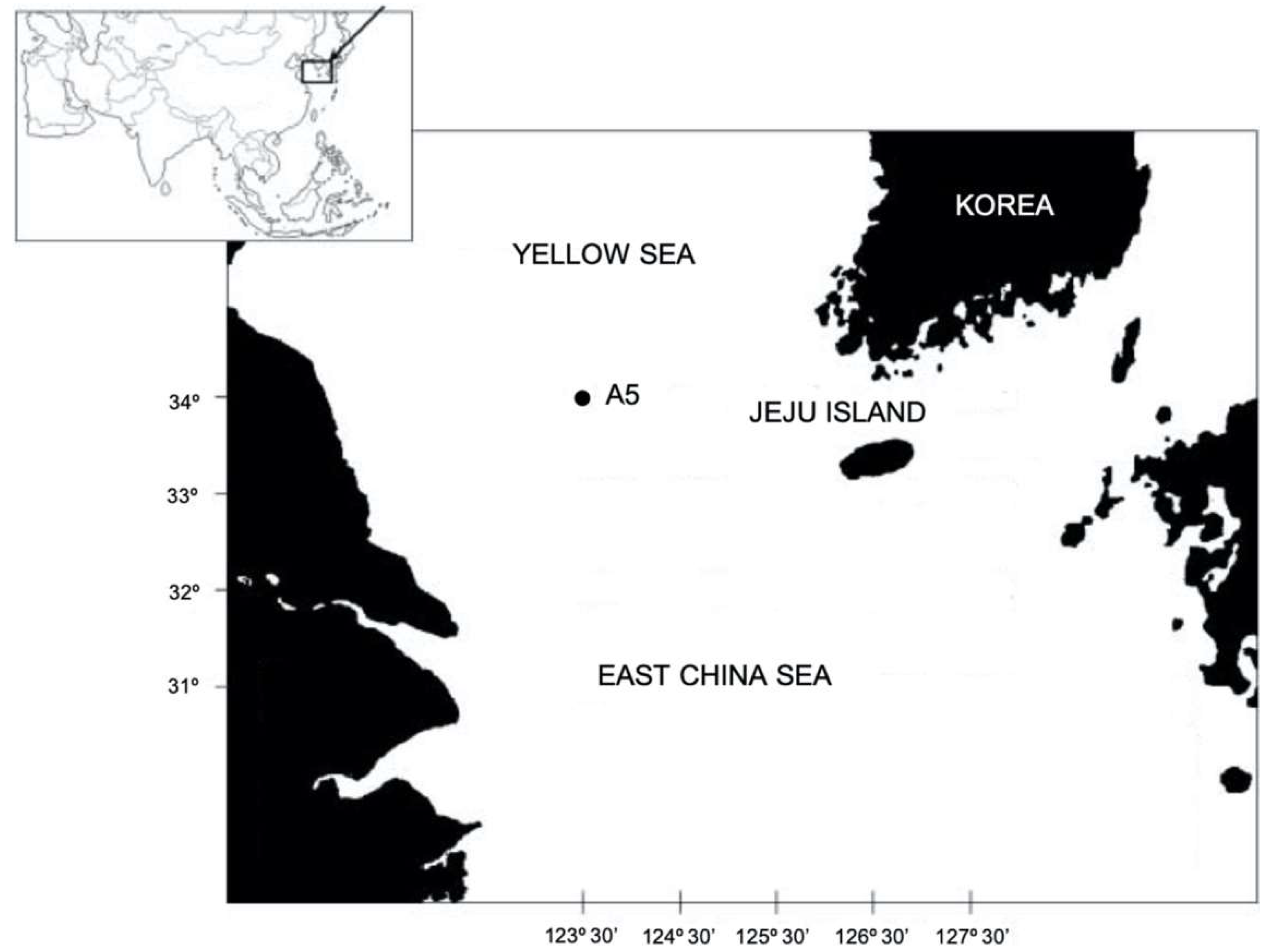
Specimen Collection and Morphological Examinations

Samples were collected at one site, A5 (34°00′ N, 123°30′ E), among 18 field sites off the coast of Jeju Island, South Korea from 24 September to 3 October 2002 (Figure 1) by the R/V Ara (Jeju National University) [5]. Sediments were collected with a box corer, and five replicates were taken from each box corer. Three replicates were used for analyses of meiofauna. Organisms were extracted from sediments by a Ludox-Am. Copepods were sorted under a dissecting microscope and stored in 70% ethanol. Dissections were applied to the specimens in lactic acid and the dissected appendages were mounted permanently on slides in lactophenol mounting medium. Preparations were sealed with transparent nail varnish. Each appendage was observed and drawn under an Olympus BX51 differential interference contrast microscope using a drawing tube. The descriptive terminology is adopted from Huys et al. [6]. Abbreviations are A1, antennule; A2, antenna; ae, aesthetasc; exp, exopod; enp, endopod; P1–P6, first to sixth thoracopod; exp(enp)-1(2, 3) to denote the proximal (middle, distal) segment of a ramus [6]. Specimens were deposited in the National Institute of Biological Resources (NIBR), Korea, and in the Marine Biodiversity Institute of Korea (MABIK). All scale bars in the figures are in µm.

3. Systematic Account

Subclass Copepoda Milne Edwards, 1830
Order Harpacticoida Sars, 1903
Family Nannopodidae Por, 1986

3.1. Genus Doolia, gen. nov.

Diagnosis. Cylindrical body densely covered by denticles. Caudal rami cylindrical with six caudal setae and one large pore. Anal operculum well-developed with a spinular row along distal margin. Rostrum defined at base, with a pair of sensilla and a row of long spinules along anterior margin. Antennule four-segmented. Antenna three-segmented with one-segmented exopod. Mandibular palp well-developed with one segmented exopod and endopod. Maxillary syncoxa with two endites. Maxilliped subchelate. Swimming legs P1–P4 with three-segmented exopods and two-segmented endopods. Female P5 fused medially, with separate exopod. P6 with two naked setae in female. Sexual dimorphism in urosome, antennule, P5, and P6. No sexual dimorphism in swimming legs. Male antennule eight-segmented, subchirocer. Male P5 baseoendopod fused with exopod, and represented by several setae. Male P6 represented by a small plate without ornamentation.
Type species: Doolia ara sp. nov. by original designation and monotype.
Etymology. The new genus was named after a famous cartoon character, “Dooly,” a baby dinosaur created in 1983 in Korea.

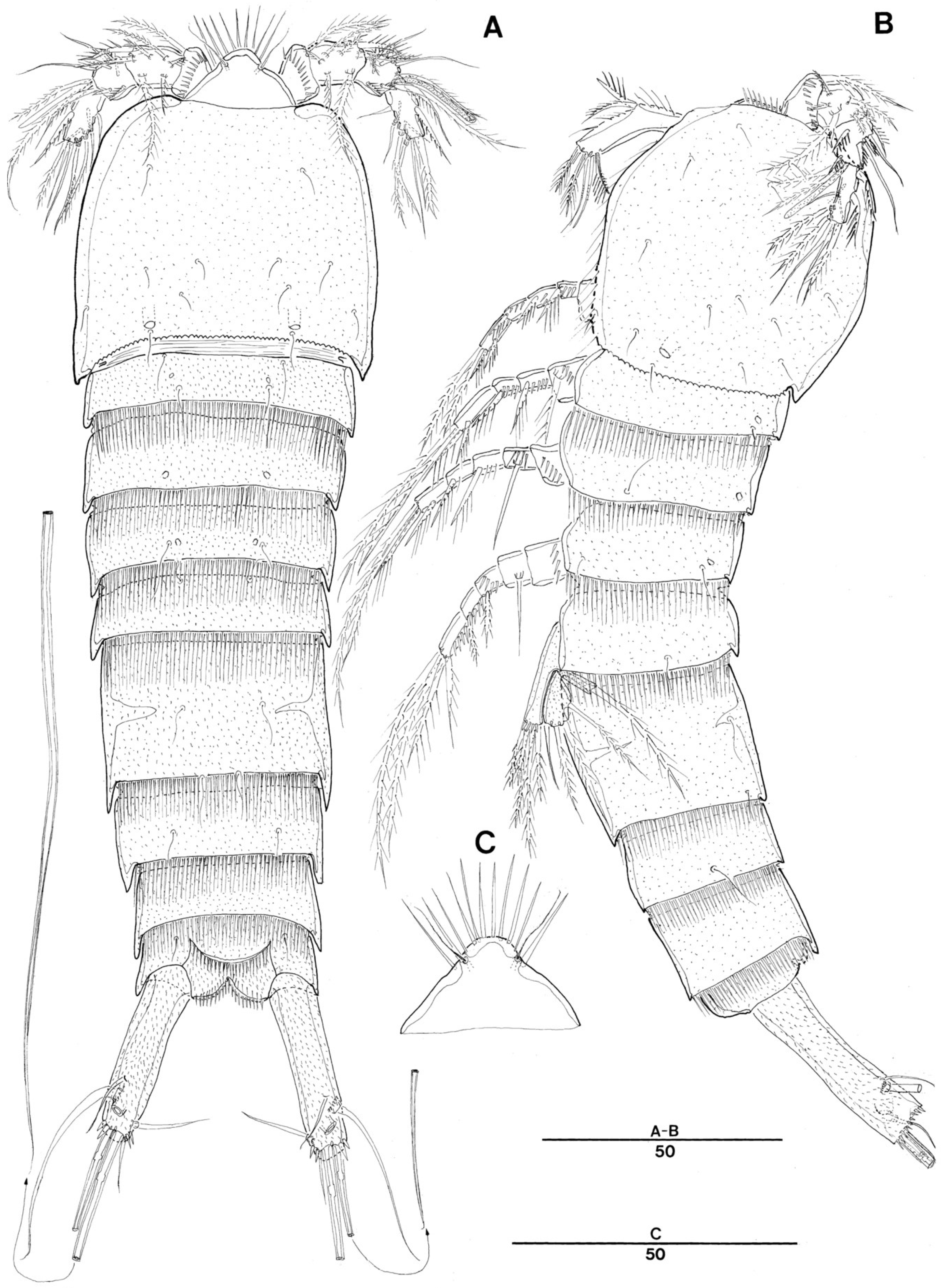
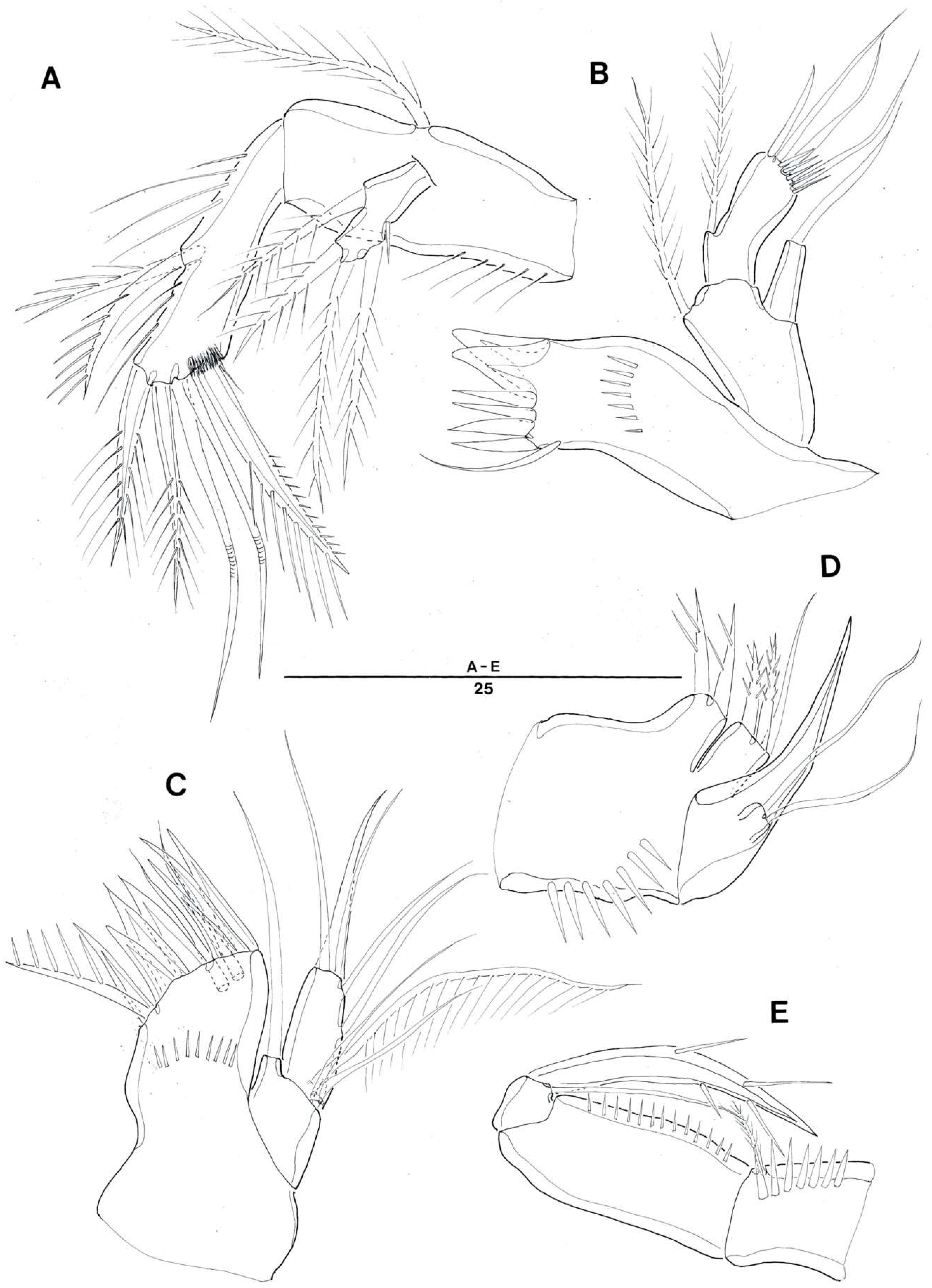
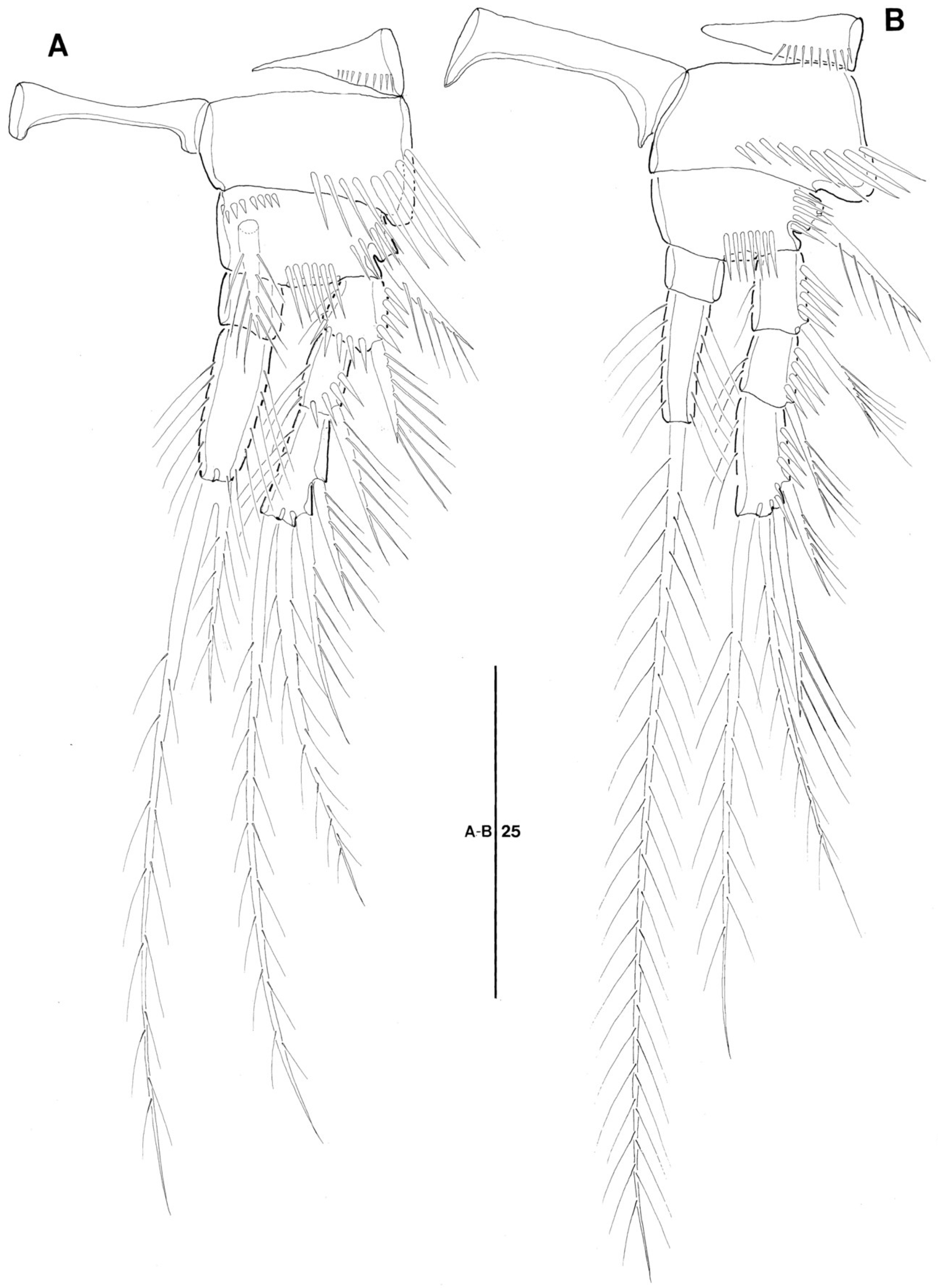
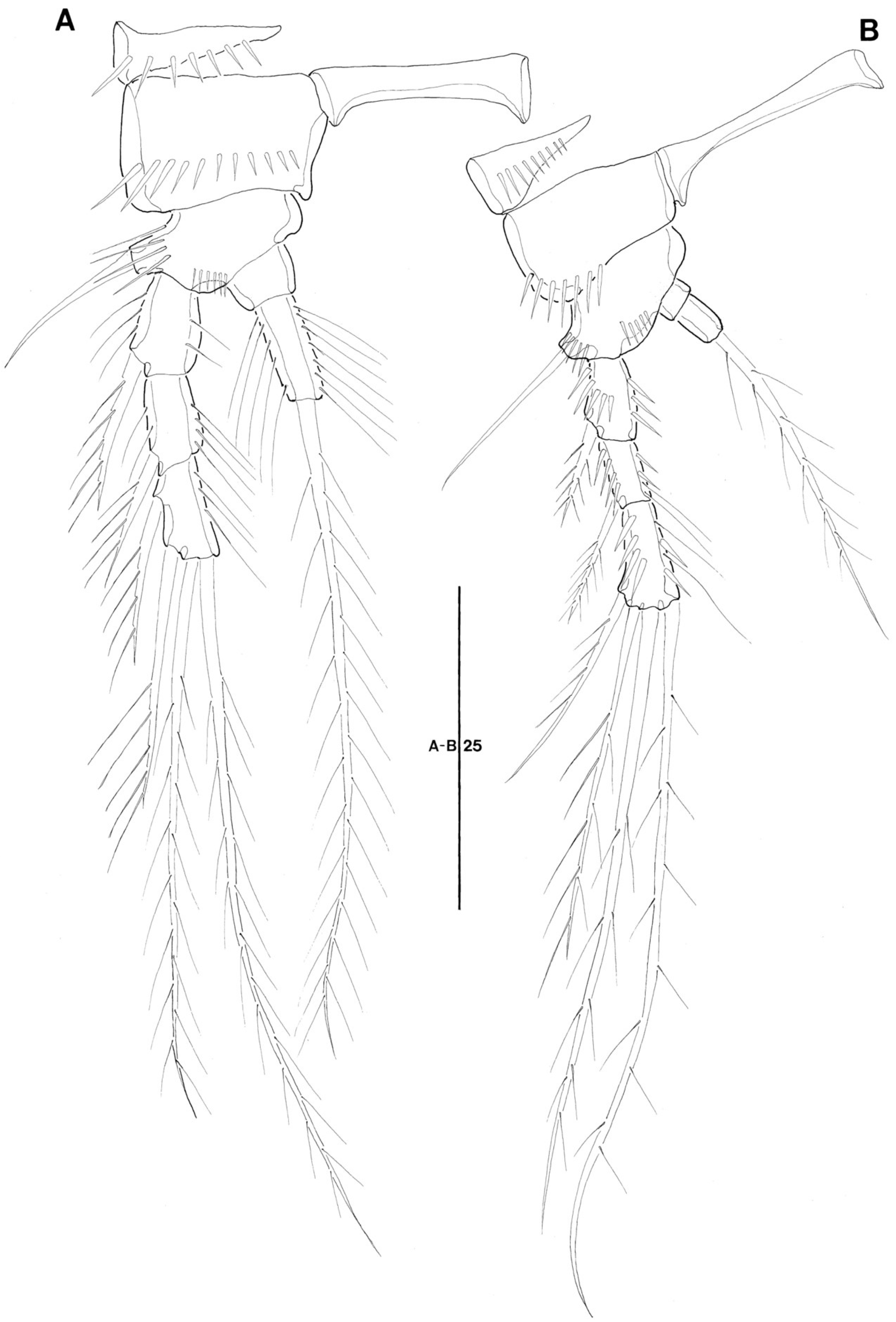
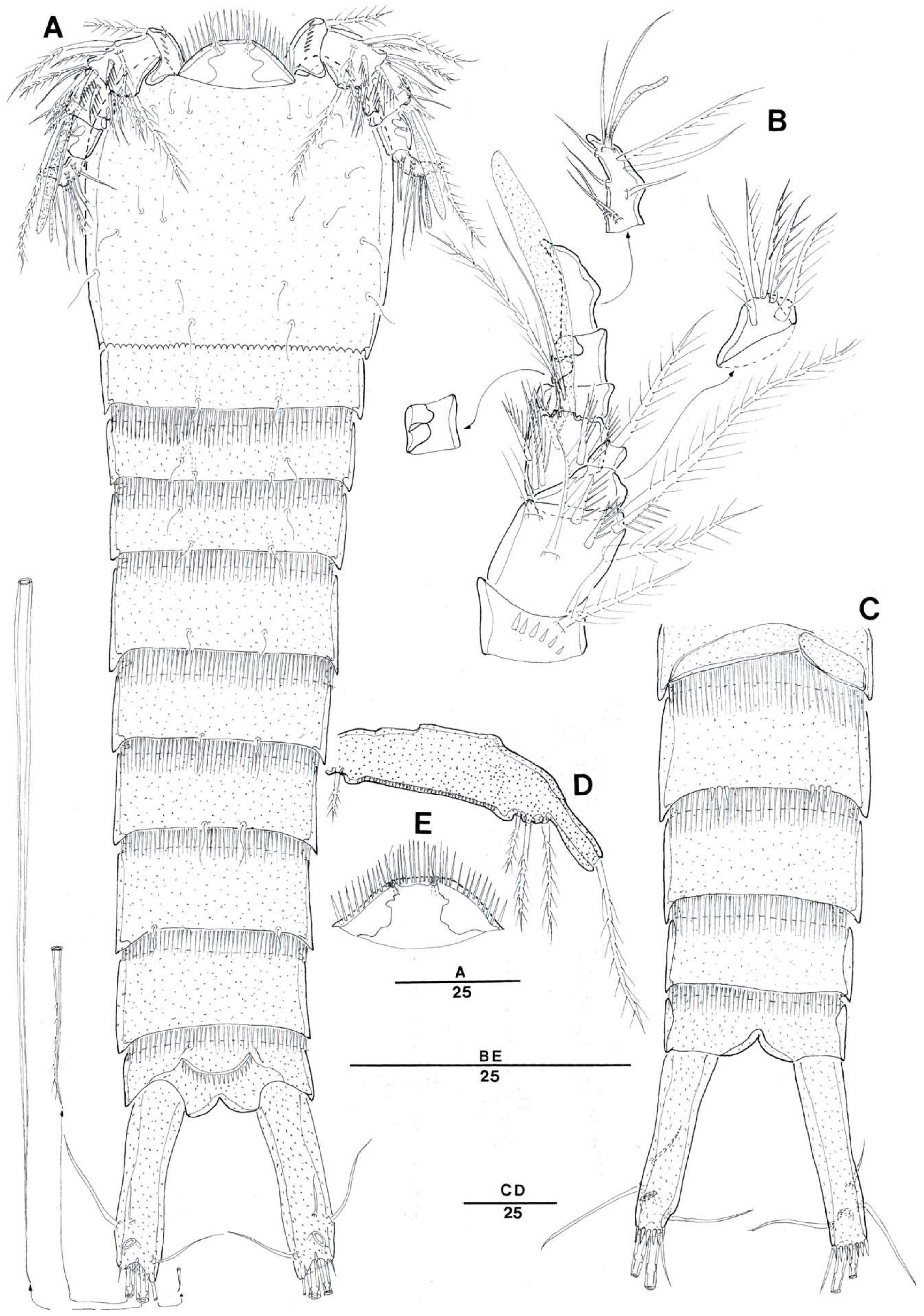
3.2. Doolia ara, sp. nov.

Type locality. Muddy sand substratum, station A5 (34°00′ N, 123°30′ E), off Jeju Island, the south west coast of Korea.
Material examined. Holotype 1♀ (NIBRIV0000862381) dissected on seven slides. Paratypes: three♀♀ and two♂♂; 2♀♀ (MABIK CR00246523 – 4) on five slides, 1♂ (MABIK CR00246525) on seven slides, 1♂ (NIBRIV0000862382) on six slides, and 1♀(NIBRIV0000862383) in 70% ethanol, all from the type locality, depth 66 m, collected by Y.H. Song and E.J. Nam during 24 September to 3 October 2002.
Etymology. The specific name refers to “Ara”, the research vessel of Jeju National University, in appreciation of the crew and Prof. Joon Baek Lee (Jeju National University). Gender, feminine.
Female. Total body length: 239 µm (measured from anterior margin of rostrum to posterior margin of caudal rami). Largest width measured at posterior margin of cephalic shield: 63 µm. Urosome gradually tapering posteriorly (Figure 2A).
Cephalothorax with serrulated posterior margin. Pleural areas rounded without lobate posterolateral angles. Entire surface covered with tiny denticles as illustrated [expressed as dots] in Figure 2A,B. Sensilla and a few pores are present as illustrated in Figure 2A,B.
Rostrum triangular-shaped (Figure 2A,C), broad at base, round anterior margin, clearly separated from cephalosome, and with a pair of sensilla and a row of long spinules along median anterior margin.
Pedigerous somites covered with minute denticles. Prosomites from P2-bearing to P4-bearing with well-developed, comb-shaped hyaline frills.
Urosome (Figure 2A,B and Figure 3B,D ) five-segmented, comprising P5-bearing somite, genital double-somite and three free abdominal somites. All urosomites covered with small denticles dorsally and ventrally and with well-developed hyaline frill along the hind margin. Genital double-somite (Figure 2A,B and Figure 3B) without distinct dorsal surface ridge but with a thick internal cuticle layer indicating original segmentation; completely fused internally and externally. Genital field located near anterior margin (Figure 3B) with a large copulatory pore located in a median depression (Figure 3B). P6 with a small protuberance bearing two bare setae, inner seta longer than outer seta, and with a small pore next to Inner seta. Anal somite (Figure 3D) quadrate with well-developed operculum; opercular distal margin with a row of long spinules, and flanked by a pair of sensilla.
Caudal rami (Figure 3B,D) long, cylindrical, three times longer than it is wide; each ramus with six setae: seta I bare, shortest; setae II bare and long; seta III absent, or presumably represented by one large pore on dorsal distal surface; setae IV and V separated basally; seta VI bare and small; seta VII tri-articulate at base. Each ramus with spinules on entire surface; additional spinular rows along distal margin.
Antennule (Figure 3A) four-segmented. Segment 1 with a row of long spinules on anterior surface. Segment 2 the largest. Segment 3 with aesthetasc fused basally to one long plumose seta. Armature formula: 1-[1 pinnate], 2-[8 pinnate], 3-[6 pinnate + 2 bare + (1 pinnate + ae)], 4-[4 pinnate + 4 bare + trithek]. Apical trithek consisting of small aesthetasc fused basally to two setae (1 bare, and 1 pinnate).
Antenna (Figure 4A) three-segmented, comprising coxa, allobasis, free one-segmented endopod and one-segmented exopod. Coxa small (not figured). Allobasis without distinct surface sutures marking original segmentation; with abexopodal pinnate seta on median inner margin and a row of setules along outer margin. Exopod small, about twice as longer as it is wide, with four well-developed pinnate setae and few spinules along outer margin. Endopod slightly shorter than allobasis, lateral armature arising in distal half, consisting of two strong pinnate spines; apical armature consisting of one strong pinnate spine, two pinnate, and two geniculate setae; with two rows of long spinules laterally; apical outer most spine basally fused to one tiny seta.
Mandible (Figure 4B) with well-developed gnathobase bearing several multicuspidate teeth around distal margin and one slender spine at dorsal corner. Palp well-developed. Basis with one plumose seta. Endopod one-segmented, rectangular with one lateral plumose seta, four naked apical setae, and one row of spinules along apical margin. Exopod one-segmented, cylindrical with one naked apical seta.
Maxillule (Figure 4D). Arthrite strongly developed, with two naked setae on anterior surface and eight spines/setae around distal margin and a row of spinules on posterior surface. Coxa with one short cylindrical endite bearing one long naked seta. Endopod and exopod incorporated into the basis, with three naked setae apically, and one plumose and four naked setae laterally.
Maxilla (Figure 4C). Syncoxa with two endites and a row of long spinules along outer margin. Each coxal endite cylindrical; proximal endite with two pinnate spines; distal endite with one naked and two pinnate spines. Allobasis drawn out into strong, slightly curved claw; endopod represented by two naked setae on anterior surface.
Maxilliped (Figure 4E) with one plumose seta and one row of spinules on syncoxa. Basis with one row of tiny spinules along palmar margin. Endopod one-segmented with long, curved, coarsely pinnate claw, and one naked slender seta.
Legs P1–P4 (Figure 5A,B and Figure 6A,B) with well-developed intercoxal plates and praecoxae bearing row of spinules along distal margin near borderline with each related coxa. Coxae and bases with surface ornamentations of spinules as figured. All legs P1–P4 with three-segmented exopods, and two-segmented endopods.
P1 (Figure 5A). Coxa moderate, with long spinular rows as figured. Basis with one strong, bipinnate inner and one stout, unipinnate outer spines. Anterior surface covered with spinules as figured. Exopod longer than endopod; exp-1 and exp-2 with unipinnate spine, respectively; exp-3 longest with two unipinnate spines and two plumose setae. Enp-2 about three times longer than enp-1; enp-1 without spine or seta; enp-2 with row of long setules along outer and inner margins; two terminal plumose setae on enp-2 fused basally, inner one more than three times longer than the outer.
P2–P4 (Figure 5B and Figure 6A,B). Coxae and bases ornamented with spinular rows along outer or distal margins and anterior surface. Outer margin of basis producing a setophore armed with pinnate spine (P2) or naked seta (P3–P4) at each distal end. All segments with rows of spinules along inner and outer margins as figured. Endopod segments of P2–P3 with long setules or spinules alonge inner and outer margins. P4 endopod without setula ornamentations. P2 enp-2 three times longer than enp-1, endopod reaching to proximal area of exp-3, and exp-3 longest. P3 enp-2 2.5 times longer than enp-1, endopod reaching to middle of exp-2, and exp-1 largest. P4 endopod small, enp-2 2.3 times longer than enp-1, endopod reaching to middle of exp-1, and exp-3 longer than exp-1.
Spine and setal formulae as follows:
  • Exopod Endopod
  • P1 0.0.022 0.020
  • P2 0.0.022 0.010
  • P3 0.0.022 0.010
  • P4 0.0.022 0.010
P5 (Figure 3C) fused medially, and exopod and baseoendopod separate; each covered with minute spinules as figured. Baseoendopod with a long outer setophore bearing one plumose basal seta. Endopodal lobe only reaching to middle of exopod, with five pinnate setae; second outermost seta longest; a row of spinules along distal margin. Exopod small, slightly longer than wide and with one naked and three pinnate setae.
Male. Body length 243 µm. Largest width measured at about median area of cephalic shield: 61 µm. Urosome gradually tapering posteriorly (Figure 7A).
Cephalothorax with serrulated posterior margin. Pleural areas well-developed and rounded without lobate posterolateral angles. Entire surface covered with tiny denticles as in female. Sensilla present as illustrated in Figure 7A.
Rostrum triangular-shaped (Figure 7A,E), broader than in female, smooth anterior margin, clearly separated from cephalosome, and with a pair of sensilla and a row of long spinules along entire anterior margin (only along median apical margin in female).
Urosome (Figure 7A) six-segmented, comprising P5-bearing somite, genital somite and four abdominal somites. All urosomites covered with small denticles dorsally and ventrally and with well-developed hyaline frill along the hind margin.
Antennule (Figure 7B) eight-segmented and subchirocer with geniculation between segments 5 and 6. Segment 1 with a row of coarse and widely spaced spinules. Segment 2 the largest. Segment 4 represented by a small sclerite along anterior margin of segment 3. Segment 5 swollen with one well-developed large aesthetasc. Segment 7 with three-dimensional process as figured in Figure 7B. Segment 8 with triangular distal half. Armature formula: 1-[1pinnate], 2-[7 pinnate], 3-[5 pinnate], 4-[1 pinnate], 5-[1 + 6 pinnate + (1 pinnate + ae)], 6-[0], 7-[2 modified process], 8-[6 + 1 pinnate + trithek]. Apical trithek consisting of a minute aesthetasc and two naked setae.
P5 (Figure 7D) fused medially, defined at base, and whole surface covered with minute spinules. Baseoendopod with a long, cylindrical setophore bearing one plumose outer basal seta. Exopod fused to endopod and represented by three pinnate setae. Minute and coarse spinules on anterior surface.
P6 (Figure 7C) asymmetrical, represented on both sides by a small plate without any additional ornamentation: fused to ventral wall of supporting somite along right side, articulating at base and covering gonopore along left side.

4. Discussion

The new species displays interesting characteristics: the basally-defined rostrum, the well-developed mandibular palp with one segmented exopod and endopod, the presence of a seta on the maxilliped syncoxa, three segmented exopods and two segmented endopods in P1–P4, and the separate exopod of P5 in the female. The family Nannopodidae Por, 1986 could harbor the combination of these character sets based on character comparisons (Table 1). Among the seven extant genera of the Nannopodidae, Nannopus Brady, 1880 is the only member having the rostrum fused to the cephalothorax, while all other genera have basally-defined rostrum, as in the new species (Table 1). The new species, Pontopolites T. Scott, 1894, and Talpacoxa Corgosinho, 2012 have a separate exopod on the mandibular palp, but in other genera, these are absent (Nannopus, and Huntemannia Poppe, 1884) or represented by a single seta (Rosacletodes Wells, 1985, Laophontisochra George, 2002, and Acuticoxa Huys & Kihara, 2010). The maxilliped syncoxa has a pinnate distal seta in the new species, as well as in Nannopus, Huntemannia, and Rosacletodes, while it is absent in Pontopolites, Laophontisochra, Acuticoxa, and Talpacoxa. The segmentation of P1–P4 in the new species is the most conservative state within the family, while Rosacletodes displays the most derived state with degenerated, or simply absent endopods in P2–P4. Finally, in the new species the P5 exopod of the female is clearly separated from the baseoendopod as in Huntemannia, Rosacletodes, Laophontisochra, Talpacoxa, and most Nannopus species. These five characters are suggested here as plesiomorphies of the Nannopodidae, and consequently, the new genus forms a basal group with Nannopus and Pontopolites within the family.
Doolia ara gen. et sp. nov. is the probable sister genus to Nannopus due several common plesiomorphic characteristics: the anterior spinular row on the apical margin of the rostrum, the presence of a single seta on the maxilliped syncoxa, the three-segmented exopod in P1—P4, a maximum two-segmented endopod in P1–P4, and the separate P5 exopod from baseoendopod in the female. However D. ara also has autapomorphies separating it easily from Nannopus: the four-segmented antennule in the female (five-segemented in Nannopus), the single abexopodal seta on the antennary allobasis (two abexopodal setae in Nannopus), no inner seta on the exp-2 in the exopods of P1–P4 (always present in P2–P3 of Nannopus), no sexually-modified P3 endopod in the male (apophysis present in Nannopus), and no seta on the P5 endopodal lobe in the male (3–4 setae in Nannopus) [7].
Pontopolites forms the basal group in Nannopodidae, with Doolia and Nannopus having primitive characters: the well-developed and defined triangular rostrum, the one-segmented mandibular exopod, the three-segmented exopods in P1–P4, and the two-segmented P1 endpod. Furthermore, this genus has a six-segmented antennule in the female, and the most primitive two-segmented antennary exopod (Table 1). As pointed out by Karanovic and Cho [9], Pontopolites are isolated from two other genera by having one plated P5 in both sexes. Acuticoxa also has a similarly-shaped P5 in the female [4]. The other genera, Huntemannia, Rosacletodes, Laophontisochra, Acuticoxa, and Talpacoxa have reduced segmentation in the legs (Table 1). Huntemannia, Rosacletodes, and Talpacoxa have sexual modifications in the male P3 endopod. Laophontisochra is the only genus having no sexual modification in the legs, except for Doolia. The male of Acuticoxa, and the status of modification in the legs are unknown.
In addition, Doolia gen. nov. has superficial similarities with Rhizothrix Sars G.O., 1909, and they share the following character sets: the presence of pinnate setae on the antennule and its segmentation, the segmentation of exopods and endopods in P1–P4, lack of sexual dimorphism in legs P1–P4, and lack of ornamentation on P6 in the male. Tryphoema Monard, 1926 shares similar characters with Doolia except for the segmentation of the legs [14]. Based on previous reports [15] and the present study, Rhizothrix has clear discrepancies from Doolia: clearly separated rostrum with anterior row of long setules (spinules) in Doolia ara (fused to cephalosome and lacking the spinules in Rhizothrix), the well-developed comb-shaped hyaline frill on the body somites in D. ara (all frills serrulate in Rhizothrix), the long and cylindrical caudal rami in D. ara (short and robust, or ovoid in Rhizothrix), absence of the brush setae in the exopod and endopod in P1 in D. ara (present in Rhizothrix), and presence of setae on the P6 of female in D. ara (absent in Rhizothrix).
Similarities between Doolia and Rizotrichidae are probably the result of parallel evolution; however, this hypothesis should be tested with additional molecular evidence in further studies. There are no related studies of lineages of Nannopodidae based on molecular data, except for an intrageneric discussion of Nannopus [16]. There are a few scattered reports of molecular data for presumably related families, Cletodidae [17] and Laophontidae [18]. Unfortunately, I was unable to rescue any DNA markers for the new species. However, the phylogenetic relationships among genera in Nannopodidae could potentially be confirmed by molecular data in a future study.
Key to genera of Nannopodidae Por, 1986 amended from [4]
  • Exopods of P2–P4 three-segmented … 2
    Exopods of P2–P4 two-segmented … 4
  • Rostrum bell-shaped, anterior margin with multiple rows of long setules; P1 endopod distinctly shorter than exopod; P2 endopod two-segmented … 3
    Rostrum triangular, without setular ornamentations; P1 endopod extending beyond distal margin of exp-3; P2 endopod one-segmented with one apical seta … Pontopolites T.Scott, 1894
  • Antennule ♀ five-segmented; P2 enp-2 with 2–3 setae; P3 endopod ♂ with apophysis … Nannopus Brady, 1880
    Antennule ♀ five-segmented; P2 enp-2 with one seta; P3 endopod ♂ with no modification … Doolia gen. nov.
  • Antennary exopod one-segmented; P1 exopod one-segmented … Talpacoxa Corgosinho, 2012
    Antennary exopod absent, or represented by a seta; P1 exopod two- or, three-segmented … 5
  • Antennule ♀ four-segmented; P1 endopod prehensile, distinctly longer than exopod … 6
    Antennule ♀ five-segmented; P1 endopod not prehensile, distinctly shorter than exopod … 7
  • P2–P4 with outer spinous projection on coxa; P5 exopod and baseoendopod fused in ♀, forming a single plate with eight setae/spines … Acuticoxa Huys & Kihara, 2010
    P2–P4 without coxal processes; P5 biramous in ♀ … Laophontisochra George, 2002
  • Rostrum with setula ornamentation around apex: antennule ♀ with aesthetasc on segment three; distal exopod segment of P1 with innermost seta much longer than inner distal spine and penicillate apically … Huntemannia Poppe, 1884
    Rostrum without setula ornamentation around apex: antennule ♀ with aesthetasc on segment four; distal exopod segment of P1 with innermost seta much shorter than inner distal spine and penicillate at tip … Rosacletodes Wells, 1985

Funding

This research was supported by the Discovery of Korean Indigenous Species Project, NIBR (National Institute of Biological Resources, grant no. 2020), the Marine Biotechnology Program (No. 20170431) of the Korea Institute of Marine Science and Technology Promotion (KIMST) funded by the Ministry of Oceans and Fisheries (MOF), and a grant (2018R1D1A1B07050117) from Korea Research Foundation.

Acknowledgments

The author would like to thank Joon Back Lee and the crew of the R/V Ara (Jeju National University) for their help in collecting samples and support during the study period. I would also like to thank Hee Jin Moon and Jisu Yeom (Hanyang University) for their valuable help in the drawings, and in the preparation of the manuscript, respectively.

Conflicts of Interest

The author declares no conflicts of interest.

References

  1. Por, F.D. A Re-evaluation of the family Cletodidae Sars, Lang (Copepoda, Harpacticoida). Syllogeus 1986, 58, 420–425. [Google Scholar]
  2. Lang, K. Die familie der Cletodidae Sars, 1909. Zool. Jahrb. Abt. Anat. Ontog. Tiere 1936, 68, 446–480. [Google Scholar]
  3. Huys, R. Unresolved cases of type fixation, synonymy and homonymy in harpacticoid copepod nomenclature (Crustacea: Copepoda). Zootaxa 2009, 2183, 1–99. [Google Scholar] [CrossRef]
  4. Huys, R.; Kihara, T.C. Systematics and phylogeny of Cristacoxidae (Copepoda, Harpacticoida): A review. Zootaxa 2010, 2568, 1–38. [Google Scholar] [CrossRef]
  5. Nam, E.-J.; Lee, W. Two new species of the genus Heteropsyllus (Crustacea, Copepoda, Harpacticoida) from Jeju Island, Korea and Devon, England. J. Nat. Hist. 2006, 40, 1719–1745. [Google Scholar] [CrossRef]
  6. Huys, R.; Gee, J.M.; Moore, C.G.; Hamond, R. Marine and Brackish Water Harpacticoid copepods. Part 1. In Synopses of the British Fauna (New Series); Field Studies Council: Shrewsbury, UK, 1996; pp. 4–19. [Google Scholar]
  7. Vakati, V.; Lee, W. Five new species of the genus Nannopus (Copepoda: Harpacticoida: Nannopodidae) from intertidal mudflats of the Korean West Coast (Yellow Sea). Zootaxa 2017, 4360, 001–066. [Google Scholar] [CrossRef] [PubMed]
  8. Song, S.J.; Rho, H.S.; Kim, W. A new species of Huntemannia (Copepoda: Harpacticoida: Huntemanniidae) from the Yellow Sea, Korea. Zootaxa 2007, 1616, 37–48. [Google Scholar] [CrossRef]
  9. Karanovic, T.; Cho, J.-L. Second members of the harpacticoid genera Pontopolites and Pseudoleptomesochra (Crustacea, Copepoda) are new species from Korean marine interstitial. Mar. Biodivers. 2018, 48, 367–393. [Google Scholar] [CrossRef]
  10. Pallares, R.E. Copépodos harpacticoides marinos de Tierra del Fuego (Argentina). IV. Bahía Tethis. Contrib. Cient. Centr. Investig. Biol. Mar. Buenos Aires 1982, 186, 3–39. [Google Scholar]
  11. Björnberg, T. Three new species of benthonic Harpacticoida (Copepoda, Crustacea) from São Sebastião Channel. Nauplius 2014, 22, 75–90. [Google Scholar] [CrossRef]
  12. George, K.H. New phylogenetic aspects of the Cristacoxidae Huys (Copepoda, Harpacticoida), including the description of a new genus from the Magellan region. Vie Milieu 2002, 52, 31–41. [Google Scholar]
  13. Corgosinho, P.H.C. Tapacoxa brandini get. et sp. nov. a new Nannopodidae Brady, 1880 (Copepoda: Harpacticoida) from submersed sands of Pontal do Sul (Paraná, Brazil). J. Nat. Hist. 2012, 46, 2865–2879. [Google Scholar] [CrossRef]
  14. Alper, A.; Sak, S.; Metin, O. First record of the family Rhizotrichidae (Copepoda, Harpacticoida) from Turkey with description of a new species. Mar. Biodivers. 2018, 48, 357–365. [Google Scholar] [CrossRef]
  15. Nam, E.J.; Lee, W. A new species of the genus Rhizothrix (Copepoda: Harpacticoida: Rhizotrichidae) from Korean waters. Proc. Biol. Soc. Wash. 2005, 118, 692–705. [Google Scholar] [CrossRef]
  16. Vakati, V.; Eyun, S.-I.; Lee, W. Unraveling the intricate biodiversity of the benthic harpacticoid genus Nannopus (Copepoda, Harpacticoida, Nannopodidae) in Korean waters. Mol. Phylogenet. Evol. 2019, 130, 366–379. [Google Scholar] [CrossRef] [PubMed]
  17. Karanovic, T.; Kim, K.; Lee, W. Concordance between molecular and morphology-based phylogenies of Korean Enhydrosoma (Copepoda: Harpacticoida: Cletodidae) highlights important synapomorphies and homoplasies in this genus globally. Zootaxa 2015, 3990, 451–496. [Google Scholar] [CrossRef] [PubMed] [Green Version]
  18. Yeom, J.; Nikitin, M.A.; Ivanenko, V.N.; Lee, W. A new minute ectosymbiotic harpacticoid copepod living on the sea cucumber Eupentacta fraudatrix in the East/Japan Sea. PeerJ 2018, 6, e4979. [Google Scholar] [CrossRef] [PubMed]
Figure 1. A map of the sampling location (reconstructed from [5]).
Diversity 12 00003 g001
Figure 2. Doolia ara gen. et. sp. nov. holotype female. (A), habitus, dorsal; (B), habitus, lateral; (C), rostrum, dorsal.
Diversity 12 00003 g002
Figure 3. Doolia ara gen. et. sp. nov. holotype female. (A), antennule; (B), urosome (excluding P5 bearing somite), ventral; (C), P5, anterior; (D), anal segment and caudal rami, dorsal.
Diversity 12 00003 g003
Figure 4. Doolia ara gen. et. sp. nov. holotype female. (A), antenna; (B), mandible; (C) maxillule; (D), maxilla; (E), maxilliped.
Diversity 12 00003 g004
Figure 5. Doolia ara gen. et. sp. nov. holotype female. (A), P1, anterior; (B), P2, anterior.
Diversity 12 00003 g005
Figure 6. Doolia ara gen. et. sp. nov. holotype female. (A), P3, anterior; (B), P4, anterior.
Diversity 12 00003 g006
Figure 7. Doolia ara gen. et. sp. nov. paratype male. (A), habitus, dorsal; (B), antennule; (C), urosome (excluding P5 bearing somite); (D), P5; (E), rostrum, dorsal.
Diversity 12 00003 g007
Table 1. Characteristic comparisons among genera of Nannopodidae.
Character\SpeciesDoolia gen. nov.Nannopus Brady, 1880Huntemannia Poppe, 1884Pontopolites T.Scott, 1894Rosacletodes Wells, 1985Laophontisochra George, 2002Acuticoxa Huys & Kihara, 2010Talpacoxa Corgosinho, 2012
Rostrumdefinedfused to cephalothoraxdefineddefineddefineddefineddefineddefined
Rostral spinulespresentpresentpresentabsentabsentpresentabsentabsent
A1 segment no., female45565445
A2 exopodal segment4 setae3–4 setae4 setae2-segmented; 1, 1203 setae1 segmented or, 1 setaabsent, or 1 minute seta3 setae
Mandibular exopod1 segmentabsentabsent1 segmentfused, 1 setafused, 1 setafused, 1 seta1 segment
Maxilliped syncoxa1 seta1 seta1 setano seta1 setano setano setano seta
P1 exp:enp segment3:23:1-23:13:22:12:22:21:2
P2 exp:enp segment3:23:21–2:13:12:02:11-2:12:1
P3 exp:enp segment3:23:1-21–2:13:11:02:11-2:12:1
P4 exp:enp segment3:23:11–2:13:11:02:1 (1:1 in male)1:12:1
P3 endopod in maleno modificationwith apophysiswith additional setawith apophysiswith apophysisno modificationunknownwith apophysis
P5 exp:enp seta in female4:54–5:3–45:4fused to 1 plate with 10 setae5:64:1–3fused to 1 plate with 8 setae5:3
P5 exp:enp seta in male3:04–5:3–44:41 plate with 8 setae5:44:2–4unknown4:2
P6 seta F:M2:01:2–31:32:20:33:32: unknown0:3
No. of valid species111521221
Referencepresent study[7][8][9][10][11,12][4][13]

