Communication

Preparative Enzymatic Desymmetrization of (Acetyl-Leu-Pro-Lys)2-R110 Using Bovine Trypsin Variant D189S


Charles Tanford Protein Center, Institute of Biochemistry and Biotechnology, Martin-Luther-University Halle-Wittenberg, 06120 Halle (Saale), Germany
*
Author to whom correspondence should be addressed.
Molbank 2026, 2026(3), M2179; https://doi.org/10.3390/M2179
Submission received: 1 April 2026 / Revised: 7 May 2026 / Accepted: 9 May 2026 / Published: 20 May 2026
(This article belongs to the Section Organic Synthesis and Biosynthesis)

Abstract

Rhodamine 110 (R110) peptide conjugates are widely used fluorogenic substrates in proteolytic assays; however, their inherent symmetry results in two identical hydrolysis sites, complicating their application as well-defined substrates. Here, we report a preparative enzymatic strategy for the desymmetrization of the symmetric derivative (Acetyl-Leu-Pro-Lys)2-R110 using the bovine trypsin variant D189S. Due to pronounced differences in the rates of the two sequential hydrolysis steps, a mono-substituted intermediate accumulates under controlled reaction conditions. On a preparative scale, Acetyl-Leu-Pro-Lys-R110 was generated by partial hydrolysis and isolated by preparative HPLC in 28.8% yield and 95.8% purity. The structure of the asymmetric product was fully characterized by NMR and high-resolution mass spectrometry. This work demonstrates that selective enzymatic hydrolysis provides a simple and effective preparative route to asymmetric Rhodamine 110 derivatives, offering a practical alternative to conventional multistep synthetic approaches and enabling improved substrate design for kinetic studies.

1. Introduction

[6-Amino-9-(2-carboxyphenyl)xanthen-3-ylidene]azanium, also known as Rhodamine 110 (R110), is a widely used fluorophore with an excellent quantum yield [1]. Due to the presence of two amino groups within its xanthene core, R110 can be utilized for peptide conjugation, resulting in significant fluorescence quenching [2]. Upon enzymatic cleavage, high fluorescence is released. These properties make Rhodamine peptide derivatives valuable substrates for proteolytic assays and related biochemical applications [3,4].
However, the symmetric structure of such derivatives introduces two identical hydrolysis sites, which complicates kinetic analysis and limits their use as well-defined substrates [5]. To address this limitation, asymmetric Rhodamine derivatives bearing only a single cleavable or protected amine group are desirable [4,5,6,7]. Existing approaches to obtain such derivatives typically involve multiple complex coupling and purification steps. Here, we present an alternative strategy based on kinetically controlled enzymatic desymmetrization. Using the bovine trypsin variant D189S, we demonstrate that a symmetric R110 derivative can be selectively converted into a mono-substituted intermediate due to pronounced differences in the rates of the two consecutive hydrolysis steps. This enables the accumulation and isolation of a defined asymmetric product under controlled reaction conditions. This enzymatic approach provides a simple and effective route to asymmetric fluorogenic Rhodamine derivatives and offers a practical alternative to conventional synthetic methods.

2. Results and Discussion

The symmetric precursor (Acetyl-Leu-Pro-Lys)2-R110 was synthesized from R110 using standard protocols. NMR spectroscopy confirmed the structure of the substrate (Supplementary Materials, Figures S1 and S2). The minor duplicated signals observed in the 1H NMR spectrum are attributed to cis/trans isomerization of the peptide bond preceding the proline residue, leading to two slowly interconverting conformations on the NMR timescale. This conformational heterogeneity is particularly reflected in residues N-terminal to proline, such as leucine, which exhibit duplicated resonances corresponding to the two isomers. The mass spectrometric analysis verified the expected molecular weight (Supplementary Materials, Figure S5). UV/Vis spectroscopy indicated that R110 absorption at 498 nm was absent due to peptide conjugation (Supplementary Materials, Figure S6).
To demonstrate the preparative applicability of the enzymatic approach, the hydrolysis of (Acetyl-Leu-Pro-Lys)2-R110 was carried out using the bovine trypsin variant D189S under controlled conditions. Due to pronounced differences in the rates of the two sequential hydrolysis steps, a mono-substituted intermediate accumulated and could be selectively obtained.
The hydrolysis reaction was quenched after 5 h by the addition of 5% acetic acid. The intermediate Acetyl-Leu-Pro-Lys-R110 was isolated by preparative HPLC in 28.8% yield (3.4 mg) and 95.8% purity. NMR analysis confirmed the structural integrity of Acetyl-Leu-Pro-Lys-R110 (Supplementary Materials, Figures S3 and S4). The minor duplicated signals observed in the 1H NMR spectrum are attributed to cis/trans isomerization of the peptide bond preceding the proline residue, leading to two slowly interconverting conformations on the NMR timescale. This conformational heterogeneity is particularly reflected in residues N-terminal to proline, such as leucine, which exhibit duplicated resonances corresponding to the two isomers. Mass spectrometry confirmed the expected molecular weight (Supplementary Materials, Figure S5). UV/Vis spectroscopy showed reduced absorption at 498 nm compared to R110 (Supplementary Materials, Figure S6). The intermediate Acetyl-Leu-Pro-Lys-R110 is stable and can be used directly in enzymatic assays or for further coupling reactions.
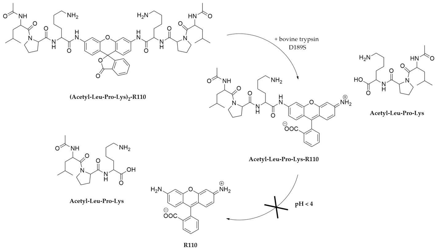
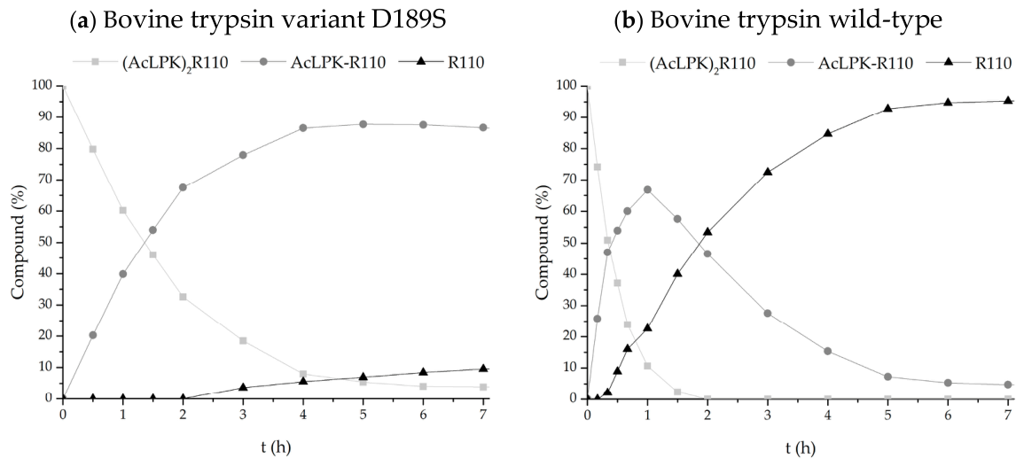
The derivative (Acetyl-Leu-Pro-Lys)2-R110 contains two identical potential hydrolysis sites corresponding to the peptide bonds between each lysine residue and the central Rhodamine 110 fluorophore. Hydrolysis studies confirmed a sequential cleavage of these two bonds (Figure 1). In reactions with the bovine trypsin variant D189S, the second hydrolysis step was significantly slower than the first, resulting in pronounced accumulation of the intermediate Acetyl-Leu-Pro-Lys-R110. Figure 2a shows that a maximum intermediate concentration of 88% was reached after 5 h. In contrast, hydrolysis with wild-type trypsin resulted in the formation of only 67% of the intermediate Acetyl-Leu-Pro-Lys-R110, which was rapidly further hydrolyzed to R110 (Figure 2b).
The enzyme activities of both trypsin variants for each hydrolysis step were determined from the slopes of their reaction curves (Table 1). The bovine trypsin variant D189S displayed a 21-fold higher activity for the first hydrolysis step compared to the second, whereas the wild-type enzyme exhibited a 5-fold difference. These results confirm the efficient accumulation of the mono-substituted intermediate under the applied conditions.
These results demonstrate that the bovine trypsin variant D189S enables kinetically controlled and preparatively useful desymmetrization of a symmetric Rhodamine 110 derivative. The isolated Acetyl-Leu-Pro-Lys-R110 represents a well-defined building block for the preparation of asymmetric fluorogenic Rhodamine derivatives.

3. Materials and Methods

3.1. General

All chemicals were purchased from commercial sources and used without additional purification unless otherwise noted. NMR spectra were recorded on a Bruker Avance III 700 MHz spectrometer (Bruker, Ettlingen, Germany) using TopSpin v3.6.2 software (Bruker, Ettlingen, Germany). High resolution mass spectra (HRMS) were recorded on a Q-Tof Premier-ESI-Q-TOFMS (Waters Corporation, Milford, MA, USA) using MassLynx v4.1 software (Waters Corporation, Milford, MA, USA). UV/Vis spectra were recorded on a NanoPhotometer® NP80 spectrophotometer (Implen, Munich, Germany) using PVC v5.5.1.2 software (Implen, Munich, Germany). Preparative HPLC was performed on a Waters® 2545 Binary Gradient Module with a Waters® 2489 UV/Vis Detector, using an XSelect Peptide CSH C18 OBD Prep Column (19 mm × 250 mm, 5 µm) (Waters Corporation, Milford, MA, USA) and MassLynx v4.1 software (Waters Corporation, Milford, MA, USA). For analytical HPLC, the Agilent 1100 Series system (Agilent Technologies, Santa Clara, CA, USA) was used with a Kinetex XB-C18 LC Column (50 mm × 3 mm, 2.6 µm) and the ChemStation Rev. B 03.02 software (Agilent Technologies, Santa Clara, CA, USA). For HPLC determination of the hydrolysis studies, the Agilent 1100 Series system (Agilent Technologies, Santa Clara, CA, USA) was used with Grace™ Vydac™ 208 MS C8 Reversed Phase HPLC Column (4.6 mm × 250 mm, 5 μm) (Grace, Columbia, MD, USA) and the ChemStation Rev. A 08.03 software (Agilent Technologies, Santa Clara, CA, USA). Unless otherwise stated, separations were performed using an ACN/water mixture containing 0.1% TFA using a gradient from 5% to 100% ACN over 6 min.

3.2. Synthesis of (Acetyl-Leu-Pro-Lys)2-R110 Derivative

Synthesis of the Rhodamine 110 derivative was carried out starting from Rhodamine 110 (R110; Synthon AcMaRi Chemie GmbH & Co. KG, Wolfen, Germany) following established protocols [2,8,9,10]. The first amino acid Lys(Boc), protected by Fmoc, was coupled after activation by EDC in DMF/pyridine (9:1) as solvent. After purification by silica gel chromatography, the Fmoc protecting group was removed using morpholine. Subsequently, the dipeptide Ac-Leu-Pro-OH was coupled, using the same activation method (EDC in DMF/pyridine, 9:1), followed by purification through silica gel chromatography. The acid-labile Boc group was removed using TFA. The product was purified by preparative HPLC and its purity was confirmed by analytical HPLC.
(Acetyl-Leu-Pro-Lys)2-R110: N,N′-(((3-oxo-3H-spiro[isobenzofuran-1,9′-xanthene]-3′,6′-diyl)bis(azanediyl))bis(6-amino-1-oxohexane-1,2-diyl))bis(1-(acetylleucyl)pyrrolidine-2-carboxamide), yield 26.5%, purity 95.5%, Rt 4.28 min (5–95% ACN/water 0.1% TFA over 6 min). 1H NMR (700.5 MHz, DMSO) δH 8.15 (t, J = 7.0 Hz, 2H), 8.06 (d, J = 7.9 Hz, 2H), 8.02 (d, J = 7.8 Hz, 1H), 7.91 (s, 1H), 7.88 (s, 1H), 7.81–7.77 (m, 3H), 7.75–7.72 (m, 6H), 7.23 (d, J = 7.4 Hz, 2H), 6.75 (s, 1H), 6.74 (s, 1H), 4.55–4.49 (m, 2H), 4.36–4.32 (m, 2H), 4.31–4.27 (m, 2H), 3.68 (q, J = 8.6 Hz, 1H), 3.51–3.48 (m, 1H), 2.80–2.75 (m, 4H), 2.49 (DMSO), 2.10–2.03 (m, 2H), 1.94–1.85 (m, 4H), 1.81 (s, 6H), 1.76–1.73 (m, 2H), 1.64–1.57 (m, 4H), 1.56–1.51 (m, 4H), 1.45–1.34 (m, 8H), 0.86–0.81 (m, 12H). 13C NMR (176.1 MHz, DMSO) δC 171.7, 171.2, 170.9, 169.1, 168.7, 158.2, 158.0, 152.6, 150.8, 141.0, 135.8, 130.3, 128.4, 125.6, 124.8, 123.9, 115.5, 113.2, 106.4, 81.8, 59.4, 53.4, 48.7, 46.8, 39.50 (DMSO), 38.7, 31.0, 29.0, 26.6, 24.5, 24.0, 23.1, 22.34, 22.26, 21.5. HRMS m/z: [M + H]+ calcd for C58H78N10O11 1091.590, found 1091.669.

3.3. Mutagenesis, Expression, Refolding and Purification of Bovine Trypsin Variant D189S

The D189S mutation was introduced into bovine trypsin by site-directed mutagenesis following the method of Kunkel as described previously [11]. The pET-21a vector containing the bovine trypsin sequence with an upstream His-Tag sequence and enteropeptidase cleavage site (Novagen, Madison, WI, USA) was used as template. The following primers were used (mismatches are underlined): GGGCGGAAAGAGCTCCTGCCAGG (forward) and TCCAGGTAGCCCGCACAG (reverse). Mutagenesis was confirmed by DNA sequencing (LGC Genomics, Berlin, Germany). Protein expression, refolding and purification were performed according to an established protocol [12].

3.4. Hydrolysis of (Acetyl-Leu-Pro-Lys)2-R110 Derivative by Bovine Trypsin Variants

For hydrolysis studies, 100 µM (Acetyl-Leu-Pro-Lys)2-R110 was incubated with either 143 nM bovine trypsin variant D189S or 1.43 nM bovine wild-type trypsin in 150 µL buffer (50 mM Tris pH 8.0, 154 mM NaCl, 10 mM CaCl2) at 37 °C for 24 h. Samples were taken at defined time points, diluted tenfold, acidified with 5% acetic acid and analysed by analytical HPLC (5–45% ACN/water + 0.1% TFA over 30 min). Peak areas of the educt and products were quantified and used for kinetic analysis.

3.5. Isolation of Acetyl-Leu-Pro-Lys-R110 Derivative

A scaled-up 1000-fold reaction with bovine trypsin variant D189S was quenched after 5 h by acidification with acetic acid. After lyophilisation, the sample was redissolved in a minimal amount of water and purified by preparative HPLC. Purity was determined by analytical HPLC.
Acetyl-Leu-Pro-Lys-R110: 2-(6-(2-(1-(acetylleucyl)pyrrolidine-2-carboxamido)-6-aminohexanamido)-3-iminio-3H-xanthen-9-yl)benzoate, yield 3.4 mg (28.8%), purity 95.8%, Rt 4.06 min (5–95% ACN/water 0.1% TFA over 6 min). 1H NMR (700.5 MHz, DMSO) δH 8.12 (d, J = 8.3 Hz, 1H), 8.05 (d, J = 8.0 Hz, 1H), 7.99 (d, J = 8.4 Hz, 1H), 7.78 (t, J = 7.5 Hz, 1H), 7.71 (t, J = 7.7 Hz, 1H), 7.65 (s, 3H), 7.23 (d, J = 7.8 Hz, 2H), 6.69 (s, 1H), 6.46 (s, 1H), 4.59–4.47 (m, 1H), 4.36–4.24 (m, 1H), 3.70–3.68 (m, 1H), 3.52–3.44 (m, 1H), 2.76 (q, J = 6.9 Hz, 2H), 2.49 (DMSO), 2.10–2.03 (m, 1H), 1.90–1.85 (m, 2H), 1.81 (s, 3H), 1.62–1.60 (m, 2H), 1.54–1.51 (m, 2H), 1.41–1.38 (m, 4H), 0.88–0.82 (m, 6H). 13C NMR (176.1 MHz, DMSO) δC 171.6, 171.1, 170.9, 169.1, 168.7, 157.7, 152.5, 152.0, 151.4, 151.1, 135.5, 130.0, 128.4, 126.3, 124.7, 124.0, 115.0, 113.9, 106.3, 99.0, 59.4, 53.3, 48.7, 46.8, 39.5 (DMSO), 38.7, 31.0, 29.0, 26.6, 24.5, 24.0, 23.1, 22.29, 22.26, 21.5. HRMS m/z: [M + H]+ calcd for C39H46N6O7 711.350; found 711.298.

4. Conclusions

The enzymatic desymmetrization of the symmetric R110 derivative (Acetyl-Leu-Pro-Lys)2-R110 using bovine trypsin variant D189S allows the selective accumulation of a mono-substituted intermediate Acetyl-Leu-Pro-Lys-R110. This asymmetric compound can be efficiently isolated and serves as a well-defined building block for further coupling reactions or kinetic studies. This approach provides a simple and practical alternative to conventional chemical synthesis of asymmetric fluorogenic Rhodamine derivatives.

Supplementary Materials

The following supporting information can be downloaded online: Figure S1: 1H NMR of (Acetyl-Leu-Pro-Lys)2-R110 in DMSO-d6; Figure S2: 13C NMR of (Acetyl-Leu-Pro-Lys)2-R110 in DMSO-d6; Figure S3: 1H NMR of Acetyl-Leu-Pro-Lys-R110 in DMSO-d6; Figure S4: 13C NMR of Acetyl-Leu-Pro-Lys-R110 in DMSO-d6; Figure S5: Overlay of the analytical HPLC chromatograms of (Acetyl-Leu-Pro-Lys)2-R110 and Acetyl-Leu-Pro-Lys-R110. HRMS spectra of (A) (Acetyl-Leu-Pro-Lys)2-R110 and (B) Acetyl-Leu-Pro-Lys-R110; Table S1: Observed ions by HRMS; Figure S6: UV/Vis spectra of (Acetyl-Leu-Pro-Lys)2-R110, Acetyl-Leu-Pro-Lys-R110 and R110.

Author Contributions

Conceptualization, S.S., M.D. and F.B.; methodology, S.S.; validation, S.S., M.H. and M.D.; investigation, S.S., M.H. and M.D.; resources, F.B.; writing—original draft preparation, S.S.; writing—review and editing, S.S. and F.B.; visualization, S.S.; supervision, F.B.; project administration, S.S. and F.B.; funding acquisition, F.B. All authors have read and agreed to the published version of the manuscript.

Funding

This work was funded by the Institut für Technische Biochemie at the Martin-Luther-University Halle-Wittenberg.

Data Availability Statement

Data presented in this study are available on request from the corresponding author.

Acknowledgments

The authors gratefully acknowledge Lars Franke for valuable discussions, continuous support, and careful proofreading of this manuscript. The authors thank Mike Schutkowski for providing access to the analytical HPLC. The authors acknowledge the use of AI-based tools to improve the wording and clarity of this manuscript. All scientific content and conclusions remain the responsibility of the authors.

Conflicts of Interest

The authors declare no conflicts of interest. The funders had no role in the design of this study; in the collection, analysis, or interpretation of data; in the writing of this manuscript; or in the decision to publish these results.

References

  1. Lavis, L.D. Teaching Old Dyes New Tricks: Biological Probes Built from Fluoresceins and Rhodamines. Annu. Rev. Biochem. 2017, 86, 825–843. [Google Scholar] [CrossRef] [PubMed]
  2. Leytus, S.P.; Melhado, L.L.; Mangel, W.F. Rhodamine-Based Compounds as Fluorogenic Substrates for Serine Proteinases. Biochem. J. 1983, 209, 299–307. [Google Scholar] [CrossRef] [PubMed]
  3. Iwatate, R.J.; Kamiya, M.; Urano, Y. Asymmetric Rhodamine-Based Fluorescent Probe for Multicolour In Vivo Imaging. Chem.—Eur. J. 2016, 22, 1696–1703. [Google Scholar] [CrossRef] [PubMed]
  4. Wang, Z.-Q.; Liao, J.; Diwu, Z. N-DEVD-N′-Morpholinecarbonyl-Rhodamine 110: Novel Caspase-3 Fluorogenic Substrates for Cell-Based Apoptosis Assay. Bioorg. Med. Chem. Lett. 2005, 15, 2335–2338. [Google Scholar] [CrossRef] [PubMed]
  5. Terentyeva, T.G.; Van Rossom, W.; Van Der Auweraer, M.; Blank, K.; Hofkens, J. Morpholinecarbonyl-Rhodamine 110 Based Substrates for the Determination of Protease Activity with Accurate Kinetic Parameters. Bioconjug. Chem. 2011, 22, 1932–1938. [Google Scholar] [CrossRef] [PubMed]
  6. Cai, S.X.; Zhang, H.-Z.; Guastella, J.; Drewe, J.; Yang, W.; Weber, E. Design and Synthesis of Rhodamine 110 Derivative and Caspase-3 Substrate for Enzyme and Cell-Based Fluorescent Assay. Bioorg. Med. Chem. Lett. 2001, 11, 39–42. [Google Scholar] [CrossRef] [PubMed]
  7. Zhang, H.-Z.; Kasibhatla, S.; Guastella, J.; Tseng, B.; Drewe, J.; Cai, S.X. N-Ac-DEVD-N‘-(Polyfluorobenzoyl)−R110: Novel Cell-Permeable Fluorogenic Caspase Substrates for the Detection of Caspase Activity and Apoptosis. Bioconjug. Chem. 2003, 14, 458–463. [Google Scholar] [CrossRef] [PubMed]
  8. Leytus, S.P.; Patterson, W.L.; Mangel, W.F. New Class of Sensitive and Selective Fluorogenic Substrates for Serine Proteinases. Amino Acid and Dipeptide Derivatives of Rhodamine. Biochem. J. 1983, 215, 253–260. [Google Scholar] [CrossRef] [PubMed]
  9. Liu, J.; Bhalgat, M.; Zhang, C.; Diwu, Z.; Hoyland, B.; Klaubert, D.H. Fluorescent Molecular Probes V: A Sensitive Caspase-3 Substrate for Fluorometric Assays. Bioorg. Med. Chem. Lett. 1999, 9, 3231–3236. [Google Scholar] [CrossRef] [PubMed]
  10. Xiong, C.; Lu, W.; Zhang, R.; Tian, M.; Tong, W.; Gelovani, J.; Li, C. Cell-Permeable [99mTc(CO3)]-Labeled Fluorogenic Caspase 3 Substrate for Dual-Modality Detection of Apoptosis. Chem.—Eur. J. 2009, 15, 8979–8984. [Google Scholar] [CrossRef] [PubMed]
  11. Kunkel, T.A. Rapid and Efficient Site-Specific Mutagenesis without Phenotypic Selection. Proc. Natl. Acad. Sci. USA 1985, 82, 488–492. [Google Scholar] [CrossRef] [PubMed]
  12. Liebscher, S.; Schöpfel, M.; Aumüller, T.; Sharkhuukhen, A.; Pech, A.; Höss, E.; Parthier, C.; Jahreis, G.; Stubbs, M.T.; Bordusa, F. N-Terminal Protein Modification by Substrate-Activated Reverse Proteolysis. Angew. Chem. Int. Ed. 2014, 53, 3024–3028. [Google Scholar] [CrossRef] [PubMed]
Figure 1. Partial hydrolysis of symmetric R110 derivative (Acetyl-Leu-Pro-Lys)2-R110 by bovine trypsin variant D189S. The sequential cleavage of the two peptide bonds allows accumulation of mono-substituted intermediate Acetyl-Leu-Pro-Lys-R110.
Molbank 2026 m2179 g001
Figure 2. Time-dependent hydrolysis of (Acetyl-Leu-Pro-Lys)2-R110 by bovine trypsin variant D189S (a) and wild-type trypsin (b). Molbank 2026 m2179 i001 (Acetyl-Leu-Pro-Lys)2-R110; Molbank 2026 m2179 i002 Acetyl-Leu-Pro-Lys-R110; Molbank 2026 m2179 i003 R110. Reactions were performed in a total volume of 150 µL containing 100 µM (Acetyl-Leu-Pro-Lys)2-R110, 143 nM bovine trypsin variant D189S or 1.43 nM bovine trypsin wild-type, respectively, and buffer (50 mM Tris, pH 8.0, 154 mM NaCl, 10 mM CaCl2) at 37 °C. Samples (10 µL) were withdrawn at defined time points, diluted 10-fold in 5% acetic acid and analyzed by HPLC. The bovine trypsin variant D189S shows pronounced accumulation of Acetyl-Leu-Pro-Lys-R110 due to slower hydrolysis of the second peptide bond, whereas wild-type trypsin converts the intermediate to R110.

Molbank 2026 m2179 g002
Table 1. Specific activities [µkat/mg] of bovine trypsin variant D189S and wild-type for the two sequential hydrolysis steps 1 and 2 of (Acetyl-Leu-Pro-Lys)2-R110, determined from the slopes of their reaction curves in Figure 2. The ratio of activities for the first vs. the second hydrolysis step highlights the kinetic preferences of the bovine trypsin variant D189S for the first cleavage.
Specific Activity [µkat/mg]
Trypsin D189STrypsin Wild-Type
Hydrolysis first site (1)
(AcLPK)2-R110 → AcLPK-R110
12552,300
Hydrolysis second site (2)
AcLPK-R110 → R110
611,600
Activity ratio (hydrolysis 1/hydrolysis 2)20.84.5
Disclaimer/Publisher’s Note: The statements, opinions and data contained in all publications are solely those of the individual author(s) and contributor(s) and not of MDPI and/or the editor(s). MDPI and/or the editor(s) disclaim responsibility for any injury to people or property resulting from any ideas, methods, instructions or products referred to in the content.