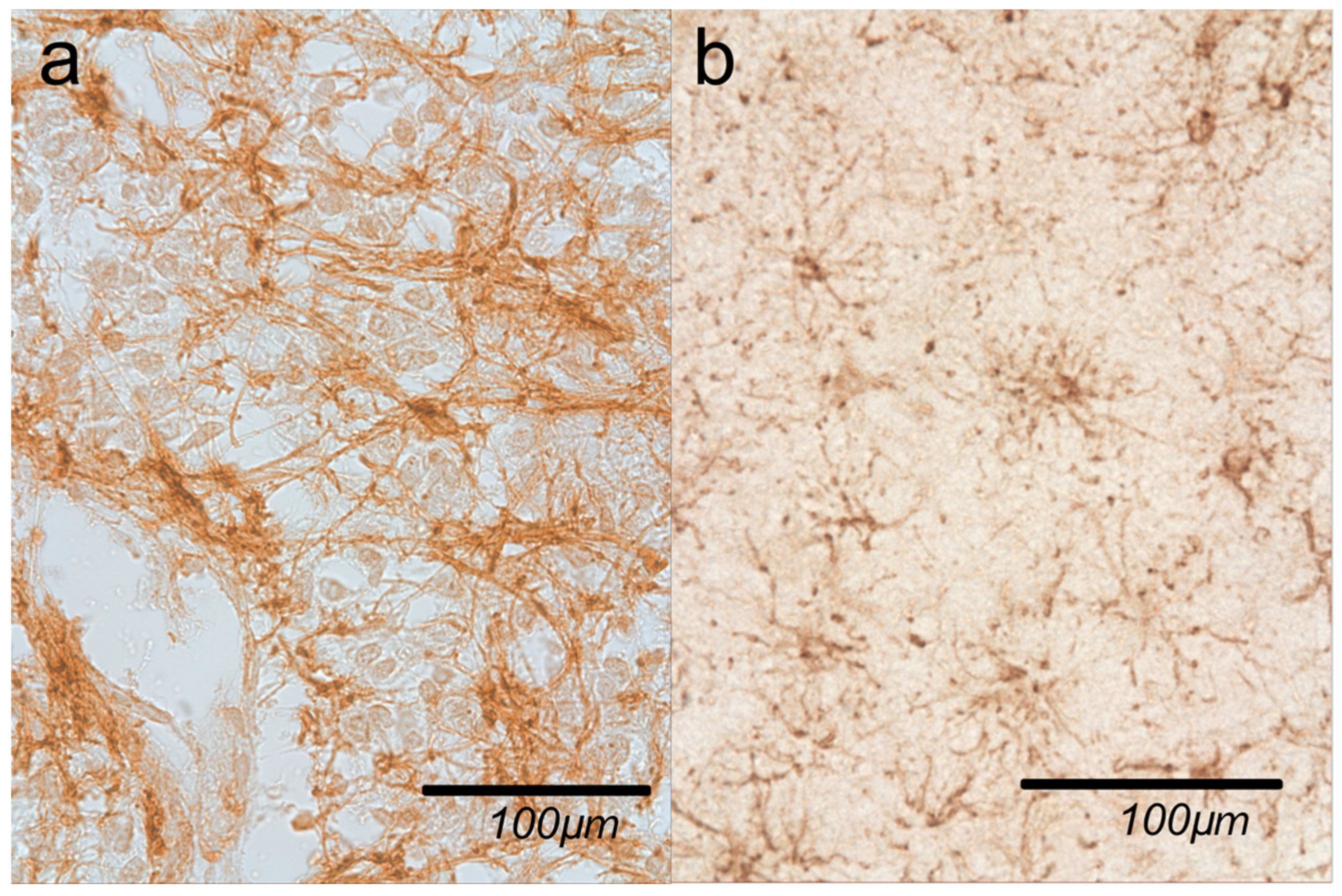
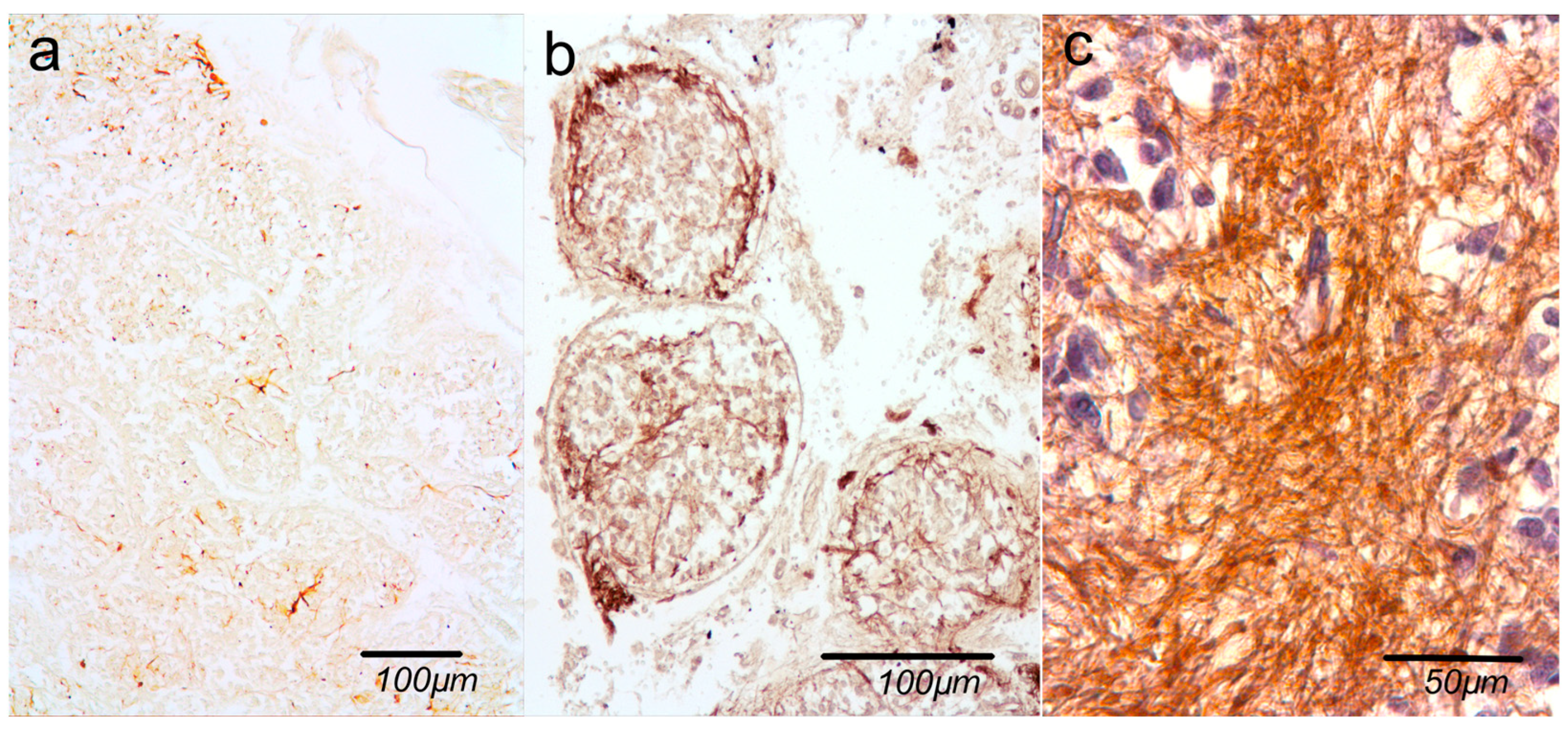
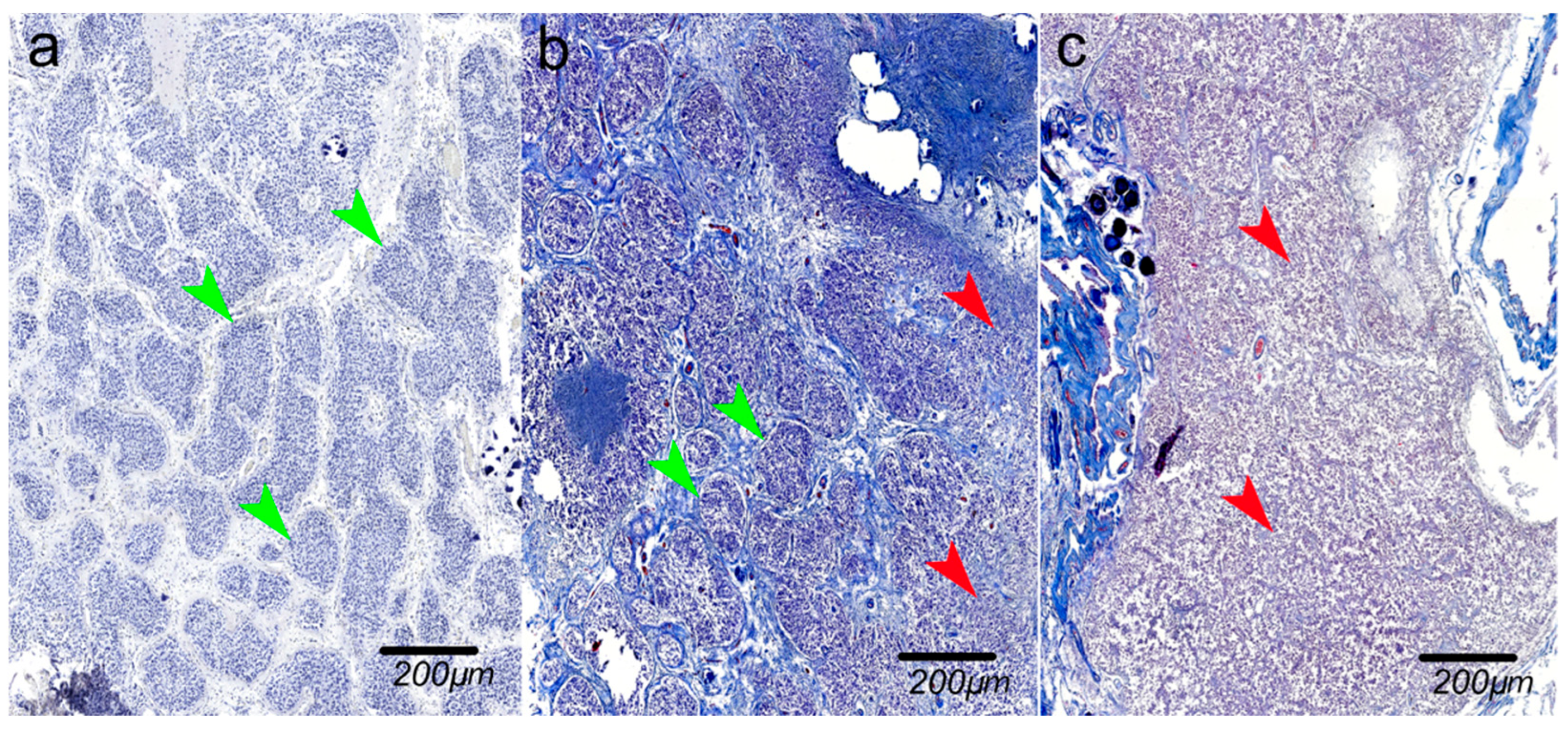
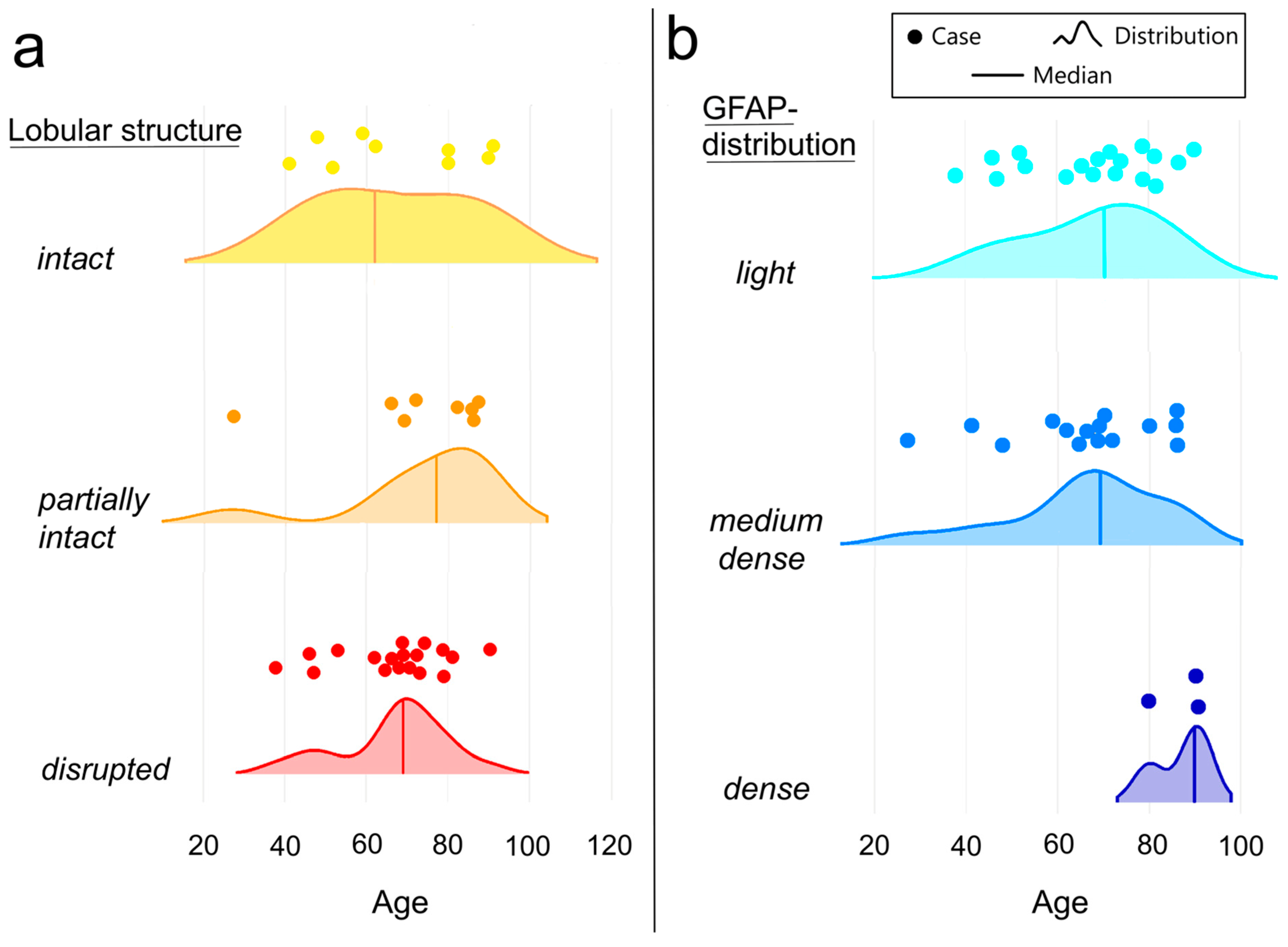
Age-Related Changes as the Primary Driver of Pineal Gland Involution—A Morphological Study in Health and Disease


Abstract
1. Introduction
2. Results
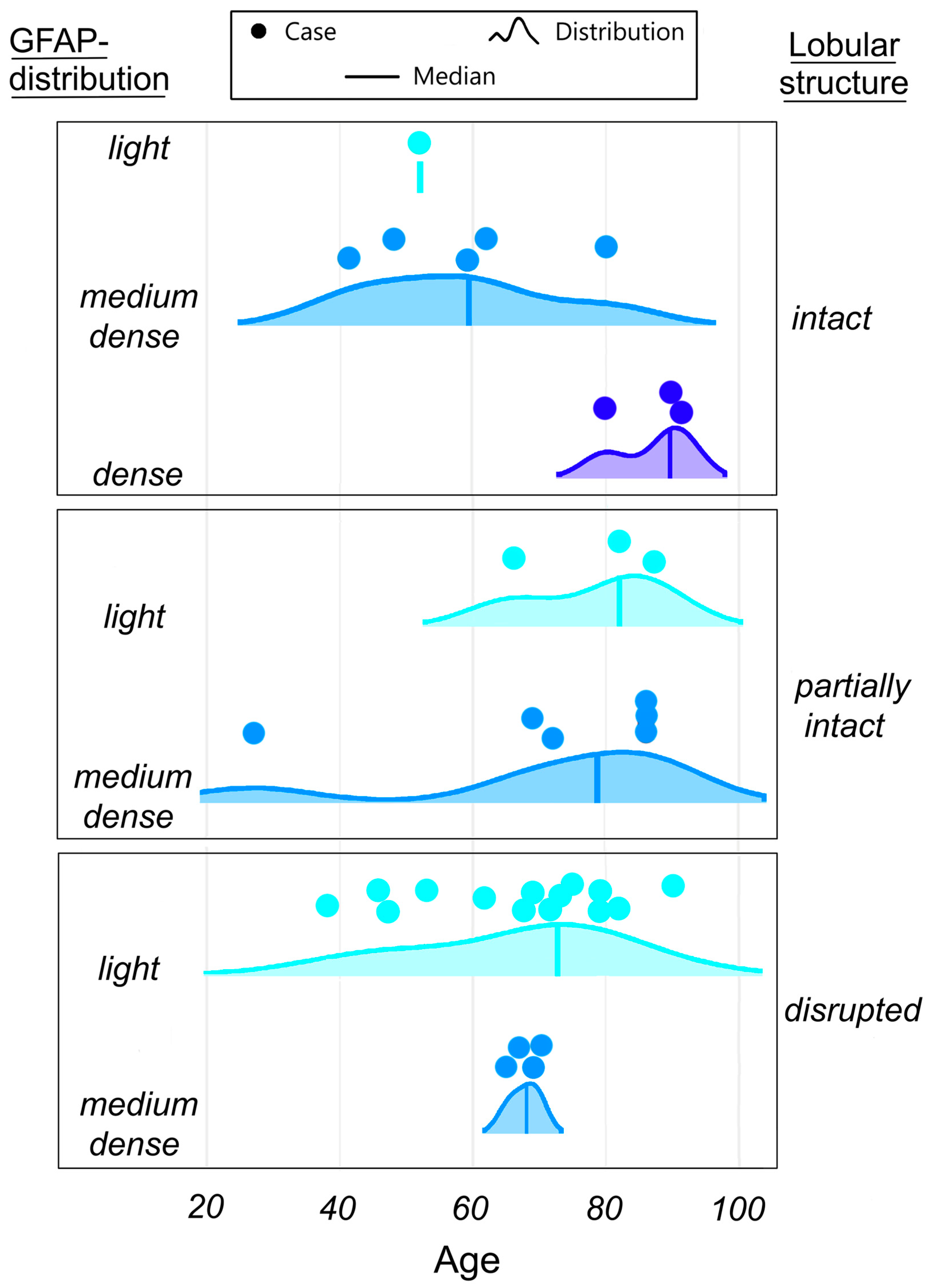
2.1. Age-Related Variability
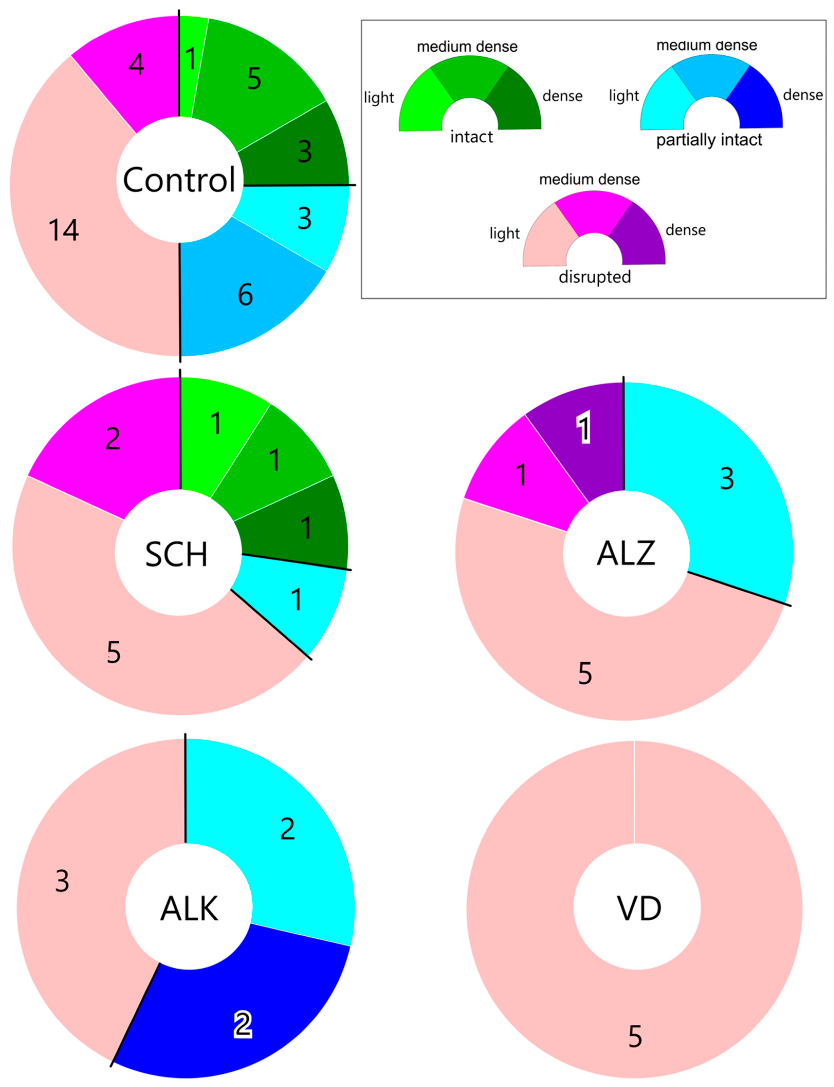
2.2. Pathological Variability
2.3. Statistical Analysis
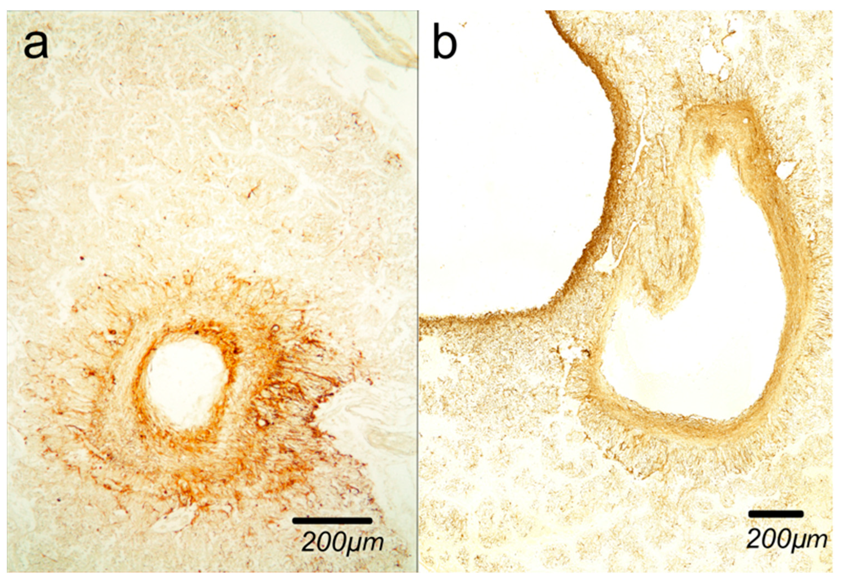
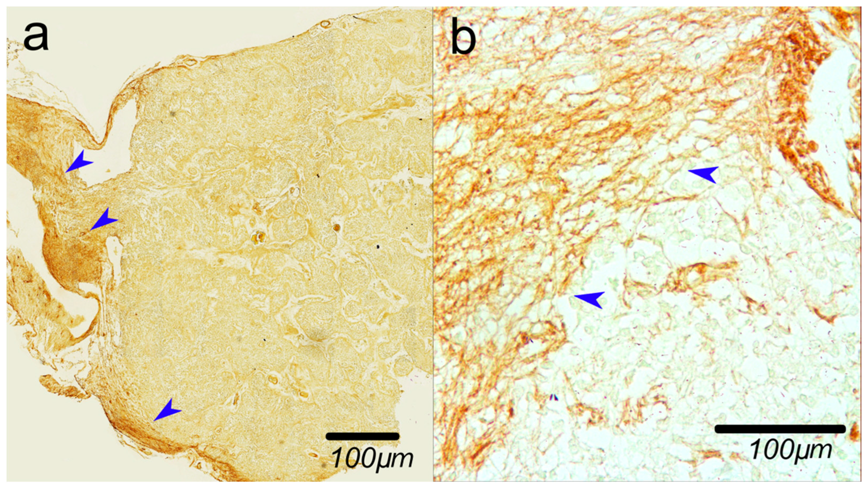
2.4. Glial Cysts
3. Discussion
3.1. Age-Related Variability
3.2. Pathological Variability
3.3. Glial Cysts
4. Materials and Methods
4.1. Materials
4.2. Methods
4.2.1. Histology
4.2.2. Immunohistochemistry
4.2.3. Analysis
4.2.4. Statistical Analysis
5. Conclusions
Author Contributions
Funding
Institutional Review Board Statement
Informed Consent Statement
Data Availability Statement
Conflicts of Interest
Abbreviations
| PG | Pineal gland |
| GFAP | Glial fibrillary acidic protein |
| ALK | Alcoholism |
| SCH | Schizophrenia |
| ALZ | Alzheimer disease |
| VD | Vascular dementia |
| CNS | Central nervous system |
| BBB | Blood–brain barrier |
References
- Ibañez Rodriguez, M.P.; Noctor, S.C.; Muñoz, E.M. Cellular Basis of Pineal Gland Development: Emerging Role of Microglia as Phenotype Regulator. PLoS ONE 2016, 11, e0167063. [Google Scholar] [CrossRef]
- Reiter, R.J. The pineal gland and melatonin in relation to aging: A summary of the theories and of the data. Exp. Gerontol. 1995, 30, 199–212. [Google Scholar] [CrossRef]
- Reiter, R.J.; Melchiorri, D.; Sewerynek, E.; Poeggeler, B.; Barlow-Walden, L.; Chuang, J.; Ortiz, G.G.; AcuñaCastroviejo, D. A review of the evidence supporting melatonin’s role as an antioxidant. J. Pineal Res. 1995, 18, 1–11. [Google Scholar] [CrossRef]
- Tan, D.X.; Manchester, L.C.; Terron, M.P.; Flores, L.J.; Reiter, R.J. One molecule, many derivatives: A never-ending interaction of melatonin with reactive oxygen and nitrogen species? J. Pineal Res. 2007, 42, 28–42. [Google Scholar] [CrossRef]
- Ponchietti, R.; Raugei, A.; Santoni, G.; Trippitelli, A.; Grechi, G. Modulation of human lymphoblastoid interferon activity by melatonin in metastatic renal cell carcinoma. A phase II study. Cancer 1994, 73, 3015–3019. [Google Scholar] [CrossRef]
- da Silveira Cruz-Machado, S.; Pinato, L.; Tamura, E.K.; Carvalho-Sousa, C.E.; Markus, R.P. Glia-Pinealocyte Network: The Paracrine Modulation of Melatonin Synthesis by Tumor Necrosis Factor (TNF). PLoS ONE 2012, 7, e40142. [Google Scholar] [CrossRef]
- Dawson, D.; Encel, N. Melatonin and sleep in humans. J. Pineal Res. 1993, 15, 1–12. [Google Scholar] [CrossRef]
- Aleandri, V.; Spina, V.; Morini, A. The pineal gland and reproduction. Hum. Reprod. Updat. 1996, 2, 225–235. [Google Scholar] [CrossRef] [PubMed]
- Macchi, M.M.; Bruce, J.N. Human pineal physiology and functional significance of melatonin. Front. Neuroendocr. 2004, 25, 177–195. [Google Scholar] [CrossRef] [PubMed]
- Reiter, R.J. The pineal and its hormones in the control of reproduction in mammals. Endocr. Rev. 1980, 1, 109–131. [Google Scholar] [CrossRef] [PubMed]
- Hu, W.; Ma, Z.; Jiang, S.; Fan, C.; Deng, C.; Yan, X.; Di, S.; Lv, J.; Reiter, R.J.; Yang, Y. Melatonin: The dawning of a treatment for fibrosis? J. Pineal Res. 2016, 60, 121–131. [Google Scholar] [CrossRef]
- Verkhratsky, A.; Nedergaard, M. Physiology of Astroglia. Physiol. Rev. 2018, 98, 239–389. [Google Scholar] [CrossRef]
- Walz, W. Role of astrocytes in the clearance of excess extracellular potassium. Neurochem. Int. 2000, 36, 291–300. [Google Scholar] [CrossRef]
- Calvo, J.; Boya, J.; Borregon, A.; Garcia-Mauriño, J.E. Presence of glial cells in the rat pineal gland: A light and electron microscopic immunohistochemical study. Anat. Rec. 1988, 220, 424–428. [Google Scholar] [CrossRef]
- Wartenberg, H. The mammalian pineal organ: Electron microscopic studies on the fine structure of pinealocytes, glial cells and on the perivascular compartment. Cell Tissue Res. 1968, 86, 74–97. [Google Scholar] [CrossRef]
- Zang, X.; Nilaver, G.; Stein, B.M.; Fetell, M.R.; Duffy, P.E. Immunocytochemistry of Pineal Astrocytes: Species Differences and Functional Implications. J. Neuropathol. Exp. Neurol. 1985, 44, 486–495. [Google Scholar] [CrossRef] [PubMed]
- Liu, Y.J.; Zhuang, J.; Zhu, H.Y.; Shen, Y.X.; Tan, Z.L.; Zhou, J.N. Cultured rat cortical astrocytes synthesize melatonin: Absence of a diurnal rhythm. J. Pineal Res. 2007, 43, 232–238. [Google Scholar] [CrossRef] [PubMed]
- Hasegawa, A.; Ohtsubo, K.; Mori, W. Pineal gland in old age; quantitative and qualitative morphological study of 168 human autopsy cases. Brain Res. 1987, 409, 343–349. [Google Scholar] [CrossRef]
- Kunz, D.; Schmitz, S.; Mahlberg, R.; Mohr, A.; Stoter, C.; Wolf, K.J.; Herrmann, W.M. A new concept for melatonin deficit: On pineal calcification and melatonin excretion. Neuropsychopharmacology 1999, 21, 765–772. [Google Scholar] [CrossRef]
- Mahlberg, R.; Kienast, T.; Hädel, S.; Heidenreich, J.O.; Schmitz, S.; Kunz, D. Degree of pineal calcification (DOC) is associated with polysomnographic sleep measures in primary insomnia patients. Sleep Med. 2009, 10, 439–445. [Google Scholar] [CrossRef] [PubMed]
- Tan, D.X.; Xu, B.; Zhou, X.; Reiter, R.J. Pineal Calcification, Melatonin Production, Aging, Associated Health Consequences and Rejuvenation of the Pineal Gland. Molecules 2018, 23, 301. [Google Scholar] [CrossRef]
- Junemann, O.; Ivanova, A.G.; Bukreeva, I.; Zolotov, D.A.; Fratini, M.; Cedola, A.; Wilde, F.; Dyachkova, I.G.; Krivonosov, Y.S.; Otlyga, D.A.; et al. Comparative study of calcification in human choroid plexus, pineal gland, and habenula. Cell Tissue Res. 2023, 393, 537–545. [Google Scholar] [CrossRef]
- Wu, Y.H.; Swaab, D.F. The human pineal gland and melatonin in aging and Alzheimer’s disease. J. Pineal Res. 2005, 38, 145–152. [Google Scholar] [CrossRef]
- Mahlberg, R.; Walther, S.; Kalus, P.; Bohner, G.; Haedel, S.; Reischies, F.M.; Kühl, K.P.; Hellweg, R.; Kunz, D. Pineal calcification in Alzheimer’s disease: An in vivo study using computed tomography. Neurobiol. Aging 2008, 29, 203–209. [Google Scholar] [CrossRef]
- Song, J. Pineal gland dysfunction in Alzheimer’s disease: Relationship with the immune-pineal axis, sleep disturbance, and neurogenesis. Mol. Neurodegener. 2019, 14, 1–10. [Google Scholar] [CrossRef]
- Bukreeva, I.; Junemann, O.; Cedola, A.; Brun, F.; Longo, E.; Tromba, G.; Wilde, F.; Chukalina, M.V.; Krivonosov, Y.S.; Dyachkova, I.G.; et al. Micromorphology of pineal gland calcification in age-related neurodegenerative diseases. Med. Phys. 2023, 50, 1601–1613. [Google Scholar] [CrossRef]
- Greiner, A.C. Schizophrenia and the pineal gland. Can. Psychiatr. Assoc. J. 1970, 15, 433–447. [Google Scholar] [CrossRef] [PubMed]
- Sandyk, R. Pineal and habenula calcification in schizophrenia. Int. J. Neurosci. 1992, 67, 19–30. [Google Scholar] [CrossRef] [PubMed]
- Sandyk, R. The relationship of thought disorder to third ventricle width and calcification of the pineal gland in chronic schizophrenia. Int. J. Neurosci. 1993, 68, 53–59. [Google Scholar] [CrossRef]
- Junemann, O.; Bukreeva, I.; Otlyga, D.A.; Cedola, A.; Fratini, M.; Saveliev, S.V. Human Pineal Gland Involutionary Process: New Findings. J. Gerontol. A Biol. Sci. Med. Sci. 2023, 78, 1558–1560. [Google Scholar] [CrossRef] [PubMed]
- Gómez Esteban, M.B.; Muñoz, M.I.; Carbajo, S.; Carvajal, J.C.; Alvarez-Morujo, A.J.; Muñoz Barragán, L. Pineal gliosis and gland ageing. The possible role of the glia in the transfer of melatonin from pinealocytes to the blood and cerebrospinal fluid. Eur. J. Anat. 2008, 12, 97–114. [Google Scholar]
- Sufieva, D.A.; Fedorova, E.A.; Yakovlev, V.S.; Grigorev, I.P. Immunohistochemical study of human pineal vessels. Med. Acad. J. 2023, 23, 109–118. [Google Scholar] [CrossRef]
- Singh, G.; Sethi, S.S.; Das, S.; Mohapatra, C. Study of the Histomorphology of Human Pineal Gland. Natl. J. Clin. Anat. 2023, 12, 110–114. [Google Scholar] [CrossRef]
- Xu, J.; Wang, L.L.; Dammer, E.B.; Li, C.B.; Xu, G.; Chen, S.D.; Wang, G. Melatonin for sleep disorders and cognition in dementia: A meta-analysis of randomized controlled trials. Am. J. Alzheimer’s Dis. Other Dementiasr 2015, 30, 439–447. [Google Scholar] [CrossRef]
- Srinivasan, V.; Pandi-Perumal, S.R.; Cardinali, D.P.; Poeggeler, B.; Hardeland, R. Melatonin in Alzheimer’s disease and other neurodegenerative disorders. Behav. Brain Funct. 2006, 2, 15. [Google Scholar] [CrossRef] [PubMed]
- Cipriani, G.; Lucetti, C.; Danti, S.; Nuti, A. Sleep disturbances and dementia. Psychogeriatrics 2014, 15, 65–74. [Google Scholar] [CrossRef] [PubMed]
- Anderson, G.; Maes, M. Melatonin: An overlooked factor in schizophrenia and in the inhibition of anti-psychotic side effects. Metab. Brain Dis. 2012, 27, 113–119. [Google Scholar] [CrossRef]
- Morera-Fumero, A.L.; Abreu-Gonzalez, P. Role of melatonin in schizophrenia. Int. J. Mol. Sci. 2013, 14, 9037–9050. [Google Scholar] [CrossRef]
- Kühlwein, E.; Hauger, R.L.; Irwin, M.R. Abnormal nocturnal melatonin secretion and disordered sleep in abstinent alcoholics. Biol. Psychiatry 2003, 54, 1437–1443. [Google Scholar] [CrossRef]
- Gendy, M.N.S.; Lagzdins, D.; Schaman, J.; Le Foll, B. Melatonin for Treatment-Seeking Alcohol Use Disorder patients with sleeping problems: A randomized clinical pilot trial. Sci. Rep. 2020, 10, 8739. [Google Scholar] [CrossRef] [PubMed]
- Krämer, G. Alzheimer Krankheit: Benennung und Abgrenzung, Ursachen und Vererbung, Veränderungen am Nervensystem, Krankheitszeichen, Untersuchungen, Behandlung und Verlauf. 3. Aufl.; TRIAS Thieme Hippokrates Enke: Stuttgart, Germany, 1996. [Google Scholar]
- Tapp, E. The histology and pathology of the human pineal gland. Prog. Brain Res. 1979, 52, 481–500. [Google Scholar] [CrossRef] [PubMed]
- Bosnjak, J.; Budisić, M.; Azman, D.; Strineka, M.; Crnjaković, M.; Demarin, V. Pineal gland cysts—An overview. Acta Clin. Croat. 2009, 48, 355–358. [Google Scholar]
- Bargmann, W.; Hellman, T.; Watzka, M. (Eds.) Die mikroskopische Anatomie der Epiphyse. In Blutgefäss- und Lymphgefässapparat Innersekretorische Drüsen. Handbuch der Mikroskopischen Anatomie des Menschen; Springer: Berlin/Heidelberg, Germany, 1943; Volume 6. [Google Scholar] [CrossRef]
- Loonen, A.J.M. Role of Neuroglia in the Habenular Connection Hub of the Dorsal Diencephalic Conduction System. Neuroglia 2023, 4, 34–51. [Google Scholar] [CrossRef]
- Luijerink, L.; Rodriguez, M.; Machaalani, R. Quantifying GFAP immunohistochemistry in the brain—Introduction of the Reactivity score (R-score) and how it compares to other methodologies. J. Neurosci. Methods 2024, 402, 110025. [Google Scholar] [CrossRef] [PubMed]
- Viaene, A.N. A role for immunohistochemical stains in perinatal brain autopsies. J. Neuropathol. Exp. Neurol. 2024, 83, 345–356. [Google Scholar] [CrossRef] [PubMed]
- Cohen, J. Statistical Power Analysis for the Behavioral Sciences; Academic Press: Cambridge, MA, USA, 2013. [Google Scholar]

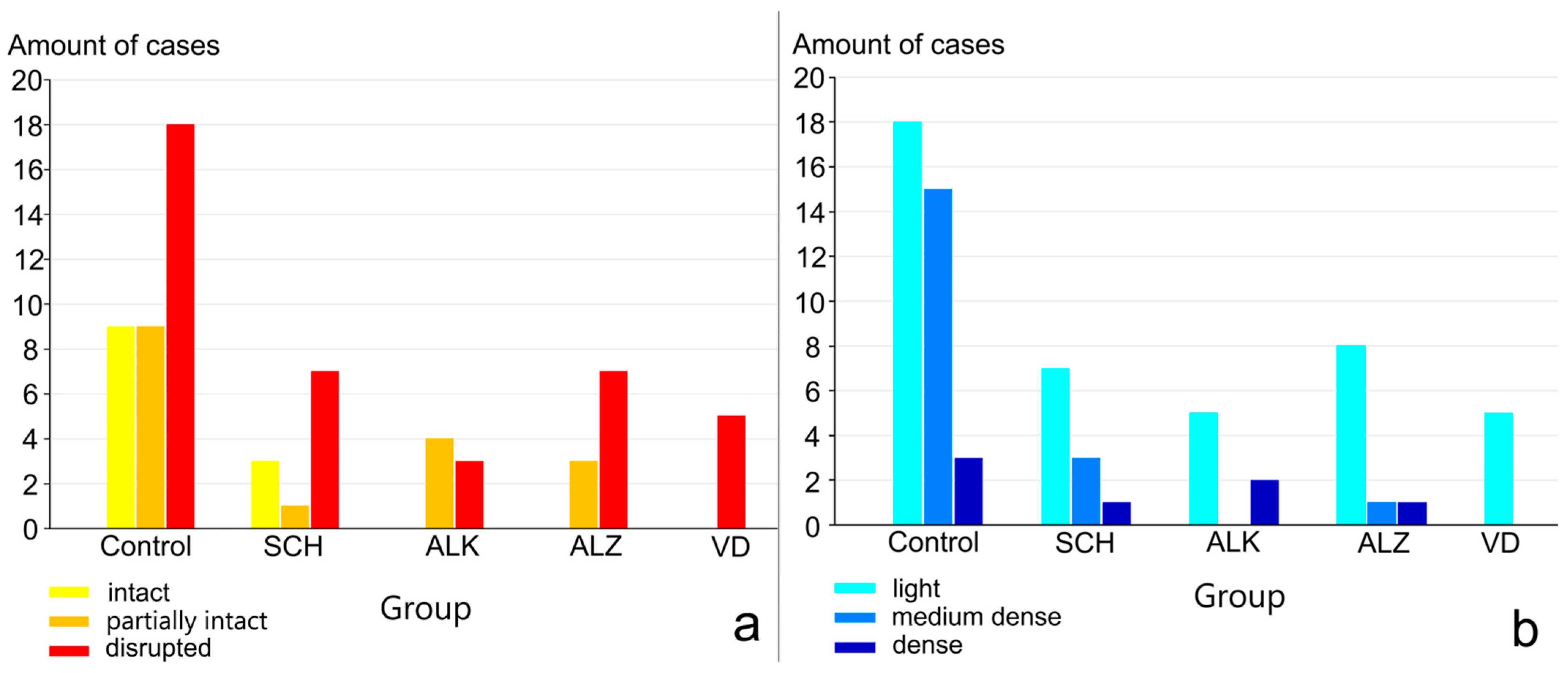



| State | Group | |||||
|---|---|---|---|---|---|---|
| Control | SCH | ALK | AD | VD | ||
| Lobular Structure | intact | 9 (25%) | 3 (27.3%) | - | - | - |
| partly intact | 9 (25%) | 1 (9.1%) | 4 (57.1%) | 3 (30%) | - | |
| disrupted | 18 (50%) | 7 (63.6%) | 3 (42.9%) | 7 (70%) | 5 (100%) | |
| GFAP-distribution | light | 18 (50%) | 7 (63.6%) | 5 (71.4%) | 8 (80%) | 5 (100%) |
| medium dense | 15 (41.7%) | 3 (27.3%) | - | 1 (10%) | - | |
| dense | 3 (8.3%) | 1 (9.1%) | 2 (28.6%) | 1 (10%) | - | |
| Lobular Structure—GFAP Distribution | Control | SCH | ALK | AD | VD |
|---|---|---|---|---|---|
| intact—light | 1 (2.8%) | 1 (9.1%) | - | - | - |
| intact—medium dense | 5 (13.9%) | 1 (9.1%) | - | - | - |
| intact—dense | 3 (8.3%) | 1 (9.1%) | - | - | - |
| partly intact—light | 3 (8.3%) | 1 (9.1%) | 2 (28.6%) | 3 (30%) | - |
| partly intact—medium dense | 6 (16.7%) | - | - | - | - |
| partly intact—dense | - | - | 2 (28.6%) | - | - |
| disrupted—light | 14 (38.9%) | 5 (45.4%) | 3 (42.8%) | 5 (50%) | 5 (100%) |
| disrupted—medium dense | 4 (11.1%) | 2 (18.2%) | - | 1 (10%) | - |
| disrupted–dense | - | - | - | 1 (10%) | - |
| Group | Number of Patients | Number of Glial Cysts | Average Age in the Group |
|---|---|---|---|
| Control | 36 | 7 | 68.5 |
| SCH | 11 | 0 | 57.5 |
| ALK | 7 | 1 | 52.6 |
| ALZ | 10 | 1 | 78.4 |
| VD | 5 | 2 | 73.8 |
Disclaimer/Publisher’s Note: The statements, opinions and data contained in all publications are solely those of the individual author(s) and contributor(s) and not of MDPI and/or the editor(s). MDPI and/or the editor(s) disclaim responsibility for any injury to people or property resulting from any ideas, methods, instructions or products referred to in the content. |
© 2026 by the authors. Licensee MDPI, Basel, Switzerland. This article is an open access article distributed under the terms and conditions of the Creative Commons Attribution (CC BY) license.
Share and Cite
Junemann, O.; Otlyga, D.; Bukreeva, I.; Saveliev, S. Age-Related Changes as the Primary Driver of Pineal Gland Involution—A Morphological Study in Health and Disease. Int. J. Mol. Sci. 2026, 27, 3093. https://doi.org/10.3390/ijms27073093
Junemann O, Otlyga D, Bukreeva I, Saveliev S. Age-Related Changes as the Primary Driver of Pineal Gland Involution—A Morphological Study in Health and Disease. International Journal of Molecular Sciences. 2026; 27(7):3093. https://doi.org/10.3390/ijms27073093
Chicago/Turabian StyleJunemann, Olga, Dmitry Otlyga, Inna Bukreeva, and Sergey Saveliev. 2026. "Age-Related Changes as the Primary Driver of Pineal Gland Involution—A Morphological Study in Health and Disease" International Journal of Molecular Sciences 27, no. 7: 3093. https://doi.org/10.3390/ijms27073093
APA StyleJunemann, O., Otlyga, D., Bukreeva, I., & Saveliev, S. (2026). Age-Related Changes as the Primary Driver of Pineal Gland Involution—A Morphological Study in Health and Disease. International Journal of Molecular Sciences, 27(7), 3093. https://doi.org/10.3390/ijms27073093