Article

Comparative Genomics Reveals Unique Genetic Determinants of Biofilm Formation in Campylobacter
Characterization and Interventions for Foodborne Pathogens Research Unit, Eastern Regional Research Center, Agricultural Research Service, United States Department of Agriculture, 600 East Mermaid Lane, Wyndmoor, PA 19038, USA
*
Author to whom correspondence should be addressed.
Int. J. Mol. Sci. 2026, 27(6), 2543; https://doi.org/10.3390/ijms27062543
Submission received: 6 February 2026 / Revised: 7 March 2026 / Accepted: 9 March 2026 / Published: 10 March 2026
(This article belongs to the Special Issue Mechanisms in Biofilm Formation, Tolerance and Control: 3rd Edition)

Abstract

A biofilm is a complex microbial community that protects bacterial cells from various stressors, including harsh environmental conditions, antimicrobial treatments, and host immune responses. This protective capability enhances Campylobacter survival during food processing and storage and facilitates transmission to humans. Despite their importance, the molecular mechanisms underlying Campylobacter biofilm formation and its impact on pathogen persistence remain poorly understood. In this study, we characterized the biofilm-forming ability of 18 C. jejuni and C. coli strains isolated from retail meat and performed whole-genome sequencing and comparative genomic analysis to identify strain-specific genes contributing to biofilm formation and maintenance. Phenotypic analysis revealed that C. jejuni strains YH001 and YH027 exhibited the strongest biofilm-forming capacity, producing the highest biomass among all isolates. Phylogenetic analysis indicated a close genetic relationship between these two strains, while pangenome analysis identified 19 unique genes/proteins specific to these strains. Functional annotation indicated their critical roles in adhesion, extracellular matrix production, and stress response. These findings demonstrate strain-specific biofilm formation in Campylobacter and highlight genetic determinants that may serve as targets for novel therapeutic approaches and intervention strategies to disrupt biofilms, improve food safety, and reduce persistent infections.

1. Introduction

Campylobacter is one of the most common foodborne pathogens worldwide and a leading cause of human gastroenteritis, with typical symptoms including diarrhea, abdominal pain, fever, and nausea. According to the Centers for Disease Control and Prevention (CDC), approximately 1.5 million cases of Campylobacter infection occur each year in the United States, making it a major public health concern [1]. Among the various species, Campylobacter jejuni and C. coli are most commonly associated with human illness [2]. These species are prevalent in poultry and other animal-derived food and can also be found in eggs, unpasteurized milk, and untreated water. The primary routes of transmission to humans include the consumption of undercooked poultry, cross-contamination in kitchens, and the ingestion of contaminated water or raw milk [3,4].
Biofilms are structured microbial communities in which cells are aggregated and embedded in a matrix of extracellular polymeric substances (EPS). This matrix, produced either by the cells themselves or by surrounding bacteria, consists of polysaccharides, structural proteins (e.g., flagella and pili), nucleic acids (extracellular DNA (eDNA) and RNA (eRNA)), lipids, and other biomolecules [5]. Polysaccharides are essential for biofilm formation contributing to cell adhesion to surfaces and the maintenance of structural integrity [6]. Biofilm proteins play important roles in biofilm development and the survival of cells by facilitating access to nutrients and regulating biofilm integrity. They are involved in attaching cells to surfaces, developing three-dimensional biofilm structures, and maintaining biofilm stability through interactions with exopolysaccharide and nucleic acids [7,8]. As a key component of biofilm structure, eDNA contributes to biofilm formation, adhesion, and structural integrity by binding and connecting cells within biofilms [9].
Biofilms formed by pathogenic bacteria present a major challenge in the food processing industry. Common foodborne pathogens, including Campylobacter, Salmonella, Escherichia coli O157:H7, Listeria monocytogenes, and Staphylococcus aureus, readily develop biofilms on both biotic and abiotic surfaces during food processing and storage. These biofilms significantly enhance bacterial resistance to environmental stresses, disinfectants, and antibiotics, making intervention and eradication efforts difficult [10]. It has been reported that biofilm-embedded cells can exhibit remarkably higher (100–1000-fold) resistance to antimicrobials compared with planktonic cells due to the protective properties of the matrix and altered physiology [11]. Consequently, biofilms contribute to persistent contamination, reduced sanitation efficacy, and elevated food safety risks across production environments.
Campylobacter is a Gram-negative, spiral-shaped, microaerophilic bacterium that thrives in low-oxygen environments, typically requiring 3–10% oxygen, 5–10% CO2 and a temperature of 37–42 °C for optimal growth. These physiological constraints favor its persistence in niches such as the gastrointestinal tract of animals, as well as in water systems and plumbing within animal husbandry facilities and food processing plants. In these environments, Campylobacter frequently forms biofilms. Studies have shown that C. jejuni cells embedded within a biofilm matrix exhibit significantly greater tolerance to environmental stresses, such as oxygen exposure and temperature fluctuations, compared to planktonic cells [12]. This enhanced resilience makes biofilm-associated Campylobacter extremely difficult to eradicate, leading to persistent contamination in food processing facilities and increasing the risk of foodborne infections [12,13,14,15].
Campylobacter biofilm formation exhibits significant strain-to-strain variation influenced by genetic factors and environmental conditions including nutrient limitation, extracellular DNA, oxygen availability, and interactions with co-cultivated bacteria [16,17]. Despite its importance for persistence and transmission, the molecular basis of biofilm development in Campylobacter remains poorly understood. Although previous studies have suggested individual genes linked to flagellar synthesis, stress response, and quorum sensing that influence biofilm formation, the broader genetic determinants and regulatory networks have yet to be systematically characterized [18,19,20].
The rapid expansion of whole-genome sequencing has greatly enhanced our understanding of genetic diversity within Campylobacter. However, genomic data have not been fully integrated into a mechanistic understanding of biofilm formation [18]. There is currently no comprehensive validated genetic framework that explains how diverse Campylobacter strains coordinate biofilm development across environmental settings. This knowledge gap limits our ability to predict biofilm phenotypes from genomic data and hinders the development of targeted interventions.
To address these limitations, we conducted a combined genomic and phenotypic analysis of biofilm formation across 18 Campylobacter isolates (nine C. jejuni and nine C. coli) recovered from retail meat products. We assessed biofilm-forming ability, performed whole-genome sequencing, and generated phylogenetic and pangenomic profiles to identify genes and proteins associated with biofilm phenotypes. The functional annotation of genes uniquely present in biofilm-forming strains provided insights into potential mechanisms underlying biofilm development and maintenance. Together, these findings expand the current understanding of the genetic basis of Campylobacter biofilms and support the development of targeted strategies to enhance food safety.

2. Results and Discussion

2.1. Determination of Biofilm-Forming Ability of C. jejuni and C. coli Food Isolates

Biofilm formation was examined in 18 food-derived Campylobacter isolates (nine C. jejuni and nine C. coli). These isolates represent the complete set of strains recovered from retail meat samples during our surveillance effort for which high-quality complete genome sequences were generated. Thus, the isolates constitute the entire dataset available for this comparative genomic analysis, rather than a subset selected based on specific phenotypic or genotypic traits. After incubating duplicate samples in polystyrene tubes under microaerobic conditions at 42 °C for five days, adherent cells were washed and stained with crystal violet solution.
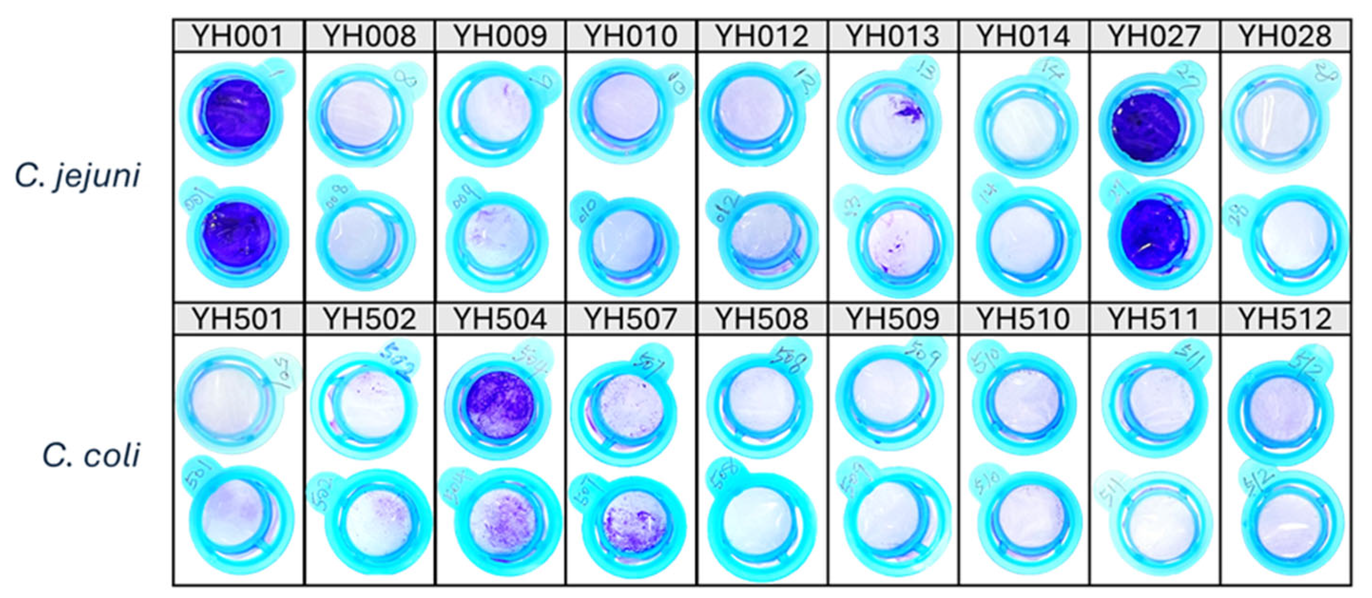
The results are shown in Figure 1. Biofilm-forming ability varied significantly among the strains: C. jejuni YH001 and YH027 produced the largest amount of biofilm biomass, as indicated by crystal violet staining, whereas C. coli YH504 and YH507 formed moderate biofilms. The remaining strains showed negligible biofilm formation under the same conditions. The stronger adhesion and aggregation observed in C. jejuni YH001 and YH027 compared to other isolates indicate that biofilm formation in Campylobacter is strain-dependent, a finding consistent with prior studies showing variability across strains and species [21].

2.2. Comparison Between Genetic Relatedness and Biofilm Formation of Campylobacter Isolates

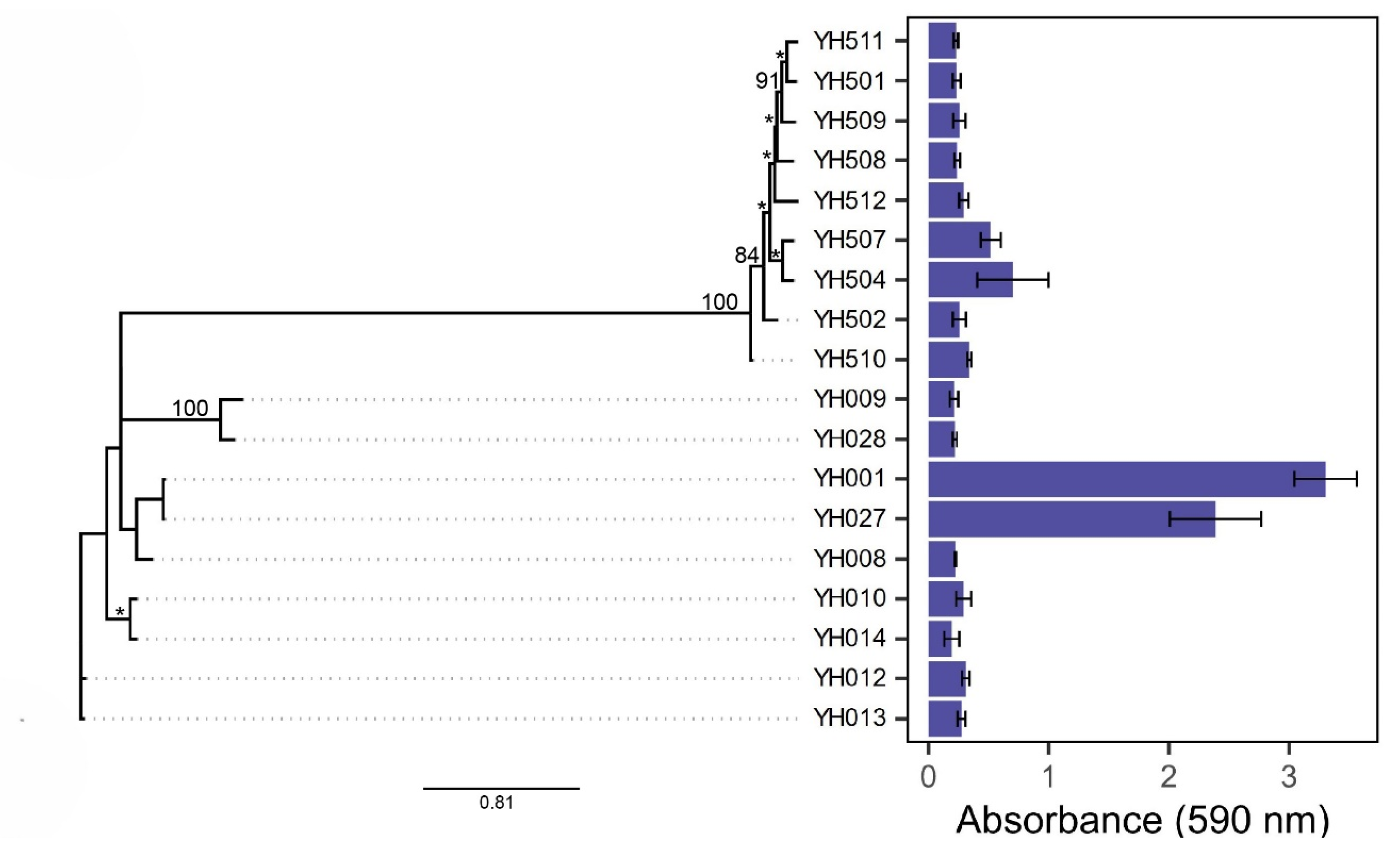
After crystal violet staining, the biofilm biomass was dislodged and quantified by measuring absorbance at 590 nm. The results confirmed that C. jejuni YH001 and YH027 were the strongest biofilm-forming strains, whereas C. coli YH504 and YH507 formed moderate biofilms among 18 Campylobacter food isolates (Figure 2, right panel).
Biofilm formation in bacteria can be influenced by genetic background, environmental adaptation, and regulatory mechanisms. To compare the genotypic traits associated with Campylobacter biofilm formation, a phylogenetic tree was constructed based on SNPs derived from whole-genome sequences of C. jejuni and C. coli isolates (Figure 2, left panel). Interestingly, C. jejuni YH001 and YH027 (the two strongest biofilm producers) were clustered together in the tree, indicating close genetic relatedness and shared genomic elements. Similarly, C. coli YH504 and YH507 (two moderate biofilm producers) were also clustered closely in the tree. These findings suggest that biofilm-forming ability may correlate with genetic similarity, supporting the observed strain-dependent variability in Campylobacter biofilm formation.

2.3. Genetic Traits Associated with Biofilm Formation and Stability

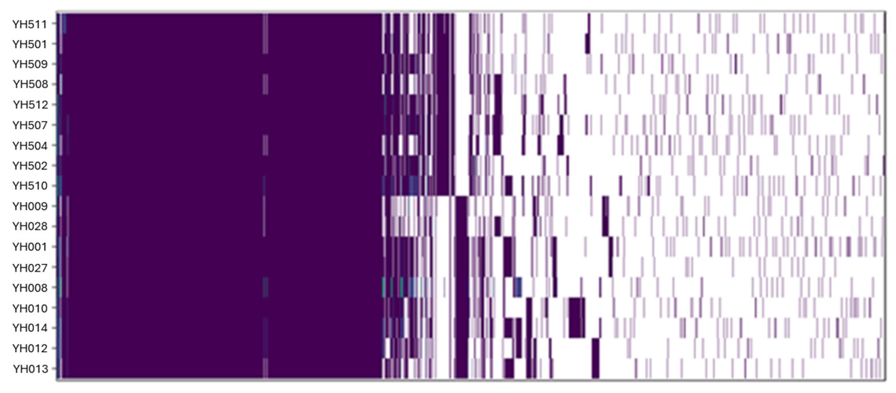
To identify core genes shared by all 18 Campylobacter isolates and variable genes unique to specific strains, a pangenome analysis was performed. The complete results are provided in Supplementary Table S1. The heatmap in Figure 3 illustrates the presence and absence of genes across C. jejuni and C. coli isolates, revealing highly diverse genetic profiles with no two strains exhibiting identical genome patterns.

2.4. Identification of Strain-Specific Genes Associated with Biofilm Formation

To identify genes potentially involved in Campylobacter biofilm formation, we searched for strain-specific genes and annotated proteins using the complete pangenome dataset (Supplementary Table S1). This analysis revealed 19 unique genes/proteins, including five hypothetical proteins, which were exclusively present in C. jejuni YH001 and YH027, and two strains characterized as strong biofilm producers (Table 1).
Additionally, two annotated genes (methyl-accepting chemotaxis signal transduction protein and cytolethal distending toxin subunit A) and five hypothetical genes were uniquely identified in C. coli YH504 and YH507, which are both moderate biofilm producers. None of these genes overlapped with those found in the strong biofilm-forming C. jejuni strains, suggesting that the moderate biofilm phenotype in C. coli may not be driven by gene content directly analogous to that associated with strong biofilm formation. Although C. jejuni and C. coli share substantial genomic homology, they remain clearly distinct species, exhibiting roughly 70–80% overlap in their core genomes and 75–85% average nucleotide identity (ANI). The observed differences in proteins associated with biofilm formation may reflect species-specific regulatory mechanisms, variations in gene expression, or functional divergence within biofilm-related pathways. The presence of multiple hypothetical genes further indicates that uncharacterized functions may contribute to biofilm formation, underscoring the need for additional functional studies to elucidate their roles.
Functional annotation suggested that the candidate genes associated with the biofilm-forming phenotype may contribute to biofilm development and stability by promoting cell adhesion, extracellular matrix production, and key regulatory signaling. In addition, we categorized the functional subsystems for the 19 unique genes associated with the observed biofilm phenotypes. Genes unique to the strong biofilm-forming strains were categorized into functional subsystems associated with virulence, disease and defense; protein, nitrogen and potassium metabolism; membrane transport; and amino acids and derivatives (Table 1).
Specifically, Dihydrolipoamide dehydrogenase (DLDH) is a central metabolic enzyme that converts pyruvate to acetyl-CoA, supporting energy production essential for bacterial survival under nutrient-limited conditions. Beyond metabolism, DLDH has been implicated in bacterial adherence, biofilm formation, structure integrity, and virulence, and has also been detected in the exopolysaccharide (EPS) matrix of Pseudomonas aeruginosa biofilms [22].
FAD-dependent NAD(P)-disulfide oxidoreductases catalyze disulfide bond formation, which stabilizes biofilm matrix proteins and enhances cohesion and resilience within the biofilm. These enzymes also participate in polysaccharide biosynthesis, contributing to the extracellular polymeric substance (EPS) that forms the structural backbone of the biofilm matrix [23,24].
DD-carboxypeptidase, a key enzyme in peptidoglycan (PG) biosynthesis, is essential for maintaining cell wall structure, surface attachment, and overall biofilm stability. Defects in PG synthesis impair cell–cell interactions and biofilm formation [25,26]. Studies in C. jejuni demonstrated that DD-carboxypeptidase mutations lead to defective PG assembly and diminished biofilm development [27].
DNA-binding protein Roi is important in structuring the biofilm matrix, which is formed largely by extracellular DNA (eDNA) together with exopolysaccharides and other components. These DNA-binding proteins act as scaffolds that cross-link eDNA to other matrix components, thereby supporting biofilm formation and maintaining matrix integrity [28].
NiFe hydrogenases, frequently detected in diverse biofilm communities, participate in hydrogen metabolism and energy conversion pathways that support microbial survival within biofilm environments [29].
L-Proline/Glycine betaine transporter (ProP) facilitates the uptake of compatible solutes necessary for osmotic balance, surface attachment, and EPS synthesis, all of which are important for biofilm formation [30].
YraQ family membrane proteins promote cell adhesion and contribute to EPS matrix production, playing a role during the early stages of biofilm establishment and structural stabilization [31].
Alpha-ketoglutarate permease (KgtP), which transports α-ketoglutarate for central carbon and nitrogen metabolism, may enhance bacterial fitness under nutrient-limited conditions typical of biofilms, thereby influencing biofilm growth and architecture [32].
Efflux proteins also play multifaceted roles in biofilm biology by exporting EPS components, quorum-sensing molecules, and other factors involved in the adhesion, aggregation, and transcriptional regulation of biofilm formation [33,34].
Ammonium transporters facilitate ammonium uptake and waste removal, both of which are critical for biofilm physiology and can influence biofilm structure and microbial survival [35].
Sodium-dependent phosphate transporters, including the PstS subunit, support phosphate acquisition and have been implicated in shaping biofilm structure and promoting biofilm formation [36].
Multi-antimicrobial extrusion proteins (MATEs) further enhance biofilm resilience by exporting antimicrobial compounds, contributing to the high tolerance commonly observed in biofilm-embedded cells [33,37].
The potassium-transporting ATPase A chain maintains intracellular potassium homeostasis, supporting membrane potential, pH regulation, and growth, which are essential functions for biofilm development and maintenance [38].
Cytochrome c family proteins are essential for electron transfer, redox balancing, and energy production in bacteria, processes critical for sustaining biofilm metabolic activity [39].
These results align with the phenotypic observations and highlight key biological processes that may underlie variation in biofilm development among isolates. Together, these functional attributes suggest roles for these proteins in supporting Campylobacter biofilm matrix production, structural integrity, and stress resilience. Future studies should focus on examining biofilm formation under diverse environmental conditions and across multiple time points, experimentally validating the candidate genes and proteins identified here, characterizing the regulatory networks that govern their activity, and exploring targeted biofilm disruption strategies to extend these findings and help mitigate Campylobacter persistence in food production environments. Furthermore, given the close association between biofilm formation and enhanced antimicrobial tolerance, additional research is needed to assess how these mechanisms contribute to antimicrobial resistance and their broader implications for public health.

3. Materials and Methods

3.1. Sample Preparation

C. jejuni and C. coli strains were isolated from retail meat, including chicken meat, chicken livers, and beef livers, collected between 2011 and 2023 using previously described methods [40]. Briefly, 450 g of meat was combined with 250 mL buffered peptone water (BPW) and homogenized using a stomacher. The homogenate was centrifuged and the pellet was enriched in Bolton broth supplemented with horse blood and selective antibiotics (cefoperazone, trimethoprim, vancomycin, and cycloheximide) at 42 °C for 24 h under microaerobic conditions (5% O2, 10% CO2, and 85% N2) using a CampyPak (Becton, Dickinson and Company, Franklin Lakes, NJ, USA) in an airtight jar. Following enrichment, passive filtration onto Brucella agar was employed for strain isolation based on the highly motile nature of Campylobacter. Colonies were re-streaked twice for strain purification, and genus and species identification was performed using a multiplex qPCR assay previously developed for differentiating C. jejuni and C. coli [41].

3.2. Biofilm Formation

Campylobacter isolates were streaked from −80 °C frozen stocks onto Mueller–Hinton (MH) agar plates and incubated overnight under microaerobic conditions (5% O2, 10% CO2, 85% N2) at 42 °C. Fresh colonies were scraped from the agar plates and resuspended in 2 mL of MH broth, followed by overnight incubation under the same conditions. Subsequently, 50 μL of the overnight culture was inoculated into 5 mL of MH broth and homogenized. Two aliquots of 2 mL of the diluted culture were transferred into 10 cm2/10 mL polystyrene tissue culture tubes with a flat surface and vent cap (Techno Plastic Products AG, Trasadingen, Canton Schaffhausen, Switzerland) and incubated horizontally for 5 days to allow biofilm development. A 5-day static incubation period was selected for the biofilm development due to the growth variation observed among the Campylobacter isolates used in our study. This extended incubation ensures that both fast- and slow-growing strains are able to reach stable biofilm development, thereby allowing consistent and comparable measurements across isolates [15,42]. All incubations were performed under microaerobic conditions at 42 °C.

3.3. Biofilm Quantification

Biofilm cultures were filtered onto a 40 μm cell strainer fitted on a 50 mL conical tube. The strainer containing aggregated cells was rinsed with 2 mL of fresh MH broth, transferred to a 6-well plate and then submerged in 6 mL 0.1% crystal violet solution. After a 30 min incubation at room temperature, the strainers and aggregates were rinsed three times with 5 mL of sterile water and photographed in a light box. The strainer was then placed in a fresh 6-well plate and submerged in 6 mL of 100% ethanol. After 30 min of incubation at room temperature with gentle shaking, a pipette was used to dislodge aggregates from the strainer surface and mix the ethanol. The absorbance of the ethanol solution (200 μL in each well, in duplicate) was measured at 590 nm using a Cytation 5 plate reader (BioTek/Agilent Life Sciences, Winooski, VT, USA).

3.4. Genome Sequencing, Assembly, and Annotation

Genomic DNA was extracted using the Qiagen genomic tip 100/G kit (Qiagen, Valencia, CA, USA) and quantified with a Qubit 3.0 fluorometer (Thermo Fisher Scientific, Waltham, MA, USA) following the manufacturers’ instructions. Whole-genome sequencing was performed using Illumina MiSeq and Pacific Biosciences (PacBio) RSII and/or Sequel platforms (Illumina, San Diego, CA, USA; Pacific Biosciences, Menlo Park, CA, USA). Genome assemblies were generated from PacBio long reads using Canu v2.2 [43]. Overhangs of assembled contigs were trimmed and reoriented using Circlator v1.5.5 [44] to produce complete circularized genomes.
In a few cases where reorientation and trimming failed, Illumina MiSeq reads were used to correct sequencing errors in assembled contigs: MiSeq reads were mapped to Canu assemblies using BWA v0.7.17-r1188 [45]; errors were corrected using Pilon v1.22 [46]; and Pilon correction was repeated iteratively until no errors were detected. Finally, the assemblies were trimmed and reoriented using Circlator v1.5.5 to generate complete circular genomes.
Table 2 summarizes the source and assembled genome information for C. jejuni and C. coli isolates. For each strain, the complete genome of approximately 1.6–1.8 Mbp, consistent with the size of previously reported Campylobacter chromosomes, was annotated using the RAST server [47,48]. Chromosomal integrity was verified by confirming the presence of the start gene dnaA, three copies of rRNA operons (23S, 16S, and 5S rRNA), and minimal repeat sequences. Smaller contigs were assessed for potential plasmid sequences.

3.5. Pangenome and Phylogenetic Analysis

The Campylobacter pangenome was constructed using the KBase web server [53]. First, genome assemblies were annotated and categorized into the functional subsystems using RASTtk v1.073, then the pangenome was constructed using Ortho-MCL v0.0.8 [54]. To investigate the relatedness of different genome clusters, we constructed a phylogenetic parsimony tree from all SNPs using kSNP4 v4.1 [55] with a k-mer size of 19. Bootstrap support values were calculated with IQ-Tree v 2.1.2 [56] on the CIPRES Scientific Gateway [57]. Briefly, a maximum likelihood consensus tree was built using a General Time Reversible (GTR) model with a correction for ascertainment bias. The kSNP4 parsimony tree served as the starting tree, and a non-parametric bootstrap analysis was performed with 1000 replicates to assess branch support. Complete genome sequences (accession numbers) used for this analysis are listed in Table 2. The resulting phylogenetic tree was visualized using Iroki [58].
To determine which genes were unique or shared among the Campylobacter genomes, we constructed a pangenome using OrthoMCL v.2.0, based on RASTtk annotations generated through the KBase Server. Heatmaps depicting gene presence/absence and the number of shared genes among the genomes were generated in R v4.4.0 (R Core Team 2024) using the ggplot2 and viridis packages [59].

4. Conclusions

This study demonstrates the strain-specific nature of biofilm formation in Campylobacter spp., identifying two strong biofilm-forming strains among 18 food isolates. By integrating whole-genome sequencing with comparative genomic analysis of biofilm-forming and non-forming strains, we uncovered key genes and pathways associated with strong biofilm formation, providing new insight into the potential genetic determinants underlying this phenotype. Phylogenetic and pangenomic analyses provided valuable insights into the genetic basis of biofilm formation and highlighted the strain-specific ability to utilize this mechanism for survival in challenging conditions, particularly those encountered during food processing and storage. Importantly, the identification of proteins linked to both biofilm formation and pathogenesis in C. jejuni strains YH001 and YH027 offers promising molecular targets for therapeutic approaches and intervention strategies aimed at disrupting biofilms. Collectively, these findings enhance our understanding of Campylobacter persistence in food environments and highlight molecular targets that may inform the development of innovative intervention strategies to reduce contamination and improve food safety.

Supplementary Materials

The following supporting information can be downloaded at: https://www.mdpi.com/article/10.3390/ijms27062543/s1.

Author Contributions

Y.H., G.D., C.-Y.C. and J.C.: designed and performed experiments, analyzed data, interpreted results, and prepared manuscript. H.K.: conducted experiments, analyzed results, and edited manuscript. All authors have read and agreed to the published version of the manuscript.

Funding

This research was supported by the U.S. Department of Agriculture, Agricultural Research Service (USDA-ARS), National Program 108, Current Research Information System numbers 8072-42000-093 and 8072-42000-094 and used resources provided by the SCINet project and/or the AI Center of Excellence of USDA-ARS, project numbers 0201-88888-003-000D and 0201-88888-002-000D.

Institutional Review Board Statement

Not applicable.

Informed Consent Statement

Not applicable.

Data Availability Statement

All the assembled genome sequences of Campylobacter isolates were deposited and are available in GenBank, NCBI under the accession numbers listed in Table 2.

Acknowledgments

The mention of trade names or commercial products in this publication is solely for the purpose of providing specific information and does not imply recommendation or endorsement by the USDA. All opinions expressed are the authors’ and do not necessarily reflect the policies or views of the USDA. The USDA is an equal opportunity provider and employer.

Conflicts of Interest

The authors declare no conflicts of interest.

References

  1. Centers for Disease Control and Prevention. About Campylobacter Infection. Available online: https://www.cdc.gov/campylobacter/about/index.html (accessed on 28 January 2026).
  2. Tikhomirova, A.; McNabb, E.R.; Petterlin, L.; Bellamy, G.L.; Lin, K.H.; Santoso, C.A.; Daye, E.S.; Alhaddad, F.M.; Lee, K.P.; Roujeinikova, A. Campylobacter jejuni virulence factors: Update on emerging issues and trends. J. Biomed. Sci. 2024, 31, 45. [Google Scholar] [CrossRef]
  3. World Health Organization (WHO) Fact Sheet on Campylobacter. Available online: https://www.who.int/news-room/fact-sheets/detail/campylobacter (accessed on 28 January 2026).
  4. Veronese, P.; Dodi, I. Campylobacter jejuni/coli Infection: Is It Still a Concern? Microorganisms 2024, 12, 2669. [Google Scholar] [CrossRef] [PubMed]
  5. Karygianni, L.; Ren, Z.; Koo, H.; Thurnheer, T. Biofilm Matrixome: Extracellular Components in Structured Microbial Communities. Trends Microbiol. 2020, 28, 668–681. [Google Scholar] [CrossRef] [PubMed]
  6. Ryder, C.; Byrd, M.; Wozniak, D.J. Role of polysaccharides in Pseudomonas aeruginosa biofilm development. Curr. Opin. Microbiol. 2007, 10, 644–648. [Google Scholar] [CrossRef] [PubMed]
  7. Lasa, I.; Penades, J.R. Bap: A family of surface proteins involved in biofilm formation. Res. Microbiol. 2006, 157, 99–107. [Google Scholar] [CrossRef]
  8. Fong, J.N.C.; Yildiz, F.H. Biofilm Matrix Proteins. Microbiol. Spectr. 2015, 3, 201–222. [Google Scholar] [CrossRef]
  9. Panlilio, H.; Rice, C.V. The role of extracellular DNA in the formation, architecture, stability, and treatment of bacterial biofilms. Biotechnol. Bioeng. 2021, 118, 2129–2141. [Google Scholar] [CrossRef]
  10. Bai, X.; Nakatsu, C.H.; Bhunia, A.K. Bacterial Biofilms and Their Implications in Pathogenesis and Food Safety. Foods 2021, 10, 2117. [Google Scholar] [CrossRef]
  11. Olsen, I. Biofilm-specific antibiotic tolerance and resistance. Eur. J. Clin. Microbiol. Infect. Dis. 2015, 34, 877–886. [Google Scholar] [CrossRef]
  12. Buswell, C.M.; Herlihy, Y.M.; Lawrence, L.M.; McGuiggan, J.T.; Marsh, P.D.; Keevil, C.W.; Leach, S.A. Extended survival and persistence of Campylobacter spp. in water and aquatic biofilms and their detection by immunofluorescent-antibody and -rRNA staining. Appl. Environ. Microbiol. 1998, 64, 733–741. [Google Scholar] [CrossRef]
  13. Teh, A.H.; Lee, S.M.; Dykes, G.A. Does Campylobacter jejuni form biofilms in food-related environments? Appl. Environ. Microbiol. 2014, 80, 5154–5160. [Google Scholar] [CrossRef] [PubMed]
  14. Reeser, R.J.; Medler, R.T.; Billington, S.J.; Jost, B.H.; Joens, L.A. Characterization of Campylobacter jejuni biofilms under defined growth conditions. Appl. Environ. Microbiol. 2007, 73, 1908–1913. [Google Scholar] [CrossRef] [PubMed]
  15. Ica, T.; Caner, V.; Istanbullu, O.; Nguyen, H.D.; Ahmed, B.; Call, D.R.; Beyenal, H. Characterization of mono- and mixed-culture Campylobacter jejuni biofilms. Appl. Environ. Microbiol. 2012, 78, 1033–1038. [Google Scholar] [CrossRef] [PubMed]
  16. Melo, R.T.; Mendonca, E.P.; Monteiro, G.P.; Siqueira, M.C.; Pereira, C.B.; Peres, P.; Fernandez, H.; Rossi, D.A. Intrinsic and Extrinsic Aspects on Campylobacter jejuni Biofilms. Front. Microbiol. 2017, 8, 1332. [Google Scholar] [CrossRef]
  17. Silha, D.; Sirotkova, S.; Svarcova, K.; Hofmeisterova, L.; Korycanova, K.; Silhova, L. Biofilm Formation Ability of Arcobacter-like and Campylobacter Strains under Different Conditions and on Food Processing Materials. Microorganisms 2021, 9, 2017. [Google Scholar] [CrossRef]
  18. Puning, C.; Su, Y.; Lu, X.; Golz, G. Molecular Mechanisms of Campylobacter Biofilm Formation and Quorum Sensing. Curr. Top. Microbiol. Immunol. 2021, 431, 293–319. [Google Scholar] [CrossRef]
  19. Korkus, J.; Salata, P.; Thompson, S.A.; Paluch, E.; Bania, J.; Walecka-Zacharska, E. The role of cydB gene in the biofilm formation by Campylobacter jejuni. Sci. Rep. 2024, 14, 26574. [Google Scholar] [CrossRef]
  20. Svensson, S.L.; Pryjma, M.; Gaynor, E.C. Flagella-mediated adhesion and extracellular DNA release contribute to biofilm formation and stress tolerance of Campylobacter jejuni. PLoS ONE 2014, 9, e106063. [Google Scholar] [CrossRef]
  21. Sulaeman, S.; Le Bihan, G.; Rossero, A.; Federighi, M.; De, E.; Tresse, O. Comparison between the biofilm initiation of Campylobacter jejuni and Campylobacter coli strains to an inert surface using BioFilm Ring Test. J. Appl. Microbiol. 2010, 108, 1303–1312. [Google Scholar] [CrossRef]
  22. Sauer, K.; Camper, A.K.; Ehrlich, G.D.; Costerton, J.W.; Davies, D.G. Pseudomonas aeruginosa displays multiple phenotypes during development as a biofilm. J. Bacteriol. 2002, 184, 1140–1154. [Google Scholar] [CrossRef]
  23. Flemming, H.C.; van Hullebusch, E.D.; Little, B.J.; Neu, T.R.; Nielsen, P.H.; Seviour, T.; Stoodley, P.; Wingender, J.; Wuertz, S. Microbial extracellular polymeric substances in the environment, technology and medicine. Nat. Rev. Microbiol. 2025, 23, 87–105. [Google Scholar] [CrossRef]
  24. Selles Vidal, L.; Kelly, C.L.; Mordaka, P.M.; Heap, J.T. Review of NAD(P)H-dependent oxidoreductases: Properties, engineering and application. Biochim. Biophys. Acta Proteins Proteom. 2018, 1866, 327–347. [Google Scholar] [CrossRef] [PubMed]
  25. Pal, S.; Jain, D.; Biswal, S.; Rastogi, S.K.; Kumar, G.; Ghosh, A.S. The physiological role of Acinetobacter baumannii DacC is exerted through influencing cell shape, biofilm formation, the fitness of survival, and manifesting DD-carboxypeptidase and beta-lactamase dual-enzyme activities. FEMS Microbiol. Lett. 2024, 371, fnae079. [Google Scholar] [CrossRef] [PubMed]
  26. Peters, K.; Kannan, S.; Rao, V.A.; Biboy, J.; Vollmer, D.; Erickson, S.W.; Lewis, R.J.; Young, K.D.; Vollmer, W. The Redundancy of Peptidoglycan Carboxypeptidases Ensures Robust Cell Shape Maintenance in Escherichia coli. mBio 2016, 7, e00819-16. [Google Scholar] [CrossRef] [PubMed]
  27. Iwata, T.; Watanabe, A.; Kusumoto, M.; Akiba, M. Peptidoglycan Acetylation of Campylobacter jejuni Is Essential for Maintaining Cell Wall Integrity and Colonization in Chicken Intestines. Appl. Environ. Microbiol. 2016, 82, 6284–6290. [Google Scholar] [CrossRef]
  28. Das, T.; Sehar, S.; Manefield, M. The roles of extracellular DNA in the structural integrity of extracellular polymeric substance and bacterial biofilm development. Environ. Microbiol. Rep. 2013, 5, 778–786. [Google Scholar] [CrossRef]
  29. Greening, C.; Biswas, A.; Carere, C.R.; Jackson, C.J.; Taylor, M.C.; Stott, M.B.; Cook, G.M.; Morales, S.E. Genomic and metagenomic surveys of hydrogenase distribution indicate H2 is a widely utilised energy source for microbial growth and survival. ISME J. 2016, 10, 761–777. [Google Scholar] [CrossRef]
  30. Kapfhammer, D.; Karatan, E.; Pflughoeft, K.J.; Watnick, P.I. Role for glycine betaine transport in Vibrio cholerae osmoadaptation and biofilm formation within microbial communities. Appl. Environ. Microbiol. 2005, 71, 3840–3847. [Google Scholar] [CrossRef]
  31. Typas, A.; Banzhaf, M.; Gross, C.A.; Vollmer, W. From the regulation of peptidoglycan synthesis to bacterial growth and morphology. Nat. Rev. Microbiol. 2011, 10, 123–136. [Google Scholar] [CrossRef]
  32. Doucette, C.D.; Schwab, D.J.; Wingreen, N.S.; Rabinowitz, J.D. α-Ketoglutarate coordinates carbon and nitrogen utilization via enzyme I inhibition. Nat. Chem. Biol. 2011, 7, 894–901. [Google Scholar] [CrossRef]
  33. Piddock, L.J. Multidrug-resistance efflux pumps—Not just for resistance. Nat. Rev. Microbiol. 2006, 4, 629–636. [Google Scholar] [CrossRef] [PubMed]
  34. Zhang, L.; Mah, T.F. Involvement of a novel efflux system in biofilm-specific resistance to antibiotics. J. Bacteriol. 2008, 190, 4447–4452. [Google Scholar] [CrossRef] [PubMed]
  35. Ardin, A.C.; Fujita, K.; Nagayama, K.; Takashima, Y.; Nomura, R.; Nakano, K.; Ooshima, T.; Matsumoto-Nakano, M. Identification and functional analysis of an ammonium transporter in Streptococcus mutans. PLoS ONE 2014, 9, e107569. [Google Scholar] [CrossRef]
  36. Neznansky, A.; Blus-Kadosh, I.; Yerushalmi, G.; Banin, E.; Opatowsky, Y. The Pseudomonas aeruginosa phosphate transport protein PstS plays a phosphate-independent role in biofilm formation. FASEB J. 2014, 28, 5223–5233. [Google Scholar] [CrossRef] [PubMed]
  37. Alav, I.; Sutton, J.M.; Rahman, K.M. Role of bacterial efflux pumps in biofilm formation. J. Antimicrob. Chemother. 2018, 73, 2003–2020. [Google Scholar] [CrossRef]
  38. Epstein, W. The roles and regulation of potassium in bacteria. Prog. Nucleic Acid. Res. Mol. Biol. 2003, 75, 293–320. [Google Scholar] [CrossRef]
  39. Gray, H.B.; Winkler, J.R. Electron flow through metalloproteins. Biochim. Biophys. Acta 2010, 1797, 1563–1572. [Google Scholar] [CrossRef]
  40. He, Y.; Capobianco, J.; Armstrong, C.M.; Chen, C.Y.; Counihan, K.; Lee, J.; Reed, S.; Tilman, S. Detection and Isolation of Campylobacter spp. from Raw Meat. J. Vis. Exp. 2024, 23, e66462. [Google Scholar] [CrossRef]
  41. He, Y.P.; Yao, X.M.; Gunther, N.W.; Xie, Y.P.; Tu, S.I.; Shi, X.M. Simultaneous Detection and Differentiation of Campylobacter jejuni, C. coli, and C. lari in Chickens Using a Multiplex Real-Time PCR Assay. Food Anal. Method. 2010, 3, 321–329. [Google Scholar] [CrossRef]
  42. Gunther, N.W.; Nunez, A.; Bagi, L.; Abdul-Wakeel, A.; Ream, A.; Liu, Y.; Uhlich, G. Butyrate decreases Campylobacter jejuni motility and biofilm partially through influence on LysR expression. Food Microbiol. 2023, 115, 104310. [Google Scholar] [CrossRef]
  43. Koren, S.; Walenz, B.P.; Berlin, K.; Miller, J.R.; Bergman, N.H.; Phillippy, A.M. Canu: Scalable and accurate long-read assembly via adaptive k-mer weighting and repeat separation. Genome Res. 2017, 27, 722–736. [Google Scholar] [CrossRef]
  44. Hunt, M.; Silva, N.D.; Otto, T.D.; Parkhill, J.; Keane, J.A.; Harris, S.R. Circlator: Automated circularization of genome assemblies using long sequencing reads. Genome Biol. 2015, 16, 294. [Google Scholar] [CrossRef] [PubMed]
  45. Li, H.; Durbin, R. Fast and accurate short read alignment with Burrows-Wheeler transform. Bioinformatics 2009, 25, 1754–1760. [Google Scholar] [CrossRef] [PubMed]
  46. Walker, B.J.; Abeel, T.; Shea, T.; Priest, M.; Abouelliel, A.; Sakthikumar, S.; Cuomo, C.A.; Zeng, Q.; Wortman, J.; Young, S.K.; et al. Pilon: An integrated tool for comprehensive microbial variant detection and genome assembly improvement. PLoS ONE 2014, 9, e112963. [Google Scholar] [CrossRef] [PubMed]
  47. Aziz, R.K.; Bartels, D.; Best, A.A.; DeJongh, M.; Disz, T.; Edwards, R.A.; Formsma, K.; Gerdes, S.; Glass, E.M.; Kubal, M.; et al. The RAST Server: Rapid annotations using subsystems technology. BMC Genom. 2008, 9, 75. [Google Scholar] [CrossRef]
  48. Overbeek, R.; Olson, R.; Pusch, G.D.; Olsen, G.J.; Davis, J.J.; Disz, T.; Edwards, R.A.; Gerdes, S.; Parrello, B.; Shukla, M.; et al. The SEED and the Rapid Annotation of microbial genomes using Subsystems Technology (RAST). Nucleic Acids Res. 2014, 42, D206–D214. [Google Scholar] [CrossRef]
  49. He, Y.; Yan, X.; Reed, S.; Xie, Y.; Chen, C.Y.; Irwin, P. Complete Genome Sequence of Campylobacter jejuni YH001 from Beef Liver, Which Contains a Novel Plasmid. Genome Announc. 2015, 3, e01492-14. [Google Scholar] [CrossRef]
  50. He, Y.; Kanrar, S.; Reed, S.; Lee, J.; Capobianco, J. Whole Genome Sequences, De Novo Assembly, and Annotation of Antibiotic Resistant Campylobacter jejuni Strains S27, S33, and S36 Newly Isolated from Chicken Meat. Microorganisms 2024, 12, 159. [Google Scholar] [CrossRef]
  51. He, Y.; Reed, S.; Yan, X.; Zhang, D.; Strobaugh, T.; Capobianco, J.; Gehring, A. Complete Genome Sequences of Multidrug-Resistant Campylobacter coli Strains YH501, YH503, and YH504, from Retail Chicken. Microbiol. Resour. Announc. 2022, 11, e0023722. [Google Scholar] [CrossRef]
  52. Ghatak, S.; He, Y.; Reed, S.; Strobaugh, T., Jr.; Irwin, P. Whole genome sequencing and analysis of Campylobacter coli YH502 from retail chicken reveals a plasmid-borne type VI secretion system. Genom. Data 2017, 11, 128–131. [Google Scholar] [CrossRef]
  53. Arkin, A.P.; Cottingham, R.W.; Henry, C.S.; Harris, N.L.; Stevens, R.L.; Maslov, S.; Dehal, P.; Ware, D.; Perez, F.; Canon, S.; et al. KBase: The United States Department of Energy Systems Biology Knowledgebase. Nat. Biotechnol. 2018, 36, 566–569. [Google Scholar] [CrossRef]
  54. Li, L.; Stoeckert, C.J., Jr.; Roos, D.S. OrthoMCL: Identification of ortholog groups for eukaryotic genomes. Genome Res. 2003, 13, 2178–2189. [Google Scholar] [CrossRef]
  55. Hall, B.G.; Nisbet, J. Building Phylogenetic Trees from Genome Sequences With kSNP4. Mol. Biol. Evol. 2023, 40, msad235. [Google Scholar] [CrossRef]
  56. Nguyen, L.T.; Schmidt, H.A.; von Haeseler, A.; Minh, B.Q. IQ-TREE: A fast and effective stochastic algorithm for estimating maximum-likelihood phylogenies. Mol. Biol. Evol. 2015, 32, 268–274. [Google Scholar] [CrossRef]
  57. Miller, M.A.; Pfeiffer, W.; Schwartz, T. Creating the CIPRES Science Gateway for inference of large phylogenetic trees. In Proceedings of the Gateway Computing Environments Workshop (GCE), New Orleans, LA, USA, 14 November 2010; IEEE: New York, NY, USA, 2010; pp. 1–8. [Google Scholar]
  58. Moore, R.M.; Harrison, A.O.; McAllister, S.M.; Polson, S.W.; Wommack, K.E. Iroki: Automatic customization and visualization of phylogenetic trees. PeerJ 2020, 8, e8584. [Google Scholar] [CrossRef]
  59. Ginestet, C. ggplot2: Elegant Graphics for Data Analysis. J. R. Stat. Soc. A 2011, 174, 245. [Google Scholar] [CrossRef]
Figure 1. Biofilms of C. jejuni (upper panel) and C. coli (low panel) strains stained with crystal violet. Biofilms were developed in duplicate samples under microaerobic conditions at 42 °C for five days and visualized after staining.
Ijms 27 02543 g001
Figure 2. SNP-based phylogenetic tree and biofilm quantification of C. jejuni and C. coli isolates. Left panel: SNP-based phylogenetic tree constructed from whole-genome sequences of C. jejuni (YH001–YH028) and C. coli (YH501–YH512) isolates. Bootstrap values on branches correspond with maximum likelihood consensus tree. * indicates a bootstrap value of 100. Right panel: Biofilm formation quantified by crystal violet staining after 5 days of growth in Mueller–Hinton broth under microaerobic conditions at 42 °C without shaking. Absorbance at 590 nm represents the average of two independent growing biofilm samples, each assayed in duplicate. Error bars represent the standard deviation, with significance defined as p < 0.05.
Ijms 27 02543 g002
Figure 3. Pangenome analysis of C. jejuni and C. coli isolates. The heatmap shows the presence (purple) and absence (white) of genes or gene clusters across C. jejuni and C. coli genomes. The x-axis represents gene clusters, but their position does not correspond to chromosomal location. This visualization highlights the genetic diversity among isolates.
Ijms 27 02543 g003
Table 1. Annotated proteins uniquely present in C. jejuni strains YH001 and YH027.
Annotated ProteinSubsystem CategoryYH001YH027Other Strains
Putative Dihydrolipoamide dehydrogenase (EC 1.8.1.4); Mercuric ion reductase (EC 1.16.1.1); PF00070 family, FAD-dependent NAD(P)-disulphide oxidoreductaseVirulence, Disease and Defense110
D-alanyl-D-alanine carboxypeptidase (EC 3.4.16.4)Protein Metabolism110
FIG00471123: hypothetical proteinNone110
DNA-binding protein RoiNone110
FIG00470265: hypothetical proteinNone110
FIG00471635: hypothetical proteinNone110
FIG00470314: hypothetical proteinNone110
Hydrogenase, (NiFe)/(NiFeSe) small subunit familyNone110
L-Proline/Glycine betaine transporter ProPAmino Acids and Derivatives110
Uncharacterized membrane protein, YraQ familyNone110
Alpha-ketoglutarate permeaseNone110
Putative efflux proteinNone110
Ammonium transporterNitrogen Metabolism110
C4-dicarboxylate transporterNone110
Sodium-dependent phosphate transporterMembrane Transport110
Multi antimicrobial extrusion protein (Na(+)/drug antiporter), MATE family of MDR efflux pumpsVirulence, Disease and Defense110
Hypothetical protein Cj0566None110
Potassium-transporting ATPase A chain (EC 3.6.3.12) (TC 3.A.3.7.1)Potassium Metabolism110
Cytochrome c family proteinNone110
Protein names and subsystem categories were assigned based on annotation using the RAST server. A designation of ‘None’ in the Subsystem Category indicates that the protein’s functional subsystem could not be identified by the RAST annotation. The values “1” and “0” indicated the presence and absence of the genes in the isolates, respectively.
Table 2. Sources and genome information of C. jejuni and C. coli isolates.
Strain and SpeciesSourceGenome Size (bp)%GCAccession No.Reference
C. jejuni YH001Veal livers1,712,36130.5CP010058[49]
C. jejuni YH008Drumsticks1,792,42430.5CP172380This work
C. jejuni (S27Cj) YH009Chicken thighs1,663,22630.5CP131444[50]
C. jejuni (S33Cj) YH010Chicken thighs1,748,76130.5CP131442[50]
C. jejuni YH012Chicken livers1,698,96330.5CP172815This work
C. jejuni YH013Chicken livers1,691,84830.5CP172379This work
C. jejuni YH014Chicken livers1,802,03930.5CP172376This work
C. jejuni YH027Calf livers1,710,95930.5CP172352This work
C. jejuni YH028Beef livers1,667,69830.5CP172351This work
C. coli YH501Drumsticks1,668,52331.5CP015528[51]
C. coli YH502Drumsticks1,718,97431.0CP018900[52]
C. coli YH504Drumsticks1,722,14331.0CP091644[51]
C. coli YH507Chicken livers1,756,09631.0CP172392This work
C. coli YH508Chicken thighs1,703,74031.5CP172391This work
C. coli YH509Chicken livers1,697,11331.5CP172390This work
C. coli YH510Chicken livers1,812,35631.0CP172387This work
C. coli YH511Chicken livers1,674,28831.5CP172385This work
C. coli YH512Chicken livers1,754,13531.5CP172384This work
Disclaimer/Publisher’s Note: The statements, opinions and data contained in all publications are solely those of the individual author(s) and contributor(s) and not of MDPI and/or the editor(s). MDPI and/or the editor(s) disclaim responsibility for any injury to people or property resulting from any ideas, methods, instructions or products referred to in the content.