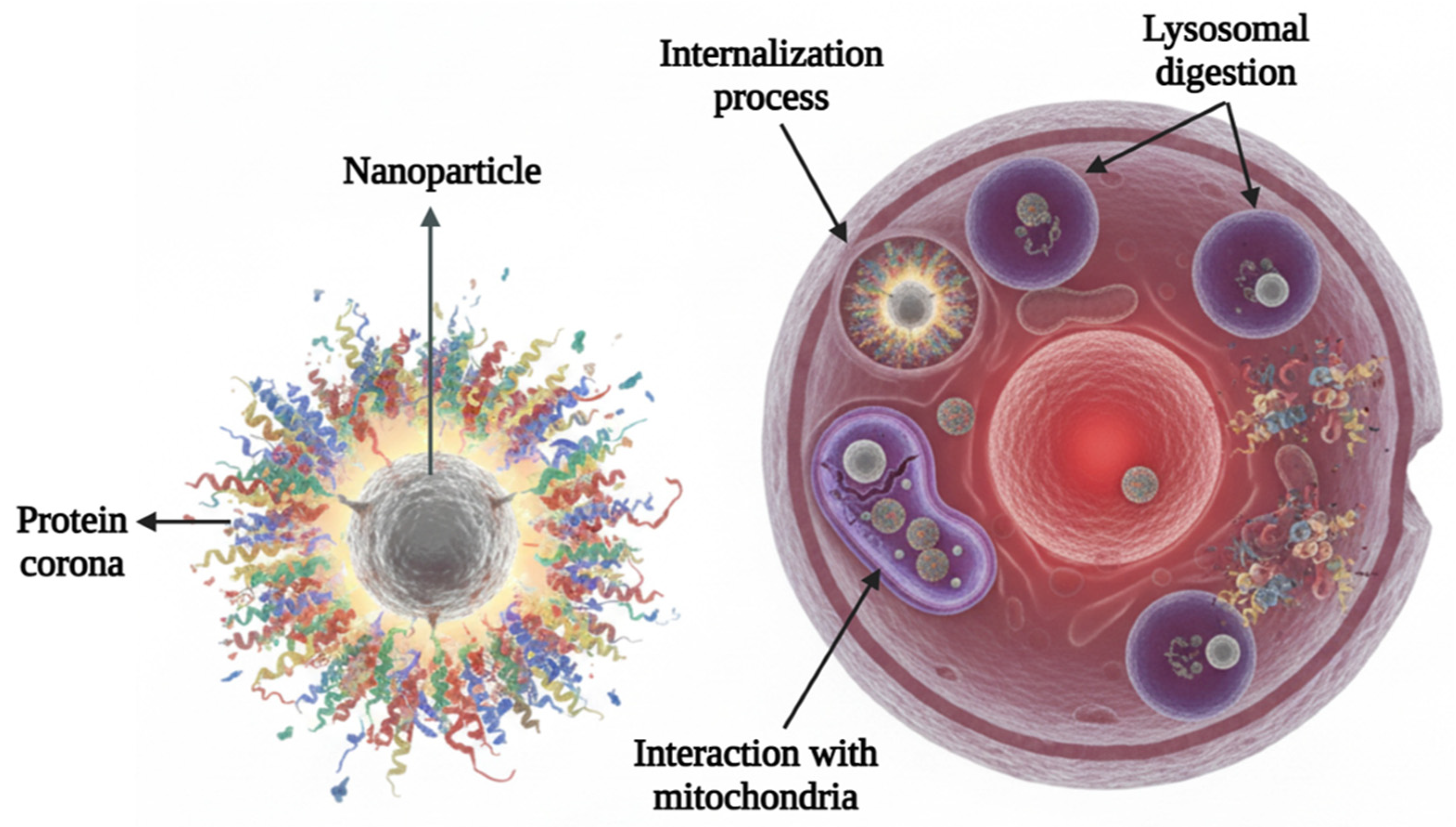
Layers of Hope: How Graphene and Nanostructures Hold Promise for Cancer Therapy
Abstract
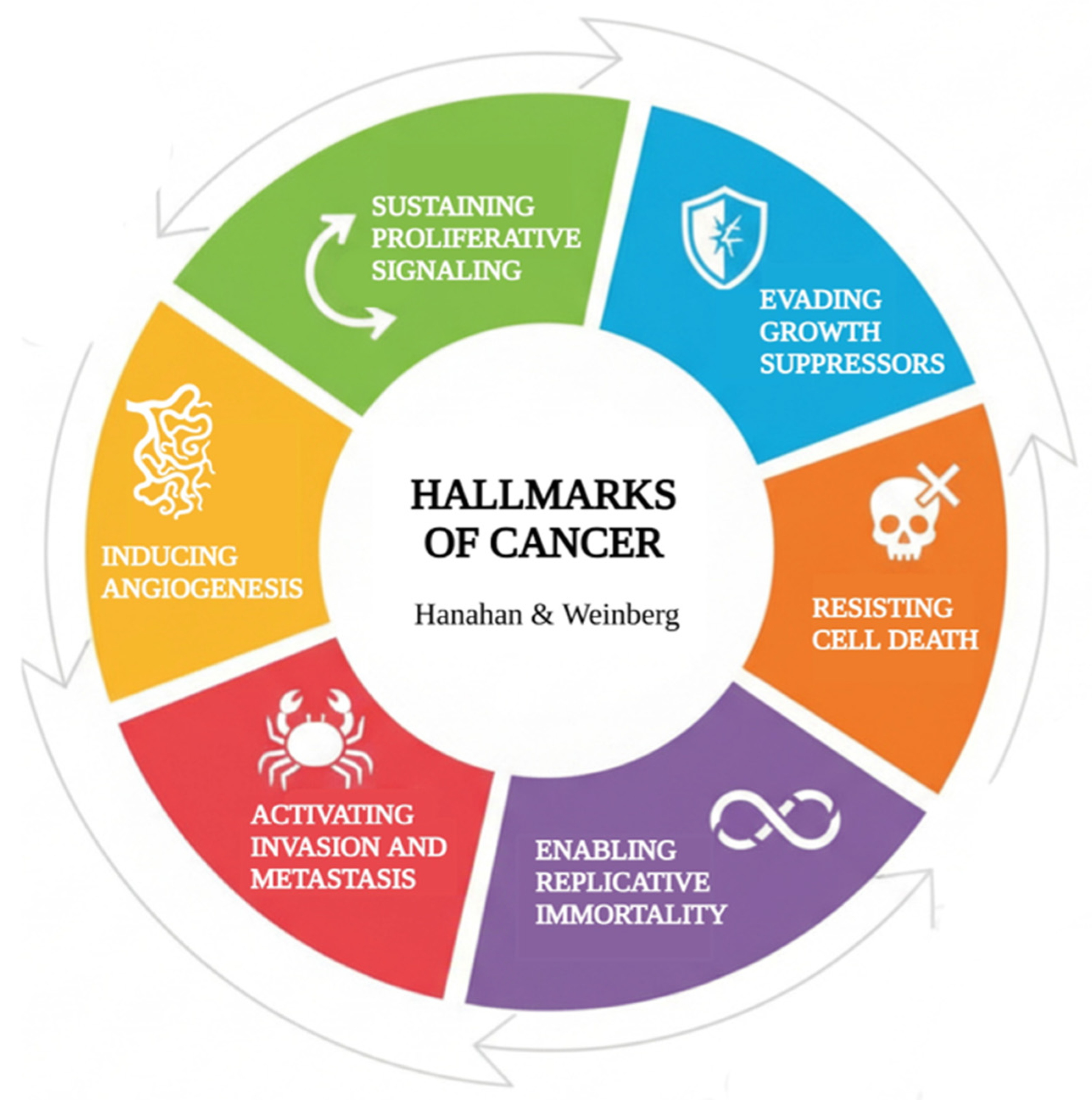
1. The Emperor of All Maladies
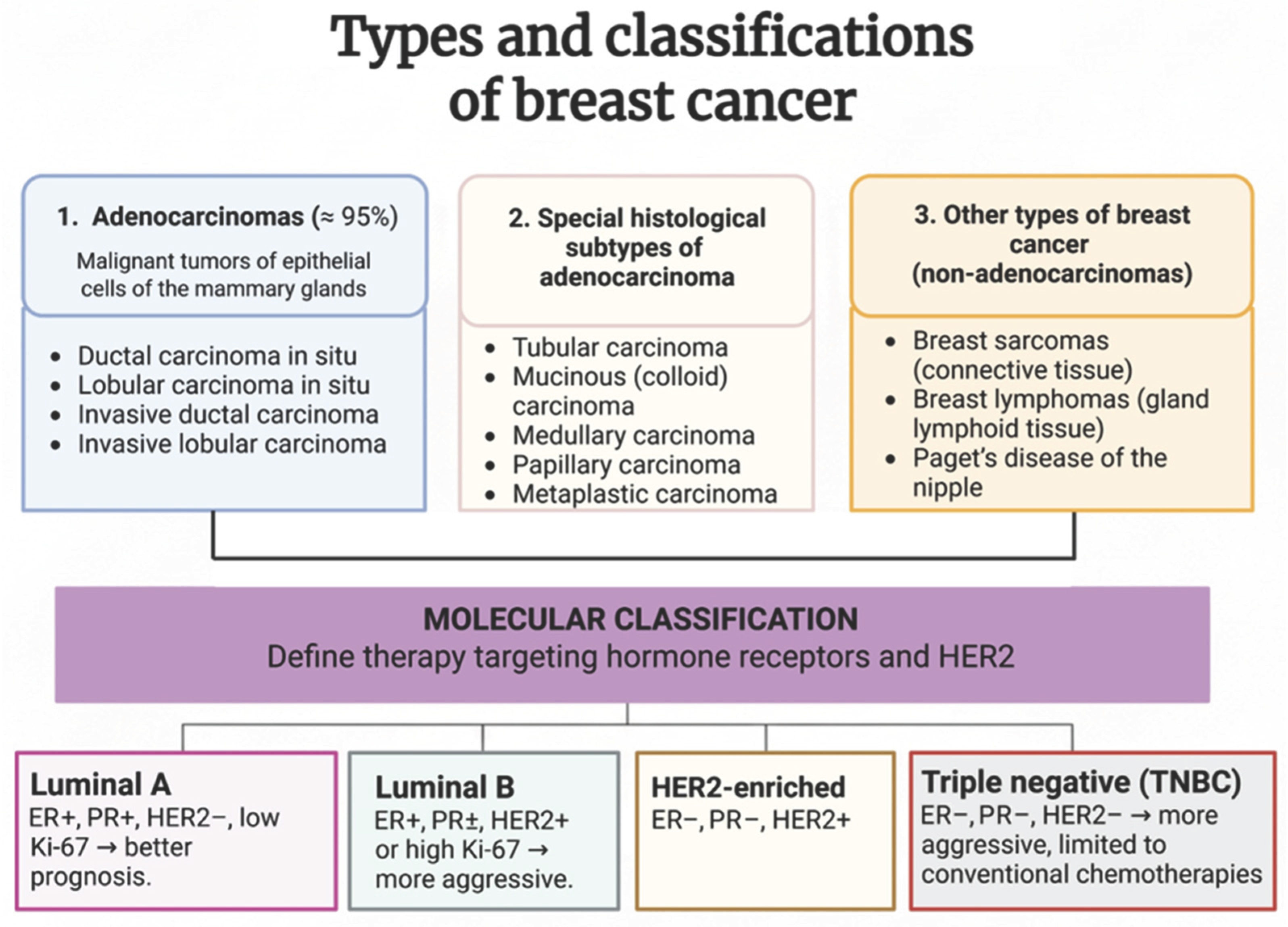
1.1. Breast Cancer
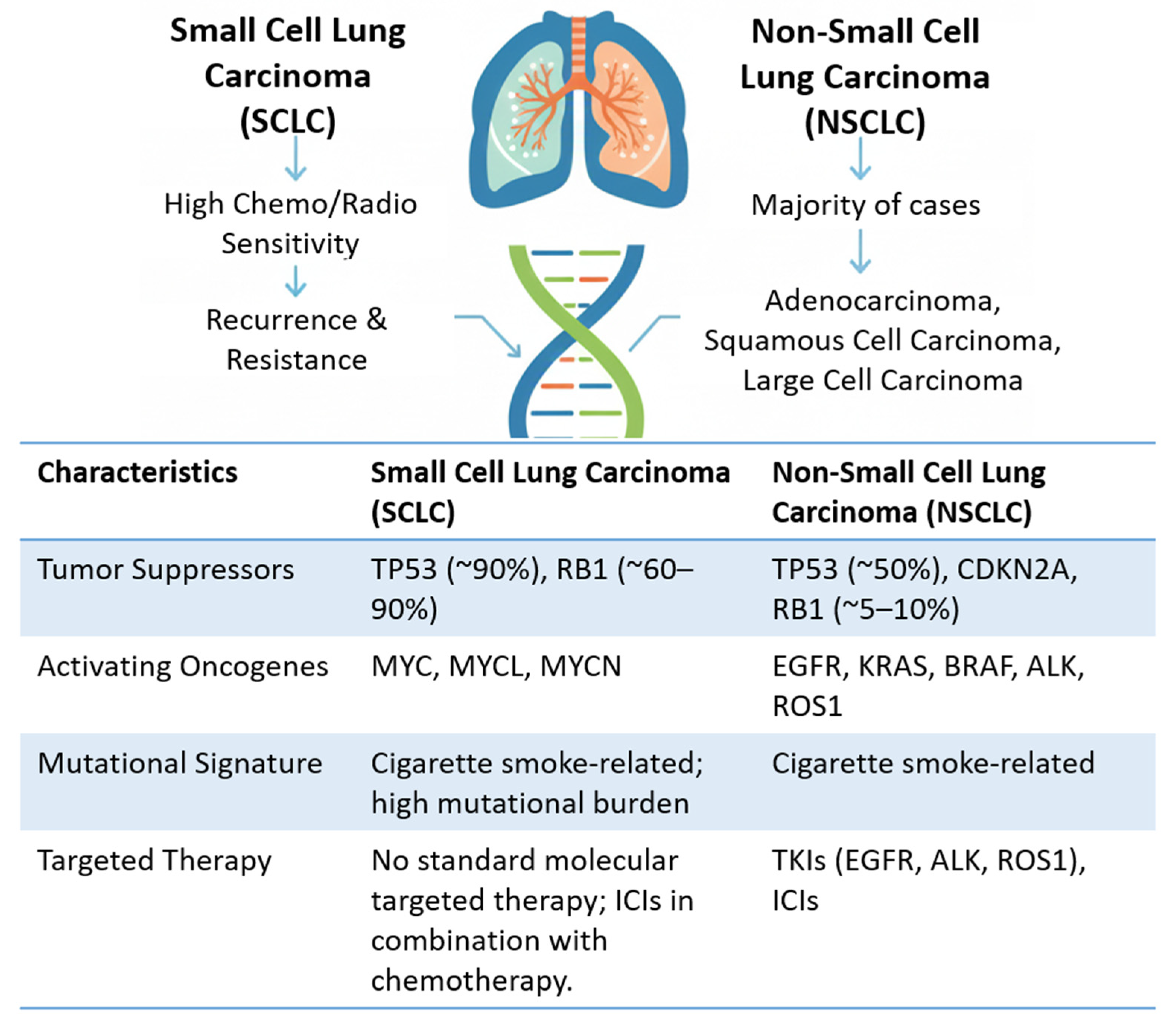
1.2. Lung Cancer
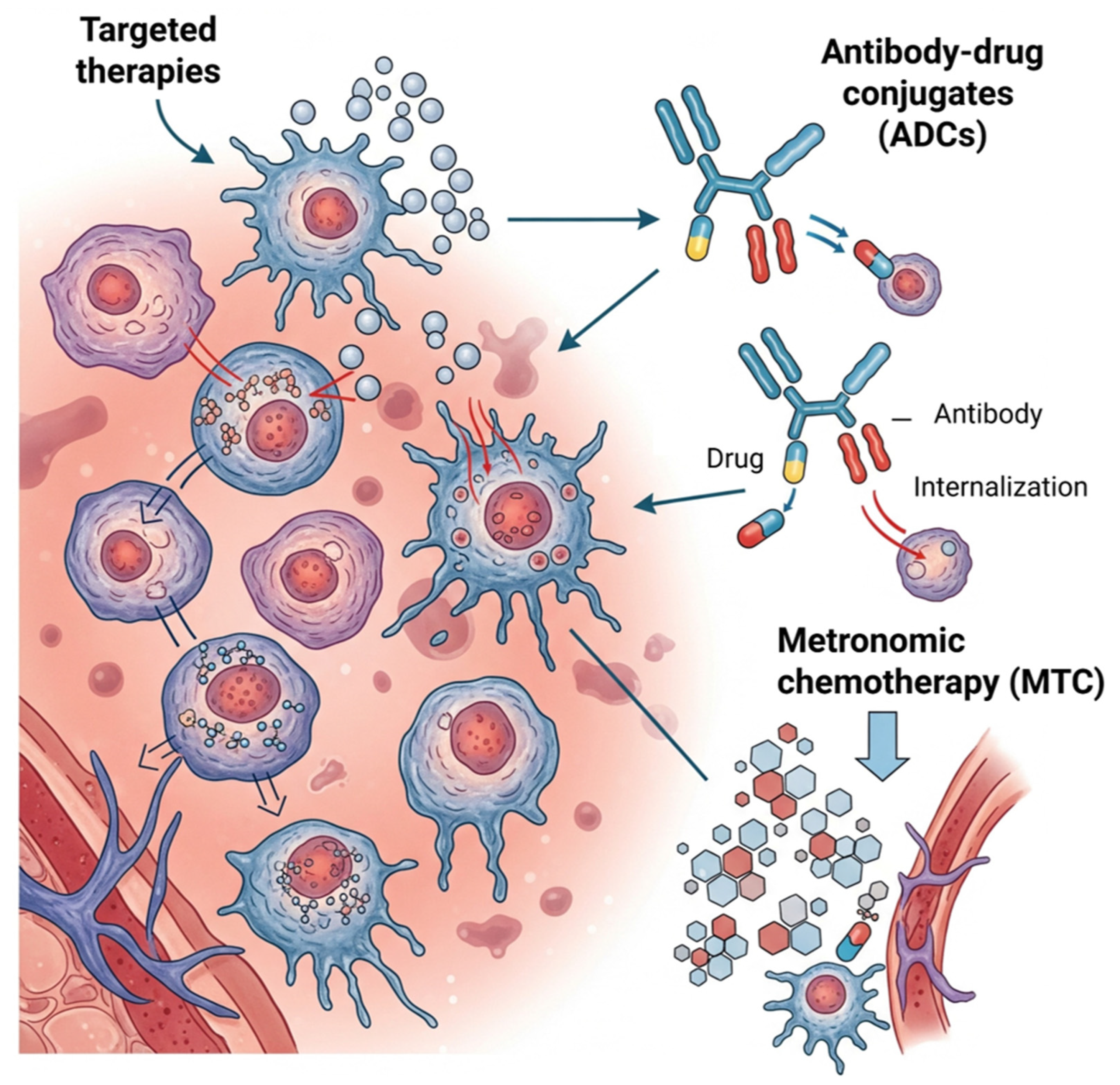
2. Emerging Therapeutic Frontiers: Novel Strategies in Oncology
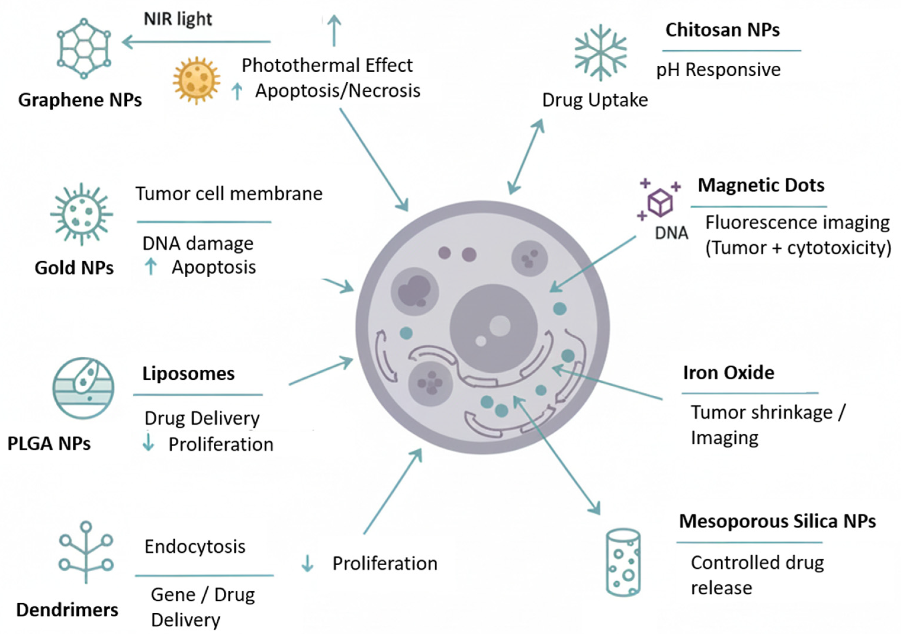
3. Subcellular Interventions: Nanomedicine as a Strategy Against Cancer
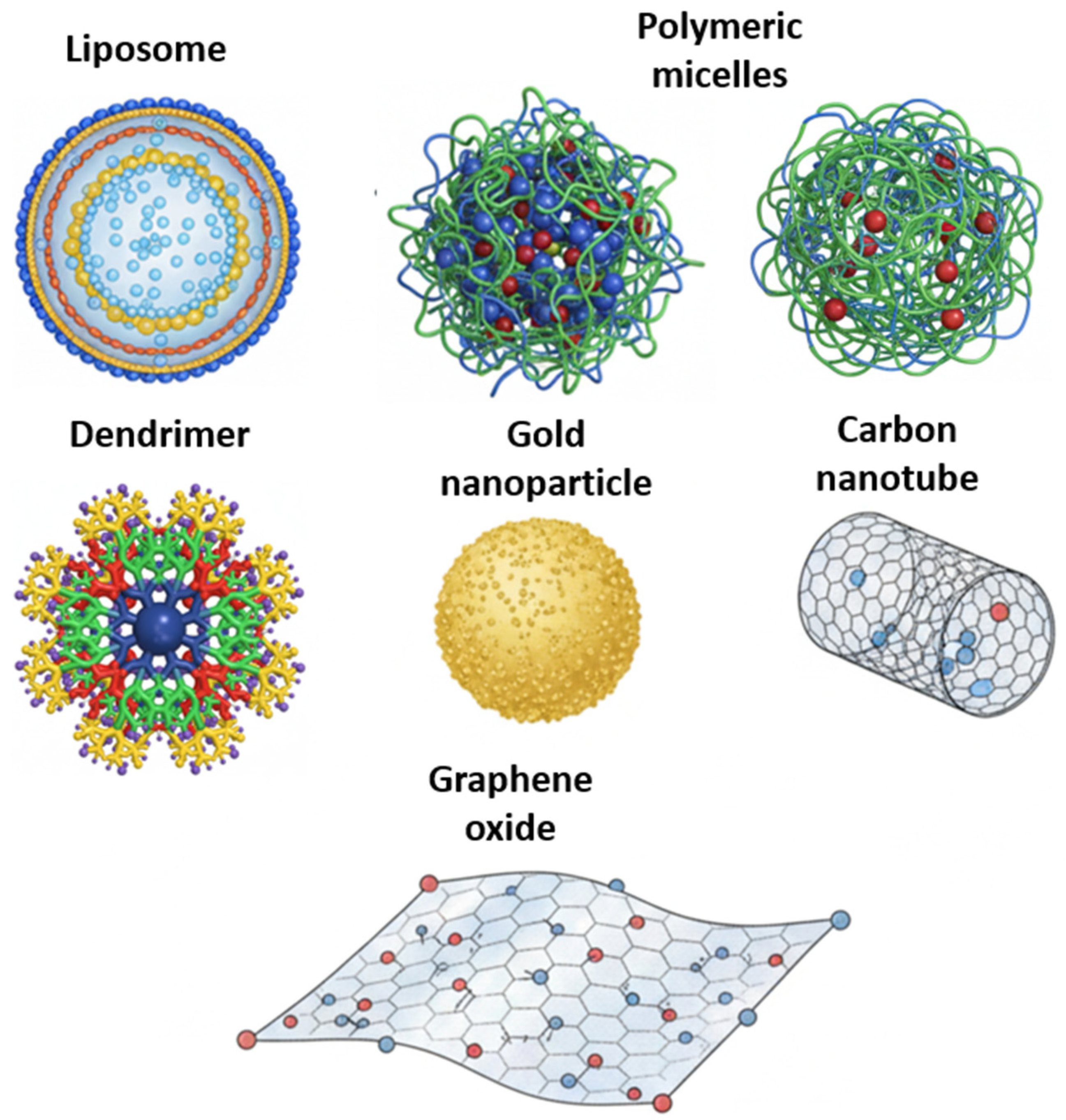

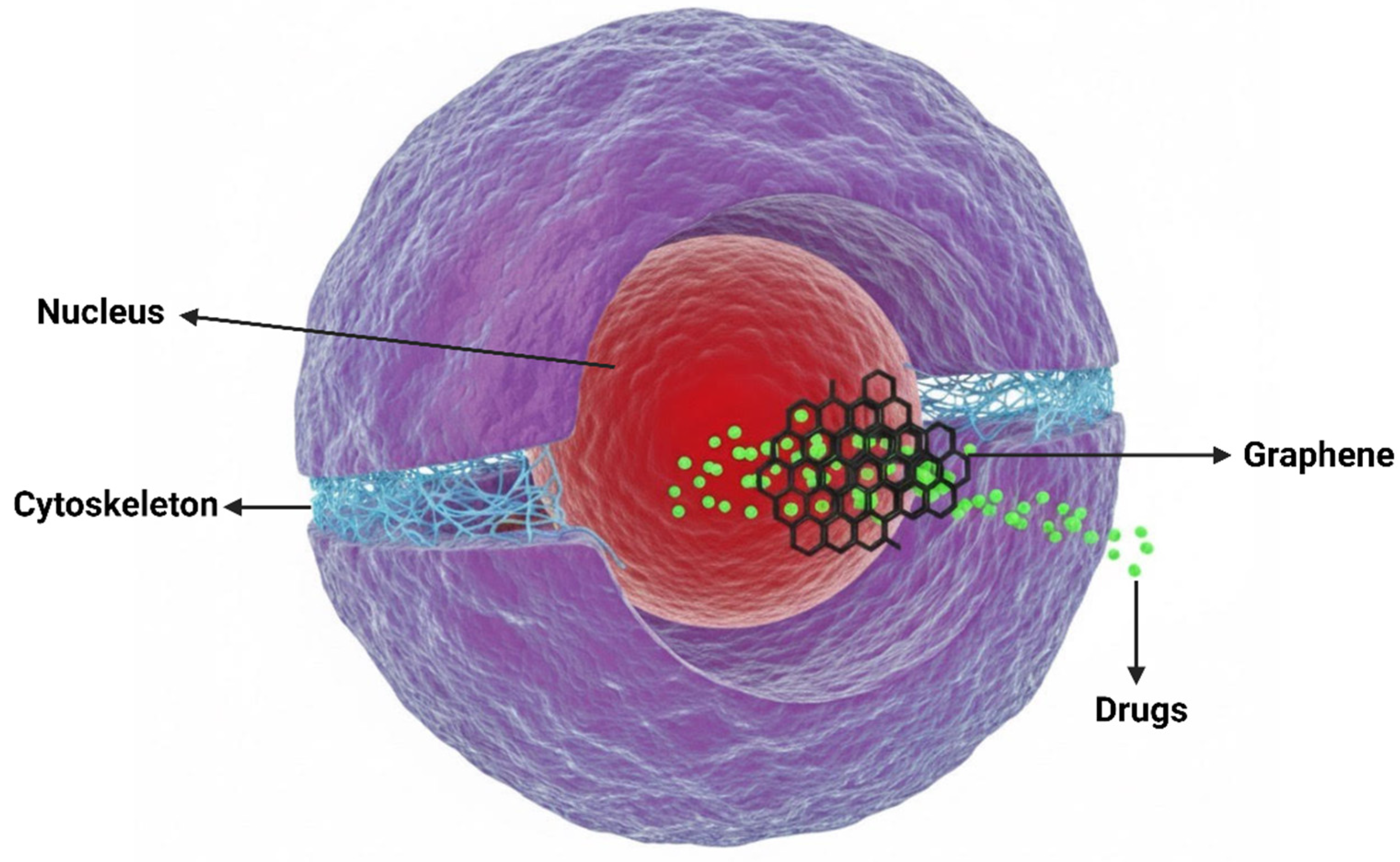
4. Modulating the Cellular Landscape: The Role of Graphene-Based Materials
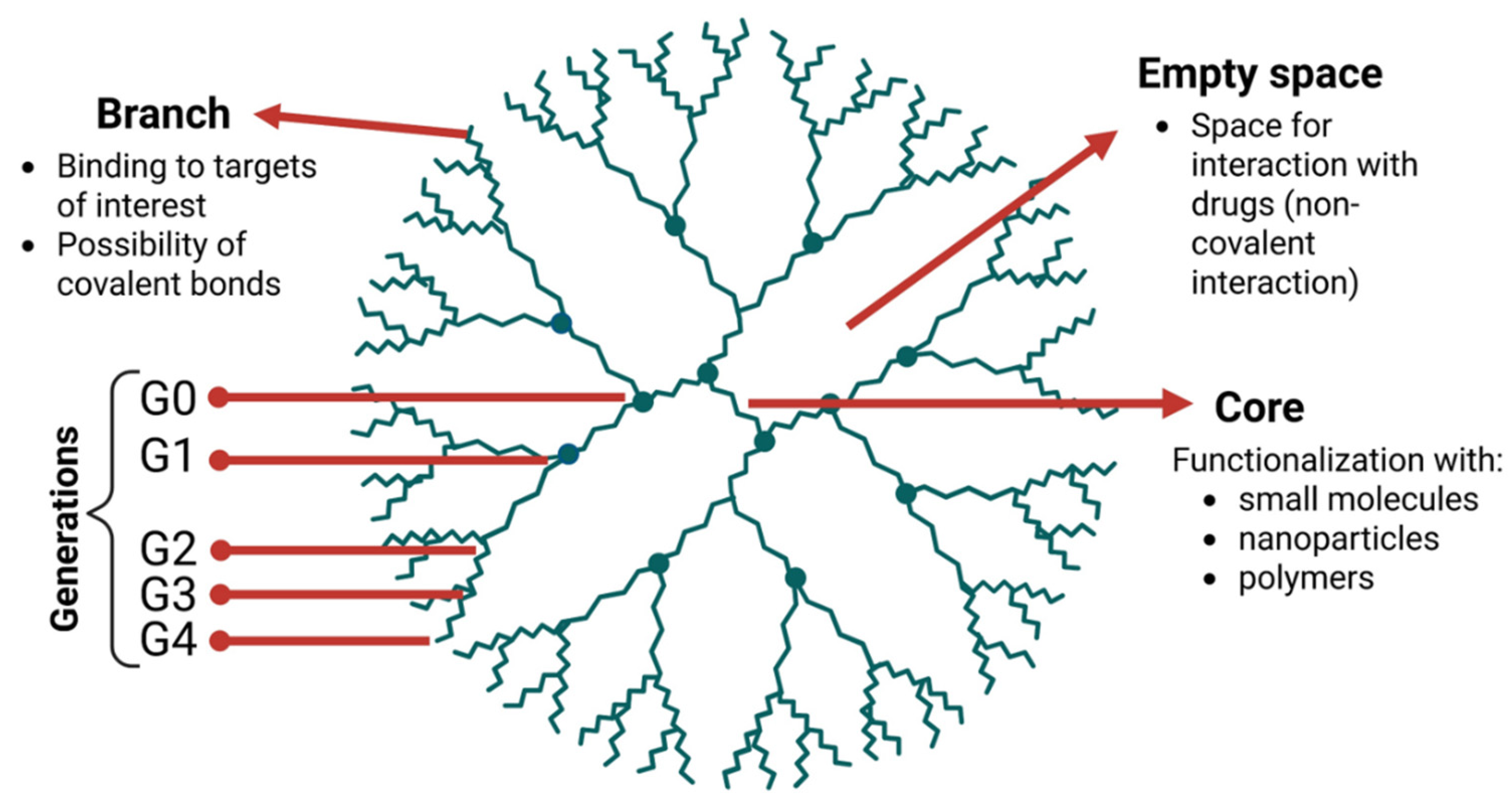
5. Dendrimers: The Power of Branching
6. Beyond Individual Components: Emergent Properties and Complementarity in Graphene-Dendrimer Hybrids
7. The Nano-Bio Interface: Deciphering the Cellular Impact of GOX and Dendrimers
8. Conclusions
9. Future Perspective
Author Contributions
Funding
Data Availability Statement
Acknowledgments
Conflicts of Interest
References
- World Health Organization (WHO). Cancer; World Health Organization: Geneva, Switzerland, 2025. [Google Scholar]
- Sung, H.; Ferlay, J.; Siegel, R.L.; Laversanne, M.; Soerjomataram, I.; Jemal, A.; Bray, F. Global Cancer Statistics 2020: GLOBOCAN Estimates of Incidence and Mortality Worldwide for 36 Cancers in 185 Countries. CA Cancer J. Clin. 2021, 71, 209–249. [Google Scholar] [CrossRef]
- Hanahan, D.; Weinberg, R.A. The hallmarks of cancer. Cell 2000, 100, 57–70. [Google Scholar] [CrossRef]
- Hanahan, D.; Weinberg, R.A. Hallmarks of cancer: The next generation. Cell 2011, 144, 646–674. [Google Scholar] [CrossRef]
- Li, Y.; Liu, F.; Cai, Q.; Deng, L.; Ouyang, Q.; Zhang, X.H.; Zheng, J. Invasion and metastasis in cancer: Molecular insights and therapeutic targets. Signal Transduct. Target. Ther. 2025, 10, 57. [Google Scholar] [CrossRef] [PubMed]
- Novikov, N.M.; Zolotaryova, S.Y.; Gautreau, A.M.; Denisov, E.V. Mutational drivers of cancer cell migration and invasion. Br. J. Cancer 2021, 124, 102–114. [Google Scholar] [CrossRef] [PubMed]
- Yadav, K.; Das, T.; Lynn, A.M. Pancancer analysis of DNA damage repair gene mutations and their impact on immune regulatory gene expression. Sci. Rep. 2025, 15, 15667. [Google Scholar] [CrossRef]
- Hanahan, D. Hallmarks of Cancer: New Dimensions. Cancer Discov. 2022, 12, 31–46. [Google Scholar] [CrossRef]
- Muzammal, M.; Awan, M.; Mukhtar, A.; Mohsin, M.; Qamar, M.A. Graphene Based Nanomaterials Are a Potent and Innovative Technology for the Treatment of Cancer: A Review. ChemistrySelect 2024, 9, e202403554. [Google Scholar] [CrossRef]
- Khan, M.Z.; Tahir, D.; Asim, M.; Israr, M.; Haider, A.; Xu, D.D. Revolutionizing Cancer Care: Advances in Carbon-Based Materials for Diagnosis and Treatment. Cureus 2024, 16, e52511. [Google Scholar] [CrossRef]
- Liu, L.; Ma, Q.; Cao, J.; Song, Y.; Sun, Y. Recent progress of graphene oxide-based multifunctional nanomaterials for cancer treatment. Cancer Nanotechnol. 2021, 12, 18. [Google Scholar] [CrossRef]
- Obalola, A.A.; Abrahamse, H.; Dhilip Kumar, S.S. Recent advances in gold nanoparticle-based targeted photodynamic and photothermal cancer therapy. Nanoscale Adv. 2025, 7, 7440–7458. [Google Scholar] [CrossRef]
- Ali, M.S.; Hooshmand, N.; El-Sayed, M.; Labouta, H.I. Microfluidics for Development of Lipid Nanoparticles: Paving the Way for Nucleic Acids to the Clinic. ACS Appl. Bio Mater. 2021, 6, 3566–3576. [Google Scholar] [CrossRef]
- Mandal, M.; Samanta, D.; Manna, D.; Bharadwaj, D.; Majumder, R.; Banerjee, I.; Mukherjee, B.; Mandal, M. Modified PAMAM Dendrimer-Based Carboplatin Delivery in Glioblastoma: In Vitro and In Vivo Studies. ACS Appl. Bio Mater. 2025, 8, 10393–10404. [Google Scholar] [CrossRef]
- Wiwatchaitawee, K.; Ebeid, K.; Quarterman, J.C.; Naguib, Y.; Ali, M.Y.; Oliva, C.; Griguer, C.; Salem, A.K. Surface Modification of Nanoparticles Enhances Drug Delivery to the Brain and Improves Survival in a Glioblastoma Multiforme Murine Model. Bioconjug. Chem. 2022, 33, 1957–1972. [Google Scholar] [CrossRef]
- Nikkhoo, A.; Rostami, N.; Farhadi, S.; Esmaily, M.; Moghadaszadeh, A.; Atyabi, F.; Baghaei, M.; Haghnavaz, N.; Yousefi, M.; Aliparasti, M.R.; et al. Codelivery of STAT3 siRNA and BV6 by carboxymethyl dextran trimethyl chitosan nanoparticles suppresses cancer cell progression. Int. J. Pharm. 2020, 581, 119236. [Google Scholar] [CrossRef]
- Liu, Y.; Cheng, W.; Xin, H.; Liu, R.; Wang, Q.; Cai, W.; Peng, X.; Yang, F.; Xin, H. Nanoparticles advanced from preclinical studies to clinical trials for lung cancer therapy. Cancer Nanotechnol. 2023, 14, 28. [Google Scholar] [CrossRef]
- Huang, J.; Lin, G.; Juenke, T.; Chung, S.; Lai, N.; Zhang, T.; Zhang, T.; Zhang, M. Iron Oxide Nanoparticle-Mediated mRNA Delivery to Hard-to-Transfect Cancer Cells. Pharmaceutics 2023, 15, 1946. [Google Scholar] [CrossRef] [PubMed]
- Ho, T.L.; Mutalik, C.; Rethi, L.; Nguyen, H.T.; Jheng, P.R.; Wong, C.C.; Yang, T.S.; Nguyen, T.T.; Mansel, B.W.; Wang, C.A.; et al. Cancer-targeted fucoidan iron oxide nanoparticles for synergistic chemotherapy/chemodynamic theranostics through amplification of P-selectin and oxidative stress. Int. J. Biol. Macromol. 2023, 235, 123821. [Google Scholar] [CrossRef] [PubMed]
- Mikaeili Ghezeljeh, S.; Salehzadeh, A.; Ataei-EJaliseh, S. Iron oxide nanoparticles coated with glucose and conjugated with safranal (Fe3O4@Glu-Safranal NPs) inducing apoptosis in liver cancer cell line (HepG2). BMC Chem. 2024, 18, 33. [Google Scholar] [CrossRef] [PubMed]
- Lan, H.; Jamil, M.; Ke, G.; Dong, N. The role of nanoparticles and nanomaterials in cancer diagnosis and treatment: A comprehensive review. Am. J. Cancer Res. 2023, 13, 5751–5784. [Google Scholar] [PubMed] [PubMed Central]
- Baranwal, J.; Barse, B.; Di Petrillo, A.; Gatto, G.; Pilia, L.; Kumar, A. Nanoparticles in Cancer Diagnosis and Treatment. Materials 2023, 16, 5354. [Google Scholar] [CrossRef]
- Taléns-Visconti, R.; Díez-Sales, O.; de Julián-Ortiz, J.V.; Nácher, A. Nanoliposomes in Cancer Therapy: Marketed Products and Current Clinical Trials. Int. J. Mol. Sci. 2022, 23, 4249. [Google Scholar] [CrossRef]
- Krauss, A.C.; Gao, X.; Li, L.; Manning, M.L.; Patel, P.; Fu, W.; Janoria, K.G.; Gieser, G.; Bateman, D.A.; Przepiorka, D.; et al. FDA Approval Summary: (Daunorubicin and Cytarabine) Liposome for Injection for the Treatment of Adults with High-Risk Acute Myeloid Leukemia. Clin. Cancer Res. 2019, 25, 2685–2690. [Google Scholar] [CrossRef] [PubMed]
- Gabizon, A.A.; Gabizon-Peretz, S.; Modaresahmadi, S.; La-Beck, N.M. Thirty years from FDA approval of pegylated liposomal doxorubicin (Doxil/Caelyx): An updated analysis and future perspective. BMJ Oncol. 2025, 4, e000573. [Google Scholar] [CrossRef] [PubMed]
- Hinshaw, D.C.; Shevde, L.A. The Tumor Microenvironment Innately Modulates Cancer Progression. Cancer Res. 2019, 79, 4557–4566. [Google Scholar] [CrossRef] [PubMed]
- Yuan, S.; Almagro, J.; Fuchs, E. Beyond genetics: Driving cancer with the tumour microenvironment behind the wheel. Nat. Rev. Cancer 2024, 24, 274–286. [Google Scholar] [CrossRef]
- Sun, L.; Zhang, H.; Gao, P. Metabolic reprogramming and epigenetic modifications on the path to cancer. Protein Cell 2022, 13, 877–919. [Google Scholar] [CrossRef]
- Ji, Y.; Xiao, C.; Fan, T.; Deng, Z.; Wang, D.; Cai, W.; Li, J.; Liao, T.; Li, C.; He, J. The epigenetic hallmarks of immune cells in cancer. Mol. Cancer 2025, 24, 66. [Google Scholar] [CrossRef]
- American Association for Cancer Research (AACR). Preventing Cancer: Identifying Risk Factors; American Association for Cancer Research (AACR): Philadelphia, PA, USA, 2022. [Google Scholar]
- Islami, F.; Marlow, E.C.; Thomson, B.; McCullough, M.L.; Rumgay, H.; Gapstur, S.M.; Patel, A.V.; Soerjomataram, I.; Jemal, A. Proportion and number of cancer cases and deaths attributable to potentially modifiable risk factors in the United States, 2019. CA Cancer J. Clin. 2024, 74, 405–432. [Google Scholar] [CrossRef]
- Pérez, R.F.; Tejedor, J.R.; Fernández, A.F.; Fraga, M.F. Aging and cancer epigenetics: Where do the paths fork? Aging Cell 2022, 21, e13709. [Google Scholar] [CrossRef]
- DeGregori, J.; Seidl, K.J.; Montano, M. Aging and Cancer-Inextricably Linked Across the Lifespan. Aging Cell 2025, 24, e14483. [Google Scholar] [CrossRef]
- Patel, M.M.; Adrada, B.E. Hereditary Breast Cancer: BRCA Mutations and Beyond. Radiol. Clin. N. Am. 2024, 62, 627–642. [Google Scholar] [CrossRef]
- Roy, M.; Fowler, A.M.; Ulaner, G.A.; Mahajan, A. Molecular Classification of Breast Cancer. PET Clin. 2023, 18, 441–458. [Google Scholar] [CrossRef] [PubMed]
- Zhu, S.; Wu, Y.; Song, B.; Yi, M.; Yan, Y.; Mei, Q.; Wu, K. Recent advances in targeted strategies for triple-negative breast cancer. J. Hematol. Oncol. 2023, 16, 100. [Google Scholar] [CrossRef]
- Schopp, J.G.; Polat, D.S.; Arjmandi, F.; Hayes, J.C.; Ahn, R.W.; Sullivan, K.; Sahoo, S.; Porembka, J.H. Imaging Challenges in Diagnosing Triple-Negative Breast Cancer. Radiographics 2023, 43, e230027. [Google Scholar] [CrossRef]
- Barzaman, K.; Karami, J.; Zarei, Z.; Hosseinzadeh, A.; Kazemi, M.H.; Moradi-Kalbolandi, S.; Safari, E.; Farahmand, L. Breast cancer: Biology, biomarkers, and treatments. Int. Immunopharmacol. 2020, 84, 106535. [Google Scholar] [CrossRef]
- Wild, C.P.; Weiderpass, E.; Stewart, B.W. World Cancer Report: Cancer Research for Cancer Prevention; International Agency for Research on Cancer: Lyon, France, 2020. [Google Scholar]
- Nicholson, A.G.; Tsao, M.S.; Beasley, M.B.; Borczuk, A.C.; Brambilla, E.; Cooper, W.A.; Dacic, S.; Jain, D.; Kerr, K.M.; Lantuejoul, S.; et al. The 2021 WHO Classification of Lung Tumors: Impact of Advances Since 2015. J. Thorac. Oncol. 2022, 17, 362–387. [Google Scholar] [CrossRef]
- Jeon, H.; Wang, S.; Song, J.; Gill, H.; Cheng, H. Update 2025: Management of Non Small-Cell Lung Cancer. Lung 2025, 203, 53. [Google Scholar] [CrossRef] [PubMed]
- Bade, B.C.; Dela Cruz, C.S. Lung Cancer 2020: Epidemiology, Etiology, and Prevention. Clin. Chest Med. 2020, 41, 1–24. [Google Scholar] [CrossRef] [PubMed]
- Mujahid, M.H.; Upadhyay, T.K.; Khan, F.; Pandey, P.; Park, M.N.; Sharangi, A.B.; Saeed, M.; Upadhye, V.J.; Kim, B. Metallic and metal oxide-derived nanohybrid as a tool for biomedical applications. Biomed. Pharmacother. 2022, 155, 113791. [Google Scholar] [CrossRef]
- Yan, C.; Wang, J.; Yang, Y.; Ma, W.; Chen, X. Molecular biomarker-guided anti-angiogenic targeted therapy for malignant glioma. J. Cell. Mol. Med. 2019, 23, 4876–4882. [Google Scholar] [CrossRef]
- Jia, J.; Wu, X.; Long, G.; Yu, J.; He, W.; Zhang, H.; Wang, D.; Ye, Z.; Tian, J. Revolutionizing cancer treatment: Nanotechnology-enabled photodynamic therapy and immunotherapy with advanced photosensitizers. Front. Immunol. 2023, 14, 1219785. [Google Scholar] [CrossRef]
- Koirala, N.; Dey, N.; Aske, J.; De, P. Targeting Cell Cycle Progression in HER2+ Breast Cancer: An Emerging Treatment Opportunity. Int. J. Mol. Sci. 2022, 23, 6547. [Google Scholar] [CrossRef] [PubMed]
- Infantino, V.; Santarsiero, A.; Convertini, P.; Todisco, S.; Iacobazzi, V. Cancer Cell Metabolism in Hypoxia: Role of HIF-1 as Key Regulator and Therapeutic Target. Int. J. Mol. Sci. 2021, 22, 5703. [Google Scholar] [CrossRef]
- Hsu, C.C.; Peng, D.; Cai, Z.; Lin, H.K. AMPK signaling and its targeting in cancer progression and treatment. Semin. Cancer Biol. 2022, 85, 52–68. [Google Scholar] [CrossRef]
- Zhu, K.; Wu, Y.; He, P.; Fan, Y.; Zhong, X.; Zheng, H.; Luo, T. PI3K/AKT/mTOR-Targeted Therapy for Breast Cancer. Cells 2022, 11, 2508. [Google Scholar] [CrossRef]
- Dong, W.; Chen, M.; Wang, J.; Xia, L.; Wang, Q.; Nie, X.; Feng, Y.; Fang, Y. rHuPH20-facilitated subcutaneous administration of monoclonal antibodies in cancer therapy. Immunotherapy 2021, 13, 79–88. [Google Scholar] [CrossRef] [PubMed]
- Delgado, M.; Garcia-Sanz, J.A. Therapeutic Monoclonal Antibodies against Cancer: Present and Future. Cells 2023, 12, 2837. [Google Scholar] [CrossRef] [PubMed]
- Galvao, T.F.; Livinalli, A.; Lopes, L.C.; Zimmermann, I.R.; Silva, M.T. Biosimilar monoclonal antibodies for cancer treatment in adults. Cochrane Database Syst. Rev. 2024, 11, CD013539. [Google Scholar] [CrossRef] [PubMed]
- Merle, G.; Friedlaender, A.; Desai, A.; Addeo, A. Antibody Drug Conjugates in Lung Cancer. Cancer J. 2022, 28, 429–435. [Google Scholar] [CrossRef]
- Najjar, M.K.; Manore, S.G.; Regua, A.T.; Lo, H.W. Antibody-Drug Conjugates for the Treatment of HER2-Positive Breast Cancer. Genes 2022, 13, 2065. [Google Scholar] [CrossRef] [PubMed]
- Feng, Y.; Lee, J.; Yang, L.; Hilton, M.B.; Morris, K.; Seaman, S.; Edupuganti, V.V.S.R.; Hsu, K.S.; Dower, C.; Yu, G.; et al. Engineering CD276/B7-H3-targeted antibody-drug conjugates with enhanced cancer-eradicating capability. Cell Rep. 2023, 42, 113503. [Google Scholar] [CrossRef]
- Mathew, D.; Marmarelis, M.E.; Foley, C.; Bauml, J.M.; Ye, D.; Ghinnagow, R.; Ngiow, S.F.; Klapholz, M.; Jun, S.; Zhang, Z.; et al. Combined JAK inhibition and PD-1 immunotherapy for non-small cell lung cancer patients. Science 2024, 384, eadf1329. [Google Scholar] [CrossRef]
- Denkert, C.; Schneeweiss, A.; Rey, J.; Karn, T.; Hattesohl, A.; Weber, K.E.; Rackakonda, S.; Braun, M.; Huober, J.; Jank, P.; et al. Molecular adaptation to neoadjuvant immunotherapy in triple-negative breast cancer. Cell Rep. Med. 2024, 5, 101825. [Google Scholar] [CrossRef]
- Cazzaniga, M.E.; Cordani, N.; Capici, S.; Cogliati, V.; Riva, F.; Cerrito, M.G. Metronomic Chemotherapy. Cancers 2021, 13, 2236. [Google Scholar] [CrossRef]
- Mo, H.; Yu, Y.; Sun, X.; Ge, H.; Yu, L.; Guan, X.; Zhai, J.; Zhu, A.; Wei, Y.; Wang, J.; et al. Metronomic chemotherapy plus anti-PD-1 in metastatic breast cancer: A Bayesian adaptive randomized phase 2 trial. Nat. Med. 2024, 30, 2528–2539. [Google Scholar] [CrossRef]
- Dumontet, C.; Reichert, J.M.; Senter, P.D.; Lambert, J.M.; Beck, A. Antibody-drug conjugates come of age in oncology. Nat. Rev. Drug Discov. 2023, 22, 641–661. [Google Scholar] [CrossRef]
- Fuentes-Antrás, J.; Genta, S.; Vijenthira, A.; Siu, L.L. Antibody-drug conjugates: In search of partners of choice. Trends Cancer 2023, 9, 339–354. [Google Scholar] [CrossRef] [PubMed]
- Grinda, T.; Rassy, E.; Pistilli, B. Antibody-Drug Conjugate Revolution in Breast Cancer: The Road Ahead. Curr. Treat. Options Oncol. 2023, 24, 442–465, Correction in Curr. Treat. Options Oncol. 2023, 24, 466–467. https://doi.org/10.1007/s11864-023-01092-1.. [Google Scholar] [CrossRef] [PubMed]
- Fu, Z.; Li, S.; Han, S.; Shi, C.; Zhang, Y. Antibody drug conjugate: The “biological missile” for targeted cancer therapy. Signal Transduct. Target. Ther. 2022, 7, 93. [Google Scholar] [CrossRef]
- Birrer, M.J.; Moore, K.N.; Betella, I.; Bates, R.C. Antibody-Drug Conjugate-Based Therapeutics: State of the Science. J. Natl. Cancer Inst. 2019, 111, 538–549. [Google Scholar] [CrossRef]
- Phuna, Z.X.; Kumar, P.A.; Haroun, E.; Dutta, D.; Lim, S.H. Antibody-drug conjugates: Principles and opportunities. Life Sci. 2024, 347, 122676. [Google Scholar] [CrossRef]
- Nguyen, T.D.; Bordeau, B.M.; Balthasar, J.P. Mechanisms of ADC Toxicity and Strategies to Increase ADC Tolerability. Cancers 2023, 15, 713. [Google Scholar] [CrossRef] [PubMed]
- Tsuchikama, K.; Anami, Y.; Ha, S.Y.Y.; Yamazaki, C.M. Exploring the next generation of antibody-drug conjugates. Nat. Rev. Clin. Oncol. 2024, 21, 203–223. [Google Scholar] [CrossRef] [PubMed]
- Han, E.L.; Tang, S.; Kim, D.; Murray, A.M.; Swingle, K.L.; Hamilton, A.G.; Mrksich, K.; Padilla, M.S.; Palanki, R.; Li, J.J.; et al. Peptide-Functionalized Lipid Nanoparticles for Targeted Systemic mRNA Delivery to the Brain. Nano Lett. 2025, 25, 800–810. [Google Scholar] [CrossRef]
- Khare, P.; Edgecomb, S.X.; Hamadani, C.M.; Tanner, E.E.L.; SManickam, D. Lipid nanoparticle-mediated drug delivery to the brain. Adv. Drug Deliv. Rev. 2023, 197, 114861. [Google Scholar] [CrossRef] [PubMed]
- Shang, H.; Xia, D.; Geng, R.; Wu, J.; Deng, W.; Tong, Y.; Ba, X.; Zhong, Z.; He, Y.; Huang, Q.; et al. Thermosensitive Resiquimod-Loaded Lipid Nanoparticles Promote the Polarization of Tumor-Associated Macrophages to Enhance Bladder Cancer Immunotherapy. ACS Nano 2025, 19, 19599–19621. [Google Scholar] [CrossRef]
- Agarwal, H.; Bynum, R.C.; Saleh, N.; Harris, D.; MacCuaig, W.M.; Kim, V.; Sanderson, E.J.; Dennahy, I.S.; Singh, R.; Behkam, B.; et al. Theranostic nanoparticles for detection and treatment of pancreatic cancer. Wiley Interdiscip. Rev. Nanomed. Nanobiotechnol. 2024, 16, e1983. [Google Scholar] [CrossRef] [PubMed]
- Palazzolo, S.; Bayda, S.; Hadla, M.; Caligiuri, I.; Corona, G.; Toffoli, G.; Rizzolio, F. The Clinical Translation of Organic Nanomaterials for Cancer Therapy: A Focus on Polymeric Nanoparticles, Micelles, Liposomes and Exosomes. Curr. Med. Chem. 2018, 25, 4224–4268. [Google Scholar] [CrossRef]
- Zhang, H.; Wu, Y.; Hu, Y.; Li, X.; Zhao, M.; Lv, Z. Targeted Nanoparticle Drug Delivery System for the Enhancement of Cancer Immunotherapy. J. Biomed. Nanotechnol. 2019, 15, 1839–1866. [Google Scholar] [CrossRef]
- Xing, Y.; Yang, J.; Peng, A.; Qian, Y.; Liu, Y.; Pan, P.; Liu, Q. Lysosome Targeted Nanoparticle Aggregation Reverses Immunosuppressive Tumor Microenvironment for Cancer Immunotherapy. Adv. Mater. 2024, 36, e2412730. [Google Scholar] [CrossRef]
- Chai, D.; Wang, J.; Lim, J.M.; Xie, X.; Yu, X.; Zhao, D.; Maza, P.A.M.; Wang, Y.; Cyril-Remirez, D.; Young, K.H.; et al. Lipid nanoparticles deliver DNA-encoded biologics and induce potent protective immunity. Mol. Cancer 2025, 24, 12. [Google Scholar] [CrossRef]
- Niero, E.L.; Rocha-Sales, B.; Lauand, C.; Cortez, B.A.; de Souza, M.M.; Rezende-Teixeira, P.; Urabayashi, M.S.; Martens, A.A.; Neves, J.H.; Machado-Santelli, G.M. The multiple facets of drug resistance: One history, different approaches. J. Exp. Clin. Cancer Res. 2014, 33, 37. [Google Scholar] [CrossRef] [PubMed]
- De Las Rivas, J.; Brozovic, A.; Izraely, S.; Casas-Pais, A.; Witz, I.P.; Figueroa, A. Cancer drug resistance induced by EMT: Novel therapeutic strategies. Arch. Toxicol. 2021, 95, 2279–2297. [Google Scholar] [CrossRef]
- Duan, C.; Yu, M.; Xu, J.; Li, B.Y.; Zhao, Y.; Kankala, R.K. Overcoming Cancer Multi-drug Resistance (MDR): Reasons, mechanisms, nanotherapeutic solutions, and challenges. Biomed. Pharmacother. 2023, 162, 114643. [Google Scholar] [CrossRef]
- Niu, X.; You, Q.; Hou, K.; Tian, Y.; Wei, P.; Zhu, Y.; Gao, B.; Ashrafizadeh, M.; Aref, A.R.; Kalbasi, A.; et al. Autophagy in cancer development, immune evasion, and drug resistance. Drug Resist. Updat. 2025, 78, 101170. [Google Scholar] [CrossRef]
- Jin, K.T.; Yao, J.Y.; Ying, X.J.; Lin, Y.; Chen, Y.F. Nanomedicine and Early Cancer Diagnosis: Molecular Imaging using Fluorescence Nanoparticles. Curr. Top. Med. Chem. 2020, 20, 2737–2761. [Google Scholar] [CrossRef]
- Fan, M.; Han, Y.; Gao, S.; Yan, H.; Cao, L.; Li, Z.; Liang, X.J.; Zhang, J. Ultrasmall gold nanoparticles in cancer diagnosis and therapy. Theranostics 2020, 10, 4944–4957. [Google Scholar] [CrossRef] [PubMed]
- Nannuri, S.H.; Nikam, A.N.; Pandey, A.; Mutalik, S.; George, S.D. Subcellular Imaging and Diagnosis of Cancer using Engineered Nanoparticles. Curr. Pharm. Des. 2022, 28, 690–710. [Google Scholar] [CrossRef] [PubMed]
- Lu, Q.; Kou, D.; Lou, S.; Ashrafizadeh, M.; Aref, A.R.; Canadas, I.; Tian, Y.; Niu, X.; Wang, Y.; Torabian, P.; et al. Nanoparticles in tumor microenvironment remodeling and cancer immunotherapy. J. Hematol. Oncol. 2024, 17, 16. [Google Scholar] [CrossRef]
- Devi, R.S.; Girigoswami, A.; Siddharth, M.; Girigoswami, K. Applications of Gold and Silver Nanoparticles in Theranostics. Appl. Biochem. Biotechnol. 2022, 194, 4187–4219. [Google Scholar] [CrossRef]
- Zheng, X.; Wu, Y.; Zuo, H.; Chen, W.; Wang, K. Metal Nanoparticles as Novel Agents for Lung Cancer Diagnosis and Therapy. Small 2023, 19, e2206624. [Google Scholar] [CrossRef]
- Yang, R.; Chen, L.; Wang, Y.; Zhang, L.; Zheng, X.; Yang, Y.; Zhu, Y. Tumor microenvironment responsive metal nanoparticles in cancer immunotherapy. Front. Immunol. 2023, 14, 1237361. [Google Scholar] [CrossRef]
- Bhattacharya, S.; Saindane, D.; Prajapati, B.G. Liposomal Drug Delivery and Its Potential Impact on Cancer Research. Anticancer Agents Med. Chem. 2022, 22, 2671–2683. [Google Scholar] [CrossRef] [PubMed]
- Liu, Y.; Han, J.; Bo, Y.; Bhatta, R.; Wang, H. Targeted delivery of liposomal chemoimmunotherapy for cancer treatment. Front. Immunol. 2022, 13, 1010021. [Google Scholar] [CrossRef] [PubMed]
- Senti, M.E.; Del Valle, L.G.; Schiffelers, R.M. mRNA delivery systems for cancer immunotherapy: Lipid nanoparticles and beyond. Adv. Drug Deliv. Rev. 2024, 206, 115190. [Google Scholar] [CrossRef] [PubMed]
- Beach, M.A.; Nayanathara, U.; Gao, Y.; Zhang, C.; Xiong, Y.; Wang, Y.; Such, G.K. Polymeric Nanoparticles for Drug Delivery. Adv. Drug Deliv. Rev. 2024, 124, 5505–5616. [Google Scholar] [CrossRef]
- Floyd, T.G.; Gurnani, P.; Rho, J.Y. Characterisation of polymeric nanoparticles for drug delivery. Nanoscale 2025, 17, 7738–7752. [Google Scholar] [CrossRef]
- Nam, S.H.; Lee, S.W.; Lee, Y.J.; Kim, Y.M. Safety and Tolerability of Weekly Genexol-PM, a Cremophor-Free Polymeric Micelle Formulation of Paclitaxel, with Carboplatin in Gynecologic Cancer: A Phase I Study. Cancer Res. Treat. 2023, 55, 1346–1354. [Google Scholar] [CrossRef] [PubMed]
- Li, X.; Shi, J.; Zhu, J.; Zhu, J.; Yan, F.; Liu, D.; Cao, G. Polymeric Micellar Paclitaxel Plus Cisplatin Combined With Tislelizumab as the First-Line Treatment of Advanced Unresectable Esophageal Squamous Cell Carcinoma: A Phase II Study. Thorac. Cancer 2025, 16, e70055. [Google Scholar] [CrossRef]
- Li, D.; Ahmed, M.; Khan, A.; Xu, L.; Walters, A.A.; Ballesteros, B.; Al-Jamal, K.T. Tailoring the Architecture of Cationic Polymer Brush-Modified Carbon Nanotubes for Efficient siRNA Delivery in Cancer Immunotherapy. ACS Appl. Mater. Interfaces 2021, 13, 30284–30294. [Google Scholar] [CrossRef] [PubMed]
- Li, Y.; Xu, Z.; Qi, Z.; Huang, X.; Li, M.; Liu, S.; Yan, Y.; Gao, M. Application of Carbon Nanomaterials to Enhancing Tumor Immunotherapy: Current Advances and Prospects. Int. J. Nanomed. 2024, 19, 10899–10915. [Google Scholar] [CrossRef] [PubMed]
- Sharma, H.; Mondal, S. Functionalized Graphene Oxide for Chemotherapeutic Drug Delivery and Cancer Treatment: A Promising Material in Nanomedicine. Int. J. Mol. Sci. 2020, 21, 6280. [Google Scholar] [CrossRef]
- Chauhan, K.; Woo, J.; Jung, W.; Kim, D.E. Poly(ethylene glycol)-Engrafted Graphene Oxide for Gene Delivery and Nucleic Acid Amplification. Materials 2023, 16, 7434. [Google Scholar] [CrossRef]
- Kuntamung, K.; Sangthong, P.; Jakmunee, J.; Ounnunkad, K. Simultaneous immunodetection of multiple cervical cancer biomarkers based on a signal-amplifying redox probes/polyethyleneimine-coated gold nanoparticles/2D tungsten disulfide/graphene oxide nanocomposite platform. Bioelectrochemistry 2024, 160, 108780. [Google Scholar] [CrossRef]
- Sadrabadi, E.A.; Benvidi, A.; Azimzadeh, M.; Asgharnejad, L.; Dezfuli, A.S.; Khashayar, P. Novel electrochemical biosensor for breast cancer detection, based on a nanocomposite of carbon nanofiber, metal-organic framework, and magnetic graphene oxide. Bioelectrochemistry 2024, 155, 108558. [Google Scholar] [CrossRef] [PubMed]
- Vimalanathan, B.; Thiyagarajan, D.; Mary, R.N.; Sachidanandam, M.; Ignacimuthu, S.; Gnanasampanthapandian, D.; Rajasingh, J.; Palaniyandi, K. Composites of Reduced Graphene Oxide Based on Silver Nanoparticles and Their Effect on Breast Cancer Stem Cells. Bioengineering 2025, 12, 508. [Google Scholar] [CrossRef]
- Zhang, Z.; Wang, J.; Hou, L.; Zhu, D.; Xiao, H.J.; Wang, K. Graphene/carbohydrate polymer composites as emerging hybrid materials in tumor therapy and diagnosis. Int. J. Biol. Macromol. 2025, 287, 138621. [Google Scholar] [CrossRef]
- Lee, S.; Suh, S.H.; Ma, S.K.; Kim, J.; Park, S.; Kim, S.W.; Lee, J.Y. ROS-responsive graphene-hyaluronic acid nanomedicine for targeted therapy in renal ischemia/reperfusion injury. Theranostics 2026, 16, 618–636. [Google Scholar] [CrossRef]
- Itoo, A.M.; Vemula, S.L.; Gupta, M.T.; Giram, M.V.; Kumar, S.A.; Ghosh, B.; Biswas, S. Multifunctional graphene oxide nanoparticles for drug delivery in cancer. J. Control. Release 2022, 350, 26–59. [Google Scholar] [CrossRef]
- Ali, M.R.; Rahman, M.A.; Wu, Y.; Han, T.; Peng, X.; Mackey, M.A.; Wang, D.; Shin, H.J.; Chen, Z.G.; Xiao, H.; et al. Efficacy, long-term toxicity, and mechanistic studies of gold nanorods photothermal therapy of cancer in xenograft mice. Proc. Natl. Acad. Sci. USA 2017, 114, E3110–E3118. [Google Scholar] [CrossRef]
- Yoshikawa, T.; Mori, Y.; Feng, H.; Phan, K.Q.; Kishimura, A.; Kang, J.H.; Mori, T.; Katayama, Y. Rapid and continuous accumulation of nitric oxide-releasing liposomes in tumors to augment the enhanced permeability and retention (EPR) effect. Int. J. Pharm. 2019, 565, 481–487. [Google Scholar] [CrossRef] [PubMed]
- Butreddy, A.; Gaddam, R.P.; Kommineni, N.; Dudhipala, N.; Voshavar, C. PLGA/PLA-Based Long-Acting Injectable Depot Microspheres in Clinical Use: Production and Characterization Overview for Protein/Peptide Delivery. Int. J. Mol. Sci. 2021, 22, 8884. [Google Scholar] [CrossRef]
- Dey, A.D.; Bigham, A.; Esmaeili, Y.; Ashrafizadeh, M.; Moghaddam, F.D.; Tan, S.C.; Yousefiasl, S.; Sharma, S.; Maleki, A.; Rabiee, N.; et al. Dendrimers as nanoscale vectors: Unlocking the bars of cancer therapy. Semin. Cancer Biol. 2022, 86, 396–419. [Google Scholar] [CrossRef]
- Jafernik, K.; Ładniak, A.; Blicharska, E.; Czarnek, K.; Ekiert, H.; Wiącek, A.E.; Szopa, A. Chitosan-Based Nanoparticles as Effective Drug Delivery Systems-A review. Molecules 2023, 28, 1963. [Google Scholar] [CrossRef]
- Adam, A.; Mertz, D. Iron Oxide@Mesoporous Silica Core-Shell Nanoparticles as Multimodal Platforms for Magnetic Resonance Imaging, Magnetic Hyperthermia, Near-Infrared Light Photothermia, and Drug Delivery. Nanomaterials 2023, 13, 1342. [Google Scholar] [CrossRef]
- Rethi, L.; Rethi, L.; Liu, C.H.; Hyun, T.V.; Chen, C.H.; Chuang, E.Y. Fortification of Iron Oxide as Sustainable Nanoparticles: An Amalgamation with Magnetic/Photo Responsive Cancer Therapies. Int. J. Nanomed. 2023, 18, 5607–5623. [Google Scholar] [CrossRef]
- Geim, A.K.; Novoselov, K.S. The rise of graphene. Nat. Mater. 2007, 6, 183–191. [Google Scholar] [CrossRef]
- Geim, A.K. Graphene: Status and prospects. Science 2009, 324, 1530–1534. [Google Scholar] [CrossRef] [PubMed]
- Wei, L.; Li, G.; Lu, T.; Wei, Y.; Nong, Z.; Wei, M.; Pan, X.; Qin, Q.; Meng, F.; Li, X. Functionalized Graphene Oxide as Drug Delivery Systems for Platinum Anticancer Drugs. J. Pharm. Sci. 2021, 110, 3631–3638. [Google Scholar] [CrossRef] [PubMed]
- Dar, M.S.; Tabish, T.A.; Thorat, N.D.; Swati, G.; Sahu, N.K. Photothermal therapy using graphene quantum dots. APL Bioeng. 2023, 7, 031502. [Google Scholar] [CrossRef] [PubMed]
- Ribeiro, B.F.M.; Souza, M.M.; Fernandes, D.S.; do Carmo, D.R.; Machado-Santelli, G.M. Graphene oxide-based nanomaterial interaction with human breast cancer cells. J. Biomed. Mater. Res. A. 2020, 108, 863–870. [Google Scholar] [CrossRef]
- Ribeiro, B.F.M.; Chaves, J.B.; De Souza, M.M.; Keppler, A.F.; Do Carmo, D.R.; Machado-Santelli, G.M. Interaction of Graphene Oxide Particles and Dendrimers with Human Breast Cancer Cells by Real-Time Microscopy. Pharmaceutics 2023, 15, 2655. [Google Scholar] [CrossRef]
- Bavorova, H.; Svadlakova, T.; Fiala, Z.; Pisal, R.; Mokry, J. The Dose- and Time-Dependent Cytotoxic Effect of Graphene Nanoplatelets: In Vitro and In Vivo Study. Nanomaterials 2022, 12, 1978. [Google Scholar] [CrossRef]
- Feito, M.J.; Cicuéndez, M.; Casarrubios, L.; Diez-Orejas, R.; Fateixa, S.; Silva, D.; Barroca, N.; Marques, P.A.A.P.; Portolés, M.T. Effects of Graphene Oxide and Reduced Graphene Oxide Nanostructures on CD4+ Th2 Lymphocytes. Int. J. Mol. Sci. 2022, 23, 10625. [Google Scholar] [CrossRef]
- Ghosh, S.; Uikey, G.; Mishra, S.; Litha Nelson, A.; Azmi, M.S. Evaluation of the Cytotoxic Effects of Graphene Oxide on Oral Squamous Carcinoma Cells. J. Pharm. Bioallied Sci. 2025, 17, S1779–S1781. [Google Scholar] [CrossRef] [PubMed]
- Donato, K.Z.; Tan, H.L.; Marangoni, V.S.; Martins, M.V.S.; Ng, P.R.; Costa, M.C.F.; Jain, P.; Lee, S.J.; Koon, G.K.W.; Donato, R.K.; et al. Graphene oxide classification and standardization. Sci. Rep. 2023, 13, 6064. [Google Scholar] [CrossRef] [PubMed]
- Kregielewski, K.; Fraczek, W.; Grodzik, M. Graphene Oxide Enhanced Cisplatin Cytotoxic Effect in Glioblastoma and Cervical Cancer. Molecules 2023, 28, 6253. [Google Scholar] [CrossRef]
- Soleymani, H.; Moghaddam, M.M.; Naderi-Manesh, H.; Taheri, R.A. Single-layer graphene oxide nanosheets induce proliferation and Osteogenesis of single-cell hBMSCs encapsulated in Alginate Microgels. Sci. Rep. 2024, 14, 25272. [Google Scholar] [CrossRef]
- Cibecchini, G.; Veronesi, M.; Catelani, T.; Bandiera, T.; Guarnieri, D.; Pompa, P.P. Antiangiogenic Effect of Graphene Oxide in Primary Human Endothelial Cells. ACS Appl. Mater. Interfaces 2020, 12, 22507–22518. [Google Scholar] [CrossRef]
- Ghorbani, M.; Soleymani, H.; Hashemzadeh, H.; Mortezazadeh, S.; Sedghi, M.; Shojaeilangari, S.; Allahverdi, A.; Naderi-Manesh, H. Microfluidic investigation of the effect of graphene oxide on mechanical properties of cell and actin cytoskeleton networks: Experimental and theoretical approaches. Sci. Rep. 2021, 11, 16216. [Google Scholar] [CrossRef]
- Bera, S.; Ghosh, S.; Ali, A.; Pal, M.; Chakrabarti, P. Inhibition of microtubule assembly and cytotoxic effect of graphene oxide on human colorectal carcinoma cell HCT116. Arch. Biochem. Biophys. 2021, 708, 108940. [Google Scholar] [CrossRef]
- Sack, S.; Zucker, B.; Yecheskel, Y.; Zucker, I. The role of size, charge, and cholesterol of cell membrane models in interactions with graphene oxide. J. Hazard. Mater. 2022, 432, 128661. [Google Scholar] [CrossRef]
- Park, J.; Kravchuk, P.; Krishnaprasad, A.; Roy, T.; Kang, E.H. Graphene Enhances Actin Filament Assembly Kinetics and Modulates NIH-3T3 Fibroblast Cell Spreading. Int. J. Mol. Sci. 2022, 23, 509. [Google Scholar] [CrossRef]
- Li, M.; Lu, H.; Ye, C.; Wang, X.; Ji, D.; Zhang, Z.; Cao, Y.; Zou, W. Graphene Oxide Nanosheets Induce Mitochondrial Toxicity in Human Ovarian Granulosa Cells: Implications for Female Reproductive Health. Int. J. Nanomed. 2025, 20, 4461–4479. [Google Scholar] [CrossRef]
- Li, Y.; Yuan, H.; von dem Bussche, A.; Creighton, M.; Hurt, R.H.; Kane, A.B.; Gao, H. Graphene microsheets enter cells through spontaneous membrane penetration at edge asperities and corner sites. Proc. Natl. Acad. Sci. USA 2013, 110, 12295–12300. [Google Scholar] [CrossRef]
- Dąbrowski, B.; Żuchowska, A.; Brzózka, Z. Graphene oxide internalization into mammalian cells—A review. Colloids Surf. B Biointerfaces 2023, 221, 112998. [Google Scholar] [CrossRef] [PubMed]
- Ristic, B.; Bosnjak, M.; Misirkic Marjanovic, M.; Stevanovic, D.; Janjetovic, K.; Harhaji-Trajkovic, L. The Exploitation of Lysosomes in Cancer Therapy with Graphene-Based Nanomaterials. Pharmaceutics 2023, 15, 1846. [Google Scholar] [CrossRef] [PubMed]
- Shao, X.; Fang, Z.; Li, M.; Zou, W.; Wang, Q.; Zhang, C.; Wei, Y.; Tian, Z.; Lai, Y.; Cao, Y.; et al. Graphene oxide nanosheets induce mitochondrial fragmentation by cutting through membrane. ACS Mater. Lett. 2023, 5, 2308–2316. [Google Scholar] [CrossRef]
- Xiaoli, F.; Yaqing, Z.; Ruhui, L.; Xuan, L.; Aijie, C.; Yanli, Z.; Chen, H.; Lili, C.; Longquan, S. Graphene oxide disrupted mitochondrial homeostasis through inducing intracellular redox deviation and autophagy-lysosomal network dysfunction in SH-SY5Y cells. J. Hazard. Mater. 2021, 416, 126158. [Google Scholar] [CrossRef] [PubMed]
- Krętowski, R.; Szynaka, B.; Borzym-Kluczyk, M.; Tyszka, N.; Jabłońska-Trypuć, A.; Gil, M.; Cechowska-Pasko, M. Effectiveness of Graphene Oxide (GO) in Activating the Mitochondrial Pathway of Oxidative Stress-Induced Apoptosis in Breast Cancer Cells. Cells 2025, 14, 1717. [Google Scholar] [CrossRef]
- Romaldini, A.; Spanò, R.; Catalano, F.; Villa, F.; Poggi, A.; Sabella, S. Sub-Lethal Concentrations of Graphene Oxide Trigger Acute-Phase Response and Impairment of Phase-I Xenobiotic Metabolism in Upcyte® Hepatocytes. Front. Bioeng. Biotechnol. 2022, 10, 867728. [Google Scholar] [CrossRef]
- Luo, Y.; Li, J.; Huang, C.; Wang, X.; Long, D.; Cao, Y. Graphene oxide links alterations of anti-viral signaling pathways with lipid metabolism via suppressing TLR3 in vascular smooth muscle cells. Mol. Omics. 2022, 18, 779–790. [Google Scholar] [CrossRef]
- Zarepour, A.; Khosravi, A.; Yücel Ayten, N.; Çakır Hatır, P.; Iravani, S.; Zarrabi, A. Innovative approaches for cancer treatment: Graphene quantum dots for photodynamic and photothermal therapies. J. Mater. Chem. B. 2024, 12, 4307–4334. [Google Scholar] [CrossRef] [PubMed]
- Wang, L.; Wang, W.; Wang, Y.; Tao, W.; Hou, T.; Cai, D.; Liu, L.; Liu, C.; Jiang, K.; Lin, J.; et al. The Graphene Quantum Dots Gated Nanoplatform for Photothermal-Enhanced Synergetic Tumor Therapy. Molecules 2024, 29, 615. [Google Scholar] [CrossRef] [PubMed]
- Kansara, V.; Tiwari, S.; Patel, M. Graphene quantum dots: A review on the effect of synthesis parameters and theranostic applications. Colloids Surf. B Biointerfaces 2022, 217, 112605. [Google Scholar] [CrossRef] [PubMed]
- Kregielewski, K.; Fraczek, W.; Grodzik, M. Graphene Quantum Dots for Glioblastoma Treatment and Detection-Systematic Review. Molecules 2025, 30, 2483. [Google Scholar] [CrossRef]
- ESilva, G.L.G.; Puejo, A.A.E.; de Almeida Bertassoni, B.; de Almeida, B.C.; Do Nascimento, T.; Ricci-Júnior, E. Graphene quantum dots for breast cancer treatment. Expert Opin. Drug Deliv. 2025, 22, 1209–1221. [Google Scholar] [CrossRef]
- Asadi, M.; Ghorbani, S.H.; Mahdavian, L.; Aghamohammadi, M. Graphene-based hybrid composites for cancer diagnostic and therapy. J. Transl. Med. 2024, 22, 611. [Google Scholar] [CrossRef]
- Khramtsov, P.; Bochkova, M.; Timganova, V.; Nechaev, A.; Uzhviyuk, S.; Shardina, K.; Maslennikova, I.; Rayev, M.; Zamorina, S. Interaction of Graphene Oxide Modified with Linear and Branched PEG with Monocytes Isolated from Human Blood. Nanomaterials 2021, 12, 126. [Google Scholar] [CrossRef]
- Timganova, V.; Usanina, D.; Bochkova, M.; Shardina, K.; Vlasova, V.; Rayev, M.; Zamorina, S.; Kurbatova, O.; Petrichuk, S.; Konyashin, M. Effect of PEGylated Graphene Oxide Nanoparticles on the Functions and Metabolism of THP-1 Cells. J. Biomed. Mater. Res. Part A 2026, 114, e70011. [Google Scholar] [CrossRef]
- Xenodochidis, C.; Hristova-Panusheva, K.; Kamenska, T.; Santhosh, P.B.; Petrov, T.; Stoychev, L.; Genova, J.; Krasteva, N. Graphene Oxide Nanoparticles for Photothermal Treatment of Hepatocellular Carcinoma Using Low-Intensity Femtosecond Laser Irradiation. Molecules 2024, 29, 5650. [Google Scholar] [CrossRef]
- Zhang, P.; Zheng, L.; Zhang, X.; Liu, M.; Li, M.; Zhang, M.; Wu, J.L.; Choi, M.M.F.; Bian, W. Mesoporous Graphene Oxide Nanocomposite Effective for Combined Chemo/Photo Therapy Against Non-Small Cell Lung Cancer. Int. J. Nanomed. 2024, 19, 7493–7508. [Google Scholar] [CrossRef]
- Zhang, Z.; Ke, J.; Dai, Y.; Fang, C.; Dai, Y.; Wang, C.; Duan, M.; Ren, J.; Chen, M.; Wang, C. Tumor cells targetable graphene oxide doped microneedle for synergistic photothermal-chemotherapy treatment of melanoma. J. Pharm. Anal. 2025, 15, 101270. [Google Scholar] [CrossRef] [PubMed]
- Feng, Y.; Jin, W.; Li, Z.; Fan, G.; Zhao, J.; Zhu, M.; Wang, S.; Zhang, Y.; Li, Y.; Dong, Z. Gallic Acid-Modified Graphene Oxide Nanocomposites Based Photothermal-Chemotherapy Enhancing Melanoma Immunotherapy. Int. J. Nanomed. 2025, 20, 13797–13813. [Google Scholar] [CrossRef] [PubMed]
- Grilli, F.; Hassan, E.M.; Variola, F.; Zou, S. Harnessing graphene oxide nanocarriers for siRNA delivery in a 3D spheroid model of lung cancer. Biomater. Sci. 2023, 11, 6635–6649. [Google Scholar] [CrossRef]
- Valimukhametova, A.R.; Lee, B.H.; Topkiran, U.C.; Gries, K.; Gonzalez-Rodriguez, R.; Coffer, J.L.; Akkaraju, G.; Naumov, A. Cancer Therapeutic siRNA Delivery and Imaging by Nitrogen- and Neodymium-Doped Graphene Quantum Dots. ACS Biomater. Sci. Eng. 2023, 9, 3425–3434. [Google Scholar] [CrossRef]
- Sadeghi, M.S.; Sangrizeh, F.H.; Jahani, N.; Abedin, M.S.; Chaleshgari, S.; Ardakan, A.K.; Baeelashaki, R.; Ranjbarpazuki, G.; Rahmanian, P.; Zandieh, M.A.; et al. Graphene oxide nanoarchitectures in cancer therapy: Drug and gene delivery, phototherapy, immunotherapy, and vaccine development. Environ. Res. 2023, 237, 117027. [Google Scholar] [CrossRef]
- Wang, J.; Wang, P.; He, Y.; Liu, X.; Wang, S.; Ma, C.; Tian, X.; Wang, J.; Wu, X. Graphene oxide inhibits cell migration and invasion by destroying actin cytoskeleton in cervical cancer cells. Aging 2020, 12, 17625–17633. [Google Scholar] [CrossRef]
- Lanai, V.; Chen, Y.; Naumovska, E.; Pandit, S.; Schröder, E.; Mijakovic, I.; Rahimi, S. Differences in interaction of graphene/graphene oxide with bacterial and mammalian cell membranes. Nanoscale 2024, 16, 1156–1166. [Google Scholar] [CrossRef] [PubMed]
- Wang, Z.; Cheng, H.; Sheng, Y.; Chen, Z.; Zhu, X.; Ren, J.; Zhang, X.; Lv, L.; Zhang, H.; Zhou, J.; et al. Biofunctionalized graphene oxide nanosheet for amplifying antitumor therapy: Multimodal high drug encapsulation, prolonged hyperthermal window, and deep-site burst drug release. Biomaterials 2022, 287, 121629. [Google Scholar] [CrossRef]
- Rahimi, S.; Chen, Y.; Zareian, M.; Pandit, S.; Mijakovic, I. Cellular and subcellular interactions of graphene-based materials with cancerous and non-cancerous cells. Adv. Drug Deliv. Rev. 2022, 189, 114467. [Google Scholar] [CrossRef]
- Chauhan, N.P.S.; Ashtari, B.; Eftekhari, B.S.; Akhshik, M.; Maria, H.J.; Khosravimelal, S.; Seifalian, N.; Thomas, S.; Gholipourmalekabadi, M.; Seifalian, A.M. Functionalization of graphene oxide and its applications in tissue engineering and regenerative medicine. Biomater. Adv. 2025, 178, 214421. [Google Scholar] [CrossRef] [PubMed]
- Chis, A.A.; Dobrea, C.; Morgovan, C.; Arseniu, A.M.; Rus, L.L.; Butuca, A.; Juncan, A.M.; Totan, M.; Vonica-Tincu, A.L.; Cormos, G.; et al. Applications and Limitations of Dendrimers in Biomedicine. Molecules 2020, 25, 3982. [Google Scholar] [CrossRef] [PubMed]
- Camacho, C.; Tomás, H.; Rodrigues, J. Use of Half-Generation PAMAM Dendrimers (G0.5-G3.5) with Carboxylate End-Groups to Improve the DACHPtCl2 and 5-FU Efficacy as Anticancer Drugs. Molecules 2021, 26, 2924. [Google Scholar] [CrossRef]
- Alami, O.; Laurent, R.; Tassé, M.; Coppel, Y.; Collière, V.; Bignon, J.; Majoral, J.-P.; El Kazzouli, S.; El Brahmi, N.; Caminade, A.-M. Functionalization of graphene oxide surfaces with phosphorus dendrimer and dendron. FlatChem 2023, 42, 100564. [Google Scholar] [CrossRef]
- Rayej, A.; Hasanbeik, N.Y.; Asgari Naghdi Olia, F.; Ehsani, M.; Khonakdar, H.A.; Ghaffari, M.; Ghadami, A.; Rahdar, A.; Pandey, S. Doping of Zinc Cation in PAMAM Dendrimer-Grafted Graphene Oxide Smart Nanoreservoir to Improve Corrosion Protection and Mechanical Properties of Vinyl Ester Coating. BioNanoScience 2025, 15, 126. [Google Scholar] [CrossRef]
- Nguyen, T.N.T.; Kumar, S.; Cao, X.T. Synthesis of dendrimer stabilized high-density silver nanoparticles on reduced graphene oxide for catalytic and antibacterial properties. Energy Adv. 2024, 3, 2399–2406. [Google Scholar] [CrossRef]
- Bober, Z.; Bartusik-Aebisher, D.; Aebisher, D. Application of Dendrimers in Anticancer Diagnostics and Therapy. Molecules 2022, 27, 3237. [Google Scholar] [CrossRef]
- Fox, L.J.; Richardson, R.M.; Briscoe, W.H. PAMAM dendrimer—Cell membrane interactions. Adv. Colloid Interface Sci. 2018, 257, 1–18. [Google Scholar] [CrossRef]
- Padnya, P.; Shiabiev, I.; Pysin, D.; Gerasimova, T.; Ranishenka, B.; Stanavaya, A.; Abashkin, V.; Shcharbin, D.; Shi, X.; Shen, M.; et al. Non-Viral Systems Based on PAMAM-Calix-Dendrimers for Regulatory siRNA Delivery into Cancer Cells. Int. J. Mol. Sci. 2024, 25, 12614. [Google Scholar] [CrossRef] [PubMed]
- An, H.; Deng, X.; Wang, F.; Xu, P.; Wang, N. Dendrimers as Nanocarriers for the Delivery of Drugs Obtained from Natural Products. Polymers 2023, 15, 2292. [Google Scholar] [CrossRef]
- Kulhari, H.; Pooja, D.; Shrivastava, S.; Kuncha, M.; Naidu, V.G.M.; Bansal, V.; Sistla, R.; Adams, D.J. Trastuzumab-grafted PAMAM dendrimers for the selective delivery of anticancer drugs to HER2-positive breast cancer. Sci. Rep. 2016, 6, 23179. [Google Scholar] [CrossRef]
- Szota, M.; Reczyńska-Kolman, K.; Pamuła, E.; Michel, O.; Kulbacka, J.; Jachimska, B. Poly(amidoamine) Dendrimers as Nanocarriers for 5-Fluorouracil: Effectiveness of Complex Formation and Cytotoxicity Studies. Int. J. Mol. Sci. 2021, 22, 11167. [Google Scholar] [CrossRef]
- Do Carmo, D.R.; Fernandes, D.S. Hybrid graphene oxide/DAB-Am-16 dendrimer: Preparation, characterization chemical reactivity and their electrocatalytic detection of l-Dopamine. Solid State Sci. 2017, 71, 33–41. [Google Scholar] [CrossRef]
- Fernandes, D.S.; Bonfim, K.S.; Do Carmo, D.R. Silver Hexacyanoferrate (III) on a Hybrid Graphene Oxide/PAMAM Dendrimer Surface and Application as an Electrocatalyst in the Detection of Isoniazid. Electroanalysis 2018, 30, 1107–1116. [Google Scholar] [CrossRef]
- Dias, A.P.; da Silva Santos, S.; da Silva, J.V.; Parise-Filho, R.; Igne Ferreira, E.; Seoud, O.E.; Giarolla, J. Dendrimers in the context of nanomedicine. Int. J. Pharm. 2020, 573, 118814. [Google Scholar] [CrossRef] [PubMed]
- Matiyani, M.; Rana, A.; Pal, M.; Dokwal, S.; Sahoo, N.G. Polyamidoamine dendrimer decorated graphene oxide as a pH-sensitive nanocarrier for the delivery of hydrophobic anticancer drug quercetin: A remedy for breast cancer. J. Pharm. Pharmacol. 2023, 75, 859–872. [Google Scholar] [CrossRef]
- Žárská, L.; Moynihan, E.; Rossi, A.; Bassi, G.; Balatková, P.; Campodoni, E.; Cameo, M.G.; Montesi, M.; Montagner, D.; Ranc, V.; et al. Dual Drug Delivery in Cancer Therapy Using Graphene Oxide-Based Nanoplatforms. Adv. NanoBiomed Res. 2024, 4, 2400026. [Google Scholar] [CrossRef]
- Hansen, C.B.; Janaszewska, A.; Dąbrzalska, M.; Marcinkowska, M.; Klajnert-Maculewicz, B.; Christensen, J.B. Core-size and geometry versus toxicity in small amino terminated PAMAM dendrimers. RSC Adv. 2024, 14, 28684–28692. [Google Scholar] [CrossRef]
- Bellier, N.; Baipaywad, P.; Ryu, N.; Lee, J.Y.; Park, H. Recent biomedical advancements in graphene oxide- and reduced graphene oxide-based nanocomposite nanocarriers. Biomater. Res. 2022, 26, 65. [Google Scholar] [CrossRef] [PubMed]
- Li, R.; Wang, Y.; Du, J.; Wang, X.; Duan, A.; Gao, R.; Liu, J.; Li, B. Graphene oxide loaded with tumor-targeted peptide and anti-cancer drugs for cancer target therapy. Sci. Rep. 2021, 11, 1725. [Google Scholar] [CrossRef]
- Bai, X.; Wang, J.; Mu, Q.; Su, G. In vivo Protein Corona Formation: Characterizations, Effects on Engineered Nanoparticles’ Biobehaviors, and Applications. Front. Bioeng. Biotechnol. 2021, 9, 646708. [Google Scholar] [CrossRef]
- Akhter, M.H.; Khalilullah, H.; Gupta, M.; Alfaleh, M.A.; Alhakamy, N.A.; Riadi, Y.; Md, S. Impact of Protein Corona on the Biological Identity of Nanomedicine: Understanding the Fate of Nanomaterials in the Biological Milieu. Biomedicines 2021, 9, 1496. [Google Scholar] [CrossRef]
- González-García, L.E.; MacGregor, M.N.; Visalakshan, R.M.; Lazarian, A.; Cavallaro, A.A.; Morsbach, S.; Mierczynska-Vasilev, A.; Mailänder, V.; Landfester, K.; Vasilev, K. Nanoparticles Surface Chemistry Influence on Protein Corona Composition and Inflammatory Responses. Nanomaterials 2022, 12, 682. [Google Scholar] [CrossRef]
- Tang, H.; Zhang, Y.; Yang, T.; Wang, C.; Zhu, Y.; Qiu, L.; Liu, J.; Song, Y.; Zhou, L.; Zhang, J.; et al. Cholesterol modulates the physiological response to nanoparticles by changing the composition of protein corona. Nat. Nanotechnol. 2023, 18, 1067–1077. [Google Scholar] [CrossRef]
- Aliyandi, A.; Reker-Smit, C.; Bron, R.; Zuhorn, I.S.; Salvati, A. Correlating Corona Composition and Cell Uptake to Identify Proteins Affecting Nanoparticle Entry into Endothelial Cells. ACS Biomater. Sci. Eng. 2021, 7, 5573–5584. [Google Scholar] [CrossRef]
- Verkhovskii, R.A.; Ivanov, A.N.; Lengert, E.V.; Tulyakova, K.A.; Shilyagina, N.Y.; Ermakov, A.V. Current Principles, Challenges, and New Metrics in pH-Responsive Drug Delivery Systems for Systemic Cancer Therapy. Pharmaceutics 2023, 15, 1566. [Google Scholar] [CrossRef]
- Awashra, M.; Młynarz, P. The toxicity of nanoparticles and their interaction with cells: An in vitro metabolomic perspective. Nanoscale Adv. 2023, 5, 2674–2723. [Google Scholar] [CrossRef] [PubMed]
- Qiu, J.; Gong, S.; Alp, Y.; Medeiros, J.; Agnello, E.; Thayumanavan, S. Membrane Fusion Drives Facile Uptake of Cell Membrane-Coated Nanocarriers. ACS Nano 2025, 19, 23001–23010. [Google Scholar] [CrossRef] [PubMed]
- Zou, D.; Wu, Z.; Yi, X.; Hui, Y.; Yang, G.; Liu, Y.; Tengjisi Wang, H.; Brooks, A.; Wang, H.; Liu, X.; et al. Nanoparticle elasticity regulates the formation of cell membrane-coated nanoparticles and their nano-bio interactions. Proc. Natl. Acad. Sci. USA 2023, 120, e2214757120. [Google Scholar] [CrossRef]
- Janaszewska, A.; Lazniewska, J.; Trzepiński, P.; Marcinkowska, M.; Klajnert-Maculewicz, B. Cytotoxicity of Dendrimers. Biomolecules 2019, 9, 330. [Google Scholar] [CrossRef]
- Lee, J.; Kwon, Y.E.; Edwards, S.D.; Guim, H.; Jae Jeong, K. Improved biocompatibility of dendrimer-based gene delivery by histidine-modified nuclear localization signals. Int. J. Pharm. 2023, 644, 123299. [Google Scholar] [CrossRef]
- Zhang, J.; Wu, Z.; Zhan, S.; Li, M.; Wang, Y.; Xu, H.; Ding, B.; Gao, J. Nano-carrier Polyamidoamine Dendrimer G4 Induces Mitochondrialdependent Apoptosis in Human Multidrug-resistant Breast Cancer Cells through G0/G1 Phase Arrest. Curr. Pharm. Biotechnol. 2023, 24, 589–598. [Google Scholar] [CrossRef] [PubMed]
- Li, X.; Naeem, A.; Xiao, S.; Hu, L.; Zhang, J.; Zheng, Q. Safety Challenges and Application Strategies for the Use of Dendrimers in Medicine. Pharmaceutics 2022, 14, 1292. [Google Scholar] [CrossRef] [PubMed]
- Qin, H.; Luo, S.; Zuo, W.; Cao, Z.; Assaraf, Y.G.; Kwok, H.F. Targeted eradication of glioblastoma via venom decapeptide-conjugated dendrimers: Inducing nuclear translocation and autophagic cell death. J. Control. Release 2025, 383, 113780. [Google Scholar] [CrossRef] [PubMed]
- Ma, H.; Zhang, X.; Liu, J.; Chen, J.; Yue, T. Phase separation-driven modulation of cell membrane interactions with benzo[a]pyrene-bearing graphene nanosheets: Molecular insights into combined toxicity. Nanoscale 2025, 17, 18345–18358. [Google Scholar] [CrossRef]
- Wei, M.; Fu, Z.; Wang, C.; Zheng, W.; Li, S.; Le, W. Graphene Oxide Nanocolloids Induce Autophagy-Lysosome Dysfunction in Mouse Embryonic Stem Cells. J. Biomed. Nanotechnol. 2019, 15, 340–351. [Google Scholar] [CrossRef]
- Uzdrowska, K.; Knap, N.; Gulczynski, J.; Kuban-Jankowska, A.; Struck-Lewicka, W.; Markuszewski, M.J.; Bączek, T.; Izycka-Swieszewska, E.; Gorska-Ponikowska, M. Chasing Graphene-Based Anticancer. Drugs: Where are We Now. on the Biomedical Graphene Roadmap? Int. J. Nanomed. 2024, 19, 3973–3989. [Google Scholar] [CrossRef]
- Dolgikh, M.D.; Bochkova, M.S.; Timganova, V.P.; Zamorina, S.A. Evaluation of the effects of graphene oxide nanoparticles on MCF-7 cells under NIR irradiation. Adv. Targets Biomed. 2025, 1, 20–30. [Google Scholar] [CrossRef]
- Alsaedi, I.I.J.; Taqi, Z.J.; Abdul Hussien, A.M.; Sulaiman, G.M.; Jabir, M.S. Graphene nanoparticles induce apoptosis in MCF-7 cells through mitochondrial damage and NF-κB pathway. Mater. Res. Express 2019, 6, 095413. [Google Scholar] [CrossRef]
- Adil, S.F.; Shaik, M.R.; Nasr, F.A.; Alqahtani, A.S.; Ahmed, M.Z.; Qamar, W.; Kuniyil, M.; Almutairi, A.; Alwarthan, A.; Siddiqui, M.R.H.; et al. Enhanced Apoptosis by Functionalized Highly Reduced Graphene Oxide and Gold Nanocomposites in MCF-7 Breast Cancer Cells. ACS Omega 2021, 6, 15147–15155. [Google Scholar] [CrossRef] [PubMed]
- Taheriazam, A.; Abad, G.G.Y.; Hajimazdarany, S.; Imani, M.H.; Ziaolhagh, S.; Zandieh, M.A.; Bayanzadeh, S.D.; Mirzaei, S.; Hamblin, M.R.; Entezari, M.; et al. Graphene oxide nanoarchitectures in cancer biology: Nano-modulators of autophagy and apoptosis. J. Control. Release 2023, 354, 503–522. [Google Scholar] [CrossRef] [PubMed]
- Arya, B.D.; Mittal, S.; Joshi, P.; Pandey, A.K.; Ramirez-Vick, J.E.; Gupta, G.; Singh, S.P. Graphene oxide-chloroquine conjugate induces DNA damage in A549 lung cancer cells through autophagy modulation. Beilstein J. Nanotechnol. 2025, 16, 316–332. [Google Scholar] [CrossRef]
- Zhou, H.; Zhang, B.; Zheng, J.; Yu, M.; Zhou, T.; Zhao, K.; Jia, Y.; Gao, X.; Chen, C.; Wei, T. The inhibition of migration and invasion of cancer cells by graphene via the impairment of mitochondrial respiration. Biomaterials 2014, 35, 1597–1607. [Google Scholar] [CrossRef]
- Su, Y.; Ye, K.; Hu, J.; Zhang, Z.; Wang, Y.; Geng, B.; Pan, D.; Shen, L. Graphene Quantum Dots Eradicate Resistant and Metastatic Cancer Cells by Enhanced Interfacial Inhibition. Adv. Healthc. Mater. 2024, 13, e2304648. [Google Scholar] [CrossRef]
- Wierzbicki, M.; Jaworski, S.; Kutwin, M.; Grodzik, M.; Strojny, B.; Kurantowicz, N.; Zdunek, K.; Chodun, R.; Chwalibog, A.; Sawosz, E. Diamond, graphite, and graphene oxide nanoparticles decrease migration and invasiveness in glioblastoma cell lines by impairing extracellular adhesion. Int. J. Nanomed. 2017, 12, 7241–7254. [Google Scholar] [CrossRef]
- Mitchell, M.J.; Billingsley, M.M.; Haley, R.M.; Wechsler, M.E.; Peppas, N.A.; Langer, R. Engineering precision nanoparticles for drug delivery. Nat. Rev. Drug Discov. 2021, 20, 101–124. [Google Scholar] [CrossRef]
- Shi, J.; Kantoff, P.W.; Wooster, R.; Farokhzad, O.C. Cancer nanomedicine: Progress, challenges and opportunities. Nat. Rev. Cancer 2017, 17, 20–37. [Google Scholar] [CrossRef]
- Ban, G.; Hou, Y.; Shen, Z.; Jia, J.; Chai, L.; Ma, C. Potential Biomedical Limitations of Graphene Nanomaterials. Int. J. Nanomed. 2023, 18, 1695–1708. [Google Scholar] [CrossRef] [PubMed]
- Nasirzadeh, N.; Azari, M.R.; Rasoulzadeh, Y.; Mohammadian, Y. An assessment of the cytotoxic effects of graphene nanoparticles on the epithelial cells of the human lung. Toxicol. Ind. Health 2019, 35, 79–87. [Google Scholar] [CrossRef] [PubMed]
- Fadeel, B.; Bussy, C.; Merino, S.; Vázquez, E.; Flahaut, E.; Mouchet, F.; Evariste, L.; Gauthier, L.; Koivisto, A.J.; Vogel, U.; et al. Safety Assessment of Graphene-Based Materials: Focus on Human Health and the Environment. ACS Nano 2018, 12, 10582–10620. [Google Scholar] [CrossRef] [PubMed]
- Dai, X.; Yu, L.; Zhao, X.; Ostrikov, K.K. Nanomaterials for oncotherapies targeting the hallmarks of cancer. Nanotechnology 2020, 31, 392001. [Google Scholar] [CrossRef] [PubMed]
- de Roode, K.E.; Hashemi, K.; Verdurmen, W.P.R.; Brock, R. Tumor-On-A-Chip Models for Predicting In Vivo Nanoparticle Behavior. Small 2024, 20, e2402311. [Google Scholar] [CrossRef]
- Fernandes, S.; Cassani, M.; Pagliari, S.; Filipensky, P.; Cavalieri, F.; Forte, G. Tumor in 3D: In Vitro Complex Cellular Models to Improve Nanodrugs Cancer Therapy. Curr. Med. Chem. 2020, 27, 7234–7255. [Google Scholar] [CrossRef]
- Mohammad-Hadi, L.; MacRobert, A.J.; Loizidou, M.; Yaghini, E. Photodynamic therapy in 3D cancer models and the utilisation of nanodelivery systems. Nanoscale 2018, 10, 1570–1581. [Google Scholar] [CrossRef]
- Rosenblum, D.; Joshi, N.; Tao, W.; Karp, J.M.; Peer, D. Progress and challenges towards targeted delivery of cancer therapeutics. Nat. Commun. 2018, 9, 1410. [Google Scholar] [CrossRef]



| Cancer Type/Model | Nanomaterial/ Derivative | Applications | Clinical Status | General Concerns |
|---|---|---|---|---|
| Multiply solid tumor (breast cancer, glioblastoma, melanoma) | Graphene Oxide (GOX) | Drug delivery, photothermal/photodynamic therapy; apoptosis induction | Preclinical (in vitro/in vivo studies) | Biocompatibility, toxicity and systemic elimination |
| Solid tumors (head and neck, lung, glioblastoma) | Gold Nanoparticles (AuNPs) | Photothermal therapy, radiosensitization, drug delivery, imaging | Clinical trials ongoing (TNF-α AuNPs Phase 1, Trial NCT03020017 studied spherical gold NPs) | Long-term safety, accumulation, immune responses |
| NSCLC adenocarcinoma | Liposomal Cisplatin (Lipoplatin) | Targeted cisplatin delivery with reduced toxicity | Completed Phase I–III trials in cancer with superior efficacy to conventional cisplatin in combination therapy | Regulatory approval pending in many regions |
| Various solid tumors | Lipid Nanoparticles (LNPs) | Nucleic acid delivery (siRNA/miRNA), chemotherapy carriers |
| Off-target effects, immune recognition |
| Advanced solid tumors | PLGA Polymeric NPs | Controlled release of chemo and immunotherapy agents | Preclinical and early clinical (not FDA-approved as monotherapy) (PRECIOUS-01 with NY-ESO-1 antigen in solid tumor) | Scale-up, reproducibility, targeting efficiency |
| Glioblastoma and leukemias | Dendrimers (e.g., PAMAM) | Drug/gene delivery, targeting ligands | Preclinical exploration; no approved products yet | Potential toxicity, clearance issues |
| Multiple cancers | Iron Oxide NPs | MRI contrast, hyperthermia, immune activation, targeted delivery |
| Accumulation, long-term effects |
| Ovarian, metastatic breast, Kaposi’s sarcoma | Liposomal Doxorubicin (Doxil/Caelyx) | Approved chemotherapeutic NP | FDA-approved | Cardiotoxicity reduced vs. free drug |
| Kaposi’s sarcoma | Liposomal Daunorubicin (DaunoXome) | Chemotherapy delivery | FDA-approved | Cardiotoxicity reduced vs. free drug |
| Acute myeloid leukemia | Liposomal Cytarabine + Daunorubicin (VYXEOS/CPX-351) | Combination chemotherapy | FDA-approved | Classic chemo side-effects |
| Properties | Graphene-Based Materials | Dendrimers | Graphene-Dendrimer Hybrids |
|---|---|---|---|
| Structural organization | Two-dimensional sheets | Three-dimensional, highly branched architecture | Hierarchical 2D-3D hybrid structure |
| Surface area | Very high | Moderate | High and accessible |
| Drug loading capacity | High (π-π stacking, adsorption) | Limited to moderate | High |
| Control over surface chemistry | Limited precision | Highly precise and programmable | Multiscale and controllable |
| Colloidal stability | Often poor due to aggregation | Generally good | Improved via steric and electrostatic stabilization |
| Targeting capability | Predominantly passive | Active (ligand conjugation) | Passive + multivalent active targeting |
| Cellular uptake | Variable and cell-type dependent | Efficient but charge-dependent | Enhanced internalization efficiency |
| Emergent properties | Intrinsic 2D-related photothermal effects, redox- and pH-responsive behavior | Architecture-driven multivalency, charge-dependent cellular interactions | Coupled stimuli-responsiveness, synergistic uptake, multifunctionality |
| Main limitations | Aggregation, nonspecific interactions | Cytotoxicity at high generations | Synthetic complexity, optimization-dependent toxicity |
Disclaimer/Publisher’s Note: The statements, opinions and data contained in all publications are solely those of the individual author(s) and contributor(s) and not of MDPI and/or the editor(s). MDPI and/or the editor(s) disclaim responsibility for any injury to people or property resulting from any ideas, methods, instructions or products referred to in the content. |
© 2026 by the authors. Licensee MDPI, Basel, Switzerland. This article is an open access article distributed under the terms and conditions of the Creative Commons Attribution (CC BY) license.
Share and Cite
Ribeiro, B.F.M.; Machado-Santelli, G.M. Layers of Hope: How Graphene and Nanostructures Hold Promise for Cancer Therapy. Int. J. Mol. Sci. 2026, 27, 2336. https://doi.org/10.3390/ijms27052336
Ribeiro BFM, Machado-Santelli GM. Layers of Hope: How Graphene and Nanostructures Hold Promise for Cancer Therapy. International Journal of Molecular Sciences. 2026; 27(5):2336. https://doi.org/10.3390/ijms27052336
Chicago/Turabian StyleRibeiro, Beatriz Fumelli Monti, and Gláucia Maria Machado-Santelli. 2026. "Layers of Hope: How Graphene and Nanostructures Hold Promise for Cancer Therapy" International Journal of Molecular Sciences 27, no. 5: 2336. https://doi.org/10.3390/ijms27052336
APA StyleRibeiro, B. F. M., & Machado-Santelli, G. M. (2026). Layers of Hope: How Graphene and Nanostructures Hold Promise for Cancer Therapy. International Journal of Molecular Sciences, 27(5), 2336. https://doi.org/10.3390/ijms27052336