The Role of NF-κB in Peritoneal Fibrosis and Adhesion in Humans and Animals: A Systematic Review
 , , ,
, , ,
Abstract
1. Introduction
2. Materials and Methods
2.1. Eligibility and Exclusion Criteria
2.2. Search Strategies
2.3. Selection Process
2.4. Data Collection and Grouping Process
2.5. Risk of Bias Assessment
3. Results
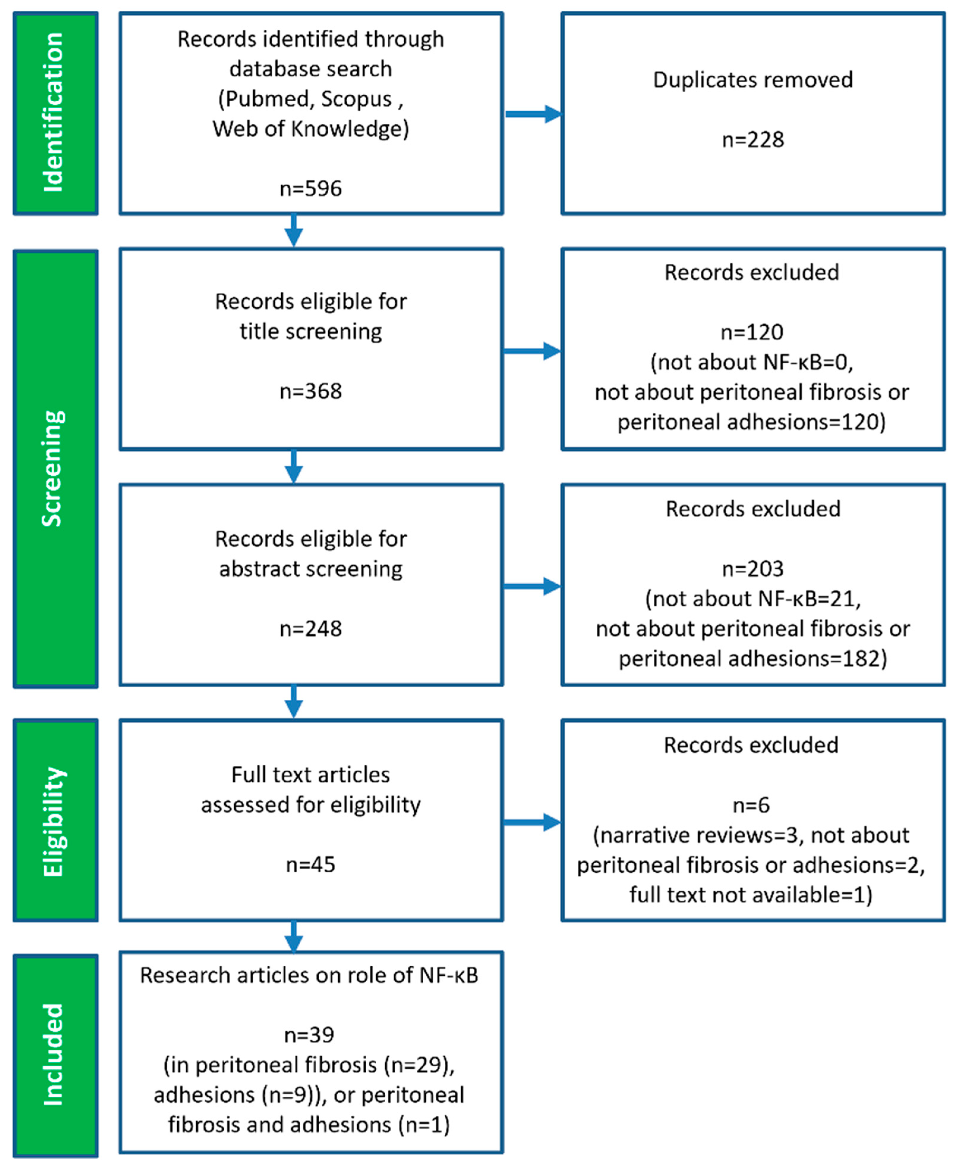
3.1. Study Selection
3.2. Research Articles on the Role of NF-κB in Peritoneal Fibrosis
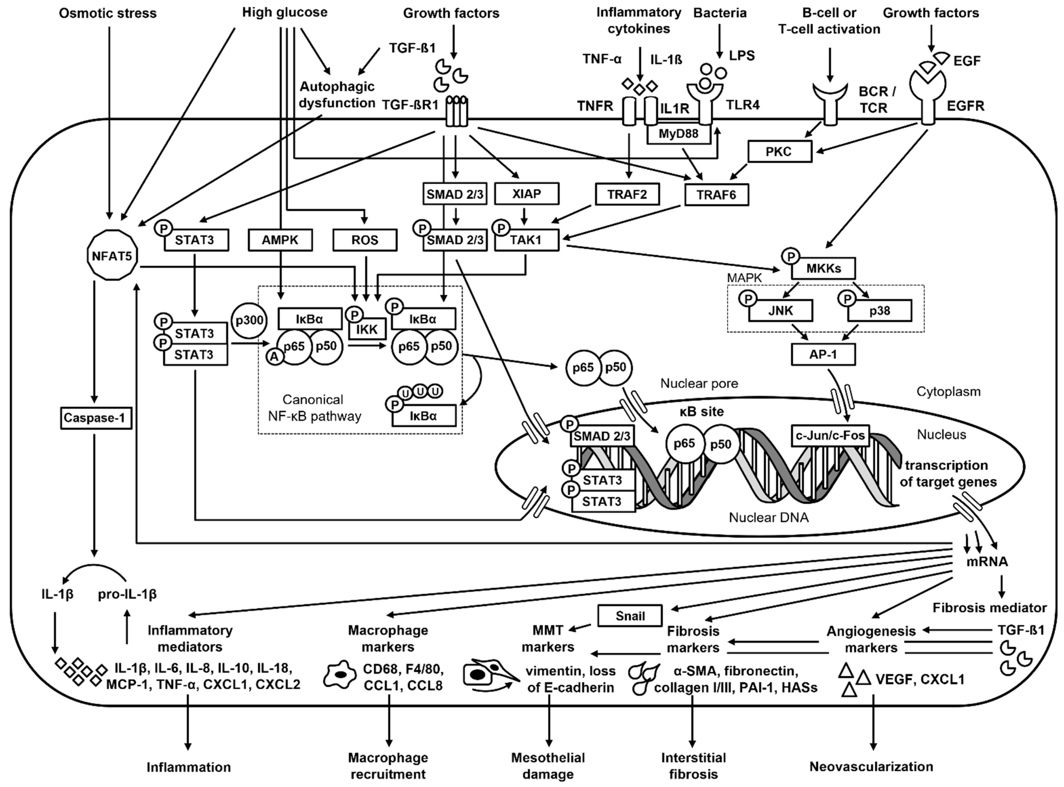
3.2.1. NF-κB-Mediated Regulation of Peritoneal Fibrosis
3.2.2. NF-κB-Targeted Therapy for the Prevention of Peritoneal Fibrosis
3.3. Research Articles on the Role of NF-κB in Peritoneal Adhesions
3.3.1. NF-κB-Mediated Regulation of Peritoneal Adhesions
3.3.2. NF-κB-Targeted Therapy for the Prevention of Peritoneal Adhesions
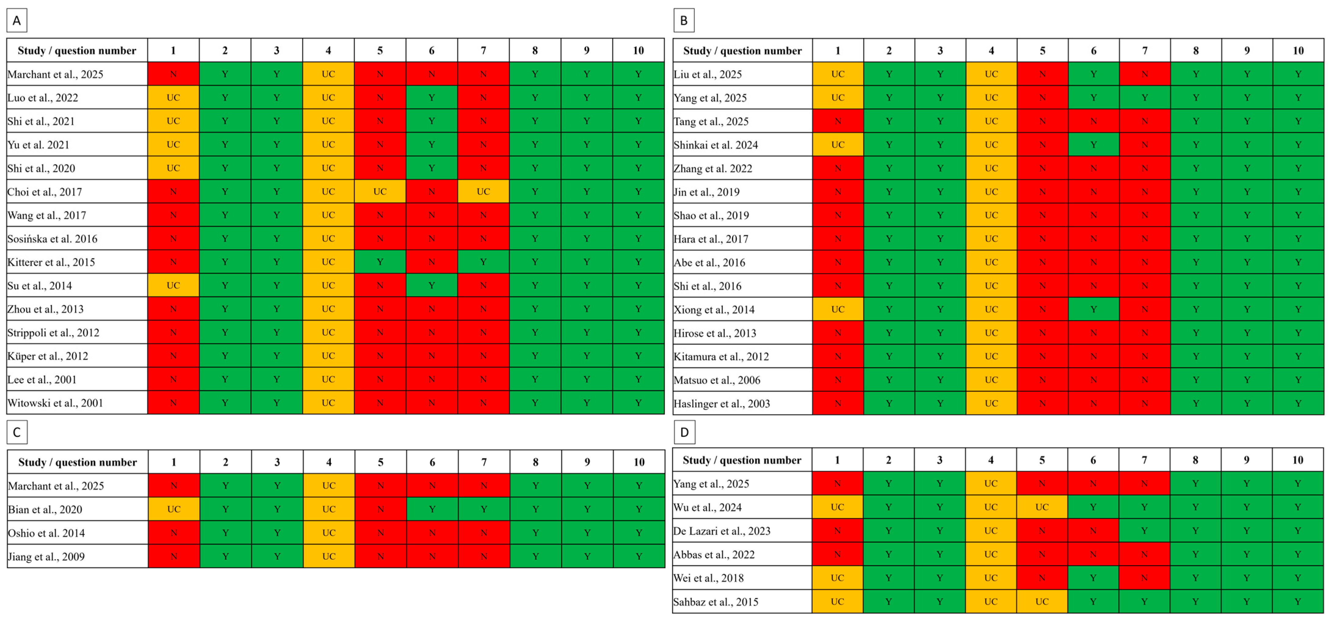
3.4. Risks of Bias Assessment
4. Discussion
5. Conclusions
Supplementary Materials
Author Contributions
Funding
Data Availability Statement
Conflicts of Interest
References
- Kramann, R.; Floege, J.; Ketteler, M.; Marx, N.; Brandenburg, V.M. Medical options to fight mortality in end-stage renal disease: A review of the literature. Nephrol. Dial. Transplant. 2012, 27, 4298–4307. [Google Scholar] [CrossRef]
- Kalantar-Zadeh, K.; Jafar, T.H.; Nitsch, D.; Neuen, B.L.; Perkovic, V. Chronic kidney disease. Lancet 2021, 398, 786–802. [Google Scholar] [CrossRef]
- Francis, A.; Harhay, M.N.; Ong, A.C.; Tummalapalli, S.L.; Ortiz, A.; Fogo, A.B.; Fliser, D.; Roy-Chaudhury, P.; Fontana, M.; Nangaku, M.; et al. Chronic kidney disease and the global public health agenda: An international consensus. Nat. Rev. Nephrol. 2024, 20, 473–485. [Google Scholar] [CrossRef]
- Purnell, T.S.; Auguste, P.; Crews, D.C.; Lamprea-Montealegre, J.; Olufade, T.; Greer, R.; Ephraim, P.; Sheu, J.; Kostecki, B.S.; Powe, N.R.; et al. Comparison of life participation activities among adults treated by hemodialysis, peritoneal dialysis, and kidney transplantation: A systematic review. Am. J. Kidney Dis. 2013, 62, 953–973. [Google Scholar] [CrossRef] [PubMed]
- Maruyama, Y.; Higuchi, C.; Io, H.; Wakabayashi, K.; Tsujimoto, H.; Tsujimoto, Y.; Yuasa, H.; Ryuzaki, M.; Ito, Y.; Nakamoto, H. Comparison of peritoneal dialysis and hemodialysis as first renal replacement therapy in patients with end-stage renal disease and diabetes: A systematic review. Ren. Replace. Ther. 2019, 5, 44. [Google Scholar] [CrossRef]
- Krediet, R.T.; Struijk, D.G. Peritoneal changes in patients on long-term peritoneal dialysis. Nat. Rev. Nephrol. 2013, 9, 419–429. [Google Scholar] [CrossRef] [PubMed]
- Fusshoeller, A. Histomorphological and functional changes of the peritoneal membrane during long-term peritoneal dialysis. Pediatr. Nephrol. 2008, 23, 19–25. [Google Scholar] [CrossRef] [PubMed]
- Morelle, J.; Lambie, M.; Öberg, C.M.; Davies, S. The peritoneal membrane and its role in peritoneal dialysis. Clin. J. Am. Soc. Nephrol. 2024, 19, 244–253. [Google Scholar] [CrossRef]
- Klingele, M.; Carstens, M.; Baerens, L.; Laschke, M.W.; Metzger, W.; Fliser, D.; Meier, C.M. A material stress test study on occurrence of leakage and material failure of peritoneal dialysis (PD) catheters. Sci. Rep. 2021, 11, 10212. [Google Scholar] [CrossRef]
- Marchant, V.; García-Giménez, J.; González-Mateo, G.T.; Sandoval, P.; Tejedor-Santamaria, L.; Rayego-Mateos, S.; Ramos, R.; Jimenez-Heffernan, J.A.; Ortiz, A.; Raby, A.C.; et al. STING inhibition alleviates experimental peritoneal damage: Potential therapeutic relevance for peritoneal dialysis. J. Pathol. 2025, 267, 196–212. [Google Scholar] [CrossRef]
- Terri, M.; Trionfetti, F.; Montaldo, C.; Cordani, M.; Tripodi, M.; Lopez-Cabrera, M.; Strippoli, R. Mechanisms of peritoneal fibrosis: Focus on immune cells–peritoneal stroma interactions. Front. Immunol. 2021, 12, 607204. [Google Scholar] [CrossRef] [PubMed]
- Ito, T.; Yorioka, N. Peritoneal damage by peritoneal dialysis solutions. Clin. Exp. Nephrol. 2008, 12, 243–249. [Google Scholar] [CrossRef]
- Low, S.; Liew, A. Peritoneal dialysis fluids. Semin. Dial. 2022, 37, 10–23. [Google Scholar] [CrossRef] [PubMed]
- Bonomini, M.; Zammit, V.; Divino-Filho, J.C.; Davies, S.J.; Di Liberato, L.; Arduini, A.; Lambie, M. The osmo-metabolic approach: A novel and tantalizing glucose-sparing strategy in peritoneal dialysis. J. Nephrol. 2021, 34, 503–519. [Google Scholar] [CrossRef]
- Liakakos, T.; Thomakos, N.; Fine, P.M.; Dervenis, C.; Young, R.L. Peritoneal adhesions: Etiology, pathophysiology, and clinical significance: Recent advances in prevention and management. Dig. Surg. 2001, 18, 260–273. [Google Scholar] [CrossRef]
- Qureshi, M.A.; Maierean, S.; Crabtree, J.H.; Clarke, A.; Armstrong, S.; Fissell, R.; Jain, A.K.; Jassal, S.V.; Hu, S.L.; Kennealey, P.; et al. The association of intra-abdominal adhesions with peritoneal dialysis catheter-related complications. Clin. J. Am. Soc. Nephrol. 2024, 19, 472–482. [Google Scholar]
- Williams, J.D.; Craig, K.J.; Topley, N.; Von Ruhland, C.; Fallon, M.; Newman, G.R.; Mackenzie, R.K.; Williams, G.T. Morphologic changes in the peritoneal membrane of patients with renal disease. J. Am. Soc. Nephrol. 2002, 13, 470–479. [Google Scholar] [CrossRef]
- Strippoli, R.; Moreno-Vicente, R.; Battistelli, C.; Cicchini, C.; Noce, V.; Amicone, L.; Marchetti, A.; del Pozo, M.A.; Tripodi, M. Molecular mechanisms underlying peritoneal EMT and fibrosis. Stem Cells Int. 2016, 2016, 3543678. [Google Scholar] [CrossRef]
- Stone, R.C.; Pastar, I.; Ojeh, N.; Chen, V.; Liu, S.; Garzon, K.I.; Tomic-Canic, M. Epithelial-mesenchymal transition in tissue repair and fibrosis. Cell Tissue. Res. 2016, 365, 495–506. [Google Scholar] [CrossRef] [PubMed]
- Puisieux, A.; Brabletz, T.; Caramel, J. Oncogenic roles of EMT-inducing transcription factors. Nat. Cell Biol. 2014, 16, 488–494. [Google Scholar] [CrossRef] [PubMed]
- López-Cabrera, M. Mesenchymal conversion of mesothelial cells is a key event in the pathophysiology of the peritoneum during peritoneal dialysis. Adv. Med. 2014, 2014, 473134. [Google Scholar] [CrossRef]
- Xu, J.; Lamouille, S.; Derynck, R. TGF-β-induced epithelial to mesenchymal transition. Cell Res. 2009, 19, 156–172. [Google Scholar] [CrossRef] [PubMed]
- de Lima, S.M.A.; Otoni, A.; de Paula Sabino, A.; Dusse, L.M.S.A.; Gomes, K.B.; Pinto, S.W.L.; Marinho, M.A.S.; Rios, D.R.A. Inflammation, neoangiogenesis and fibrosis in peritoneal dialysis. Clin. Chim. Acta 2013, 421, 46–50. [Google Scholar] [CrossRef]
- Kalluri, R.; Weinberg, R.A. The basics of epithelial-mesenchymal transition. J. Clin. Investig. 2009, 119, 1420–1428. [Google Scholar] [CrossRef]
- Li, M.; Luan, F.; Zhao, Y.; Hao, H.; Zhou, Y.; Han, W.; Fu, X. Epithelial-mesenchymal transition: An emerging target in tissue fibrosis. Exp. Biol. Med. 2016, 241, 1–13. [Google Scholar] [CrossRef]
- Williams, J.D.; Craig, K.J.; von Ruhland, C.; Topley, N.; Williams, G.T.; Biopsy Registry Study Group. The natural course of peritoneal membrane biology during peritoneal dialysis. Kidney Int. 2003, 64, S43–S49. [Google Scholar]
- Biernacka, A.; Dobaczewski, M.; Frangogiannis, N.G. TGF-β signaling in fibrosis. Growth Factors 2011, 29, 196–202. [Google Scholar] [CrossRef]
- Davies, S.J.; Mushahar, L.; Yu, Z.; Lambie, M. Determinants of Peritoneal Membrane Function over Time. Semin. Nephrol. 2011, 31, 172–182. [Google Scholar] [CrossRef]
- Seo, M.J.; Oh, S.J.; Kim, S.I.; Cho, K.W.; Jo, I.; Schaub, T.; Schilling, H.; Passlick-Deetjen, J.; Han, D.C. High glucose dialysis solutions increase synthesis of vascular endothelial growth factors by peritoneal vascular endothelial cells. Perit. Dial. Int. 2001, 21, 35–40. [Google Scholar]
- Kariya, T.; Nishimura, H.; Mizuno, M.; Suzuki, Y.; Matsukawa, Y.; Sakata, F.; Maruyama, S.; Takei, Y.; Ito, Y. TGF-β1-VEGF-A pathway induces neoangiogenesis with peritoneal fibrosis in patients undergoing peritoneal dialysis. Am. J. Physiol. Renal Physiol. 2018, 314, F167–F180. [Google Scholar] [CrossRef] [PubMed]
- Davies, S.J. Longitudinal relationship between solute transport and ultrafiltration capacity in peritoneal dialysis patients. Kidney Int. 2004, 66, 2437–2445. [Google Scholar] [CrossRef] [PubMed]
- Kinashi, H.; Ito, Y.; Mizuno, M.; Suzuki, Y.; Terabayashi, T.; Nagura, F.; Hattori, R.; Matsukawa, Y.; Mizuno, T.; Noda, Y.; et al. TGF-β1 promotes lymphangiogenesis during peritoneal fibrosis. J. Am. Soc. Nephrol. 2013, 24, 1627–1642. [Google Scholar] [CrossRef]
- Wynn, T.A.; Vannella, K.M. Macrophages in tissue repair, regeneration, and fibrosis. Immunity 2016, 44, 450–462. [Google Scholar] [CrossRef] [PubMed]
- Deshmane, S.L.; Kremlev, S.; Amini, S.; Sawaya, B.E. Monocyte chemoattractant protein-1 (MCP-1): An overview. J. Interferon Cytokine Res. 2009, 29, 313–326. [Google Scholar] [CrossRef]
- Li, F.K.; Davenport, A.; Robson, R.L.; Loetscher, P.; Rothlein, R.; Williams, J.D.; Topley, N. Leukocyte migration across human peritoneal mesothelial cells is dependent on directed chemokine secretion and ICAM-1 expression. Kidney Int. 1998, 54, 2170–2183. [Google Scholar] [CrossRef]
- Yung, S.; Chan, T.M. Pathophysiological changes to the peritoneal membrane during PD-related peritonitis: The role of mesothelial cells. Mediat. Inflamm. 2012, 2012, 484167. [Google Scholar] [CrossRef]
- Rollins, B.J.; Walz, A.; Baggiolini, M. Recombinant human MCP-1/JE induces chemotaxis, calcium flux, and the respiratory burst in human monocytes. Blood 1991, 78, 1112–1116. [Google Scholar] [CrossRef] [PubMed]
- Strippoli, R.; Benedicto, I.; Perez Lozano, M.L.; Cerezo, A.; López-Cabrera, M.; del Pozo, M.A. Epithelial-to-mesenchymal transition of peritoneal mesothelial cells is regulated by an ERK/NF-κB/Snail1 pathway. Dis. Models Mech. 2008, 1, 264–274. [Google Scholar] [CrossRef]
- Strippoli, R.; Benedicto, I.; Foronda, M.; Perez-Lozano, M.L.; Sánchez-Perales, S.; López-Cabrera, M.; Del Pozo, M.Á. p38 maintains E-cadherin expression by modulating TAK1-NF-κB during epithelial-to-mesenchymal transition. J. Cell Sci. 2010, 123, 4321–4331. [Google Scholar] [CrossRef]
- Kitterer, D.; Latus, J.; Ulmer, C.; Fritz, P.; Biegger, D.; Ott, G.; Alscher, M.D.; Witowski, J.; Kawka, E.; Jorres, A.; et al. Activation of nuclear factor of activated T cells 5 in the peritoneal membrane of uremic patients. Am. J. Physiol. Renal Physiol. 2015, 308, F1247–F1258. [Google Scholar]
- Zhang, Y.; Feng, W.; Peng, X.; Zhu, L.; Wang, Z.; Shen, H.; Chen, C.; Xiao, L.; Li, S.; Zhao, Y.; et al. Parthenolide alleviates peritoneal fibrosis by inhibiting inflammation via the NF-κB/TGF-β/Smad signaling axis. Lab. Investig. 2022, 102, 1346–1354. [Google Scholar] [CrossRef]
- Karin, M.; Ben-Neriah, Y. Phosphorylation meets ubiquitination: The control of NF-κB activity. Annu. Rev. Immunol. 2000, 18, 621–663. [Google Scholar] [CrossRef] [PubMed]
- Napetschnig, J.; Wu, H. Molecular basis of NF-κB signaling. Annu. Rev. Biophys. 2013, 42, 443–468. [Google Scholar] [CrossRef] [PubMed]
- Li, Z.; Zhang, L.; He, W.; Zhu, C.; Yang, J.; Sheng, M. Astragalus membranaceus inhibits peritoneal fibrosis via monocyte chemoattractant protein (MCP)-1 and the transforming growth factor-β1 (TGF-β1) pathway in rats submitted to peritoneal dialysis. Int. J. Mol. Sci. 2014, 15, 12959–12971. [Google Scholar] [CrossRef] [PubMed]
- Xu, L.; Liu, N.A.; Gu, H.; Wang, H.; Shi, Y.; Ma, X.; Ma, S.; Ni, J.; Tao, M.; Qio, A.; et al. Histone deacetylase 6 inhibition counteracts the epithelial-mesenchymal transition of peritoneal mesothelial cells and prevents peritoneal fibrosis. Oncotarget 2017, 8, 88730. [Google Scholar] [CrossRef]
- Sun, M.; Dawood, F.; Wen, W.H.; Chen, M.; Dixon, I.; Kirshenbaum, L.A.; Liu, P.P. Excessive tumor necrosis factor activation after infarction contributes to susceptibility of myocardial rupture and left ventricular dysfunction. Circulation 2004, 110, 3221–3228. [Google Scholar] [CrossRef]
- Hoesel, B.; Schmid, J.A. The complexity of NF-κB signaling in inflammation and cancer. Mol. Cancer 2013, 12, 86. [Google Scholar]
- Ghosh, S.; Hayden, M.S. New regulators of NF-κB in inflammation. Nat. Rev. Immunol. 2008, 8, 837–848. [Google Scholar] [CrossRef]
- Luedde, T.; Schwabe, R.F. NF-κB in the liver-linking injury, fibrosis and hepatocellular carcinoma. Nat. Rev. Gastroenterol. Hepatol. 2011, 8, 108–118. [Google Scholar] [CrossRef]
- Alharbi, K.S.; Fuloria, N.K.; Fuloria, S.; Rahman, S.B.; Al-Malki, W.H.; Shaikh, M.A.J.; Thangavelu, L.; Singh, S.K.; Allam, V.S.R.R.; Jha, N.K.; et al. Nuclear factor-kappa B and its role in inflammatory lung disease. Chem. Biol. Interact. 2021, 345, 109568. [Google Scholar] [CrossRef]
- Zdrojkowski, Ł.; Jasiński, T.; Ferreira-Dias, G.; Pawliński, B.; Domino, M. The role of NF-κB in endometrial diseases in humans and animals: A review. Int. J. Mol. Sci. 2023, 24, 2901. [Google Scholar] [CrossRef]
- Giridharan, S.; Srinivasan, M. Mechanisms of NF-κB p65 and strategies for therapeutic manipulation. J. Inflamm. Res. 2018, 11, 407–419. [Google Scholar] [CrossRef] [PubMed]
- Margetts, P.J.; Churchill, D.N. Acquired ultrafiltration dysfunction in peritoneal dialysis patients. J. Am. Soc. Nephrol. 2002, 13, 2787–2794. [Google Scholar] [CrossRef] [PubMed]
- Mujais, S.; Nolph, K.; Gokal, R.; Blake, P.; Burkart, J.; Coles, G.; Kawaquchi, Y.; Kawanishi, H.; Korbet, S.; Krediet, R.; et al. Evaluation and management of ultrafiltration problems in peritoneal dialysis. Perit. Dial. Int. 2000, 20, 5–21. [Google Scholar] [CrossRef]
- Descœudres, B.; Koller, M.T.; Garzoni, D.; Wolff, T.; Steiger, J.; Schaub, S.; Mayr, M. Contribution of early failure to outcome on peritoneal dialysis. Perit. Dial. Int. 2008, 28, 259–267. [Google Scholar] [CrossRef]
- Zhou, Q.; Bajo, M.A.; Del Peso, G.; Yu, X.; Selgas, R. Preventing peritoneal membrane fibrosis in peritoneal dialysis patients. Kidney Int. 2016, 90, 515–524. [Google Scholar] [CrossRef]
- Cheng, B.C.; Tsai, N.W.; Lai, Y.R.; Huang, C.C.; Lu, C.H. Impact of intra-abdominal adhesion on dialysis outcome in peritoneal dialysis patients. Biomed Res. Int. 2018, 2018, 1978765. [Google Scholar] [CrossRef]
- Filler, G.; Haig, A.; Merritt, N.; Alvarez-Elias, A.C.; Teoh, C.W.; Filler, T.J.; Díaz-González de Ferris, M.E. Histopathological changes of long-term peritoneal dialysis using physiological solutions: A case report and review of the literature. Blood Purif. 2022, 51, 60–67. [Google Scholar] [CrossRef]
- Moher, D.; Liberati, A.; Tetzlaff, J.; Altman, D.G. Preferred reporting items for systematic reviews and meta-analyses: The PRISMA statement. BMJ 2009, 339, b2535. [Google Scholar] [CrossRef]
- Ryan, R.; Synnot, A.; Prictor, M.; Hill, S.; Cochrane Consumers and Communication Group. Data Extraction Template for Included Studies; The Cochrane Collaboration: London, UK, 2016; pp. 1–25. Available online: https://cccrg.cochrane.org/sites/cccrg.cochrane.org/files/uploads/det_2015_revised_final_june_20_2016_nov_29_revised.doc (accessed on 25 April 2025).
- Hooijmans, C.R.; Rovers, M.M.; De Vries, R.B.; Leenaars, M.; Ritskes-Hoitinga, M.; Langendam, M.W. SYRCLE’s risk of bias tool for animal studies. BMC Med. Res. Methodol. 2014, 14, 43. [Google Scholar] [CrossRef]
- Mondello, S.; Mazzon, E.; Di Paola, R.; Crisafulli, C.; Mondello, P.; Buemi, M.; Aloisi, C.; Cuzzocrea, S. Thalidomide suppresses sclerosing encapsulating peritonitis in a rat experimental model. Shock 2009, 32, 332–339. [Google Scholar] [CrossRef]
- Wu, J.; Yang, X.; Zhang, Y.F.; Zhou, S.F.; Zhang, R.; Dong, X.Q.; Fan, J.J.; Liu, M.; Yu, X.Q. Angiotensin II upregulates Toll-like receptor 4 and enhances lipopolysaccharide-induced CD40 expression in rat peritoneal mesothelial cells. Inflamm. Res. 2009, 58, 473–482. [Google Scholar] [CrossRef]
- Morinelli, T.A.; Luttrell, L.M.; Strungs, E.G.; Ullian, M.E. Angiotensin II receptors and peritoneal dialysis-induced peritoneal fibrosis. Int. J. Biochem. Cell Biol. 2016, 77, 240–250. [Google Scholar] [CrossRef] [PubMed]
- Artunc, F.; Lang, F. Mineralocorticoid and SGK1-sensitive inflammation and tissue fibrosis. Nephron Physiol. 2014, 128, 35–39. [Google Scholar] [CrossRef] [PubMed]
- Aliakbarian, M.; Khodashahi, R.; Tavakkoli, M.; Ashrafzadeh, K.; Rahimi, H.; Khaleghi, E.; Ghayour-Mobarhan, M.; Arjmand, M.H. Review the Role of Metabolism Reprogramming in the Pathogenesis of Post-surgical Adhesion: A New Therapeutic Strategy. Curr. Top. Med. Chem. 2023, 23, 2527–2534. [Google Scholar] [CrossRef]
- Liu, H.; Xu, H.; Sun, H.; Xu, H.; Han, J.; Zhao, L.; Li, X.; Sun, X.X.; Dong, X.; Xu, R.; et al. Tetrahydroberberrubine prevents peritoneal adhesion by suppressing inflammation and extracellular matrix accumulation. Eur. J. Pharmacol. 2023, 954, 175803. [Google Scholar] [CrossRef]
- Witowski, J.; Thiel, A.; Dechend, R.; Dunkel, K.; Fouquet, N.; Bender, T.O.; Langrehr, J.M.; Gahl, G.M.; Frei, U.; Jorres, A. Synthesis of CXC and CC chemokines by human peritoneal fibroblasts: Induction by macrophage-derived cytokines. Am. J. Pathol. 2001, 158, 1441–1450. [Google Scholar] [CrossRef] [PubMed]
- Sosińska, P.; Baum, E.; Maćkowiak, B.; Staniszewski, R.; Jasinski, T.; Umezawa, K.; Bręborowicz, A. Inhibition of NF-kappaB with Dehydroxymethylepoxyquinomicin modifies the function of human peritoneal mesothelial cells. Am. J. Transl. Res. 2016, 8, 5756. [Google Scholar]
- Lee, S.K.; Kim, B.S.; Yang, W.S.; Kim, S.B.; Park, S.K.; Park, J.S. High glucose induces MCP-1 expression partly via tyrosine kinase-AP-1 pathway in peritoneal mesothelial cells. Kidney Int. 2001, 60, 55–64. [Google Scholar] [CrossRef]
- Küper, C.; Beck, F.X.; Neuhofer, W. NFAT5 Contributes to Osmolality-Induced MCP-1 Expression in Mesothelial Cells. Mediators Inflamm. 2012, 2012, 513015. [Google Scholar] [CrossRef]
- Choi, S.Y.; Ryu, H.M.; Choi, J.Y.; Cho, J.H.; Kim, C.D.; Kim, Y.L.; Park, S.H. The role of Toll-like receptor 4 in high-glucose-induced inflammatory and fibrosis markers in human peritoneal mesothelial cells. Int. Urol. Nephrol. 2017, 49, 171–181. [Google Scholar] [CrossRef] [PubMed]
- Zhou, G.; Su, X.; Ma, J.; Wang, L.; Li, D. Pioglitazone inhibits high glucose-induced synthesis of extracellular matrix by NF-κB and AP-1 pathways in rat peritoneal mesothelial cells. Mol. Med. Rep. 2013, 7, 1336–1342. [Google Scholar] [CrossRef]
- Su, X.; Zhou, G.; Wang, Y.; Yang, X.; Li, L.; Yu, R.; Li, D. The PPARβ/δ agonist GW501516 attenuates peritonitis in peritoneal fibrosis via inhibition of TAK1-NFκB pathway in rats. Inflammation 2014, 37, 729–737. [Google Scholar] [CrossRef]
- Luo, Q.; Liu, M.; Tan, Y.; Chen, J.; Zhang, W.; Zhong, S.; Pan, J.; Zheng, Q.; Gong, L.; Su, L.; et al. Blockade of prostaglandin E2 receptor 4 ameliorates peritoneal dialysis-associated peritoneal fibrosis. Front. Pharmacol. 2022, 13, 1004619. [Google Scholar] [CrossRef]
- Strippoli, R.; Benedicto, I.; Perez Lozano, M.L.; Pellinen, T.; Sandoval, P.; Lopez-Cabrera, M.; Del Pozo, M.A. Inhibition of transforming growth factor-activated kinase 1 (TAK1) blocks and reverses epithelial to mesenchymal transition of mesothelial cells. PLoS ONE 2012, 7, e31492. [Google Scholar] [CrossRef]
- Wang, J.; Wang, L.; Xu, L.; Shi, Y.; Liu, F.; Qi, H.; Liu, N.; Zhuang, S. Targeting Src attenuates peritoneal fibrosis and inhibits the epithelial to mesenchymal transition. Oncotarget 2017, 8, 83872. [Google Scholar] [CrossRef]
- Shi, Y.; Hu, Y.; Wang, Y.; Ma, X.; Tang, L.; Tao, M.; Qiu, A.; Zhuang, S.; Liu, N. Blockade of autophagy prevents the development and progression of peritoneal fibrosis. Front. Pharmacol. 2021, 12, 724141. [Google Scholar] [CrossRef]
- Shi, Y.; Tao, M.; Wang, Y.; Zang, X.; Ma, X.; Qiu, A.; Zhuang, S.; Liu, N. Genetic or pharmacologic blockade of enhancer of zeste homolog 2 inhibits the progression of peritoneal fibrosis. J. Pathol. 2020, 250, 79–94. [Google Scholar] [PubMed]
- Yu, C.; Yang, N.; Wang, W.; Du, X.; Tang, Q.; Lin, H.; Li, L. Blocking core fucosylation of epidermal growth factor (EGF) receptor prevents peritoneal fibrosis progression. Ren. Fail. 2021, 43, 869–877. [Google Scholar] [CrossRef]
- Haslinger, B.; Kleemann, R.; Toet, K.H.; Kooistra, T. Simvastatin suppresses tissue factor expression and increases fibrinolytic activity in tumor necrosis factor-α-activated human peritoneal mesothelial cells. Kidney Int. 2003, 63, 2065–2074. [Google Scholar] [CrossRef] [PubMed]
- Matsuo, H.; Tamura, M.; Kabashima, N.; Serino, R.; Tokunaga, M.; Shibata, T.; Matsumoto, M.; Aijima, M.; Oikawa, S.; Anai, H.; et al. Prednisolone inhibits hyperosmolarity-induced expression of MCP-1 via NF-κB in peritoneal mesothelial cells. Kidney Int. 2006, 69, 736–746. [Google Scholar] [CrossRef] [PubMed]
- Hara, K.; Hamada, C.; Wakabayashi, K.; Kanda, R.; Kaneko, K.; Horikoshi, S.; Tomino, Y.; Suzuki, Y. Scavenging of reactive oxygen species by astaxanthin inhibits epithelial-mesenchymal transition in high glucose-stimulated mesothelial cells. PLoS ONE 2017, 12, e0184332. [Google Scholar] [CrossRef]
- Liu, K.; Huang, Y.; Xiao, W.; Liao, J.; Ouyang, S.; Liang, Y.; Fu, J. Resveratrol glycoside inhibits NLRP3/IL-1β/NF-κB to alleviate peritoneal fibrosis in peritoneal dialysis. Clin. Exp. Nephrol. 2025, 29, 1331–1341. [Google Scholar] [CrossRef]
- Kitamura, M.; Nishino, T.; Obata, Y.; Furusu, A.; Hishikawa, Y.; Koji, T.; Kohno, S. Epigallocatechin gallate suppresses peritoneal fibrosis in mice. Chem. Biol. Interact. 2012, 195, 95–104. [Google Scholar] [CrossRef]
- Tang, Y.; Wang, Y.; Sun, X.; Zheng, J.; Fang, J.; Liu, S.; Luo, J.; Liao, Y.; Cheng, Y.; Lu, J.; et al. Shenbing Decoction III and Apigenin Improve Peritoneal Fibrosis Mediated by Epithelial-Mesenchymal Transition Through TAK1/p38MAPK/NF-κB Pathways. J. Food Biochem. 2025, 2025, 7841565. [Google Scholar] [CrossRef]
- Jin, G.; Su, Y.; Dong, Q.; Zhao, X.; Zhang, L.; Yan, X. Arctigenin alleviates TGF-β1-induced epithelial-mesenchymal transition and PAI-1 expression via AMPK/NF-κB pathway in peritoneal mesothelial cells. Biochem. Biophys. Res. Commun. 2019, 520, 413–419. [Google Scholar] [PubMed]
- Shinkai, Y.; Sasaki, K.; Tamura, R.; Ike, T.; Takahashi, A.; Osaki, Y.; Ishiuchi, N.; Maeoka, Y.; Nakashima, A.; Masaki, T. Selective activation of PPARα by pemafibrate mitigates peritoneal inflammation and fibrosis through suppression of NLRP3 inflammasome and modulation of inflammation. Sci. Rep. 2024, 14, 23816. [Google Scholar] [CrossRef]
- Hirose, M.; Nishino, T.; Obata, Y.; Nakazawa, M.; Nakazawa, Y.; Furusu, A.; Abe, K.; Miyazaki, M.; Koji, T.; Kohno, S. 22-Oxacalcitriol prevents progression of peritoneal fibrosis in a mouse model. Perit. Dial. Int. 2013, 33, 132–142. [Google Scholar] [CrossRef]
- Xiong, C.; Liu, N.; Fang, L.; Zhuang, S.; Yan, H. Suramin inhibits the development and progression of peritoneal fibrosis. J. Pharmacol. Exp. Ther. 2014, 351, 373–382. [Google Scholar] [CrossRef]
- Abe, S.; Obata, Y.; Oka, S.; Koji, T.; Nishino, T.; Izumikawa, K. Chondroitin sulfate prevents peritoneal fibrosis in mice by suppressing NF-κB activation. Med. Mol. Morphol. 2016, 49, 144–153. [Google Scholar] [CrossRef] [PubMed]
- Yang, C.C.; Yue, Y.; Wang, Y.T.; Chiang, J.Y.; Cheng, B.C.; Hsu, T.W.; Chen, Y.L.; Li, Y.C.; Yip, H.K. Dulaglutide markedly prevents peritoneal fibrosis in a rodent model of chronic kidney disease: Insights into the pathogenesis. Int. J. Mol. Sci. 2025, 56, 151. [Google Scholar] [CrossRef]
- Shi, S.; Zhang, Y.; Wen, W.; Zhao, Y.; Sun, L. Molecular mechanisms of melatonin in the reversal of LPS-induced EMT in peritoneal mesothelial cells. Mol. Med. Rep. 2016, 14, 4342–4348. [Google Scholar] [CrossRef]
- Shao, Q.; Jiang, C.; Xia, Y.; Zhao, M.; Zhang, Q.; Jin, B.; Liu, J. Dioscin ameliorates peritoneal fibrosis by inhibiting epithelial-to-mesenchymal transition of human peritoneal mesothelial cells via the TLR4/MyD88/NF-κB signaling pathway. Int. J. Clin. Exp. Pathol. 2019, 12, 867. [Google Scholar] [PubMed]
- Jiang, Z.L.; Fletcher, N.M.; Diamond, M.P.; Abu-Soud, H.M.; Saed, G.M. Hypoxia regulates iNOS expression in human normal peritoneal and adhesion fibroblasts through nuclear factor kappa B activation mechanism. Fertil. Steril. 2009, 91, 616–621. [Google Scholar] [CrossRef]
- Oshio, T.; Kawashima, R.; Kawamura, Y.I.; Hagiwara, T.; Mizutani, N.; Okada, T.; Otsubo, T.; Inagaki-Ohara, K.; Matsukawa, A.; Haga, T.; et al. Chemokine receptor CCR8 is required for lipopolysaccharide-triggered cytokine production in mouse peritoneal macrophages. PLoS ONE 2014, 9, e94445. [Google Scholar] [CrossRef]
- Bian, Y.Y.; Yang, L.L.; Yan, Y.; Zhao, M.; Chen, Y.Q.; Zhou, Y.Q.; Wang, Z.X.; Li, W.L.; Zeng, L. Identification of candidate biomarkers correlated with pathogenesis of postoperative peritoneal adhesion by using microarray analysis. World J. Gastrointest. Oncol. 2020, 12, 54. [Google Scholar] [CrossRef] [PubMed]
- Sahbaz, A.; Aynioglu, O.; Isik, H.; Gulle, K.; Ferah, M.A.; Sahbaz, H.C. Cholecalciferol (vitamin D3) prevents postoperative adhesion formation by inactivating the nuclear factor kappa B pathway: A randomized experimental study. J. Surg. Res. 2015, 198, 252–259. [Google Scholar] [CrossRef]
- Wei, G.; Wu, Y.; Gao, Q.; Shen, C.; Chen, Z.; Wang, K.; Yu, J.; Li, X.; Sun, X. Gallic acid attenuates postoperative intra-abdominal adhesion by inhibiting inflammatory reaction in a rat model. Med. Sci. Monit. 2018, 24, 827. [Google Scholar] [CrossRef]
- Abbas, N.A.; Hassan, H.A. The protective and therapeutic effects of 5-androstene3β, 17β-diol (ADIOL) in abdominal post-operative adhesions in rat: Suppressing TLR4/NFκB/HMGB1/TGF1 β/α SMA pathway. Int. Immunopharmacol. 2022, 109, 108801. [Google Scholar] [CrossRef]
- De Lazari, M.G.T.; Viana, C.T.R.; Pereira, L.X.; Orellano, L.A.A.; Ulrich, H.; Andrade, S.P.; Campos, P.P. Sodium butyrate attenuates peritoneal fibroproliferative process in mice. Exp. Physiol. 2023, 108, 146–157. [Google Scholar] [CrossRef] [PubMed]
- Wu, S.; Yuan, Z.; Xie, P.; Shafiq, M.; Hou, J.; Liang, Y.; Hashim, R.; Zhang, W.; Yang, R.; Mo, X.; et al. Lecithin-complexed oregano essential oil-encapsulated fibrous barriers prevent postoperative adhesions by regulating Nrf2/NF-κB signaling pathways. Appl. Mater. Today 2024, 38, 102185. [Google Scholar] [CrossRef]
- Yang, S.; Qu, C.; Yuan, X.; Liu, K.; Deng, K.; Du, J.; Wang, Y.; Xu, M.; Zhang, L.; Zhang, M.; et al. Biomimetic hydrogel microsphere encapsulated superoxide dismutase enzymes alleviate postoperative peritoneal adhesion by modulating oxidative stress-inflammation cycles via Piezo1 mechanosensitive channels. Chem. Eng. J. 2025, 524, 169275. [Google Scholar] [CrossRef]
- Leavesley, D.I.; Stanley, J.M.; Faull, R.J. Epidermal growth factor modifies the expression and function of extracellular matrix adhesion receptors expressed by peritoneal mesothelial cells from patients on CAPD. Nephrol. Dial. Transplant. 1999, 14, 1208–1216. [Google Scholar] [CrossRef]
- Yang, X.; Bao, M.; Fang, Y.; Yu, X.; Ji, J.; Ding, X. STAT3/HIF-1α signaling activation mediates peritoneal fibrosis induced by high glucose. J. Transl. Med. 2021, 19, 283. [Google Scholar] [CrossRef]
- Roth, I.; Leroy, V.; Kwon, H.M.; Martin, P.Y.; Féraille, E.; Hasler, U. Osmoprotective transcription factor NFAT5/TonEBP modulates nuclear factor-κB activity. Mol. Biol. Cell 2010, 21, 3459–3474. [Google Scholar]
- Lee, C.; Cheung, S.T. STAT3: An emerging therapeutic target for hepatocellular carcinoma. Cancers 2019, 11, 1646. [Google Scholar] [CrossRef]
- Hirata, Y.; Takahashi, M.; Morishita, T.; Noguchi, T.; Matsuzawa, A. Post-translational modifications of the TAK1-TAB complex. Int. J. Mol. Sci. 2017, 18, 205. [Google Scholar] [CrossRef] [PubMed]
- Luo, K. Signaling cross talk between TGF-β/Smad and other signaling pathways. Cold Spring Harb. Perspect. Biol. 2017, 9, a022137. [Google Scholar] [CrossRef]
- Ishinaga, H.; Jono, H.; Lim, J.H.; Kweon, S.M.; Xu, H.; Ha, U.H.; Xu, H.; Koga, T.; Yan, C.; Feng, X.H.; et al. TGF-β induces p65 acetylation to enhance bacteria-induced NF-κB activation. EMBO J. 2007, 26, 1150–1162. [Google Scholar] [CrossRef] [PubMed]
- Yeh, Y.Y.; Chiao, C.C.; Kuo, W.Y.; Hsiao, Y.C.; Chen, Y.J.; Wei, Y.Y.; Lai, T.H.; Fong, Y.C.; Tang, C.H. TGF-β1 increases motility and αvβ3 integrin up-regulation via PI3K, Akt and NF-κB-dependent pathway in human chondrosarcoma cells. Biochem. Pharmacol. 2008, 75, 1292–1301. [Google Scholar] [CrossRef]
- Brandl, M.; Seidler, B.; Haller, F.; Adamski, J.; Schmid, R.M.; Saur, D.; Schneider, G. IKKα controls canonical TGFβ-SMAD signaling to regulate genes expressing SNAIL and SLUG during EMT in Panc1 cells. J. Cell Sci. 2010, 123, 4231–4239. [Google Scholar] [CrossRef]
- Monteleone, G.; Mann, J.; Monteleone, I.; Vavassori, P.; Bremner, R.; Fantini, M.; Blanco, G.D.V.; Tersigni, R.; Alessandroni, L.; Mann, D.; et al. A failure of transforming growth factor-β1 negative regulation maintains sustained NF-κB activation in gut inflammation. J. Biol. Chem. 2004, 279, 3925–3932. [Google Scholar]
- Freudlsperger, C.; Bian, Y.; Contag Wise, S.; Burnett, J.; Coupar, J.; Yang, X.; Chen, Z.; Van Waes, C. TGF-β and NF-κB signal pathway cross-talk is mediated through TAK1 and SMAD7 in a subset of head and neck cancers. Oncogene 2013, 32, 1549–1559. [Google Scholar] [CrossRef] [PubMed]
- Wang, J.; Yu, C.; Lyu, H.; Liu, T.; Xie, R. Macrophage extracellular traps promote peritoneal fibrosis through the ROS/TGF-β/Smad pathway under high-glucose dialysis conditions. Int. Immunopharmacol. 2025, 167, 115748. [Google Scholar] [PubMed]
- Baldwin, A.S., Jr. The NF-κB and IκB proteins: New discoveries and insights. Annu. Rev. Immunol. 1996, 14, 649–681. [Google Scholar] [CrossRef]
- Johno, H.; Ogata, R.; Nakajima, S.; Hiramatsu, N.; Kobayashi, T.; Hara, H.; Kitamura, M. Acidic stress-ER stress axis for blunted activation of NF-κB in mesothelial cell sexposed to perito neal dialysis fluid. Nephrol. Dial. Transplant. 2012, 27, 4053–4060. [Google Scholar] [CrossRef]
- Bartges, J.W. Chronic kidney disease in dogs and cats. Vet. Clin. N. Am. Small Anim. Pract. 2012, 42, 669–692. [Google Scholar] [CrossRef] [PubMed]
- O’Neill, D.G.; Elliott, J.; Church, D.B.; McGreevy, P.D.; Thomson, P.C.; Brodbelt, D.C. Chronic kidney disease in dogs in UK veterinary practices: Prevalence, risk factors, and survival. J. Vet. Intern. Med. 2013, 27, 814–821. [Google Scholar] [CrossRef]
- Olsen, E.; van Galen, G. Chronic renal failure-causes, clinical findings, treatments and prognosis. Vet. Clin. N. Am. Equine Pract. 2022, 38, 25–46. [Google Scholar] [CrossRef]
- Galen, G.V.; Olsen, E.; Siwinska, N. Biomarkers of kidney disease in horses: A review of the current literature. Animals 2022, 12, 2678. [Google Scholar] [CrossRef]
- Bersenas, A.M. A clinical review of peritoneal dialysis. J. Vet. Emerg. Crit. Care 2011, 21, 605–617. [Google Scholar] [CrossRef]
- O’Mara, L.A.; Fischetti, A.J.; Spector, D.I.; Weltman, J.G. Accuracy of Computed Tomographic Signs for the Detection of Severe Adhesions in Dogs with Abdominal Masses. Vet. Radiol. Ultrasound 2025, 66, e70058. [Google Scholar] [CrossRef] [PubMed]
- Hadala, A.; Lavallée, J. Partial volvulus, entrapment, and extraluminal obstruction of the jejunum in a cat. Can. Vet. J. 2023, 64, 742–746. [Google Scholar] [PubMed]
- Alonso, J.D.M.; Alves, A.L.G.; Watanabe, M.J.; Rodrigues, C.A.; Hussni, C.A. Peritoneal response to abdominal surgery: The role of equine abdominal adhesions and current prophylactic strategies. Vet. Med. Int. 2014, 2014, 279730. [Google Scholar] [CrossRef]
- Rizzo, A.; Spedicato, M.; Mutinati, M.; Minoia, G.; Angioni, S.; Jirillo, F.; Pantaleo, M.; Sciorsci, R.L. Peritoneal adhesions in human and veterinary medicine: From pathogenesis to therapy. A review. Immunopharmacol. Immunotoxicol. 2010, 32, 481–494. [Google Scholar] [CrossRef] [PubMed]



| Paper | Treatment | In Vitro Model | Cell Stimulation | In Vivo Model | Fibrosis Induction | Molecular Markers | Molecular Methods | Outcome |
|---|---|---|---|---|---|---|---|---|
| Marchant et al., 2025 [10] | STING gene deletion or inhibitors | human peritoneal biopsies/human peritoneal MC culture | AMCM/LPS | CKD (5/6 nephrectomy) and non-CKD mouse models | CG solution/bacteria | Fibronectin, α-SMA, MCP-1, IL-1β, IL-6, TNF-α TGF-β1, VEGF, CXCL1, STING, NF-κB, IκBα, IκBε, IKKε * | RNA-seq, qPCR, WB, IF **, IHC **, FC ** | STING genetic deletion and inhibition downregulates: predominately inflammatory markers by inhibiting NF-κB activation; MMT and fibrosis markers by blocking activated macrophages and inhibiting TGF-β1-driven signaling pathway; angiogenesis markers via independent mechanism |
| Kitterer et al., 2015 [40] | not applied | human peritoneal MC culture/human peritoneal biopsies | glucose/ effluent PD dialysate | not applied | not applied | MCP-1, CD68, NFAT5, NF-κB * | IHC, qPCR ** | Glucose upregulates MCP-1 secretion by activating NFAT5/NF-κB pathway both in MC culture and biopsy promoting macrophages migration to peritoneum |
| Witowski et al., 2001 [68] | IL-1 inhibitor | human peritoneal MC culture | effluent PD dialysate or TNF-α/IL-1β | not applied | not applied | MCP-1, IL-8, NF-κB, IκBα * | qPCR, EMSA ELISA ** | Peritoneal MCs are capable to secrete MCP-1 and IL-8 probably by activating NF-κB |
| Sosińska et al., 2016 [69] | NF-κB inhibitor | human peritoneal MC culture | effluent PD dialysate | not applied | not applied | MCP-1, IL-6, HAS 1-3 | qPCR **, ELISA ** | MCP-1, IL-6, and HAS secretion by peritoneal MCs is suppress by NF-κB inhibition |
| Lee et al., 2001 [70] | MCP-1, PTK AP-1/NF-κB inhibitor | human peritoneal MC culture | glucose/mannitol | not applied | not applied | MCP-1, AP-1, NF-κB * | EMSA, qPCR **, ELISA ** | Glucose upregulates MCP-1 secretion by activating PTK/AP-1 pathway but not PKC/NF-κB pathway |
| Küper et al., 2012 [71] | NF-κB inhibitor | human peritoneal MC culture | glucose/mannitol | not applied | not applied | MCP-1, NFAT5, NF-κB, p-NF-κB * | WB, SEAP, qPCR **, ELISA ** | High osmolality upregulates MCP-1 secretion by activating NFAT5/NF-κB pathway |
| Choi et al., 2017 [72] | not applied | human peritoneal MC culture | glucose/mannitol | not applied | not applied | Fibronectin, α-SMA, MCP-1, TGF-β1, TLR 1-6, MyD88, NF-κB * | WB, qPCR **, ELISA ** | Glucose upregulates inflammatory and fibrosis markers by activating TLR4/MyD88/NF-κB pathway |
| Zhou et al., 2013 [73] | PPARγ activator/ AP-1 inhibitor/ NF-κB inhibitor | rat peritoneal MC culture | glucose | not applied | not applied | Fibronectin, collagen I, PAI-1, PPARγ, AP-1, NF-κB, IκBα * | WB, LUC, qPCR **, ELISA ** | Glucose upregulates fibrosis markers and downregulates PPARγ expression. PPARγ activation downregulates fibrosis markers by inhibiting AP-1 and NF-κB activity |
| Su et al., 2014 [74] | PPARβ/δ activator | rat peritoneal MC culture | glucose | CKD (5/6 nephrectomy) rat model | glucose- based PD solution/LPS | MCP-1, IL-6, TNF-α, TGF-β1, TAK1, p-TAK1, p-NF-κB, IκBα, p-IκBα * | WB, IHC | Glucose upregulates inflammatory markers. PPARβ/δ activation downregulates inflammatory markers by inhibiting TAK1/NF-κB pathway |
| Luo et al., 2022 [75] | PGE2 receptor subtype 4 inhibitor | human peritoneal biopsies/ rat peritoneal MC culture | glucose/mannitol | non-CKD rat model | glucose- based PD solution | Vimentin, collagen I, fibronectin, MCP-1, IL-1β, TNF-α, EP4, NLRP3, NF-κB, p-NF-κB * | WB, IHC **, qPCR **, ELISA ** | Glucose upregulates PGE2 receptor subtype 4 expression. PGE2 receptor subtype 4 inhibition downregulates MMT and fibrosis markers by inhibiting NLRP3 activation and inflammatory markers by suppressing NF-κB activation |
| Strippoli et al., 2012 [76] | TAK1 gene silencing | human peritoneal MC culture/human effluent- derived MC culture | TGF-β1/ IL-1β | not applied | not applied | E-cadherin, vimentin, fibronectin, PAI-1, TAK1, p-TAK1, Smad1-5, p-Smad3, p-c-Jun, Snail1, NF-κB * | LUC, WB **, IF **, PCR ** | TAK1 inhibition downregulates MMT and fibrosis markers by reducing activity of Smads, AP-1, NF-κB, and Snail |
| Wang et al., 2017 [77] | Src inhibitor | human peritoneal MC culture | TGF-β1 | non-CKD rat model | CG solution | Fibronectin, collagen I, α-SMA, MCP-1, IL-1β, IL-6, TNF-α, TGF-β1, p-Smad3, Smad3, p-Src, Src, p-NF-κB, NF-κB * | WB, IHC **, siRNA **, ELISA ** | Src inhibition downregulates fibrosis markers by inhibiting TGF-β/Smad pathway and downregulates inflammatory markers by inhibiting NF-κB activation |
| Shi et al., 2021 [78] | Autophagy inhibitor | human peritoneal MC culture | TGF-β1 | non-CKD rat model | glucose- based PD solution/ CG solution | E-cadherin, fibronectin, collagen I, MCP-1, IL-1β, IL-6, CD68, TGF-β1, TGF-βR1, Smad3, p-Smad3, p-STAT3, STAT3, NF-κB, p-NF-κB * | WB, IF, IHC, siRNA **, ELISA ** | TGF-β1 stimulates autophagic activity. Blockade of autophagy prevents MMT and fibrosis markers upregulation by suppressing TGF-β1/Smad pathway and decreases inflammatory markers upregulation by suppressing STAT3/NF-κB crosstalk |
| Shi et al., 2020 [79] | EZH2 gene deletion or inhibitor | human peritoneal MC culture/human effluent/ human effluent- derived MC culture | TGF-β1 | non-CKD mouse model | glucose- based PD solution/ CG solution | E-cadherin, collagen I, α-SMA, MCP-1, IL-1β, IL-6, TNF-α, TGF-β1, VEGF, EZH2, Smad3, TGF-βR1, p-STAT3, STAT3, p-NF-κB, NF-κB * | WB, IF **, IHC **, siRNA **, ELISA ** | EZH2 genetic deletion and inhibition downregulates MMT and fibrosis markers by inhibiting TGF-β/Smad pathway and downregulates inflammatory and angiogenesis markers by suppressing STAT3 and NF-κB activation |
| Yu et al. 2021 [80] | Fut8- knockdown of EGFR | not applied | not applied | non-CKD rat model | glucose- based PD solution | Collagen I, MCP-1, Fut8, EGF, EGFR, p-STAT3, STAT3, p-NF-κB, NF-κB | WB, IHC, ELISA ** | EGFR knockdown downregulates fibrosis and inflammatory markers by suppressing STAT3 and NF-κB activation |
| Paper | Treatment | In Vitro Model | Cell Stimulation | In Vivo Model | Fibrosis Induction | Molecular Markers | Molecular Methods | Outcome |
|---|---|---|---|---|---|---|---|---|
| Zhang et al., 2022 [41] | Parthenolide (plant NF-κB inhibitor) | human peritoneal biopsies/ human and rat peritoneal MC culture | glucose/mannitol | non-CKD mouse model | glucose- based PD solution | E-cadherin, fibronectin, collagen I, α-SMA, MCP-1, IL-6, TNF-α, TGF-β1, Smad2/3, NF-κB, p-NF-κB, IκBα, p-IκBα * | WB, LUC, IF, qPCR ** ELISA ** | Parthenolide suppresses glucose-induced MMT, fibrosis, and inflammatory markers upregulation by inhibiting TGF-β1/Smad pathway, NF-κB activation, and TGF-β/NF-κB crosstalk |
| Haslinger et al., 2003 [81] | Simvastatin (statin) | human peritoneal MC culture | TNF-α | not applied | not applied | t-PA, PAI-1, c-Jun, c-Fos, NF-κB | WB, LUC, ELISA ** | Simvastatin enhances t-PA and suppresses PAI-1 synthesis by inhibiting AP-1 and NF-κB activity |
| Matsuo et al., 2006 [82] | Prednisolone (corticosteroid) | rat peritoneal MC culture | glucose/mannitol | not applied | not applied | MCP-1, GR, PKC, NF-κB, IκBα * | WB, qPCR ** ELISA ** | Prednisolone suppresses glucose- and osmotic-induced MCP-1 secretion by inhibiting PKC/NF-κB pathway |
| Hara et al., 2017 [83] | Astaxanthin (plant carotenoid) | rat peritoneal MC culture | glucose | not applied | not applied | E-cadherin, α-SMA, TNF-α, TGF-β, VEGF, ROS, NF-κB * | IF, ELISA, qPCR **, ROS assay ** | Astaxanthin prevents glucose-induced MMT, fibrosis, and inflammatory markers upregulation by inhibiting ROS/NF-κB pathway |
| Liu et al., 2025 [84] | Polydatin (plant glycoside) | human peritoneal MC culture | glucose/mannitol | non-CKD rat model | glucose- based PD solution | E-cadherin, collagen I, α-SMA, IL-1β, IL-18, TGF-β, VEGF, ROS, NLRP3, NF-κB, p-NF-κB * | WB, IHC **, IF ** | Resveratrol glycoside mitigates glucose-induced MMT, fibrosis, angiogenesis, inflammatory markers upregulation and ROS production by inhibiting NLRP3/NF-κB pathway |
| Kitamura et al., 2012 [85] | EGCG (plant polyphenol) | not applied | not applied | non-CKD mouse model | glucose- based PD solution/glucose DP | MCP-1, TGF-β, VEGF, ROS, NF-κB * | SWH, IHC ** | EGCG reduces glucose DP-induced angiogenesis and inflammatory markers and ROS production by inhibiting NF-κB activity |
| Tang et al., 2025 [86] | Apigenin (plant flavonoid)/SBD III (plant extract) | human peritoneal MC culture | glucose- based PD solution | CKD (5/6 nephrectomy) mouse model | glucose- based PD solution | E-cadherin, fibronectin, collagen I, α-SMA, TGF-β1, TAK1, p38MAPK, NF-κB | qPCR, WB, IF ** | Apigenin and SBD III reduces glucose-induced MMT and fibrosis markers upregulation by inhibiting TAK1/p38MAPK/NF-κB pathway |
| Jin et al., 2019 [87] | Arctigenin (plant lignan) | human peritoneal MC culture | TGF-β1 | not applied | not applied | E-cadherin, fibronectin, collagen I, α-SMA, PAI-1, AMPK, p-AMPK, NF-κB, p-NF-κB, IκBα, p-IκBα * | WB, LUC, IF, qPCR **, ELISA ** | Arctigenin suppresses TGF-β-induced MMT and fibrosis markers by inhibiting IκBα phosphorylation and activating AMPK/NF-κB pathway |
| Shinkai et al., 2024 [88] | Pemafibrate (PPARα activator) | human peritoneal MC culture | IFN-γ | non-CKD mouse model ** | glucose DP ** | Fibronectin, IL-1β, IL-6, TNF-α, TGF-β1, p-c-Jun, NF-κB, p-NF-κB, p-IκBα * | WB, IHC **, ELISA **, qPCR ** | Pemafibrate inhibits IFN-γ-induced MMT, fibrosis, and inflammatory markers upregulation by inhibiting AP-1 and NF-κB activity |
| Hirose et al., 2013 [89] | Calcitriol (vitamin D) | not applied | not applied | non-CKD mouse model | CG solution | Collagen III, α-SMA, MCP-1, TGF-β, F4/80, VDR, p-Smad2/3, NF-κB | SWH, IHC ** | Calcitriol reduces CG-induced fibrosis and inflammatory markers upregulation and macrophage infiltration by inhibiting TGF-β/Smad pathway and NF-κB activation |
| Xiong et al., 2014 [90] | Suramin (growth factor inhibitor) | not applied | not applied | non-CKD rat model | CG solution | Fibronectin, collagen I, α-SMA, MCP-1, IL-1β, IL-6, TNF-α, TGF-β1, VEGF, CD68, p-Smad3, Smad3, NF-κB, p-NF-κB, IκBα, p-IκBα | WB, IHC **, ELISA ** | Suramin reduces CG-induced fibrosis, inflammatory, and angiogenesis markers upregulation and macrophage infiltration by inhibiting TGF-β1/Smad pathway and NF-κB activation |
| Abe et al., 2016 [91] | Chondroitin sulfate (glycosaminoglycan) | not applied | not applied | non-CKD mouse model | CG solution | MCP-1, IL-1β, F4/80, p-Smad2/3, NF-κB, IκBα * | SWH, IF **, IHC **, ELISA ** | Chondroitin sulfate suppresses CG- induced inflammatory markers upregulation and macrophage infiltration by inhibiting TGF-β/Smad pathway and NF-κB activation |
| Yang et al., 2025 [92] | Dulaglutide (GLP-1 receptor activator) | human pleural MC culture | uremic toxin/LPS | non-CKD and CKD (uremic toxin- induced) rat models | CG solution | Fibronectin, collagen I, α-SMA, TNF-α, TGF-β, p-Smad3, Smad3, ROS, DPP4, GLP-1, GLP-1R, Nrf2, TLR2, TLR4, NF-κB, p-NF-κB * | WB, FC **, siRNA ** | Dulaglutide reduces CG-induced, uremic-induced, and LPS-induced fibrosis markers upregulation by inhibiting TGF-β/Smad pathway; oxidative stress by inhibiting DPP4/GLP-1R/Nrf2/ROS pathway; and inflammatory markers upregulation by DPP4/GLP-1R/NF-κB pathway and TGF-β/NF-κB crosstalk |
| Shi et al., 2016 [93] | Melatonin (amino acid derived hormone) | human peritoneal MC culture | LPS | not applied | not applied | E-cadherin, vimentin, α-SMA, TLR4, c-Jun, Snail, NF-κB * | qPCR, IF **, WB ** | Melatonin suppresses LPS-induced MMT and fibrosis markers upregulation by inhibiting TLR4/AP-1 and TLR4/NF-κB/Snail pathways |
| Shao et al., 2019 [94] | Dioscin (plant steroid saponin) | human peritoneal MC culture | LPS | not applied | not applied | E-cadherin, vimentin, fibronectin, collagen I, α-SMA, IL-1β, IL-6, TNF-α, TGF-β1, p-Smad2, Smad2, TLR4, MyD88, NF-κB * | WB | Dioscin attenuates LPS-induced MMT, fibrosis, and inflammatory markers upregulation by inhibiting TGF-β1/Smad and TLR4/MyD88/NF-κB pathways |
| Paper | Treatment | In Vitro Model | Cell Stimulation | In Vivo Model | Adhesion Induction | Tissue and Molecular Markers | Molecular Methods | Outcome |
|---|---|---|---|---|---|---|---|---|
| Marchant et al., 2025 [10] | STING gene deletion or inhibitors | human peritoneal biopsies ***/ human peritoneal MC culture *** | AMCM ***/LPS *** | intra- abdominal adhesion mouse model | ischemic buttons | Adhesion score, MCP-1, RANTES, IP-10, IFIT1, USP18, Mx2, STING, TBK1, IRF3, p-IκBα * | RNA-seq, qPCR, WB, IF **, IHC **, FC **, HP ** | STING genetic deletion and inhibition reduce inflammatory markers and interferon-induced proteins upregulation and adhesion formation by inhibiting TBK1/IRF3 pathway and NF-κB activation |
| Jiang et al., 2009 [95] | not applied | normal/ adhesion-derived human peritoneal fibroblast culture | hypoxia | not applied | not applied | iNOS, NF-κB, IκBα, p-IκBα | WB, qPCR | Normal and adhesion peritoneal MCs are capable of increasing iNOS expression by a hypoxia-induced mechanism involving NF-κB activation |
| Oshio et al., 2014 [96] | CCR8 gene deletion or inhibitor/NF-κB inhibitor * | mouse peritoneal macrophage culture | LPS | three intra- abdominal adhesion mouse models | cauterization of cecum/ abrasion of cecum/ ischemic buttons | Adhesion score, IL-6, IL-10, TNF-α, CCL1, CCL8, TLR4, ERK, JNK, p38, p-JNK, p-c-Jun, p-IκBα * | PA, IF **, qPCR **, ELISA **, HP ** | CCR8 inhibition decreases LPS- induced inflammatory markers upregulation and macrophage migration as well as injury-induced adhesion formation by inhibiting TLR4/MAPK/AP-1 and TLR4/NF-κB pathways |
| Bian et al., 2020 [97] | not applied | not applied | not applied | intra- abdominal adhesion rat model | cauterization of cecum | Adhesion score, IL-6, TNF-α, CXCL1, CXCL2, TLR4, MyD88, NF-κB * | DEGs identification, WB, qPCR **, HP ** | Peritoneal adhesion formation is stimulated by inflammatory markers upregulation by activating TLR4/MyD88/NF-κB pathway |
| Paper | Treatment | In Vitro Model | Cell Stimulation | In Vivo Model | Adhesion Induction | Tissue and Molecular Markers | Molecular Methods | Outcome |
|---|---|---|---|---|---|---|---|---|
| Sahbaz et al., 2015 [98] | Chole- calciferol (vitamin D) | not applied | not applied | intra- abdominal adhesion rat model | cauterization of uterus | Adhesion score, inflammation score, NF-κB | IHC, HP ** | Cholecalciferol reduces injury-induced inflammation and adhesion formation by inhibiting NF-κB activity |
| Wei et al., 2018 [99] | Gallic acid (plant phenolic complex) | not applied | not applied | intra- abdominal adhesion rat model | abrasion of cecum and parietal peritoneum | Adhesion score, IL-6, TNF-α, TGF-β, NF-κB, p-NF-κB | WB, IHC **, ELISA **, HP ** | Gallic acid reduces injury-induced inflammatory markers upregulation and adhesion formation by inhibiting NF-κB activation |
| Abbas et al., 2022 [100] | Androstenediol (steroid) | not applied | not applied | intra- abdominal adhesion rat model | abrasion of cecum | Adhesion score, α-SMA, MAD, SOD, HMGB1, TGF-1β, TLR4, NF-κB | ELISA | Androstenediol reduces injury- induced inflammatory and fibrosis markers upregulation, oxidative stress, and adhesion formation by inhibiting TLR4/NF-κB pathway |
| De Lazari et al., 2022 [101] | Sodium butyrate (fatty acid) | not applied | not applied | intra- abdominal adhesion mouse model | abdominal implant | Vascular score, MCP-1, TNF-α, TGF-1β, VEGF, CXCL1, NF-κB | WB, ELISA **, HP ** | Sodium butyrate reduces injury-induced inflammatory and angiogenesis markers upregulation by inhibiting NF-κB activity |
| Wu et al., 2024 [102] | PLGA with plant oils (anti- adhesion membrane) | mouse macrophage culture *** | LPS *** | intra- abdominal adhesion rat model | injuring of peritoneum with a point hemorrhage | Adhesion score, collagen I, collagen III, α-SMA, p-Nrf2, p-NF-κB | WB, HP ** | PLGA-based membrane reduces injury-induced fibrosis markers upregulation and adhesion formation by activating Nrf2 phosphorylation and inhibiting NF-κB activation |
| Yang et al., 2025 [103] | L-CMH/CD (hydrogel enzyme) | mouse peritoneal fibroblast culture | LPS/H2O2 | intra- abdominal adhesion mouse model | abrasion of cecum and parietal peritoneum | Adhesion score, α-SMA, IL-1β, IL-18, TNF-α, ROS, Piezo1, NF-κB, p-NF-κB * | RNA-seq, qPCR, WB, IF **, HP **, ROS assay ** | L-CMH/CD reduces LPS- and injury-induced inflammatory and fibrosis markers upregulation, oxidative stress, and adhesion formation by inhibiting Piezo1/NF-κB pathway |
| Species | In Vitro Studies | In Vivo Studies | ||
|---|---|---|---|---|
| NF-κB-Mediated Regulation | NF-κB-Targeted Therapy | NF-κB-Mediated Regulation | NF-κB-Targeted Therapy | |
| Human | peritoneal MC cultures [10,40,68,69,70,71,72,76,77,78,79]/ peritoneal biopsies [10,40,75]/ effluent-derived MC cultures [76,79] | peritoneal MC cultures [41,81,84,86,87,88,93,94]/ peritoneal biopsies [41]/ pleural MC cultures [92] | not applied | not applied |
| Mouse | not applied | not applied | non-CKD [10,79]/ CKD (5/6 nephrectomy) [10] | non-CKD [41,85,89,91]/ CKD (5/6 nephrectomy) [85] |
| Rat | peritoneal MC cultures [73,74,75] | peritoneal MC cultures [41,82,83] | non-CKD [75,77,78,80]/ CKD (5/6 nephrectomy) [74] | non-CKD [84,90,92] CKD (uremic toxin) [92] |
| Species | In Vitro Studies | In Vivo Studies | ||
|---|---|---|---|---|
| NF-κB-Mediated Regulation | NF-κB-Targeted Therapy | NF-κB-Mediated Regulation | NF-κB-Targeted Therapy | |
| Human | adhesion-derived peritoneal fibroblast cultures [95]/ peritoneal fibroblast cultures [95] | not applied | not applied | not applied |
| Mouse | effluent-derived macrophage cultures [96] | peritoneal fibroblast cultures [102] | ischemic buttons [10,96]/ abrasion of cecum [96]/ cauterization of cecum [96] | abdominal implant [101]/ abrasion of cecum [103] |
| Rat | not applied | not applied | cauterization of cecum [97] | cauterization of uterus [98]/abrasion of cecum [99,100]/injuring of peritoneum with a point hemorrhage [102] |
Disclaimer/Publisher’s Note: The statements, opinions and data contained in all publications are solely those of the individual author(s) and contributor(s) and not of MDPI and/or the editor(s). MDPI and/or the editor(s) disclaim responsibility for any injury to people or property resulting from any ideas, methods, instructions or products referred to in the content. |
© 2026 by the authors. Licensee MDPI, Basel, Switzerland. This article is an open access article distributed under the terms and conditions of the Creative Commons Attribution (CC BY) license.
Share and Cite
Jasiński, T.; Kozłowska, N.; Zdrojkowski, Ł.; Bręborowicz, A.; Rey, B.; Domino, M. The Role of NF-κB in Peritoneal Fibrosis and Adhesion in Humans and Animals: A Systematic Review. Int. J. Mol. Sci. 2026, 27, 2199. https://doi.org/10.3390/ijms27052199
Jasiński T, Kozłowska N, Zdrojkowski Ł, Bręborowicz A, Rey B, Domino M. The Role of NF-κB in Peritoneal Fibrosis and Adhesion in Humans and Animals: A Systematic Review. International Journal of Molecular Sciences. 2026; 27(5):2199. https://doi.org/10.3390/ijms27052199
Chicago/Turabian StyleJasiński, Tomasz, Natalia Kozłowska, Łukasz Zdrojkowski, Andrzej Bręborowicz, Barbara Rey, and Małgorzata Domino. 2026. "The Role of NF-κB in Peritoneal Fibrosis and Adhesion in Humans and Animals: A Systematic Review" International Journal of Molecular Sciences 27, no. 5: 2199. https://doi.org/10.3390/ijms27052199
APA StyleJasiński, T., Kozłowska, N., Zdrojkowski, Ł., Bręborowicz, A., Rey, B., & Domino, M. (2026). The Role of NF-κB in Peritoneal Fibrosis and Adhesion in Humans and Animals: A Systematic Review. International Journal of Molecular Sciences, 27(5), 2199. https://doi.org/10.3390/ijms27052199