Mesenchymal Stromal Cells and Extracellular Vesicles: A Novel Therapeutic Paradigm for Mitochondrial Dysfunctions
 , , and
Abstract
1. Introduction
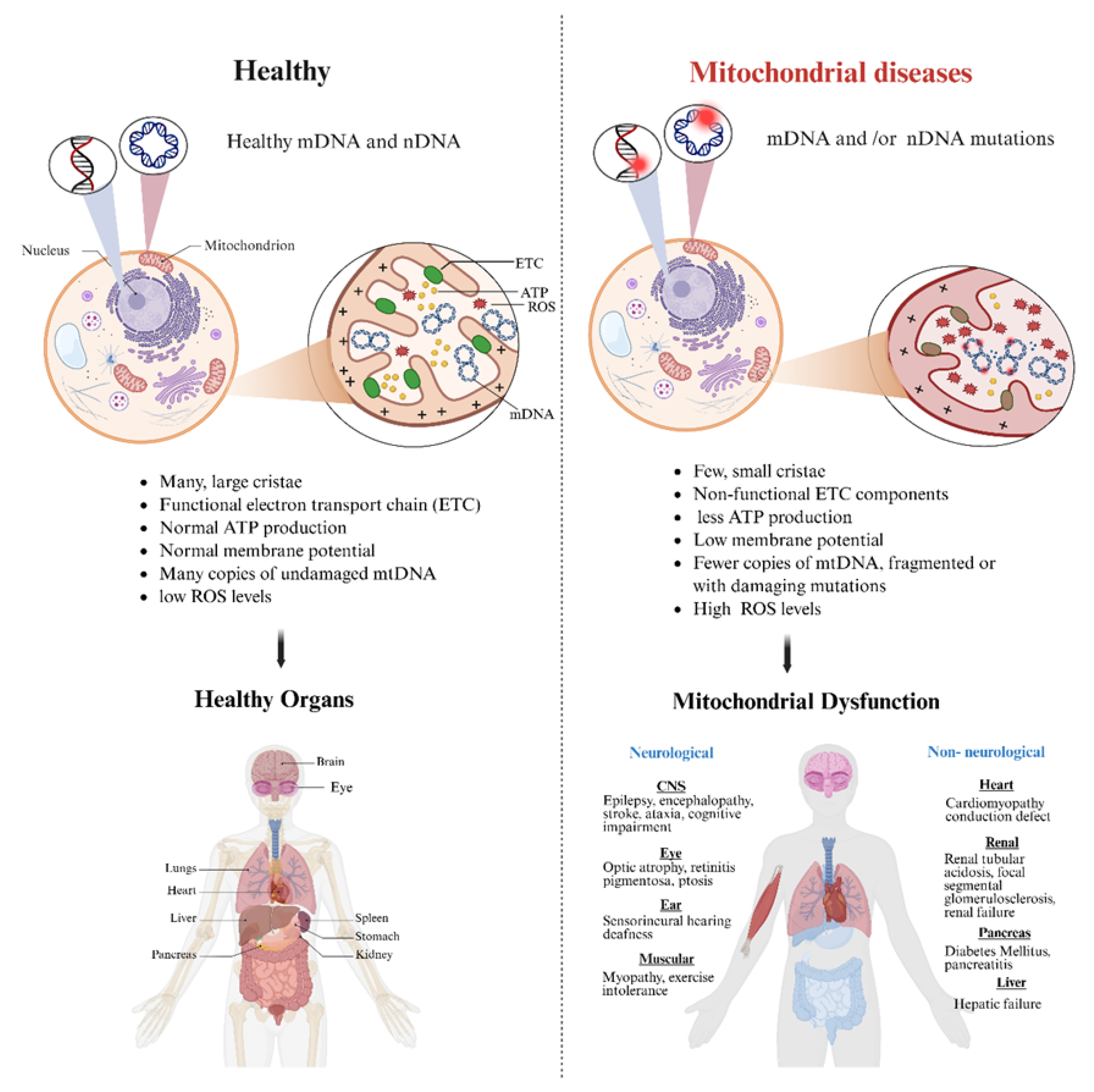
2. Genetics of Mitochondrial Dysfunctions
3. Diagnosis of Mitochondrial Dysfunctions
4. Treatment and Prognosis of Mitochondrial Dysfunctions
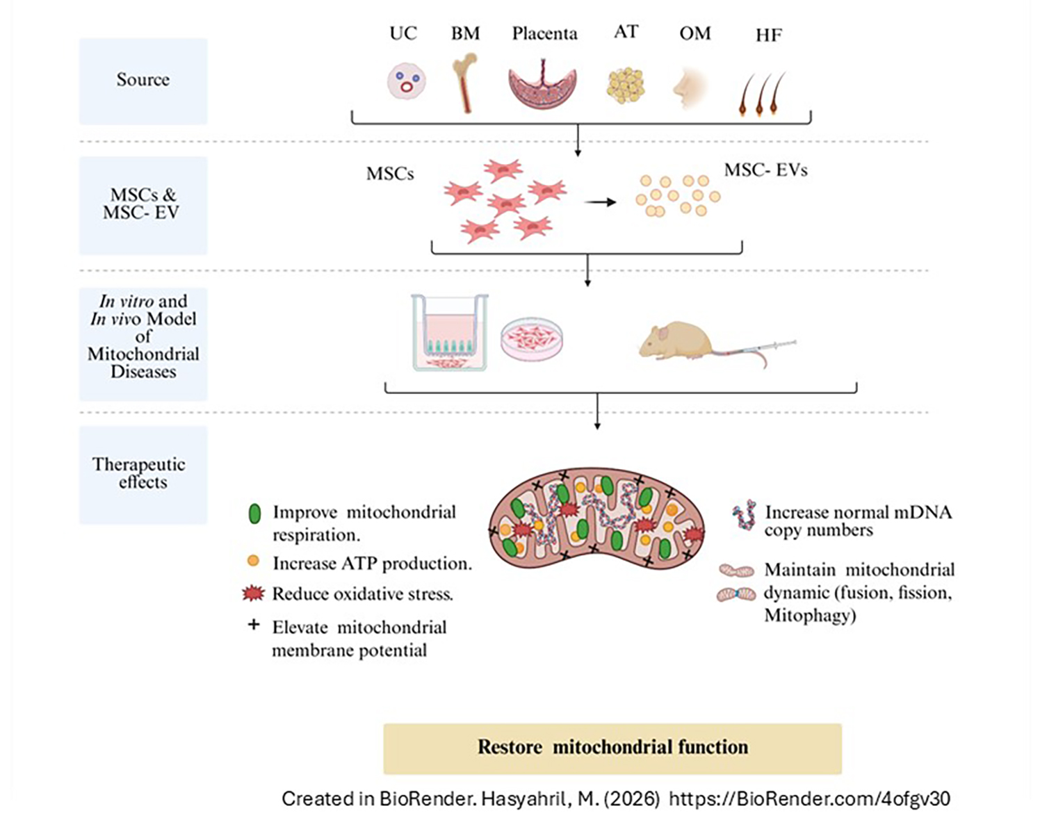
5. Therapeutic Potential of Mesenchymal Stromal Cells and Their Extracellular Vesicles in Mitochondrial Dysfunctions
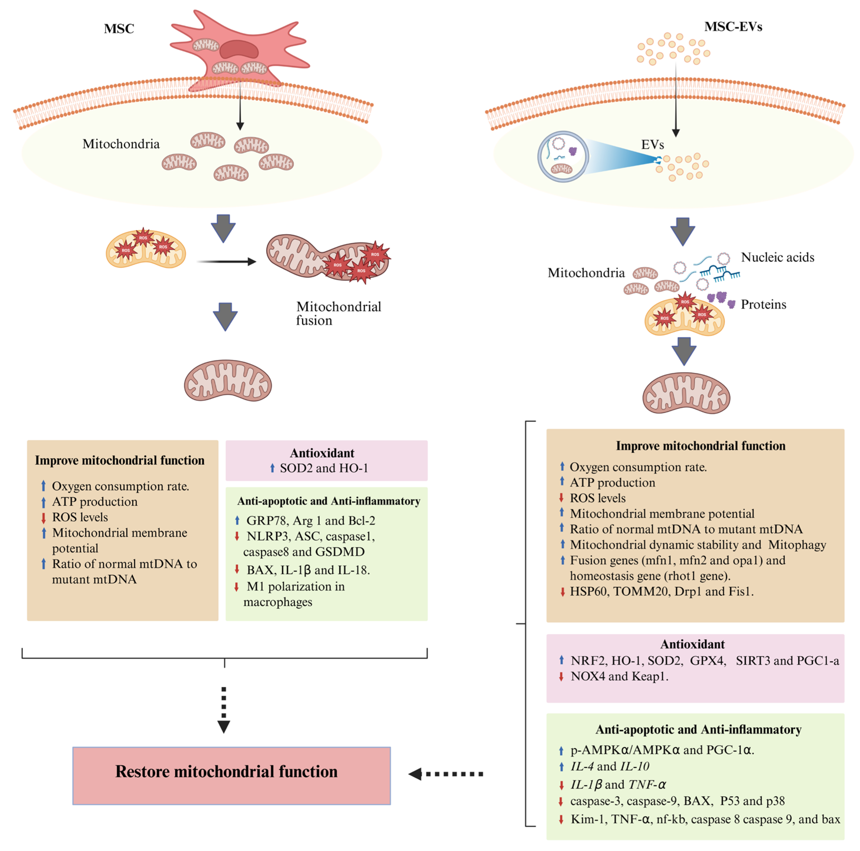
5.1. Therapeutic Potential of Mesenchymal Stromal Cells in Mitochondrial Dysfunctions
5.2. Therapeutic Potential of MSC-Derived Extracellular Vesicles in Mitochondrial Dysfunctions

| Source of MSCs/MSC-EVs | Targeted Disease | Targeted Cells/Animal Model | Dosage/Route of Administration | Therapeutic Effects | Mechanism | Ref. |
|---|---|---|---|---|---|---|
| Human BM-MSCs & highly purified MSCs (RECs) | MELAS syndrome | iPSC-derived MELAS neurons | Not specified | - Restore mitochondrial membrane potential - Improve ATP production - Reduce ROS levels - Restore intracellular calcium storage - Restore oxygen consumption rate | - Mitochondria donation | [83] |
| Human MSCs | Leber’s hereditary optic neuropathy (LHON) | LHON iPSC-derived NPCs | 1:1 ratio MSC: NPCs | - Increase mitochondrial respiration and ATP production | - Mitochondria donation - Increase the ratio of normal mtDNA to mutant mtDNA | [85] |
| Human BM-MSCs | Complex I deficiency | Human fibroblast with MT-ND3 & MT-ND6 | Not specified | - Improve mitochondrial respiration - Reduce ROS levels | - Mitochondria donation - Upregulation of cellular antioxidant, SOD2 and HO-1 | [84] |
| Human UC-MSCs | Diabetic nephropathy | Murine macrophage cell line (RAW264.7 cells) | 1:2 ratio MSCs: RAW264.7 cells | - Anti-inflammatory effect - Improve mitochondrial function - Reverse albuminuria and prevent the progression of diabetic nephropathy | - Mitochondria donation - Increase Arg1 expression and suppressed M1 polarization in macrophages - Reverse cytokine-mediated mitochondrial dysfunction | [86] |
| 8-week-old male CD1 mouse model of diabetic nephropathy | Mice were injected intravenously with 5.0 × 10⁵ UC-MSCs thrice every 4 weeks | |||||
| Human and murine BM-MSCs | Diabetic nephropathy | Human podocytes | 1:1 ratio MSCs: podocytes | - Improve mitochondrial function - Improve renal function | - Mitochondria donation - Reduce mitochondrial damage - Reduce apoptosis and inflammation - Increase nephrin gene expression | [87] |
| 8-week-old male C57BL6 mouse model of diabetic nephropathy | Mice were injected via tail vein, with BM-MSCs (1.0 × 10⁴ cells/g body weight) once a week for 6 consecutive weeks | |||||
| Human OM- MSCs | Cerebral ischemia/reperfusion injury | Neuron (SH-SY5Y) cells | Not specified | - Improve neuron mitochondrial function (increase MMP and decrease ROS) - Inhibit apoptosis and pyroptosis of neurons - Reduce damaged areas of the infarct cortices and improve rat motor function | - Increase GRP78 and Bcl-2 proteins - Decrease NLRP3 inflammasome and pyroptosis-associated proteins, ASC, caspase1, caspase8 and GSDMD - Decrease BAX, IL-1β and IL-18 | [89] |
| Adult Sprague–Dawley rat model of Cerebral ischemia/reperfusion injury | IhOM-MSCs (1 × 10⁶) were injected into the rat tail vein | |||||
| Mouse Ad-MSC-EVs | Leber’s hereditary optic neuropathy (LHON) | LHON model cells (GM10742 cells) | 6 μg EV-Mito protein/well | - Restore mitochondrial functions (MMP, ATP production, mitochondrial ROS levels, and mPTP opening) - Enhance the proliferative capacity of LHON model cells - Enhance visual recovery in LHON mice | - Mitochondria donation - Increase expression of ND4 and COX IV proteins | [99] |
| 3-month-old mutant mtND4R340H mtTg LHON male mice | Mice received an intravitreal injection of 1 μg EV-Mito protein/eye, 2 times administrations | |||||
| Human UC-MSC-EV | Cardiac hypertrophy | Neonatal rat cardiac myocytes (from 1- to 3-day-old Sprague–Dawley rats) as model of cardiac hypertrophy | - 100 ug/mL Nor-EVs or Hypo-EVs | - Reduce the cardiomyocyte size - Improvement of mitochondrial function - Attenuate heart size, ventricular wall thickening and cardiomyocyte cross-sectional areas | - Transfer DJ-1 protein to cardiomyocytes - Decrease mRNA expression of hypertrophic indicators (BNP, ANP, and β-MHC) - Enhance expression of antioxidant-related proteins, such as NRF2, HO-1, SOD2 and GPX4, while downregulate expression of NOX4 - Upregulate the expression of p-AMPKα/AMPKα and PGC-1α - Upregulate the expression of ATRAP, which inhibits the activation of p38 and ERK1/2 signaling pathways | [100] |
| Male 8-week-old adult C57BL/6 mice as model of cardiac hypertrophy | 200 μg/100 μL of Nor-EVs or Hy-EVs were injected into mice through caudal vein once a week, from one week after surgery for 3 weeks | |||||
| Human UC-MSC-Exos | Premature ovarian insufficiency | KGN cells as model of POI | Nor-Exos and hy-Exos (50 µg/mL) | - Enhance mitochondrial function and regulate mitochondrial oxidative stress - Improve body weight, ovarian weight coefficient, estrous cycles, ovarian morphology, ovulation count, and sex hormone levels in POI rats | - Increase expression of SOD2, SIRT3, PGC1-a, and TFAM - Decrease cell apoptosis by downregulation of caspase-3, caspase-9, BAX, and P53 | [101] |
| 8-week-old SD female rats’ model of POI | 200 µL of Hy-Exos or Nor-Exos (1 × 10⁹ cells) was transplanted into each ovary for two weeks (once a week) | |||||
| Mouse MSC /MSC-Exo | Cigarette smoking induced mitochondrial dysfunction | lung epithelial cells (BEAS2B cells) exposure to cigarette smoke (CS) | Not specified | - Protective response against the CSE-altered mitochondrial respiration | - Increase the expression of fusion genes (mfn1, mfn2 and opa1) and mitochondrial homeostasis gene (rhot1 gene) | [102] |
| Mouse HF-MSC-Exos | Ulcerative colitis (with high mitochondrial fission/fusion) | LPS-treated mouse MODE-K cells as a model of ulcerative colitis | Not specified | - Alleviate mitochondrial dysfunction and oxidative stress - Maintain mitochondrial dynamic stability and enhance mitophagy - Ameliorate colonic mucosal damage and inflammatory cell infiltration | - Reduce the expression of HSP60, TOMM20, Drp1 and Fis1 - Increase expression of Mfn1, Mfn2 and OPA1 - increase LC3 expression and colocalization with COX IV - Reduce IL-1β and TNF-α expression, and increase IL-4 and IL-10 expression - miR-214-3p-mediated inhibition of the PI3K/AKT/mTOR signaling pathway | [103] |
| 4–6 weeks C57BL/6J mice | 100 μg of Nor-Exos and Hy-Exos were injected via the tail vein | |||||
| Human Ad-MSC-Exos | ALI | MH-S mouse macrophage cells | Exosomes (10 μg/mL) | - Improve macrophages’ mitochondrial integrity and oxidative phosphorylation level - Mitigate lung inflammatory pathology | - Transfer mitochondrial components - Increase mtDNA and MMP | [104] |
| Human UC-MSC-Exos | IVDD | Human degenerative Nucleus pulposus cells (NPCs) | UCMSC-exos (1011 particles/mL) | - Improve viability of NPCs - Improve mitochondrial function - Delay the progression of IVDD in rats | - Reduce ROS and mitochondrial superoxide levels - Increase MMP - Restore the expression of the extracellular matrix proteins, COL2A1 and matrix metalloproteinase-13 | [94,95] |
| 23-month-old male Sprague–Dawley rats as IVDD model (by IVD puncture) | 10 uL of UCMSC-Exos (1011 particles/mL) were injected into the punctured discs every 2 weeks for 2 months | |||||
| Human P-MSC-EVs | CKD | HK-2 as induced model of CKD | Norm-EVs or Hypo-EVs (100 μg/mL) | - Reduce renal fibrosis - Enhance mitochondrial fatty acid oxidation (FAO) - Restore mitochondrial homeostasis | - Reduce collagen I and α-SMA - Restore expression of a FAO key rate-limiting enzyme, carnitine palmitoyl-transferase 1A (CPT1A) - Repair mitochondrial structure, restore mitochondrial mass and ATP production, inhibit oxidative stress, and increase mitochondrial membrane potential | [98] |
| Male C57BL/6 SPF mice (7–9 weeks old) were used as a model of ischemia–reperfusion (I/R)-induced renal fibrosis | 100 μg of Norm-EVs or Hypo-EVs in 0.15 mL of PBS solvent was administered by tail vein injection immediately after the surgery and on day (D) 1 postsurgery in the Norm-EVs and Hypo-EVs groups, respectively | |||||
| Human P-MSC-EVs | Acute kidney injury | TEC line (HK-2) as model of oxidative stress | 40–120 μg P-MSC-EVs | - Restore renal function - Restore mitochondrial function (elevate ATP production, reduce mitochondrial ROS and restore mitochondrial mass) | - Modulate inflammation and inhibit apoptosis (downregulation of Kim-1, TNF-α, nf-kb, caspase 8, caspase 9, and bax) - Increase the expression of Nrf2 and SOD2 and decrease the expression of Keap1 - Reduce mitochondrial fragmentation and normalize the mitochondrial potential - Increase mtDNA copy number | [97] |
| 6–8-week-old male FVB mice as a model of ischemia/reperfusion-induced AKI | 0.1 mL of 80 μg of EVs was injected intravenously into AKI mice |
5.3. Summary of Experimental Evidence on MSC- and MSC-EV-Mediated Restoration of Mitochondrial Function
6. Challenges and Prospects of MSC- and MSC-EV-Based Therapies for Mitochondrial Dysfunctions
- Defining the molecular mechanisms of mitochondrial repair mediated by MSCs and EVs.
- Developing standardized protocols for MSC/EV production, storage, and administration.
- Establishing multi-organ disease models to evaluate homing capacity, dosing requirements, and biodistribution.
- Conducting long-term safety and efficacy studies to assess immune tolerance and functional recovery.
7. Conclusions
Author Contributions
Funding
Institutional Review Board Statement
Informed Consent Statement
Data Availability Statement
Acknowledgments
Conflicts of Interest
References
- Al Ojaimi, M.; Salah, A.; El-Hattab, A.W. Mitochondrial fission and fusion: Molecular mechanisms, biological functions, and related disorders. Membranes 2022, 12, 893. [Google Scholar] [CrossRef] [Scilit]
- Spinelli, J.B.; Haigis, M.C. The multifaceted contributions of mitochondria to cellular metabolism. Nat. Cell Biol. 2018, 20, 745–754. [Google Scholar] [CrossRef] [Scilit]
- El-Hattab, A.W.; Suleiman, J.; Almannai, M.; Scaglia, F. Mitochondrial dynamics: Biological roles, molecular machinery, and related diseases. Mol. Genet. Metab. 2018, 125, 315–321. [Google Scholar] [CrossRef] [Scilit]
- Niyazov, D.M.; Kahler, S.G.; Frye, R.E. Primary Mitochondrial Disease and Secondary Mitochondrial Dysfunction: Importance of Distinction for Diagnosis and Treatment. Mol. Syndromol. 2016, 7, 122–137. [Google Scholar] [CrossRef] [Scilit]
- Gorman, G.; Chinnery, P.; DiMauro, S.; Hirano, M.; Koga, Y.; McFarland, R.; Suomalainen, A.; Thorburn, D.R.; Zeviani, M.; Turnbull, D.M. Mitochondrial Diseases. Nat. Rev. Dis. Primers 2016, 2, 16080. [Google Scholar] [CrossRef] [Scilit]
- Rambani, V.; Hromnikova, D.; Gasperikova, D.; Skopkova, M. Mitochondria and Mitochondrial Disorders: An Overview Update. Endocr. Regul. 2022, 56, 232–248. [Google Scholar] [CrossRef] [Scilit]
- Zhao, X.; Yu, M.; Zhang, W.; Hou, Y.; Yuan, Y.; Wang, Z. Demographic Characteristics, Diagnostic Challenges, Treatment Patterns, and Caregiver Burden of Mitochondrial Diseases: A Retrospective Cross-Sectional Study. Orphanet J. Rare Dis. 2024, 19, 287. [Google Scholar] [CrossRef] [Scilit] [PubMed]
- Ahuja, A.S. Understanding Mitochondrial Myopathies: A Review. PeerJ 2018, 6, e4790. [Google Scholar] [CrossRef] [Scilit] [PubMed]
- Merimi, M.; El-Majzoub, R.; Lagneaux, L.; Moussa Agha, D.; Bouhtit, F.; Meuleman, N.; Fahmi, H.; Lewalle, P.; Fayyad-Kazan, M.; Najar, M. The Therapeutic Potential of Mesenchymal Stromal Cells for Regenerative Medicine: Current Knowledge and Future Understandings. Front. Cell Dev. Biol. 2021, 9, 661532. [Google Scholar] [CrossRef] [Scilit]
- Peng, F.; Chen, X.; Wu, L.; He, J.; Li, Z.; Hong, Q.; Zhao, Q.; Qian, M.; Wang, X.; Shen, W.; et al. Nitric oxide-primed engineered extracellular vesicles restore bioenergetics in acute kidney injury via mitochondrial transfer. Theranostics 2025, 15, 5499. [Google Scholar] [CrossRef] [Scilit] [PubMed]
- Yu, M.; Liu, W.; Li, J.; Lu, J.; Lu, H.; Jia, W.; Liu, F. Exosomes Derived from Atorvastatin-Pretreated MSC Accelerate Diabetic Wound Repair by Enhancing Angiogenesis via AKT/eNOS Pathway. Stem Cell Res. Ther. 2020, 11, 350. [Google Scholar] [CrossRef] [Scilit]
- Wang, X.; Bai, L.; Liu, X.; Shen, W.; Tian, H.; Liu, W.; Yu, B. Cardiac Microvascular Functions Improved by MSC-Derived Exosomes Attenuate Cardiac Fibrosis after Ischemia–Reperfusion via PDGFR-β Modulation. Int. J. Cardiol. 2021, 344, 13–24. [Google Scholar] [CrossRef] [Scilit]
- Anderson, S.; Bankier, A.T.; Barrell, B.G.; de Bruijn, M.H.; Coulson, A.R.; Drouin, J.; Eperon, I.C.; Nierlich, D.P.; Roe, B.A.; Sanger, F.; et al. Sequence and organization of the human mitochondrial genome. Nature 1981, 290, 457–465. [Google Scholar] [CrossRef] [Scilit]
- Wallace, D.C. Mitochondrial DNA variation in human radiation and disease. Cell 2015, 163, 33–38. [Google Scholar] [CrossRef] [Scilit] [PubMed]
- Schon, K.R.; Ratnaike, T.; van den Ameele, J.; Horvath, R.; Chinnery, P.F. Mitochondrial Diseases: A Diagnostic Revolution. Trends Genet. 2020, 36, 702–717. [Google Scholar] [CrossRef] [Scilit] [PubMed]
- Stenton, S.L.; Prokisch, H. Genetics of Mitochondrial Diseases: Identifying Mutations to Help Diagnosis. EBioMedicine 2020, 56, 102784. [Google Scholar] [CrossRef] [Scilit]
- Powell, C.A.; Nicholls, T.J.; Minczuk, M. Nuclear-Encoded Factors Involved in Post-Transcriptional Processing and Modification of Mitochondrial tRNAs in Human Disease. Front. Genet. 2015, 6, 79. [Google Scholar] [CrossRef] [Scilit]
- Gusic, M.; Prokisch, H. Genetic Basis of Mitochondrial Diseases. FEBS Lett. 2021, 595, 1132–1158. [Google Scholar] [CrossRef] [Scilit]
- Soldatov, V.O.; Kubekina, M.V.; Skorkina, M.Y.; Belykh, A.E.; Egorova, T.V.; Korokin, M.V.; Pokrovskiy, M.V.; Deykin, A.V.; Angelova, P.R. Current Advances in Gene Therapy of Mitochondrial Diseases. J. Transl. Med. 2022, 20, 562, Correction in J. Transl. Med 2023, 21, 96. [Google Scholar] [CrossRef] [Scilit] [PubMed]
- Craven, L.; Alston, C.L.; Taylor, R.W.; Turnbull, D.M. Recent advances in mitochondrial disease. Annu. Rev. Genomics Hum. Genet. 2017, 18, 257–275. [Google Scholar] [CrossRef] [Scilit]
- Wen, H.; Deng, H.; Li, B.; Chen, J.; Zhu, J.; Zhang, X.; Yoshida, S.; Zhou, Y. Mitochondrial Diseases: From Molecular Mechanisms to Therapeutic Advances. Signal Transduct. Target. Ther. 2025, 10, 9. [Google Scholar] [CrossRef] [Scilit]
- Yoshimi, A.; Ishikawa, K.; Niemeyer, C.; Grünert, S.C. Pearson Syndrome: A Multisystem Mitochondrial Disease with Bone Marrow Failure. Orphanet J. Rare Dis. 2022, 17, 379. [Google Scholar] [CrossRef] [Scilit] [PubMed]
- Ng, Y.S.; Lax, N.Z.; Maddison, P.; Alston, C.L.; Blakely, E.L.; Hepplewhite, P.D.; Riordan, G.; Meldau, S.; Chinnery, P.F.; Pierre, G.; et al. MT-ND5 Mutation Exhibits Highly Variable Neurological Manifestations at Low Mutant Load. EBioMedicine 2018, 30, 86–93. [Google Scholar] [CrossRef] [Scilit]
- Nunnari, J.; Suomalainen, A. Mitochondria: In Sickness and in Health. Cell 2012, 148, 1145–1159. [Google Scholar] [CrossRef] [Scilit]
- Stepien, K.M.; Heaton, R.; Rankin, S.; Murphy, A.; Bentley, J.; Sexton, D.; Hargreaves, I.P. Evidence of oxidative stress and secondary mitochondrial dysfunction in metabolic and non-metabolic disorders. J. Clin. Med. 2017, 6, 71. [Google Scholar] [CrossRef] [Scilit] [PubMed]
- Zong, Y.; Li, H.; Liao, P.; Chen, L.; Pan, Y.; Zheng, Y.; Zhang, C.; Liu, D.; Zheng, M.; Gao, J. Mitochondrial dysfunction: Mechanisms and advances in therapy. Signal Transduct. Target. Ther. 2024, 9, 124. [Google Scholar] [CrossRef] [Scilit] [PubMed]
- Yoon, Y.; Galloway, C.A.; Jhun, B.S.; Yu, T. Mitochondrial Dynamics in Diabetes. Antioxid. Redox Signal. 2011, 14, 439–457. [Google Scholar] [CrossRef] [Scilit]
- Ong, S.B.; Hall, A.R.; Hausenloy, D.J. Mitochondrial Dynamics in Cardiovascular Health and Disease. Antioxid. Redox Signal. 2013, 19, 400–414. [Google Scholar] [CrossRef] [Scilit]
- Boland, M.L.; Chourasia, A.H.; Macleod, K.F. Mitochondrial Dysfunction in Cancer. Front. Oncol. 2013, 3, 292. [Google Scholar] [CrossRef] [Scilit]
- Jiang, Z.; Wang, W.; Perry, G.; Zhu, X.; Wang, X. Mitochondrial Dynamic Abnormalities in Amyotrophic Lateral Sclerosis. Transl. Neurodegener. 2015, 4, 14. [Google Scholar] [CrossRef] [Scilit]
- Chen, H.; Chan, D.C. Mitochondrial Dynamics—Fusion, Fission, Movement, and Mitophagy in Neurodegenerative Diseases. Hum. Mol. Genet. 2009, 18, R169–R176. [Google Scholar] [CrossRef] [Scilit] [PubMed]
- Charif, M.; Wong, Y.C.; Kim, S.; Guichet, A.; Vignal, C.; Zanlonghi, X.; Bensaid, P.; Procaccio, V.; Bonneau, D.; Amati-Bonneau, P.; et al. Dominant mutations in MIEF1 affect mitochondrial dynamics and cause a singular late-onset optic neuropathy. Mol. Neurodegener. 2021, 16, 12. [Google Scholar] [CrossRef] [Scilit]
- Pickrell, A.M.; Youle, R.J. The Roles of PINK1, Parkin, and Mitochondrial Fidelity in Parkinson’s Disease. Neuron 2015, 85, 257–273. [Google Scholar] [CrossRef] [Scilit]
- Deas, E.; Wood, N.W.; Plun-Favreau, H. Mitophagy and Parkinson’s Disease: The PINK1–Parkin Link. Biochim. Biophys. Acta 2011, 1813, 623–633. [Google Scholar] [CrossRef] [Scilit]
- Puccio, H.; Kœnig, M. Friedreich Ataxia: A Paradigm for Mitochondrial Diseases. Trends Mol. Med. 2002, 8, 223–227. [Google Scholar] [CrossRef] [Scilit]
- Copeland, W.C. Defects in Mitochondrial DNA Replication and Human Disease. Crit. Rev. Biochem. Mol. Biol. 2012, 47, 64–74. [Google Scholar] [CrossRef] [Scilit] [PubMed]
- Wu, Z.; Puigserver, P.; Andersson, U.; Zhang, C.; Adelmant, G.; Mootha, V.; Troy, A.; Cinti, S.; Lowell, B.; Scarpulla, R.C.; et al. Mechanisms Controlling Mitochondrial Biogenesis and Respiration through the Thermogenic Coactivator PGC-1. Cell 1999, 98, 115–124. [Google Scholar] [CrossRef] [Scilit] [PubMed]
- Klemmensen, M.M.; Borrowman, S.H.; Pearce, C.; Pyles, B.; Chandra, B. Mitochondrial dysfunction in neurodegenerative disorders. Neurotherapeutics 2024, 21, e00292. [Google Scholar] [CrossRef] [Scilit]
- Hardie, D.G. AMPK: A Key Regulator of Energy Balance in the Single Cell and the Whole Organism. Int. J. Obes. 2008, 32, S7–S12. [Google Scholar] [CrossRef] [Scilit]
- Saxton, R.A.; Sabatini, D.M. mTOR Signaling in Growth, Metabolism, and Disease. Cell 2017, 168, 960–976. [Google Scholar] [CrossRef] [Scilit]
- Zhou, R.; Yazdi, A.S.; Menu, P.; Tschopp, J. A Role for Mitochondria in NLRP3 Inflammasome Activation. Nature 2011, 469, 221–225. [Google Scholar] [CrossRef] [Scilit]
- Magro, G.; Laterza, V.; Tosto, F. Leigh Syndrome: A Comprehensive Review of the Disease and Present and Future Treatments. Biomedicines 2025, 13, 733. [Google Scholar] [CrossRef] [Scilit] [PubMed]
- El-Hattab, A.W.; Adesina, A.M.; Jones, J.; Scaglia, F. MELAS Syndrome: Clinical Manifestations, Pathogenesis, and Treatment Options. Mol. Genet. Metab. 2015, 116, 4–12. [Google Scholar] [CrossRef] [Scilit] [PubMed]
- Kim, U.S.; Jurkute, N.; Yu-Wai-Man, P. Leber Hereditary Optic Neuropathy—Light at the End of the Tunnel? Asia-Pac. J. Ophthalmol. 2018, 7, 242–245. [Google Scholar] [PubMed]
- Finsterer, J. Neuropathy, Ataxia, and Retinitis Pigmentosa Syndrome. J. Clin. Neuromuscul. Dis. 2023, 24, 140–146. [Google Scholar] [CrossRef] [Scilit] [PubMed]
- Grigalionienė, K.; Burnytė, B.; Balkelienė, D.; Ambrozaitytė, L.; Utkus, A. Kearns–Sayre Syndrome Case: Novel 5.9 kb mtDNA Deletion. Mol. Genet. Genom. Med. 2023, 11, e2059. [Google Scholar] [CrossRef] [Scilit]
- Salvador, C.L.; Oppebøen, M.; Vassli, A.Ø.; Pfeiffer, H.C.; Varhaug, K.N.; Elgstøen, K.B.; Yazdani, M. Increased Sphingomyelin and Free Sialic Acid in Cerebrospinal Fluid of Kearns–Sayre Syndrome: New Findings Using Untargeted Metabolomics. Pediatr. Neurol. 2023, 143, 68–76. [Google Scholar] [CrossRef] [Scilit]
- Hirano, M.; Pitceathly, R.D. Progressive External Ophthalmoplegia. Handb. Clin. Neurol. 2023, 194, 9–21. [Google Scholar]
- Alberti, C.; Rizzo, F.; Anastasia, A.; Comi, G.; Corti, S.; Abati, E. Charcot–Marie–Tooth disease type 2A: An update on pathogenesis and therapeutic perspectives. Neurobiol. Dis. 2024, 193, 106467. [Google Scholar] [CrossRef] [Scilit] [PubMed]
- Muench, N.A.; Patel, S.; Maes, M.E.; Donahue, R.J.; Ikeda, A.; Nickells, R.W. The influence of mitochondrial dynamics and function on retinal ganglion cell susceptibility in optic nerve disease. Cells 2021, 10, 1593. [Google Scholar] [CrossRef] [Scilit]
- San-Millán, I. The Key Role of Mitochondrial Function in Health and Disease. Antioxidants 2023, 12, 782. [Google Scholar] [CrossRef] [Scilit] [PubMed]
- Gorman, G.S.; Schaefer, A.M.; Ng, Y.; Gomez, N.; Blakely, E.L.; Alston, C.L.; Feeney, C.; Horvath, R.; Yu-Wai-Man, P.; Chinnery, P.F.; et al. Prevalence of Nuclear and Mitochondrial DNA Mutations Related to Adult Mitochondrial Disease. Ann. Neurol. 2015, 77, 753–759. [Google Scholar] [CrossRef] [Scilit]
- Aldossary, A.M.; Tawfik, E.A.; Alomary, M.N.; Alsudir, S.A.; Alfahad, A.J.; Alshehri, A.A.; Almughem, F.A.; Mohammed, R.Y.; Alzaydi, M.M. Recent Advances in Mitochondrial Diseases: From Molecular Insights to Therapeutic Perspectives. Saudi Pharm. J. 2022, 30, 1065–1078. [Google Scholar] [CrossRef] [Scilit]
- Rahman, S. Mitochondrial Disease in Children. J. Intern. Med. 2020, 287, 609–633. [Google Scholar] [CrossRef] [Scilit]
- Ng, Y.S.; Bindoff, L.A.; Gorman, G.S.; Klopstock, T.; Kornblum, C.; Mancuso, M.; McFarland, R.; Sue, C.M.; Suomalainen, A.; Taylor, R.W.; et al. Mitochondrial Disease in Adults: Recent Advances and Future Promise. Lancet Neurol. 2021, 20, 573–584. [Google Scholar] [CrossRef] [Scilit] [PubMed]
- Lightowlers, R.N.; Taylor, R.W.; Turnbull, D.M. Mutations Causing Mitochondrial Disease: What Is New and What Challenges Remain? Science 2015, 349, 1494–1499. [Google Scholar] [CrossRef] [Scilit]
- Pérez-Albert, P.; de Lucas Collantes, C.; Fernández-García, M.Á.; de Rojas, T.; Aparicio López, C.; Gutiérrez-Solana, L. Mitochondrial Disease in Children: The Nephrologist’s Perspective. JIMD Rep. 2018, 42, 61–70. [Google Scholar] [PubMed]
- Russell, O.M.; Gorman, G.S.; Lightowlers, R.N.; Turnbull, D.M. Mitochondrial diseases: Hope for the future. Cell 2020, 181, 168–188. [Google Scholar] [CrossRef] [Scilit] [PubMed]
- Davis, R.L.; Kumar, K.R.; Puttick, C.; Liang, C.; Ahmad, K.E.; Edema-Hildebrand, F.; Park, J.S.; Minoche, A.E.; Gayevskiy, V.; Mallawaarachchi, A.C.; et al. Use of whole-genome sequencing for mitochondrial disease diagnosis. Neurology 2022, 99, e730–e742. [Google Scholar] [CrossRef] [Scilit]
- Stenton, S.L.; Prokisch, H. Advancing genomic approaches to the diagnosis of mitochondrial disease. J. Inherit. Metab. Dis. 2020, 43, 1067–1077. [Google Scholar]
- Parikh, S.; Goldstein, A.; Koenig, M.K.; Scaglia, F.; Enns, G.M.; Saneto, R.; Anselm, I.; Cohen, B.H.; Falk, M.J.; Greene, C.; et al. Diagnosis and management of mitochondrial disease: A consensus statement from the Mitochondrial Medicine Society. Genet. Med. 2015, 17, 689–701. [Google Scholar] [CrossRef] [Scilit]
- Liufu, T.; Wang, Z. Treatment for mitochondrial diseases. Rev. Neurosci. 2021, 32, 35–47. [Google Scholar] [CrossRef] [Scilit]
- Jauhari, P.; Sankhyan, N.; Vyas, S.; Singhi, P. Thiamine responsive pyruvate dehydrogenase complex deficiency: A potentially treatable cause of Leigh’s disease. J. Pediatr. Neurosci. 2017, 12, 265–267. [Google Scholar]
- Sahel, J.A.; Newman, N.J.; Yu-Wai-Man, P.; Vignal-Clermont, C.; Carelli, V.; Biousse, V.; Moster, M.L.; Sergott, R.; Klopstock, T.; Sadun, A.A.; et al. Gene therapies for the treatment of Leber hereditary optic neuropathy. Int. Ophthalmol. Clin. 2021, 61, 195–208. [Google Scholar] [CrossRef] [Scilit] [PubMed]
- Olsen, R.K.; Koňaříková, E.; Giancaspero, T.A.; Mosegaard, S.; Boczonadi, V.; Mataković, L.; Veauville-Merllié, A.; Terrile, C.; Schwarzmayr, T.; Haack, T.B.; et al. Riboflavin-responsive and non-responsive mutations in FAD synthase cause multiple acyl-CoA dehydrogenase and combined respiratory-chain deficiency. Am. J. Hum. Genet. 2016, 98, 1130–1145. [Google Scholar] [CrossRef] [Scilit]
- Koňaříková, E.; Marković, A.; Korandová, Z.; Houštěk, J.; Mráček, T. Current progress in the therapeutic options for mitochondrial disorders. Physiol. Res. 2020, 69, 967. [Google Scholar] [CrossRef] [Scilit] [PubMed]
- Karaa, A.; Haas, R.; Goldstein, A.; Vockley, J.; Cohen, B.H. A randomized crossover trial of elamipretide in adults with primary mitochondrial myopathy. J. Cachexia Sarcopenia Muscle 2020, 11, 909–918. [Google Scholar] [CrossRef] [Scilit] [PubMed]
- El-Hattab, A.W.; Almannai, M.; Scaglia, F. Arginine and citrulline for the treatment of MELAS syndrome. J. Inborn Errors Metab. Screen. 2017, 5, 2326409817697399. [Google Scholar] [CrossRef] [Scilit]
- Fan, H.C.; Lee, H.F.; Yue, C.T.; Chi, C.S. Clinical characteristics of mitochondrial encephalomyopathy, lactic acidosis, and stroke-like episodes. Life 2021, 11, 1111. [Google Scholar] [CrossRef] [Scilit]
- Martinelli, D.; Catteruccia, M.; Piemonte, F.; Pastore, A.; Tozzi, G.; Dionisi-Vici, C.; Pontrelli, G.; Corsetti, T.; Livadiotti, S.; Kheifets, V.; et al. EPI-743 reverses the progression of the pediatric mitochondrial disease—genetically defined Leigh syndrome. Mol. Genet. Metab. 2012, 107, 383–388. [Google Scholar] [CrossRef] [Scilit]
- Janssen, M.C.; Koene, S.; de Laat, P.; Hemelaar, P.; Pickkers, P.; Spaans, E.; Beukema, R.; Beyrath, J.; Groothuis, J.; Verhaak, C.; et al. The KHENERGY study: Safety and efficacy of KH176 in mitochondrial m.3243A>G spectrum disorders. Clin. Pharmacol. Ther. 2019, 105, 101–111. [Google Scholar] [CrossRef] [Scilit]
- Newman, N.J.; Yu-Wai-Man, P.; Subramanian, P.S.; Moster, M.L.; Wang, A.G.; Donahue, S.P.; Leroy, B.P.; Carelli, V.; Biousse, V.; Vignal-Clermont, C.; et al. Randomized trial of bilateral gene therapy injection for m.11778G>A MT-ND4 Leber optic neuropathy. Brain 2023, 146, 1328–1341. [Google Scholar] [CrossRef] [Scilit]
- Bouquet, C.; Vignal-Clermont, C.; Galy, A.; Fitoussi, S.; Blouin, L.; Munk, M.R.; Valero, S.; Meunier, S.; Katz, B.; Sahel, J.A.; et al. Immune response and intraocular inflammation in patients with Leber hereditary optic neuropathy treated with intravitreal injection of recombinant adeno-associated virus 2 carrying the ND4 gene: A secondary analysis of a phase 1/2 clinical trial. JAMA Ophthalmol. 2019, 137, 399–406. [Google Scholar] [CrossRef] [Scilit]
- Gammage, P.A.; Rorbach, J.; Vincent, A.I.; Rebar, E.J.; Minczuk, M. Mitochondrially targeted ZFNs for selective degradation of pathogenic mitochondrial genomes bearing large-scale deletions or point mutations. EMBO Mol. Med. 2014, 6, 458–466. [Google Scholar] [CrossRef] [Scilit]
- Shoop, W.K.; Lape, J.; Trum, M.; Powell, A.; Sevigny, E.; Mischler, A.; Bacman, S.R.; Fontanesi, F.; Smith, J.; Jantz, D.; et al. Efficient elimination of MELAS-associated m.3243G mutant mitochondrial DNA by an engineered mitoARCUS nuclease. Nat. Metab. 2023, 5, 2169–2183. [Google Scholar] [CrossRef] [Scilit]
- Keshavan, N.; Rahman, S. Natural History of Mitochondrial Disorders: A Systematic Review. Essays Biochem. 2018, 62, 423–442. [Google Scholar] [CrossRef] [Scilit] [PubMed]
- Ebihara, T.; Nagatomo, T.; Sugiyama, Y.; Tsuruoka, T.; Osone, Y.; Shimura, M.; Tajika, M.; Matsuhashi, T.; Ichimoto, K.; Matsunaga, A.; et al. Neonatal-Onset Mitochondrial Disease: Clinical Features, Molecular Diagnosis and Prognosis. Arch. Dis. Child. Fetal Neonatal Ed. 2022, 107, 329–334. [Google Scholar] [CrossRef] [Scilit] [PubMed]
- Izadi, M.; Sadr Hashemi Nejad, A.; Moazenchi, M.; Masoumi, S.; Rabbani, A.; Kompani, F.; Hedayati Asl, A.A.; Abbasi Kakroodi, F.; Jaroughi, N.; Mohseni Meybodi, M.A.; et al. Mesenchymal Stem Cell Transplantation in Newly Diagnosed Type-1 Diabetes Patients: A Phase I/II Randomized Placebo-Controlled Clinical Trial. Stem Cell Res. Ther. 2022, 13, 264. [Google Scholar] [CrossRef] [Scilit]
- Yang, Y.; Wu, Y.; Yang, D.; Neo, S.H.; Kadir, N.D.; Goh, D.; Tan, J.X.; Denslin, V.; Lee, E.H.; Yang, Z. Secretome Derived from Hypoxia Preconditioned Mesenchymal Stem Cells Promote Cartilage Regeneration and Mitigate Joint Inflammation via Extracellular Vesicles. Bioact. Mater. 2023, 27, 98–112. [Google Scholar] [CrossRef] [Scilit] [PubMed]
- Perez-Cruet, M.; Beeravolu, N.; McKee, C.; Brougham, J.; Khan, I.; Bakshi, S.; Chaudhry, G.R. Potential of Human Nucleus Pulposus-Like Cells Derived from Umbilical Cord to Treat Degenerative Disc Disease. Neurosurgery 2019, 84, 272–283. [Google Scholar] [CrossRef] [Scilit]
- Wang, Y.; Zhang, D.; Shen, B.; Zhang, Y.; Gu, P. Stem/Progenitor Cells and Biodegradable Scaffolds in the Treatment of Retinal Degenerative Diseases. Curr. Stem Cell Res. Ther. 2018, 13, 160–173. [Google Scholar] [CrossRef] [Scilit]
- Song, Y.; Zhang, J.; Xu, H.; Lin, Z.; Chang, H.; Liu, W.; Kong, L. Mesenchymal Stem Cells in Knee Osteoarthritis Treatment: A Systematic Review and Meta-Analysis. J. Orthop. Transl. 2020, 24, 121–130. [Google Scholar] [CrossRef] [Scilit]
- Liu, L.; Yang, J.; Otani, Y.; Shiga, T.; Yamaguchi, A.; Oda, Y.; Hattori, M.; Goto, T.; Ishibashi, S.; Kawashima-Sonoyama, Y.; et al. MELAS-Derived Neurons Functionally Improve by Mitochondrial Transfer from Highly Purified Mesenchymal Stem Cells (REC). Int. J. Mol. Sci. 2023, 24, 17186. [Google Scholar] [CrossRef] [Scilit]
- Navaratnarajah, T.; Bellmann, M.; Seibt, A.; Anand, R.; Degistirici, Ö.; Meisel, R.; Mayatepek, E.; Reichert, A.; Baertling, F.; Distelmaier, F. Mesenchymal Stem Cells Improve Redox Homeostasis and Mitochondrial Respiration in Fibroblast Cell Lines with Pathogenic MT-ND3 and MT-ND6 Variants. Stem Cell Res. Ther. 2022, 13, 256. [Google Scholar] [CrossRef] [Scilit] [PubMed]
- Wang, R.; Bao, F.; Lu, M.; Jia, X.; Xiao, J.; Wu, Y.; Zhang, Q.; Liu, X. MSC-Mediated Mitochondrial Transfer Restores Mitochondrial DNA and Function in Neural Progenitor Cells of Leber’s Hereditary Optic Neuropathy. Sci. China Life Sci. 2024, 67, 2511–2519. [Google Scholar] [CrossRef] [Scilit] [PubMed]
- Lee, S.E.; Jang, J.E.; Kim, H.S.; Jung, M.K.; Ko, M.S.; Kim, M.O.; Park, H.S.; Oh, W.; Choi, S.J.; Jin, H.J.; et al. Mesenchymal Stem Cells Prevent the Progression of Diabetic Nephropathy by Improving Mitochondrial Function in Tubular Epithelial Cells. Exp. Mol. Med. 2019, 51, 1–4. [Google Scholar] [CrossRef] [Scilit]
- Barutta, F.; Corbetta, B.; Bellini, S.; Gambino, R.; Bruno, S.; Kimura, S.; Hase, K.; Ohno, H.; Gruden, G. Protective Effect of Mesenchymal Stromal Cells in Diabetic Nephropathy: The In Vitro and In Vivo Role of the M-Sec-Tunneling Nanotubes. Clin. Sci. 2024, 138, 1537–1559. [Google Scholar] [CrossRef] [Scilit]
- Li, Z.; Xing, J. The role of ubiquitylation in cerebral ischemia-reperfusion injury to reduce mitochondrial dysfunction and programmed cell death. Eur. J. Pharmacol. 2025, 178337. [Google Scholar] [CrossRef] [Scilit]
- Zhuo, Y.; Chen, W.; Li, W.; Huang, Y.; Duan, D.; Ge, L.; He, J.; Liu, J.; Hu, Z.; Lu, M. Ischemic-Hypoxic Preconditioning Enhances the Mitochondrial Function Recovery of Transplanted Olfactory Mucosa Mesenchymal Stem Cells via miR-181a Signaling in Ischemic Stroke. Aging 2021, 13, 11234. [Google Scholar] [CrossRef] [Scilit] [PubMed]
- Toft-Kehler, A.K.; Skytt, D.M.; Svare, A.; Lefevere, E.; Van Hove, I.; Moons, L.; Waagepetersen, H.S.; Kolko, M. Mitochondrial function in Müller cells-Does it matter? Mitochondrion 2017, 36, 43–51. [Google Scholar] [CrossRef] [Scilit]
- Huang, X.; Luodan, A.; Gao, H.; He, J.; Ge, L.; Cha, Z.; Gong, H.; Lin, X.; Li, H.; Tang, Y.; et al. Mitochondrial Transfer between BMSCs and Müller Promotes Mitochondrial Fusion and Suppresses Gliosis in Degenerative Retina. iScience 2024, 27, 110309. [Google Scholar] [CrossRef] [Scilit]
- Ma, H.; Xie, C.; Chen, Z.; He, G.; Dai, Z.; Cai, H.; Zhang, H.; Lu, H.; Wu, H.; Hu, X.; et al. MFG-E8 Alleviates Intervertebral Disc Degeneration by Suppressing Pyroptosis and Extracellular Matrix Degradation in Nucleus Pulposus Cells via Nrf2/TXNIP/NLRP3 Axis. Cell Death Discov. 2022, 8, 209. [Google Scholar] [CrossRef] [Scilit] [PubMed]
- Zhang, Z.; Wu, J.; Teng, C.; Wang, J.; Yu, J.; Jin, C.; Wang, L.; Wu, L.; Lin, Z.; Yu, Z.; et al. Orientin Downregulating Oxidative Stress-Mediated Endoplasmic Reticulum Stress and Mitochondrial Dysfunction through AMPK/SIRT1 Pathway in Rat Nucleus Pulposus Cells In Vitro and Attenuated Intervertebral Disc Degeneration In Vivo. Apoptosis 2022, 27, 1031–1048. [Google Scholar] [CrossRef] [Scilit]
- Jia, S.; Yang, T.; Gao, S.; Bai, L.; Zhu, Z.; Zhao, S.; Wang, Y.; Liang, X.; Li, Y.; Gao, L.; et al. Exosomes from Umbilical Cord Mesenchymal Stem Cells Ameliorate Intervertebral Disc Degeneration via Repairing Mitochondrial Dysfunction. J. Orthop. Transl. 2024, 46, 103–115. [Google Scholar] [CrossRef] [Scilit] [PubMed]
- Zhou, X.; Xia, T.; Xiao, J.; Xiang, J.; Fang, H.; Zhou, H.; Hu, Y.; Xiong, L. Exosomes in Intervertebral Disc Regeneration: Roles, Opportunities, and Challenges. Adv. NanoBiomed Res. 2025, 5, 2500074. [Google Scholar] [CrossRef] [Scilit]
- Jiang, M.; Bai, M.; Lei, J.; Xie, Y.; Xu, S.; Jia, Z.; Zhang, A. Mitochondrial Dysfunction and the AKI-to-CKD Transition. Am. J. Physiol. Ren. Physiol. 2020, 319, F1105–F1116. [Google Scholar] [CrossRef] [Scilit]
- Cao, H.; Cheng, Y.; Gao, H.; Zhuang, J.; Zhang, W.; Bian, Q.; Wang, F.; Du, Y.; Li, Z.; Kong, D.; et al. In Vivo Tracking of Mesenchymal Stem Cell-Derived Extracellular Vesicles Improving Mitochondrial Function in Renal Ischemia–Reperfusion Injury. ACS Nano 2020, 14, 4014–4026. [Google Scholar] [CrossRef] [Scilit]
- Gao, Z.; Zhang, C.; Peng, F.; Chen, Q.; Zhao, Y.; Chen, L.; Wang, X.; Chen, X. Hypoxic Mesenchymal Stem Cell-Derived Extracellular Vesicles Ameliorate Renal Fibrosis after Ischemia–Reperfusion Injury by Restoring CPT1A Mediated Fatty Acid Oxidation. Stem Cell Res. Ther. 2022, 13, 191. [Google Scholar] [CrossRef] [Scilit]
- Wang, Y.; Yu, H.Y.; Yi, Z.J.; Qi, L.Y.; Yang, J.S.; Xie, H.X.; Zhao, M.; Liu, N.H.; Chen, J.Q.; Zhou, T.J.; et al. Super mitochondria-enriched extracellular vesicles enable enhanced mitochondria transfer. Nat. Commun. 2025, 16, 9448. [Google Scholar] [CrossRef] [Scilit]
- Lu, Y.; Zhang, J.; Han, B.; Yu, Y.; Zhao, W.; Wu, T.; Mao, Y.; Zhang, F. Extracellular Vesicles DJ-1 Derived from Hypoxia-Conditioned hMSCs Alleviate Cardiac Hypertrophy by Suppressing Mitochondrial Dysfunction and Preventing ATRAP Degradation. Pharmacol. Res. 2023, 187, 106607. [Google Scholar] [CrossRef] [Scilit] [PubMed]
- Zhang, S.; Zou, X.; Feng, X.; Shi, S.; Zheng, Y.; Li, Q.; Wu, Y. Exosomes Derived from Hypoxic Mesenchymal Stem Cells Ameliorate Premature Ovarian Insufficiency by Reducing Mitochondrial Oxidative Stress. Sci. Rep. 2025, 15, 8235. [Google Scholar] [CrossRef] [Scilit]
- Maremanda, K.P.; Sundar, I.K.; Rahman, I. Protective Role of Mesenchymal Stem Cells and Mesenchymal Stem Cell-Derived Exosomes in Cigarette Smoke-Induced Mitochondrial Dysfunction in Mice. Toxicol. Appl. Pharmacol. 2019, 385, 114788. [Google Scholar] [CrossRef] [Scilit]
- Li, N.; Zhao, L.; Geng, X.; Liu, J.; Zhang, X.; Hu, Y.; Qi, J.; Chen, H.; Qiu, J.; Zhang, X.; et al. Stimulation by Exosomes from Hypoxia-Preconditioned Hair Follicle Mesenchymal Stem Cells Facilitates Mitophagy by Inhibiting the PI3K/AKT/mTOR Signaling Pathway to Alleviate Ulcerative Colitis. Theranostics 2024, 14, 4278. [Google Scholar] [CrossRef] [Scilit]
- Xia, L.; Zhang, C.; Lv, N.; Liang, Z.; Ma, T.; Cheng, H.; Xia, Y.; Shi, L. AdMSC-Derived Exosomes Alleviate Acute Lung Injury via Transferring Mitochondrial Component to Improve Homeostasis of Alveolar Macrophages. Theranostics 2022, 12, 2928. [Google Scholar] [CrossRef] [Scilit] [PubMed]
- Chen, J.; Xie, Z.; Zhou, H.; Ou, Y.; Tan, W.; Zhang, A.; Li, Y.; Fan, X. Mitochondria Transfer in Mesenchymal Stem Cells: Unraveling the Mechanism and Therapeutic Potential. Curr. Stem Cell Res. Ther. 2025, 20, 1153–1173. [Google Scholar] [CrossRef] [Scilit] [PubMed]
- Wang, Y.; Li, Y.J.; Li, C.C.; Pu, L.; Geng, W.L.; Gao, F.; Zhang, Q. GRP78 mediates mitochondrial fusion and fission in cigarette smoke-induced inflammatory responses in airway epithelial cells. Inhal. Toxicol. 2024, 36, 511–520. [Google Scholar] [CrossRef] [Scilit]
- Tran, Q.; Lee, H.; Jung, J.H.; Chang, S.H.; Shrestha, R.; Kong, G.; Park, J.; Kim, S.H.; Park, K.S.; Rhee, H.W.; et al. Emerging role of LETM1/GRP78 axis in lung cancer. Cell Death Dis. 2022, 13, 543. [Google Scholar] [CrossRef] [Scilit] [PubMed]
- Zhou, J.B.; Wei, T.P.; Wu, D.; Zhou, F.; Wang, R.X. DJ-1 as a novel therapeutic target for mitigating myocardial ischemia–reperfusion injury. Cardiovasc. Ther. 2024, 2024, 6615720. [Google Scholar] [CrossRef] [Scilit]
- Shahannaz, D.C.; Sugiura, T.; Yoshida, T. Mitochondria-enriched extracellular vesicles (EVs) for cardiac bioenergetics restoration: A scoping review of preclinical mechanisms and source-specific strategies. Int. J. Mol. Sci. 2025, 26, 11052. [Google Scholar] [CrossRef] [Scilit]
- Miceli, V.; Zito, G.; Bulati, M.; Gallo, A.; Busà, R.; Iannolo, G.; Conaldi, P.G. Different Priming Strategies Improve Distinct Therapeutic Capabilities of Mesenchymal Stromal/Stem Cells: Potential Implications for Their Clinical Use. World J. Stem Cells 2023, 15, 400. [Google Scholar] [CrossRef] [Scilit]
- Miceli, V. Use of Priming Strategies to Advance the Clinical Application of Mesenchymal Stromal/Stem Cell-Based Therapy. World J. Stem Cells 2024, 16, 7. [Google Scholar] [CrossRef] [Scilit]
- Kou, M.; Huang, L.; Yang, J.; Chiang, Z.; Chen, S.; Liu, J.; Guo, L.; Zhang, X.; Zhou, X.; Xu, X.; et al. Mesenchymal Stem Cell-Derived Extracellular Vesicles for Immunomodulation and Regeneration: A Next Generation Therapeutic Tool? Cell Death Dis. 2022, 13, 580. [Google Scholar] [CrossRef] [Scilit] [PubMed]
- Yang, J.; Liu, L.; Oda, Y.; Wada, K.; Ago, M.; Matsuda, S.; Hattori, M.; Goto, T.; Kawashima, Y.; Matsuzaki, Y.; et al. Highly-Purified Rapidly Expanding Clones, RECs, Are Superior for Functional-Mitochondrial Transfer. Stem Cell Res. Ther. 2023, 14, 40. [Google Scholar] [CrossRef] [Scilit]
- Ramirez-Barbieri, G.; Moskowitzova, K.; Shin, B.; Blitzer, D.; Orfany, A.; Guariento, A.; Iken, K.; Friehs, I.; Zurakowski, D.; Del Nido, P.J.; et al. Alloreactivity and Allorecognition of Syngeneic and Allogeneic Mitochondria. Mitochondrion 2019, 46, 103–115. [Google Scholar] [CrossRef] [Scilit] [PubMed]
- Vega-Letter, A.M.; García-Guerrero, C.; Yantén-Fuentes, L.; Pradenas, C.; Herrera-Luna, Y.; Lara-Barba, E.; Bustamante-Barrientos, F.A.; Rojas, M.; Araya, M.J.; Jeraldo, N.; et al. Safety and Efficacy of Mesenchymal Stromal Cells Mitochondria Transplantation as a Cell-Free Therapy for Osteoarthritis. J. Transl. Med. 2025, 23, 26. [Google Scholar] [CrossRef] [Scilit] [PubMed]
- Weiss, J.N.; Levy, S.; Benes, S.C. Stem Cell Ophthalmology Treatment Study (SCOTS): Bone Marrow-Derived Stem Cells in the Treatment of Leber’s Hereditary Optic Neuropathy. Neural Regen. Res. 2016, 11, 1685–1694. [Google Scholar]
- Weiss, J.N.; Levy, S. Stem Cell Ophthalmology Treatment Study (SCOTS): Bone Marrow-Derived Stem Cells in the Treatment of Stargardt Disease. Medicines 2021, 8, 10. [Google Scholar] [CrossRef] [Scilit]
- Weiss, J.N.; Levy, S. Stem Cell Ophthalmology Treatment Study: Bone Marrow Derived Stem Cells in the Treatment of Retinitis Pigmentosa. Stem Cell Investig. 2018, 5, 18. [Google Scholar] [CrossRef] [Scilit]
- Weiss, J.N.; Levy, S. Stem Cell Ophthalmology Treatment Study (SCOTS): Bone Marrow Derived Stem Cells in the Treatment of Dominant Optic Atrophy. Stem Cell Investig. 2019, 6, 41. [Google Scholar] [CrossRef] [Scilit]
- Wu, L.; Zhang, L.; Huang, M.; Wu, Y.; Jin, S.; Zhang, Y.; Gan, X.; Yu, T.; Yu, G.; Zhang, J.; et al. Mesenchymal Stem Cell-Derived Exosomes: Emerging as a Promising Cell-Free Therapeutic Strategy for Autoimmune Hepatitis. Biomolecules 2024, 14, 1353. [Google Scholar] [CrossRef] [Scilit]
- Song, Y.; Liang, F.; Tian, W.; Rayhill, E.; Ye, L.; Tian, X. Optimizing Therapeutic Outcomes: Preconditioning Strategies for MSC-Derived Extracellular Vesicles. Front. Pharmacol. 2025, 16, 1509418. [Google Scholar] [CrossRef] [Scilit] [PubMed]
- Bagno, L.L.; Salerno, A.G.; Balkan, W.; Hare, J.M. Mechanism of Action of Mesenchymal Stem Cells (MSCs): Impact of Delivery Method. Expert Opin. Biol. Ther. 2022, 22, 449–463. [Google Scholar] [CrossRef] [Scilit] [PubMed]


| MD Class | Disease/Syndrome | Key Genetic Basis | Core Pathophysiological Mechanisms | Key Clinical Features | Refs. |
|---|---|---|---|---|---|
| PMD | Leigh syndrome | Mutation in mtDNA genes (e.g., MT-ND1, MT-ND3, MT-ND4, and MT-ND6) and/or nDNA genes (e.g., NDUFS1) | Impaired respiratory chain complexes synthesis (often complex I), impaired ATP, lactic acidosis, neurodegeneration | Infant/child onset; developmental regression, seizures, brainstem/basal ganglia signs | [4,42] |
| MELAS | mtDNA mutation in MT-TL1 gene | Impaired respiratory chain complexes synthesis, particularly complexes I and IV, impaired ATP, oxidative stress and reduced mitophagy | Stroke-like episodes (paralysis, vision loss, cortical blindness or deafness), migraine-like headaches, vomiting, seizures, myopathy, fatigue, and psychopathology (depression, psychosis, anxiety, and cognitive decline) | [43] | |
| LHON | MT-ND1 m.3460G>A; MT-ND4 m.11778G>A; MT-ND6 m.14484T>C | Complex I dysfunction, OXPHOS impairment, impaired ATP, increased mtROS, retinal ganglion cell (RGC) degeneration | Subacute painless central vision loss; bilateral involvement; optic neuropathy | [44] | |
| NARP | mtDNA mutation in MT-ATP6 gene (m.8993 T>G or m.8993 T>C) | Impaired proton translocation mechanism of ATP synthase (complex V) and subsequent disturbance of OXPHOS | Muscle weakness, sensory neuropathy, ataxia, seizures, dementia, retinitis pigmentosa, optic atrophy, and developmental delay | [45] | |
| Single large-scale mtDNA deletion syndromes (KSS and Pearson syndrome) | Sporadic single large-scale mtDNA deletions (e.g., 4977 bp deletion) | Impaired respiratory chain complexes synthesis (complexes I, III, IV) as well as multiple mt-tRNAs, OXPHOS impairment, increased oxidative stress | KSS: ophthalmoplegia/ptosis, retinopathy and heart block; Pearson syndrome: sideroblastic anemia, intracerebral bleeding, pancreatic exocrine insufficiency, lactic acidosis, and congenital malformations | [46,47] | |
| PEO | Single or multiple large-scale mtDNA deletion | OXPHOS disturbance | Adult-onset, progressive bilateral ptosis and diffuse, symmetric ophthalmoparesis | [48] | |
| SMD | Friedreich ataxia | FXN gene defect | Impaired mitochondrial iron metabolism, secondary respiratory chain dysfunction and oxidative stress | Progressive ataxia, neuropathy, cardiomyopathy common | [35] |
| Charcot-Marie-Tooth disease type 2A (axonal peripheral neuropathy) | nDNA mutation in MFN2 gene | Impaired mitochondrial fusion, transport, and mitophagy | early childhood onset, peripheral neuropathy; variable optic/CNS involvement | [49] | |
| Secondary PEO | mtDNA depletion or deletions secondary to mutations in nDNA genes, responsible for mtDNA maintenance, including POLG, POLG2, SLC25A4, C10orf2, SPG7, DNA2, RNASEH1, TOP3A, TK2, DGUOK, RRM2B, GMPR, LIG3, and RRM1 | Secondary OXPHOS defects | Progressive bilateral ptosis and diffuse, symmetric ophthalmoparesis | [48] | |
| Dominant optic atrophy (DOA) | Mutations in OPA1 gene | Mitochondrial fragmentation, impaired OXPHOS, reduced ATP, and increased ROS, resulting in RGC apoptosis | Childhood-onset, progressive bilateral vision loss and color vision deficits | [50] | |
| Parkinson disease | PINK1, Parkin, LRRK2 and SNCA | Disrupted mitochondrial fusion/fission balance and impaired mitochondrial mitophagy and quality control lead to mitochondrial fragmentation, accumulation of damaged mitochondria, impaired OXPHOS and neurodegeneration | Early-onset parkinsonism, resting tremors, bradykinesia, rigidity, and postural instability | [33,34] | |
| Chronic diseases such as DM, CVD, cancer, and neurodegenerative disorders and aging-related diseases such as Alzheimer’s disease (AD) | Polygenic and /or environment (no single-gene) | Increased oxidative stress, abnormal mitochondrial dynamics, impaired biogenesis, and autophagy defects | Disease-related clinical features | [51] |
| Therapy/Drug | Mechanism of Action | Clinical Development Stage/Status | Refs. |
|---|---|---|---|
| Coenzyme Q10 (CoQ10) | Electron carrier in ETC; antioxidant; improves ATP production | Widely used supportive therapy; limited RCT evidence | [5,61,62,63,64,65] |
| Idebenone | Synthetic CoQ10 analog; bypasses complex I defects | Approved in EU for LHON; Phase III trials completed | [64] |
| Riboflavin (Vitamin B2) | Cofactor for complex I/II flavoproteins | Supportive clinical therapy | [65] |
| Thiamine (Vitamin B1) | Cofactor for pyruvate dehydrogenase | Supportive clinical therapy | [66] |
| Elamipretide | Cardiolipin binding; stabilizes inner mitochondrial membrane | Phase III trials in mitochondrial diseases | [67] |
| Arginine/Citrulline | Enhances nitric oxide; reduces MELAS stroke-like episodes | Supportive clinical therapy | [68] |
| L-Carnitine | Facilitates fatty acid transport into mitochondria | Supportive clinical therapy | [69] |
| EPI-743 | Synthetic analog of vitamin E; reduce ROS production by affecting redox state of intracellular glutathione | Phase I–II clinical trials | [70] |
| Sonlicromanol (KH176) | Redox modulator targeting thioredoxin system | Phase I–II clinical trials | [71] |
| AAV-ND4 Gene Therapy (LHON) | AAV-mediated nuclear expression of ND4 gene | Phase I–III clinical trials | [71,72,73] |
| mtDNA Editing (mtZFNs, mitoARCUS nuclease) | Selective elimination/correction of mutant mtDNA | Preclinical research stage | [74,75] |
Disclaimer/Publisher’s Note: The statements, opinions and data contained in all publications are solely those of the individual author(s) and contributor(s) and not of MDPI and/or the editor(s). MDPI and/or the editor(s) disclaim responsibility for any injury to people or property resulting from any ideas, methods, instructions or products referred to in the content. |
© 2026 by the authors. Licensee MDPI, Basel, Switzerland. This article is an open access article distributed under the terms and conditions of the Creative Commons Attribution (CC BY) license.
Share and Cite
Algariri, E.S.; Nordin, F.; Ng, M.H.; Mohd Idris, I.; Abdul Karim, N.; Tye, G.J.; Wan Kamarul Zaman, W.S. Mesenchymal Stromal Cells and Extracellular Vesicles: A Novel Therapeutic Paradigm for Mitochondrial Dysfunctions. Int. J. Mol. Sci. 2026, 27, 1981. https://doi.org/10.3390/ijms27041981
Algariri ES, Nordin F, Ng MH, Mohd Idris I, Abdul Karim N, Tye GJ, Wan Kamarul Zaman WS. Mesenchymal Stromal Cells and Extracellular Vesicles: A Novel Therapeutic Paradigm for Mitochondrial Dysfunctions. International Journal of Molecular Sciences. 2026; 27(4):1981. https://doi.org/10.3390/ijms27041981
Chicago/Turabian StyleAlgariri, Eman Salem, Fazlina Nordin, Min Hwei Ng, Izyan Mohd Idris, Norwahidah Abdul Karim, Gee Jun Tye, and Wan Safwani Wan Kamarul Zaman. 2026. "Mesenchymal Stromal Cells and Extracellular Vesicles: A Novel Therapeutic Paradigm for Mitochondrial Dysfunctions" International Journal of Molecular Sciences 27, no. 4: 1981. https://doi.org/10.3390/ijms27041981
APA StyleAlgariri, E. S., Nordin, F., Ng, M. H., Mohd Idris, I., Abdul Karim, N., Tye, G. J., & Wan Kamarul Zaman, W. S. (2026). Mesenchymal Stromal Cells and Extracellular Vesicles: A Novel Therapeutic Paradigm for Mitochondrial Dysfunctions. International Journal of Molecular Sciences, 27(4), 1981. https://doi.org/10.3390/ijms27041981