Molecular and Cellular Mechanisms of Synaptogenesis and Synaptic Refinement During Cerebellar Circuit Formation


Abstract
1. Introduction
2. Core Principles of Synapse Formation and Refinement in the CNS
2.1. Axon Guidance and Target Recognition
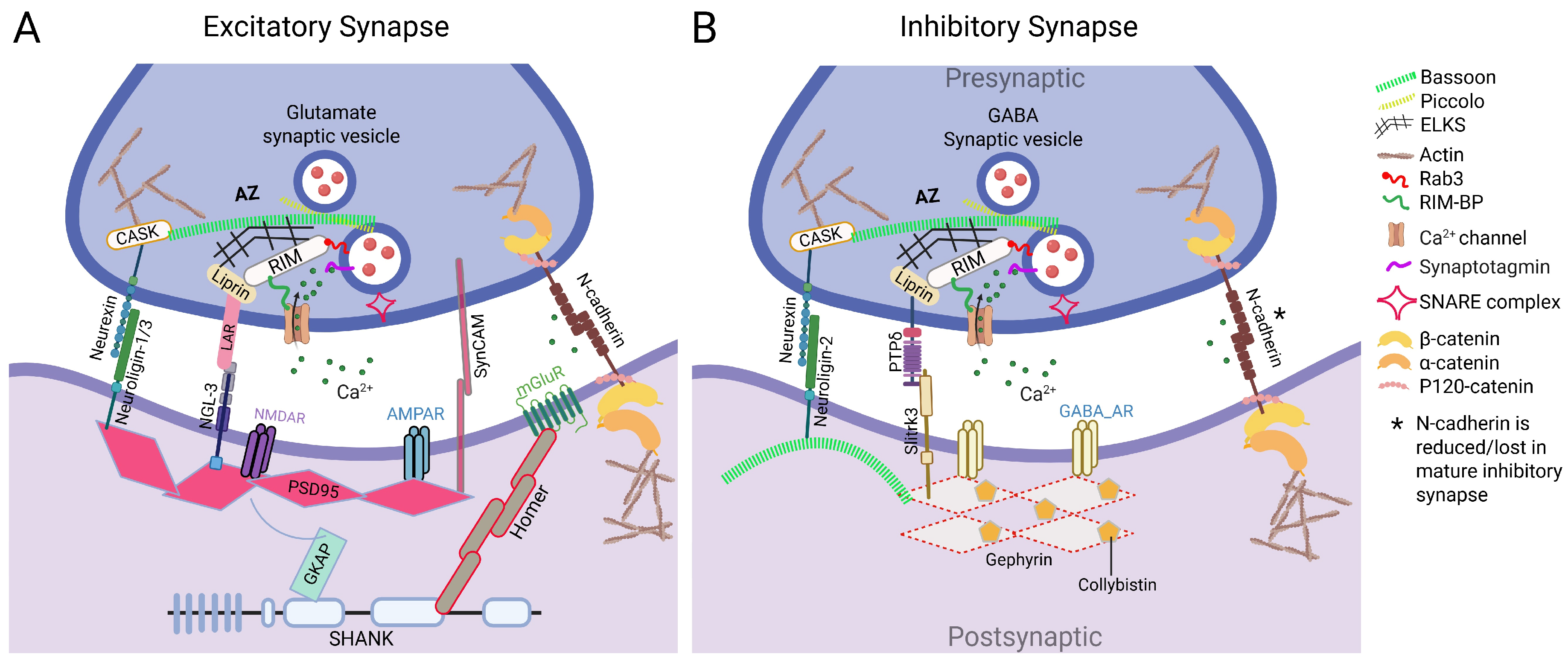
2.2. Synapse Formation: Adhesion Molecules/Organizers and Synaptic Scaffold Proteins
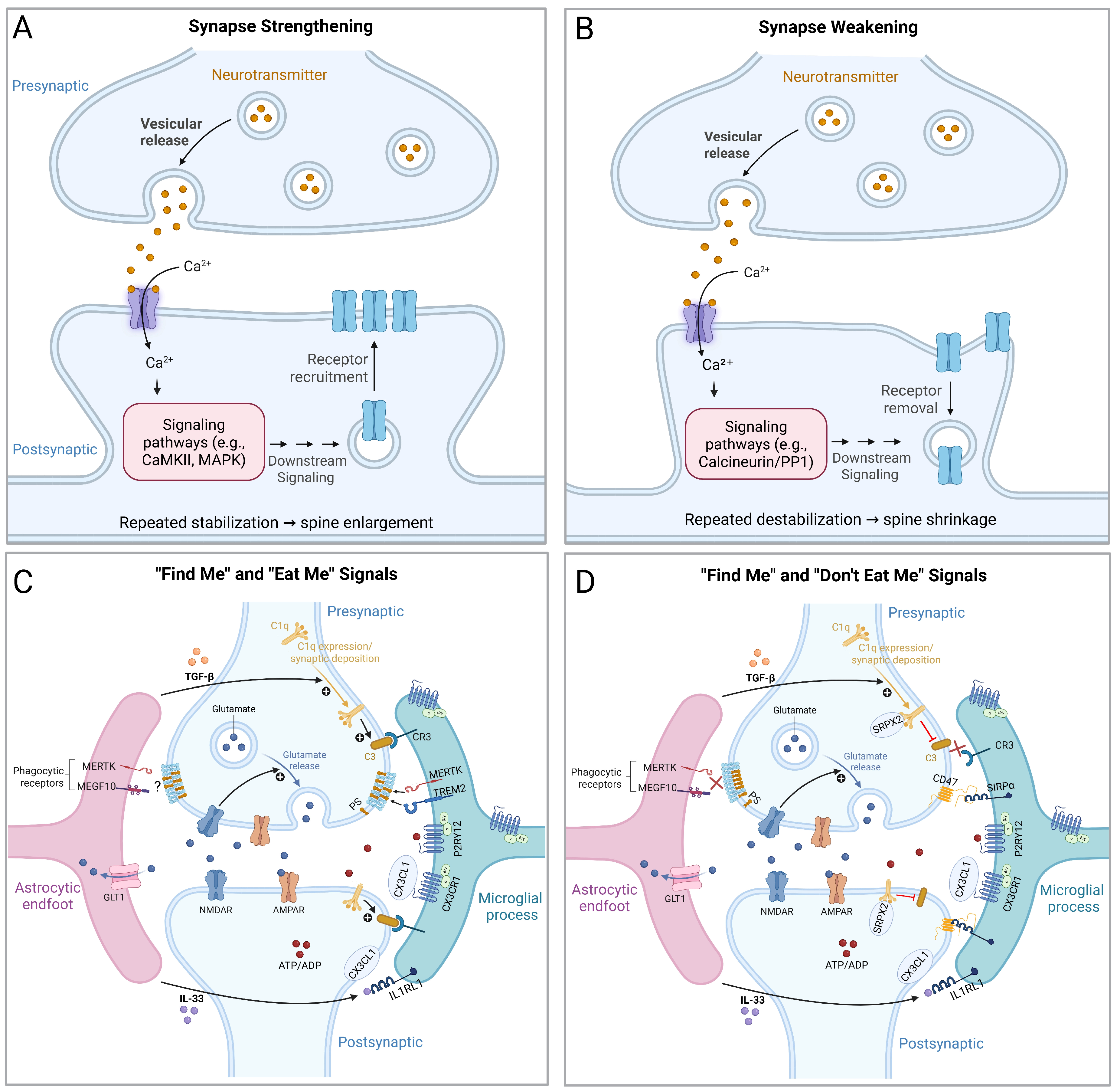
2.3. Activity-Dependent Synaptic Refinement
2.4. Glia-Mediated Synaptic Pruning
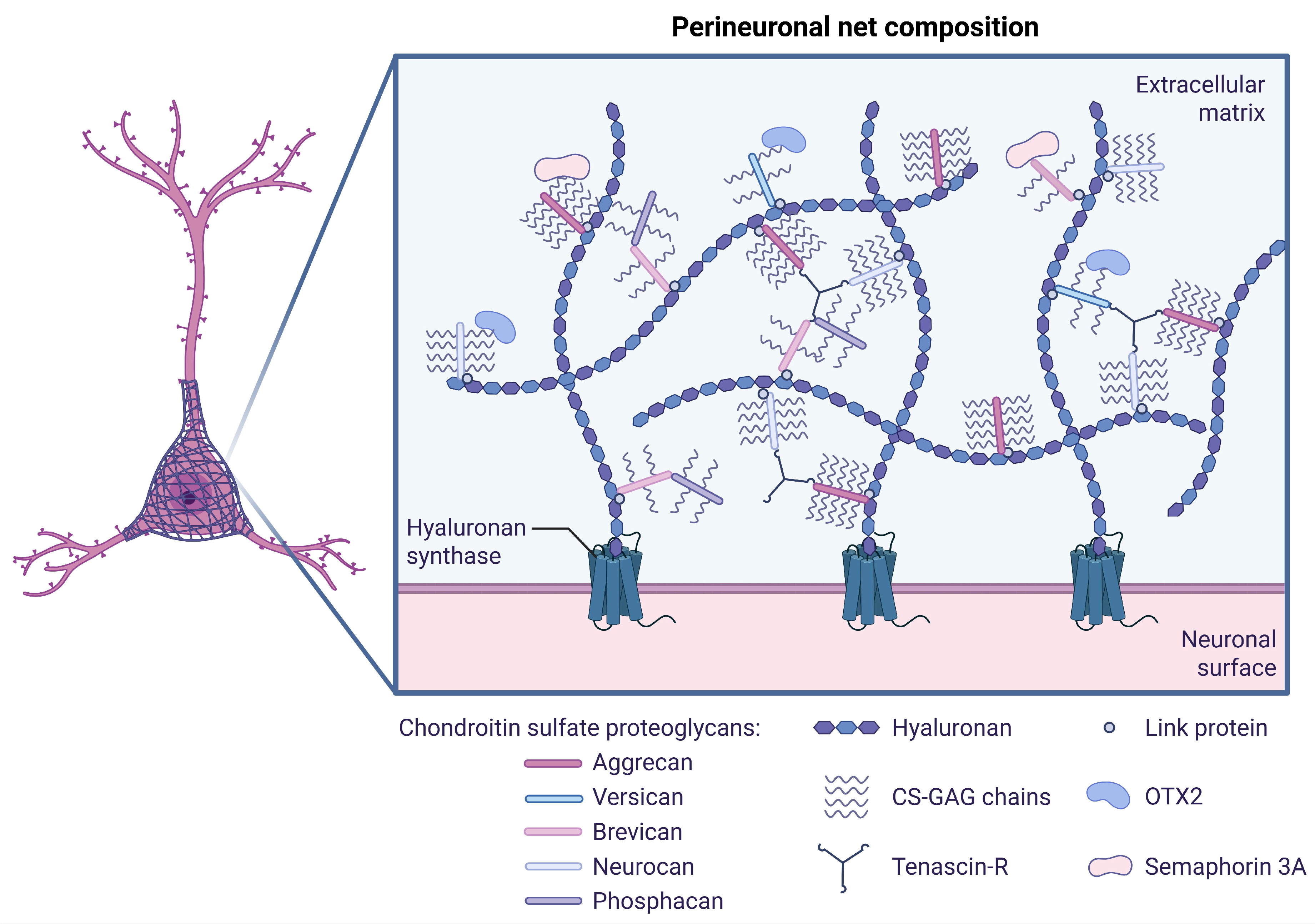
2.5. Experience-Dependent Plasticity and Transition to Synaptic Stabilization
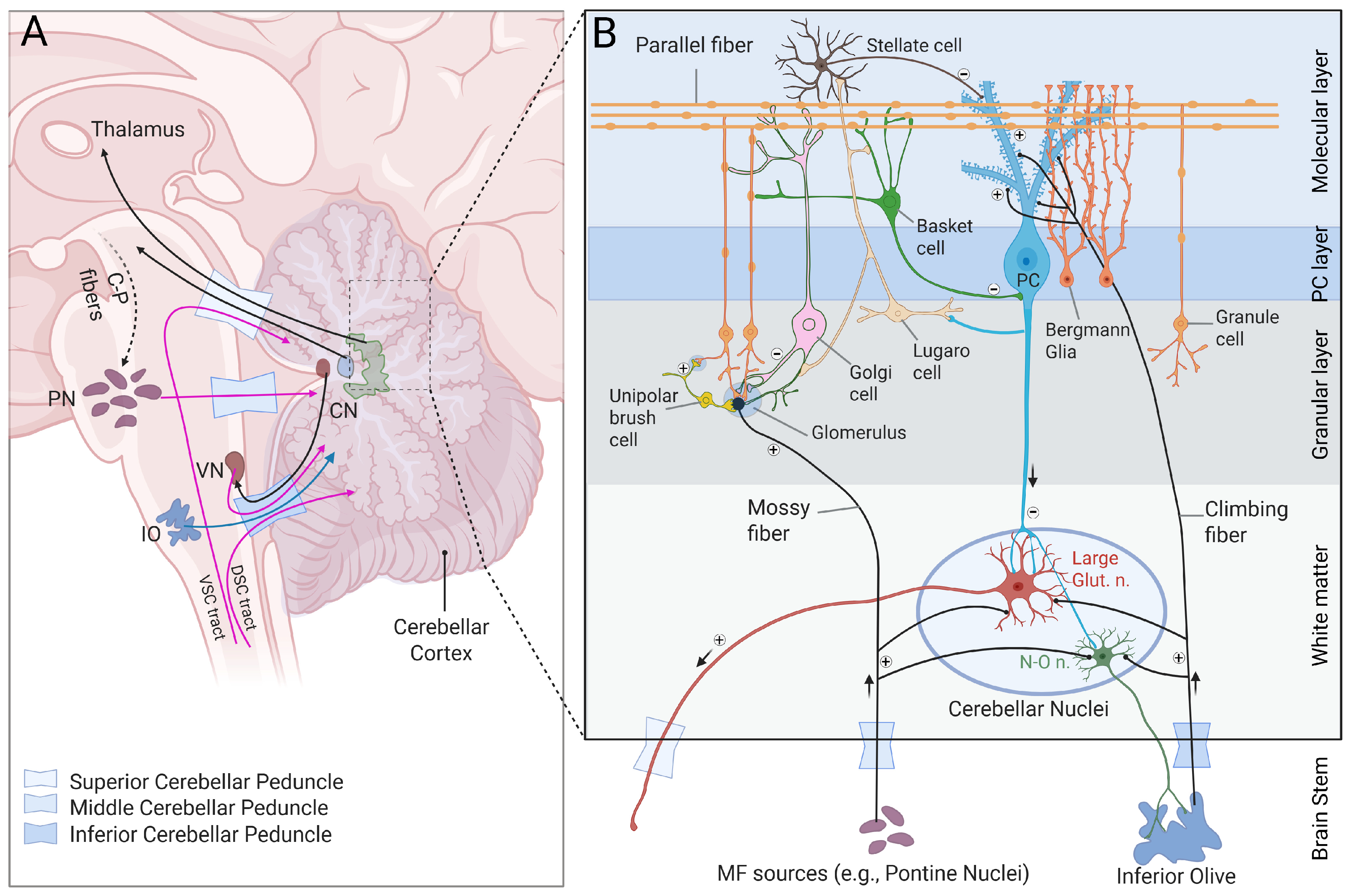
3. Synapse Formation and Refinement Within the Cerebellar Circuit
3.1. MF Afferent Pathway: From Transient MF–PC Contacts to Mature MF–GC–PC Axis
3.1.1. Transient MF–PC Synaptic Stage
3.1.2. Elimination of MF–PC Synapses and Formation of MF–GC Synapses
3.1.3. PF–PC Synapse Formation
3.1.4. Unipolar Brush Cells: Amplification of MF Signal
3.1.5. Golgi Cells: Feedforward and Feedback Inhibition of GCs
3.2. CF Afferent Pathway Targeting PCs
3.2.1. CF–PC Synapse Formation
3.2.2. CF–PC Synaptic Pruning
3.2.3. Glial Contribution to CF Pruning
3.3. Basket and Stellate Cells: Inhibition of PCs
3.4. Purkinje Cells (PCs): Principal Output Neurons of the Cerebellar Cortex
3.5. CN Efferent Pathway: Integration of CF/MF Excitation and PC Inhibition
3.5.1. Excitatory Inputs to CN Neurons from MF and CF Collaterals
3.5.2. Inhibitory Inputs to CN Neurons from PCs and Local Interneurons
3.5.3. CN Output Stabilization
4. Conclusions and Future Direction
Author Contributions
Funding
Data Availability Statement
Conflicts of Interest
Abbreviations
| AMPA | α-Amino-3-hydroxy-5-methyl-4-isoxazolepropionic acid |
| BDNF | Brain-derived neurotrophic factor |
| BMP | Bone morphogenetic protein |
| CBLN | Cerebellin |
| CF | Climbing fiber |
| CN | Cerebellar nuclei |
| GABA | Gamma-aminobutyric acid |
| Gαq | G-protein alpha q subunit |
| GC | Granule cell |
| GluD | Glutamate receptor delta |
| MF | Mossy fiber |
| mGluR | Metabotropic glutamate receptor |
| NMDA | N-methyl-D-aspartate |
| PC | Purkinje cell |
| PF | Parallel fiber |
| PKC | Protein kinase C |
| PLCβ | Phospholipase C beta |
| PNN | Perineuronal net |
| PSD | Postsynaptic density |
| Trk | Tropomyosin receptor kinase |
| VGLUT | Vesicular glutamate transporter |
References
- Veyrie, M.; Beffara, B.; Richard, N.; Mottolese, C.; Szathmari, A.; Di Rocco, F.; Faure Conter, C.; Leblond, P.; Desmurget, M.; Cristofori, I.; et al. Causally Mapping the Cerebellum in Children and Young Adults: From Motor to Cognition. Cerebellum 2025, 24, 31. [Google Scholar] [CrossRef] [Scilit] [PubMed]
- Koziol, L.F.; Budding, D.; Andreasen, N.; D’Arrigo, S.; Bulgheroni, S.; Imamizu, H.; Ito, M.; Manto, M.; Marvel, C.; Parker, K.; et al. Consensus paper: The cerebellum’s role in movement and cognition. Cerebellum 2014, 13, 151–177. [Google Scholar] [CrossRef] [Scilit]
- Alan, A.; Ennabe, M.; Alsarafandi, M.; Malik, N.; Laws, E.R.; Weinand, M. Redefining cerebellar assessment: A comprehensive review of the cerebellum’s cognitive and affective roles and the efficacy of CCAS scales. Surg. Neurol. Int. 2024, 15, 141. [Google Scholar] [CrossRef] [Scilit] [PubMed]
- Rudolph, S.; Badura, A.; Lutzu, S.; Pathak, S.S.; Thieme, A.; Verpeut, J.L.; Wagner, M.J.; Yang, Y.M.; Fioravante, D. Cognitive-Affective Functions of the Cerebellum. J. Neurosci. 2023, 43, 7554–7564. [Google Scholar] [CrossRef] [Scilit] [PubMed]
- Haldipur, P.; Aldinger, K.A.; Bernardo, S.; Deng, M.; Timms, A.E.; Overman, L.M.; Winter, C.; Lisgo, S.N.; Razavi, F.; Silvestri, E.; et al. Spatiotemporal expansion of primary progenitor zones in the developing human cerebellum. Science 2019, 366, 454–460. [Google Scholar] [CrossRef] [Scilit] [PubMed]
- Rueda-Alaña, E.; García-Moreno, F. Time in Neurogenesis: Conservation of the Developmental Formation of the Cerebellar Circuitry. Brain Behav. Evol. 2022, 97, 33–47. [Google Scholar] [CrossRef] [Scilit] [PubMed]
- Galas, L.; Bénard, M.; Lebon, A.; Komuro, Y.; Schapman, D.; Vaudry, H.; Vaudry, D.; Komuro, H. Postnatal Migration of Cerebellar Interneurons. Brain Sci. 2017, 7, 62. [Google Scholar] [CrossRef] [Scilit] [PubMed]
- Rahimi-Balaei, M.; Amiri, S.; Lamonerie, T.; Wu, S.R.; Zoghbi, H.Y.; Consalez, G.G.; Goldowitz, D.; Marzban, H. Identification of an early subset of cerebellar nuclei neurons in mice. eLife 2024, 13, RP93778. [Google Scholar] [CrossRef] [PubMed]
- Ghiyamihoor, F.; Rad, A.A.; Marzban, H. The Nuclear Transitory Zone: A Key Player in the Cerebellar Development. Cerebellum 2025, 24, 92. [Google Scholar] [CrossRef] [Scilit] [PubMed]
- Sarnat, H.B.; Flores-Sarnat, L.; Auer, R.N. Sequence of synaptogenesis in the fetal and neonatal cerebellar system—Part 1: Guillain-Mollaret triangle (dentato-rubro-olivo-cerebellar circuit). Dev. Neurosci. 2013, 35, 69–81. [Google Scholar] [CrossRef] [Scilit] [PubMed]
- Uusisaari, M.; De Schutter, E. The mysterious microcircuitry of the cerebellar nuclei. J. Physiol. 2011, 589, 3441–3457. [Google Scholar] [CrossRef] [Scilit] [PubMed]
- Mishina, M.; Uemura, T.; Yasumura, M.; Yoshida, T. Molecular mechanism of parallel fiber-Purkinje cell synapse formation. Front. Neural Circuits 2012, 6, 90. [Google Scholar] [CrossRef] [Scilit] [PubMed]
- Kano, M.; Watanabe, T.; Uesaka, N.; Watanabe, M. Multiple Phases of Climbing Fiber Synapse Elimination in the Developing Cerebellum. Cerebellum 2018, 17, 722–734. [Google Scholar] [CrossRef] [Scilit] [PubMed]
- Nakayama, H.; Abe, M.; Morimoto, C.; Iida, T.; Okabe, S.; Sakimura, K.; Hashimoto, K. Microglia permit climbing fiber elimination by promoting GABAergic inhibition in the developing cerebellum. Nat. Commun. 2018, 9, 2830. [Google Scholar] [CrossRef] [Scilit] [PubMed]
- Miyazaki, T.; Yamasaki, M.; Hashimoto, K.; Kohda, K.; Yuzaki, M.; Shimamoto, K.; Tanaka, K.; Kano, M.; Watanabe, M. Glutamate transporter GLAST controls synaptic wrapping by Bergmann glia and ensures proper wiring of Purkinje cells. Proc. Natl. Acad. Sci. USA 2017, 114, 7438–7443. [Google Scholar] [CrossRef] [Scilit] [PubMed]
- Hao, S.; Zhu, X.; Huang, Z.; Yang, Q.; Liu, H.; Wu, Y.; Zhan, Y.; Dong, Y.; Li, C.; Wang, H.; et al. Cross-species single-cell spatial transcriptomic atlases of the cerebellar cortex. Science 2024, 385, eado3927. [Google Scholar] [CrossRef] [Scilit] [PubMed]
- Haldipur, P.; Millen, K.J.; Aldinger, K.A. Human Cerebellar Development and Transcriptomics: Implications for Neurodevelopmental Disorders. Annu. Rev. Neurosci. 2022, 45, 515–531. [Google Scholar] [CrossRef] [Scilit] [PubMed]
- Sepp, M.; Leiss, K.; Murat, F.; Okonechnikov, K.; Joshi, P.; Leushkin, E.; Spänig, L.; Mbengue, N.; Schneider, C.; Schmidt, J.; et al. Cellular development and evolution of the mammalian cerebellum. Nature 2024, 625, 788–796. [Google Scholar] [CrossRef] [Scilit] [PubMed]
- Liu, Y.; McDaniel, J.A.; Chen, C.; Yang, L.; Kipcak, A.; Savier, E.L.; Erisir, A.; Cang, J.; Campbell, J.N. Co-Conservation of synaptic gene expression and circuitry in collicular neurons. Nat. Commun. 2025, 16, 9146. [Google Scholar] [CrossRef] [Scilit] [PubMed]
- Giagtzoglou, N.; Ly, C.V.; Bellen, H.J. Cell adhesion, the backbone of the synapse: “vertebrate” and “invertebrate” perspectives. Cold Spring Harb. Perspect. Biol. 2009, 1, a003079. [Google Scholar] [CrossRef] [Scilit] [PubMed]
- Nithianantharajah, J.; Komiyama, N.H.; McKechanie, A.; Johnstone, M.; Blackwood, D.H.; St Clair, D.; Emes, R.D.; van de Lagemaat, L.N.; Saksida, L.M.; Bussey, T.J.; et al. Synaptic scaffold evolution generated components of vertebrate cognitive complexity. Nat. Neurosci. 2013, 16, 16–24. [Google Scholar] [CrossRef] [Scilit] [PubMed]
- Huang, Z.J.; Zeng, H. Genetic approaches to neural circuits in the mouse. Annu. Rev. Neurosci. 2013, 36, 183–215. [Google Scholar] [CrossRef] [Scilit] [PubMed]
- Mudge, J.; Miller, N.A.; Khrebtukova, I.; Lindquist, I.E.; May, G.D.; Huntley, J.J.; Luo, S.; Zhang, L.; van Velkinburgh, J.C.; Farmer, A.D.; et al. Genomic convergence analysis of schizophrenia: mRNA sequencing reveals altered synaptic vesicular transport in post-mortem cerebellum. PLoS ONE 2008, 3, e3625. [Google Scholar] [CrossRef] [Scilit] [PubMed]
- Bayés, A.; van de Lagemaat, L.N.; Collins, M.O.; Croning, M.D.; Whittle, I.R.; Choudhary, J.S.; Grant, S.G. Characterization of the proteome, diseases and evolution of the human postsynaptic density. Nat. Neurosci. 2011, 14, 19–21. [Google Scholar] [CrossRef] [Scilit] [PubMed]
- Koopmans, F.; van Nierop, P.; Andres-Alonso, M.; Byrnes, A.; Cijsouw, T.; Coba, M.P.; Cornelisse, L.N.; Farrell, R.J.; Goldschmidt, H.L.; Howrigan, D.P.; et al. SynGO: An Evidence-Based, Expert-Curated Knowledge Base for the Synapse. Neuron 2019, 103, 217–234.e214. [Google Scholar] [CrossRef] [Scilit] [PubMed]
- Lin, C.Y.; Louis, E.D.; Faust, P.L.; Koeppen, A.H.; Vonsattel, J.P.; Kuo, S.H. Abnormal climbing fibre-Purkinje cell synaptic connections in the essential tremor cerebellum. Brain 2014, 137, 3149–3159. [Google Scholar] [CrossRef] [Scilit] [PubMed]
- Nandi, S.; de Deus, J.L.; Faborode, O.S.; Nandi, S. Synaptic Pruning by Microglia: Lessons from Genetic Studies in Mice. Dev. Neurosci. 2025, 47, 362–382. [Google Scholar] [CrossRef] [Scilit] [PubMed]
- Zeiss, C.J. Comparative Milestones in Rodent and Human Postnatal Central Nervous System Development. Toxicol. Pathol. 2021, 49, 1368–1373. [Google Scholar] [CrossRef] [Scilit] [PubMed]
- Yamamoto, H.; Demura, T.; Morita, M.; Banker, G.A.; Tanii, T.; Nakamura, S. Differential neurite outgrowth is required for axon specification by cultured hippocampal neurons. J. Neurochem. 2012, 123, 904–910. [Google Scholar] [CrossRef] [Scilit] [PubMed]
- Sainath, R.; Gallo, G. Cytoskeletal and signaling mechanisms of neurite formation. Cell Tissue Res. 2015, 359, 267–278. [Google Scholar] [CrossRef] [Scilit] [PubMed]
- Polleux, F.; Snider, W. Initiating and growing an axon. Cold Spring Harb. Perspect. Biol. 2010, 2, a001925. [Google Scholar] [CrossRef] [Scilit] [PubMed]
- Winans, A.M.; Collins, S.R.; Meyer, T. Waves of actin and microtubule polymerization drive microtubule-based transport and neurite growth before single axon formation. Elife 2016, 5, e12387. [Google Scholar] [CrossRef] [Scilit] [PubMed]
- Lowery, L.A.; Van Vactor, D. The trip of the tip: Understanding the growth cone machinery. Nat. Rev. Mol. Cell Biol. 2009, 10, 332–343. [Google Scholar] [CrossRef] [Scilit] [PubMed]
- Vitriol, E.A.; Zheng, J.Q. Growth cone travel in space and time: The cellular ensemble of cytoskeleton, adhesion, and membrane. Neuron 2012, 73, 1068–1081. [Google Scholar] [CrossRef] [Scilit] [PubMed]
- Doherty, P.; Rowett, L.H.; Moore, S.E.; Mann, D.A.; Walsh, F.S. Neurite outgrowth in response to transfected N-CAM and N-cadherin reveals fundamental differences in neuronal responsiveness to CAMs. Neuron 1991, 6, 247–258. [Google Scholar] [CrossRef] [Scilit] [PubMed]
- Wiencken-Barger, A.E.; Mavity-Hudson, J.; Bartsch, U.; Schachner, M.; Casagrande, V.A. The role of L1 in axon pathfinding and fasciculation. Cereb. Cortex 2004, 14, 121–131. [Google Scholar] [CrossRef] [Scilit] [PubMed][Green Version]
- Soler-Llavina, G.J.; Fuccillo, M.V.; Ko, J.; Südhof, T.C.; Malenka, R.C. The neurexin ligands, neuroligins and leucine-rich repeat transmembrane proteins, perform convergent and divergent synaptic functions in vivo. Proc. Natl. Acad. Sci. USA 2011, 108, 16502–16509. [Google Scholar] [CrossRef] [Scilit] [PubMed]
- Reissner, C.; Runkel, F.; Missler, M. Neurexins. Genome Biol. 2013, 14, 213. [Google Scholar] [CrossRef] [Scilit] [PubMed]
- Qi, C.; Luo, L.D.; Feng, I.; Ma, S. Molecular mechanisms of synaptogenesis. Front. Synaptic Neurosci. 2022, 14, 939793. [Google Scholar] [CrossRef] [Scilit] [PubMed]
- Südhof, T.C. Towards an Understanding of Synapse Formation. Neuron 2018, 100, 276–293. [Google Scholar] [CrossRef] [Scilit] [PubMed]
- Roppongi, R.T.; Dhume, S.H.; Padmanabhan, N.; Silwal, P.; Zahra, N.; Karimi, B.; Bomkamp, C.; Patil, C.S.; Champagne-Jorgensen, K.; Twilley, R.E.; et al. LRRTMs Organize Synapses through Differential Engagement of Neurexin and PTPσ. Neuron 2020, 106, 108–125.e12, Erratum in Neuron 2020, 106, 701. [Google Scholar] [CrossRef] [Scilit] [PubMed]
- Liouta, K.; Lubas, M.; Venugopal, V.; Chabbert, J.; Jeannière, C.; Diaz, C.; Munier, M.; Tessier, B.; Claverol, S.; Favereaux, A.; et al. LRRTM2 controls presynapse nano-organization and AMPA receptor sub-positioning through Neurexin-binding interface. Nat. Commun. 2024, 15, 8807. [Google Scholar] [CrossRef] [Scilit] [PubMed]
- Siddiqui, T.J.; Pancaroglu, R.; Kang, Y.; Rooyakkers, A.; Craig, A.M. LRRTMs and neuroligins bind neurexins with a differential code to cooperate in glutamate synapse development. J. Neurosci. 2010, 30, 7495–7506. [Google Scholar] [CrossRef] [Scilit] [PubMed]
- Yim, Y.S.; Kwon, Y.; Nam, J.; Yoon, H.I.; Lee, K.; Kim, D.G.; Kim, E.; Kim, C.H.; Ko, J. Slitrks control excitatory and inhibitory synapse formation with LAR receptor protein tyrosine phosphatases. Proc. Natl. Acad. Sci. USA 2013, 110, 4057–4062, Erratum in Proc. Natl. Acad. Sci. USA 2024, 121, e2410441121. [Google Scholar] [CrossRef] [Scilit] [PubMed]
- Südhof, T.C. The cell biology of synapse formation. J. Cell Biol. 2021, 220, e202103052. [Google Scholar] [CrossRef] [Scilit] [PubMed]
- Cooper, J.N.; Mittal, J.; Sangadi, A.; Klassen, D.L.; King, A.M.; Zalta, M.; Mittal, R.; Eshraghi, A.A. Landscape of NRXN1 Gene Variants in Phenotypic Manifestations of Autism Spectrum Disorder: A Systematic Review. J. Clin. Med. 2024, 13, 2067. [Google Scholar] [CrossRef] [Scilit] [PubMed]
- Montalbano, S.; Krebs, M.D.; Rosengren, A.; Vaez, M.; Hellberg, K.G.; Mortensen, P.B.; Børglum, A.D.; Geschwind, D.H.; Raznahan, A.; Thompson, W.K.; et al. Analysis of exonic deletions in a large population study provides novel insights into NRXN1 pathology. npj Genom. Med. 2024, 9, 67. [Google Scholar] [CrossRef] [Scilit] [PubMed]
- Nguyen, T.A.; Lehr, A.W.; Roche, K.W. Neuroligins and Neurodevelopmental Disorders: X-Linked Genetics. Front. Synaptic Neurosci. 2020, 12, 33. [Google Scholar] [CrossRef] [PubMed]
- Ali, H.; Marth, L.; Krueger-Burg, D. Neuroligin-2 as a central organizer of inhibitory synapses in health and disease. Sci. Signal 2020, 13, eabd8379. [Google Scholar] [CrossRef] [Scilit] [PubMed]
- Kim, S.; Shin, J.J.; Kang, M.; Yi, Y.; Kim, E. Synaptic and Non-Synaptic Functions of PTPRD: A Receptor Tyrosine Phosphatase at the Crossroads of Neural Circuitry and Metabolism. J. Neurochem. 2025, 169, e70292. [Google Scholar] [CrossRef] [Scilit] [PubMed]
- Cortés, B.I.; Meza, R.C.; Ancatén-González, C.; Ardiles, N.M.; Aránguiz, M.I.; Tomita, H.; Kaplan, D.R.; Cornejo, F.; Nunez-Parra, A.; Moya, P.R.; et al. Loss of protein tyrosine phosphatase receptor delta PTPRD increases the number of cortical neurons, impairs synaptic function and induces autistic-like behaviors in adult mice. Biol. Res. 2024, 57, 40. [Google Scholar] [CrossRef] [Scilit] [PubMed]
- Kim, S.; Shin, J.J.; Kang, M.; Yang, Y.; Cho, Y.S.; Paik, H.; Kim, J.; Yi, Y.; Lee, S.; Koo, H.Y.; et al. Alternatively spliced mini-exon B in PTPδ regulates excitatory synapses through cell-type-specific trans-synaptic PTPδ-IL1RAP interaction. Nat. Commun. 2025, 16, 4415. [Google Scholar] [CrossRef] [Scilit] [PubMed]
- Arikkath, J.; Reichardt, L.F. Cadherins and catenins at synapses: Roles in synaptogenesis and synaptic plasticity. Trends Neurosci. 2008, 31, 487–494. [Google Scholar] [CrossRef] [Scilit] [PubMed]
- Chen, W.V.; Maniatis, T. Clustered protocadherins. Development 2013, 140, 3297–3302. [Google Scholar] [CrossRef] [Scilit] [PubMed]
- Okuda, T.; Yu, L.M.; Cingolani, L.A.; Kemler, R.; Goda, Y. β-Catenin regulates excitatory postsynaptic strength at hippocampal synapses. Proc. Natl. Acad. Sci. USA 2007, 104, 13479–13484. [Google Scholar] [CrossRef] [Scilit] [PubMed]
- Nikitczuk, J.S.; Patil, S.B.; Matikainen-Ankney, B.A.; Scarpa, J.; Shapiro, M.L.; Benson, D.L.; Huntley, G.W. N-cadherin regulates molecular organization of excitatory and inhibitory synaptic circuits in adult hippocampus in vivo. Hippocampus 2014, 24, 943–962. [Google Scholar] [CrossRef] [Scilit] [PubMed]
- Benson, D.L.; Tanaka, H. N-cadherin redistribution during synaptogenesis in hippocampal neurons. J. Neurosci. 1998, 18, 6892–6904. [Google Scholar] [CrossRef] [Scilit] [PubMed]
- Fletcher, T.L.; Cameron, P.; De Camilli, P.; Banker, G. The distribution of synapsin I and synaptophysin in hippocampal neurons developing in culture. J. Neurosci. 1991, 11, 1617–1626. [Google Scholar] [CrossRef] [Scilit] [PubMed]
- Sarnat, H.B.; Flores-Sarnat, L.; Auer, R.N. Synaptogenesis in the foetal and neonatal cerebellar system. 2. Pontine nuclei and cerebellar cortex. Dev. Neurosci. 2013, 35, 317–325. [Google Scholar] [CrossRef] [Scilit] [PubMed]
- Castagna, C.; Merighi, A.; Lossi, L. Decreased Expression of Synaptophysin 1 (SYP1 Major Synaptic Vesicle Protein p38) and Contactin 6 (CNTN6/NB3) in the Cerebellar Vermis of reln Haplodeficient Mice. Cell. Mol. Neurobiol. 2019, 39, 833–856. [Google Scholar] [CrossRef] [Scilit] [PubMed]
- Sheng, M.; Kim, E. The postsynaptic organization of synapses. Cold Spring Harb. Perspect. Biol. 2011, 3, a005678. [Google Scholar] [CrossRef] [Scilit] [PubMed]
- Chen, X.; Levy, J.M.; Hou, A.; Winters, C.; Azzam, R.; Sousa, A.A.; Leapman, R.D.; Nicoll, R.A.; Reese, T.S. PSD-95 family MAGUKs are essential for anchoring AMPA and NMDA receptor complexes at the postsynaptic density. Proc. Natl. Acad. Sci. USA 2015, 112, E6983–E6992. [Google Scholar] [CrossRef] [Scilit] [PubMed]
- Shiraishi-Yamaguchi, Y.; Furuichi, T. The Homer family proteins. Genome Biol. 2007, 8, 206. [Google Scholar] [CrossRef] [Scilit] [PubMed]
- Durand, C.M.; Betancur, C.; Boeckers, T.M.; Bockmann, J.; Chaste, P.; Fauchereau, F.; Nygren, G.; Rastam, M.; Gillberg, I.C.; Anckarsäter, H.; et al. Mutations in the gene encoding the synaptic scaffolding protein SHANK3 are associated with autism spectrum disorders. Nat. Genet. 2007, 39, 25–27. [Google Scholar] [CrossRef] [Scilit] [PubMed]
- Monteiro, P.; Feng, G. SHANK proteins: Roles at the synapse and in autism spectrum disorder. Nat. Rev. Neurosci. 2017, 18, 147–157. [Google Scholar] [CrossRef] [Scilit] [PubMed]
- Xing, J.; Kimura, H.; Wang, C.; Ishizuka, K.; Kushima, I.; Arioka, Y.; Yoshimi, A.; Nakamura, Y.; Shiino, T.; Oya-Ito, T.; et al. Resequencing and Association Analysis of Six PSD-95-Related Genes as Possible Susceptibility Genes for Schizophrenia and Autism Spectrum Disorders. Sci. Rep. 2016, 6, 27491. [Google Scholar] [CrossRef] [Scilit] [PubMed]
- Levy, A.M.; Gomez-Puertas, P.; Tümer, Z. Neurodevelopmental Disorders Associated with PSD-95 and Its Interaction Partners. Int. J. Mol. Sci. 2022, 23, 4390. [Google Scholar] [CrossRef] [Scilit] [PubMed]
- Feyder, M.; Karlsson, R.M.; Mathur, P.; Lyman, M.; Bock, R.; Momenan, R.; Munasinghe, J.; Scattoni, M.L.; Ihne, J.; Camp, M.; et al. Association of mouse Dlg4 (PSD-95) gene deletion and human DLG4 gene variation with phenotypes relevant to autism spectrum disorders and Williams’ syndrome. Am. J. Psychiatry 2010, 167, 1508–1517. [Google Scholar] [CrossRef] [Scilit] [PubMed]
- Macha, A.; Liebsch, F.; Bruckisch, E.H.W.; Burdina, N.; von Stülpnagel, I.; Benting, K.; Gunkel, M.; Behrmann, E.; Schwarz, G. Gephyrin filaments represent the molecular basis of inhibitory postsynaptic densities. Nat. Commun. 2025, 16, 8293. [Google Scholar] [CrossRef] [Scilit] [PubMed]
- Hoffmann, C.; Milovanovic, D. Gephyrin: A scaffold that builds a phase at the inhibitory postsynapses. Cell Res. 2021, 31, 245–246, Erratum in Cell Res. 2021. https://doi.org/10.1038/s41422-021-00547-0. [Google Scholar] [CrossRef] [Scilit] [PubMed]
- Poulopoulos, A.; Aramuni, G.; Meyer, G.; Soykan, T.; Hoon, M.; Papadopoulos, T.; Zhang, M.; Paarmann, I.; Fuchs, C.; Harvey, K.; et al. Neuroligin 2 drives postsynaptic assembly at perisomatic inhibitory synapses through gephyrin and collybistin. Neuron 2009, 63, 628–642. [Google Scholar] [CrossRef] [Scilit] [PubMed]
- Reshetniak, S.; Bogaciu, C.A.; Bonn, S.; Brose, N.; Cooper, B.H.; D’Este, E.; Fauth, M.; Fernández-Busnadiego, R.; Fiosins, M.; Fischer, A.; et al. The synaptic vesicle cluster as a controller of pre- and postsynaptic structure and function. J. Physiol. 2025, 603, 5935–5964. [Google Scholar] [CrossRef] [Scilit] [PubMed]
- Ehrlich, I.; Malinow, R. Postsynaptic density 95 controls AMPA receptor incorporation during long-term potentiation and experience-driven synaptic plasticity. J. Neurosci. 2004, 24, 916–927. [Google Scholar] [CrossRef] [Scilit] [PubMed]
- Huang, X.; Stodieck, S.K.; Goetze, B.; Cui, L.; Wong, M.H.; Wenzel, C.; Hosang, L.; Dong, Y.; Löwel, S.; Schlüter, O.M. Progressive maturation of silent synapses governs the duration of a critical period. Proc. Natl. Acad. Sci. USA 2015, 112, E3131–E3140. [Google Scholar] [CrossRef] [Scilit] [PubMed]
- Fu, Z.; Vicini, S. Neuroligin-2 accelerates GABAergic synapse maturation in cerebellar granule cells. Mol. Cell. Neurosci. 2009, 42, 45–55. [Google Scholar] [CrossRef] [Scilit] [PubMed]
- Misra, C.; Brickley, S.G.; Wyllie, D.J.; Cull-Candy, S.G. Slow deactivation kinetics of NMDA receptors containing NR1 and NR2D subunits in rat cerebellar Purkinje cells. J. Physiol. 2000, 525, 299–305. [Google Scholar] [CrossRef] [Scilit] [PubMed]
- Renzi, M.; Farrant, M.; Cull-Candy, S.G. Climbing-fibre activation of NMDA receptors in Purkinje cells of adult mice. J. Physiol. 2007, 585, 91–101. [Google Scholar] [CrossRef] [Scilit] [PubMed]
- Piochon, C.; Irinopoulou, T.; Brusciano, D.; Bailly, Y.; Mariani, J.; Levenes, C. NMDA receptor contribution to the climbing fiber response in the adult mouse Purkinje cell. J. Neurosci. 2007, 27, 10797–10809. [Google Scholar] [CrossRef] [Scilit] [PubMed]
- Piochon, C.; Levenes, C.; Ohtsuki, G.; Hansel, C. Purkinje cell NMDA receptors assume a key role in synaptic gain control in the mature cerebellum. J. Neurosci. 2010, 30, 15330–15335. [Google Scholar] [CrossRef] [Scilit] [PubMed]
- Liu, H.; Lan, Y.; Bing, Y.H.; Chu, C.P.; Qiu, D.L. N-methyl-D-Aspartate Receptors Contribute to Complex Spike Signaling in Cerebellar Purkinje Cells: An In vivo Study in Mice. Front. Cell. Neurosci. 2016, 10, 172. [Google Scholar] [CrossRef] [Scilit] [PubMed]
- Casado, M.; Dieudonné, S.; Ascher, P. Presynaptic N-methyl-D-aspartate receptors at the parallel fiber-Purkinje cell synapse. Proc. Natl. Acad. Sci. USA 2000, 97, 11593–11597. [Google Scholar] [CrossRef] [Scilit] [PubMed]
- Faust, T.E.; Gunner, G.; Schafer, D.P. Mechanisms governing activity-dependent synaptic pruning in the developing mammalian CNS. Nat. Rev. Neurosci. 2021, 22, 657–673. [Google Scholar] [CrossRef] [Scilit] [PubMed]
- Bi, G.; Poo, M. Synaptic modification by correlated activity: Hebb’s postulate revisited. Annu. Rev. Neurosci. 2001, 24, 139–166. [Google Scholar] [CrossRef] [Scilit] [PubMed]
- Nagappan-Chettiar, S.; Burbridge, T.J.; Umemori, H. Activity-Dependent Synapse Refinement: From Mechanisms to Molecules. Neuroscientist 2024, 30, 673–689. [Google Scholar] [CrossRef] [Scilit] [PubMed]
- Camus, C.; Leval, L.; Villicana-Munoz, V.; Jelinkova, S.; Compans, B.; Gambino, F.; Herzog, E.; Choquet, D.; Hosy, E. Synaptic pruning following NMDAR-dependent LTD preferentially affects isolated synapses. iScience 2025, 28, 113093. [Google Scholar] [CrossRef] [Scilit] [PubMed]
- Henson, M.A.; Tucker, C.J.; Zhao, M.; Dudek, S.M. Long-term depression-associated signaling is required for an in vitro model of NMDA receptor-dependent synapse pruning. Neurobiol. Learn. Mem. 2017, 138, 39–53. [Google Scholar] [CrossRef] [Scilit] [PubMed][Green Version]
- Piochon, C.; Kano, M.; Hansel, C. LTD-like molecular pathways in developmental synaptic pruning. Nat. Neurosci. 2016, 19, 1299–1310. [Google Scholar] [CrossRef] [Scilit] [PubMed]
- Cohen, S.; Greenberg, M.E. Communication between the synapse and the nucleus in neuronal development, plasticity, and disease. Annu. Rev. Cell Dev. Biol. 2008, 24, 183–209. [Google Scholar] [CrossRef] [Scilit] [PubMed]
- Stein, I.S.; Zito, K. Dendritic Spine Elimination: Molecular Mechanisms and Implications. Neuroscientist 2019, 25, 27–47. [Google Scholar] [CrossRef] [Scilit] [PubMed]
- Lu, B. BDNF and activity-dependent synaptic modulation. Learn. Mem. 2003, 10, 86–98. [Google Scholar] [CrossRef] [Scilit] [PubMed]
- Nagappan-Chettiar, S.; Yasuda, M.; Johnson-Venkatesh, E.M.; Umemori, H. The molecular signals that regulate activity-dependent synapse refinement in the brain. Curr. Opin. Neurobiol. 2023, 79, 102692. [Google Scholar] [CrossRef] [Scilit] [PubMed]
- Yasuda, M.; Nagappan-Chettiar, S.; Johnson-Venkatesh, E.M.; Umemori, H. An activity-dependent determinant of synapse elimination in the mammalian brain. Neuron 2021, 109, 1333–1349.e1336. [Google Scholar] [CrossRef] [Scilit] [PubMed]
- Duffy, A.S.; Eyo, U.B. Microglia and Astrocytes in Postnatal Neural Circuit Formation. Glia 2025, 73, 232–250. [Google Scholar] [CrossRef] [Scilit] [PubMed]
- Paolicelli, R.C.; Bolasco, G.; Pagani, F.; Maggi, L.; Scianni, M.; Panzanelli, P.; Giustetto, M.; Ferreira, T.A.; Guiducci, E.; Dumas, L.; et al. Synaptic pruning by microglia is necessary for normal brain development. Science 2011, 333, 1456–1458. [Google Scholar] [CrossRef] [Scilit] [PubMed]
- Beiter, R.M.; Sheehan, P.W.; Schafer, D.P. Microglia phagocytic mechanisms: Development informing disease. Curr. Opin. Neurobiol. 2024, 86, 102877. [Google Scholar] [CrossRef] [Scilit] [PubMed]
- Schafer, D.P.; Lehrman, E.K.; Kautzman, A.G.; Koyama, R.; Mardinly, A.R.; Yamasaki, R.; Ransohoff, R.M.; Greenberg, M.E.; Barres, B.A.; Stevens, B. Microglia sculpt postnatal neural circuits in an activity and complement-dependent manner. Neuron 2012, 74, 691–705. [Google Scholar] [CrossRef] [Scilit] [PubMed]
- Stevens, B.; Allen, N.J.; Vazquez, L.E.; Howell, G.R.; Christopherson, K.S.; Nouri, N.; Micheva, K.D.; Mehalow, A.K.; Huberman, A.D.; Stafford, B.; et al. The classical complement cascade mediates CNS synapse elimination. Cell 2007, 131, 1164–1178. [Google Scholar] [CrossRef] [Scilit] [PubMed]
- Guedes, J.R.; Ferreira, P.A.; Costa, J.M.; Cardoso, A.L.; Peça, J. Microglia-dependent remodeling of neuronal circuits. J. Neurochem. 2022, 163, 74–93. [Google Scholar] [CrossRef] [Scilit] [PubMed]
- Vainchtein, I.D.; Chin, G.; Cho, F.S.; Kelley, K.W.; Miller, J.G.; Chien, E.C.; Liddelow, S.A.; Nguyen, P.T.; Nakao-Inoue, H.; Dorman, L.C.; et al. Astrocyte-derived interleukin-33 promotes microglial synapse engulfment and neural circuit development. Science 2018, 359, 1269–1273. [Google Scholar] [CrossRef] [Scilit] [PubMed]
- Lee, E.; Chung, W.S. Glial Control of Synapse Number in Healthy and Diseased Brain. Front. Cell. Neurosci. 2019, 13, 42. [Google Scholar] [CrossRef] [Scilit] [PubMed]
- Chung, W.S.; Clarke, L.E.; Wang, G.X.; Stafford, B.K.; Sher, A.; Chakraborty, C.; Joung, J.; Foo, L.C.; Thompson, A.; Chen, C.; et al. Astrocytes mediate synapse elimination through MEGF10 and MERTK pathways. Nature 2013, 504, 394–400. [Google Scholar] [CrossRef] [Scilit] [PubMed]
- Konishi, H.; Koizumi, S.; Kiyama, H. Phagocytic astrocytes: Emerging from the shadows of microglia. Glia 2022, 70, 1009–1026. [Google Scholar] [CrossRef] [Scilit] [PubMed]
- Iram, T.; Ramirez-Ortiz, Z.; Byrne, M.H.; Coleman, U.A.; Kingery, N.D.; Means, T.K.; Frenkel, D.; El Khoury, J. Megf10 Is a Receptor for C1Q That Mediates Clearance of Apoptotic Cells by Astrocytes. J. Neurosci. 2016, 36, 5185–5192. [Google Scholar] [CrossRef] [Scilit] [PubMed]
- Nelson, N.; Miller, V.; Broadie, K. Neuron-to-glia and glia-to-glia signaling directs critical period experience-dependent synapse pruning. Front. Cell Dev. Biol. 2025, 13, 1540052. [Google Scholar] [CrossRef] [Scilit] [PubMed]
- Xu, W.; Löwel, S.; Schlüter, O.M. Silent Synapse-Based Mechanisms of Critical Period Plasticity. Front. Cell. Neurosci. 2020, 14, 213. [Google Scholar] [CrossRef] [Scilit] [PubMed]
- Ackerman, J.M.; Ford, T.J.L.; Kattewar, S.S.; Kim, W.Y. Perineuronal nets in the developing brain: Implications for neurodevelopmental disorders. Mol. Brain 2025, 18, 88. [Google Scholar] [CrossRef] [Scilit]
- Sigal, Y.M.; Bae, H.; Bogart, L.J.; Hensch, T.K.; Zhuang, X. Structural maturation of cortical perineuronal nets and their perforating synapses revealed by superresolution imaging. Proc. Natl. Acad. Sci. USA 2019, 116, 7071–7076. [Google Scholar] [CrossRef] [Scilit] [PubMed]
- Reichelt, A.C.; Hare, D.J.; Bussey, T.J.; Saksida, L.M. Perineuronal Nets: Plasticity, Protection, and Therapeutic Potential. Trends Neurosci. 2019, 42, 458–470. [Google Scholar] [CrossRef] [Scilit] [PubMed]
- Hensch, T.K. Critical period plasticity in local cortical circuits. Nat. Rev. Neurosci. 2005, 6, 877–888. [Google Scholar] [CrossRef] [Scilit]
- Pizzorusso, T.; Medini, P.; Berardi, N.; Chierzi, S.; Fawcett, J.W.; Maffei, L. Reactivation of ocular dominance plasticity in the adult visual cortex. Science 2002, 298, 1248–1251. [Google Scholar] [CrossRef] [Scilit] [PubMed]
- Holtmaat, A.; Svoboda, K. Experience-dependent structural synaptic plasticity in the mammalian brain. Nat. Rev. Neurosci. 2009, 10, 647–658, Erratum in Nat. Rev. Neurosci. 2009, 10, 759. [Google Scholar] [CrossRef] [Scilit]
- van Welie, I.; Smith, I.T.; Watt, A.J. The metamorphosis of the developing cerebellar microcircuit. Curr. Opin. Neurobiol. 2011, 21, 245–253. [Google Scholar] [CrossRef] [Scilit] [PubMed][Green Version]
- Toledo, A.; Lang, F.; Doengi, M.; Morrison, H.; Stein, V.; Baader, S.L. Merlin modulates process outgrowth and synaptogenesis in the cerebellum. Brain Struct. Funct. 2019, 224, 2121–2142. [Google Scholar] [CrossRef] [Scilit] [PubMed]
- Rahimi-Balaei, M.; Afsharinezhad, P.; Bailey, K.; Buchok, M.; Yeganeh, B.; Marzban, H. Embryonic stages in cerebellar afferent development. Cerebellum Ataxias 2015, 2, 7. [Google Scholar] [CrossRef] [Scilit] [PubMed]
- Gebre, S.A.; Reeber, S.L.; Sillitoe, R.V. Parasagittal compartmentation of cerebellar mossy fibers as revealed by the patterned expression of vesicular glutamate transporters VGLUT1 and VGLUT2. Brain Struct. Funct. 2012, 217, 165–180. [Google Scholar] [CrossRef] [Scilit] [PubMed]
- Rahimi-Balaei, M.; Marzban, H.; Hawkes, R. Early Cerebellar Development in Relation to the Trigeminal System. Cerebellum 2022, 21, 784–790. [Google Scholar] [CrossRef] [Scilit] [PubMed]
- Marzban, H.; Rahimi-Balaei, M.; Hawkes, R. Early trigeminal ganglion afferents enter the cerebellum before the Purkinje cells are born and target the nuclear transitory zone. Brain Struct. Funct. 2019, 224, 2421–2436. [Google Scholar] [CrossRef] [Scilit] [PubMed]
- Kalinovsky, A.; Boukhtouche, F.; Blazeski, R.; Bornmann, C.; Suzuki, N.; Mason, C.A.; Scheiffele, P. Development of axon-target specificity of ponto-cerebellar afferents. PLoS Biol. 2011, 9, e1001013. [Google Scholar] [CrossRef] [Scilit] [PubMed]
- Sillitoe, R.V. Mossy Fibers Terminate Directly within Purkinje Cell Zones During Mouse Development. Cerebellum 2016, 15, 14–17. [Google Scholar] [CrossRef] [Scilit] [PubMed]
- Takeda, T.; Maekawa, K. Transient direct connection of vestibular mossy fibers to the vestibulocerebellar Purkinje cells in early postnatal development of kittens. Neuroscience 1989, 32, 99–111. [Google Scholar] [CrossRef] [Scilit] [PubMed]
- White, J.J.; Sillitoe, R.V. Postnatal development of cerebellar zones revealed by neurofilament heavy chain protein expression. Front. Neuroanat. 2013, 7, 9. [Google Scholar] [CrossRef] [Scilit] [PubMed]
- Lackey, E.P.; Sillitoe, R.V. Eph/ephrin Function Contributes to the Patterning of Spinocerebellar Mossy Fibers into Parasagittal Zones. Front. Syst. Neurosci. 2020, 14, 7. [Google Scholar] [CrossRef] [Scilit] [PubMed]
- Marzban, H.; Del Bigio, M.R.; Alizadeh, J.; Ghavami, S.; Zachariah, R.M.; Rastegar, M. Cellular commitment in the developing cerebellum. Front. Cell. Neurosci. 2014, 8, 450. [Google Scholar] [CrossRef] [Scilit] [PubMed]
- Ito, S.; Takeichi, M. Dendrites of cerebellar granule cells correctly recognize their target axons for synaptogenesis in vitro. Proc. Natl. Acad. Sci. USA 2009, 106, 12782–12787. [Google Scholar] [CrossRef] [Scilit] [PubMed]
- Kim, T.; Park, H.; Tanaka-Yamamoto, K.; Yamamoto, Y. Developmental timing-dependent organization of synaptic connections between mossy fibers and granule cells in the cerebellum. Commun. Biol. 2023, 6, 446. [Google Scholar] [CrossRef] [Scilit] [PubMed]
- Ahmad-Annuar, A.; Ciani, L.; Simeonidis, I.; Herreros, J.; Fredj, N.B.; Rosso, S.B.; Hall, A.; Brickley, S.; Salinas, P.C. Signaling across the synapse: A role for Wnt and Dishevelled in presynaptic assembly and neurotransmitter release. J. Cell Biol. 2006, 174, 127–139. [Google Scholar] [CrossRef] [Scilit] [PubMed]
- Mulligan, K.A.; Cheyette, B.N. Neurodevelopmental Perspectives on Wnt Signaling in Psychiatry. Mol. Neuropsychiatry 2017, 2, 219–246. [Google Scholar] [CrossRef] [Scilit] [PubMed]
- Hall, A.C.; Lucas, F.R.; Salinas, P.C. Axonal remodeling and synaptic differentiation in the cerebellum is regulated by WNT-7a signaling. Cell 2000, 100, 525–535. [Google Scholar] [CrossRef] [Scilit] [PubMed]
- Lucas, F.R.; Salinas, P.C. WNT-7a induces axonal remodeling and increases synapsin I levels in cerebellar neurons. Dev. Biol. 1997, 192, 31–44. [Google Scholar] [CrossRef] [Scilit] [PubMed]
- Teo, S.; Salinas, P.C. Wnt-Frizzled Signaling Regulates Activity-Mediated Synapse Formation. Front. Mol. Neurosci. 2021, 14, 683035. [Google Scholar] [CrossRef] [Scilit] [PubMed]
- Kim, M.; Jun, S.; Park, H.; Tanaka-Yamamoto, K.; Yamamoto, Y. Regulation of cerebellar network development by granule cells and their molecules. Front. Mol. Neurosci. 2023, 16, 1236015. [Google Scholar] [CrossRef] [Scilit] [PubMed]
- Northcott, P.A.; Korshunov, A.; Witt, H.; Hielscher, T.; Eberhart, C.G.; Mack, S.; Bouffet, E.; Clifford, S.C.; Hawkins, C.E.; French, P.; et al. Medulloblastoma comprises four distinct molecular variants. J. Clin. Oncol. 2011, 29, 1408–1414. [Google Scholar] [CrossRef] [Scilit] [PubMed]
- Northcott, P.A.; Dubuc, A.M.; Pfister, S.; Taylor, M.D. Molecular subgroups of medulloblastoma. Expert. Rev. Neurother. 2012, 12, 871–884. [Google Scholar] [CrossRef] [Scilit] [PubMed]
- Umemori, H.; Linhoff, M.W.; Ornitz, D.M.; Sanes, J.R. FGF22 and its close relatives are presynaptic organizing molecules in the mammalian brain. Cell 2004, 118, 257–270. [Google Scholar] [CrossRef] [Scilit] [PubMed]
- Stoeckli, E.T. Neural circuit formation in the cerebellum is controlled by cell adhesion molecules of the Contactin family. Cell Adhes. Migr. 2010, 4, 523–526. [Google Scholar] [CrossRef] [Scilit] [PubMed]
- Masoli, S.; Rizza, M.F.; Moccia, F.; D’Angelo, E. The spiny relationship between parallel fibers, climbing fibers, and Purkinje cells. Front. Physiol. 2025, 16, 1671271. [Google Scholar] [CrossRef] [Scilit] [PubMed]
- Shepherd, G.M.; Raastad, M. Axonal varicosity distributions along parallel fibers: A new angle on a cerebellar circuit. Cerebellum 2003, 2, 110–113. [Google Scholar] [CrossRef] [Scilit] [PubMed]
- Takeo, Y.H.; Shuster, S.A.; Jiang, L.; Hu, M.C.; Luginbuhl, D.J.; Rülicke, T.; Contreras, X.; Hippenmeyer, S.; Wagner, M.J.; Ganguli, S.; et al. GluD2- and Cbln1-mediated competitive interactions shape the dendritic arbors of cerebellar Purkinje cells. Neuron 2021, 109, 629–644.e628. [Google Scholar] [CrossRef] [Scilit] [PubMed]
- Hirai, H.; Pang, Z.; Bao, D.; Miyazaki, T.; Li, L.; Miura, E.; Parris, J.; Rong, Y.; Watanabe, M.; Yuzaki, M.; et al. Cbln1 is essential for synaptic integrity and plasticity in the cerebellum. Nat. Neurosci. 2005, 8, 1534–1541. [Google Scholar] [CrossRef] [Scilit] [PubMed]
- Matsuda, K.; Miura, E.; Miyazaki, T.; Kakegawa, W.; Emi, K.; Narumi, S.; Fukazawa, Y.; Ito-Ishida, A.; Kondo, T.; Shigemoto, R.; et al. Cbln1 is a ligand for an orphan glutamate receptor delta2, a bidirectional synapse organizer. Science 2010, 328, 363–368. [Google Scholar] [CrossRef] [Scilit] [PubMed]
- Fujita, E.; Tanabe, Y.; Imhof, B.A.; Momoi, M.Y.; Momoi, T. A complex of synaptic adhesion molecule CADM1, a molecule related to autism spectrum disorder, with MUPP1 in the cerebellum. J. Neurochem. 2012, 123, 886–894. [Google Scholar] [CrossRef] [Scilit] [PubMed]
- Fujita, E.; Tanabe, Y.; Imhof, B.A.; Momoi, M.Y.; Momoi, T. Cadm1-expressing synapses on Purkinje cell dendrites are involved in mouse ultrasonic vocalization activity. PLoS ONE 2012, 7, e30151. [Google Scholar] [CrossRef] [Scilit] [PubMed]
- Sakurai, K.; Toyoshima, M.; Ueda, H.; Matsubara, K.; Takeda, Y.; Karagogeos, D.; Shimoda, Y.; Watanabe, K. Contribution of the neural cell recognition molecule NB-3 to synapse formation between parallel fibers and Purkinje cells in mouse. Dev. Neurobiol. 2009, 69, 811–824. [Google Scholar] [CrossRef] [Scilit] [PubMed]
- Guedes, J.R.; Ferreira, P.A.; Costa, J.; Laranjo, M.; Pinto, M.J.; Reis, T.; Cardoso, A.M.; Lebre, C.; Casquinha, M.; Gomes, M.; et al. IL-4 shapes microglia-dependent pruning of the cerebellum during postnatal development. Neuron 2023, 111, 3435–3449.e3438. [Google Scholar] [CrossRef] [Scilit] [PubMed]
- Mugnaini, E.; Sekerková, G.; Martina, M. The unipolar brush cell: A remarkable neuron finally receiving deserved attention. Brain Res. Rev. 2011, 66, 220–245. [Google Scholar] [CrossRef] [Scilit] [PubMed]
- Houston, C.M.; Brickley, S.G. Cerebellar Golgi cell inhibition gets slowly more complicated. J. Physiol. 2011, 589, 4403. [Google Scholar] [CrossRef] [Scilit] [PubMed]
- Duguid, I.; Branco, T.; Chadderton, P.; Arlt, C.; Powell, K.; Häusser, M. Control of cerebellar granule cell output by sensory-evoked Golgi cell inhibition. Proc. Natl. Acad. Sci. USA 2015, 112, 13099–13104. [Google Scholar] [CrossRef] [Scilit] [PubMed]
- Cesana, E.; Pietrajtis, K.; Bidoret, C.; Isope, P.; D’Angelo, E.; Dieudonné, S.; Forti, L. Granule cell ascending axon excitatory synapses onto Golgi cells implement a potent feedback circuit in the cerebellar granular layer. J. Neurosci. 2013, 33, 12430–12446. [Google Scholar] [CrossRef] [Scilit] [PubMed]
- D’Angelo, E.; Solinas, S.; Mapelli, J.; Gandolfi, D.; Mapelli, L.; Prestori, F. The cerebellar Golgi cell and spatiotemporal organization of granular layer activity. Front. Neural Circuits 2013, 7, 93. [Google Scholar] [CrossRef] [Scilit] [PubMed]
- Simat, M.; Ambrosetti, L.; Lardi-Studler, B.; Fritschy, J.M. GABAergic synaptogenesis marks the onset of differentiation of basket and stellate cells in mouse cerebellum. Eur. J. Neurosci. 2007, 26, 2239–2256. [Google Scholar] [CrossRef] [Scilit] [PubMed]
- Holtzman, T.; Sivam, V.; Zhao, T.; Frey, O.; van der Wal, P.D.; de Rooij, N.F.; Dalley, J.W.; Edgley, S.A. Multiple extra-synaptic spillover mechanisms regulate prolonged activity in cerebellar Golgi cell-granule cell loops. J. Physiol. 2011, 589, 3837–3854. [Google Scholar] [CrossRef] [Scilit] [PubMed]
- Nietz, A.K.; Vaden, J.H.; Coddington, L.T.; Overstreet-Wadiche, L.; Wadiche, J.I. Non-synaptic signaling from cerebellar climbing fibers modulates Golgi cell activity. Elife 2017, 6, e29215. [Google Scholar] [CrossRef] [Scilit] [PubMed]
- Watanabe, D.; Nakanishi, S. mGluR2 postsynaptically senses granule cell inputs at Golgi cell synapses. Neuron 2003, 39, 821–829. [Google Scholar] [CrossRef] [Scilit] [PubMed]
- Okuno, Y.; Sakoori, K.; Matsuyama, K.; Yamasaki, M.; Watanabe, M.; Hashimoto, K.; Watanabe, T.; Kano, M. PTPδ is a presynaptic organizer for the formation and maintenance of climbing fiber to Purkinje cell synapses in the developing cerebellum. Front. Mol. Neurosci. 2023, 16, 1206245. [Google Scholar] [CrossRef] [Scilit] [PubMed]
- Wilson, A.M.; Schalek, R.; Suissa-Peleg, A.; Jones, T.R.; Knowles-Barley, S.; Pfister, H.; Lichtman, J.W. Developmental Rewiring between Cerebellar Climbing Fibers and Purkinje Cells Begins with Positive Feedback Synapse Addition. Cell Rep. 2019, 29, 2849–2861.e2846. [Google Scholar] [CrossRef] [Scilit] [PubMed]
- Hashimoto, K.; Tsujita, M.; Miyazaki, T.; Kitamura, K.; Yamazaki, M.; Shin, H.S.; Watanabe, M.; Sakimura, K.; Kano, M. Postsynaptic P/Q-type Ca2+ channel in Purkinje cell mediates synaptic competition and elimination in developing cerebellum. Proc. Natl. Acad. Sci. USA 2011, 108, 9987–9992. [Google Scholar] [CrossRef] [Scilit] [PubMed]
- Hashimoto, K.; Kano, M. Functional differentiation of multiple climbing fiber inputs during synapse elimination in the developing cerebellum. Neuron 2003, 38, 785–796. [Google Scholar] [CrossRef] [Scilit] [PubMed]
- Kawamura, Y.; Nakayama, H.; Hashimoto, K.; Sakimura, K.; Kitamura, K.; Kano, M. Spike timing-dependent selective strengthening of single climbing fibre inputs to Purkinje cells during cerebellar development. Nat. Commun. 2013, 4, 2732. [Google Scholar] [CrossRef] [Scilit] [PubMed][Green Version]
- Ducros, A.; Denier, C.; Joutel, A.; Cecillon, M.; Lescoat, C.; Vahedi, K.; Darcel, F.; Vicaut, E.; Bousser, M.G.; Tournier-Lasserve, E. The clinical spectrum of familial hemiplegic migraine associated with mutations in a neuronal calcium channel. N. Engl. J. Med. 2001, 345, 17–24. [Google Scholar] [CrossRef] [Scilit] [PubMed]
- Spadaro, M.; Ursu, S.; Lehmann-Horn, F.; Veneziano, L.; Antonini, G.; Giunti, P.; Frontali, M.; Jurkat-Rott, K. A G301R Na+/K+ -ATPase mutation causes familial hemiplegic migraine type 2 with cerebellar signs. Neurogenetics 2004, 5, 177–185. [Google Scholar] [CrossRef] [Scilit] [PubMed]
- Robbins, M.S.; Lipton, R.B.; Laureta, E.C.; Grosberg, B.M. CACNA1A nonsense mutation is associated with basilar-type migraine and episodic ataxia type 2. Headache 2009, 49, 1042–1046. [Google Scholar] [CrossRef] [Scilit] [PubMed]
- Alonso, I.; Barros, J.; Tuna, A.; Coelho, J.; Sequeiros, J.; Silveira, I.; Coutinho, P. Phenotypes of spinocerebellar ataxia type 6 and familial hemiplegic migraine caused by a unique CACNA1A missense mutation in patients from a large family. Arch. Neurol. 2003, 60, 610–614. [Google Scholar] [CrossRef] [Scilit] [PubMed][Green Version]
- Kao, T.H.; Okuno, Y.; Matsuyama, K.; Watanabe, T.; Uesaka, N.; Kano, M. Synaptic transmission is dispensable for selecting the winner input but is crucial for the subsequent events of synapse elimination. Proc. Natl. Acad. Sci. USA 2025, 122, e2416797122. [Google Scholar] [CrossRef] [Scilit] [PubMed]
- Hashimoto, K.; Ichikawa, R.; Kitamura, K.; Watanabe, M.; Kano, M. Translocation of a “winner” climbing fiber to the Purkinje cell dendrite and subsequent elimination of “losers” from the soma in developing cerebellum. Neuron 2009, 63, 106–118. [Google Scholar] [CrossRef] [Scilit] [PubMed]
- Carrillo, J.; Nishiyama, N.; Nishiyama, H. Dendritic translocation establishes the winner in cerebellar climbing fiber synapse elimination. J. Neurosci. 2013, 33, 7641–7653. [Google Scholar] [CrossRef] [Scilit] [PubMed]
- Sherrard, R.M.; Dixon, K.J.; Bakouche, J.; Rodger, J.; Lemaigre-Dubreuil, Y.; Mariani, J. Differential expression of TrkB isoforms switches climbing fiber-Purkinje cell synaptogenesis to selective synapse elimination. Dev. Neurobiol. 2009, 69, 647–662. [Google Scholar] [CrossRef] [Scilit] [PubMed]
- Hansel, C.; Linden, D.J.; D’Angelo, E. Beyond parallel fiber LTD: The diversity of synaptic and non-synaptic plasticity in the cerebellum. Nat. Neurosci. 2001, 4, 467–475. [Google Scholar] [CrossRef] [Scilit] [PubMed]
- Ito, M. Cerebellar long-term depression: Characterization, signal transduction, and functional roles. Physiol. Rev. 2001, 81, 1143–1195. [Google Scholar] [CrossRef] [Scilit] [PubMed]
- Hashimoto, K.; Ichikawa, R.; Takechi, H.; Inoue, Y.; Aiba, A.; Sakimura, K.; Mishina, M.; Hashikawa, T.; Konnerth, A.; Watanabe, M.; et al. Roles of glutamate receptor delta 2 subunit (GluRdelta 2) and metabotropic glutamate receptor subtype 1 (mGluR1) in climbing fiber synapse elimination during postnatal cerebellar development. J. Neurosci. 2001, 21, 9701–9712. [Google Scholar] [CrossRef] [Scilit] [PubMed]
- van Woerden, G.M.; Hoebeek, F.E.; Gao, Z.; Nagaraja, R.Y.; Hoogenraad, C.C.; Kushner, S.A.; Hansel, C.; De Zeeuw, C.I.; Elgersma, Y. βCaMKII controls the direction of plasticity at parallel fiber-Purkinje cell synapses. Nat. Neurosci. 2009, 12, 823–825. [Google Scholar] [CrossRef] [Scilit] [PubMed]
- Belmeguenai, A.; Hansel, C. A role for protein phosphatases 1, 2A, and 2B in cerebellar long-term potentiation. J. Neurosci. 2005, 25, 10768–10772. [Google Scholar] [CrossRef] [Scilit] [PubMed]
- Pinto, T.M.; Schilstra, M.J.; Steuber, V.; Roque, A.C. βCaMKII regulates bidirectional long-term plasticity in cerebellar Purkinje cells by a CaMKII/PP2B switch mechanism. BMC Neurosci. 2014, 15, P58. [Google Scholar] [CrossRef] [Scilit]
- Choo, M.; Miyazaki, T.; Yamazaki, M.; Kawamura, M.; Nakazawa, T.; Zhang, J.; Tanimura, A.; Uesaka, N.; Watanabe, M.; Sakimura, K.; et al. Retrograde BDNF to TrkB signaling promotes synapse elimination in the developing cerebellum. Nat. Commun. 2017, 8, 195. [Google Scholar] [CrossRef] [Scilit] [PubMed]
- Uesaka, N.; Uchigashima, M.; Mikuni, T.; Nakazawa, T.; Nakao, H.; Hirai, H.; Aiba, A.; Watanabe, M.; Kano, M. Retrograde semaphorin signaling regulates synapse elimination in the developing mouse brain. Science 2014, 344, 1020–1023. [Google Scholar] [CrossRef] [Scilit] [PubMed]
- Kano, M.; Hashimoto, K.; Watanabe, M.; Kurihara, H.; Offermanns, S.; Jiang, H.; Wu, Y.; Jun, K.; Shin, H.S.; Inoue, Y.; et al. Phospholipase cbeta4 is specifically involved in climbing fiber synapse elimination in the developing cerebellum. Proc. Natl. Acad. Sci. USA 1998, 95, 15724–15729. [Google Scholar] [CrossRef] [Scilit] [PubMed]
- Kakizawa, S.; Yamasaki, M.; Watanabe, M.; Kano, M. Critical period for activity-dependent synapse elimination in developing cerebellum. J. Neurosci. 2000, 20, 4954–4961. [Google Scholar] [CrossRef] [Scilit] [PubMed]
- Yamasaki, M.; Miyazaki, T.; Hashimoto, K.; Takei, N.; Aiba, A.; Kano, M.; Watanabe, M. mGluR1 signaling is necessary for strengthening winner climbing fiber inputs in the developing mouse cerebellum. Proc. Natl. Acad. Sci. USA 2026, 123, e2425460123. [Google Scholar] [CrossRef] [Scilit] [PubMed]
- Ichikawa, R.; Hashimoto, K.; Miyazaki, T.; Uchigashima, M.; Yamasaki, M.; Aiba, A.; Kano, M.; Watanabe, M. Territories of heterologous inputs onto Purkinje cell dendrites are segregated by mGluR1-dependent parallel fiber synapse elimination. Proc. Natl. Acad. Sci. USA 2016, 113, 2282–2287. [Google Scholar] [CrossRef] [Scilit] [PubMed]
- Uesaka, N.; Abe, M.; Konno, K.; Yamazaki, M.; Sakoori, K.; Watanabe, T.; Kao, T.H.; Mikuni, T.; Watanabe, M.; Sakimura, K.; et al. Retrograde Signaling from Progranulin to Sort1 Counteracts Synapse Elimination in the Developing Cerebellum. Neuron 2018, 97, 796–805.e795. [Google Scholar] [CrossRef] [Scilit] [PubMed]
- Kakegawa, W.; Mitakidis, N.; Miura, E.; Abe, M.; Matsuda, K.; Takeo, Y.H.; Kohda, K.; Motohashi, J.; Takahashi, A.; Nagao, S.; et al. Anterograde C1ql1 signaling is required in order to determine and maintain a single-winner climbing fiber in the mouse cerebellum. Neuron 2015, 85, 316–329. [Google Scholar] [CrossRef] [Scilit] [PubMed]
- Sigoillot, S.M.; Iyer, K.; Binda, F.; González-Calvo, I.; Talleur, M.; Vodjdani, G.; Isope, P.; Selimi, F. The Secreted Protein C1QL1 and Its Receptor BAI3 Control the Synaptic Connectivity of Excitatory Inputs Converging on Cerebellar Purkinje Cells. Cell Rep. 2015, 10, 820–832. [Google Scholar] [CrossRef] [Scilit] [PubMed]
- Kakegawa, W.; Paternain, A.V.; Matsuda, K.; Aller, M.I.; Iida, I.; Miura, E.; Nozawa, K.; Yamasaki, T.; Sakimura, K.; Yuzaki, M.; et al. Kainate receptors regulate synaptic integrity and plasticity by forming a complex with synaptic organizers in the cerebellum. Cell Rep. 2024, 43, 114427. [Google Scholar] [CrossRef] [Scilit] [PubMed]
- Aimi, T.; Matsuda, K.; Yuzaki, M. C1ql1-Bai3 signaling is necessary for climbing fiber synapse formation in mature Purkinje cells in coordination with neuronal activity. Mol. Brain 2023, 16, 61. [Google Scholar] [CrossRef] [Scilit] [PubMed]
- Busch, S.E.; Hansel, C. Climbing fiber multi-innervation of mouse Purkinje dendrites with arborization common to human. Science 2023, 381, 420–427. [Google Scholar] [CrossRef] [Scilit] [PubMed]
- Busch, S.E.; Hansel, C. Non-allometric expansion and enhanced compartmentalization of Purkinje cell dendrites in the human cerebellum. eLife 2025, 14, RP105013. [Google Scholar] [CrossRef] [PubMed]
- Simmons, D.H.; Busch, S.E.; Titley, H.K.; Grasselli, G.; Shih, J.; Du, X.; Wei, C.; Gomez, C.M.; Piochon, C.; Hansel, C. Sensory Over-responsivity and Aberrant Plasticity in Cerebellar Cortex in a Mouse Model of Syndromic Autism. Biol. Psychiatry Glob. Open Sci. 2022, 2, 450–459. [Google Scholar] [CrossRef] [Scilit] [PubMed]
- Hills, L.B.; Masri, A.; Konno, K.; Kakegawa, W.; Lam, A.T.; Lim-Melia, E.; Chandy, N.; Hill, R.S.; Partlow, J.N.; Al-Saffar, M.; et al. Deletions in GRID2 lead to a recessive syndrome of cerebellar ataxia and tonic upgaze in humans. Neurology 2013, 81, 1378–1386. [Google Scholar] [CrossRef] [Scilit] [PubMed]
- Utine, G.E.; Haliloğlu, G.; Salanci, B.; Çetinkaya, A.; Kiper, P.; Alanay, Y.; Aktas, D.; Boduroğlu, K.; Alikaşifoğlu, M. A homozygous deletion in GRID2 causes a human phenotype with cerebellar ataxia and atrophy. J. Child. Neurol. 2013, 28, 926–932. [Google Scholar] [CrossRef] [Scilit] [PubMed]
- Koh, K.; Shimazaki, H.; Ogawa, M.; Takiyama, Y. A heterozygous GRID2 mutation in autosomal dominant cerebellar ataxia. Hum. Genome Var. 2022, 9, 27. [Google Scholar] [CrossRef] [Scilit] [PubMed]
- Coutelier, M.; Burglen, L.; Mundwiller, E.; Abada-Bendib, M.; Rodriguez, D.; Chantot-Bastaraud, S.; Rougeot, C.; Cournelle, M.A.; Milh, M.; Toutain, A.; et al. GRID2 mutations span from congenital to mild adult-onset cerebellar ataxia. Neurology 2015, 84, 1751–1759. [Google Scholar] [CrossRef] [Scilit] [PubMed]
- Allen, J.P.; Garber, K.B.; Perszyk, R.; Khayat, C.T.; Kell, S.A.; Kaneko, M.; Quindipan, C.; Saitta, S.; Ladda, R.L.; Hewson, S.; et al. Clinical features, functional consequences, and rescue pharmacology of missense GRID1 and GRID2 human variants. Hum. Mol. Genet. 2024, 33, 355–373. [Google Scholar] [CrossRef] [Scilit] [PubMed]
- Lan, S.C.; Chang, Y.Y.; Lin, T.K.; Lan, M.Y. Spinocerebellar Ataxia 44 Caused by a Novel GRM1 Variant: Reviewing the Contrasting Pathogenic Mechanisms Underlying Two GRM1-Associated Hereditary Ataxias. Cerebellum 2025, 24, 84. [Google Scholar] [CrossRef] [Scilit] [PubMed]
- Watson, L.M.; Bamber, E.; Schnekenberg, R.P.; Williams, J.; Bettencourt, C.; Lickiss, J.; Jayawant, S.; Fawcett, K.; Clokie, S.; Wallis, Y.; et al. Dominant Mutations in GRM1 Cause Spinocerebellar Ataxia Type 44. Am. J. Hum. Genet. 2017, 101, 451–458, Erratum in Am. J. Hum. Genet. 2017, 101, 866. https://doi.org/10.1016/j.ajhg.2017.10.008. [Google Scholar] [CrossRef] [Scilit] [PubMed]
- Cesaroni, C.A.; Pisanò, G.; Trimarchi, G.; Caraffi, S.G.; Scandolo, G.; Gnazzo, M.; Frattini, D.; Spagnoli, C.; Rizzi, S.; Dittadi, C.; et al. Severe Neurodevelopmental Disorder in Autosomal Recessive Spinocerebellar Ataxia 13 (SCAR13) Caused by Two Novel Frameshift Variants in GRM1. Cerebellum 2024, 23, 1768–1771. [Google Scholar] [CrossRef] [Scilit] [PubMed]
- Yabe, I.; Sasaki, H.; Chen, D.H.; Raskind, W.H.; Bird, T.D.; Yamashita, I.; Tsuji, S.; Kikuchi, S.; Tashiro, K. Spinocerebellar ataxia type 14 caused by a mutation in protein kinase C gamma. Arch. Neurol. 2003, 60, 1749–1751. [Google Scholar] [CrossRef] [Scilit] [PubMed]
- Fahey, M.C.; Knight, M.A.; Shaw, J.H.; Gardner, R.J.; du Sart, D.; Lockhart, P.J.; Delatycki, M.B.; Gates, P.C.; Storey, E. Spinocerebellar ataxia type 14: Study of a family with an exon 5 mutation in the PRKCG gene. J. Neurol. Neurosurg. Psychiatry 2005, 76, 1720–1722. [Google Scholar] [CrossRef] [Scilit] [PubMed]
- Scuderi, C.; Saccuzzo, L.; Vinci, M.; Castiglia, L.; Galesi, O.; Salemi, M.; Mattina, T.; Borgione, E.; Città, S.; Romano, C.; et al. Biallelic intragenic duplication in ADGRB3 (BAI3) gene associated with intellectual disability, cerebellar atrophy, and behavioral disorder. Eur. J. Hum. Genet. 2019, 27, 594–602. [Google Scholar] [CrossRef] [Scilit] [PubMed]
- Stoessel, M.B.; Majewska, A.K. Little cells of the little brain: Microglia in cerebellar development and function. Trends Neurosci. 2021, 44, 564–578. [Google Scholar] [CrossRef] [Scilit] [PubMed]
- Kaiser, N.; Pätz, C.; Brachtendorf, S.; Eilers, J.; Bechmann, I. Undisturbed climbing fiber pruning in the cerebellar cortex of CX(3) CR1-deficient mice. Glia 2020, 68, 2316–2329. [Google Scholar] [CrossRef] [Scilit] [PubMed]
- Brown, A.M.; Arancillo, M.; Lin, T.; Catt, D.R.; Zhou, J.; Lackey, E.P.; Stay, T.L.; Zuo, Z.; White, J.J.; Sillitoe, R.V. Molecular layer interneurons shape the spike activity of cerebellar Purkinje cells. Sci. Rep. 2019, 9, 1742. [Google Scholar] [CrossRef] [Scilit] [PubMed]
- Sassoè-Pognetto, M.; Frola, E.; Pregno, G.; Briatore, F.; Patrizi, A. Understanding the molecular diversity of GABAergic synapses. Front. Cell. Neurosci. 2011, 5, 4. [Google Scholar] [CrossRef] [Scilit] [PubMed]
- Ango, F.; Wu, C.; Van der Want, J.J.; Wu, P.; Schachner, M.; Huang, Z.J. Bergmann glia and the recognition molecule CHL1 organize GABAergic axons and direct innervation of Purkinje cell dendrites. PLoS Biol. 2008, 6, e103. [Google Scholar] [CrossRef] [Scilit] [PubMed]
- Patrizi, A.; Scelfo, B.; Viltono, L.; Briatore, F.; Fukaya, M.; Watanabe, M.; Strata, P.; Varoqueaux, F.; Brose, N.; Fritschy, J.M.; et al. Synapse formation and clustering of neuroligin-2 in the absence of GABAA receptors. Proc. Natl. Acad. Sci. USA 2008, 105, 13151–13156. [Google Scholar] [CrossRef] [Scilit] [PubMed]
- Gambino, F.; Kneib, M.; Pavlowsky, A.; Skala, H.; Heitz, S.; Vitale, N.; Poulain, B.; Khelfaoui, M.; Chelly, J.; Billuart, P.; et al. IL1RAPL1 controls inhibitory networks during cerebellar development in mice. Eur. J. Neurosci. 2009, 30, 1476–1486. [Google Scholar] [CrossRef] [Scilit] [PubMed]
- Rinaldi, A.; Defterali, C.; Mialot, A.; Garden, D.L.; Beraneck, M.; Nolan, M.F. HCN1 channels in cerebellar Purkinje cells promote late stages of learning and constrain synaptic inhibition. J. Physiol. 2013, 591, 5691–5709. [Google Scholar] [CrossRef] [Scilit] [PubMed]
- Nolan, M.F.; Malleret, G.; Lee, K.H.; Gibbs, E.; Dudman, J.T.; Santoro, B.; Yin, D.; Thompson, R.F.; Siegelbaum, S.A.; Kandel, E.R.; et al. The hyperpolarization-activated HCN1 channel is important for motor learning and neuronal integration by cerebellar Purkinje cells. Cell 2003, 115, 551–564. [Google Scholar] [CrossRef] [Scilit] [PubMed]
- Masoli, S.; Rizza, M.F.; Soda, T.; Sánchez-Ponce, D.; Munoz, A.; Prestori, F.; D’Angelo, E. Cerebellar basket cell filtering of Purkinje cell responses elicited by low frequency parallel fibre transmission. Sci. Rep. 2025, 15, 25192. [Google Scholar] [CrossRef] [Scilit] [PubMed]
- Dieudonné, S.; Dumoulin, A. Serotonin-driven long-range inhibitory connections in the cerebellar cortex. J. Neurosci. 2000, 20, 1837–1848. [Google Scholar] [CrossRef] [Scilit] [PubMed]
- Witter, L.; Rudolph, S.; Pressler, R.T.; Lahlaf, S.I.; Regehr, W.G. Purkinje Cell Collaterals Enable Output Signals from the Cerebellar Cortex to Feed Back to Purkinje Cells and Interneurons. Neuron 2016, 91, 312–319. [Google Scholar] [CrossRef] [Scilit] [PubMed]
- Dean, I.; Robertson, S.J.; Edwards, F.A. Serotonin drives a novel GABAergic synaptic current recorded in rat cerebellar purkinje cells: A Lugaro cell to Purkinje cell synapse. J. Neurosci. 2003, 23, 4457–4469. [Google Scholar] [CrossRef] [Scilit] [PubMed]
- Miyata, T.; Ono, Y.; Okamoto, M.; Masaoka, M.; Sakakibara, A.; Kawaguchi, A.; Hashimoto, M.; Ogawa, M. Migration, early axonogenesis, and Reelin-dependent layer-forming behavior of early/posterior-born Purkinje cells in the developing mouse lateral cerebellum. Neural Dev. 2010, 5, 23. [Google Scholar] [CrossRef] [Scilit] [PubMed]
- Garin, N.; Escher, G. The development of inhibitory synaptic specializations in the mouse deep cerebellar nuclei. Neuroscience 2001, 105, 431–441. [Google Scholar] [CrossRef] [Scilit] [PubMed]
- Garin, N.; Hornung, J.P.; Escher, G. Distribution of postsynaptic GABA(A) receptor aggregates in the deep cerebellar nuclei of normal and mutant mice. J. Comp. Neurol. 2002, 447, 210–217. [Google Scholar] [CrossRef] [Scilit] [PubMed]
- Barron, T.; Saifetiarova, J.; Bhat, M.A.; Kim, J.H. Myelination of Purkinje axons is critical for resilient synaptic transmission in the deep cerebellar nucleus. Sci. Rep. 2018, 8, 1022. [Google Scholar] [CrossRef] [Scilit] [PubMed]
- Matsuno, H.; Kudoh, M.; Watakabe, A.; Yamamori, T.; Shigemoto, R.; Nagao, S. Distribution and Structure of Synapses on Medial Vestibular Nuclear Neurons Targeted by Cerebellar Flocculus Purkinje Cells and Vestibular Nerve in Mice: Light and Electron Microscopy Studies. PLoS ONE 2016, 11, e0164037. [Google Scholar] [CrossRef] [Scilit] [PubMed]
- Sekirnjak, C.; Vissel, B.; Bollinger, J.; Faulstich, M.; du Lac, S. Purkinje cell synapses target physiologically unique brainstem neurons. J. Neurosci. 2003, 23, 6392–6398. [Google Scholar] [CrossRef] [Scilit] [PubMed]
- Voogd, J.; Epema, A.H.; Rubertone, J.A. Cerebello-vestibular connections of the anterior vermis. A retrograde tracer study in different mammals including primates. Arch. Ital. Biol. 1991, 129, 3–19. [Google Scholar] [PubMed]
- Kebschull, J.M.; Casoni, F.; Consalez, G.G.; Goldowitz, D.; Hawkes, R.; Ruigrok, T.J.H.; Schilling, K.; Wingate, R.; Wu, J.; Yeung, J.; et al. Cerebellum Lecture: The Cerebellar Nuclei-Core of the Cerebellum. Cerebellum 2024, 23, 620–677. [Google Scholar] [CrossRef] [Scilit] [PubMed]
- Wu, Y.; Raman, I.M. Facilitation of mossy fibre-driven spiking in the cerebellar nuclei by the synchrony of inhibition. J. Physiol. 2017, 595, 5245–5264. [Google Scholar] [CrossRef] [Scilit] [PubMed]
- De Zeeuw, C.I.; Van Alphen, A.M.; Hawkins, R.K.; Ruigrok, T.J. Climbing fibre collaterals contact neurons in the cerebellar nuclei that provide a GABAergic feedback to the inferior olive. Neuroscience 1997, 80, 981–986. [Google Scholar] [CrossRef] [Scilit] [PubMed]
- Najac, M.; Raman, I.M. Synaptic excitation by climbing fibre collaterals in the cerebellar nuclei of juvenile and adult mice. J. Physiol. 2017, 595, 6703–6718. [Google Scholar] [CrossRef] [Scilit] [PubMed]
- Pickford, J.; Apps, R. Collateral impact: A dual role for climbing fibre collaterals to the cerebellar nuclei? J. Physiol. 2017, 595, 6589–6590. [Google Scholar] [CrossRef] [Scilit] [PubMed]
- Bengtsson, F.; Jörntell, H. Specific relationship between excitatory inputs and climbing fiber receptive fields in deep cerebellar nuclear neurons. PLoS ONE 2014, 9, e84616. [Google Scholar] [CrossRef] [Scilit] [PubMed]
- Brown, S.T.; Medina-Pizarro, M.; Holla, M.; Vaaga, C.E.; Raman, I.M. Simple spike patterns and synaptic mechanisms encoding sensory and motor signals in Purkinje cells and the cerebellar nuclei. Neuron 2024, 112, 1848–1861.e1844. [Google Scholar] [CrossRef] [Scilit] [PubMed]
- Person, A.L.; Raman, I.M. Purkinje neuron synchrony elicits time-locked spiking in the cerebellar nuclei. Nature 2011, 481, 502–505. [Google Scholar] [CrossRef] [Scilit] [PubMed]
- Lang, E.J.; Blenkinsop, T.A. Control of cerebellar nuclear cells: A direct role for complex spikes? Cerebellum 2011, 10, 694–701. [Google Scholar] [CrossRef] [Scilit] [PubMed][Green Version]
- Zheng, N.; Raman, I.M. Ca currents activated by spontaneous firing and synaptic disinhibition in neurons of the cerebellar nuclei. J. Neurosci. 2009, 29, 9826–9838. [Google Scholar] [CrossRef] [Scilit] [PubMed]
- Aizenman, C.D.; Linden, D.J. Regulation of the rebound depolarization and spontaneous firing patterns of deep nuclear neurons in slices of rat cerebellum. J. Neurophysiol. 1999, 82, 1697–1709. [Google Scholar] [CrossRef] [Scilit] [PubMed]
- Molineux, M.L.; McRory, J.E.; McKay, B.E.; Hamid, J.; Mehaffey, W.H.; Rehak, R.; Snutch, T.P.; Zamponi, G.W.; Turner, R.W. Specific T-type calcium channel isoforms are associated with distinct burst phenotypes in deep cerebellar nuclear neurons. Proc. Natl. Acad. Sci. USA 2006, 103, 5555–5560. [Google Scholar] [CrossRef] [Scilit] [PubMed]
- Telgkamp, P.; Padgett, D.E.; Ledoux, V.A.; Woolley, C.S.; Raman, I.M. Maintenance of high-frequency transmission at purkinje to cerebellar nuclear synapses by spillover from boutons with multiple release sites. Neuron 2004, 41, 113–126. [Google Scholar] [CrossRef] [Scilit] [PubMed]
- Najac, M.; Raman, I.M. Integration of Purkinje cell inhibition by cerebellar nucleo-olivary neurons. J. Neurosci. 2015, 35, 544–549. [Google Scholar] [CrossRef] [Scilit] [PubMed]
- Husson, Z.; Rousseau, C.V.; Broll, I.; Zeilhofer, H.U.; Dieudonné, S. Differential GABAergic and glycinergic inputs of inhibitory interneurons and Purkinje cells to principal cells of the cerebellar nuclei. J. Neurosci. 2014, 34, 9418–9431. [Google Scholar] [CrossRef] [Scilit] [PubMed]
- Carulli, D.; Rhodes, K.E.; Brown, D.J.; Bonnert, T.P.; Pollack, S.J.; Oliver, K.; Strata, P.; Fawcett, J.W. Composition of perineuronal nets in the adult rat cerebellum and the cellular origin of their components. J. Comp. Neurol. 2006, 494, 559–577. [Google Scholar] [CrossRef] [Scilit] [PubMed]
- Bekku, Y.; Saito, M.; Moser, M.; Fuchigami, M.; Maehara, A.; Nakayama, M.; Kusachi, S.; Ninomiya, Y.; Oohashi, T. Bral2 is indispensable for the proper localization of brevican and the structural integrity of the perineuronal net in the brainstem and cerebellum. J. Comp. Neurol. 2012, 520, 1721–1736. [Google Scholar] [CrossRef] [Scilit] [PubMed]
- Mpai, R.; Hercher, C.; Belliveau, C.; Rahimian, R.; Kotsiopoulos, J.; Huang, A.; Fakhfouri, G.; Davoli, M.A.; Turecki, G.; Mechawar, N. Characterization of Perineuronal Nets in the Human Cerebellum. Cerebellum 2025, 24, 168. [Google Scholar] [CrossRef] [Scilit] [PubMed]
- Schreurs, B.G.; O’Dell, D.E.; Wang, D. The Role of Cerebellar Intrinsic Neuronal Excitability, Synaptic Plasticity, and Perineuronal Nets in Eyeblink Conditioning. Biology 2024, 13, 200. [Google Scholar] [CrossRef] [Scilit] [PubMed]
- Asemi-Rad, A.; Ghiyamihoor, F.; Consalez, G.G.; Marzban, H. Ablation of Projection Glutamatergic Neurons in the Lateral Cerebellar Nuclei Alters Motor Coordination in Vglut2-Cre+ Mice. Cerebellum 2024, 23, 1313–1320. [Google Scholar] [CrossRef] [Scilit] [PubMed]
- Fujita, H.; Kodama, T.; du Lac, S. Modular output circuits of the fastigial nucleus for diverse motor and nonmotor functions of the cerebellar vermis. eLife 2020, 9, e58613. [Google Scholar] [CrossRef] [Scilit] [PubMed]
- Dumas, D.B.; Gornati, S.V.; Adolfs, Y.; Shimogori, T.; Pasterkamp, R.J.; Hoebeek, F.E. Anatomical Development of the Cerebellothalamic Tract in Embryonic Mice. Cells 2022, 11, 3800. [Google Scholar] [CrossRef] [Scilit] [PubMed]
- Bohne, P.; Schwarz, M.K.; Herlitze, S.; Mark, M.D. A New Projection From the Deep Cerebellar Nuclei to the Hippocampus via the Ventrolateral and Laterodorsal Thalamus in Mice. Front. Neural Circuits 2019, 13, 51. [Google Scholar] [CrossRef] [Scilit] [PubMed]
- Wu, X.M.; Lu, B.; He, J.Y.; Zhang, Y.X.; Wu, Z.Y.; Xiong, Z.Q. Aberrant outputs of glutamatergic neurons in deep cerebellar nuclei mediate dystonic movements. Sci. Adv. 2025, 11, eadp2377. [Google Scholar] [CrossRef] [Scilit] [PubMed]
- Turecek, J.; Regehr, W.G. Cerebellar and vestibular nuclear synapses in the inferior olive have distinct release kinetics and neurotransmitters. eLife 2020, 9, e61672. [Google Scholar] [CrossRef] [Scilit] [PubMed]
- de Zeeuw, C.I.; Holstege, J.C.; Calkoen, F.; Ruigrok, T.J.; Voogd, J. A new combination of WGA-HRP anterograde tracing and GABA immunocytochemistry applied to afferents of the cat inferior olive at the ultrastructural level. Brain Res. 1988, 447, 369–375. [Google Scholar] [CrossRef] [Scilit] [PubMed]
- Wang, X.; Liu, Z.; Angelov, M.; Feng, Z.; Li, X.; Li, A.; Yang, Y.; Gong, H.; Gao, Z. Excitatory nucleo-olivary pathway shapes cerebellar outputs for motor control. Nat. Neurosci. 2023, 26, 1394–1406. [Google Scholar] [CrossRef] [Scilit] [PubMed]
- Judd, E.N.; Lewis, S.M.; Person, A.L. Diverse inhibitory projections from the cerebellar interposed nucleus. eLife 2021, 10, e66231. [Google Scholar] [CrossRef] [Scilit] [PubMed]
- Bagnall, M.W.; Zingg, B.; Sakatos, A.; Moghadam, S.H.; Zeilhofer, H.U.; du Lac, S. Glycinergic projection neurons of the cerebellum. J. Neurosci. 2009, 29, 10104–10110. [Google Scholar] [CrossRef] [Scilit] [PubMed]
- Uusisaari, M.; Knöpfel, T. GlyT2+ neurons in the lateral cerebellar nucleus. Cerebellum 2010, 9, 42–55. [Google Scholar] [CrossRef] [Scilit] [PubMed]
- Robinson, K.; Delhaye, M.; Craig, A.M. Mapping proteomic composition of excitatory postsynaptic sites in the cerebellar cortex. Front. Mol. Neurosci. 2024, 17, 1381534. [Google Scholar] [CrossRef] [Scilit] [PubMed]
- Ibrahimi, M.; Gray, M.T.; Rochon, P.-L.; Eiras, S.M.; Pietraszkiewicz, P.; Dabiri, S.; Hicks, J.L.; Salter, M.W.; Boisvert, M.; Paquet, M.-E. TRACR: An anterograde transneuronal tracing system for genetic access across synapses and longitudinal circuit analysis. bioRxiv 2026. preprint. [Google Scholar] [CrossRef] [Scilit] [PubMed]
- Ajongbolo, A.O.; Langhans, S.A. Brain Organoids and Assembloids—From Disease Modeling to Drug Discovery. Cells 2025, 14, 842. [Google Scholar] [CrossRef] [Scilit] [PubMed]


Disclaimer/Publisher’s Note: The statements, opinions and data contained in all publications are solely those of the individual author(s) and contributor(s) and not of MDPI and/or the editor(s). MDPI and/or the editor(s) disclaim responsibility for any injury to people or property resulting from any ideas, methods, instructions or products referred to in the content. |
© 2026 by the authors. Licensee MDPI, Basel, Switzerland. This article is an open access article distributed under the terms and conditions of the Creative Commons Attribution (CC BY) license.
Share and Cite
Ghiyamihoor, F.; Asemi Rad, A.; Marzban, H. Molecular and Cellular Mechanisms of Synaptogenesis and Synaptic Refinement During Cerebellar Circuit Formation. Int. J. Mol. Sci. 2026, 27, 7576. https://doi.org/10.3390/ijms27177576
Ghiyamihoor F, Asemi Rad A, Marzban H. Molecular and Cellular Mechanisms of Synaptogenesis and Synaptic Refinement During Cerebellar Circuit Formation. International Journal of Molecular Sciences. 2026; 27(17):7576. https://doi.org/10.3390/ijms27177576
Chicago/Turabian StyleGhiyamihoor, Farshid, Azam Asemi Rad, and Hassan Marzban. 2026. "Molecular and Cellular Mechanisms of Synaptogenesis and Synaptic Refinement During Cerebellar Circuit Formation" International Journal of Molecular Sciences 27, no. 17: 7576. https://doi.org/10.3390/ijms27177576
APA StyleGhiyamihoor, F., Asemi Rad, A., & Marzban, H. (2026). Molecular and Cellular Mechanisms of Synaptogenesis and Synaptic Refinement During Cerebellar Circuit Formation. International Journal of Molecular Sciences, 27(17), 7576. https://doi.org/10.3390/ijms27177576