1. Introduction
Skin aging is broadly categorized into intrinsic aging, which occurs naturally with advancing age, and extrinsic aging, primarily driven by chronic environmental factors such as ultraviolet radiation [
1]. Despite these etiological differences, both types share a common clinical hallmark: progressive wrinkle formation and elasticity loss due to dermal atrophy [
2]. A fundamental mechanism underlying this dermal atrophy is a substantial reduction in the extracellular matrix (ECM), particularly collagen types I and III [
3]. In aged skin, ECM homeostasis is disrupted because collagen synthesis substantially declines while enzymatic degradation increases [
4,
5,
6]. This net loss of collagen integrity is directly attributed to functional impairment of dermal fibroblasts, the primary cells responsible for ECM production. Crucially, senescent fibroblasts undergo morphological changes, including reduced cell size and spreading, which are closely associated with diminished biosynthetic capacity [
7]. These structural alterations increase mitochondrial reactive oxygen species production [
8]; they also lead to mechanical downregulation of the transforming growth factor (TGF)-β type II receptor [
9]. Consequently, TGF-β signaling axis impairment serves as a central mediator of reduced ECM production, ultimately leading to structural deterioration of the dermis and the clinical manifestation of skin wrinkles [
10].
Platelet-rich plasma (PRP) has been widely adopted in regenerative dermatology to enhance tissue repair by delivering a concentrated mixture of platelet-derived mediators and other blood-borne soluble factors [
11]. Randomized clinical trial evidence indicates that intradermal PRP injections improve clinical parameters of photoaged facial skin relative to control injections [
12]. PRP has also been used as an adjunct to device-based procedures (e.g., microneedling) for dermal remodeling, such as the approach utilized in atrophic acne scars, where meta-analytic data suggests additional benefit compared with microneedling alone [
13]. Despite these applications, outcomes remain heterogeneous across studies, reflecting variability in PRP preparation methods and cellular composition [
14].
A major source of PRP heterogeneity is leukocyte content. Leukocyte- and platelet-rich plasma (L-PRP) is defined by the presence of concentrated platelets and a substantial leukocyte fraction compared with leukocyte-poor preparations and platelet-poor plasma (PPP) [
14]. Leukocyte-rich PRP preparations exhibit higher levels of inflammatory cytokines than pure PRP in comparative analyses, indicating that leukocyte inclusion can meaningfully alter the bioactive milieu delivered to tissue [
15].
Accordingly, the biological effects of L-PRP may qualitatively differ from those of leukocyte-poor PRP, particularly in tissues where inflammatory chemokines couple immune signals to stromal cells, such as fibroblast activation [
16]. Chemokine ligand/receptor signaling is increasingly recognized as a mechanism by which immune-derived cues directly regulate stromal cells. CCR8 is a CC chemokine receptor with CCL1 as a well-established ligand [
17].
CCL1-CCR8 signaling has been implicated in tissue remodeling and fibrotic programs through macrophage recruitment and polarization, as well as direct effects on mesenchymal effector cells, with downstream engagement of pathways including JAK/STAT [
18,
19]. CCL1-CCR8 signaling has also been shown to induce hepatic stellate cell activation via the JAK/STAT pathway, promoting liver fibrosis [
18]. Lung fibroblast proliferation and activation are driven in part by macrophage-derived mediators, including interleukin-10, CCL1, and TGF-β [
20,
21,
22]. This interaction leads to excessive ECM production, accelerating the clinical progression of pulmonary fibrosis [
23,
24].
Recent evidence indicates that CCL1 can directly act on fibroblasts through CCR8, promoting anti-apoptotic signaling and metabolic reprogramming via pyruvate kinase M2 (PKM2), thereby influencing fibroblast survival and matrix production [
25]. PKM2 is unique among glycolytic enzymes in that it exists in multiple oligomeric states—primarily tetrameric and dimeric/monomeric forms—each conferring distinct biological functions [
26]. The tetrameric form exhibits high pyruvate kinase activity and primarily supports glycolytic flux and adenosine triphosphate production in the cytoplasm, favoring energy metabolism over transcriptional regulation [
26,
27].
In contrast, the dimeric or low-activity form of PKM2 displays reduced catalytic activity but acquires non-metabolic signaling functions, including nuclear translocation and interaction with transcriptional regulators [
26,
27]. Nuclear PKM2 can function as a protein kinase or transcriptional co-regulator, phosphorylating or binding to factors such as STAT3, β-catenin, and hypoxia-inducible factor-1α, thus promoting anti-apoptotic gene expression and cell cycle progression [
28].
Recently, the PKM2 tetramer has been implicated in TGF-β signaling through a direct interaction with SMAD7, whereby PKM2 binding interferes with SMAD7-mediated negative regulation of TGF-β receptor signaling [
29].
Based on the above findings, PKM2 appears to play a multifaceted role in fibrosis through its distinct oligomeric states. Specifically, the dimeric form of PKM2 may drive fibrotic progression by translocating to the nucleus and activating the STAT3 signaling pathway, which promotes cell survival and proliferation. Concurrently, the tetrameric form of PKM2 may exacerbate fibrosis by interacting with SMAD7, thus relieving TGF-β signaling inhibition and enhancing ECM synthesis.
L-PRP is characterized by the presence of monocytes, lymphocytes, and neutrophils, which results in higher levels of inflammatory and immunomodulatory mediators (e.g., chemokines such as CCL1, CCL2, and CCL5) compared with PPP [
17].
We hypothesized that the high concentration of CCL1 in L-PRP promotes binding to CCR8 on senescent fibroblasts, triggering activation and structural modulation of PKM2. This signaling axis may facilitate the formation of PKM2 dimers, which readily undergo nuclear translocation due to their structural flexibility. Once in the nucleus, PKM2 dimers activate the JAK/STAT3 pathway, upregulating anti-apoptotic markers (Bcl-2, Bcl-xL) and proliferative factors (Cyclin D1) to rescue fibroblasts from growth arrest. In contrast, the tetrameric form of PKM2 primarily remains in the cytoplasm, where it interacts with SMAD7. This interaction neutralizes SMAD7-mediated inhibition of TGF-β signaling, restoring the collagen synthesis capacity of aged fibroblasts. To validate this dual mechanism, we utilized H2O2-induced senescent human dermal fibroblasts (HDFs) for in vitro mechanistic studies; we also performed intradermal injections of L-PRP in aged mice to compare their effects on in vivo collagen production.
3. Discussion
PRP has been widely used across regenerative indications, including dermatologic applications, based on the prevailing mechanistic paradigm that concentrated platelets release growth factors and cytokines (e.g., platelet-derived growth factor, epidermal growth factor, fibroblast growth factor, insulin-like growth factors, vascular endothelial growth factor, and TGF-β) that promote fibroblast chemotaxis, proliferation, angiogenesis, and ECM remodeling [
12]. In esthetic dermatology, clinical and translational studies suggest that intradermal PRP improves the clinical parameters of photoaged skin; PRP is frequently used as an adjunct to procedures such as microneedling for dermal remodeling [
13,
14]. Nevertheless, PRP outcomes remain heterogeneous, largely because PRP is not a standardized product; preparation methods yield formulations that differ in terms of platelet concentration, leukocyte content, fibrin architecture, and soluble mediator composition [
15]. Although PPP is commonly used as a comparator or control, it may retain biologically active plasma proteins and residual platelets. Activated PPP reportedly influences dermal fibroblast behavior and ECM-related outputs in vitro, indicating that it may possess inherent biological activity rather than serving as a strictly inert baseline [
17,
30].
A major source of PRP variability is leukocyte content. L-PRP incorporates neutrophils, monocytes, and lymphocytes, which can substantially reshape the injected secretome by adding inflammatory and immunomodulatory cytokines and chemokines [
15,
16]. Comparative studies in noncutaneous tissues indicate that leukocyte enrichment can increase proinflammatory cytokine levels and may enhance or counteract regenerative outcomes, depending on the microenvironment and target cell state [
16,
18]. Accordingly, although much of the dermatologic PRP literature has emphasized platelet-derived growth factors, L-PRP may exert additional effects through immune–chemokine signaling that directly influences stromal cell fate [
16,
31]. Although leukocytes are sometimes considered undesirable due to their proinflammatory potential, accumulating evidence suggests that leukocyte-derived mediators can play context-dependent regenerative roles, particularly in tissues where immune–stromal crosstalk is critical for repair. Nevertheless, the molecular mechanisms by which L-PRP exerts regenerative effects in aged skin remain poorly defined.
Most of the literature concerning PRP and PPP has primarily focused on the role of growth factors in skin rejuvenation. The present study explores an alternative perspective by investigating the CCL1-CCR8 chemokine receptor axis and the metabolic enzyme PKM2 as potential mediators of L-PRP activity in aged skin and senescent fibroblasts.
CCR8 is a CC chemokine receptor with CCL1 as a well-established ligand; CCR8 signaling has been linked to immune cell trafficking and inflammatory programs that interact with tissue remodeling [
19]. In fibrotic disease models, CCL1-CCR8 signaling has been implicated in macrophage recruitment and M2 polarization. Macrophage-derived CCL1 can act on CCR8 expressed by mesenchymal effector cells (e.g., hepatic stellate cells), promoting fibrogenic activation via pathways that include JAK/STAT signaling [
20,
26]. More broadly, stromal–immune crosstalk is regarded as a key determinant of fibroblast activation states; immune-derived mediators (e.g., chemokines) regulate fibroblast proliferation, survival, and matrix production [
31].
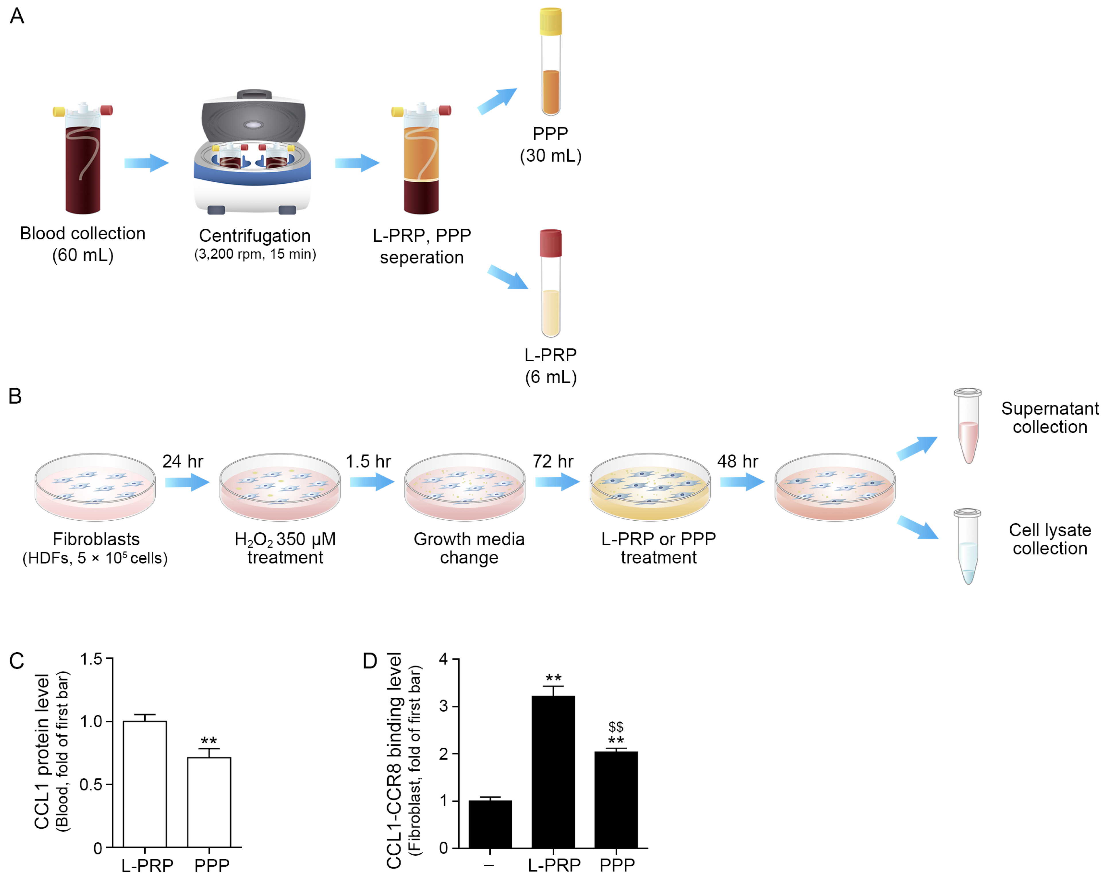
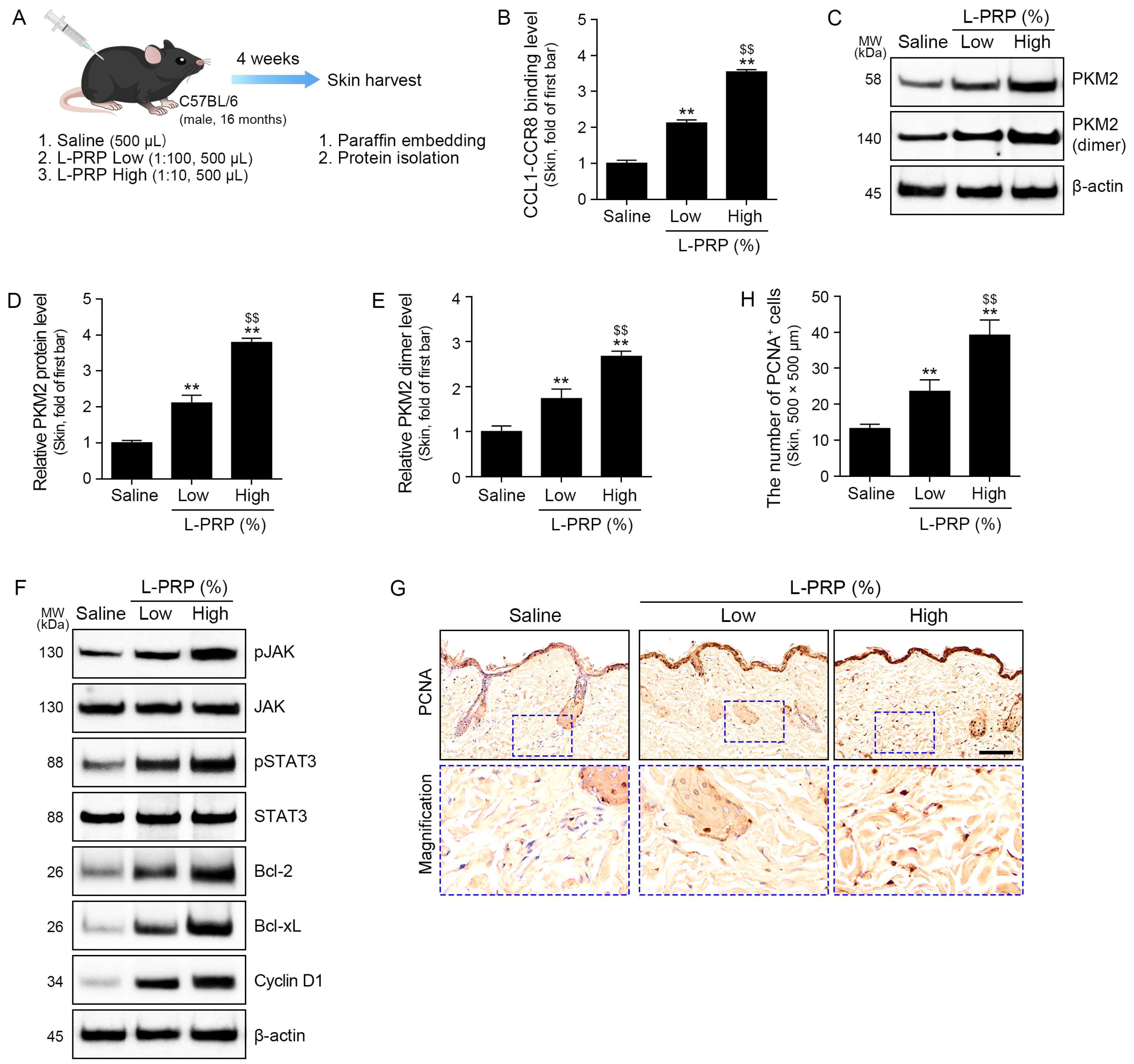
In the present study, L-PRP contained higher concentrations of CCL1 compared with PPP and induced stronger CCL1-CCR8 binding in senescent fibroblasts, accompanied by increased nuclearPKM2 (dimer). Similarly, intradermal administration of low- and high-dose L-PRP led to increased CCL1-CCR8 binding and nuclear PKM2 (dimer) levels in a dose-dependent manner in aged mice.
PKM2 functions in distinct oligomeric states. In its typical role, it exists as a high-activity tetramer that drives glycolytic flux in the cytosol. It can also exist as lower-activity dimers or monomers [
4,
32,
33,
34]. These forms facilitate nonmetabolic signaling, including nuclear translocation and transcriptional regulation or protein kinase activity [
14,
23,
24,
25]. Because the dimeric state is less efficient with regard to pyruvate processing, it permits diversion of glycolytic intermediates toward biosynthetic pathways that support cell growth and proliferation [
4,
32,
33,
34]. In contrast, the tetrameric form is enzymatically more active and less likely to translocate to the nucleus. However, even in the cytosol, tetrameric PKM2 can participate in signaling complexes that influence fibrotic pathways [
4,
32,
33,
34].
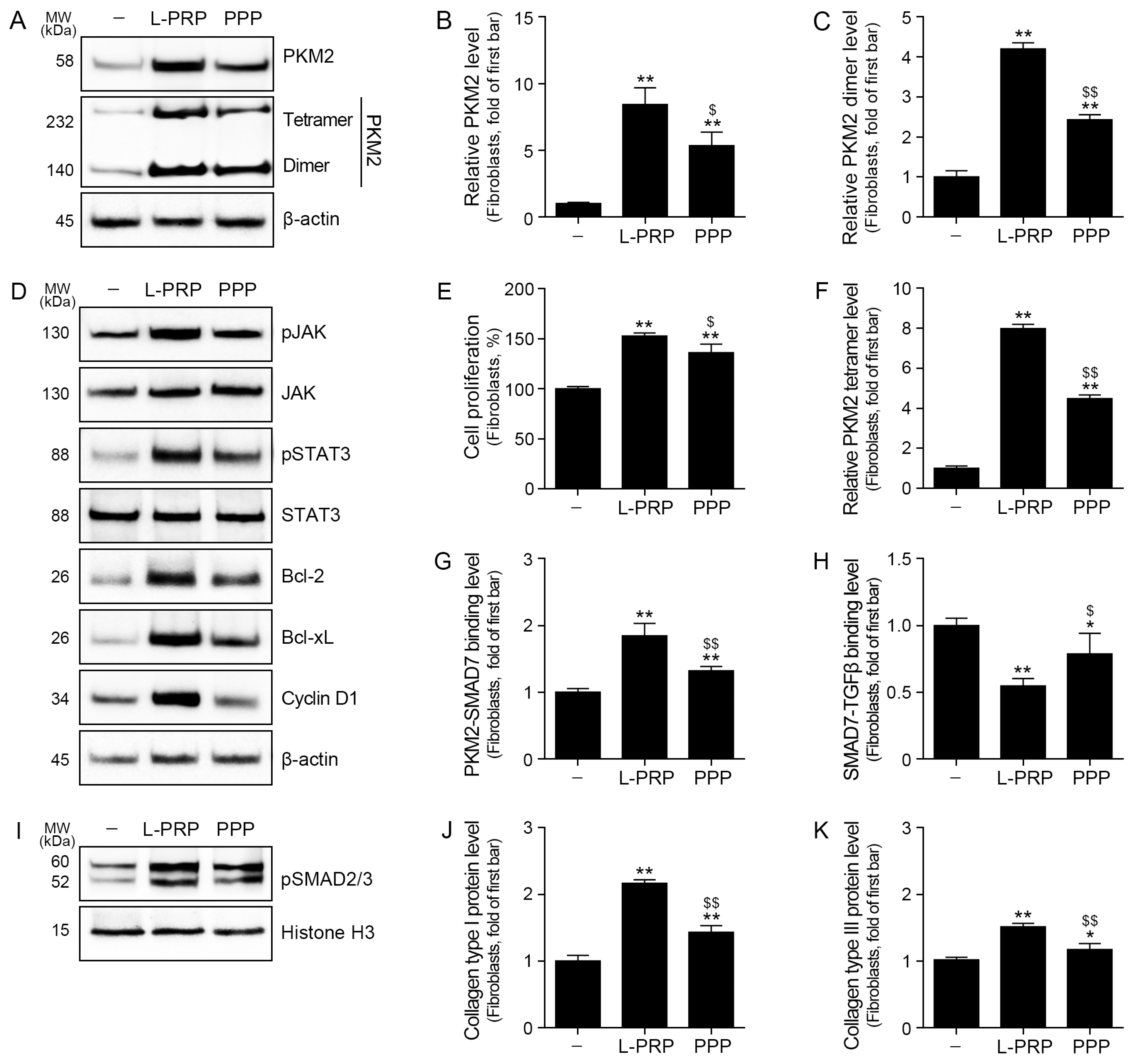
In our in vitro senescent fibroblast model, L-PRP increased phosphorylation of JAK and STAT3. This pattern is consistent with the concept that receptor-coupled signals can converge on PKM2 and STAT3; nuclear PKM2 reportedly interacts with transcription factors such as STAT3 and supports STAT3-dependent transcriptional programs [
35]. STAT3 activation is well known to promote cell survival and proliferation by inducing anti-apoptotic genes (including Bcl-2 and Bcl-XL) and cell cycle regulators such as cyclin D1 [
36]. Consistent with these effects, L-PRP increased Bcl-2, Bcl-xL, and cyclin D1 levels and enhanced fibroblast proliferation relative to PPP in our study.
These observations are biologically relevant to dermal aging because aged skin exhibits reduced fibroblast functional capacity and disrupted homeostasis. Senescent fibroblasts display impaired proliferative potential and contribute to ECM loss [
4,
37]. Chronologically aged human skin reportedly exhibits reduced fibroblast numbers and diminished collagen production capacity, contributing to dermal thinning and wrinkle formation [
4,
37]. In this context, an intervention that restores survival and proliferative signaling in senescent fibroblasts could expand the pool of matrix-producing cells and support ECM re-accumulation, provided that fibrosis or scarring is not excessively induced [
4].
Importantly, the present study demonstrates an association—rather than definitive causality—between CCR8 engagement and PKM2 dimer formation. We did not directly assess whether CCR8 activation is necessary and sufficient for the observed increase in nuclear PKM2 (dimer) under L-PRP conditions. To establish causality, future studies should incorporate functional perturbations such as CCL1 neutralization or depletion in L-PRP, CCR8 blockade or antagonism, or CCR8 knockdown or knockout in fibroblasts, followed by direct assessment of PKM2 oligomeric state and nuclear translocation. These approaches would clarify whether CCL1-CCR8 signaling acts upstream of PKM2 dimerization in this context or whether both are co-regulated by another L-PRP-induced pathway.
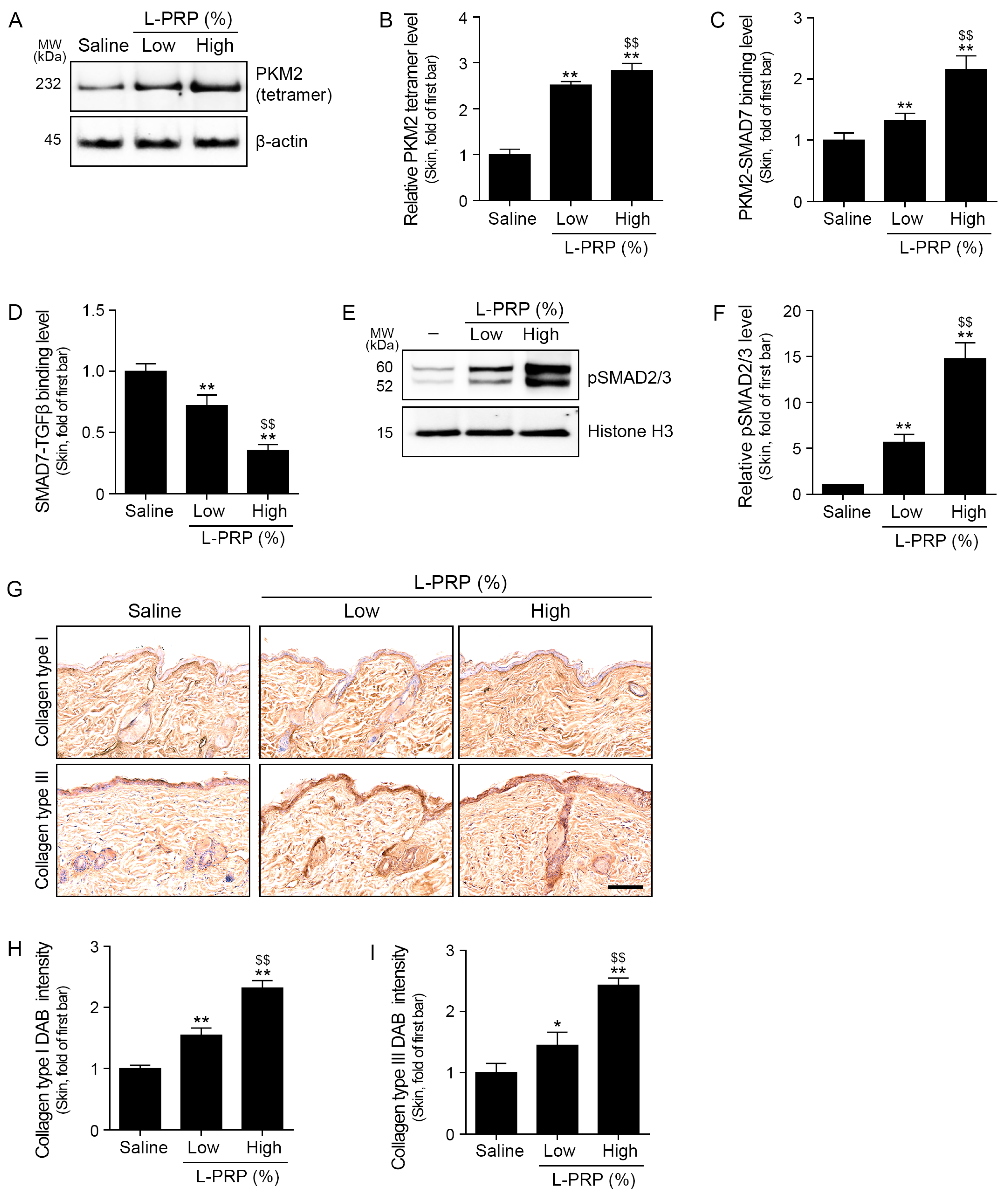
A second major finding of our study is that L-PRP increased PKM2 (tetramer) and shifted SMAD7-associated complexes toward enhanced TGF-β signaling in fibroblasts and aged skin. SMAD7 is a canonical inhibitory SMAD that negatively regulates TGF-β receptor signaling; disruption of SMAD7-mediated inhibition can amplify TGF-β/SMAD2/3 activity and fibrotic ECM transcription [
4].
Recent mechanistic studies indicate that PKM2 tetramer can directly interact with SMAD7 and interfere with SMAD7-mediated negative regulation of TGF-β signaling, thus enhancing downstream signaling and fibrosis [
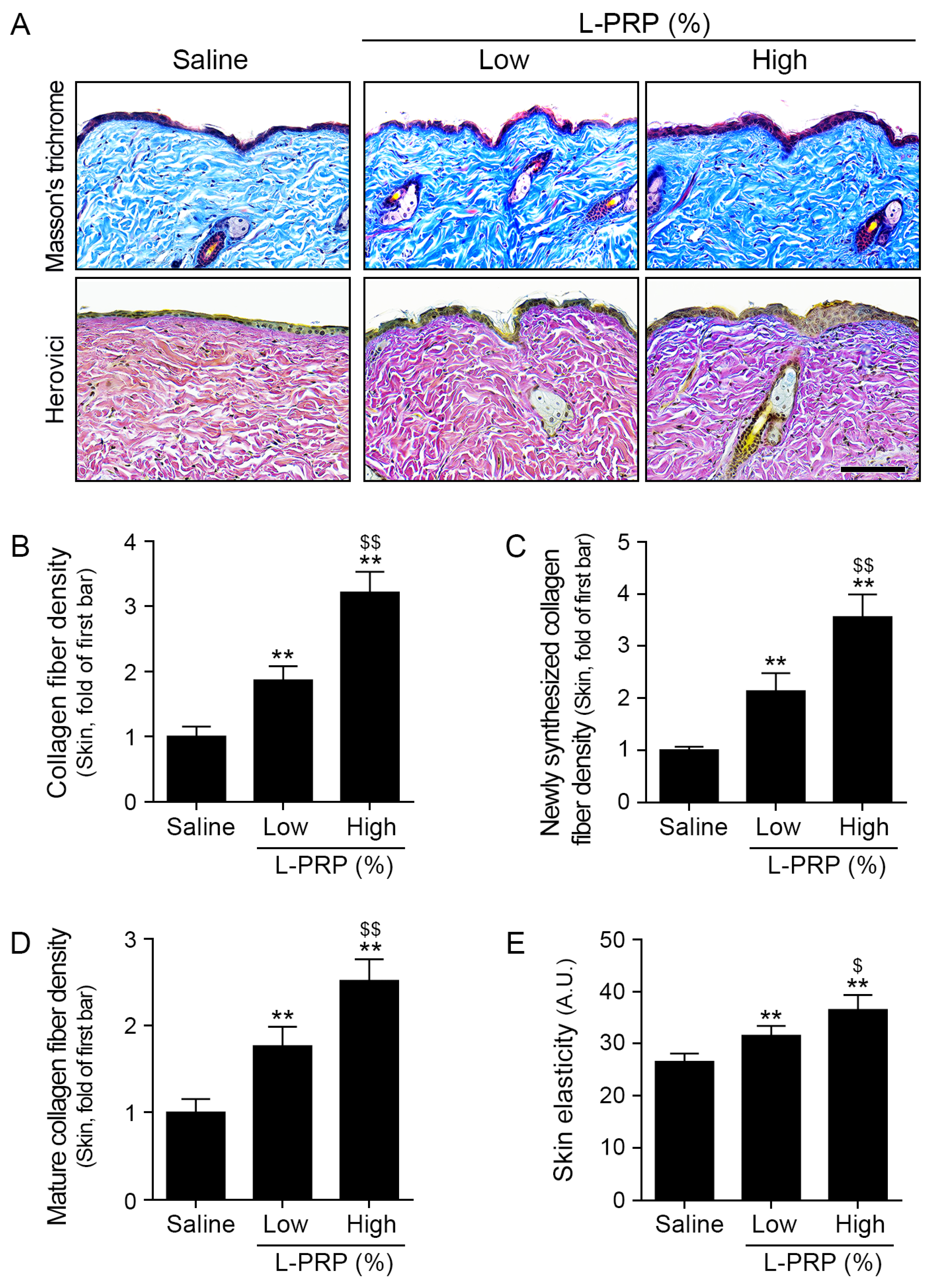
4]. Consistent with this model, L-PRP increased PKM2 tetramer levels, enhanced PKM2-SMAD7 binding, reduced SMAD7-TGF-β binding, and increased pSMAD2/3 levels. These changes coincided with increased expression of collagens I and III in vitro and enhanced dermal collagen deposition in vivo.
A key mechanistic gap revealed by these data concerns how L-PRP increases PKM2 tetramer levels. Although emerging evidence suggests that CCL1-CCR8 signaling can promote PKM2 induction, there is limited direct evidence that this pathway specifically drives PKM2 tetramerization, rather than PKM2 expression or dimer-associated nuclear functions. Furthermore, our data do not clarify whether CCL1-CCR8 signaling directly promotes PKM2 tetramerization. Accordingly, the tetramer arm of this model should be considered an empirically supported downstream effect of L-PRP treatment with an unresolved proximal mechanism.
Mechanistic analyses indicate that PKM2 oligomerization is regulated by multiple inputs, including allosteric metabolites (e.g., fructose-1,6-bisphosphate), post-translational modifications (phosphorylation, acetylation, and oxidation), and protein–protein interactions. These factors suggest several plausible mechanisms by which L-PRP may promote PKM2 tetramer formation [
38].
Our animal data demonstrate a dose-dependent pattern, in which high-dose L-PRP more strongly increased proliferative signaling (PCNA), ECM signaling (pSMAD2/3), and structural outcomes (collagen fiber and elasticity) compared with low-dose L-PRP. This dose–response relationship supports biological plausibility and conflicts with a purely nonspecific injection effect, although it leaves unresolved the components of L-PRP (platelets, leukocytes, or plasma factors) that predominantly drive each signaling pathway.
Previous studies have shown that leukocyte content can influence whether PRP promotes inflammation or tissue repair, depending on the microenvironment [
16]. Our findings suggest that CCL1, a chemokine associated with leukocytes, can serve as a key parameter for optimizing PRP formulations intended to restore the dermal matrix in older patients.
This study has several limitations. First, a direct cause–effect relationship between CCL1-CCR8 engagement and PKM2 dimer formation or STAT3 activation was not established; future loss-of-function studies (e.g., CCL1 neutralization or silencing, CCR8 blockade) are required. Second, the upstream mechanism underlying PKM2 tetramer accumulation after L-PRP treatment remains undefined, and further investigation of PKM2 oligomer regulation in senescent fibroblasts is needed. Third, human L-PRP was administered to mice. Although the consistent dose-dependent effects suggest that xenogeneic inflammation did not substantially confound the findings, species-specific immune responses cannot be excluded. Future studies should incorporate matched mouse-derived PRP controls. Finally, this work should be regarded as a mechanistic proof of concept demonstrating that L-PRP can engage a PKM2-linked pathway to promote ECM restoration in an aging context, rather than definitive evidence of clinical anti-aging efficacy, which will require controlled human studies with standardized PRP characterization. Finally, in this study, leukocyte-high and leukocyte-low PRP preparations were not directly compared. In the present in vivo experiments, the low- and high-dose groups represented different dilutions of the same L-PRP preparation rather than PRP formulations with different leukocyte contents. Therefore, although our data support the biological activity of L-PRP, they do not determine the specific contribution of leukocyte abundance to the observed effects. Future studies should compare leukocyte-rich and leukocyte-poor PRP preparations with matched platelet concentrations and standardized PRP characterization.
Despite these limitations, the findings expand the mechanistic framework through which L-PRP may promote dermal ECM regeneration. Beyond the conventional growth factor-centric paradigm, our data supports a model in which L-PRP enhances ECM production by (i) promoting fibroblast survival and proliferation through a CCL1-CCR8-associated PKM2 (dimer) JAK/STAT3 pathway and (ii) amplifying TGF-β/SMAD2/3 signaling via PKM2 (tetramer) interactions with SMAD7.
Conceptually, these results position L-PRP as an immuno-regenerative intervention that can modulate senescent fibroblast fate via chemokine–metabolic coupling. This framework may help explain why leukocyte content substantially influences PRP responses; it suggests that mediator-guided PRP formulation, or combination strategies targeting CCR8 or PKM2, could represent future approaches for skin rejuvenation in aging.
4. Materials and Methods
4.1. Study Participants and Ethical Approval
This study was reviewed and approved by the Public Institutional Review Board designated by the Ministry of Health and Welfare of Korea (approval no. P01-202507-02-011). The study was conducted in accordance with the Declaration of Helsinki and relevant Korean bioethics regulations for human-derived materials. Written informed consent was obtained from all participants prior to blood collection. A total of five eligible participants were enrolled in this study. Eligible participants were adults aged 20–65 years who voluntarily agreed to donate peripheral venous blood for this study. Individuals were excluded if they had a history of anemia, were currently taking antiplatelet or anticoagulant medications, had a previous history of syncope after blood collection, had skin disease at peripheral blood collection sites such as the arms or legs, or were pregnant. Participant confidentiality was maintained throughout the study, and no personally identifiable information was used in subsequent analyses.
4.2. Blood Collection
Peripheral venous blood was collected under sterile conditions using a standard venipuncture procedure. For each participant, 52 mL of whole blood was collected and mixed with 8 mL of acid–citrate–dextrose solution A (ACD-A) as an anticoagulant. The anticoagulated blood mixture was immediately processed for PRP preparation using the GPS® III Platelet Concentration System. To minimize risks associated with blood collection, participant identity and general condition were checked before venipuncture, an appropriate peripheral vein was selected, and standard aseptic procedures were followed. After blood collection, adequate compression was applied to the puncture site, and participants were monitored for possible adverse events such as hematoma, bleeding, dizziness, vasovagal reaction, or other rare complications.
4.3. Preparation of L-PRP and PPP Using an Automated Platelet Concentration System
L-PRP and PPP were prepared using a commercially available automated platelet concentration system (GPS® III Platelet Concentration System; Zimmer Biomet, Warsaw, IN, USA) according to the manufacturer’s instructions. Briefly, 52 mL of whole blood was collected and mixed with 8 mL ACD-A. The anticoagulated whole blood was then transferred into the GPS® III separation tube and centrifuged at 3200 rpm for 15 min. After centrifugation, the blood components were separated into distinct layers, including a lower red blood cell layer, an intermediate buffy coat layer corresponding to L-PRP, and an upper plasma layer corresponding to PPP. The PPP fraction was first aspirated from the upper plasma layer, and the L-PRP fraction was subsequently obtained from the buffy coat-containing layer. The obtained L-PRP and PPP were transferred into sterile tubes containing 1% bovine serum albumin in phosphate-buffered saline (PBS), stored at 4 °C, and used for subsequent analyses and in vivo experiments within 3 h of preparation.
4.4. In Vivo Study
4.4.1. Animal Housing and Ethics Statement
Male C57BL/6 mice obtained from Orient Bio (Seongnam, Republic of Korea) were housed under standard laboratory conditions at 20–24 °C and 45–55% humidity, with a 12 h light/dark cycle and free access to food and water. After a 1-week acclimation period, mice were used at 16 months of age as an aging model. All animal procedures were conducted in accordance with the guidelines of the Institutional Animal Care and Use Committee of Gachon University (IACUC no. LCDI-2025-0073) and complied with the ethical standards of AAALAC International (Frederick, MD, USA) and the ARRIVE guidelines.
4.4.2. Experimental Design and L-PRP Administration
Aged mice (16 months old) were randomly allocated into three groups: Aging/Saline, Aging/L-PRP (Low; 1:100 dilution), and Aging/L-PRP (High; 1:10 dilution) (n = 5 per group). Each mouse served as an experimental unit. All injections and evaluations were performed by investigators who had been blinded to group allocation to minimize bias. No animals met the predefined exclusion criteria (e.g., severe intercurrent illness, substantial weight loss, or unexpected adverse events), and all animals completed the study.
For all procedures, mice were anesthetized with inhalational isoflurane. The dorsal skin was shaved and disinfected with 70% ethanol prior to injection. In the Aging/Saline group, 500 µL of sterile saline was intradermally injected into the dorsal dermis. In the Aging/L-PRP (Low) group, L-PRP was diluted 1:100 in saline, and 500 µL of diluted L-PRP were intradermally injected into the dorsal dermis. In the Aging/L-PRP (High) group, L-PRP was diluted 1:10 in saline, and 500 µL of the diluted L-PRP was intradermally injected into the same region. In all groups, the total volume was delivered as multiple small intradermal blebs evenly distributed across the dorsal area.
Animals were evaluated 4 weeks after injection. At the endpoint, full thickness dorsal skin samples encompassing the injection sites were harvested for molecular and histological analyses. Histological evaluation and quantitative image analysis were performed by investigators who had been blinded to treatment group to minimize observer bias.
4.4.3. Skin Elasticity Measurement
Skin elasticity was assessed using an API 100 Skin Analyzer (Aram Huvis, Seongnam, Republic of Korea). High-resolution images of the skin surface were captured using a noncontact optical method, and elasticity was quantified using the accompanying software (Solutionist v1.7.10). Each mouse was measured five times immediately prior to sampling, and the average value was calculated.
4.5. In Vitro Experiments
4.5.1. Cell Culture
HDFs were purchased from CEFO Bio (Seoul, Republic of Korea) and cultured using the CEFO™ Human Dermal Fibroblast Cell Kit, in accordance with the manufacturer’s instructions. The culture medium was replaced every 2–3 days. Cells were maintained at 37 °C in a humidified atmosphere containing 5% CO2. Cells between passages 4 and 7 were used for all experiments; all experiments were performed in triplicate.
4.5.2. Induction of Cellular Senescence and L-PRP/PPP Treatment in HDFs
Cellular senescence in HDFs was induced by treatment with 350 μM H
2O
2 for 1.5 h, followed by medium replacement and 72 h of culture [
39]. Senescent HDFs were used for all subsequent experiments. L-PRP and PPP were prepared from human peripheral blood as described above and diluted in culture medium immediately before use.
For cytotoxicity assessment, senescent HDFs were seeded in 96-well plates at a density of 5 × 10⁴ cells/well and treated with L-PRP at final concentrations of 0%, 1%, 2%, 5%, or 10% (v/v) for 48 h. Cell viability was evaluated using cell counting kit (CCK)-8, in accordance with the manufacturer’s instructions. To determine the optimal L-PRP concentration, senescent HDFs were treated with 0%, 2.5%, 5%, or 7.5% (v/v) L-PRP for 48 h under the same conditions; 5% (v/v) L-PRP was identified as the optimal concentration, providing maximal efficacy without detectable cytotoxicity.
Based on these results, all subsequent experiments were performed using 5% (v/v) L-PRP. For comparison at an equivalent volume fraction, PPP was also diluted to a final concentration of 5% (v/v) in culture medium. In all experiments, L-PRP or PPP was added 72 h after H2O2 treatment. Senescent HDFs were incubated with 5% (v/v) L-PRP or PPP for 48 h prior to sample collection for molecular and functional analyses.
4.6. Cell Death and Proliferation Assays
Cell proliferation and viability were assessed via CCK-8 (TransGen Biotech, Beijing, China). For cytotoxicity assessment, senescent HDFs were seeded at a density of 5 × 10⁴ cells/well and treated with L-PRP at concentrations of 0%, 1%, 2%, 5%, or 10% (v/v) for 48 h. For proliferation assays, senescent HDFs were seeded in 96-well plates at a density of 1 × 10⁴ cells/well and allowed to attach overnight, then treated with L-PRP at final concentrations of 0%, 2.5%, 5%, or 7.5% (v/v) for 48 h to determine the optimal concentration, as described above. In all experiments, CCK 8 solution was added to each well for the final 2 h of incubation, and absorbance at 450 nm was measured using a microplate reader. Cell proliferation or viability was expressed as a percentage of the untreated control.
4.7. Protein Isolation and Quantification
Tissue and cell samples were lysed using the EzRIPA Lysis Kit (ATTO Corporation, Tokyo, Japan), which contains radioimmunoprecipitation assay buffer supplemented with protease and phosphatase inhibitors (each at 100× dilution). For tissue samples, excised skin tissues were washed once with PBS, and lysis buffer was added at a ratio of 1 mL per 40 mg of tissue. Samples were incubated on ice at 4 °C for 10 min, then sonicated for 15 min (10 s on, 1 min off cycles). For cell samples, the culture medium was removed, and cells were washed three times with PBS. Lysis buffer (500 μL per 10 cm dish) was added, and cells were incubated on ice at 4 °C for 5 min. Cells were then scraped and disrupted by sonication for 10 min (10 s on, 1 min off cycles). Tissue and cell lysates were centrifuged at 14,000× g for 15 min at 4 °C, and supernatants were collected. For nuclear protein extraction, cell lysates were processed via NE-PER™ Nuclear and Cytoplasmic Extraction Reagents (Invitrogen/Thermo Fisher Scientific, Waltham, MA, USA), using the manufacturer’s protocol to separate cytoplasmic and nuclear fractions. Protein concentrations were determined using the BCA Protein Assay Kit (Thermo Fisher Scientific) with bovine serum albumin as the standard. Absorbance was measured at 562 nm using a Multiskan SkyHigh microplate spectrophotometer (Thermo Fisher Scientific). Protein samples were stored at −80 °C until use.
4.8. Indirect ELISA
For CCL1 measurement, L-PRP and PPP samples were diluted to a final concentration of 5% (
v/
v) in carbonate–bicarbonate coating buffer (pH 9.6), and 96 well plates (SPL Life Sciences, Pocheon, Republic of Korea) were coated with the diluted samples by overnight incubation at 4 °C. After plates had been coated, they were washed three times with PBS containing 0.1% Tween 20, then blocked with 5% skim milk (LPS Solution, Daejeon, Republic of Korea) in PBS for 1 h at room temperature. A primary antibody against CCL1 (details listed in
Table S1) was added to each well and incubated overnight at 4 °C; this was followed by three washes with PBS containing 0.1% Tween 20. Horseradish peroxidase-conjugated secondary antibodies (1:1000; Vector Laboratories, Burlingame, CA, USA) were then applied for 1 h at room temperature. Color development was achieved using 3,3′,5,5′ tetramethylbenzidine (TMB) substrate solution (Sigma Aldrich, St. Louis, MO, USA), and the reaction was terminated with 1 M sulfuric acid. Absorbance was measured at 450 nm using a Multiskan SkyHigh microplate spectrophotometer (Thermo Fisher Scientific). CCL1 levels were expressed as absorbance values relative to the control group.
For collagens I and III, indirect ELISA was performed using cell lysates. Protein samples extracted from HDFs were diluted in carbonate–bicarbonate coating buffer (pH 9.6), adhered to 96-well plates by overnight incubation at 4 °C, and processed as described above using primary antibodies against collagens I and III (
Table S1), followed by horseradish peroxidase-conjugated secondary antibodies and TMB substrate. Absorbance at 450 nm was recorded. Collagen levels were normalized to the control group or to total protein content, as indicated in the figure legends.
4.9. Sandwich ELISA
Sandwich ELISA was performed to comparatively evaluate target protein levels in cell culture supernatants and skin tissue extracts. Ninety six-well plates were coated with capture antibodies specific for the target proteins diluted in carbonate–bicarbonate coating buffer (pH 9.6), then incubated overnight at 4 °C. After three washes with PBS containing 0.1% Tween 20, wells were blocked with 5% skim milk in PBS for 1 h at room temperature.
Cell culture supernatants from HDFs or homogenized skin tissue lysates were added to the wells and incubated for 24 h at room temperature. This incubation step was followed by washing and further incubation with detection antibodies against the corresponding targets for 24 h. Horseradish peroxidase-conjugated secondary antibodies were then applied for 2 h at room temperature. Color development was achieved using TMB substrate, and the reaction was terminated with 1 M sulfuric acid. Absorbance was measured at 450 nm using a microplate spectrophotometer. Relative protein levels were expressed as absorbance values normalized to the control group.
4.10. Western Blot Analysis
Protein samples were denatured with 4× lithium dodecyl sulfate sample buffer and 10× reducing reagent (Thermo Fisher Scientific) at 70 °C for 10 min. Thirty micrograms of total protein per lane were resolved on 10% sodium dodecyl sulfate–polyacrylamide gels using MOPS buffer (Thermo Fisher Scientific) at 200 V for 25 min, in conjunction with appropriate molecular weight markers (EasySee Western Marker and EasySee II Western Marker; TransGen Biotech). Proteins were transferred to polyvinylidene fluoride membranes (Millipore, Burlington, MA, USA) using a semi-dry transfer system (ATTO Corporation) at 1 A for 10 min. Membranes were blocked with 5% skim milk (LPS Solution) in Tris-buffered saline plus Tween (TBST) for 1 h at room temperature, then probed with primary antibodies (
Table S1) overnight at 4 °C. After three 10 min washes with TBST, membranes were incubated with horseradish peroxidase-conjugated secondary antibodies (1:2000–1:10,000; Vector Laboratories) for 1 h at room temperature. Following three additional 10 min washes with TBST, immunoreactive bands were visualized using an enhanced chemiluminescence reagent (Cytiva, Marlborough, MA, USA) on a ChemiDoc system (Bio-Rad Laboratories, Hercules, CA, USA). Band intensities were quantified using ImageJ (v1.53s; National Institutes of Health, Bethesda, MD, USA), normalized to histone H3 or β-actin, and expressed as fold change relative to the control group.
4.11. Blue Native PAGE (BN-PAGE) Analysis of PKM2 Oligomerization
BN-PAGE was performed to analyze PKM2 oligomerization in L-PRP-treated cells and skin tissues. After 48 h of treatment with 5% (v/v) L-PRP or PPP, cells were washed twice with cold PBS. Skin tissue from the corresponding treatment groups were collected and homogenized. Cells and tissue homogenates were lysed on ice for 30 min in BN-PAGE lysis buffer (50 mM Bis Tris HCl, 0.5 M 6-aminohexanoic acid, 10% glycerol, and 1% digitonin, pH 7.0) supplemented with protease and phosphatase inhibitors (ATTO Corporation). Lysates were centrifuged at 16,000× g for 15 min at 4 °C, and protein concentrations in the supernatants were determined using a BCA Protein Assay Kit (Thermo Fisher Scientific). BN-PAGE loading buffer containing Coomassie brilliant blue G-250 (Sigma-Aldrich) was then added to prepare samples for electrophoresis.
Equal amounts of protein were loaded onto native polyacrylamide gels; electrophoresis was carried out at a constant voltage of 100 V using an inner buffer (0.05 M Tricine, 15 mM Bis Tris, pH 7.0) and an outer buffer (0.05 M Bis Tris HCl, pH 7.0). Proteins were then transferred onto polyvinylidene fluoride membranes at a constant current of 300 mA [
40]. After transfer, membranes were briefly destained with methanol, washed twice with TBST, and blocked with 10% skim milk in TBST for 1 h at room temperature. Membranes were incubated overnight at 4 °C with primary antibodies against PKM2, then incubated with horseradish peroxidase-conjugated secondary antibodies and subjected to chemiluminescent detection as described for conventional Western blotting (antibody details are listed in
Table S1). Band intensities corresponding to PKM2 tetramers and dimers were quantified using ImageJ software (v1.53s) and expressed relative to the control group.
4.12. Histological and Immunohistochemistry Analysis
4.12.1. Tissue Fixation, Processing, and Paraffin Embedding
Skin tissues were fixed in 4% paraformaldehyde (Sigma-Aldrich) at 4 °C for 72 h using a fixative-to-tissue volume ratio of 10:1. Fixed tissues were processed using an automated tissue processor (Tissue-Tek VIP 5 Jr., SAKURA, Tokyo, Japan), in accordance with the manufacturer’s protocol for dehydration, clearing, and paraffin infiltration. Tissues were then embedded in paraffin (Leica, Wetzlar, Germany) using an embedding center (Tissue-Tek TEC, SAKURA). Serial sections (7 μm thick) were cut from paraffin blocks using a microtome (Thermo Fisher Scientific), mounted on coated slides (Muto Pure Chemicals Co., Ltd., Tokyo, Japan), and incubated overnight at 60 °C to ensure adhesion.
4.12.2. Masson’s Trichrome Staining
Paraffin-embedded tissue sections (7 μm) were deparaffinized in xylene and rehydrated through a graded ethanol series (100%, 95%, 90%, 80%, and 70%) to distilled water. Sections were incubated in Bouin’s fluid (ScyTek Laboratories Inc., Logan, UT, USA) at 56–64 °C for 60 min, then incubated at room temperature for 10 min. Sections were rinsed in running tap water for 20 min, then stained with Weigert’s iron hematoxylin (ScyTek Laboratories) for 5 min and rinsed in running tap water for 10 min. Sections were subsequently incubated in Biebrich scarlet/acid fuchsin solution (ScyTek Laboratories) for 2 min, rinsed in running tap water for 10 min, treated with phosphomolybdic/phosphotungstic acid solution (ScyTek Laboratories) for 10 min, and stained with aniline blue solution (ScyTek Laboratories) for 5 min. After they had been rinsed in running tap water for 10 min, sections were differentiated in 1% acetic acid solution for 3 min. Stained sections were dehydrated through a graded ethanol series (95% for 1 min twice, 100% for 1 min twice), cleared in xylene, and mounted.
Images were captured using a slide scanner (Motic Scan Infinity 100; Motic, Hong Kong, China). Collagen content in the dermal area was quantified as the relative intensity of blue-stained regions using ImageJ software (v1.53s). Multiple fields per sample were analyzed.
4.12.3. Herovici Staining
Herovici staining was performed using a commercial Herovici Stain Kit (ScyTek Laboratories), in accordance with the manufacturer’s instructions. Sections were stained with Weigert’s iron hematoxylin followed by Herovici solution, then dehydrated and mounted. Stained slides were scanned using a slide scanner (Motic Scan Infinity 100; Motic), and representative images were captured. The densities of newly synthesized collagen fibers (blue) and mature collagen fibers (red) were separately quantified using ImageJ software (v1.53s). Specifically, young (blue) and mature (red) collagen were separated by color deconvolution [
41,
42]; the density of each collagen subtype was measured and expressed as fold change relative to the Saline group.
4.12.4. Immunohistochemical Staining
For immunohistochemical staining, paraffin-embedded sections were deparaffinized in xylene and rehydrated through a graded ethanol series. Permeabilization was performed using 0.5% Triton X-100 for 5 min, followed by PBS washes. To block nonspecific binding, sections were incubated with normal serum blocking solution for 1 h at room temperature. Slides were then incubated overnight at 4 °C with primary antibodies (
Table S1) diluted in blocking solution. After sections had been washed, they were incubated with biotin-conjugated secondary antibodies (Vector Laboratories) for 1 h at room temperature. Signal amplification was achieved using an avidin–biotin complex reagent (Vector Laboratories), and immunoreactivity was visualized with 3,3′-diaminobenzidine solution (DAB; Sigma-Aldrich) for 5 min to develop a brown reaction product. Nuclei were counterstained with hematoxylin (KPNT, Cheongju, Republic of Korea). Sections were then dehydrated, cleared in xylene, and mounted with DPX mounting medium. Stained tissues were scanned using a Motic Scan Infinity 100 slide scanner (Motic), and representative images were captured. Quantification of DAB-positive staining was performed using ImageJ software (v1.53s) by measuring the number of brown-stained nuclei or density of brown-stained fibers within the dermis. The resulting values were expressed as fold change relative to the control group.
4.13. Statistical Analysis
Statistical analyses were performed using SPSS version 26 (IBM Corp., Armonk, NY, USA). All analyses were conducted using nonparametric methods. The Kruskal–Wallis test was used for comparisons among multiple independent groups, followed by the Mann–Whitney U test for pairwise comparisons. Data are presented as mean ± standard deviation. A p-value < 0.05 was considered statistically significant. The definitions of statistical symbols used in the graphs are provided in the corresponding figure legends. All experiments were performed in triplicate (n = 3 biological replicates), and animal experiments included five animals per group (n = 5).





 ,
,