Figure 1.
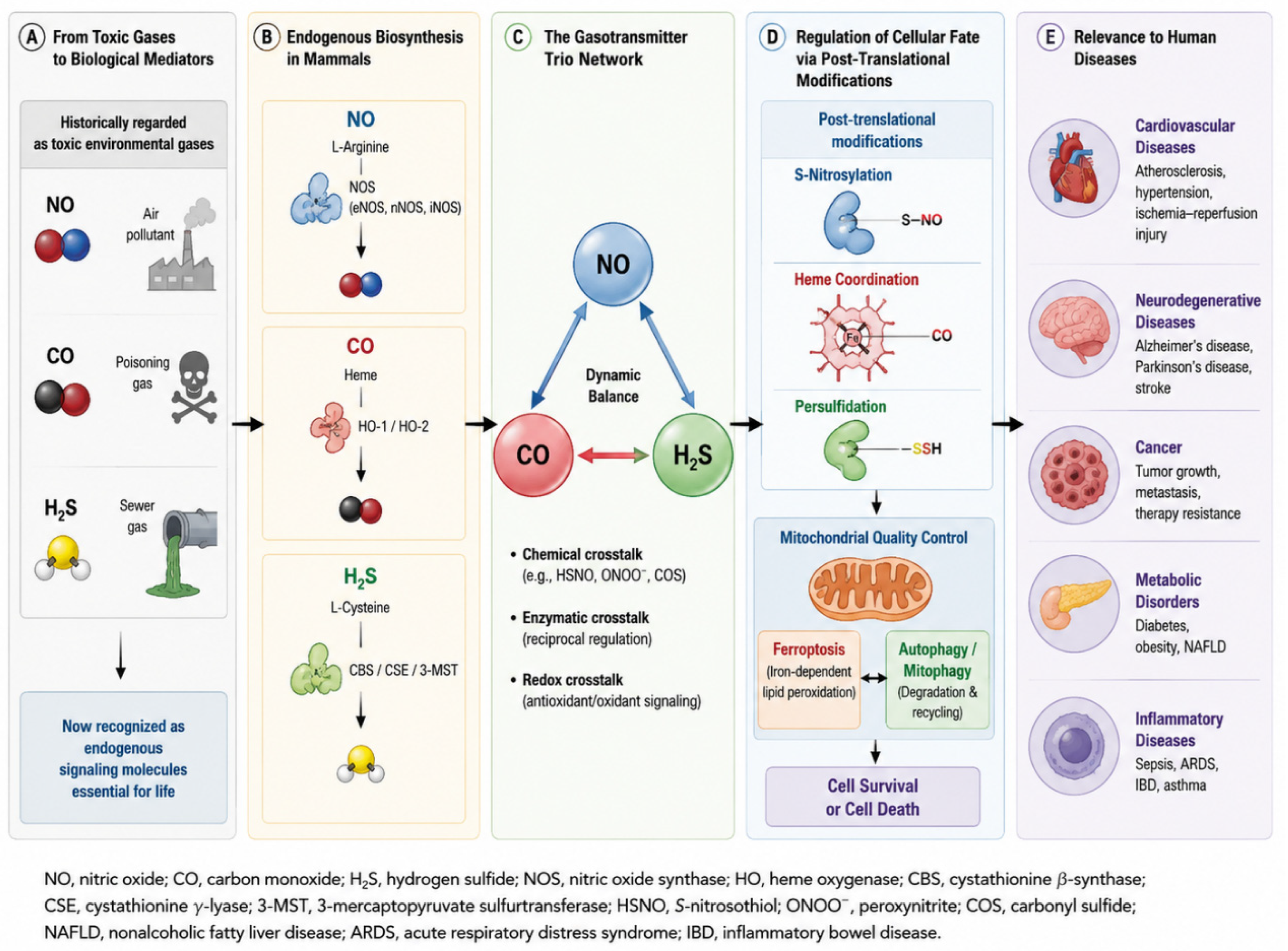
Evolution of the Gasotransmitter Trio from Toxic Gases to Master Regulators of Cellular Fate. Nitric oxide (NO), carbon monoxide (CO), and hydrogen sulfide (H2S) were historically regarded as toxic environmental gases. Subsequent discoveries demonstrated that these molecules are enzymatically synthesized in mammalian tissues through nitric oxide synthases (NOS), heme oxygenases (HO-1/HO-2), and sulfur-metabolizing enzymes, including cystathionine β-synthase (CBS), cystathionine γ-lyase (CSE), and 3-mercaptopyruvate sulfurtransferase (3-MST). Increasing evidence indicates that NO, CO, and H2S form an integrated gasotransmitter network through direct chemical interactions, reciprocal enzymatic regulation, and shared redox signaling pathways. Their biological effects are mediated primarily through post-translational modifications, including S-nitrosylation, heme-dependent coordination, and persulfidation, which collectively regulate mitochondrial quality control, ferroptosis, autophagy, and mitophagy. Dysregulation of these pathways contributes to the pathogenesis of cardiovascular, neurodegenerative, metabolic, inflammatory, and malignant diseases.
Figure 1.
Evolution of the Gasotransmitter Trio from Toxic Gases to Master Regulators of Cellular Fate. Nitric oxide (NO), carbon monoxide (CO), and hydrogen sulfide (H2S) were historically regarded as toxic environmental gases. Subsequent discoveries demonstrated that these molecules are enzymatically synthesized in mammalian tissues through nitric oxide synthases (NOS), heme oxygenases (HO-1/HO-2), and sulfur-metabolizing enzymes, including cystathionine β-synthase (CBS), cystathionine γ-lyase (CSE), and 3-mercaptopyruvate sulfurtransferase (3-MST). Increasing evidence indicates that NO, CO, and H2S form an integrated gasotransmitter network through direct chemical interactions, reciprocal enzymatic regulation, and shared redox signaling pathways. Their biological effects are mediated primarily through post-translational modifications, including S-nitrosylation, heme-dependent coordination, and persulfidation, which collectively regulate mitochondrial quality control, ferroptosis, autophagy, and mitophagy. Dysregulation of these pathways contributes to the pathogenesis of cardiovascular, neurodegenerative, metabolic, inflammatory, and malignant diseases.

Figure 2.
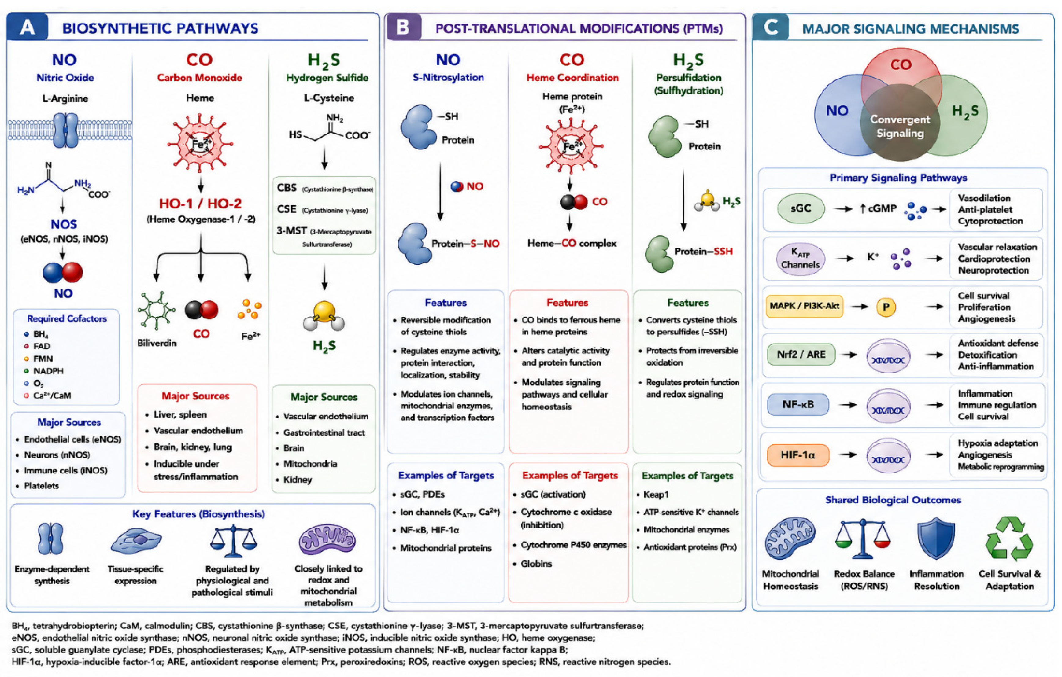
Biosynthetic pathways, post-translational modifications, and major signaling mechanisms of the gasotransmitter trio. Nitric oxide (NO), carbon monoxide (CO), and hydrogen sulfide (H2S) are synthesized through distinct enzymatic pathways but ultimately converge on shared signaling networks that regulate cellular homeostasis. (A) Biosynthetic pathways. NO is generated from L-arginine by nitric oxide synthase (NOS) isoforms, including endothelial NOS (eNOS), neuronal NOS (nNOS), and inducible NOS (iNOS), in the presence of cofactors such as tetrahydrobiopterin (BH4), flavin adenine dinucleotide (FAD), flavin mononucleotide (FMN), and NADPH. CO is produced during heme degradation by heme oxygenase-1 (HO-1) and heme oxygenase-2 (HO-2), generating CO, biliverdin, and ferrous iron (Fe2+). H2S is synthesized primarily from sulfur-containing amino acids by cystathionine β-synthase (CBS), cystathionine γ-lyase (CSE), and 3-mercaptopyruvate sulfurtransferase (3-MST), with tissue-specific expression patterns. (B) Gasotransmitter-mediated post-translational modifications (PTMs). NO regulates protein function through reversible S-nitrosylation of reactive cysteine residues, thereby influencing enzymatic activity, protein stability, intracellular trafficking, and signal transduction. CO exerts its biological effects primarily through heme coordination, in which binding to ferrous heme centers modulates the activity of heme-containing proteins, including soluble guanylate cyclase (sGC), cytochrome c oxidase, cytochrome P450 enzymes, and globins. H2S mediates protein persulfidation (sulfhydration), converting cysteine thiols (-SH) into persulfides (-SSH), thereby protecting proteins from irreversible oxidation and regulating diverse cellular signaling pathways. (C) Major signaling mechanisms and downstream targets. Despite their distinct chemical properties, the three gasotransmitters converge on several shared signaling pathways. Direct targets include soluble guanylate cyclase (sGC), ATP-sensitive potassium (KATP) channels, calcium-sensitive ion channels, and multiple heme-containing proteins. These interactions modulate cyclic guanosine monophosphate (cGMP) signaling, membrane excitability, mitochondrial respiration, and cellular metabolism. Furthermore, NO, CO, and H2S regulate redox-sensitive pathways including Nrf2/ARE, NF-κB, and HIF-1α signaling, thereby influencing antioxidant defense, inflammatory responses, hypoxic adaptation, and mitochondrial quality control. Through coordinated regulation of mitochondrial biogenesis, electron transport chain activity, and reactive oxygen/nitrogen species (ROS/RNS) homeostasis, the gasotransmitter trio serves as a central regulator of cellular adaptation and survival under physiological and pathological conditions. ARE, antioxidant response element; BH4, tetrahydrobiopterin; CBS, cystathionine β-synthase; CSE, cystathionine γ-lyase; FMN, flavin mononucleotide; FAD, flavin adenine dinucleotide; HIF-1α, hypoxia-inducible factor-1α; HO, heme oxygenase; KATP, ATP-sensitive potassium channel; NF-κB, nuclear factor kappa B; Nrf2, nuclear factor erythroid 2-related factor 2; NOS, nitric oxide synthase; ROS, reactive oxygen species; RNS, reactive nitrogen species; sGC, soluble guanylate cyclase; 3-MST, 3-mercaptopyruvate sulfurtransferase.
Figure 2.
Biosynthetic pathways, post-translational modifications, and major signaling mechanisms of the gasotransmitter trio. Nitric oxide (NO), carbon monoxide (CO), and hydrogen sulfide (H2S) are synthesized through distinct enzymatic pathways but ultimately converge on shared signaling networks that regulate cellular homeostasis. (A) Biosynthetic pathways. NO is generated from L-arginine by nitric oxide synthase (NOS) isoforms, including endothelial NOS (eNOS), neuronal NOS (nNOS), and inducible NOS (iNOS), in the presence of cofactors such as tetrahydrobiopterin (BH4), flavin adenine dinucleotide (FAD), flavin mononucleotide (FMN), and NADPH. CO is produced during heme degradation by heme oxygenase-1 (HO-1) and heme oxygenase-2 (HO-2), generating CO, biliverdin, and ferrous iron (Fe2+). H2S is synthesized primarily from sulfur-containing amino acids by cystathionine β-synthase (CBS), cystathionine γ-lyase (CSE), and 3-mercaptopyruvate sulfurtransferase (3-MST), with tissue-specific expression patterns. (B) Gasotransmitter-mediated post-translational modifications (PTMs). NO regulates protein function through reversible S-nitrosylation of reactive cysteine residues, thereby influencing enzymatic activity, protein stability, intracellular trafficking, and signal transduction. CO exerts its biological effects primarily through heme coordination, in which binding to ferrous heme centers modulates the activity of heme-containing proteins, including soluble guanylate cyclase (sGC), cytochrome c oxidase, cytochrome P450 enzymes, and globins. H2S mediates protein persulfidation (sulfhydration), converting cysteine thiols (-SH) into persulfides (-SSH), thereby protecting proteins from irreversible oxidation and regulating diverse cellular signaling pathways. (C) Major signaling mechanisms and downstream targets. Despite their distinct chemical properties, the three gasotransmitters converge on several shared signaling pathways. Direct targets include soluble guanylate cyclase (sGC), ATP-sensitive potassium (KATP) channels, calcium-sensitive ion channels, and multiple heme-containing proteins. These interactions modulate cyclic guanosine monophosphate (cGMP) signaling, membrane excitability, mitochondrial respiration, and cellular metabolism. Furthermore, NO, CO, and H2S regulate redox-sensitive pathways including Nrf2/ARE, NF-κB, and HIF-1α signaling, thereby influencing antioxidant defense, inflammatory responses, hypoxic adaptation, and mitochondrial quality control. Through coordinated regulation of mitochondrial biogenesis, electron transport chain activity, and reactive oxygen/nitrogen species (ROS/RNS) homeostasis, the gasotransmitter trio serves as a central regulator of cellular adaptation and survival under physiological and pathological conditions. ARE, antioxidant response element; BH4, tetrahydrobiopterin; CBS, cystathionine β-synthase; CSE, cystathionine γ-lyase; FMN, flavin mononucleotide; FAD, flavin adenine dinucleotide; HIF-1α, hypoxia-inducible factor-1α; HO, heme oxygenase; KATP, ATP-sensitive potassium channel; NF-κB, nuclear factor kappa B; Nrf2, nuclear factor erythroid 2-related factor 2; NOS, nitric oxide synthase; ROS, reactive oxygen species; RNS, reactive nitrogen species; sGC, soluble guanylate cyclase; 3-MST, 3-mercaptopyruvate sulfurtransferase.

Figure 3.
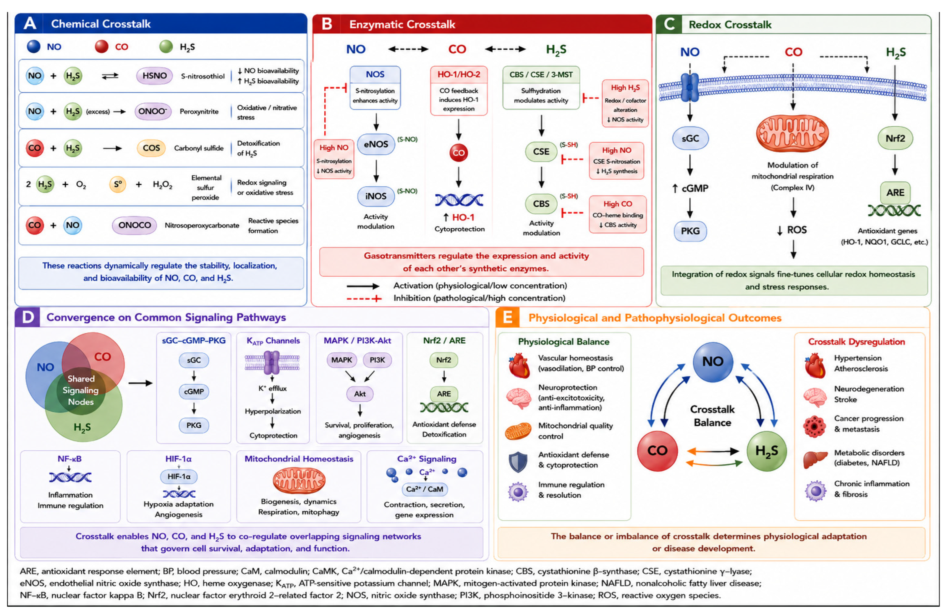
Molecular crosstalk network among nitric oxide (NO), carbon monoxide (CO), and hydrogen sulfide (H2S). The biological activities of NO, CO, and H2S are coordinated through multiple layers of molecular crosstalk, including direct chemical reactions, reciprocal enzymatic regulation, integration of redox signaling, and convergence on shared downstream pathways. (A) Chemical crosstalk. NO and H2S react to form a variety of reactive sulfur–nitrogen intermediates, including nitrosopersulfide (SSNO−), nitroxyl (HNO), and other sulfur–nitrogen hybrid species. These intermediates exhibit distinct biological activities compared with their parent molecules and may serve as prolonged reservoirs of signaling capacity. Interactions among NO, CO, and H2S further influence gasotransmitter bioavailability, redox balance, and reactive species formation. (B) Enzymatic crosstalk. Gasotransmitter-generating enzymes are regulated by bidirectional feedback mechanisms that depend on local gas concentrations. Under physiological conditions, activating interactions dominate: H2S enhances eNOS activity through sulfhydration-dependent mechanisms and the preservation of tetrahydrobiopterin (BH4); NO induces HO-1 expression via Nrf2/HIF-1α-dependent pathways to increase CO generation; and H2S further promotes HO-1 activation through redox-sensitive signaling. In contrast, excessive accumulation of gasotransmitters triggers negative feedback regulation. High NO levels inhibit NOS activity via S-nitrosylation-mediated enzyme regulation and suppress CSE-dependent H2S generation, whereas elevated CO levels inhibit CBS activity through direct heme binding. Excessive H2S may also negatively regulate NOS activity through redox- and cofactor-dependent mechanisms. Thus, the CBS/CSE–HO–NOS network functions as a concentration-sensitive regulatory circuit that maintains gasotransmitter homeostasis while preventing pathological overactivation. (C) Redox crosstalk. All three gasotransmitters participate in the regulation of cellular redox homeostasis. NO activates soluble guanylate cyclase (sGC) and cGMP signaling, CO modulates mitochondrial respiration through interactions with cytochrome c oxidase (Complex IV), and H2S promotes activation of the Nrf2/ARE antioxidant pathway through persulfidation of Keap1. Together, these mechanisms coordinate antioxidant defense, mitochondrial adaptation, and cellular stress responses. (D) Convergence on common signaling pathways. Despite their distinct biosynthetic origins, NO, CO, and H2S converge on several shared signaling nodes, including the sGC–cGMP–PKG axis, ATP-sensitive potassium (KATP) channels, PI3K/Akt signaling, MAPK pathways, Nrf2-mediated antioxidant responses, NF-κB-dependent inflammatory signaling, HIF-1α-mediated hypoxic adaptation, mitochondrial quality control pathways, and calcium signaling networks. Through these shared pathways, gasotransmitters collectively regulate cell survival, metabolism, angiogenesis, inflammation, and stress adaptation. (E) Physiological and pathophysiological outcomes. Balanced gasotransmitter crosstalk supports vascular homeostasis, neuroprotection, mitochondrial fitness, antioxidant defense, and immune regulation. In contrast, dysregulation of the gasotransmitter network contributes to the development of cardiovascular disease, neurodegeneration, metabolic disorders, chronic inflammation, fibrosis, and cancer progression. Thus, cellular adaptation and disease susceptibility are determined not only by the abundance of individual gasotransmitters but also by the integrity of the overall gasotransmitter trio network. ARE, antioxidant response element; BH4, tetrahydrobiopterin; CBS, cystathionine β-synthase; CSE, cystathionine γ-lyase; eNOS, endothelial nitric oxide synthase; HIF-1α, hypoxia-inducible factor-1α; HO, heme oxygenase; KATP, ATP-sensitive potassium channel; MAPK, mitogen-activated protein kinase; NF-κB, nuclear factor kappa B; Nrf2, nuclear factor erythroid 2-related factor 2; NOS, nitric oxide synthase; PI3K, phosphoinositide 3-kinase; PKG, protein kinase G; ROS, reactive oxygen species; sGC, soluble guanylate cyclase; SSNO−, nitrosopersulfide; HNO, nitroxyl; 3-MST, 3-mercaptopyruvate sulfurtransferase.
Figure 3.
Molecular crosstalk network among nitric oxide (NO), carbon monoxide (CO), and hydrogen sulfide (H2S). The biological activities of NO, CO, and H2S are coordinated through multiple layers of molecular crosstalk, including direct chemical reactions, reciprocal enzymatic regulation, integration of redox signaling, and convergence on shared downstream pathways. (A) Chemical crosstalk. NO and H2S react to form a variety of reactive sulfur–nitrogen intermediates, including nitrosopersulfide (SSNO−), nitroxyl (HNO), and other sulfur–nitrogen hybrid species. These intermediates exhibit distinct biological activities compared with their parent molecules and may serve as prolonged reservoirs of signaling capacity. Interactions among NO, CO, and H2S further influence gasotransmitter bioavailability, redox balance, and reactive species formation. (B) Enzymatic crosstalk. Gasotransmitter-generating enzymes are regulated by bidirectional feedback mechanisms that depend on local gas concentrations. Under physiological conditions, activating interactions dominate: H2S enhances eNOS activity through sulfhydration-dependent mechanisms and the preservation of tetrahydrobiopterin (BH4); NO induces HO-1 expression via Nrf2/HIF-1α-dependent pathways to increase CO generation; and H2S further promotes HO-1 activation through redox-sensitive signaling. In contrast, excessive accumulation of gasotransmitters triggers negative feedback regulation. High NO levels inhibit NOS activity via S-nitrosylation-mediated enzyme regulation and suppress CSE-dependent H2S generation, whereas elevated CO levels inhibit CBS activity through direct heme binding. Excessive H2S may also negatively regulate NOS activity through redox- and cofactor-dependent mechanisms. Thus, the CBS/CSE–HO–NOS network functions as a concentration-sensitive regulatory circuit that maintains gasotransmitter homeostasis while preventing pathological overactivation. (C) Redox crosstalk. All three gasotransmitters participate in the regulation of cellular redox homeostasis. NO activates soluble guanylate cyclase (sGC) and cGMP signaling, CO modulates mitochondrial respiration through interactions with cytochrome c oxidase (Complex IV), and H2S promotes activation of the Nrf2/ARE antioxidant pathway through persulfidation of Keap1. Together, these mechanisms coordinate antioxidant defense, mitochondrial adaptation, and cellular stress responses. (D) Convergence on common signaling pathways. Despite their distinct biosynthetic origins, NO, CO, and H2S converge on several shared signaling nodes, including the sGC–cGMP–PKG axis, ATP-sensitive potassium (KATP) channels, PI3K/Akt signaling, MAPK pathways, Nrf2-mediated antioxidant responses, NF-κB-dependent inflammatory signaling, HIF-1α-mediated hypoxic adaptation, mitochondrial quality control pathways, and calcium signaling networks. Through these shared pathways, gasotransmitters collectively regulate cell survival, metabolism, angiogenesis, inflammation, and stress adaptation. (E) Physiological and pathophysiological outcomes. Balanced gasotransmitter crosstalk supports vascular homeostasis, neuroprotection, mitochondrial fitness, antioxidant defense, and immune regulation. In contrast, dysregulation of the gasotransmitter network contributes to the development of cardiovascular disease, neurodegeneration, metabolic disorders, chronic inflammation, fibrosis, and cancer progression. Thus, cellular adaptation and disease susceptibility are determined not only by the abundance of individual gasotransmitters but also by the integrity of the overall gasotransmitter trio network. ARE, antioxidant response element; BH4, tetrahydrobiopterin; CBS, cystathionine β-synthase; CSE, cystathionine γ-lyase; eNOS, endothelial nitric oxide synthase; HIF-1α, hypoxia-inducible factor-1α; HO, heme oxygenase; KATP, ATP-sensitive potassium channel; MAPK, mitogen-activated protein kinase; NF-κB, nuclear factor kappa B; Nrf2, nuclear factor erythroid 2-related factor 2; NOS, nitric oxide synthase; PI3K, phosphoinositide 3-kinase; PKG, protein kinase G; ROS, reactive oxygen species; sGC, soluble guanylate cyclase; SSNO−, nitrosopersulfide; HNO, nitroxyl; 3-MST, 3-mercaptopyruvate sulfurtransferase.

Figure 4.
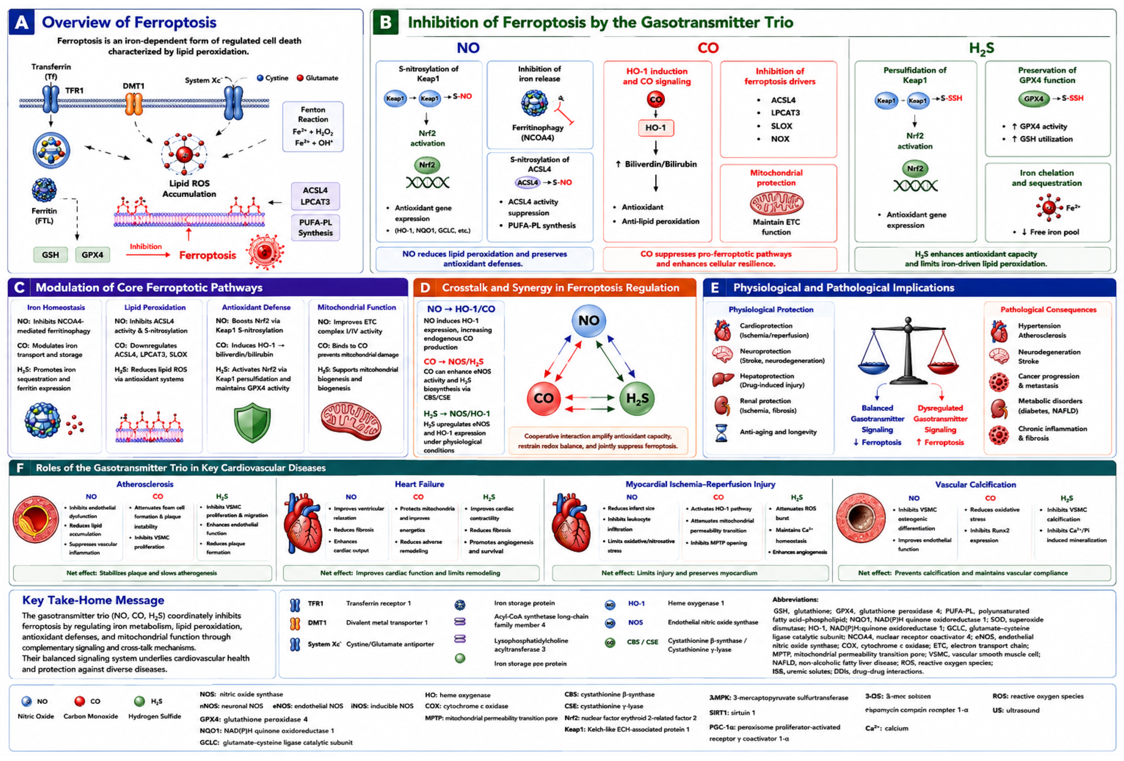
Regulation of ferroptosis by the gasotransmitter trio (NO, CO, and H2S). This figure illustrates how the gasotransmitter trio, nitric oxide (NO), carbon monoxide (CO), and hydrogen sulfide (H2S), suppresses ferroptosis and protects against major cardiovascular diseases. (A) Overview of ferroptosis. Ferroptosis is an iron-dependent, regulated form of cell death driven by excessive lipid peroxidation. Increased intracellular Fe2+ (via TfR1/DMT1 uptake and ferritin turnover) generates ROS through the Fenton reaction. ACSL4- and LPCAT3-mediated synthesis of polyunsaturated phospholipids (PUFA-PL) promotes lipid peroxide accumulation, while the GSH-GPX4 axis counteracts this process. (B) Inhibition of ferroptosis by the gasotransmitter trio. NO suppresses ferroptosis via S-nitrosylation of Keap1 and ACSL4, activation of Nrf2, inhibition of ferritinophagy, and reduced PUFA-PL synthesis. CO induces HO-1, generates biliverdin/bilirubin, suppresses pro-ferroptotic enzymes (ACSL4, LPCAT3, 5-LOX, NOX), and preserves mitochondrial function. H2S inhibits ferroptosis through Keap1 persulfidation, Nrf2 activation, GPX4 preservation, enhanced GSH utilization, and free iron sequestration. (C) Modulation of core ferroptotic pathways. The gasotransmitter trio coordinately regulates iron homeostasis, lipid peroxidation, antioxidant defense, and mitochondrial function, thereby reducing iron-driven oxidative stress, membrane lipid oxidation, and mitochondrial dysfunction. (D) Crosstalk and synergistic regulation. NO, CO, and H2S exhibit extensive bidirectional interactions: NO induces HO-1 and CO production; CO enhances eNOS activity and H2S biosynthesis; H2S promotes eNOS activation and HO-1 expression. This network amplifies antioxidant defenses and resistance to ferroptosis. (E) Physiological and pathological implications. Balanced gasotransmitter signaling suppresses ferroptosis and confers cardioprotection, neuroprotection, and healthy aging. Dysregulated signaling promotes ferroptosis-associated diseases, including atherosclerosis, hypertension, stroke, neurodegeneration, fibrosis, and cancer. (F) Roles in major cardiovascular diseases. In atherosclerosis, the trio improves endothelial function, reduces foam-cell formation and oxidative stress, and inhibits plaque progression. In heart failure, it enhances cardiac performance, preserves mitochondrial energetics, and promotes cardiomyocyte survival. During ischemia–reperfusion injury, it limits oxidative damage, mPTP opening, and infarct size. In vascular calcification, it suppresses VSMC osteogenic differentiation and calcium deposition, preserving vascular compliance. ACSL4, acyl-CoA synthetase long-chain family member 4; DMT1, divalent metal transporter 1; GSH, glutathione; GPX4, glutathione peroxidase 4; HO-1, heme oxygenase-1; LPCAT3, lysophosphatidylcholine acyltransferase 3; Nrf2, nuclear factor erythroid 2-related factor 2; NOX, NADPH oxidase; PUFA-PL, polyunsaturated fatty acid-containing phospholipid; ROS, reactive oxygen species; TfR1, transferrin receptor 1.
Figure 4.
Regulation of ferroptosis by the gasotransmitter trio (NO, CO, and H2S). This figure illustrates how the gasotransmitter trio, nitric oxide (NO), carbon monoxide (CO), and hydrogen sulfide (H2S), suppresses ferroptosis and protects against major cardiovascular diseases. (A) Overview of ferroptosis. Ferroptosis is an iron-dependent, regulated form of cell death driven by excessive lipid peroxidation. Increased intracellular Fe2+ (via TfR1/DMT1 uptake and ferritin turnover) generates ROS through the Fenton reaction. ACSL4- and LPCAT3-mediated synthesis of polyunsaturated phospholipids (PUFA-PL) promotes lipid peroxide accumulation, while the GSH-GPX4 axis counteracts this process. (B) Inhibition of ferroptosis by the gasotransmitter trio. NO suppresses ferroptosis via S-nitrosylation of Keap1 and ACSL4, activation of Nrf2, inhibition of ferritinophagy, and reduced PUFA-PL synthesis. CO induces HO-1, generates biliverdin/bilirubin, suppresses pro-ferroptotic enzymes (ACSL4, LPCAT3, 5-LOX, NOX), and preserves mitochondrial function. H2S inhibits ferroptosis through Keap1 persulfidation, Nrf2 activation, GPX4 preservation, enhanced GSH utilization, and free iron sequestration. (C) Modulation of core ferroptotic pathways. The gasotransmitter trio coordinately regulates iron homeostasis, lipid peroxidation, antioxidant defense, and mitochondrial function, thereby reducing iron-driven oxidative stress, membrane lipid oxidation, and mitochondrial dysfunction. (D) Crosstalk and synergistic regulation. NO, CO, and H2S exhibit extensive bidirectional interactions: NO induces HO-1 and CO production; CO enhances eNOS activity and H2S biosynthesis; H2S promotes eNOS activation and HO-1 expression. This network amplifies antioxidant defenses and resistance to ferroptosis. (E) Physiological and pathological implications. Balanced gasotransmitter signaling suppresses ferroptosis and confers cardioprotection, neuroprotection, and healthy aging. Dysregulated signaling promotes ferroptosis-associated diseases, including atherosclerosis, hypertension, stroke, neurodegeneration, fibrosis, and cancer. (F) Roles in major cardiovascular diseases. In atherosclerosis, the trio improves endothelial function, reduces foam-cell formation and oxidative stress, and inhibits plaque progression. In heart failure, it enhances cardiac performance, preserves mitochondrial energetics, and promotes cardiomyocyte survival. During ischemia–reperfusion injury, it limits oxidative damage, mPTP opening, and infarct size. In vascular calcification, it suppresses VSMC osteogenic differentiation and calcium deposition, preserving vascular compliance. ACSL4, acyl-CoA synthetase long-chain family member 4; DMT1, divalent metal transporter 1; GSH, glutathione; GPX4, glutathione peroxidase 4; HO-1, heme oxygenase-1; LPCAT3, lysophosphatidylcholine acyltransferase 3; Nrf2, nuclear factor erythroid 2-related factor 2; NOX, NADPH oxidase; PUFA-PL, polyunsaturated fatty acid-containing phospholipid; ROS, reactive oxygen species; TfR1, transferrin receptor 1.

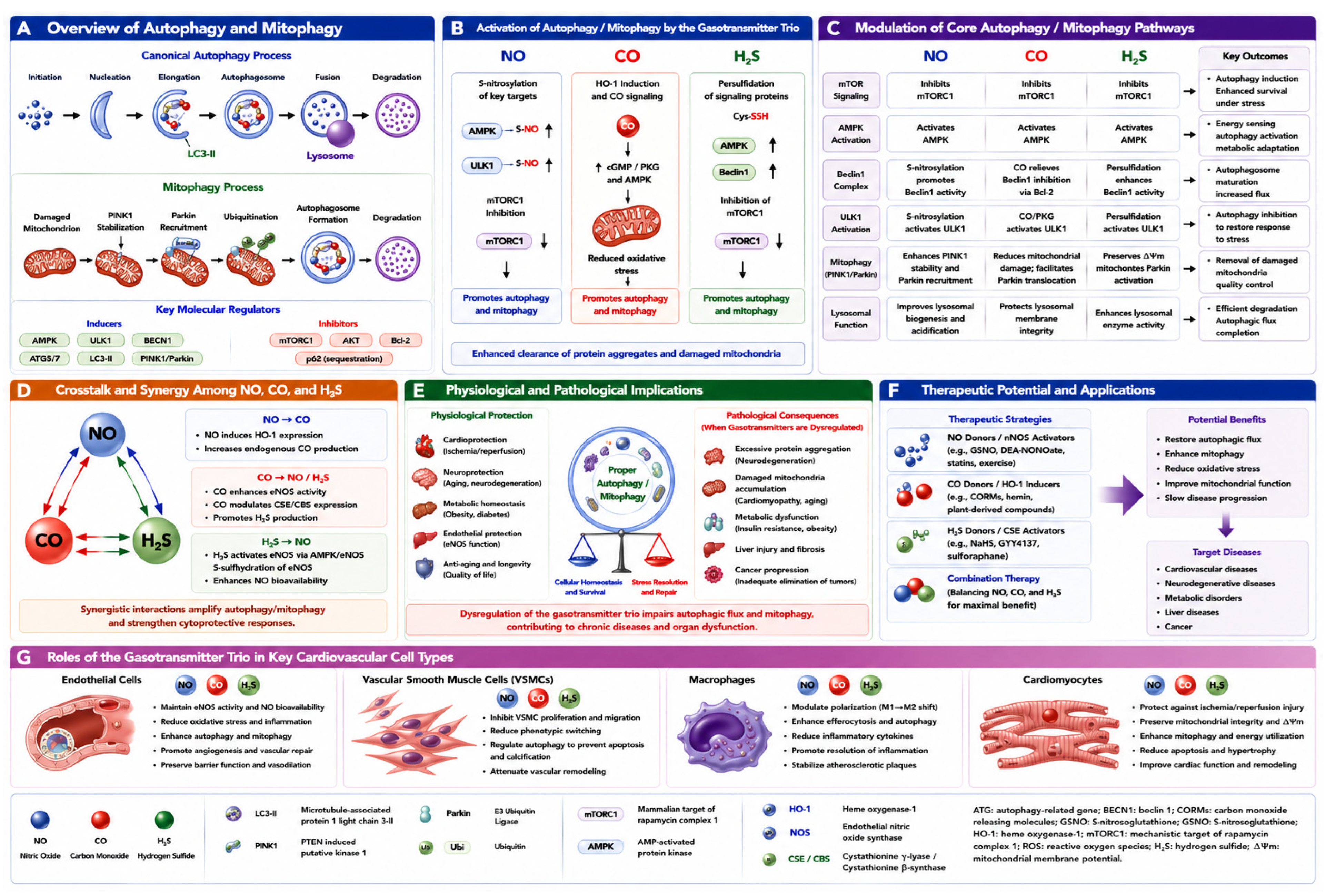
Figure 5.
Regulation of autophagy and mitophagy by the gasotransmitter trio (NO, CO, and H2S). This figure illustrates the mechanisms by which nitric oxide (NO), carbon monoxide (CO), and hydrogen sulfide (H2S) regulate autophagy and mitophagy and their implications in cardiovascular and metabolic diseases. (A) Overview of canonical autophagy and mitophagy. Autophagy involves initiation, nucleation, elongation, autophagosome formation, fusion with lysosomes, and cargo degradation. Mitophagy selectively clears damaged mitochondria via the PINK1/Parkin pathway. Key activators include AMPK, ULK1, Beclin-1, ATG proteins, and LC3-II, while mTORC1, AKT, and Bcl-2 act as major inhibitors. (B) Activation by individual gasotransmitters. NO promotes autophagy via S-nitrosylation of the AMPK/ULK1 pathways and suppression of mTORC1. CO, generated by HO-1, activates cGMP/PKG and AMPK signaling, reduces oxidative stress, and improves mitochondrial quality control. H2S induces protein persulfidation, activates AMPK/Beclin-1, inhibits mTORC1, and enhances autophagic flux and mitophagy. Together, they promote clearance of damaged proteins and dysfunctional mitochondria. (C) Modulation of core signaling networks. The gasotransmitter trio converges on key nodes (mTORC1, AMPK, ULK1, Beclin-1, PINK1/Parkin, and lysosomes) to enhance autophagosome formation, mitochondrial turnover, and cargo degradation. (D) Molecular crosstalk and synergy. Bidirectional interactions amplify responses: NO induces HO-1 and CO production; CO enhances eNOS and H2S biosynthesis; H2S activates eNOS/AMPK to increase NO bioavailability. This synergy strengthens cytoprotective autophagy under stress. (E) Physiological and pathological implications. Balanced gasotransmitter signaling supports cardioprotection, neuroprotection, metabolic homeostasis, endothelial function, and healthy aging by maintaining autophagic flux and mitochondrial quality control. Dysregulation impairs autophagy/mitophagy, leading to protein aggregation, mitochondrial dysfunction, fibrosis, and disease progression. (F) Therapeutic potential. Strategies include NO donors/eNOS activators, CO-releasing molecules (CORMs)/HO-1 inducers, H2S donors/CSE activators, and combination therapies. These enhance autophagy, mitophagy, and mitochondrial function, and may slow the progression of cardiovascular and metabolic disease. (G) Roles in cardiovascular cell types. In endothelial cells, the trio maintains vascular homeostasis, reduces oxidative stress, and promotes angiogenesis. In vascular smooth muscle cells, it inhibits proliferation, phenotypic switching, and calcification via autophagy. In macrophages, it drives M2 polarization, efferocytosis, and resolution of inflammation. In cardiomyocytes, it enhances mitophagy, preserves mitochondrial integrity, and protects against ischemia–reperfusion injury and heart failure.
Figure 5.
Regulation of autophagy and mitophagy by the gasotransmitter trio (NO, CO, and H2S). This figure illustrates the mechanisms by which nitric oxide (NO), carbon monoxide (CO), and hydrogen sulfide (H2S) regulate autophagy and mitophagy and their implications in cardiovascular and metabolic diseases. (A) Overview of canonical autophagy and mitophagy. Autophagy involves initiation, nucleation, elongation, autophagosome formation, fusion with lysosomes, and cargo degradation. Mitophagy selectively clears damaged mitochondria via the PINK1/Parkin pathway. Key activators include AMPK, ULK1, Beclin-1, ATG proteins, and LC3-II, while mTORC1, AKT, and Bcl-2 act as major inhibitors. (B) Activation by individual gasotransmitters. NO promotes autophagy via S-nitrosylation of the AMPK/ULK1 pathways and suppression of mTORC1. CO, generated by HO-1, activates cGMP/PKG and AMPK signaling, reduces oxidative stress, and improves mitochondrial quality control. H2S induces protein persulfidation, activates AMPK/Beclin-1, inhibits mTORC1, and enhances autophagic flux and mitophagy. Together, they promote clearance of damaged proteins and dysfunctional mitochondria. (C) Modulation of core signaling networks. The gasotransmitter trio converges on key nodes (mTORC1, AMPK, ULK1, Beclin-1, PINK1/Parkin, and lysosomes) to enhance autophagosome formation, mitochondrial turnover, and cargo degradation. (D) Molecular crosstalk and synergy. Bidirectional interactions amplify responses: NO induces HO-1 and CO production; CO enhances eNOS and H2S biosynthesis; H2S activates eNOS/AMPK to increase NO bioavailability. This synergy strengthens cytoprotective autophagy under stress. (E) Physiological and pathological implications. Balanced gasotransmitter signaling supports cardioprotection, neuroprotection, metabolic homeostasis, endothelial function, and healthy aging by maintaining autophagic flux and mitochondrial quality control. Dysregulation impairs autophagy/mitophagy, leading to protein aggregation, mitochondrial dysfunction, fibrosis, and disease progression. (F) Therapeutic potential. Strategies include NO donors/eNOS activators, CO-releasing molecules (CORMs)/HO-1 inducers, H2S donors/CSE activators, and combination therapies. These enhance autophagy, mitophagy, and mitochondrial function, and may slow the progression of cardiovascular and metabolic disease. (G) Roles in cardiovascular cell types. In endothelial cells, the trio maintains vascular homeostasis, reduces oxidative stress, and promotes angiogenesis. In vascular smooth muscle cells, it inhibits proliferation, phenotypic switching, and calcification via autophagy. In macrophages, it drives M2 polarization, efferocytosis, and resolution of inflammation. In cardiomyocytes, it enhances mitophagy, preserves mitochondrial integrity, and protects against ischemia–reperfusion injury and heart failure.

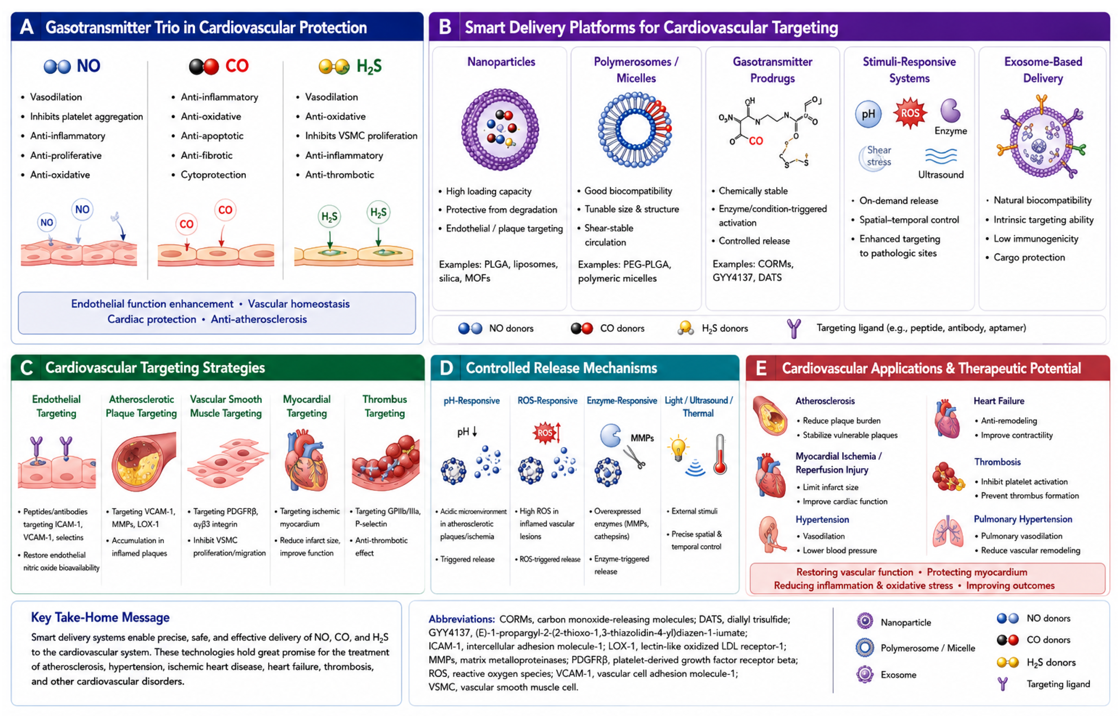
Figure 6.
Smart Delivery Systems for the Gasotransmitter Trio (NO, CO, H2S) in Cardiovascular Disease. (A) Cardiovascular protective actions of individual gasotransmitters. NO primarily promotes vasodilation, inhibits platelet aggregation, suppresses vascular inflammation, and preserves endothelial function. CO exerts anti-inflammatory, antioxidant, anti-apoptotic, and cytoprotective effects by modulating mitochondrial function and stress signaling. H2S contributes to vasorelaxation, antioxidant defense, inhibition of vascular smooth muscle cell proliferation, anti-inflammatory responses, and antithrombotic activity. Collectively, these gasotransmitters maintain vascular homeostasis and protect against cardiovascular injury. (B) Representative smart delivery platforms for cardiovascular-targeted gasotransmitter administration, including nanoparticles, polymeric micelles/polymersomes, gasotransmitter prodrugs, stimuli-responsive systems, and exosome-based carriers. These platforms improve drug stability, prolong circulation time, enable controlled and site-specific release, and reduce systemic toxicity while enhancing delivery to diseased cardiovascular tissues. (C) Targeting strategies for precision cardiovascular therapy, including endothelial, atherosclerotic plaque, vascular smooth muscle cell, ischemic myocardium, and thrombus targeting via disease-specific ligands or pathological biomarkers. Such approaches increase therapeutic accumulation at lesion sites while minimizing off-target effects. (D) Controlled-release mechanisms that exploit pathological microenvironments characteristic of cardiovascular disease, including acidic pH, elevated reactive oxygen species (ROS), disease-associated enzymes (e.g., matrix metalloproteinases and cathepsins), and externally applied stimuli such as light, ultrasound, or thermal activation, thereby enabling spatiotemporally regulated gasotransmitter release. (E) Major cardiovascular applications of gasotransmitter-based nanomedicine, including atherosclerosis, myocardial ischemia–reperfusion injury, hypertension, heart failure, thrombosis, and pulmonary hypertension. Through restoration of endothelial function, suppression of oxidative stress and inflammation, modulation of vascular remodeling, inhibition of platelet activation, and preservation of mitochondrial homeostasis, smart delivery systems substantially enhance the therapeutic potential of NO-, CO-, and H2S-based interventions while reducing systemic adverse effects. Collectively, these next-generation delivery technologies provide a promising translational strategy for precision cardiovascular medicine by integrating targeted delivery, controlled release, and disease-responsive activation to maximize therapeutic efficacy and safety. Collectively, these advanced delivery platforms are transforming gasotransmitter research from fundamental signaling biology into a clinically translatable strategy for precision cardiovascular medicine. Future smart nanomedicines capable of disease-responsive, spatiotemporally controlled, and multi-gas release may enable personalized modulation of the gasotransmitter network to treat complex cardiovascular disorders, thereby maximizing therapeutic efficacy while minimizing systemic toxicity. CORM, carbon monoxide-releasing molecule; EPR, enhanced permeability and retention; GSH, glutathione; MOF, metal–organic framework; NORM, nitric oxide-releasing molecule; PLGA, poly(lactic-co-glycolic acid); ROS, reactive oxygen species; TPP+, triphenylphosphonium.
Figure 6.
Smart Delivery Systems for the Gasotransmitter Trio (NO, CO, H2S) in Cardiovascular Disease. (A) Cardiovascular protective actions of individual gasotransmitters. NO primarily promotes vasodilation, inhibits platelet aggregation, suppresses vascular inflammation, and preserves endothelial function. CO exerts anti-inflammatory, antioxidant, anti-apoptotic, and cytoprotective effects by modulating mitochondrial function and stress signaling. H2S contributes to vasorelaxation, antioxidant defense, inhibition of vascular smooth muscle cell proliferation, anti-inflammatory responses, and antithrombotic activity. Collectively, these gasotransmitters maintain vascular homeostasis and protect against cardiovascular injury. (B) Representative smart delivery platforms for cardiovascular-targeted gasotransmitter administration, including nanoparticles, polymeric micelles/polymersomes, gasotransmitter prodrugs, stimuli-responsive systems, and exosome-based carriers. These platforms improve drug stability, prolong circulation time, enable controlled and site-specific release, and reduce systemic toxicity while enhancing delivery to diseased cardiovascular tissues. (C) Targeting strategies for precision cardiovascular therapy, including endothelial, atherosclerotic plaque, vascular smooth muscle cell, ischemic myocardium, and thrombus targeting via disease-specific ligands or pathological biomarkers. Such approaches increase therapeutic accumulation at lesion sites while minimizing off-target effects. (D) Controlled-release mechanisms that exploit pathological microenvironments characteristic of cardiovascular disease, including acidic pH, elevated reactive oxygen species (ROS), disease-associated enzymes (e.g., matrix metalloproteinases and cathepsins), and externally applied stimuli such as light, ultrasound, or thermal activation, thereby enabling spatiotemporally regulated gasotransmitter release. (E) Major cardiovascular applications of gasotransmitter-based nanomedicine, including atherosclerosis, myocardial ischemia–reperfusion injury, hypertension, heart failure, thrombosis, and pulmonary hypertension. Through restoration of endothelial function, suppression of oxidative stress and inflammation, modulation of vascular remodeling, inhibition of platelet activation, and preservation of mitochondrial homeostasis, smart delivery systems substantially enhance the therapeutic potential of NO-, CO-, and H2S-based interventions while reducing systemic adverse effects. Collectively, these next-generation delivery technologies provide a promising translational strategy for precision cardiovascular medicine by integrating targeted delivery, controlled release, and disease-responsive activation to maximize therapeutic efficacy and safety. Collectively, these advanced delivery platforms are transforming gasotransmitter research from fundamental signaling biology into a clinically translatable strategy for precision cardiovascular medicine. Future smart nanomedicines capable of disease-responsive, spatiotemporally controlled, and multi-gas release may enable personalized modulation of the gasotransmitter network to treat complex cardiovascular disorders, thereby maximizing therapeutic efficacy while minimizing systemic toxicity. CORM, carbon monoxide-releasing molecule; EPR, enhanced permeability and retention; GSH, glutathione; MOF, metal–organic framework; NORM, nitric oxide-releasing molecule; PLGA, poly(lactic-co-glycolic acid); ROS, reactive oxygen species; TPP+, triphenylphosphonium.

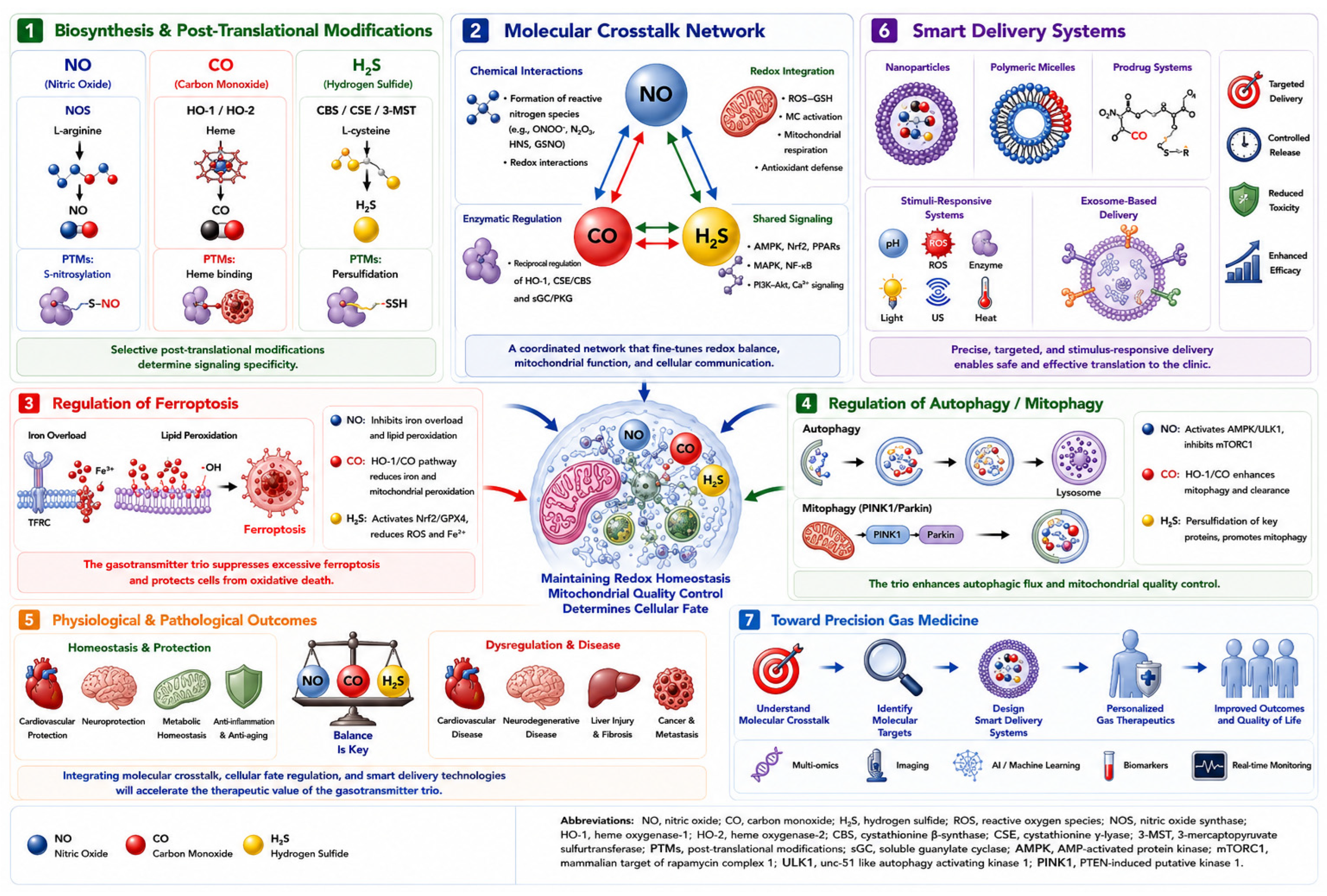
Figure 7.
Toward precision cardiovascular gas medicine: integrating molecular crosstalk, cellular fate regulation, and smart delivery technologies for the gasotransmitter trio (NO, CO, and H2S). This figure illustrates a future framework for precision gas medicine in which nitric oxide (NO), carbon monoxide (CO), and hydrogen sulfide (H2S) are viewed as components of an integrated signaling network rather than individual therapeutic agents. (1) Biosynthesis and post-translational modifications (PTMs). Endogenous production of NO, CO, and H2S is mediated by nitric oxide synthases (NOS), heme oxygenases (HO-1/HO-2), and sulfur-metabolizing enzymes including cystathionine β-synthase (CBS), cystathionine γ-lyase (CSE), and 3-mercaptopyruvate sulfurtransferase (3-MST), respectively. These gasotransmitters exert their biological actions through S-nitrosylation, heme-dependent coordination, and persulfidation, thereby modulating protein function and signaling specificity. (2) Molecular crosstalk network. NO, CO, and H2S engage in direct chemical interactions, reciprocal enzymatic regulation, and shared redox signaling pathways. These interactions generate hybrid signaling intermediates and coordinate key pathways involving sGC–cGMP signaling, Nrf2 activation, mitochondrial adaptation, inflammatory regulation, and cellular stress responses. (3) Regulation of ferroptosis. The gasotransmitter trio collectively suppresses excessive ferroptosis by modulating iron metabolism, inhibiting lipid peroxidation, activating antioxidant defense systems, and preserving GPX4 activity. Balanced gasotransmitter signaling protects tissues from oxidative injury and ferroptotic cell death. (4) Regulation of autophagy and mitophagy. NO, CO, and H2S promote mitochondrial quality control by activating AMPK–ULK1 signaling, inhibiting mTORC1, enhancing PINK1–Parkin-dependent mitophagy, and maintaining lysosomal function. These adaptive mechanisms facilitate removal of damaged mitochondria and support cellular survival. (5) Physiological and pathological outcomes. Homeostatic gasotransmitter signaling contributes to cardiovascular protection, neuroprotection, metabolic balance, anti-inflammatory responses, and healthy aging. In contrast, disruption of the gasotransmitter network promotes oxidative stress, mitochondrial dysfunction, inflammation, fibrosis, neurodegeneration, metabolic disorders, and cancer progression. (6) Smart delivery systems. Emerging nanotechnologies, including nanoparticles, liposomes, polymeric micelles, metal–organic frameworks (MOFs), stimuli-responsive carriers, and exosome-based systems, enable controlled, targeted, and programmable delivery of gasotransmitters. These platforms improve therapeutic efficacy while minimizing systemic toxicity. (7) Future precision gas medicine. Integration of gasotransmitter biology with redox proteomics, spatial multi-omics, artificial intelligence-assisted biomarker discovery, and programmable multi-gas delivery systems may enable personalized gasotherapeutics. Future therapeutic strategies are expected to combine real-time disease sensing with spatiotemporally controlled release of NO, CO, and H2S, thereby optimizing treatment outcomes in cardiovascular, neurodegenerative, inflammatory, metabolic, and malignant diseases. The central concept highlighted in this figure is that cellular fate is determined by coordinated regulation of redox homeostasis and mitochondrial quality control. Future precision gas medicine will therefore depend on the ability to quantitatively monitor and therapeutically modulate the gasotransmitter trio network in a disease-specific and patient-specific manner. AI, artificial intelligence; AMPK, AMP-activated protein kinase; CBS, cystathionine β-synthase; CSE, cystathionine γ-lyase; GPX4, glutathione peroxidase 4; HO, heme oxygenase; MOF, metal–organic framework; mTORC1, mechanistic target of rapamycin complex 1; Nrf2, nuclear factor erythroid 2-related factor 2; NOS, nitric oxide synthase; PINK1, PTEN-induced kinase 1; PTM, post-translational modification; sGC, soluble guanylate cyclase; ULK1, unc-51-like kinase 1; 3-MST, 3-mercaptopyruvate sulfurtransferase.
Figure 7.
Toward precision cardiovascular gas medicine: integrating molecular crosstalk, cellular fate regulation, and smart delivery technologies for the gasotransmitter trio (NO, CO, and H2S). This figure illustrates a future framework for precision gas medicine in which nitric oxide (NO), carbon monoxide (CO), and hydrogen sulfide (H2S) are viewed as components of an integrated signaling network rather than individual therapeutic agents. (1) Biosynthesis and post-translational modifications (PTMs). Endogenous production of NO, CO, and H2S is mediated by nitric oxide synthases (NOS), heme oxygenases (HO-1/HO-2), and sulfur-metabolizing enzymes including cystathionine β-synthase (CBS), cystathionine γ-lyase (CSE), and 3-mercaptopyruvate sulfurtransferase (3-MST), respectively. These gasotransmitters exert their biological actions through S-nitrosylation, heme-dependent coordination, and persulfidation, thereby modulating protein function and signaling specificity. (2) Molecular crosstalk network. NO, CO, and H2S engage in direct chemical interactions, reciprocal enzymatic regulation, and shared redox signaling pathways. These interactions generate hybrid signaling intermediates and coordinate key pathways involving sGC–cGMP signaling, Nrf2 activation, mitochondrial adaptation, inflammatory regulation, and cellular stress responses. (3) Regulation of ferroptosis. The gasotransmitter trio collectively suppresses excessive ferroptosis by modulating iron metabolism, inhibiting lipid peroxidation, activating antioxidant defense systems, and preserving GPX4 activity. Balanced gasotransmitter signaling protects tissues from oxidative injury and ferroptotic cell death. (4) Regulation of autophagy and mitophagy. NO, CO, and H2S promote mitochondrial quality control by activating AMPK–ULK1 signaling, inhibiting mTORC1, enhancing PINK1–Parkin-dependent mitophagy, and maintaining lysosomal function. These adaptive mechanisms facilitate removal of damaged mitochondria and support cellular survival. (5) Physiological and pathological outcomes. Homeostatic gasotransmitter signaling contributes to cardiovascular protection, neuroprotection, metabolic balance, anti-inflammatory responses, and healthy aging. In contrast, disruption of the gasotransmitter network promotes oxidative stress, mitochondrial dysfunction, inflammation, fibrosis, neurodegeneration, metabolic disorders, and cancer progression. (6) Smart delivery systems. Emerging nanotechnologies, including nanoparticles, liposomes, polymeric micelles, metal–organic frameworks (MOFs), stimuli-responsive carriers, and exosome-based systems, enable controlled, targeted, and programmable delivery of gasotransmitters. These platforms improve therapeutic efficacy while minimizing systemic toxicity. (7) Future precision gas medicine. Integration of gasotransmitter biology with redox proteomics, spatial multi-omics, artificial intelligence-assisted biomarker discovery, and programmable multi-gas delivery systems may enable personalized gasotherapeutics. Future therapeutic strategies are expected to combine real-time disease sensing with spatiotemporally controlled release of NO, CO, and H2S, thereby optimizing treatment outcomes in cardiovascular, neurodegenerative, inflammatory, metabolic, and malignant diseases. The central concept highlighted in this figure is that cellular fate is determined by coordinated regulation of redox homeostasis and mitochondrial quality control. Future precision gas medicine will therefore depend on the ability to quantitatively monitor and therapeutically modulate the gasotransmitter trio network in a disease-specific and patient-specific manner. AI, artificial intelligence; AMPK, AMP-activated protein kinase; CBS, cystathionine β-synthase; CSE, cystathionine γ-lyase; GPX4, glutathione peroxidase 4; HO, heme oxygenase; MOF, metal–organic framework; mTORC1, mechanistic target of rapamycin complex 1; Nrf2, nuclear factor erythroid 2-related factor 2; NOS, nitric oxide synthase; PINK1, PTEN-induced kinase 1; PTM, post-translational modification; sGC, soluble guanylate cyclase; ULK1, unc-51-like kinase 1; 3-MST, 3-mercaptopyruvate sulfurtransferase.

Table 1.
Comparative Overview of Biosynthesis, Molecular Targets, and Signaling Mechanisms of NO, CO, and H2S.
Table 1.
Comparative Overview of Biosynthesis, Molecular Targets, and Signaling Mechanisms of NO, CO, and H2S.
| Feature | NO | CO | H2S |
| Major biosynthetic enzymes | eNOS, nNOS, iNOS | HO-1, HO-2 | CBS, CSE, 3-MST |
| Principal substrate | L-arginine | Heme | L-cysteine and sulfur-containing amino acids |
| Major signaling mechanism | S-nitrosylation; sGC–cGMP activation | Heme coordination; modulation of heme proteins | Persulfidation/sulfhydration |
| Representative molecular targets | sGC, Drp1, NF-κB, mitochondrial proteins | sGC, cytochrome c oxidase, cytochrome P450 enzymes, globins | Keap1, KATP channels, mitochondrial enzymes, antioxidant proteins |
| Major biological effects | Vasodilation, antiplatelet activity, neurotransmission, and immune regulation | Anti-inflammation, mitochondrial adaptation, cytoprotection | Antioxidant defense, vasorelaxation, mitochondrial bioenergetics, stress adaptation |
| Pathological risk when excessive | Nitrosative stress, peroxynitrite formation, mitochondrial dysfunction | Hypoxia, mitochondrial inhibition, potential iron dysregulation | Inhibition of cytochrome c oxidase, mitochondrial toxicity |
| Key references | [19,20,21,31,58,59,60,61] | [8,9,10,18,53,54] | [25,33,55,56,62,63,64,65] |
Table 2.
Molecular crosstalk mechanisms among NO, CO, and H2S.
Table 2.
Molecular crosstalk mechanisms among NO, CO, and H2S.
| Interaction | Major Mechanism | Representative Outcome | Key References |
|---|
| NO–H2S | Formation of sulfur–nitrogen hybrid species, including SSNO− | Sustained NO-like signaling and prolonged vasorelaxation | [36,37,70] |
| NO–H2S | Generation of nitroxyl (HNO) | Cardioprotection and positive inotropic effects | [71,72] |
| H2S → NO | Enhancement of eNOS phosphorylation, dimerization, and NO bioavailability | Improved endothelial function and vasorelaxation | [35,38,73,74,75,76] |
| CO → NO | Modulation of endothelial NO release and eNOS-related signaling | Vasodilation and vascular adaptation | [77] |
| NO → CO | Induction of HO-1 expression through redox- and stress-responsive transcriptional pathways | Increased endogenous CO generation and cytoprotection | [78,79] |
| H2S → CO | Nrf2-dependent HO-1 induction | Antioxidant defense and stress adaptation | [64,80] |
| NO/CO/H2S convergence | Shared regulation of sGC–cGMP–PKG signaling | Vascular relaxation, antiplatelet activity, cardioprotection | [19,67,81] |
| NO/CO/H2S convergence | Regulation of KATP channels, Nrf2, NF-κB, HIF-1α, and mitochondrial pathways | Cytoprotection, redox balance, inflammation control | [22,64,68,80,82,83,84] |
Table 3.
Bidirectional Regulation Among CBS, CSE, HO, and NOS.
Table 3.
Bidirectional Regulation Among CBS, CSE, HO, and NOS.
| Enzyme Pair | Physiological (Activating) Effect | Pathological/High-Concentration (Inhibitory) Effect | Proposed Mechanism | Key References |
|---|
| H2S → NOS | Enhances eNOS phosphorylation (Ser1177), dimerization, BH4 preservation | Suppresses NOS activity | Cofactor interaction/redox alteration | [73,74,75,76] |
| NO → NOS (self) | — | Product inhibition of eNOS | S-nitrosylation-induced dimer collapse (loss of zinc-tetrathiolate cluster) | [52] |
| CO → CBS | — (CO not a physiological CBS activator) | Blocks CBS catalytic activity | Heme binding displaces Cys ligand; competitive with homocysteine | [18] |
| NO → CSE | — | Suppresses H2S-generating activity | S-nitrosation of Cys229 | [26] |
| CO → eNOS | Activates eNOS, enhances NO production | — | PI3K/Akt-dependent signaling | [77] |
| NO → HO-1 | Induces HO-1 transcription (Nrf2/HIF-1α), concentration-dependent increase | — | ARE/Nrf2-dependent transcription | [54,78,79] |
| H2S → HO-1 | Induces HO-1 via Nrf2 | — | Persulfidation of Keap1 | [80] |
| NO → heme-protein maturation (general) | Facilitates heme incorporation into apo-hemoproteins at low levels | Reversibly inhibits heme insertion into apo-hemoproteins | Direct NO-heme binding competes with apoprotein maturation | [53] |
Table 4.
Regulatory roles of NO, CO, and H2S in ferroptosis, autophagy, and mitophagy.
Table 4.
Regulatory roles of NO, CO, and H2S in ferroptosis, autophagy, and mitophagy.
| Gasotransmitter | Effects on Ferroptosis | Effects on Autophagy/Mitophagy | Major Pathways or Targets | Disease Relevance | Key References |
|---|
| NO | Suppresses lipid peroxidation by terminating lipid radical chain reactions; preserves GPX4 activity under physiological conditions | Regulates autophagy through redox signaling and S-nitrosylation-dependent mechanisms | Lipid peroxyl radicals, GPX4, ferritin, Drp1, AMPK-related pathways | Cardiovascular injury, neurodegeneration, oxidative stress | [31,58,86,87] |
| CO | Context-dependent; moderate HO-1/CO signaling is protective, whereas excessive HO-1 activity may increase labile iron and promote ferroptosis | Activates autophagy through mitochondrial ROS and adaptive stress signaling | HO-1, biliverdin/bilirubin, Fe2+, cytochrome c oxidase, mitochondrial ROS | Ischemia–reperfusion injury, cancer, inflammation | [9,22,66,88,89] |
| H2S | Strongly inhibits ferroptosis by preserving GSH homeostasis, activating Nrf2, and maintaining GPX4 activity | Promotes autophagy and mitophagy through persulfidation, AMPK activation, and mitochondrial protection | Keap1–Nrf2, System Xc−, GSH, GPX4, mitochondrial enzymes | Cardiomyocyte injury, sepsis-associated injury, metabolic disease | [33,43,64,90,91,92] |
| Integrated trio network | Coordinately limits iron-driven lipid peroxidation and maintains redox balance | Supports mitochondrial quality control and cellular adaptation | Nrf2, GPX4, AMPK–mTOR, PINK1–Parkin, mitochondrial homeostasis | Cardiovascular, neurodegenerative, hepatic, renal, inflammatory, and malignant diseases | [39,40,41,42,43,44,45,93,94,95,96,97,98] |
Table 5.
Representative gasotransmitter donors and smart delivery platforms for therapeutic applications.
Table 5.
Representative gasotransmitter donors and smart delivery platforms for therapeutic applications.
| Platform/Donor Type | Gas Delivered | Trigger or Release Mechanism | Major Advantages | Major Limitations | Clinical/Translational Bottleneck | Representative Applications | Key References |
|---|
| Organic NO donors (NONOates, etc.) | NO | Spontaneous, pH-dependent, or chemical decomposition | Rapid NO release; useful experimental tools | Burst release kinetics; short half-life; tolerance with repeated dosing | Poor spatial control leads to systemic hypotension; limited disease-site selectivity | Vascular regulation, wound healing, antimicrobial therapy | [102,103,104] |
| Light-responsive NO-releasing materials | NO | Photoactivation | Spatial and temporal control of NO release | Requires external light source; limited tissue penetration of activating wavelengths | Impractical for deep/internal cardiovascular targets without invasive light delivery | Local vascular modulation, cancer therapy, biomaterials | [105,106] |
| CORMs | CO | Chemical or ligand-exchange release | Controlled CO administration without inhaled CO exposure | Off-target reactivity of metal scaffold; some CO release independent of intended trigger | Metal byproduct toxicity/clearance concerns; inconsistent release kinetics across compartments | Anti-inflammatory therapy, cytoprotection, cancer therapy | [10,107,108] |
| PhotoCORMs | CO | Light-triggered CO release | On-demand and localized CO delivery | Same light-penetration constraint as above; photobleaching limits repeat dosing | Not yet validated for non-superficial cardiovascular lesions | Cancer therapy, inflammatory diseases, mechanistic studies | [109] |
| Enzyme-triggered CORMs | CO | Disease- or enzyme-responsive activation | Improved selectivity and reduced systemic exposure | Dependent on disease-specific enzyme expression, variable between patients | Patient-to-patient variability in trigger-enzyme activity complicates dose standardization | Targeted CO therapy | [110] |
| Slow-releasing H2S donors (GYY4137-type) | H2S | Hydrolysis or slow chemical release | Sustained H2S exposure and reduced toxicity | Slow onset limits utility in acute settings; hydrolysis rate hard to fine-tune in vivo | Long-term safety and optimal dosing regimens undefined in humans | Cardioprotection, anti-inflammatory therapy, metabolic disease | [111,112,113,114] |
| pH- and ROS-responsive H2S donors | H2S | Acidic pH or oxidative microenvironment | Disease-responsive H2S release | Release depends on sufficiently acidic/oxidative microenvironment, which is heterogeneous within lesions | Unpredictable release in early-stage or mixed-microenvironment lesions | Inflammation, cancer, oxidative injury | [114] |
| Nanoparticles and MOFs | NO, CO, H2S, or multi-gas | Encapsulation, adsorption, or stimulus-responsive release | High loading capacity, tunable release, improved targeting | Complex synthesis/scale-up; potential immunogenicity or long-term accumulation | Regulatory pathway for inorganic/metal–organic nanomaterials less established than small molecules | Cancer, inflammation, cardiovascular disease | [115,116,117] |
| Polymeric micelles, liposomes, and hydrogels | NO, CO, H2S | Sustained, local, or injectable delivery | Improved stability, local retention, biocompatibility | Batch-to-batch variability; limited long-term storage stability | Manufacturing reproducibility and GMP scale-up remain major hurdles | Regenerative medicine, wound healing, local inflammation | [118,119,120] |
| Cell membrane-coated or exosome-based systems | NO, CO, H2S | Biomimetic membrane camouflage (e.g., macrophage-, cardiomyocyte-, or platelet-derived) enabling immune evasion and passive/active disease-site targeting | Prolonged circulation, reduced immune clearance, inherent disease-relevant targeting (e.g., inflamed or ischemic tissue) via source-cell surface markers | Complex, low-yield production; donor-source and batch variability; challenging drug/gas loading efficiency | High manufacturing cost, lack of standardized production protocols, and unresolved long-term biodistribution/immunogenicity data limit clinical scalability | Atherosclerosis-targeted therapy, myocardial ischemia–reperfusion injury, inflammation-targeted delivery | [115,121] |
Table 6.
Future Perspectives and Clinical Translation of Precision Gas Medicine.
Table 6.
Future Perspectives and Clinical Translation of Precision Gas Medicine.
| Challenge | Current Limitation | Emerging Technology/Strategy | Potential Clinical Application | Future Direction | Key References |
|---|
| Biomarker Identification | Lack of reliable biomarkers reflecting local gasotransmitter activity and signaling status | Multi-omics profiling, spatial transcriptomics, metabolomics, redox proteomics, liquid biopsy | Early disease detection, patient stratification, therapeutic monitoring | Personalized biomarker-guided gasotherapy | [121,122,123,124] |
| Quantification of Endogenous Gases | Difficulty measuring NO, CO, and H2S concentrations in specific tissues in real time | Fluorescent probes, electrochemical biosensors, molecular imaging, wearable sensing devices | Dynamic monitoring of disease progression and treatment response | Real-time precision gas monitoring systems | [123,124] |
| Dose Optimization | Narrow therapeutic window and concentration-dependent toxicity | Programmable release systems, AI-assisted dose prediction, pharmacokinetic modeling | Individualized dosing regimens | Adaptive precision gas medicine | [103,107,111,113,114] |
| Tissue-Specific Delivery | Rapid diffusion and lack of target specificity | MOFs, liposomes, polymeric micelles, exosomes, cell membrane-coated nanoparticles | Organ-specific delivery to heart, brain, liver, kidney, and tumors | Precision tissue-targeted therapy | [115,116,117,118,119,120,121,122,123] |
| Multi-Gas Integration | Most current approaches focus on a single gasotransmitter | Multi-gas co-delivery platforms and stimuli-responsive nanomaterials | Reconstitution of physiological gasotransmitter networks | Programmable gasotransmitter network therapy | [115,116,117,118,119,120,121,122,123] |
| Disease Heterogeneity | Variable responses among patients and disease stages | Molecular classification, machine learning, systems biology approaches | Patient stratification and treatment selection | Precision medicine-based intervention | [121,122,123,124] |
| Monitoring Cellular Fate | Lack of biomarkers reflecting ferroptosis, autophagy, and mitophagy status | Ferroptosis-associated lipidomics, circulating miRNAs, mitochondrial biomarkers | Monitoring therapeutic efficacy and disease progression | Cell-fate-guided therapy optimization | [93,94,95,96,97,98,99,100,101] |
| Clinical Translation | Limited clinical trials and regulatory frameworks | GMP-compliant gas donors, scalable nanomedicine manufacturing, regulatory harmonization | Translation into cardiovascular, neurodegenerative, inflammatory, metabolic, and cancer therapies | Evidence-based gasotransmitter therapeutics | [118,119,120,121,122,123,124] |
| Artificial Intelligence Integration | Massive and complex gasotransmitter-related datasets | AI-assisted multi-omics integration, digital twin modeling, predictive analytics | Predictive diagnosis and therapeutic planning | Digital twin-guided precision gasotherapy | [121,122,123,124] |
| Long-Term Safety | Incomplete understanding of chronic exposure effects | Longitudinal cohort studies and real-world evidence platforms | Improved safety assessment and risk prediction | Personalized long-term treatment management | [118,119,120,121,122,123,124] |