


Amyloid Precursor Protein Processing Links Female Urgency Urinary Incontinence with Alzheimer’s Disease: Implications for Treatment

 , , , ,
  , , , ,
  , , , and
Abstract
1. Introduction
2. Results
2.1. Landscape Input Genes
2.2. Enriched Networks
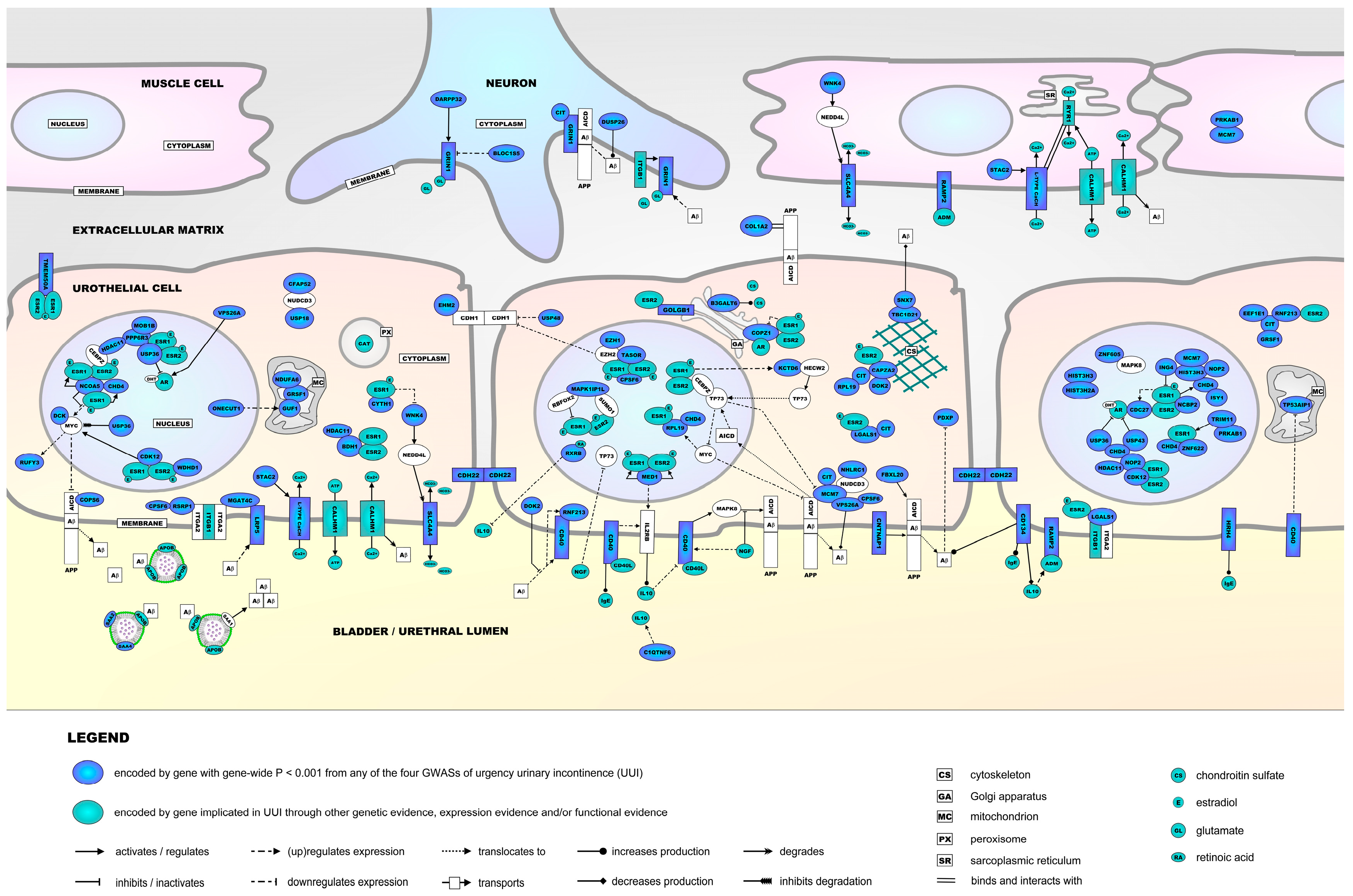
2.3. The Molecular Landscape of UUI
2.4. PRS-Based Analyses
3. Discussion
4. Materials and Methods
4.1. Selection of Landscape Input Genes
4.2. Network Enrichment Analysis
4.3. Building the Molecular Landscape of UUI
4.4. PRS-Based Analyses
5. Conclusions
Supplementary Materials
Author Contributions
Funding
Institutional Review Board Statement
Informed Consent Statement
Data Availability Statement
Acknowledgments
Conflicts of Interest
Abbreviations
| AD | Alzheimer’s disease |
| AICD | APP intracellular domain |
| APP | Amyloid Precursor Protein |
| ATP | Adenosine triphosphate |
| Aβ | Amyloid beta |
| CNS | Central nervous system |
| eQTL | expression quantitative trait loci |
| ESR1 | Estrogen receptor 1 |
| ESR2 | Estrogen receptor 2 |
| GARNET | Women’s Health Initiative Genomics & Randomized Trials Network |
| GWAS | Genome-wide association study |
| HUNT | Trøndelag Health Study |
| IPA | Ingenuity pathway analysis |
| kb | kilobase |
| OAB | Overactive bladder syndrome |
| PRS | Polygenic risk score |
| SNP | Single nucleotide polymorphism |
| UUI | Urgency urinary incontinence |
References
- Haylen, B.T.; de Ridder, D.; Freeman, R.M.; Swift, S.E.; Berghmans, B.; Lee, J.; Monga, A.; Petri, E.; Rizk, D.E.; Sand, P.K.; et al. An International Urogynecological Association (IUGA)/International Continence Society (ICS) joint report on the terminology for female pelvic floor dysfunction. Int. Urogynecol. J. 2010, 21, 5–26. [Google Scholar] [CrossRef] [PubMed]
- Peyronnet, B.; Mironska, E.; Chapple, C.; Cardozo, L.; Oelke, M.; Dmochowski, R.; Amarenco, G.; Game, X.; Kirby, R.; Van Der Aa, F.; et al. A Comprehensive Review of Overactive Bladder Pathophysiology: On the Way to Tailored Treatment. Eur. Urol. 2019, 75, 988–1000. [Google Scholar] [CrossRef] [PubMed]
- Milsom, I.; Coyne, K.S.; Nicholson, S.; Kvasz, M.; Chen, C.I.; Wein, A.J. Global prevalence and economic burden of urgency urinary incontinence: A systematic review. Eur. Urol. 2014, 65, 79–95. [Google Scholar] [CrossRef] [PubMed]
- Wennberg, A.L.; Altman, D.; Lundholm, C.; Klint, A.; Iliadou, A.; Peeker, R.; Fall, M.; Pedersen, N.L.; Milsom, I. Genetic influences are important for most but not all lower urinary tract symptoms: A population-based survey in a cohort of adult Swedish twins. Eur. Urol. 2011, 59, 1032–1038. [Google Scholar] [CrossRef] [PubMed]
- Chung, E.; Katz, D.J.; Love, C. Adult male stress and urge urinary incontinence—A review of pathophysiology and treatment strategies for voiding dysfunction in men. Aust. Fam. Physician 2017, 46, 661–666. [Google Scholar] [PubMed]
- Chen, L.C.; Kuo, H.C. Pathophysiology of refractory overactive bladder. Low. Urin. Tract. Symptoms 2019, 11, 177–181. [Google Scholar] [CrossRef] [PubMed]
- Soria Lopez, J.A.; Gonzalez, H.M.; Leger, G.C. Alzheimer’s disease. Handb. Clin. Neurol. 2019, 167, 231–255. [Google Scholar] [CrossRef] [PubMed]
- Lee, S.H.; Cho, S.T.; Na, H.R.; Ko, S.B.; Park, M.H. Urinary incontinence in patients with Alzheimer’s disease: Relationship between symptom status and urodynamic diagnoses. Int. J. Urol. 2014, 21, 683–687. [Google Scholar] [CrossRef] [PubMed]
- Hellstrom, L.; Ekelund, P.; Milsom, I.; Skoog, I. The influence of dementia on the prevalence of urinary and faecal incontinence in 85-year-old men and women. Arch. Gerontol. Geriatr. 1994, 19, 11–20. [Google Scholar] [CrossRef] [PubMed]
- Orme, S.; Morris, V.; Gibson, W.; Wagg, A. Managing Urinary Incontinence in Patients with Dementia: Pharmacological Treatment Options and Considerations. Drugs Aging 2015, 32, 559–567. [Google Scholar] [CrossRef] [PubMed]
- Divo, M.J.; Martinez, C.H.; Mannino, D.M. Ageing and the epidemiology of multimorbidity. Eur. Respir. J. 2014, 44, 1055–1068. [Google Scholar] [CrossRef] [PubMed]
- Akpinar Soylemez, B.; Kucukguclu, O.; Akyol, M.A.; Isik, A.T. Quality of life and factors affecting it in patients with Alzheimer’s disease: A cross-sectional study. Health Qual. Life Outcomes 2020, 18, 304. [Google Scholar] [CrossRef] [PubMed]
- Astrom, Y.; Asklund, I.; Lindam, A.; Sjostrom, M. Quality of life in women with urinary incontinence seeking care using e-health. BMC Womens Health 2021, 21, 337. [Google Scholar] [CrossRef] [PubMed]
- Lee, H.Y.; Li, C.C.; Juan, Y.S.; Chang, Y.H.; Yeh, H.C.; Tsai, C.C.; Chueh, K.S.; Wu, W.J.; Yang, Y.H. Urinary Incontinence in Alzheimer’s Disease. Am. J. Alzheimers Dis. Other Demen 2017, 32, 51–55. [Google Scholar] [CrossRef] [PubMed]
- Richter, H.E.; Whitehead, N.; Arya, L.; Ridgeway, B.; Allen-Brady, K.; Norton, P.; Sung, V.; Shepherd, J.P.; Komesu, Y.; Gaddis, N.; et al. Genetic contributions to urgency urinary incontinence in women. J. Urol. 2015, 193, 2020–2027. [Google Scholar] [CrossRef] [PubMed]
- Penney, K.L.; Townsend, M.K.; Turman, C.; Glass, K.; Staller, K.; Kraft, P.; Grodstein, F.; Minassian, V.A. Genome-Wide Association Study for Urinary and Fecal Incontinence in Women. J. Urol. 2020, 203, 978–983. [Google Scholar] [CrossRef] [PubMed]
- Cartwright, R.; Franklin, L.; Tikkinen, K.A.O.; Kalliala, I.; Miotla, P.; Rechberger, T.; Offiah, I.; McMahon, S.; O’Reilly, B.; Lince, S.; et al. Genome-Wide Association Study Identifies Two Novel Loci Associated with Female Stress and Urgency Urinary Incontinence. J. Urol. 2021, 206, 679–687, Correction in J. Urol. 2023, 210, 569. https://doi.org/10.1097/JU.0000000000003604. [Google Scholar] [CrossRef] [PubMed]
- Asvold, B.O.; Langhammer, A.; Rehn, T.A.; Kjelvik, G.; Grontvedt, T.V.; Sorgjerd, E.P.; Fenstad, J.S.; Heggland, J.; Holmen, O.; Stuifbergen, M.C.; et al. Cohort Profile Update: The HUNT Study, Norway. Int. J. Epidemiol. 2023, 52, e80–e91. [Google Scholar] [CrossRef] [PubMed]
- Balat, A.; Sarica, K.; Cekmen, M.; Yurekli, M.; Yagci, F.; Erbagci, A. Adrenomedullin and nitric oxide in children with detrusor instability. Pediatr. Nephrol. 2003, 18, 422–425. [Google Scholar] [CrossRef] [PubMed]
- UniProt, C. UniProt: The Universal Protein Knowledgebase in 2025. Nucleic Acids Res. 2025, 53, D609–D617. [Google Scholar] [CrossRef] [PubMed]
- Baytaroglu, C.; Sevgili, E. Association of Metabolic Syndrome Components and Overactive Bladder in Women. Cureus 2021, 13, e14765. [Google Scholar] [CrossRef] [PubMed]
- Cornu, J.N.; Merlet, B.; Cussenot, O.; Cancel-Tassin, G.; Ciofu, C.; Amarenco, G.; Haab, F. Genetic susceptibility to urinary incontinence: Implication of polymorphisms of androgen and oestrogen pathways. World J. Urol. 2011, 29, 239–242. [Google Scholar] [CrossRef] [PubMed]
- Sana-Ur-Rehman, H.; Markus, I.; Moore, K.H.; Mansfield, K.J.; Liu, L. Expression and localization of pannexin-1 and CALHM1 in porcine bladder and their involvement in modulating ATP release. Am. J. Physiol. Regul. Integr. Comp. Physiol. 2017, 312, R763–R772. [Google Scholar] [CrossRef] [PubMed]
- Kumar, V.; Chapple, C.R.; Rosario, D.; Tophill, P.R.; Chess-Williams, R. In vitro release of adenosine triphosphate from the urothelium of human bladders with detrusor overactivity, both neurogenic and idiopathic. Eur. Urol. 2010, 57, 1087–1092. [Google Scholar] [CrossRef] [PubMed]
- Badawi, J.K.; Langbein, S. Selective beta-adrenoceptor agonists, calcium antagonists and potassium channel openers as a possible medical treatment of the overactive bladder and urge incontinence. Pharmazie 2006, 61, 175–178. [Google Scholar] [PubMed]
- Antunes-Lopes, T.; Cruz, F. Urinary Biomarkers in Overactive Bladder: Revisiting the Evidence in 2019. Eur. Urol. Focus 2019, 5, 329–336. [Google Scholar] [CrossRef] [PubMed]
- Masuda, H.; Kihara, K.; Saito, K.; Matsuoka, Y.; Yoshida, S.; Chancellor, M.B.; de Groat, W.C.; Yoshimura, N. Reactive oxygen species mediate detrusor overactivity via sensitization of afferent pathway in the bladder of anaesthetized rats. BJU Int. 2008, 101, 775–780. [Google Scholar] [CrossRef] [PubMed]
- Tyagi, P.; Barclay, D.; Zamora, R.; Yoshimura, N.; Peters, K.; Vodovotz, Y.; Chancellor, M. Urine cytokines suggest an inflammatory response in the overactive bladder: A pilot study. Int. Urol. Nephrol. 2010, 42, 629–635. [Google Scholar] [CrossRef] [PubMed]
- Robinson, D.; Cardozo, L. Managing overactive bladder. Climacteric 2019, 22, 250–256. [Google Scholar] [CrossRef] [PubMed]
- Hillard, T. The postmenopausal bladder. Post Reprod. Health 2010, 16, 74–80. [Google Scholar] [CrossRef] [PubMed]
- Robinson, D.; Toozs-Hobson, P.; Cardozo, L. The effect of hormones on the lower urinary tract. Post Reprod. Health 2013, 19, 155–162. [Google Scholar] [CrossRef] [PubMed]
- Kullmann, F.A.; Limberg, B.J.; Artim, D.E.; Shah, M.; Downs, T.R.; Contract, D.; Wos, J.; Rosenbaum, J.S.; de Groat, W.C. Effects of beta3-adrenergic receptor activation on rat urinary bladder hyperactivity induced by ovariectomy. J. Pharmacol. Exp. Ther. 2009, 330, 704–717. [Google Scholar] [CrossRef] [PubMed]
- Liang, W.; Afshar, K.; Stothers, L.; Laher, I. The influence of ovariectomy and estrogen replacement on voiding patterns and detrusor muscarinic receptor affinity in the rat. Life Sci. 2002, 71, 351–362. [Google Scholar] [CrossRef] [PubMed]
- Juliato, C.R.; Baccaro, L.F.; Pedro, A.O.; Costa-Paiva, L.; Lui-Filho, J.; Pinto-Neto, A.M. Subjective urinary urgency in middle age women: A population-based study. Maturitas 2016, 85, 82–87. [Google Scholar] [CrossRef] [PubMed]
- Pillalamarri, N.; Shalom, D.F.; Pilkinton, M.L.; Winkler, H.A.; Chatterjee, P.K.; Solanki, M.; Metz, C.N. Inflammatory Urinary Cytokine Expression and Quality of Life in Patients with Overactive Bladder. Female Pelvic Med. Reconstr. Surg. 2018, 24, 449–453. [Google Scholar] [CrossRef] [PubMed]
- Kanasaki, K.; Yu, W.; von Bodungen, M.; Larigakis, J.D.; Kanasaki, M.; Ayala de la Pena, F.; Kalluri, R.; Hill, W.G. Loss of beta1-integrin from urothelium results in overactive bladder and incontinence in mice: A mechanosensory rather than structural phenotype. FASEB J. 2013, 27, 1950–1961. [Google Scholar] [CrossRef] [PubMed]
- Post, W.M.; Ruiz-Zapata, A.M.; Grens, H.; de Vries, R.B.M.; Poelmans, G.; Coenen, M.J.H.; Janssen, D.A.W.; Heesakkers, J.; Oosterwijk, E.; Kluivers, K.B. Genetic variants and expression changes in urgency urinary incontinence: A systematic review. Neurourol. Urodyn. 2020, 39, 2089–2110. [Google Scholar] [CrossRef] [PubMed]
- Jiang, H.H.; Song, B.; Lu, G.S.; Wen, Q.J.; Jin, X.Y. Loss of ryanodine receptor calcium-release channel expression associated with overactive urinary bladder smooth muscle contractions in a detrusor instability model. BJU Int. 2005, 96, 428–433. [Google Scholar] [CrossRef] [PubMed]
- Sonmez, M.G.; Goger, Y.E.; Ecer, G.; Atici, A.; Ozkent, M.S.; Ozturk, A. Effects of urine alkalinization with sodium bicarbonate orally on lower urinary tract symptoms in female patients: A pilot study. Int. Urogynecol. J. 2018, 29, 1029–1033. [Google Scholar] [CrossRef] [PubMed]
- Sonmez, M.G.; Ecer, G.; Atici, A.; Ozkent, M.S.; Iyisoy, M.S.; Ozturk, A. Comparison of Oral Sodium Bicarbonate and Solifenacin Treatment in Female Overactive Bladder Patients With Acidic Urine pH. Female Pelvic Med. Reconstr. Surg. 2020, 26, 649–653. [Google Scholar] [CrossRef] [PubMed]
- Irkilata, L.; Aydin, M.; Riza Aydin, H.; Cihan Demirel, H.; Kadihasanoglu, M.; Kemal Atilla, M. Intravesical Sodium Chondroitin Sulphate to Treat Overactive Bladder: Preliminary Result. Int. Neurourol. J. 2015, 19, 85–89. [Google Scholar] [CrossRef] [PubMed]
- Morelli, M.; Mocciaro, R.; Venturella, R.; Albano, A.; Sacchinelli, A.; Zullo, F. Hyaluronic acid-chondroitin sulfate: A potential factor to select pure stress urinary incontinence in patients with interstitial cystitispainful bladder syndrome and mixed incontinence symptoms. Minerva Ginecol. 2015, 67, 121–125. [Google Scholar] [PubMed]
- de Groat, W.C.; Yoshimura, N. Afferent nerve regulation of bladder function in health and disease. In Handbook of Experimental Pharmacology; Springer: Berlin/Heidelberg, Germany, 2009; pp. 91–138. [Google Scholar] [CrossRef] [PubMed]
- Schwen, Z.; Matsuta, Y.; Shen, B.; Wang, J.; Roppolo, J.R.; de Groat, W.C.; Tai, C. Involvement of 5-HT3 receptors in pudendal inhibition of bladder overactivity in cats. Am. J. Physiol.-Ren. Physiol. 2013, 305, F663–F671. [Google Scholar] [CrossRef] [PubMed]
- Larson, J.A.; Ogagan, P.D.; Chen, G.; Shen, B.; Wang, J.; Roppolo, J.R.; de Groat, W.C.; Tai, C. Involvement of metabotropic glutamate receptor 5 in pudendal inhibition of nociceptive bladder activity in cats. J. Physiol. 2011, 589, 5833–5843. [Google Scholar] [CrossRef] [PubMed]
- Yin, L.; Zhang, Z.; Zheng, Y.; Hou, L.; Zhao, C.G.; Wang, X.L.; Jiang, K.L.; Du, Y. Clinical Correlation Between Overactive Bladder and Allergy in Children. Front. Pediatr. 2021, 9, 813161. [Google Scholar] [CrossRef] [PubMed]
- Zheng, Y.; Zhang, Z.; Hou, L.; Wang, X.; Jiang, K.; Zhang, S.; Du, Y. Clinical Observation of the Correlation Between Overactive Bladder and Atopic Constitution in Children. Front. Pediatr. 2021, 9, 646118. [Google Scholar] [CrossRef] [PubMed]
- Wrobel, A.; Rechberger, E.; Rechberger, T. The influence of duloxetine on detrusor overactivity in rats with depression induced by 13-cis-retinoic acid. Int. Urogynecol. J. 2018, 29, 987–995. [Google Scholar] [CrossRef] [PubMed]
- Birder, L.A.; de Groat, W.C. Mechanisms of disease: Involvement of the urothelium in bladder dysfunction. Nat. Clin. Pract. Urol. 2007, 4, 46–54. [Google Scholar] [CrossRef] [PubMed]
- Baumkotter, F.; Schmidt, N.; Vargas, C.; Schilling, S.; Weber, R.; Wagner, K.; Fiedler, S.; Klug, W.; Radzimanowski, J.; Nickolaus, S.; et al. Amyloid precursor protein dimerization and synaptogenic function depend on copper binding to the growth factor-like domain. J. Neurosci. 2014, 34, 11159–11172. [Google Scholar] [CrossRef] [PubMed]
- Cha, H.J.; Shen, J.; Kang, J. Regulation of gene expression by the APP family in the adult cerebral cortex. Sci. Rep. 2022, 12, 66. [Google Scholar] [CrossRef] [PubMed]
- Sabo, S.L.; Ikin, A.F.; Buxbaum, J.D.; Greengard, P. The Alzheimer amyloid precursor protein (APP) and FE65, an APP-binding protein, regulate cell movement. J. Cell Biol. 2001, 153, 1403–1414. [Google Scholar] [CrossRef] [PubMed]
- Cao, X.; Sudhof, T.C. A transcriptionally [correction of transcriptively] active complex of APP with Fe65 and histone acetyltransferase Tip60. Science 2001, 293, 115–120. [Google Scholar] [CrossRef] [PubMed]
- Chen, G.F.; Xu, T.H.; Yan, Y.; Zhou, Y.R.; Jiang, Y.; Melcher, K.; Xu, H.E. Amyloid beta: Structure, biology and structure-based therapeutic development. Acta Pharmacol. Sin. 2017, 38, 1205–1235. [Google Scholar] [CrossRef] [PubMed]
- Konietzko, U. AICD nuclear signaling and its possible contribution to Alzheimer’s disease. Curr. Alzheimer Res. 2012, 9, 200–216. [Google Scholar] [CrossRef] [PubMed]
- Bukhari, H.; Glotzbach, A.; Kolbe, K.; Leonhardt, G.; Loosse, C.; Muller, T. Small things matter: Implications of APP intracellular domain AICD nuclear signaling in the progression and pathogenesis of Alzheimer’s disease. Prog. Neurobiol. 2017, 156, 189–213. [Google Scholar] [CrossRef] [PubMed]
- Benosman, S.; Meng, X.; Von Grabowiecki, Y.; Palamiuc, L.; Hritcu, L.; Gross, I.; Mellitzer, G.; Taya, Y.; Loeffler, J.P.; Gaiddon, C. Complex regulation of p73 isoforms after alteration of amyloid precursor polypeptide (APP) function and DNA damage in neurons. J. Biol. Chem. 2011, 286, 43013–43025. [Google Scholar] [CrossRef] [PubMed]
- Fuentes, N.; Silveyra, P. Estrogen receptor signaling mechanisms. Adv. Protein Chem. Struct. Biol. 2019, 116, 135–170. [Google Scholar] [CrossRef] [PubMed]
- Wittmann, B.M.; Sherk, A.; McDonnell, D.P. Definition of functionally important mechanistic differences among selective estrogen receptor down-regulators. Cancer Res. 2007, 67, 9549–9560. [Google Scholar] [CrossRef] [PubMed]
- Frasor, J.; Stossi, F.; Danes, J.M.; Komm, B.; Lyttle, C.R.; Katzenellenbogen, B.S. Selective estrogen receptor modulators: Discrimination of agonistic versus antagonistic activities by gene expression profiling in breast cancer cells. Cancer Res. 2004, 64, 1522–1533. [Google Scholar] [CrossRef] [PubMed]
- Bai, Y.; Giguere, V. Isoform-selective interactions between estrogen receptors and steroid receptor coactivators promoted by estradiol and ErbB-2 signaling in living cells. Mol. Endocrinol. 2003, 17, 589–599. [Google Scholar] [CrossRef] [PubMed]
- Ogawa, S.; Inoue, S.; Watanabe, T.; Hiroi, H.; Orimo, A.; Hosoi, T.; Ouchi, Y.; Muramatsu, M. The complete primary structure of human estrogen receptor beta (hER beta) and its heterodimerization with ER alpha in vivo and in vitro. Biochem. Biophys. Res. Commun. 1998, 243, 122–126. [Google Scholar] [CrossRef] [PubMed]
- Wu, Q.; Burghardt, R.; Safe, S. Vitamin D-interacting protein 205 (DRIP205) coactivation of estrogen receptor alpha (ERalpha) involves multiple domains of both proteins. J. Biol. Chem. 2004, 279, 53602–53612. [Google Scholar] [CrossRef] [PubMed]
- Yun, J.; Son, C.H.; Um, S.J.; Kwon, H.C.; Lee, K.E.; Choi, P.J.; Roh, M.S. A different TRAP220 expression in distinct histologic subtypes of lung adenocarcinoma and the prognostic significance. Lung Cancer 2011, 71, 312–318. [Google Scholar] [CrossRef] [PubMed]
- Fujita, T.; Kobayashi, Y.; Wada, O.; Tateishi, Y.; Kitada, L.; Yamamoto, Y.; Takashima, H.; Murayama, A.; Yano, T.; Baba, T.; et al. Full activation of estrogen receptor alpha activation function-1 induces proliferation of breast cancer cells. J. Biol. Chem. 2003, 278, 26704–26714. [Google Scholar] [CrossRef] [PubMed]
- Burakov, D.; Wong, C.W.; Rachez, C.; Cheskis, B.J.; Freedman, L.P. Functional interactions between the estrogen receptor and DRIP205, a subunit of the heteromeric DRIP coactivator complex. J. Biol. Chem. 2000, 275, 20928–20934. [Google Scholar] [CrossRef] [PubMed]
- Sauve, F.; McBroom, L.D.; Gallant, J.; Moraitis, A.N.; Labrie, F.; Giguere, V. CIA, a novel estrogen receptor coactivator with a bifunctional nuclear receptor interacting determinant. Mol. Cell. Biol. 2001, 21, 343–353. [Google Scholar] [CrossRef] [PubMed]
- Yue, X.; Izcue, A.; Borggrefe, T. Essential role of Mediator subunit Med1 in invariant natural killer T-cell development. Proc. Natl. Acad. Sci. USA 2011, 108, 17105–17110. [Google Scholar] [CrossRef] [PubMed]
- Oda, Y.; Chalkley, R.J.; Burlingame, A.L.; Bikle, D.D. The transcriptional coactivator DRIP/mediator complex is involved in vitamin D receptor function and regulates keratinocyte proliferation and differentiation. J. Investig. Dermatol. 2010, 130, 2377–2388. [Google Scholar] [CrossRef] [PubMed]
- Jiang, C.; Ito, M.; Piening, V.; Bruck, K.; Roeder, R.G.; Xiao, H. TIP30 interacts with an estrogen receptor alpha-interacting coactivator CIA and regulates c-myc transcription. J. Biol. Chem. 2004, 279, 27781–27789. [Google Scholar] [CrossRef] [PubMed]
- Hoffmeister, H.; Fuchs, A.; Erdel, F.; Pinz, S.; Grobner-Ferreira, R.; Bruckmann, A.; Deutzmann, R.; Schwartz, U.; Maldonado, R.; Huber, C.; et al. CHD3 and CHD4 form distinct NuRD complexes with different yet overlapping functionality. Nucleic Acids Res. 2017, 45, 10534–10554. [Google Scholar] [CrossRef] [PubMed]
- Tang, J.; Luo, Y.; Tian, Z.; Liao, X.; Cui, Q.; Yang, Q.; Wu, G. TRIM11 promotes breast cancer cell proliferation by stabilizing estrogen receptor alpha. Neoplasia 2020, 22, 343–351. [Google Scholar] [CrossRef] [PubMed]
- Huttlin, E.L.; Bruckner, R.J.; Navarrete-Perea, J.; Cannon, J.R.; Baltier, K.; Gebreab, F.; Gygi, M.P.; Thornock, A.; Zarraga, G.; Tam, S.; et al. Dual proteome-scale networks reveal cell-specific remodeling of the human interactome. Cell 2021, 184, 3022–3040.e3028. [Google Scholar] [CrossRef] [PubMed]
- Keenen, M.M.; Kim, S. Tumor suppressor ING4 inhibits estrogen receptor activity in breast cancer cells. Breast Cancer (Dove Med. Press) 2016, 8, 211–221. [Google Scholar] [CrossRef] [PubMed]
- Norris, J.D.; Fan, D.; Sherk, A.; McDonnell, D.P. A negative coregulator for the human ER. Mol. Endocrinol. 2002, 16, 459–468. [Google Scholar] [CrossRef] [PubMed]
- Rolland, T.; Tasan, M.; Charloteaux, B.; Pevzner, S.J.; Zhong, Q.; Sahni, N.; Yi, S.; Lemmens, I.; Fontanillo, C.; Mosca, R.; et al. A proteome-scale map of the human interactome network. Cell 2014, 159, 1212–1226. [Google Scholar] [CrossRef] [PubMed]
- Adachi, J.; Kumar, C.; Zhang, Y.; Olsen, J.V.; Mann, M. The human urinary proteome contains more than 1500 proteins, including a large proportion of membrane proteins. Genome Biol. 2006, 7, R80. [Google Scholar] [CrossRef] [PubMed]
- Notas, G.; Kampa, M.; Pelekanou, V.; Troullinaki, M.; Jacquot, Y.; Leclercq, G.; Castanas, E. Whole transcriptome analysis of the ERalpha synthetic fragment P295-T311 (ERalpha17p) identifies specific ERalpha-isoform (ERalpha, ERalpha36)-dependent and -independent actions in breast cancer cells. Mol. Oncol. 2013, 7, 595–610. [Google Scholar] [CrossRef] [PubMed]
- Stender, J.D.; Kim, K.; Charn, T.H.; Komm, B.; Chang, K.C.; Kraus, W.L.; Benner, C.; Glass, C.K.; Katzenellenbogen, B.S. Genome-wide analysis of estrogen receptor alpha DNA binding and tethering mechanisms identifies Runx1 as a novel tethering factor in receptor-mediated transcriptional activation. Mol. Cell. Biol. 2010, 30, 3943–3955. [Google Scholar] [CrossRef] [PubMed]
- Ye, Y.; Xiao, Y.; Wang, W.; Yearsley, K.; Gao, J.X.; Shetuni, B.; Barsky, S.H. ERalpha signaling through slug regulates E-cadherin and EMT. Oncogene 2010, 29, 1451–1462. [Google Scholar] [CrossRef] [PubMed]
- Liu, X.F.; Bagchi, M.K. Recruitment of distinct chromatin-modifying complexes by tamoxifen-complexed estrogen receptor at natural target gene promoters in vivo. J. Biol. Chem. 2004, 279, 15050–15058. [Google Scholar] [CrossRef] [PubMed]
- Jin, L.; Williamson, A.; Banerjee, S.; Philipp, I.; Rape, M. Mechanism of ubiquitin-chain formation by the human anaphase-promoting complex. Cell 2008, 133, 653–665. [Google Scholar] [CrossRef] [PubMed]
- Bott, L.C.; Salomons, F.A.; Maric, D.; Liu, Y.; Merry, D.; Fischbeck, K.H.; Dantuma, N.P. The polyglutamine-expanded androgen receptor responsible for spinal and bulbar muscular atrophy inhibits the APC/C(Cdh1) ubiquitin ligase complex. Sci. Rep. 2016, 6, 27703. [Google Scholar] [CrossRef] [PubMed]
- Giurato, G.; Nassa, G.; Salvati, A.; Alexandrova, E.; Rizzo, F.; Nyman, T.A.; Weisz, A.; Tarallo, R. Quantitative mapping of RNA-mediated nuclear estrogen receptor beta interactome in human breast cancer cells. Sci. Data 2018, 5, 180031. [Google Scholar] [CrossRef] [PubMed]
- Shi, Y.; Xu, X.; Zhang, Q.; Fu, G.; Mo, Z.; Wang, G.S.; Kishi, S.; Yang, X.L. tRNA synthetase counteracts c-Myc to develop functional vasculature. eLife 2014, 3, e02349. [Google Scholar] [CrossRef] [PubMed]
- Guo, Q.M.; Malek, R.L.; Kim, S.; Chiao, C.; He, M.; Ruffy, M.; Sanka, K.; Lee, N.H.; Dang, C.V.; Liu, E.T. Identification of c-myc responsive genes using rat cDNA microarray. Cancer Res. 2000, 60, 5922–5928. [Google Scholar] [PubMed]
- Hsu, T.Y.; Simon, L.M.; Neill, N.J.; Marcotte, R.; Sayad, A.; Bland, C.S.; Echeverria, G.V.; Sun, T.; Kurley, S.J.; Tyagi, S.; et al. The spliceosome is a therapeutic vulnerability in MYC-driven cancer. Nature 2015, 525, 384–388. [Google Scholar] [CrossRef] [PubMed]
- Zelaya, M.V.; Perez-Valderrama, E.; de Morentin, X.M.; Tunon, T.; Ferrer, I.; Luquin, M.R.; Fernandez-Irigoyen, J.; Santamaria, E. Olfactory bulb proteome dynamics during the progression of sporadic Alzheimer’s disease: Identification of common and distinct olfactory targets across Alzheimer-related co-pathologies. Oncotarget 2015, 6, 39437–39456. [Google Scholar] [CrossRef] [PubMed]
- O’Connell, B.C.; Cheung, A.F.; Simkevich, C.P.; Tam, W.; Ren, X.; Mateyak, M.K.; Sedivy, J.M. A large scale genetic analysis of c-Myc-regulated gene expression patterns. J. Biol. Chem. 2003, 278, 12563–12573. [Google Scholar] [CrossRef] [PubMed]
- Heidelberger, J.B.; Voigt, A.; Borisova, M.E.; Petrosino, G.; Ruf, S.; Wagner, S.A.; Beli, P. Proteomic profiling of VCP substrates links VCP to K6-linked ubiquitylation and c-Myc function. EMBO Rep. 2018, 19, EMBR201744754. [Google Scholar] [CrossRef] [PubMed]
- Uhlen, M.; Fagerberg, L.; Hallstrom, B.M.; Lindskog, C.; Oksvold, P.; Mardinoglu, A.; Sivertsson, A.; Kampf, C.; Sjostedt, E.; Asplund, A.; et al. Proteomics. Tissue-based map of the human proteome. Science 2015, 347, 1260419. [Google Scholar] [CrossRef] [PubMed]
- Mohammed, H.; D’Santos, C.; Serandour, A.A.; Ali, H.R.; Brown, G.D.; Atkins, A.; Rueda, O.M.; Holmes, K.A.; Theodorou, V.; Robinson, J.L.; et al. Endogenous purification reveals GREB1 as a key estrogen receptor regulatory factor. Cell Rep. 2013, 3, 342–349. [Google Scholar] [CrossRef] [PubMed]
- Vermeulen, M.; Eberl, H.C.; Matarese, F.; Marks, H.; Denissov, S.; Butter, F.; Lee, K.K.; Olsen, J.V.; Hyman, A.A.; Stunnenberg, H.G.; et al. Quantitative interaction proteomics and genome-wide profiling of epigenetic histone marks and their readers. Cell 2010, 142, 967–980. [Google Scholar] [CrossRef] [PubMed]
- Marques, J.G.; Gryder, B.E.; Pavlovic, B.; Chung, Y.; Ngo, Q.A.; Frommelt, F.; Gstaiger, M.; Song, Y.; Benischke, K.; Laubscher, D.; et al. NuRD subunit CHD4 regulates super-enhancer accessibility in rhabdomyosarcoma and represents a general tumor dependency. eLife 2020, 9, e54993. [Google Scholar] [CrossRef] [PubMed]
- Sowa, M.E.; Bennett, E.J.; Gygi, S.P.; Harper, J.W. Defining the human deubiquitinating enzyme interaction landscape. Cell 2009, 138, 389–403. [Google Scholar] [CrossRef] [PubMed]
- He, L.; Liu, X.; Yang, J.; Li, W.; Liu, S.; Liu, X.; Yang, Z.; Ren, J.; Wang, Y.; Shan, L.; et al. Imbalance of the reciprocally inhibitory loop between the ubiquitin-specific protease USP43 and EGFR/PI3K/AKT drives breast carcinogenesis. Cell Res. 2018, 28, 934–951. [Google Scholar] [CrossRef] [PubMed]
- Nassa, G.; Giurato, G.; Salvati, A.; Gigantino, V.; Pecoraro, G.; Lamberti, J.; Rizzo, F.; Nyman, T.A.; Tarallo, R.; Weisz, A. The RNA-mediated estrogen receptor alpha interactome of hormone-dependent human breast cancer cell nuclei. Sci. Data 2019, 6, 173. [Google Scholar] [CrossRef] [PubMed]
- Shi, B.; Liang, J.; Yang, X.; Wang, Y.; Zhao, Y.; Wu, H.; Sun, L.; Zhang, Y.; Chen, Y.; Li, R.; et al. Integration of estrogen and Wnt signaling circuits by the polycomb group protein EZH2 in breast cancer cells. Mol. Cell. Biol. 2007, 27, 5105–5119. [Google Scholar] [CrossRef] [PubMed]
- Shin, E.M.; Huynh, V.T.; Neja, S.A.; Liu, C.Y.; Raju, A.; Tan, K.; Tan, N.S.; Gunaratne, J.; Bi, X.; Iyer, L.M.; et al. GREB1: An evolutionarily conserved protein with a glycosyltransferase domain links ERalpha glycosylation and stability to cancer. Sci. Adv. 2021, 7, eabe2470. [Google Scholar] [CrossRef] [PubMed]
- Tarallo, R.; Bamundo, A.; Nassa, G.; Nola, E.; Paris, O.; Ambrosino, C.; Facchiano, A.; Baumann, M.; Nyman, T.A.; Weisz, A. Identification of proteins associated with ligand-activated estrogen receptor alpha in human breast cancer cell nuclei by tandem affinity purification and nano LC-MS/MS. Proteomics 2011, 11, 172–179. [Google Scholar] [CrossRef] [PubMed]
- Iorns, E.; Martens-de Kemp, S.R.; Lord, C.J.; Ashworth, A. CRK7 modifies the MAPK pathway and influences the response to endocrine therapy. Carcinogenesis 2009, 30, 1696–1701. [Google Scholar] [CrossRef] [PubMed]
- Davidson, L.; Muniz, L.; West, S. 3′ end formation of pre-mRNA and phosphorylation of Ser2 on the RNA polymerase II CTD are reciprocally coupled in human cells. Genes. Dev. 2014, 28, 342–356. [Google Scholar] [CrossRef] [PubMed]
- Toyoshima, M.; Howie, H.L.; Imakura, M.; Walsh, R.M.; Annis, J.E.; Chang, A.N.; Frazier, J.; Chau, B.N.; Loboda, A.; Linsley, P.S.; et al. Functional genomics identifies therapeutic targets for MYC-driven cancer. Proc. Natl. Acad. Sci. USA 2012, 109, 9545–9550. [Google Scholar] [CrossRef] [PubMed]
- Ruepp, M.D.; Aringhieri, C.; Vivarelli, S.; Cardinale, S.; Paro, S.; Schumperli, D.; Barabino, S.M. Mammalian pre-mRNA 3′ end processing factor CF I m 68 functions in mRNA export. Mol. Biol. Cell 2009, 20, 5211–5223. [Google Scholar] [CrossRef] [PubMed]
- Du, L.; Fakih, M.G.; Rosen, S.T.; Chen, Y. SUMOylation of E2F1 Regulates Expression of EZH2. Cancer Res. 2020, 80, 4212–4223. [Google Scholar] [CrossRef] [PubMed]
- Margueron, R.; Li, G.; Sarma, K.; Blais, A.; Zavadil, J.; Woodcock, C.L.; Dynlacht, B.D.; Reinberg, D. Ezh1 and Ezh2 maintain repressive chromatin through different mechanisms. Mol. Cell 2008, 32, 503–518. [Google Scholar] [CrossRef] [PubMed]
- Oliviero, G.; Brien, G.L.; Waston, A.; Streubel, G.; Jerman, E.; Andrews, D.; Doyle, B.; Munawar, N.; Wynne, K.; Crean, J.; et al. Dynamic Protein Interactions of the Polycomb Repressive Complex 2 during Differentiation of Pluripotent Cells. Mol. Cell. Proteom. 2016, 15, 3450–3460. [Google Scholar] [CrossRef] [PubMed]
- Joshi, P.; Greco, T.M.; Guise, A.J.; Luo, Y.; Yu, F.; Nesvizhskii, A.I.; Cristea, I.M. The functional interactome landscape of the human histone deacetylase family. Mol. Syst. Biol. 2013, 9, 672. [Google Scholar] [CrossRef] [PubMed]
- Fasci, D.; van Ingen, H.; Scheltema, R.A.; Heck, A.J.R. Histone Interaction Landscapes Visualized by Crosslinking Mass Spectrometry in Intact Cell Nuclei. Mol. Cell. Proteom. 2018, 17, 2018–2033. [Google Scholar] [CrossRef] [PubMed]
- Pichler, G.; Jack, A.; Wolf, P.; Hake, S.B. Versatile toolbox for high throughput biochemical and functional studies with fluorescent fusion proteins. PLoS ONE 2012, 7, e36967. [Google Scholar] [CrossRef] [PubMed]
- Varier, R.A.; Outchkourov, N.S.; de Graaf, P.; van Schaik, F.M.; Ensing, H.J.; Wang, F.; Higgins, J.M.; Kops, G.J.; Timmers, H.T. A phospho/methyl switch at histone H3 regulates TFIID association with mitotic chromosomes. EMBO J. 2010, 29, 3967–3978. [Google Scholar] [CrossRef] [PubMed]
- Campos, E.I.; Fillingham, J.; Li, G.; Zheng, H.; Voigt, P.; Kuo, W.H.; Seepany, H.; Gao, Z.; Day, L.A.; Greenblatt, J.F.; et al. The program for processing newly synthesized histones H3.1 and H4. Nat. Struct. Mol. Biol. 2010, 17, 1343–1351. [Google Scholar] [CrossRef] [PubMed]
- Twells, R.C.; Metzker, M.L.; Brown, S.D.; Cox, R.; Garey, C.; Hammond, H.; Hey, P.J.; Levy, E.; Nakagawa, Y.; Philips, M.S.; et al. The sequence and gene characterization of a 400-kb candidate region for IDDM4 on chromosome 11q13. Genomics 2001, 72, 231–242. [Google Scholar] [CrossRef] [PubMed]
- Stefansson, B.; Brautigan, D.L. Protein phosphatase 6 subunit with conserved Sit4-associated protein domain targets IkappaBepsilon. J. Biol. Chem. 2006, 281, 22624–22634. [Google Scholar] [CrossRef] [PubMed]
- Couzens, A.L.; Knight, J.D.; Kean, M.J.; Teo, G.; Weiss, A.; Dunham, W.H.; Lin, Z.Y.; Bagshaw, R.D.; Sicheri, F.; Pawson, T.; et al. Protein interaction network of the mammalian Hippo pathway reveals mechanisms of kinase-phosphatase interactions. Sci. Signal 2013, 6, rs15. [Google Scholar] [CrossRef] [PubMed]
- Zhang, Y.; Yao, Y.; Wang, Z.; Lu, D.; Zhang, Y.; Adetula, A.A.; Liu, S.; Zhu, M.; Yang, Y.; Fan, X.; et al. MiR-743a-5p regulates differentiation of myoblast by targeting Mob1b in skeletal muscle development and regeneration. Genes. Dis. 2022, 9, 1038–1048. [Google Scholar] [CrossRef] [PubMed]
- Sun, X.X.; He, X.; Yin, L.; Komada, M.; Sears, R.C.; Dai, M.S. The nucleolar ubiquitin-specific protease USP36 deubiquitinates and stabilizes c-Myc. Proc. Natl. Acad. Sci. USA 2015, 112, 3734–3739. [Google Scholar] [CrossRef] [PubMed]
- Hsiao, J.J.; Smits, M.M.; Ng, B.H.; Lee, J.; Wright, M.E. Discovery Proteomics Identifies a Molecular Link between the Coatomer Protein Complex I and Androgen Receptor-dependent Transcription. J. Biol. Chem. 2016, 291, 18818–18842. [Google Scholar] [CrossRef] [PubMed]
- Uramoto, H.; Izumi, H.; Nagatani, G.; Ohmori, H.; Nagasue, N.; Ise, T.; Yoshida, T.; Yasumoto, K.; Kohno, K. Physical interaction of tumour suppressor p53/p73 with CCAAT-binding transcription factor 2 (CTF2) and differential regulation of human high-mobility group 1 (HMG1) gene expression. Biochem. J. 2003, 371, 301–310. [Google Scholar] [CrossRef] [PubMed]
- Flores, E.R.; Tsai, K.Y.; Crowley, D.; Sengupta, S.; Yang, A.; McKeon, F.; Jacks, T. p63 and p73 are required for p53-dependent apoptosis in response to DNA damage. Nature 2002, 416, 560–564. [Google Scholar] [CrossRef] [PubMed]
- Scian, M.J.; Carchman, E.H.; Mohanraj, L.; Stagliano, K.E.; Anderson, M.A.; Deb, D.; Crane, B.M.; Kiyono, T.; Windle, B.; Deb, S.P.; et al. Wild-type p53 and p73 negatively regulate expression of proliferation related genes. Oncogene 2008, 27, 2583–2593. [Google Scholar] [CrossRef] [PubMed]
- Odom, D.T.; Zizlsperger, N.; Gordon, D.B.; Bell, G.W.; Rinaldi, N.J.; Murray, H.L.; Volkert, T.L.; Schreiber, J.; Rolfe, P.A.; Gifford, D.K.; et al. Control of pancreas and liver gene expression by HNF transcription factors. Science 2004, 303, 1378–1381. [Google Scholar] [CrossRef] [PubMed]
- Li, M.; Messaddeq, N.; Teletin, M.; Pasquali, J.L.; Metzger, D.; Chambon, P. Retinoid X receptor ablation in adult mouse keratinocytes generates an atopic dermatitis triggered by thymic stromal lymphopoietin. Proc. Natl. Acad. Sci. USA 2005, 102, 14795–14800. [Google Scholar] [CrossRef] [PubMed]
- Kawahara, K.; Suenobu, M.; Ohtsuka, H.; Kuniyasu, A.; Sugimoto, Y.; Nakagomi, M.; Fukasawa, H.; Shudo, K.; Nakayama, H. Cooperative therapeutic action of retinoic acid receptor and retinoid x receptor agonists in a mouse model of Alzheimer’s disease. J. Alzheimers Dis. 2014, 42, 587–605. [Google Scholar] [CrossRef] [PubMed]
- Lee, H.P.; Casadesus, G.; Zhu, X.; Lee, H.G.; Perry, G.; Smith, M.A.; Gustaw-Rothenberg, K.; Lerner, A. All-trans retinoic acid as a novel therapeutic strategy for Alzheimer’s disease. Expert. Rev. Neurother. 2009, 9, 1615–1621. [Google Scholar] [CrossRef] [PubMed]
- Vinayagam, A.; Stelzl, U.; Foulle, R.; Plassmann, S.; Zenkner, M.; Timm, J.; Assmus, H.E.; Andrade-Navarro, M.A.; Wanker, E.E. A directed protein interaction network for investigating intracellular signal transduction. Sci. Signal 2011, 4, rs8. [Google Scholar] [CrossRef] [PubMed]
- Monje, P.; Zanello, S.; Holick, M.; Boland, R. Differential cellular localization of estrogen receptor alpha in uterine and mammary cells. Mol. Cell. Endocrinol. 2001, 181, 117–129. [Google Scholar] [CrossRef] [PubMed]
- Zhang, Y.; Li, C.; Li, W.; Zhao, Y. Estrogen regulation of human with-no-lysine (K) kinase-4 gene expression involves AP-1 transcription factor. Mol. Cell. Endocrinol. 2011, 332, 140–148. [Google Scholar] [CrossRef] [PubMed]
- Heise, C.J.; Xu, B.E.; Deaton, S.L.; Cha, S.K.; Cheng, C.J.; Earnest, S.; Sengupta, S.; Juang, Y.C.; Stippec, S.; Xu, Y.; et al. Serum and glucocorticoid-induced kinase (SGK) 1 and the epithelial sodium channel are regulated by multiple with no lysine (WNK) family members. J. Biol. Chem. 2010, 285, 25161–25167. [Google Scholar] [CrossRef] [PubMed]
- Tu, J.; Zhang, B.; Fang, G.; Chang, W.; Zhao, Y. Neddylation-mediated Nedd4-2 activation regulates ubiquitination modification of renal NBCe1. Exp. Cell Res. 2020, 390, 111958. [Google Scholar] [CrossRef] [PubMed]
- Su, N.; Choi, H.P.; Wang, F.; Su, H.; Fei, Z.; Yang, J.H.; Azadzoi, K.M. Quantitative Proteomic Analysis of Differentially Expressed Proteins and Downstream Signaling Pathways in Chronic Bladder Ischemia. J. Urol. 2016, 195, 515–523. [Google Scholar] [CrossRef] [PubMed]
- Handlogten, M.E.; Osis, G.; Lee, H.W.; Romero, M.F.; Verlander, J.W.; Weiner, I.D. NBCe1 expression is required for normal renal ammonia metabolism. Am. J. Physiol.-Ren. Physiol. 2015, 309, F658–F666. [Google Scholar] [CrossRef] [PubMed]
- Sarvari, M.; Hrabovszky, E.; Kallo, I.; Galamb, O.; Solymosi, N.; Liko, I.; Molnar, B.; Tihanyi, K.; Szombathelyi, Z.; Liposits, Z. Gene expression profiling identifies key estradiol targets in the frontal cortex of the rat. Endocrinology 2010, 151, 1161–1176. [Google Scholar] [CrossRef] [PubMed]
- Comstock, C.E.S.; Augello, M.A.; Schiewer, M.J.; Karch, J.; Burd, C.J.; Ertel, A.; Knudsen, E.S.; Jessen, W.J.; Aronow, B.J.; Knudsen, K.E. Cyclin D1 is a selective modifier of androgen-dependent signaling and androgen receptor function. J. Biol. Chem. 2011, 286, 8117–8127. [Google Scholar] [CrossRef] [PubMed]
- Shi, H.; Belbin, O.; Medway, C.; Brown, K.; Kalsheker, N.; Carrasquillo, M.; Proitsi, P.; Powell, J.; Lovestone, S.; Goate, A.; et al. Genetic variants influencing human aging from late-onset Alzheimer’s disease (LOAD) genome-wide association studies (GWAS). Neurobiol. Aging 2012, 33, 1849.e5–1849.e18. [Google Scholar] [CrossRef] [PubMed]
- Lee, E.; Giovanello, K.S.; Saykin, A.J.; Xie, F.; Kong, D.; Wang, Y.; Yang, L.; Ibrahim, J.G.; Doraiswamy, P.M.; Zhu, H. Single-nucleotide polymorphisms are associated with cognitive decline at Alzheimer’s disease conversion within mild cognitive impairment patients. Alzheimers Dement (Amst) 2017, 8, 86–95. [Google Scholar] [CrossRef] [PubMed]
- Yamazaki, S.; Harashima, S.; Sakaguchi, M.; Mihara, K. Identification and functional characterization of yeast zeta-COP. J. Biochem. 1997, 121, 8–14. [Google Scholar] [CrossRef] [PubMed]
- Kennedy, S.A.; Jarboui, M.A.; Srihari, S.; Raso, C.; Bryan, K.; Dernayka, L.; Charitou, T.; Bernal-Llinares, M.; Herrera-Montavez, C.; Krstic, A.; et al. Extensive rewiring of the EGFR network in colorectal cancer cells expressing transforming levels of KRAS(G13D). Nat. Commun. 2020, 11, 499. [Google Scholar] [CrossRef] [PubMed]
- Capalbo, L.; Bassi, Z.I.; Geymonat, M.; Todesca, S.; Copoiu, L.; Enright, A.J.; Callaini, G.; Riparbelli, M.G.; Yu, L.; Choudhary, J.S.; et al. The midbody interactome reveals unexpected roles for PP1 phosphatases in cytokinesis. Nat. Commun. 2019, 10, 4513. [Google Scholar] [CrossRef] [PubMed]
- Nakajima, M.; Mizumoto, S.; Miyake, N.; Kogawa, R.; Iida, A.; Ito, H.; Kitoh, H.; Hirayama, A.; Mitsubuchi, H.; Miyazaki, O.; et al. Mutations in B3GALT6, which encodes a glycosaminoglycan linker region enzyme, cause a spectrum of skeletal and connective tissue disorders. Am. J. Hum. Genet. 2013, 92, 927–934. [Google Scholar] [CrossRef] [PubMed]
- Bai, X.; Zhou, D.; Brown, J.R.; Crawford, B.E.; Hennet, T.; Esko, J.D. Biosynthesis of the linkage region of glycosaminoglycans: Cloning and activity of galactosyltransferase II, the sixth member of the beta 1,3-galactosyltransferase family (beta 3GalT6). J. Biol. Chem. 2001, 276, 48189–48195. [Google Scholar] [CrossRef] [PubMed]
- Nell, H.J.; Au, J.L.; Giordano, C.R.; Terlecky, S.R.; Walton, P.A.; Whitehead, S.N.; Cechetto, D.F. Targeted Antioxidant, Catalase-SKL, Reduces Beta-Amyloid Toxicity in the Rat Brain. Brain Pathol. 2017, 27, 86–94. [Google Scholar] [CrossRef] [PubMed]
- Antonicka, H.; Lin, Z.Y.; Janer, A.; Aaltonen, M.J.; Weraarpachai, W.; Gingras, A.C.; Shoubridge, E.A. A High-Density Human Mitochondrial Proximity Interaction Network. Cell Metab. 2020, 32, 479–497.e479. [Google Scholar] [CrossRef] [PubMed]
- Lyons, A.; Downer, E.J.; Costello, D.A.; Murphy, N.; Lynch, M.A. Dok2 mediates the CD200Fc attenuation of Abeta-induced changes in glia. J. Neuroinflamm. 2012, 9, 107. [Google Scholar] [CrossRef] [PubMed]
- Lu, L.; Hu, S.; Wei, R.; Qiu, X.; Lu, K.; Fu, Y.; Li, H.; Xing, G.; Li, D.; Peng, R.; et al. The HECT type ubiquitin ligase NEDL2 is degraded by anaphase-promoting complex/cyclosome (APC/C)-Cdh1, and its tight regulation maintains the metaphase to anaphase transition. J. Biol. Chem. 2013, 288, 35637–35650. [Google Scholar] [CrossRef] [PubMed]
- Miyazaki, K.; Ozaki, T.; Kato, C.; Hanamoto, T.; Fujita, T.; Irino, S.; Watanabe, K.; Nakagawa, T.; Nakagawara, A. A novel HECT-type E3 ubiquitin ligase, NEDL2, stabilizes p73 and enhances its transcriptional activity. Biochem. Biophys. Res. Commun. 2003, 308, 106–113. [Google Scholar] [CrossRef] [PubMed]
- Taipale, M.; Tucker, G.; Peng, J.; Krykbaeva, I.; Lin, Z.Y.; Larsen, B.; Choi, H.; Berger, B.; Gingras, A.C.; Lindquist, S. A quantitative chaperone interaction network reveals the architecture of cellular protein homeostasis pathways. Cell 2014, 158, 434–448. [Google Scholar] [CrossRef] [PubMed]
- Silva, F.P.; Hamamoto, R.; Nakamura, Y.; Furukawa, Y. WDRPUH, a novel WD-repeat-containing protein, is highly expressed in human hepatocellular carcinoma and involved in cell proliferation. Neoplasia 2005, 7, 348–355. [Google Scholar] [CrossRef] [PubMed]
- Virok, D.P.; Simon, D.; Bozso, Z.; Rajko, R.; Datki, Z.; Balint, E.; Szegedi, V.; Janaky, T.; Penke, B.; Fulop, L. Protein array based interactome analysis of amyloid-beta indicates an inhibition of protein translation. J. Proteome Res. 2011, 10, 1538–1547. [Google Scholar] [CrossRef] [PubMed]
- Huttlin, E.L.; Bruckner, R.J.; Paulo, J.A.; Cannon, J.R.; Ting, L.; Baltier, K.; Colby, G.; Gebreab, F.; Gygi, M.P.; Parzen, H.; et al. Architecture of the human interactome defines protein communities and disease networks. Nature 2017, 545, 505–509. [Google Scholar] [CrossRef] [PubMed]
- Arimoto, K.I.; Lochte, S.; Stoner, S.A.; Burkart, C.; Zhang, Y.; Miyauchi, S.; Wilmes, S.; Fan, J.B.; Heinisch, J.J.; Li, Z.; et al. STAT2 is an essential adaptor in USP18-mediated suppression of type I interferon signaling. Nat. Struct. Mol. Biol. 2017, 24, 279–289. [Google Scholar] [CrossRef] [PubMed]
- Huttlin, E.L.; Ting, L.; Bruckner, R.J.; Gebreab, F.; Gygi, M.P.; Szpyt, J.; Tam, S.; Zarraga, G.; Colby, G.; Baltier, K.; et al. The BioPlex Network: A Systematic Exploration of the Human Interactome. Cell 2015, 162, 425–440. [Google Scholar] [CrossRef] [PubMed]
- Hein, M.Y.; Hubner, N.C.; Poser, I.; Cox, J.; Nagaraj, N.; Toyoda, Y.; Gak, I.A.; Weisswange, I.; Mansfeld, J.; Buchholz, F.; et al. A human interactome in three quantitative dimensions organized by stoichiometries and abundances. Cell 2015, 163, 712–723. [Google Scholar] [CrossRef] [PubMed]
- Wright, S.; Malinin, N.L.; Powell, K.A.; Yednock, T.; Rydel, R.E.; Griswold-Prenner, I. Alpha2beta1 and alphaVbeta1 integrin signaling pathways mediate amyloid-beta-induced neurotoxicity. Neurobiol. Aging 2007, 28, 226–237. [Google Scholar] [CrossRef] [PubMed]
- Anton, Z.; Betin, V.M.S.; Simonetti, B.; Traer, C.J.; Attar, N.; Cullen, P.J.; Lane, J.D. A heterodimeric SNX4--SNX7 SNX-BAR autophagy complex coordinates ATG9A trafficking for efficient autophagosome assembly. J. Cell Sci. 2020, 133, jcs246306. [Google Scholar] [CrossRef] [PubMed]
- Xu, S.; Zhang, L.; Brodin, L. Overexpression of SNX7 reduces Abeta production by enhancing lysosomal degradation of APP. Biochem. Biophys. Res. Commun. 2018, 495, 12–19. [Google Scholar] [CrossRef] [PubMed]
- Luck, K.; Kim, D.K.; Lambourne, L.; Spirohn, K.; Begg, B.E.; Bian, W.; Brignall, R.; Cafarelli, T.; Campos-Laborie, F.J.; Charloteaux, B.; et al. A reference map of the human binary protein interactome. Nature 2020, 580, 402–408. [Google Scholar] [CrossRef] [PubMed]
- Lin, Y.H.; Lin, Y.M.; Kuo, Y.C.; Wang, Y.Y.; Kuo, P.L. Identification and characterization of a novel Rab GTPase-activating protein in spermatids. Int. J. Androl. 2011, 34, e358–e367. [Google Scholar] [CrossRef] [PubMed]
- Muhammad, A.; Flores, I.; Zhang, H.; Yu, R.; Staniszewski, A.; Planel, E.; Herman, M.; Ho, L.; Kreber, R.; Honig, L.S.; et al. Retromer deficiency observed in Alzheimer’s disease causes hippocampal dysfunction, neurodegeneration, and Abeta accumulation. Proc. Natl. Acad. Sci. USA 2008, 105, 7327–7332. [Google Scholar] [CrossRef] [PubMed]
- Curtis, M.E.; Yu, D.; Pratico, D. Dysregulation of the Retromer Complex System in Down Syndrome. Ann. Neurol. 2020, 88, 137–147. [Google Scholar] [CrossRef] [PubMed]
- Chung, C.W.; Zhang, Q.L.; Qiao, L.Y. Endogenous nerve growth factor regulates collagen expression and bladder hypertrophy through Akt and MAPK pathways during cystitis. J. Biol. Chem. 2010, 285, 4206–4212. [Google Scholar] [CrossRef] [PubMed]
- Kushida, N.; Yamaguchi, O.; Kawashima, Y.; Akaihata, H.; Hata, J.; Ishibashi, K.; Aikawa, K.; Kojima, Y. Uni-axial stretch induces actin stress fiber reorganization and activates c-Jun NH2 terminal kinase via RhoA and Rho kinase in human bladder smooth muscle cells. BMC Urol. 2016, 16, 9. [Google Scholar] [CrossRef] [PubMed]
- Scheinfeld, M.H.; Ghersi, E.; Davies, P.; D’Adamio, L. Amyloid beta protein precursor is phosphorylated by JNK-1 independent of, yet facilitated by, JNK-interacting protein (JIP)-1. J. Biol. Chem. 2003, 278, 42058–42063. [Google Scholar] [CrossRef] [PubMed]
- Taru, H.; Suzuki, T. Facilitation of stress-induced phosphorylation of beta-amyloid precursor protein family members by X11-like/Mint2 protein. J. Biol. Chem. 2004, 279, 21628–21636. [Google Scholar] [CrossRef] [PubMed]
- Watanabe, T.; Hikichi, Y.; Willuweit, A.; Shintani, Y.; Horiguchi, T. FBL2 regulates amyloid precursor protein (APP) metabolism by promoting ubiquitination-dependent APP degradation and inhibition of APP endocytosis. J. Neurosci. 2012, 32, 3352–3365. [Google Scholar] [CrossRef] [PubMed]
- Watanabe, T.; von der Kammer, H.; Wang, X.; Shintani, Y.; Horiguchi, T. Neuronal expression of F-box and leucine-rich-repeat protein 2 decreases over Braak stages in the brains of Alzheimer’s disease patients. Neurodegener. Dis. 2013, 11, 1–12. [Google Scholar] [CrossRef] [PubMed]
- Hur, J.Y.; Teranishi, Y.; Kihara, T.; Yamamoto, N.G.; Inoue, M.; Hosia, W.; Hashimoto, M.; Winblad, B.; Frykman, S.; Tjernberg, L.O. Identification of novel gamma-secretase-associated proteins in detergent-resistant membranes from brain. J. Biol. Chem. 2012, 287, 11991–12005. [Google Scholar] [CrossRef] [PubMed]
- Del Prete, D.; Rice, R.C.; Rajadhyaksha, A.M.; D’Adamio, L. Amyloid Precursor Protein (APP) May Act as a Substrate and a Recognition Unit for CRL4CRBN and Stub1 E3 Ligases Facilitating Ubiquitination of Proteins Involved in Presynaptic Functions and Neurodegeneration. J. Biol. Chem. 2016, 291, 17209–17227. [Google Scholar] [CrossRef] [PubMed]
- Young-Pearse, T.L.; Chen, A.C.; Chang, R.; Marquez, C.; Selkoe, D.J. Secreted APP regulates the function of full-length APP in neurite outgrowth through interaction with integrin beta1. Neural Dev. 2008, 3, 15. [Google Scholar] [CrossRef] [PubMed]
- Beher, D.; Hesse, L.; Masters, C.L.; Multhaup, G. Regulation of amyloid protein precursor (APP) binding to collagen and mapping of the binding sites on APP and collagen type I. J. Biol. Chem. 1996, 271, 1613–1620. [Google Scholar] [CrossRef] [PubMed]
- Georgopoulos, N.T.; Steele, L.P.; Thomson, M.J.; Selby, P.J.; Southgate, J.; Trejdosiewicz, L.K. A novel mechanism of CD40-induced apoptosis of carcinoma cells involving TRAF3 and JNK/AP-1 activation. Cell Death Differ. 2006, 13, 1789–1801. [Google Scholar] [CrossRef] [PubMed]
- Jiang, Y.; Chen, G.; Zhang, Y.; Lu, L.; Liu, S.; Cao, X. Nerve growth factor promotes TLR4 signaling-induced maturation of human dendritic cells in vitro through inducible p75NTR 1. J. Immunol. 2007, 179, 6297–6304. [Google Scholar] [CrossRef] [PubMed]
- Qin, H.; Wilson, C.A.; Roberts, K.L.; Baker, B.J.; Zhao, X.; Benveniste, E.N. IL-10 inhibits lipopolysaccharide-induced CD40 gene expression through induction of suppressor of cytokine signaling-3. J. Immunol. 2006, 177, 7761–7771. [Google Scholar] [CrossRef] [PubMed]
- de Totero, D.; Meazza, R.; Capaia, M.; Fabbi, M.; Azzarone, B.; Balleari, E.; Gobbi, M.; Cutrona, G.; Ferrarini, M.; Ferrini, S. The opposite effects of IL-15 and IL-21 on CLL B cells correlate with differential activation of the JAK/STAT and ERK1/2 pathways. Blood 2008, 111, 517–524. [Google Scholar] [CrossRef] [PubMed]
- Hase, H.; Kanno, Y.; Kojima, H.; Morimoto, C.; Okumura, K.; Kobata, T. CD27 and CD40 inhibit p53-independent mitochondrial pathways in apoptosis of B cells induced by B cell receptor ligation. J. Biol. Chem. 2002, 277, 46950–46958. [Google Scholar] [CrossRef] [PubMed]
- Sutherland, C.L.; Heath, A.W.; Pelech, S.L.; Young, P.R.; Gold, M.R. Differential activation of the ERK, JNK, and p38 mitogen-activated protein kinases by CD40 and the B cell antigen receptor. J. Immunol. 1996, 157, 3381–3390. [Google Scholar] [CrossRef] [PubMed]
- Schwabe, R.F.; Schnabl, B.; Kweon, Y.O.; Brenner, D.A. CD40 activates NF-kappa B and c-Jun N-terminal kinase and enhances chemokine secretion on activated human hepatic stellate cells. J. Immunol. 2001, 166, 6812–6819. [Google Scholar] [CrossRef] [PubMed]
- Ensminger, S.M.; Spriewald, B.M.; Sorensen, H.V.; Witzke, O.; Flashman, E.G.; Bushell, A.; Morris, P.J.; Rose, M.L.; Rahemtulla, A.; Wood, K.J. Critical role for IL-4 in the development of transplant arteriosclerosis in the absence of CD40-CD154 costimulation. J. Immunol. 2001, 167, 532–541. [Google Scholar] [CrossRef] [PubMed]
- Woodling, N.S.; Wang, Q.; Priyam, P.G.; Larkin, P.; Shi, J.; Johansson, J.U.; Zagol-Ikapitte, I.; Boutaud, O.; Andreasson, K.I. Suppression of Alzheimer-associated inflammation by microglial prostaglandin-E2 EP4 receptor signaling. J. Neurosci. 2014, 34, 5882–5894. [Google Scholar] [CrossRef] [PubMed]
- Triaca, V.; Sposato, V.; Bolasco, G.; Ciotti, M.T.; Pelicci, P.; Bruni, A.C.; Cupidi, C.; Maletta, R.; Feligioni, M.; Nistico, R.; et al. NGF controls APP cleavage by downregulating APP phosphorylation at Thr668: Relevance for Alzheimer’s disease. Aging Cell 2016, 15, 661–672. [Google Scholar] [CrossRef] [PubMed]
- Zhang, J.; Chen, X. DeltaNp73 modulates nerve growth factor-mediated neuronal differentiation through repression of TrkA. Mol. Cell. Biol. 2007, 27, 3868–3880. [Google Scholar] [CrossRef] [PubMed]
- Hwang, E.S.; White, I.A.; Ho, I.C. An IL-4-independent and CD25-mediated function of c-maf in promoting the production of Th2 cytokines. Proc. Natl. Acad. Sci. USA 2002, 99, 13026–13030. [Google Scholar] [CrossRef] [PubMed]
- Williams, M.E.; Feldman, D.H.; McCue, A.F.; Brenner, R.; Velicelebi, G.; Ellis, S.B.; Harpold, M.M. Structure and functional expression of alpha 1, alpha 2, and beta subunits of a novel human neuronal calcium channel subtype. Neuron 1992, 8, 71–84. [Google Scholar] [CrossRef] [PubMed]
- Brust, P.F.; Simerson, S.; McCue, A.F.; Deal, C.R.; Schoonmaker, S.; Williams, M.E.; Velicelebi, G.; Johnson, E.C.; Harpold, M.M.; Ellis, S.B. Human neuronal voltage-dependent calcium channels: Studies on subunit structure and role in channel assembly. Neuropharmacology 1993, 32, 1089–1102. [Google Scholar] [CrossRef] [PubMed]
- Olsen, S.M.; Stover, J.D.; Nagatomi, J. Examining the role of mechanosensitive ion channels in pressure mechanotransduction in rat bladder urothelial cells. Ann. Biomed. Eng. 2011, 39, 688–697. [Google Scholar] [CrossRef] [PubMed]
- Polster, A.; Perni, S.; Bichraoui, H.; Beam, K.G. Stac adaptor proteins regulate trafficking and function of muscle and neuronal L-type Ca2+ channels. Proc. Natl. Acad. Sci. USA 2015, 112, 602–606. [Google Scholar] [CrossRef] [PubMed]
- Campiglio, M.; Coste de Bagneaux, P.; Ortner, N.J.; Tuluc, P.; Van Petegem, F.; Flucher, B.E. STAC proteins associate to the IQ domain of CaV1.2 and inhibit calcium-dependent inactivation. Proc. Natl. Acad. Sci. USA 2018, 115, 1376–1381. [Google Scholar] [CrossRef] [PubMed]
- Darblade, B.; Behr-Roussel, D.; Oger, S.; Hieble, J.P.; Lebret, T.; Gorny, D.; Benoit, G.; Alexandre, L.; Giuliano, F. Effects of potassium channel modulators on human detrusor smooth muscle myogenic phasic contractile activity: Potential therapeutic targets for overactive bladder. Urology 2006, 68, 442–448. [Google Scholar] [CrossRef] [PubMed]
- Chen, H.; Vandorpe, D.H.; Xie, X.; Alper, S.L.; Zeidel, M.L.; Yu, W. Disruption of Cav1.2-mediated signaling is a pathway for ketamine-induced pathology. Nat. Commun. 2020, 11, 4328. [Google Scholar] [CrossRef] [PubMed]
- Dreses-Werringloer, U.; Lambert, J.C.; Vingtdeux, V.; Zhao, H.; Vais, H.; Siebert, A.; Jain, A.; Koppel, J.; Rovelet-Lecrux, A.; Hannequin, D.; et al. A polymorphism in CALHM1 influences Ca2+ homeostasis, Abeta levels, and Alzheimer’s disease risk. Cell 2008, 133, 1149–1161. [Google Scholar] [CrossRef] [PubMed]
- Lei, F.; Song, J.; Haque, R.; Xiong, X.; Fang, D.; Wu, Y.; Lens, S.M.; Croft, M.; Song, J. Transgenic expression of survivin compensates for OX40-deficiency in driving Th2 development and allergic inflammation. Eur. J. Immunol. 2013, 43, 1914–1924. [Google Scholar] [CrossRef] [PubMed]
- Lambracht-Washington, D.; Rosenberg, R.N. Co-stimulation with TNF receptor superfamily 4/25 antibodies enhances in-vivo expansion of CD4+CD25+Foxp3+ T cells (Tregs) in a mouse study for active DNA Abeta42 immunotherapy. J. Neuroimmunol. 2015, 278, 90–99. [Google Scholar] [CrossRef] [PubMed]
- Wang, H.C.; Klein, J.R. Multiple levels of activation of murine CD8(+) intraepithelial lymphocytes defined by OX40 (CD134) expression: Effects on cell-mediated cytotoxicity, IFN-gamma, and IL-10 regulation. J. Immunol. 2001, 167, 6717–6723. [Google Scholar] [CrossRef] [PubMed]
- Paris, D.; Ait-Ghezala, G.; Mathura, V.S.; Patel, N.; Quadros, A.; Laporte, V.; Mullan, M. Anti-angiogenic activity of the mutant Dutch A(beta) peptide on human brain microvascular endothelial cells. Brain Res. Mol. Brain Res. 2005, 136, 212–230. [Google Scholar] [CrossRef] [PubMed]
- Sebollela, A.; Freitas-Correa, L.; Oliveira, F.F.; Paula-Lima, A.C.; Saraiva, L.M.; Martins, S.M.; Mota, L.D.; Torres, C.; Alves-Leon, S.; de Souza, J.M.; et al. Amyloid-beta oligomers induce differential gene expression in adult human brain slices. J. Biol. Chem. 2012, 287, 7436–7445. [Google Scholar] [CrossRef] [PubMed]
- Zoudilova, M.; Kumar, P.; Ge, L.; Wang, P.; Bokoch, G.M.; DeFea, K.A. Beta-arrestin-dependent regulation of the cofilin pathway downstream of protease-activated receptor-2. J. Biol. Chem. 2007, 282, 20634–20646. [Google Scholar] [CrossRef] [PubMed]
- Wrobel, A. Overactive bladder syndrome pharmacotherapy: Future treatment options. Prz. Menopauzalny 2015, 14, 211–217. [Google Scholar] [CrossRef] [PubMed]
- Ma, E.; Vetter, J.; Bliss, L.; Lai, H.H.; Mysorekar, I.U.; Jain, S. A multiplexed analysis approach identifies new association of inflammatory proteins in patients with overactive bladder. Am. J. Physiol. Ren. Physiol. 2016, 311, F28–F34. [Google Scholar] [CrossRef] [PubMed]
- Wilson, P.G.; Thompson, J.C.; Shridas, P.; McNamara, P.J.; de Beer, M.C.; de Beer, F.C.; Webb, N.R.; Tannock, L.R. Serum Amyloid A Is an Exchangeable Apolipoprotein. Arterioscler. Thromb. Vasc. Biol. 2018, 38, 1890–1900. [Google Scholar] [CrossRef] [PubMed]
- Jahangiri, A.; Wilson, P.G.; Hou, T.; Brown, A.; King, V.L.; Tannock, L.R. Serum amyloid A is found on ApoB-containing lipoproteins in obese humans with diabetes. Obesity 2013, 21, 993–996. [Google Scholar] [CrossRef] [PubMed]
- Pieper, R.; Gatlin, C.L.; McGrath, A.M.; Makusky, A.J.; Mondal, M.; Seonarain, M.; Field, E.; Schatz, C.R.; Estock, M.A.; Ahmed, N.; et al. Characterization of the human urinary proteome: A method for high-resolution display of urinary proteins on two-dimensional electrophoresis gels with a yield of nearly 1400 distinct protein spots. Proteomics 2004, 4, 1159–1174. [Google Scholar] [CrossRef] [PubMed]
- Spahr, C.S.; Davis, M.T.; McGinley, M.D.; Robinson, J.H.; Bures, E.J.; Beierle, J.; Mort, J.; Courchesne, P.L.; Chen, K.; Wahl, R.C.; et al. Towards defining the urinary proteome using liquid chromatography-tandem mass spectrometry. I. Profiling an unfractionated tryptic digest. Proteomics 2001, 1, 93–107. [Google Scholar] [CrossRef]
- Nelson, T.J.; Alkon, D.L. Protection against beta-amyloid-induced apoptosis by peptides interacting with beta-amyloid. J. Biol. Chem. 2007, 282, 31238–31249. [Google Scholar] [CrossRef] [PubMed]
- Jang, S.; Jang, W.Y.; Choi, M.; Lee, J.; Kwon, W.; Yi, J.; Park, S.J.; Yoon, D.; Lee, S.; Kim, M.O.; et al. Serum amyloid A1 is involved in amyloid plaque aggregation and memory decline in amyloid beta abundant condition. Transgenic Res. 2019, 28, 499–508. [Google Scholar] [CrossRef] [PubMed]
- Bosanquet, D.C.; Ye, L.; Harding, K.G.; Jiang, W.G. Expressed in high metastatic cells (Ehm2) is a positive regulator of keratinocyte adhesion and motility: The implication for wound healing. J. Dermatol. Sci. 2013, 71, 115–121. [Google Scholar] [CrossRef] [PubMed]
- Guo, Z.; Neilson, L.J.; Zhong, H.; Murray, P.S.; Zanivan, S.; Zaidel-Bar, R. E-cadherin interactome complexity and robustness resolved by quantitative proteomics. Sci. Signal 2014, 7, rs7. [Google Scholar] [CrossRef] [PubMed]
- Tzimas, C.; Michailidou, G.; Arsenakis, M.; Kieff, E.; Mosialos, G.; Hatzivassiliou, E.G. Human ubiquitin specific protease 31 is a deubiquitinating enzyme implicated in activation of nuclear factor-kappaB. Cell Signal 2006, 18, 83–92. [Google Scholar] [CrossRef] [PubMed]
- Li, S.; Wang, D.; Zhao, J.; Weathington, N.M.; Shang, D.; Zhao, Y. The deubiquitinating enzyme USP48 stabilizes TRAF2 and reduces E-cadherin-mediated adherens junctions. FASEB J. 2018, 32, 230–242. [Google Scholar] [CrossRef] [PubMed]
- Dunford, P.J.; O’Donnell, N.; Riley, J.P.; Williams, K.N.; Karlsson, L.; Thurmond, R.L. The histamine H4 receptor mediates allergic airway inflammation by regulating the activation of CD4+ T cells. J. Immunol. 2006, 176, 7062–7070. [Google Scholar] [CrossRef] [PubMed]
- Nishimura, J.; Seguchi, H.; Sakihara, C.; Kureishi, Y.; Yoshimura, H.; Kobayashi, S.; Kanaide, H. The relaxant effect of adrenomedullin on particular smooth muscles despite a general expression of its mRNA in smooth muscle, endothelial and epithelial cells. Br. J. Pharmacol. 1997, 120, 193–200. [Google Scholar] [CrossRef] [PubMed]
- Herrero, C.; Hu, X.; Li, W.P.; Samuels, S.; Sharif, M.N.; Kotenko, S.; Ivashkiv, L.B. Reprogramming of IL-10 activity and signaling by IFN-gamma. J. Immunol. 2003, 171, 5034–5041. [Google Scholar] [CrossRef] [PubMed]
- Kim, M.J.; Lee, W.; Park, E.J.; Park, S.Y. C1qTNF-related protein-6 increases the expression of interleukin-10 in macrophages. Mol. Cells 2010, 30, 59–64. [Google Scholar] [CrossRef] [PubMed]
- Avanes, A.; Lenz, G.; Momand, J. Darpp-32 and t-Darpp protein products of PPP1R1B: Old dogs with new tricks. Biochem. Pharmacol. 2019, 160, 71–79. [Google Scholar] [CrossRef] [PubMed]
- Snyder, G.L.; Fienberg, A.A.; Huganir, R.L.; Greengard, P. A dopamine/D1 receptor/protein kinase A/dopamine- and cAMP-regulated phosphoprotein (Mr 32 kDa)/protein phosphatase-1 pathway regulates dephosphorylation of the NMDA receptor. J. Neurosci. 1998, 18, 10297–10303. [Google Scholar] [CrossRef] [PubMed]
- Larimore, J.; Zlatic, S.A.; Gokhale, A.; Tornieri, K.; Singleton, K.S.; Mullin, A.P.; Tang, J.; Talbot, K.; Faundez, V. Mutations in the BLOC-1 subunits dysbindin and muted generate divergent and dosage-dependent phenotypes. J. Biol. Chem. 2014, 289, 14291–14300. [Google Scholar] [CrossRef] [PubMed]
- Gonzalez-Cadavid, N.F.; Ryndin, I.; Vernet, D.; Magee, T.R.; Rajfer, J. Presence of NMDA receptor subunits in the male lower urogenital tract. J. Androl. 2000, 21, 566–578. [Google Scholar] [CrossRef] [PubMed]
- Liu, M.; Shen, S.; Kendig, D.M.; Mahavadi, S.; Murthy, K.S.; Grider, J.R.; Qiao, L.Y. Inhibition of NMDAR reduces bladder hypertrophy and improves bladder function in cyclophosphamide induced cystitis. J. Urol. 2015, 193, 1676–1683. [Google Scholar] [CrossRef] [PubMed]
- Leung, Y.F.; Ma, P.; Dowling, J.E. Gene expression profiling of zebrafish embryonic retinal pigment epithelium in vivo. Investig. Ophthalmol. Vis. Sci. 2007, 48, 881–890. [Google Scholar] [CrossRef] [PubMed]
- Jurgensen, S.; Antonio, L.L.; Mussi, G.E.; Brito-Moreira, J.; Bomfim, T.R.; De Felice, F.G.; Garrido-Sanabria, E.R.; Cavalheiro, E.A.; Ferreira, S.T. Activation of D1/D5 dopamine receptors protects neurons from synapse dysfunction induced by amyloid-beta oligomers. J. Biol. Chem. 2011, 286, 3270–3276. [Google Scholar] [CrossRef] [PubMed]
- Hinton, A.O., Jr.; He, Y.; Xia, Y.; Xu, P.; Yang, Y.; Saito, K.; Wang, C.; Yan, X.; Shu, G.; Henderson, A.; et al. Estrogen Receptor-alpha in the Medial Amygdala Prevents Stress-Induced Elevations in Blood Pressure in Females. Hypertension 2016, 67, 1321–1330. [Google Scholar] [CrossRef] [PubMed]
- Liu, W.T.; Han, Y.; Liu, Y.P.; Song, A.A.; Barnes, B.; Song, X.J. Spinal matrix metalloproteinase-9 contributes to physical dependence on morphine in mice. J. Neurosci. 2010, 30, 7613–7623. [Google Scholar] [CrossRef] [PubMed]
- Cousins, S.L.; Hoey, S.E.; Anne Stephenson, F.; Perkinton, M.S. Amyloid precursor protein 695 associates with assembled NR2A- and NR2B-containing NMDA receptors to result in the enhancement of their cell surface delivery. J. Neurochem. 2009, 111, 1501–1513. [Google Scholar] [CrossRef] [PubMed]
- Husi, H.; Ward, M.A.; Choudhary, J.S.; Blackstock, W.P.; Grant, S.G. Proteomic analysis of NMDA receptor-adhesion protein signaling complexes. Nat. Neurosci. 2000, 3, 661–669. [Google Scholar] [CrossRef] [PubMed]
- Jung, S.; Nah, J.; Han, J.; Choi, S.G.; Kim, H.; Park, J.; Pyo, H.K.; Jung, Y.K. Dual-specificity phosphatase 26 (DUSP26) stimulates Abeta42 generation by promoting amyloid precursor protein axonal transport during hypoxia. J. Neurochem. 2016, 137, 770–781. [Google Scholar] [CrossRef] [PubMed]
- Ewing, R.M.; Chu, P.; Elisma, F.; Li, H.; Taylor, P.; Climie, S.; McBroom-Cerajewski, L.; Robinson, M.D.; O’Connor, L.; Li, M.; et al. Large-scale mapping of human protein-protein interactions by mass spectrometry. Mol. Syst. Biol. 2007, 3, 89. [Google Scholar] [CrossRef] [PubMed]
- Bruemmer, D.; Yin, F.; Liu, J.; Kiyono, T.; Fleck, E.; Van Herle, A.; Graf, K.; Law, R.E. Atorvastatin inhibits expression of minichromosome maintenance proteins in vascular smooth muscle cells. Eur. J. Pharmacol. 2003, 462, 15–23. [Google Scholar] [CrossRef] [PubMed]
- Rebbeck, R.T.; Karunasekara, Y.; Gallant, E.M.; Board, P.G.; Beard, N.A.; Casarotto, M.G.; Dulhunty, A.F. The beta(1a) subunit of the skeletal DHPR binds to skeletal RyR1 and activates the channel via its 35-residue C-terminal tail. Biophys. J. 2011, 100, 922–930. [Google Scholar] [CrossRef] [PubMed]
- Tilgen, N.; Zorzato, F.; Halliger-Keller, B.; Muntoni, F.; Sewry, C.; Palmucci, L.M.; Schneider, C.; Hauser, E.; Lehmann-Horn, F.; Muller, C.R.; et al. Identification of four novel mutations in the C-terminal membrane spanning domain of the ryanodine receptor 1: Association with central core disease and alteration of calcium homeostasis. Hum. Mol. Genet. 2001, 10, 2879–2887. [Google Scholar] [CrossRef] [PubMed]
- Monnier, N.; Kozak-Ribbens, G.; Krivosic-Horber, R.; Nivoche, Y.; Qi, D.; Kraev, N.; Loke, J.; Sharma, P.; Tegazzin, V.; Figarella-Branger, D.; et al. Correlations between genotype and pharmacological, histological, functional, and clinical phenotypes in malignant hyperthermia susceptibility. Hum. Mutat. 2005, 26, 413–425. [Google Scholar] [CrossRef] [PubMed]
- Yuan, Q.; Dridi, H.; Clarke, O.B.; Reiken, S.; Melville, Z.; Wronska, A.; Kushnir, A.; Zalk, R.; Sittenfeld, L.; Marks, A.R. RyR1-related myopathy mutations in ATP and calcium binding sites impair channel regulation. Acta Neuropathol. Commun. 2021, 9, 186. [Google Scholar] [CrossRef] [PubMed]
- Oules, B.; Del Prete, D.; Greco, B.; Zhang, X.; Lauritzen, I.; Sevalle, J.; Moreno, S.; Paterlini-Brechot, P.; Trebak, M.; Checler, F.; et al. Ryanodine receptor blockade reduces amyloid-beta load and memory impairments in Tg2576 mouse model of Alzheimer disease. J. Neurosci. 2012, 32, 11820–11834. [Google Scholar] [CrossRef] [PubMed]
- Kristensen, J.M.; Kristensen, M.; Juel, C. Expression of Na+/HCO3− co-transporter proteins (NBCs) in rat and human skeletal muscle. Acta Physiol. Scand. 2004, 182, 69–76. [Google Scholar] [CrossRef] [PubMed]
- Parker, M.D.; Qin, X.; Williamson, R.C.; Toye, A.M.; Boron, W.F. HCO3−-independent conductance with a mutant Na+/HCO3− cotransporter (SLC4A4) in a case of proximal renal tubular acidosis with hypokalaemic paralysis. J. Physiol. 2012, 590, 2009–2034. [Google Scholar] [CrossRef] [PubMed]
- Tietge, U.J.; Bakillah, A.; Maugeais, C.; Tsukamoto, K.; Hussain, M.; Rader, D.J. Hepatic overexpression of microsomal triglyceride transfer protein (MTP) results in increased in vivo secretion of VLDL triglycerides and apolipoprotein B. J. Lipid Res. 1999, 40, 2134–2139. [Google Scholar] [CrossRef]
- Dikkers, A.; Annema, W.; de Boer, J.F.; Iqbal, J.; Hussain, M.M.; Tietge, U.J. Differential impact of hepatic deficiency and total body inhibition of MTP on cholesterol metabolism and RCT in mice. J. Lipid Res. 2014, 55, 816–825. [Google Scholar] [CrossRef] [PubMed]
- Bellenguez, C.; Kucukali, F.; Jansen, I.E.; Kleineidam, L.; Moreno-Grau, S.; Amin, N.; Naj, A.C.; Campos-Martin, R.; Grenier-Boley, B.; Andrade, V.; et al. New insights into the genetic etiology of Alzheimer’s disease and related dementias. Nat. Genet. 2022, 54, 412–436. [Google Scholar] [CrossRef] [PubMed]
- Damotte, V.; van der Lee, S.J.; Chouraki, V.; Grenier-Boley, B.; Simino, J.; Adams, H.; Tosto, G.; White, C.; Terzikhan, N.; Cruchaga, C.; et al. Plasma amyloid beta levels are driven by genetic variants near APOE, BACE1, APP, PSEN2: A genome-wide association study in over 12,000 non-demented participants. Alzheimers Dement. 2021, 17, 1663–1674. [Google Scholar] [CrossRef] [PubMed]
- Jansen, I.E.; Savage, J.E.; Watanabe, K.; Bryois, J.; Williams, D.M.; Steinberg, S.; Sealock, J.; Karlsson, I.K.; Hagg, S.; Athanasiu, L.; et al. Genome-wide meta-analysis identifies new loci and functional pathways influencing Alzheimer’s disease risk. Nat. Genet. 2019, 51, 404–413, Correction in Nat. Genet. 2020, 52, 354. https://doi.org/10.1038/s41588-019-0573-x. [Google Scholar] [CrossRef] [PubMed]
- Czeczor, J.K.; McGee, S.L. Emerging roles for the amyloid precursor protein and derived peptides in the regulation of cellular and systemic metabolism. J. Neuroendocrinol. 2017, 29, 1–8. [Google Scholar] [CrossRef] [PubMed]
- Wang, X.; Zhou, X.; Li, G.; Zhang, Y.; Wu, Y.; Song, W. Modifications and Trafficking of APP in the Pathogenesis of Alzheimer’s Disease. Front. Mol. Neurosci. 2017, 10, 294. [Google Scholar] [CrossRef] [PubMed]
- Khachornsakkul, K.; Tiangtrong, A.; Suwannasom, A.; Sangkharoek, W.; Jamjumrus, O.; Dungchai, W. Distance-based beta-amyloid protein detection on PADs for the scanning and subsequent follow-up of Alzheimer’s disease in human urine samples. Analyst 2022, 147, 695–703. [Google Scholar] [CrossRef] [PubMed]
- Tian, D.Y.; Cheng, Y.; Zhuang, Z.Q.; He, C.Y.; Pan, Q.G.; Tang, M.Z.; Hu, X.L.; Shen, Y.Y.; Wang, Y.R.; Chen, S.H.; et al. Physiological clearance of amyloid-beta by the kidney and its therapeutic potential for Alzheimer’s disease. Mol. Psychiatry 2021, 26, 6074–6082. [Google Scholar] [CrossRef] [PubMed]
- Hardy, C.C.; Ramasamy, R.; Rosenberg, D.A.; Kuchel, G.A.; Yan, R.; Hu, X.; Smith, P.P. Alzheimer’s disease amyloidogenesis is linked to altered lower urinary tract physiology. Neurourol. Urodyn. 2022, 41, 1344–1354. [Google Scholar] [CrossRef] [PubMed]
- Vaucher, E.; Aumont, N.; Pearson, D.; Rowe, W.; Poirier, J.; Kar, S. Amyloid beta peptide levels and its effects on hippocampal acetylcholine release in aged, cognitively-impaired and -unimpaired rats. J. Chem. Neuroanat. 2001, 21, 323–329. [Google Scholar] [CrossRef] [PubMed]
- German-Castelan, L.; Shanks, H.R.C.; Gros, R.; Saito, T.; Saido, T.C.; Saksida, L.M.; Bussey, T.J.; Prado, M.A.M.; Schmitz, T.W.; Prado, V.F.; et al. Sex-dependent cholinergic effects on amyloid pathology: A translational study. Alzheimers Dement. 2024, 20, 995–1012. [Google Scholar] [CrossRef] [PubMed]
- Bartolone, S.N.; Sharma, P.; Chancellor, M.B.; Lamb, L.E. Urinary Incontinence and Alzheimer’s Disease: Insights From Patients and Preclinical Models. Front. Aging Neurosci. 2021, 13, 777819. [Google Scholar] [CrossRef] [PubMed]
- Dmochowski, R.R.; Thai, S.; Iglay, K.; Enemchukwu, E.; Tee, S.; Varano, S.; Girman, C.; Radican, L.; Mudd, P.N., Jr.; Poole, C. Increased risk of incident dementia following use of anticholinergic agents: A systematic literature review and meta-analysis. Neurourol. Urodyn. 2021, 40, 28–37. [Google Scholar] [CrossRef] [PubMed]
- Jewart, R.D.; Green, J.; Lu, C.J.; Cellar, J.; Tune, L.E. Cognitive, behavioral, and physiological changes in Alzheimer disease patients as a function of incontinence medications. Am. J. Geriatr. Psychiatry 2005, 13, 324–328. [Google Scholar] [CrossRef] [PubMed]
- Rahn, D.D.; Richter, H.E.; Sung, V.W.; Hynan, L.S.; Pruszynski, J.E. Effects of preoperative intravaginal estrogen on pelvic floor disorder symptoms in postmenopausal women with pelvic organ prolapse. Am. J. Obstet. Gynecol. 2023, 229, 309.e1–309.e10. [Google Scholar] [CrossRef] [PubMed]
- Tecalco-Cruz, A.C.; Lopez-Canovas, L.; Azuara-Liceaga, E. Estrogen signaling via estrogen receptor alpha and its implications for neurodegeneration associated with Alzheimer’s disease in aging women. Metab. Brain Dis. 2023, 38, 783–793. [Google Scholar] [CrossRef] [PubMed]
- Pritchard, J.K.; Stephens, M.; Donnelly, P. Inference of population structure using multilocus genotype data. Genetics 2000, 155, 945–959. [Google Scholar] [CrossRef] [PubMed]
- Zhan, X.; Hu, Y.; Li, B.; Abecasis, G.R.; Liu, D.J. RVTESTS: An efficient and comprehensive tool for rare variant association analysis using sequence data. Bioinformatics 2016, 32, 1423–1426. [Google Scholar] [CrossRef] [PubMed]
- Watanabe, K.; Taskesen, E.; van Bochoven, A.; Posthuma, D. Functional mapping and annotation of genetic associations with FUMA. Nat. Commun. 2017, 8, 1826. [Google Scholar] [CrossRef] [PubMed]
- Krokstad, S.; Langhammer, A.; Hveem, K.; Holmen, T.L.; Midthjell, K.; Stene, T.R.; Bratberg, G.; Heggland, J.; Holmen, J. Cohort Profile: The HUNT Study, Norway. Int. J. Epidemiol. 2013, 42, 968–977. [Google Scholar] [CrossRef] [PubMed]
- Jun, G.; Flickinger, M.; Hetrick, K.N.; Romm, J.M.; Doheny, K.F.; Abecasis, G.R.; Boehnke, M.; Kang, H.M. Detecting and estimating contamination of human DNA samples in sequencing and array-based genotype data. Am. J. Hum. Genet. 2012, 91, 839–848. [Google Scholar] [CrossRef] [PubMed]
- Guo, Y.; He, J.; Zhao, S.; Wu, H.; Zhong, X.; Sheng, Q.; Samuels, D.C.; Shyr, Y.; Long, J. Illumina human exome genotyping array clustering and quality control. Nat. Protoc. 2014, 9, 2643–2662. [Google Scholar] [CrossRef] [PubMed]
- Consortium, E.P. An integrated encyclopedia of DNA elements in the human genome. Nature 2012, 489, 57–74. [Google Scholar] [CrossRef] [PubMed]
- Li, J.Z.; Absher, D.M.; Tang, H.; Southwick, A.M.; Casto, A.M.; Ramachandran, S.; Cann, H.M.; Barsh, G.S.; Feldman, M.; Cavalli-Sforza, L.L.; et al. Worldwide human relationships inferred from genome-wide patterns of variation. Science 2008, 319, 1100–1104. [Google Scholar] [CrossRef] [PubMed]
- Wang, C.; Zhan, X.; Bragg-Gresham, J.; Kang, H.M.; Stambolian, D.; Chew, E.Y.; Branham, K.E.; Heckenlively, J.; Study, F.; Fulton, R.; et al. Ancestry estimation and control of population stratification for sequence-based association studies. Nat. Genet. 2014, 46, 409–415. [Google Scholar] [CrossRef] [PubMed]
- Chang, C.C.; Chow, C.C.; Tellier, L.C.; Vattikuti, S.; Purcell, S.M.; Lee, J.J. Second-generation PLINK: Rising to the challenge of larger and richer datasets. Gigascience 2015, 4, 7. [Google Scholar] [CrossRef] [PubMed]
- Loh, P.R.; Danecek, P.; Palamara, P.F.; Fuchsberger, C.; Reshef, Y.A.; Finucane, H.K.; Schoenherr, S.; Forer, L.; McCarthy, S.; Abecasis, G.R.; et al. Reference-based phasing using the Haplotype Reference Consortium panel. Nat. Genet. 2016, 48, 1443–1448. [Google Scholar] [CrossRef] [PubMed]
- Das, S.; Forer, L.; Schonherr, S.; Sidore, C.; Locke, A.E.; Kwong, A.; Vrieze, S.I.; Chew, E.Y.; Levy, S.; McGue, M.; et al. Next-generation genotype imputation service and methods. Nat. Genet. 2016, 48, 1284–1287. [Google Scholar] [CrossRef] [PubMed]
- McCarthy, S.; Das, S.; Kretzschmar, W.; Delaneau, O.; Wood, A.R.; Teumer, A.; Kang, H.M.; Fuchsberger, C.; Danecek, P.; Sharp, K.; et al. A reference panel of 64,976 haplotypes for genotype imputation. Nat. Genet. 2016, 48, 1279–1283. [Google Scholar] [CrossRef] [PubMed]
- Willer, C.J.; Li, Y.; Abecasis, G.R. METAL: Fast and efficient meta-analysis of genomewide association scans. Bioinformatics 2010, 26, 2190–2191. [Google Scholar] [CrossRef] [PubMed]
- de Leeuw, C.A.; Mooij, J.M.; Heskes, T.; Posthuma, D. MAGMA: Generalized gene-set analysis of GWAS data. PLoS Comput. Biol. 2015, 11, e1004219. [Google Scholar] [CrossRef] [PubMed]
- Wang, X.; Tucker, N.R.; Rizki, G.; Mills, R.; Krijger, P.H.; de Wit, E.; Subramanian, V.; Bartell, E.; Nguyen, X.X.; Ye, J.; et al. Discovery and validation of sub-threshold genome-wide association study loci using epigenomic signatures. eLife 2016, 5, e10557. [Google Scholar] [CrossRef] [PubMed]
- Hammond, R.K.; Pahl, M.C.; Su, C.; Cousminer, D.L.; Leonard, M.E.; Lu, S.; Doege, C.A.; Wagley, Y.; Hodge, K.M.; Lasconi, C.; et al. Biological constraints on GWAS SNPs at suggestive significance thresholds reveal additional BMI loci. eLife 2021, 10, e62206. [Google Scholar] [CrossRef] [PubMed]
- Klemann, C.; Martens, G.J.M.; Sharma, M.; Martens, M.B.; Isacson, O.; Gasser, T.; Visser, J.E.; Poelmans, G. Integrated molecular landscape of Parkinson’s disease. NPJ Park. Dis. 2017, 3, 14. [Google Scholar] [CrossRef] [PubMed]
- Chatterjee, P.; Pedrini, S.; Doecke, J.D.; Thota, R.; Villemagne, V.L.; Dore, V.; Singh, A.K.; Wang, P.; Rainey-Smith, S.; Fowler, C.; et al. Plasma Abeta42/40 ratio, p-tau181, GFAP, and NfL across the Alzheimer’s disease continuum: A cross-sectional and longitudinal study in the AIBL cohort. Alzheimers Dement. 2023, 19, 1117–1134. [Google Scholar] [CrossRef] [PubMed]
- Liu, J.Z.; Erlich, Y.; Pickrell, J.K. Case-control association mapping by proxy using family history of disease. Nat. Genet. 2017, 49, 325–331. [Google Scholar] [CrossRef] [PubMed]
- Euesden, J.; Lewis, C.M.; O’Reilly, P.F. PRSice: Polygenic Risk Score software. Bioinformatics 2015, 31, 1466–1468. [Google Scholar] [CrossRef] [PubMed]
- Bralten, J.; van Hulzen, K.J.; Martens, M.B.; Galesloot, T.E.; Arias Vasquez, A.; Kiemeney, L.A.; Buitelaar, J.K.; Muntjewerff, J.W.; Franke, B.; Poelmans, G. Autism spectrum disorders and autistic traits share genetics and biology. Mol. Psychiatry 2018, 23, 1205–1212, Erratum in Mol. Psychiatry 2017. https://doi.org/10.1038/mp.2017.127. [Google Scholar] [CrossRef] [PubMed]
- Xicoy, H.; Klemann, C.J.; De Witte, W.; Martens, M.B.; Martens, G.J.; Poelmans, G. Shared genetic etiology between Parkinson’s disease and blood levels of specific lipids. NPJ Park. Dis. 2021, 7, 23. [Google Scholar] [CrossRef] [PubMed]
- Nyholt, D.R. SECA: SNP effect concordance analysis using genome-wide association summary results. Bioinformatics 2014, 30, 2086–2088. [Google Scholar] [CrossRef] [PubMed]

| Best p Values | |||||
|---|---|---|---|---|---|
| Gene Name | Richter et al. (Update) | Penney et al. | Cartwright et al. | HUNT | Meta GWAS |
| ACSS3 | 9.42 × 10⁻¹ | 9.26 × 10⁻¹ | 9.95 × 10⁻⁴ * | 5.65 × 10⁻¹ | 4.36 × 10⁻¹ (b) |
| ADAMTSL3 | 9.65 × 10⁻¹ | 8.12 × 10⁻⁴ * | 2.10 × 10⁻¹ | 3.87 × 10⁻¹ | 4.22 × 10⁻¹ |
| ARHGAP24 | 9.42 × 10⁻¹ | 5.91 × 10⁻⁴ * | 9.43 × 10⁻¹ | 1.17 × 10⁻¹ | 5.65 × 10⁻³ † (a) |
| ASB4 | 4.67 × 10⁻¹ | 1.96 × 10⁻⁴ * | 5.60 × 10⁻¹ | 3.38 × 10⁻¹ | 2.77 × 10⁻² † |
| B3GALT6 | 2.29 × 10⁻¹ | 2.10 × 10⁻¹ | 4.51 × 10⁻⁴ * | 6.80 × 10⁻¹ | 3.94 × 10⁻² † |
| BCOX1 | 3.03 × 10⁻¹ | 8.52 × 10⁻⁴ * | 8.74 × 10⁻¹ | NA | NA |
| BDH1 | 7.37 × 10⁻¹ | 6.94 × 10⁻¹ | 4.86 × 10⁻² † | 8.06 × 10⁻⁴ * | 2.59 × 10⁻² † |
| BLOC1S5 | 2.07 × 10⁻¹ | 1.05 × 10⁻¹ | 6.21 × 10⁻¹ | 7.15 × 10⁻⁴ * | 6.58 × 10⁻¹ |
| BTNL10 | 1.10 × 10⁻⁴ * | 3.62 × 10⁻¹ | 3.81 × 10⁻¹ | 5.59 × 10⁻¹ | NA |
| C11orf16 | 5.08 × 10⁻¹ | 3.57 × 10⁻¹ | 7.38 × 10⁻¹ | 3.39 × 10⁻⁴ * | 2.21 × 10⁻¹ |
| C1QTNF6 | 8.18 × 10⁻¹ | 4.39 × 10⁻¹ | 6.24 × 10⁻⁴ * | 9.83 × 10⁻¹ | 1.22 × 10⁻¹ |
| C2orf70 | 3.08 × 10⁻¹ | 8.77 × 10⁻¹ | 4.30 × 10⁻² † | 1.33 × 10⁻⁴ * | NA |
| CACNB1 | 2.52 × 10⁻¹ | 9.06 × 10⁻⁴ * | 9.24 × 10⁻² | 1.25 × 10⁻¹ | 2.42 × 10⁻² † |
| CADM2 | 9.90 × 10⁻² | 9.13 × 10⁻⁴ * | 1.99 × 10⁻¹ | 9.92 × 10⁻¹ | 5.75 × 10⁻² |
| CAPN13 | 6.14 × 10⁻¹ | 8.33 × 10⁻² | 5.68 × 10⁻¹ | 8.20 × 10⁻⁴ * | 1.99 × 10⁻² † |
| CAPZA2 | 2.25 × 10⁻¹ | 5.87 × 10⁻⁴* | 6.30 × 10⁻¹ | 3.91 × 10⁻¹ | 6.04 × 10⁻³ † |
| CCDC151 | 5.30 × 10⁻⁵ * | 1.17 × 10⁻¹ | 2.99 × 10⁻¹ | 5.50 × 10⁻¹ | NA |
| CCDC66 | 9.63 × 10⁻¹ | 9.70 × 10⁻⁴ * | 7.09 × 10⁻¹ | 7.19 × 10⁻¹ | 7.74 × 10⁻² |
| CD40 | 5.38 × 10⁻¹ | 1.97 × 10⁻¹ | 2.04 × 10⁻⁴ * | 3.55 × 10⁻¹ | 1.48 × 10⁻¹ |
| CDC27 | 6.05 × 10⁻¹ | 1.26 × 10⁻¹ | 7.20 × 10⁻¹ | 6.76 × 10⁻⁴ * | 2.62 × 10⁻² † |
| CDH22 | 2.17 × 10⁻¹ | 6.66 × 10⁻¹ | 8.07 × 10⁻⁵ * | 2.69 × 10⁻¹ | 2.24 × 10⁻¹ |
| CDK12 | 4.01 × 10⁻² † | 8.48 × 10⁻⁵ * | 5.72 × 10⁻¹ | 7.94 × 10⁻¹ | 6.29 × 10⁻³ † |
| CFAP52 | 3.28 × 10⁻¹ | 2.12 × 10⁻¹ | 6.89 × 10⁻⁴ * | 7.22 × 10⁻¹ | 1.10 × 10⁻¹ |
| CHD4 | 2.09 × 10⁻¹ | 2.57 × 10⁻¹ | 1.46 × 10⁻¹ | 5.71 × 10⁻⁴ * | 2.86 × 10⁻² † |
| CIT | 2.36 × 10⁻⁵ * | 4.85 × 10⁻¹ | 5.02 × 10⁻¹ | 8.35 × 10⁻¹ | 6.29 × 10⁻¹ |
| CLCA4 | 8.69 × 10⁻⁴ * | 1.13 × 10⁻³ † | 2.51 × 10⁻¹ | 1.00 × 10⁻¹ | 1.65 × 10⁻³ † |
| CNTNAP1 | 3.42 × 10⁻¹ | 7.49 × 10⁻⁵ * | 8.64 × 10⁻¹ | 2.75 × 10⁻¹ | 4.41 × 10⁻³ † |
| COL1A2 | 1.92 × 10⁻² † | 4.38 × 10⁻¹ | 1.84 × 10⁻¹ | 7.66 × 10⁻⁴ * | 1.14 × 10⁻² † |
| COPS6 | 3.93 × 10⁻¹ | 4.13 × 10⁻¹ | 5.87 × 10⁻⁴ * | 1.47 × 10⁻¹ | 1.74 × 10⁻¹ |
| COPZ1 | 3.83 × 10⁻¹ | 2.30 × 10⁻¹ | 8.49 × 10⁻¹ | 9.59 × 10⁻⁴ * | 8.30 × 10⁻¹ |
| CPSF6 | 8.49 × 10⁻⁴ * | 1.13 × 10⁻¹ | 7.58 × 10⁻¹ | 2.03 × 10⁻¹ | 5.38 × 10⁻¹ |
| CYTH1 | 2.53 × 10⁻¹ | 3.75 × 10⁻⁴ * | 8.61 × 10⁻¹ | 1.36 × 10⁻¹ | 5.94 × 10⁻² |
| DCK | 1.11 × 10⁻⁴ * | 6.16 × 10⁻¹ | 4.38 × 10⁻¹ | 1.88 × 10⁻¹ | 6.99 × 10⁻¹ |
| DEAF1 | 2.88 × 10⁻⁴ * | 6.90 × 10⁻¹ | 6.19 × 10⁻¹ | 2.62 × 10⁻¹ | 7.04 × 10⁻¹ |
| DGAT2 | 3.68 × 10⁻¹ | 2.00 × 10⁻¹ | 6.18 × 10⁻¹ | 3.07 × 10⁻⁴ * | 2.30 × 10⁻² † |
| DIRAS1 | 7.75 × 10⁻⁴ * | 2.66 × 10⁻¹ | 2.17 × 10⁻¹ | 4.53 × 10⁻¹ | 8.59 × 10⁻¹ |
| DOK2 | 5.54 × 10⁻² | 4.06 × 10⁻⁴ * | 5.11 × 10⁻¹ | 6.75 × 10⁻¹ | 4.68 × 10⁻¹ |
| DUSP26 | 6.00 × 10⁻⁴ * | 8.53 × 10⁻¹ | 3.08 × 10⁻¹ | 1.43 × 10⁻¹ | 8.78 × 10⁻¹ |
| EEF1E1 | 4.67 × 10⁻² † | 1.58 × 10⁻¹ | 8.80 × 10⁻¹ | 8.10 × 10⁻⁴ * | 4.18 × 10⁻¹ |
| ELK3 | 7.38 × 10⁻¹ | 6.57 × 10⁻¹ | 1.27 × 10⁻¹ | 3.45 × 10⁻⁴ * | 3.99 × 10⁻¹ |
| EPB41L4B | 5.58 × 10⁻¹ | 8.89 × 10⁻⁴ * | 3.46 × 10⁻¹ | 6.86 × 10⁻² | 1.62 × 10⁻³ † |
| EPS8L2 | 2.43 × 10⁻⁴ * | 2.91 × 10⁻¹ | 4.89 × 10⁻¹ | 4.29 × 10⁻¹ | 2.53 × 10⁻¹ |
| EZH1 | 2.65 × 10⁻¹ | 4.87 × 10⁻⁴ * | 7.69 × 10⁻¹ | 1.44 × 10⁻¹ | 1.26 × 10⁻³ † |
| FBXL20 | 2.75 × 10⁻² † | 4.54 × 10⁻⁵ * | 4.22 × 10⁻¹ | 6.09 × 10⁻¹ | 3.24 × 10⁻³ † |
| FBXO33 | 3.94 × 10⁻¹ | 9.85 × 10⁻⁴ * | 7.72 × 10⁻¹ | 6.64 × 10⁻² | 1.85 × 10⁻¹ |
| GC | 1.64 × 10⁻¹ | 3.60 × 10⁻⁵ * | 2.70 × 10⁻¹ | 3.11 × 10⁻¹ | 6.79 × 10⁻³ † |
| GOLGB1 | 9.21 × 10⁻⁴ * | 2.49 × 10⁻¹ | 4.05 × 10⁻² † | 7.36 × 10⁻¹ | 6.99 × 10⁻¹ |
| GRIN1 | 7.97 × 10⁻² | 1.58 × 10⁻⁴ * | 4.44 × 10⁻¹ | 2.44 × 10⁻¹ | 2.91 × 10⁻² † |
| GRSF1 | 1.69 × 10⁻⁴ * | 5.95 × 10⁻¹ | 1.01 × 10⁻¹ | 2.99 × 10⁻¹ | 2.11 × 10⁻¹ |
| GUF1 | 7.75 × 10⁻⁴ * | 5.33 × 10⁻¹ | 7.69 × 10⁻¹ | 2.86 × 10⁻¹ | 8.31 × 10⁻¹ |
| HDAC11 | 6.90 × 10⁻¹ | 1.67 × 10⁻¹ | 8.41 × 10⁻⁴ * | 3.91 × 10⁻¹ | 4.63 × 10⁻² † |
| HIST3H2A | 5.95 × 10⁻⁵ * | 6.98 × 10⁻¹ | 8.26 × 10⁻¹ | 4.95 × 10⁻¹ | NA |
| HIST3H2BB | 6.32 × 10⁻⁵ * | 6.95 × 10⁻¹ | 8.35 × 10⁻¹ | 3.51 × 10⁻¹ | NA |
| HIST3H3 | 8.92 × 10⁻⁵ * | 6.38 × 10⁻¹ | 8.22 × 10⁻¹ | 3.02 × 10⁻¹ | NA |
| HRH4 | 4.96 × 10⁻¹ | 2.11 × 10⁻¹ | 2.25 × 10⁻¹ | 6.09 × 10⁻⁴ * | 1.98 × 10⁻¹ |
| IFFO1 | 3.86 × 10⁻¹ | 2.65 × 10⁻¹ | 3.22 × 10⁻² † | 7.23 × 10⁻⁴ * | 5.59 × 10⁻² |
| ING4 | 6.61 × 10⁻¹ | 7.61 × 10⁻² | 3.52 × 10⁻¹ | 8.45 × 10⁻⁴ * | 2.41 × 10⁻² † |
| IQCF2 | 2.22 × 10⁻¹ | 7.44 × 10⁻¹ | 8.94 × 10⁻⁴ * | 6.47 × 10⁻¹ | 1.06 × 10⁻² † |
| IQCF5 | 7.99 × 10⁻¹ | 7.57 × 10⁻¹ | 8.25 × 10⁻⁴ * | 6.24 × 10⁻¹ | 1.12 × 10⁻² † |
| ISY1 | 8.77 × 10⁻⁴ * | 5.23 × 10⁻¹ | 1.74 × 10⁻¹ | 1.26 × 10⁻¹ | 7.96 × 10⁻³ † |
| KCTD6 | 6.44 × 10⁻¹ | 5.08 × 10⁻¹ | 7.62 × 10⁻⁴ * | 3.78 × 10⁻¹ | 8.36 × 10⁻² |
| LGALS1 | 2.11 × 10⁻¹ | 9.24 × 10⁻¹ | 5.32 × 10⁻⁴ * | 1.68 × 10⁻¹ | 2.64 × 10⁻² † |
| LMCD1 | 1.58 × 10⁻¹ | 7.54 × 10⁻¹ | 2.61 × 10⁻¹ | 9.47 × 10⁻⁴* | 2.00 × 10⁻¹ |
| LRP5 | 1.35 × 10⁻¹ | 5.33 × 10⁻⁴ * | 1.31 × 10⁻¹ | 1.90 × 10⁻¹ | 5.52 × 10⁻² |
| MAPK1IP1L | 3.89 × 10⁻¹ | 2.48 × 10⁻¹ | 1.62 × 10⁻⁶ * | 5.86 × 10⁻¹ | 1.01 × 10⁻² † |
| MCM7 | 4.12 × 10⁻¹ | 3.31 × 10⁻¹ | 8.40 × 10⁻⁴ * | 1.18 × 10⁻¹ | 2.57 × 10⁻¹ |
| MED1 | 3.94 × 10⁻² † | 5.11 × 10⁻⁵ * | 6.67 × 10⁻¹ | 7.39 × 10⁻¹ | 3.16 × 10⁻³ † |
| MGAT4C | 6.03 × 10⁻¹ | 2.50 × 10⁻⁴ * | 8.34 × 10⁻¹ | 6.22 × 10⁻¹ | 1.10 × 10⁻¹ |
| MOB1B | 1.86 × 10⁻⁵ * | 4.97 × 10⁻¹ | 1.67 × 10⁻¹ | 2.74 × 10⁻¹ | 5.59 × 10⁻¹ |
| MRGPRX3 | 5.27 × 10⁻¹ | 2.61 × 10⁻¹ | 2.36 × 10⁻⁴ * | 5.36 × 10⁻¹ | 4.49 × 10⁻² † |
| MRGPRX4 | 4.15 × 10⁻¹ | 5.06 × 10⁻¹ | 2.15 × 10⁻⁴ * | 3.81 × 10⁻¹ | 2.34 × 10⁻² † |
| MTTP | 3.14 × 10⁻¹ | 1.07 × 10⁻¹ | 9.35 × 10⁻⁴ * | 3.37 × 10⁻¹ | 1.01 × 10⁻² † |
| NAGA | 4.07 × 10⁻¹ | 2.18 × 10⁻⁴ * | 2.59 × 10⁻¹ | 9.18 × 10⁻² | 1.78 × 10⁻² † |
| NAGLU | 8.22 × 10⁻² | 8.70 × 10⁻⁴ * | 8.55 × 10⁻¹ | 2.47 × 10⁻¹ | 1.43 × 10⁻¹ |
| NCBP2 | 6.98 × 10⁻¹ | 2.22 × 10⁻¹ | 1.29 × 10⁻¹ | 4.40 × 10⁻⁴ * | 1.17 × 10⁻¹ |
| NCOA5 | 5.06 × 10⁻¹ | 3.01 × 10⁻¹ | 9.95 × 10⁻⁴ * | 4.48 × 10⁻¹ | 2.11 × 10⁻² † |
| NDUFA6 | 6.19 × 10⁻¹ | 3.25 × 10⁻⁴* | 2.21 × 10⁻¹ | 2.00 × 10⁻¹ | 2.80 × 10⁻² † |
| NEUROD2 | 1.19 × 10⁻¹ | 1.37 × 10⁻⁴* | 1.22 × 10⁻¹ | 6.95 × 10⁻¹ | 1.38 × 10⁻² † |
| NHLRC1 | 4.03 × 10⁻⁴ * | 5.94 × 10⁻¹ | 4.28 × 10⁻⁴* | 7.39 × 10⁻¹ | 9.91 × 10⁻³ † |
| NLRP12 | 3.78 × 10⁻¹ | 4.71 × 10⁻¹ | 2.78 × 10⁻¹ | 8.80 × 10⁻⁴* | 2.36 × 10⁻¹ |
| NOP2 | 4.40 × 10⁻¹ | 2.24 × 10⁻¹ | 1.33 × 10⁻¹ | 8.19 × 10⁻⁴* | 2.58 × 10⁻² † |
| NOX3 | 7.82 × 10⁻⁴ * | 5.62 × 10⁻¹ | 8.02 × 10⁻¹ | 2.51 × 10⁻² † | 3.89 × 10⁻¹ |
| NUBP1 | 1.02 × 10⁻² † | 4.91 × 10⁻¹ | 9.88 × 10⁻⁵ * | 2.44 × 10⁻¹ | 2.28 × 10⁻¹ |
| ONECUT1 | 9.57 × 10⁻³ † | 2.79 × 10⁻¹ | 4.03 × 10⁻⁴ * | 1.23 × 10⁻¹ | NA |
| OR1M1 | 3.55 × 10⁻¹ | 3.44 × 10⁻¹ | 2.25 × 10⁻⁴ * | 5.97 × 10⁻² | 2.61 × 10⁻² † |
| OR51E1 | 4.80 × 10⁻¹ | 8.04 × 10⁻¹ | 8.80 × 10⁻⁴ * | 7.94 × 10⁻¹ | 6.48 × 10⁻² |
| OR7G1 | 4.52 × 10⁻¹ | 3.07 × 10⁻¹ | 6.24 × 10⁻⁴ * | 6.57 × 10⁻² | 1.39 × 10⁻² † |
| OR7G2 | 4.81 × 10⁻¹ | 2.16 × 10⁻¹ | 8.70 × 10⁻⁴ * | 6.55 × 10⁻² | 3.97 × 10⁻² † |
| OR7G3 | 4.52 × 10⁻¹ | 3.81 × 10⁻¹ | 5.09 × 10⁻⁴ * | 4.50 × 10⁻² † | 7.69 × 10⁻² |
| PDDC1 | 9.87 × 10⁻⁴ * | 8.29 × 10⁻¹ | 5.27 × 10⁻¹ | 7.71 × 10⁻¹ | NA |
| PDXP | 7.38 × 10⁻² | 8.75 × 10⁻¹ | 4.15 × 10⁻⁴ * | 3.15 × 10⁻¹ | 2.13 × 10⁻² † |
| PHETA2 | 5.96 × 10⁻¹ | 4.26 × 10⁻⁴ * | 2.53 × 10⁻¹ | 1.44 × 10⁻¹ | 2.34 × 10⁻² † |
| PPP1R1B | 1.43 × 10⁻¹ | 6.08 × 10⁻⁴ * | 1.29 × 10⁻¹ | 2.30 × 10⁻¹ | 1.84 × 10⁻² † |
| PPP6R3 | 3.21 × 10⁻² † | 5.20 × 10⁻⁴ * | 2.01 × 10⁻¹ | 4.83 × 10⁻¹ | 3.81 × 10⁻² † |
| PRKAB1 | 8.46 × 10⁻⁵ * | 5.89 × 10⁻¹ | 1.32 × 10⁻¹ | 8.58 × 10⁻¹ | 2.07 × 10⁻¹ |
| PYGO2 | 8.56 × 10⁻¹ | 7.39 × 10⁻¹ | 1.31 × 10⁻⁴ * | 3.20 × 10⁻¹ | 1.34 × 10⁻¹ |
| RAB43 | 7.48 × 10⁻⁴ * | 7.88 × 10⁻¹ | 1.96 × 10⁻¹ | 1.53 × 10⁻¹ | 2.42 × 10⁻² † |
| RAMP2 | 5.07 × 10⁻¹ | 4.58 × 10⁻⁴ * | 8.77 × 10⁻¹ | 7.91 × 10⁻³ † | 1.98 × 10⁻³ † |
| RAPH1 | 1.14 × 10⁻¹ | 1.69 × 10⁻¹ | 6.45 × 10⁻¹ | 8.15 × 10⁻⁴ * | 1.26 × 10⁻¹ |
| RHCE | 5.19 × 10⁻¹ | 6.48 × 10⁻¹ | 3.29 × 10⁻⁴ * | 9.39 × 10⁻¹ | 1.00 × 10⁻¹ |
| RHD | 7.15 × 10⁻¹ | 8.61 × 10⁻¹ | 2.86 × 10⁻⁴ * | 9.32 × 10⁻¹ | 2.43 × 10⁻¹ |
| RNF180 | 5.88 × 10⁻⁴ * | 2.39 × 10⁻¹ | 3.91 × 10⁻¹ | 3.99 × 10⁻¹ | 5.13 × 10⁻¹ |
| RNF187 | 8.39 × 10⁻⁵ * | 1.96 × 10⁻¹ | 6.83 × 10⁻¹ | 5.19 × 10⁻¹ | 5.33 × 10⁻¹ |
| RNF213 | 4.66 × 10⁻⁴ * | 6.56 × 10⁻¹ | 5.78 × 10⁻¹ | 2.28 × 10⁻¹ | 5.16 × 10⁻¹ |
| RPL19 | 2.55 × 10⁻¹ | 5.33 × 10⁻⁴ * | 3.12 × 10⁻¹ | 2.29 × 10⁻¹ | 2.38 × 10⁻² † |
| RSRP1 | 6.32 × 10⁻¹ | 9.26 × 10⁻¹ | 9.80 × 10⁻⁴ * | 3.43 × 10⁻¹ | 6.19 × 10⁻² |
| RUFY3 | 4.15 × 10⁻⁴ * | 4.63 × 10⁻¹ | 6.87 × 10⁻² | 2.37 × 10⁻¹ | 3.68 × 10⁻¹ |
| RXRB | 3.92 × 10⁻² † | 9.79 × 10⁻⁴ * | 3.67 × 10⁻¹ | NA | 9.96 × 10⁻² |
| SAA2 | 4.34 × 10⁻¹ | 5.41 × 10⁻¹ | 2.07 × 10⁻⁴ * | 4.97 × 10⁻¹ | 9.86 × 10⁻² |
| SAA4 | 4.59 × 10⁻¹ | 4.43 × 10⁻¹ | 1.50 × 10⁻⁴ * | 3.74 × 10⁻¹ | 8.49 × 10⁻² |
| SAAL1 | 5.24 × 10⁻¹ | 7.45 × 10⁻² | 3.38 × 10⁻⁴ * | 4.53 × 10⁻¹ | 4.58 × 10⁻² † |
| SERTAD4 | 5.53 × 10⁻¹ | 1.12 × 10⁻¹ | 6.97 × 10⁻¹ | 8.18 × 10⁻⁴ * | 2.39 × 10⁻² † |
| SHISA7 | 2.24 × 10⁻² † | 3.45 × 10⁻⁴ * | 1.53 × 10⁻¹ | 3.23 × 10⁻¹ | 3.42 × 10⁻³ † |
| SLC14A2 | 6.01 × 10⁻¹ | 1.12 × 10⁻¹ | 3.59 × 10⁻¹ | 7.97 × 10⁻⁴ * | 3.12 × 10⁻³ † |
| SLC39A3 | 8.21 × 10⁻⁴ * | 5.88 × 10⁻¹ | 2.07 × 10⁻¹ | 1.39 × 10⁻¹ | 8.46 × 10⁻¹ |
| SLC4A4 | 1.30 × 10⁻² † | 2.39 × 10⁻¹ | 6.78 × 10⁻⁴ * | 5.22 × 10⁻¹ | 4.30 × 10⁻² † |
| SMDT1 | 4.78 × 10⁻¹ | 3.14 × 10⁻⁴ * | 2.59 × 10⁻¹ | 5.42 × 10⁻² | 2.52 × 10⁻² † |
| SNX7 | 7.20 × 10⁻¹ | 2.70 × 10⁻¹ | 2.37 × 10⁻¹ | 7.06 × 10⁻⁴ * | 1.12 × 10⁻³ † |
| SOCS4 | 6.45 × 10⁻¹ | 2.81 × 10⁻¹ | 1.43 × 10⁻⁴ * | 6.63 × 10⁻¹ | 2.08 × 10⁻² † |
| SOX1 | 5.38 × 10⁻¹ | 1.68 × 10⁻¹ | 7.51 × 10⁻³ † | 4.23 × 10⁻⁴ * | 1.62 × 10⁻¹ |
| SPDYE1 | 4.67 × 10⁻² † | 3.72 × 10⁻¹ | 1.61 × 10⁻⁴* | 2.87 × 10⁻¹ | 4.51 × 10⁻² † |
| SSNA1 | 7.69 × 10⁻¹ | 6.63 × 10⁻⁴ * | 7.89 × 10⁻¹ | 3.49 × 10⁻¹ | 1.02 × 10⁻¹ |
| SSTR3 | 6.14 × 10⁻¹ | 6.77 × 10⁻¹ | 7.78 × 10⁻⁴ * | 9.58 × 10⁻¹ | 1.49 × 10⁻¹ |
| STAC2 | 2.47 × 10⁻¹ | 3.55 × 10⁻⁴ * | 4.37 × 10⁻² † | 2.94 × 10⁻¹ | 2.43 × 10⁻² † |
| STARD3 | 1.55 × 10⁻¹ | 2.78 × 10⁻⁴ * | 4.14 × 10⁻¹ | 6.00 × 10⁻¹ | 2.46 × 10⁻² † |
| TASOR | 8.68 × 10⁻¹ | 4.80 × 10⁻⁴ * | 6.89 × 10⁻¹ | 7.37 × 10⁻¹ | 3.31 × 10⁻² † |
| TBC1D21 | 2.27 × 10⁻¹ | 7.98 × 10⁻⁴ * | 5.33 × 10⁻¹ | 6.68 × 10⁻¹ | 3.65 × 10⁻² † |
| TEKT5 | 2.30 × 10⁻² † | 6.87 × 10⁻¹ | 1.21 × 10⁻⁴ * | 1.54 × 10⁻² † | 1.06 × 10⁻¹ |
| TLE1 | 1.04 × 10⁻¹ | 2.27 × 10⁻¹ | 9.22 × 10⁻⁵ * | 3.01 × 10⁻¹ | 8.95 × 10⁻² |
| TLL1 | 1.91 × 10⁻¹ | 9.98 × 10⁻² | 4.21 × 10⁻⁵ * | 7.54 × 10⁻¹ | 3.99 × 10⁻² † |
| TMEM225 | 1.52 × 10⁻⁴ * | 7.03 × 10⁻¹ | 3.82 × 10⁻² † | 1.60 × 10⁻² † | 3.63 × 10⁻¹ |
| TMEM233 | 1.47 × 10⁻⁴ * | 6.69 × 10⁻¹ | 4.32 × 10⁻¹ | 5.30 × 10⁻¹ | 5.57 × 10⁻¹ |
| TMEM50A | 6.01 × 10⁻¹ | 5.90 × 10⁻¹ | 2.64 × 10⁻⁴ * | 6.98 × 10⁻¹ | 6.29 × 10⁻² |
| TMEM80 | 2.92 × 10⁻⁴ * | 3.12 × 10⁻¹ | 3.48 × 10⁻¹ | 1.64 × 10⁻¹ | 1.31 × 10⁻² † |
| TNFRSF4 | 3.46 × 10⁻¹ | 1.55 × 10⁻¹ | 8.65 × 10⁻⁴ * | 7.17 × 10⁻¹ | 3.28 × 10⁻¹ |
| TP53AIP1 | 9.75 × 10⁻⁴ * | 8.03 × 10⁻¹ | 7.48 × 10⁻¹ | 6.01 × 10⁻¹ | 4.02 × 10⁻¹ |
| TPMT | 6.55 × 10⁻⁴ * | 1.62 × 10⁻¹ | 1.34 × 10⁻³ † | 8.20 × 10⁻¹ | 9.73 × 10⁻³ † |
| TRIM11 | 1.25 × 10⁻⁴ * | 4.69 × 10⁻¹ | 8.66 × 10⁻¹ | 2.35 × 10⁻¹ | 2.99 × 10⁻¹ |
| TRIM17 | 1.48 × 10⁻⁴ * | 2.81 × 10⁻¹ | 8.64 × 10⁻¹ | 2.60 × 10⁻¹ | 1.50 × 10⁻¹ |
| TRIM49D2 | 4.92 × 10⁻¹ | 8.80 × 10⁻¹ | 1.41 × 10⁻¹ | 9.78 × 10⁻⁴ * | 9.53 × 10⁻² |
| TVP23A | 1.57 × 10⁻² † | 6.00 × 10⁻¹ | 1.39 × 10⁻⁴ * | 4.42 × 10⁻¹ | 1.41 × 10⁻¹ |
| USP18 | 1.15 × 10⁻¹ | 5.88 × 10⁻¹ | 7.73 × 10⁻⁴ * | 6.11 × 10⁻¹ | 2.71 × 10⁻¹ |
| USP36 | 2.09 × 10⁻¹ | 9.27 × 10⁻⁴ * | 9.67 × 10⁻¹ | 1.84 × 10⁻¹ | 2.34 × 10⁻² † |
| USP43 | 1.06 × 10⁻¹ | 5.59 × 10⁻² | 7.00 × 10⁻⁴ * | 9.41 × 10⁻¹ | 2.80 × 10⁻² † |
| USP48 | 6.18 × 10⁻⁴ * | 2.36 × 10⁻¹ | 2.22 × 10⁻¹ | 1.18 × 10⁻¹ | 1.31 × 10⁻¹ |
| VPS25 | 5.08 × 10⁻¹ | 4.54 × 10⁻⁴ * | 6.87 × 10⁻¹ | 1.88 × 10⁻¹ | 1.79 × 10⁻³ † |
| VPS26A | 2.72 × 10⁻¹ | 3.96 × 10⁻⁴ * | 9.62 × 10⁻¹ | 5.53 × 10⁻¹ | 2.31 × 10⁻² † |
| WBP2NL | 6.34 × 10⁻¹ | 6.08 × 10⁻⁴ * | 2.36 × 10⁻¹ | 6.27 × 10⁻² | 6.77 × 10⁻³ † |
| WDHD1 | 8.64 × 10⁻¹ | 4.30 × 10⁻¹ | 7.05 × 10⁻⁴ * | 8.06 × 10⁻¹ | 4.87 × 10⁻² † |
| WNK4 | 2.17 × 10⁻¹ | 4.63 × 10⁻⁴ * | 8.59 × 10⁻¹ | 2.22 × 10⁻¹ | 1.58 × 10⁻³ † |
| XKR4 | 4.30 × 10⁻¹ | 6.73 × 10⁻¹ | 2.07 × 10⁻⁴* | 5.85 × 10⁻¹ | 4.93 × 10⁻¹ |
| ZNF605 | 5.81 × 10⁻¹ | 1.44 × 10⁻¹ | 9.85 × 10⁻⁵ * | 2.22 × 10⁻¹ | 5.80 × 10⁻² |
| ZNF622 | 8.20 × 10⁻¹ | 1.61 × 10⁻¹ | 1.52 × 10⁻¹ | 1.29 × 10⁻⁴ * | 2.40 × 10⁻¹ |
| ZSCAN20 | 3.62 × 10⁻¹ | 4.04 × 10⁻⁴ * | 6.75 × 10⁻¹ | 7.03 × 10⁻² | 6.48 × 10⁻³ † |
| Network | Genes | Score | Focus Molecules | Top Diseases and Functions |
|---|---|---|---|---|
| 1 | B3GALT6, CD40, CDC27, CDK12, CYTH1, DOK2, EPS8L2, EZH1, GC, LGALS1, LRP5, MED1, MGAT4C, MTTP, NAGLU, NOX3, OR51E1, RUFY3, SAA2, SAA4, SAAL1, SOX1, TLL1, TP53AIP1, TRIM11, WNK4 | 1.00 × 10−55 | 26 | Cellular Movement, Hematological System Development and Function, and Immune Cell Trafficking |
| 2 | BDH1, BLOC1S5, C1QTNF6, CHD4, COPS6, GRIN1, HIST3H2A, HIST3H3, HDAC11, ING4, ISY1, MOB1B, NCOA5, NEUROD2, NHLRC1, NOP2, PPP1R1B, PPP6R3, RNF213, SOCS4, STARD3, USP18, USP36, USP43, USP48, ZNF622 | 1.00 × 10−55 | 26 | Cardiovascular Disease, Cell-To-Cell Signaling and Interaction, Nervous System Development and Function |
| 3 * | ACSS3, ASB4, CAPZA2, CIT, CPSF6, DCK, DGAT2, EEF1E1, GRSF1, HIST3H2BB, MCM7, NDUFA6, ONECUT1, PRKAB1, RPL19, RSRP1, SLC14A2, TEKT5, TLE1, VPS26A, WDHD1 | 1.00 × 10−41 | 21 | Developmental Disorder, DNA Replication, Recombination, and Repair, Hereditary Disorder |
| 4 * | ADAMTSL3, BCOX1, CADM2, CFAP52, CNTNAP1, COPZ1, EPB41L4B, GOLGB1, IQCF2, KCTD6, NCBP2, OR1M1, PHETA2, SSNA1, TASOR, TMEM50A | 1.00 × 10⁻²⁹ | 16 | Cell Cycle, Developmental Disorder, Organismal Injury and Abnormalities |
| 5 | CDH22, COL1A2, DIRAS1, ELK3, GUF1, HRH4, NLRP12, RAMP2, RNF180, RNF187, RXRB, SSTR3, TNFRSF4, TPMT | 1.00 × 10⁻²⁴ | 14 | Developmental Disorder, Hereditary Disorder, and Neurological Disease |
| 6 * | CACNB1, DUSP26, IFFO1, LMCD1, MRGPRX3, OR7G1, RAB43, RHCE, RHD, SNX7, TBC1D21, TMEM80, WBP2NL, ZSCAN20 | 1.00 × 10⁻²² | 14 | Cellular Compromise, Hypersensitivity Response, and Inflammatory Response |
| 7 * | C11orf16, CAPN13, CCDC151, CLCA4, DEAF1, FBXL20, IQCF5, MAPK1IP1L, MRGPRX4, NAGA, SHISA7, SMDT1, TMEM225 | 1.00 × 10⁻²² | 13 | Cell Cycle, Glomerular Injury, Organismal Injury, and Abnormalities |
| 8 * | ARHGAP24, FBXO33, OR7G2, OR7G3, PDXP, PYGO2, SERTAD4, SLC4A4, STAC2, TRIM17, VPS25, XKR4 | 1.00 × 10⁻²⁰ | 12 | Amino Acid Metabolism, Drug Metabolism, Endocrine System Development and Function |
| 9 * | CCDC66, NUBP1, PDDC1, RAPH1, SLC39A3, TVP23A | 1.00 × 10⁻⁸ | 6 | Digestive System Development and Function, Gastrointestinal Disease, and Hepatic System Development and Function |
| FinnGen | Best pT | N SNPs | p Value | Variance Explained R2 |
|---|---|---|---|---|
| Richter et al. (update) [15] | 0.001 | 1918 | 3.96 × 10⁻³ | 0.0022438348 |
| Penney et al. [16] | 0.3 | 146,587 | 1.15 × 10⁻³ | 0.0013751369 |
| Cartwright et al. [17] | 0.05 | 55,274 | 3.10 × 10⁻³ | 0.0008344947 |
| HUNT | 0.1 | 169,806 | 2.21 × 10⁻¹ | 0.0000319658 |
| Jansen et al. [238] | ||||
| Richter et al. (update) [15] | 0.5 | 445,721 | 3.45 × 10⁻¹ | 0.0000503918 |
| Penney et al. [16] | 0.5 | 248,600 | 5.98 × 10⁻³ | 0.0009349944 |
| Cartwright et al. [17] | 0.4 | 367,515 | 4.35 × 10⁻³ | 0.0007669877 |
| HUNT | 0.001 | 3472 | 3.37 × 10⁻³ | 0.0003959924 |
| Bellenguez et al. [236] | ||||
| Richter et al. (update) [15] | 0.2 | 228,958 | 9.55 × 10⁻² | 0.0005443668 |
| Penney et al. [16] | 0.4 | 198,042 | 2.13 × 10⁻² | 0.0006091996 |
| Cartwright et al. [17] | 0.05 | 70,273 | 4.85 × 10⁻⁸ † | 0.0031641935 |
| HUNT | 0.05 | 121,328 | 9.93 × 10⁻² | 0.0000891644 |
| Damotte et al. [237] | Best pT | N SNPs | p Value | Variance Explained R2 |
| Penney et al. [16] | 0.001 | 1247 | 5.29 × 10⁻² | 0.000387 |
| Cartwright et al. [17] | 0.1 | 108,908 | 2.31 × 10⁻³ † | 0.000894 |
| HUNT | 0.5 | 588,577 | 1.62 × 10⁻¹ | 0.000079 |
| GWAS Dataset | Population | UUI Patients | Controls | Diagnosis | Definition of UUI | Definition of Controls | Adjusted for Confounding Factors |
|---|---|---|---|---|---|---|---|
| Richter et al. (update) [15] | USA | 2322 | 817 | Self-reported via questionnaire | UUI more than once a month and who leaked sufficiently to wet or soak their underpants or clothes | No UUI | PCs, age, obesity (BMI ≥ 30), diabetes, and parity |
| Penney et al. [16] | USA | 1942 | 4811 | Self-reported on (seven) biennial questionnaires | At least weekly leakage of urine and most leaking episodes related to a feeling of urgency | Never or no more than leaking a few drops less than once a month on all questionnaires | PCs, age, BMI, parity, and type II diabetes |
| Cartwright et al. [17] | European | 870 | 8127 | Self-reported via questionnaire | Self-reported ‘UUI’, leakage of urine when ‘rushing to the toilet’ | No UUI | PCs, age, BMI, and parity |
| HUNT | European | 693 | 23,055 | Self-reported via questionnaire | Urgency urinary leakage ≥1×/month (no SUI) | No UUI | Birth year, BMI, and parity |
Disclaimer/Publisher’s Note: The statements, opinions and data contained in all publications are solely those of the individual author(s) and contributor(s) and not of MDPI and/or the editor(s). MDPI and/or the editor(s) disclaim responsibility for any injury to people or property resulting from any ideas, methods, instructions or products referred to in the content. |
© 2026 by the authors. Licensee MDPI, Basel, Switzerland. This article is an open access article distributed under the terms and conditions of the Creative Commons Attribution (CC BY) license.
Share and Cite
Post, W.M.; Widomska, J.; Oosterwijk, E.; De Witte, W.; Tiemessen, D.M.; Klemann, C.J.H.M.; Ruisch, I.H.; Coenen, M.J.H.; Janssen, D.A.W.; Martens, F.; et al. Amyloid Precursor Protein Processing Links Female Urgency Urinary Incontinence with Alzheimer’s Disease: Implications for Treatment. Int. J. Mol. Sci. 2026, 27, 6156. https://doi.org/10.3390/ijms27146156
Post WM, Widomska J, Oosterwijk E, De Witte W, Tiemessen DM, Klemann CJHM, Ruisch IH, Coenen MJH, Janssen DAW, Martens F, et al. Amyloid Precursor Protein Processing Links Female Urgency Urinary Incontinence with Alzheimer’s Disease: Implications for Treatment. International Journal of Molecular Sciences. 2026; 27(14):6156. https://doi.org/10.3390/ijms27146156
Chicago/Turabian StylePost, Wilke M., Joanna Widomska, Egbert Oosterwijk, Ward De Witte, Dorien M. Tiemessen, Cornelius J. H. M. Klemann, Il Hyun Ruisch, Marieke J. H. Coenen, Dick A. W. Janssen, Frank Martens, and et al. 2026. "Amyloid Precursor Protein Processing Links Female Urgency Urinary Incontinence with Alzheimer’s Disease: Implications for Treatment" International Journal of Molecular Sciences 27, no. 14: 6156. https://doi.org/10.3390/ijms27146156
APA StylePost, W. M., Widomska, J., Oosterwijk, E., De Witte, W., Tiemessen, D. M., Klemann, C. J. H. M., Ruisch, I. H., Coenen, M. J. H., Janssen, D. A. W., Martens, F., Carnes, M. U., Marks, J. A., Page, G. P., Richter, H. E., Cartwright, R., Minassian, V. A., Thomas, L. F., Skogholt, A. H., Stafne, S. N., ... Poelmans, G. (2026). Amyloid Precursor Protein Processing Links Female Urgency Urinary Incontinence with Alzheimer’s Disease: Implications for Treatment. International Journal of Molecular Sciences, 27(14), 6156. https://doi.org/10.3390/ijms27146156

