Spatial Transcriptomics for Dissecting Cellular and Molecular Heterogeneity in the Aging and Diseased Brain


 and
and
Abstract
1. Introduction
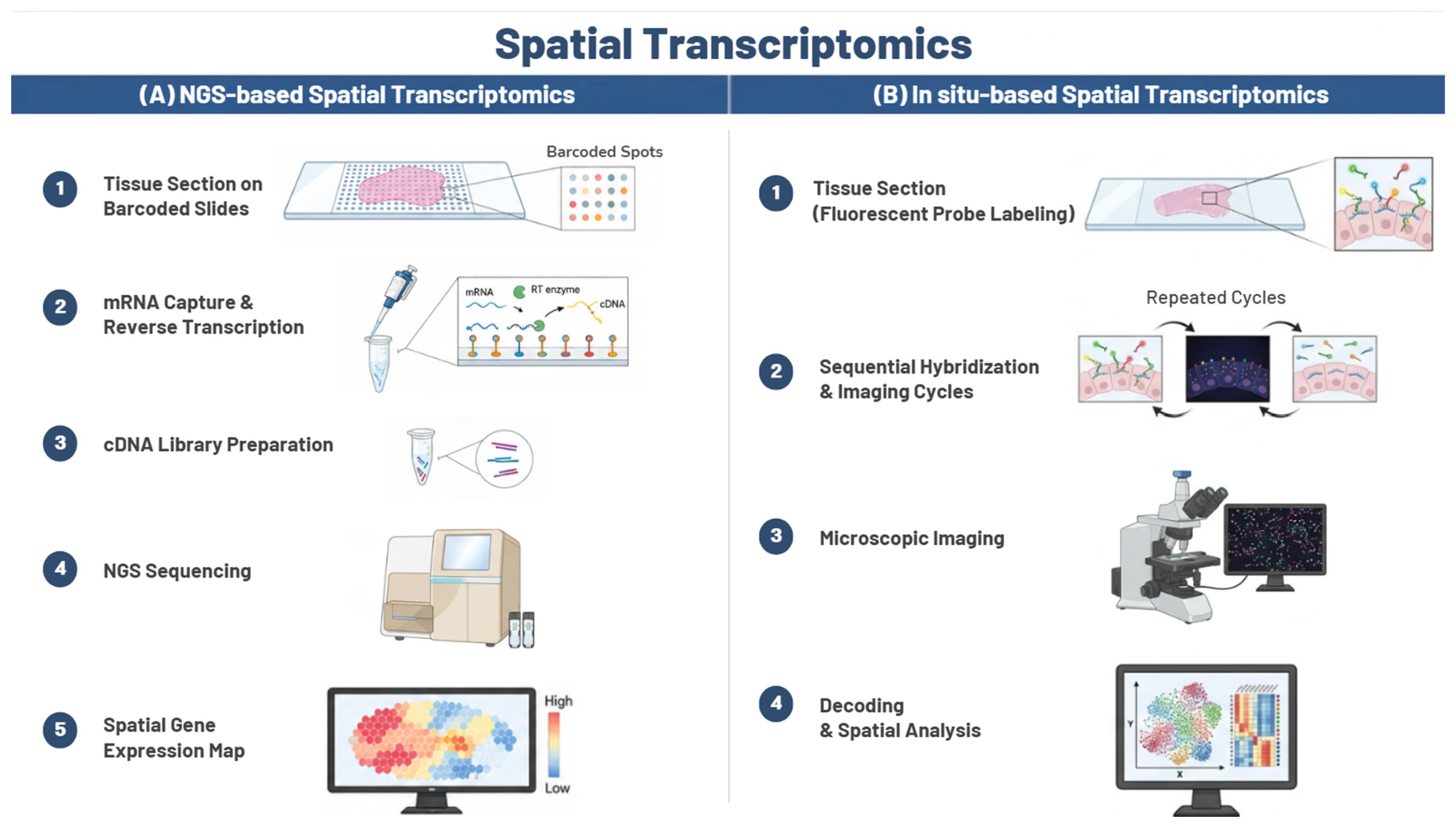
2. Spatial Transcriptomics Technologies
2.1. Overview and Classification
2.2. NGS-Based Platforms
2.3. In Situ-Based Platforms
2.4. Computational Analysis of Spatial Transcriptomic Data
3. Spatial Transcriptomics of the Aging and Diseased Brain
3.1. Brain Aging
3.2. Neurodegenerative Disease
3.3. Glioblastoma
3.4. Multiple Sclerosis
4. Brain Spatial Transcriptomics Data Resources and Current Challenges
5. Conclusions
Author Contributions
Funding
Institutional Review Board Statement
Informed Consent Statement
Data Availability Statement
Conflicts of Interest
Abbreviations
References
- Stark, R.; Grzelak, M.; Hadfield, J. RNA sequencing: The teenage years. Nat. Rev. Genet. 2019, 20, 631–656. [Google Scholar] [CrossRef] [PubMed]
- Longo, S.K.; Guo, M.G.; Ji, A.L.; Khavari, P.A. Integrating single-cell and spatial transcriptomics to elucidate intercellular tissue dynamics. Nat. Rev. Genet. 2021, 22, 627–644. [Google Scholar] [CrossRef] [PubMed]
- Marx, V. Method of the Year: Spatially resolved transcriptomics. Nat. Methods 2021, 18, 9–14, Correction in Nat. Methods 2021, 18, 219. [Google Scholar] [CrossRef] [PubMed]
- Rodriques, S.G.; Stickels, R.R.; Goeva, A.; Martin, C.A.; Murray, E.; Vanderburg, C.R.; Welch, J.; Chen, L.M.; Chen, F.; Macosko, E.Z. Slide-seq: A scalable technology for measuring genome-wide expression at high spatial resolution. Science 2019, 363, 1463–1467. [Google Scholar] [CrossRef] [PubMed]
- Chen, K.H.; Boettiger, A.N.; Moffitt, J.R.; Wang, S.; Zhuang, X. Spatially resolved, highly multiplexed RNA profiling in single cells. Science 2015, 348, aaa6090. [Google Scholar] [CrossRef] [PubMed]
- Rao, A.; Barkley, D.; França, G.S.; Yanai, I. Exploring tissue architecture using spatial transcriptomics. Nature 2021, 596, 211–220. [Google Scholar] [CrossRef] [PubMed]
- Moses, L.; Pachter, L. Museum of spatial transcriptomics. Nat. Methods 2022, 19, 534–546. [Google Scholar] [CrossRef] [PubMed]
- Park, H.E.; Jo, S.H.; Lee, R.H.; Macks, C.P.; Ku, T.; Park, J.; Lee, C.W.; Hur, J.K.; Sohn, C.H. Spatial transcriptomics: Technical aspects of recent developments and their applications in neuroscience and cancer research. Adv. Sci. 2023, 10, 2206939. [Google Scholar] [CrossRef]
- Fu, H.; Hardy, J.; Duff, K.E. Selective vulnerability in neurodegenerative diseases. Nat. Neurosci. 2018, 21, 1350–1358. [Google Scholar] [CrossRef] [PubMed]
- Williams, C.G.; Lee, H.J.; Asatsuma, T.; Vento-Tormo, R.; Haque, A. An introduction to spatial transcriptomics for biomedical research. Genome Med. 2022, 14, 68. [Google Scholar] [CrossRef] [PubMed]
- Ståhl, P.L.; Salmén, F.; Vickovic, S.; Lundmark, A.; Navarro, J.F.; Magnusson, J.; Giacomello, S.; Asp, M.; Westholm, J.O.; Huss, M.; et al. Visualization and analysis of gene expression in tissue sections by spatial transcriptomics. Science 2016, 353, 78–82. [Google Scholar] [CrossRef] [PubMed]
- Asp, M.; Bergenstråhle, J.; Lundeberg, J. Spatially resolved transcriptomes—Next generation tools for tissue exploration. BioEssays 2020, 42, 1900221. [Google Scholar] [CrossRef]
- Chen, A.; Liao, S.; Cheng, M.; Ma, K.; Wu, L.; Lai, Y.; Qiu, X.; Yang, J.; Xu, J.; Hao, S.; et al. Spatiotemporal transcriptomic atlas of mouse organogenesis using DNA nanoball-patterned arrays. Cell 2022, 185, 1777–1792. [Google Scholar] [CrossRef] [PubMed]
- Zhao, Y.; Li, Y.; He, Y.; Wu, J.; Liu, Y.; Li, X.; Li, Z.; Yuan, Q.; Li, J.; Zhang, X.; et al. Stereo-seq V2: Spatial mapping of total RNA on FFPE sections with high resolution. Cell 2025, 188, 6554–6571. [Google Scholar] [CrossRef] [PubMed]
- Stickels, R.R.; Murray, E.; Kumar, P.; Li, J.; Marshall, J.L.; Di Bella, D.J.; Arlotta, P.; Macosko, E.Z.; Chen, F. Highly sensitive spatial transcriptomics at near-cellular resolution with Slide-seqV2. Nat. Biotechnol. 2021, 39, 313–319. [Google Scholar] [PubMed]
- Moffitt, J.R.; Zhuang, X. RNA imaging with multiplexed error-robust fluorescence in situ hybridization (MERFISH). Methods Enzymol. 2016, 572, 1–49. [Google Scholar] [CrossRef] [PubMed]
- Eng, C.H.L.; Lawson, M.; Zhu, Q.; Dries, R.; Koulena, N.; Takei, Y.; Yun, J.; Cronin, C.; Karp, C.; Yuan, G.C.; et al. Transcriptome-scale super-resolved imaging in tissues by RNA seqFISH+. Nature 2019, 568, 235–239. [Google Scholar] [CrossRef] [PubMed]
- Janesick, A.; Shelansky, R.; Gottscho, A.D.; Wagner, F.; Williams, S.R.; Rouault, M.; Beliakoff, G.; Morrison, C.A.; Oliveira, M.F.; Sicherman, J.T.; et al. High-resolution mapping of the tumor microenvironment using integrated single-cell, spatial and in situ analysis. Nat. Commun. 2023, 14, 8353. [Google Scholar] [CrossRef] [PubMed]
- Ren, P.; Zhang, R.; Wang, Y.; Zhang, P.; Luo, C.; Wang, S.; Li, X.; Zhang, Z.; Zhao, Y.; He, Y.; et al. Systematic benchmarking of high-throughput subcellular spatial transcriptomics platforms across human tumors. Nat. Commun. 2025, 16, 9232. [Google Scholar] [CrossRef] [PubMed]
- 10x Genomics. Visium Spatial Gene Expression User Guide; 10x Genomics: Pleasanton, CA, USA, 2020; Available online: https://www.10xgenomics.com (accessed on 1 June 2026).
- Moncada, R.; Barkley, D.; Wagner, F.; Chiodin, M.; Devlin, J.C.; Baron, M.; Hajdu, C.H.; Simeone, D.M.; Yanai, I. Integrating microarray-based spatial transcriptomics and single-cell RNA-seq reveals tissue architecture in pancreatic ductal adenocarcinomas. Nat. Biotechnol. 2020, 38, 333–342, Correction in Nat. Biotechnol. 2020, 38, 1476. [Google Scholar] [CrossRef] [PubMed]
- Lim, H.J.; Wang, Y.; Buzdin, A.; Li, X. A practical guide for choosing an optimal spatial transcriptomics technology from seven major commercially available options. BMC Genom. 2025, 26, 47. [Google Scholar] [CrossRef]
- Villacampa, E.G.; Larsson, L.; Mirzazadeh, R.; Kvastad, L.; Andersson, A.; Mollbrink, A.; Kokaraki, G.; Monteil, V.; Schultz, N.; Appelberg, K.S.; et al. Genome-wide spatial expression profiling in formalin-fixed tissues. Cell Genom. 2021, 1, 100065. [Google Scholar] [CrossRef]
- Marshall, J.L.; Noel, T.; Wang, Q.S.; Chen, H.; Murray, E.; Subramanian, A.; Vernon, K.A.; Bazua-Valenti, S.; Liguori, K.; Keller, K.; et al. High-resolution Slide-seqV2 spatial transcriptomics enables discovery of disease-specific cell neighborhoods and pathways. iScience 2022, 25, 104097. [Google Scholar] [CrossRef] [PubMed]
- Moffitt, J.R.; Hao, J.; Wang, G.; Chen, K.H.; Babcock, H.P.; Zhuang, X. High-throughput single-cell gene-expression profiling with multiplexed error-robust fluorescence in situ hybridization. Proc. Natl. Acad. Sci. USA 2016, 113, 11046–11051. [Google Scholar] [CrossRef] [PubMed]
- Moffitt, J.R.; Bambah-Mukku, D.; Eichhorn, S.W.; Vaughn, E.; Shekhar, K.; Perez, J.D.; Rubinstein, N.D.; Hao, J.; Regev, A.; Dulac, C.; et al. Molecular, spatial, and functional single-cell profiling of the hypothalamic preoptic region. Science 2018, 362, eaau5324. [Google Scholar] [CrossRef] [PubMed]
- Lee, J.H.; Daugharthy, E.R.; Scheiman, J.; Kalhor, R.; Yang, J.L.; Ferrante, T.C.; Terry, R.; Jeanty, S.S.F.; Li, C.; Amamoto, R.; et al. Highly multiplexed subcellular RNA sequencing in situ. Science 2014, 343, 1360–1363. [Google Scholar] [CrossRef] [PubMed]
- Crosetto, N.; Bienko, M.; van Oudenaarden, A. Spatially resolved transcriptomics and beyond. Nat. Rev. Genet. 2015, 16, 57–66. [Google Scholar] [PubMed]
- Lubeck, E.; Coskun, A.F.; Zhiyentayev, T.; Ahmad, M.; Cai, L. Single-cell in situ RNA profiling by sequential hybridization. Nat. Methods 2014, 11, 360–361. [Google Scholar] [CrossRef] [PubMed]
- Zhu, Q.; Shah, S.; Dries, R.; Cai, L.; Yuan, G.C. Identification of spatially associated subpopulations by combining scRNAseq and sequential fluorescence in situ hybridization data. Nat. Biotechnol. 2018, 36, 1183–1190. [Google Scholar] [CrossRef]
- Wang, H.; Huang, R.; Nelson, J.; Gao, C.; Tran, M.; Yeaton, A.; Krishna, S.; Felt, K.; Pfaff, K.L.; Bowman, T.; et al. Systematic benchmarking of imaging spatial transcriptomics platforms in FFPE tissues. Nat. Commun. 2025, 16, 10215. [Google Scholar] [CrossRef] [PubMed]
- He, S.; Bhatt, R.; Brown, C.; Brown, E.A.; Buhr, C.A.; Chantranuvatana, K.; Danaher, P.; Dunaway, D.; Garrison, R.G.; Geiss, G.; et al. High-plex imaging of RNA and proteins at subcellular resolution in fixed tissue by spatial molecular imaging. Nat. Biotechnol. 2022, 40, 1794–1806. [Google Scholar] [CrossRef] [PubMed]
- Merritt, C.R.; Ong, G.T.; Church, S.E.; Barker, K.; Danaher, P.; Geiss, G.; Hoang, M.; Jung, J.; Liang, Y.; McKay-Fleisch, J.; et al. Multiplex digital spatial profiling of proteins and RNA in fixed tissue. Nat. Biotechnol. 2020, 38, 586–599. [Google Scholar] [CrossRef] [PubMed]
- Kleshchevnikov, V.; Shmatko, A.; Dann, E.; Aivazidis, A.; King, H.W.; Li, T.; Elmentaite, R.; Lomakin, A.; Kedlian, V.; Gayoso, A.; et al. Cell2location maps fine-grained cell types in spatial transcriptomics. Nat. Biotechnol. 2022, 40, 661–671. [Google Scholar] [CrossRef] [PubMed]
- Cable, D.M.; Murray, E.; Zou, L.S.; Goeva, A.; Macosko, E.Z.; Chen, F.; Irizarry, R.A. Robust decomposition of cell type mixtures in spatial transcriptomics. Nat. Biotechnol. 2022, 40, 517–526. [Google Scholar] [PubMed]
- Elosua-Bayes, M.; Nieto, P.; Mereu, E.; Gut, I.; Heyn, H. SPOTlight: Seeded NMF regression to deconvolute spatial transcriptomics spots with single-cell transcriptomes. Nucleic Acids Res. 2021, 49, e50. [Google Scholar] [CrossRef] [PubMed]
- Biancalani, T.; Scalia, G.; Buffoni, L.; Avasthi, R.; Lu, Z.; Sanger, A.; Tokcan, N.; Vanderburg, C.R.; Segerstolpe, Å.; Zhang, M.; et al. Deep learning and alignment of spatially resolved single-cell transcriptomes with Tangram. Nat. Methods 2021, 18, 1352–1362. [Google Scholar] [CrossRef] [PubMed]
- Zhao, E.; Stone, M.R.; Ren, X.; Guenthoer, J.; Smythe, K.S.; Pulliam, T.; Williams, S.R.; Uytingco, C.R.; Taylor, S.E.B.; Nghiem, P.; et al. Spatial transcriptomics at subspot resolution with BayesSpace. Nat. Biotechnol. 2021, 39, 1375–1384. [Google Scholar] [CrossRef] [PubMed]
- Hu, J.; Li, X.; Coleman, K.; Schroeder, A.; Ma, N.; Irwin, D.J.; Lee, E.B.; Shinohara, R.T.; Li, M. SpaGCN: Integrating gene expression, spatial location and histology to identify spatial domains and spatially variable genes by graph convolutional network. Nat. Methods 2021, 18, 1342–1351. [Google Scholar] [CrossRef] [PubMed]
- Jin, S.; Guerrero-Juarez, C.F.; Zhang, L.; Chang, I.; Ramos, R.; Kuan, C.H.; Myung, P.; Plikus, M.V.; Nie, Q. Inference and analysis of cell-cell communication using CellChat. Nat. Commun. 2021, 12, 1088. [Google Scholar] [CrossRef] [PubMed]
- Efremova, M.; Vento-Tormo, M.; Teichmann, S.A.; Vento-Tormo, R. CellPhoneDB: Inferring cell-cell communication from combined expression of multi-subunit ligand-receptor complexes. Nat. Protoc. 2020, 15, 1484–1506. [Google Scholar] [CrossRef] [PubMed]
- Jiang, M.; Cai, S.; Zhang, K.; Li, M.; Tian, Y.; Wang, T.; Alarcón Rodríguez, R.; Wang, Z.; Wu, S.; He, J.; et al. Deep learning in medical image analysis for bone tumor: A comprehensive survey. Inf. Fusion 2026, 133, 104305. [Google Scholar] [CrossRef]
- Liang, X.; Lai, G.; Yu, J.; Lin, T.; Wang, C.; Wang, W. Herbal ingredient-target interaction prediction via multi-modal learning. Inf. Sci. 2025, 711, 122115. [Google Scholar] [CrossRef]
- Safaiyan, S.; Kannaiyan, N.; Snaidero, N.; Brioschi, S.; Biber, K.; Yona, S.; Edinger, A.L.; Jung, S.; Rossner, M.J.; Simons, M. Age-related myelin degradation burdens the clearance function of microglia during aging. Nat. Neurosci. 2016, 19, 995–998. [Google Scholar] [CrossRef] [PubMed]
- Clarke, L.E.; Liddelow, S.A.; Chakraborty, C.; Münch, A.E.; Heiman, M.; Barres, B.A. Normal aging induces A1-like astrocyte reactivity. Proc. Natl. Acad. Sci. USA 2018, 115, E1896–E1905. [Google Scholar] [CrossRef] [PubMed]
- Wang, L.; Cui, C.Y.; Lee, C.T.; Bodogai, M.; Yang, N.; Shi, C.; Irfanoglu, M.O.; Occean, J.R.; Afrin, S.; Sarker, N.; et al. Spatial transcriptomics of the aging mouse brain reveals origins of inflammation in the white matter. Nat. Commun. 2025, 16, 3231. [Google Scholar] [CrossRef] [PubMed]
- Deng, H.W.; Gong, Y.; Zhang, Q.L.; Wu, D.; Liu, A.; Li, T.; Xiao, Z.; Li, Y.; Haeri, M.; Swerdlow, R.; et al. Novel pathological mechanisms revealed by spatial transcriptomic analysis of hippocampus in aged control, primary age-related tauopathy, and Alzheimer’s disease. Res. Sq. 2025; preprint. [CrossRef] [PubMed]
- Garg, A.; Vo, S.; De Souza, I.D.; Castro Brant, A.; Brase, L.; Aladyeva, E.; Albanus, R.D.O.; Nallapu, A.; Fu, H.; Harari, O.; et al. Exploring cellular heterogeneity: Single-cell and spatial transcriptomics of Alzheimer’s disease brains and iPSC-derived microglia. Alzheimer’s Res. Ther. 2025, 18, 22. [Google Scholar] [CrossRef] [PubMed]
- Surmeier, D.J.; Obeso, J.A.; Halliday, G.M. Selective neuronal vulnerability in Parkinson disease. Nat. Rev. Neurosci. 2017, 18, 101–113. [Google Scholar] [CrossRef] [PubMed]
- Tansey, M.G.; Wallings, R.L.; Houser, M.C.; Herrick, M.K.; Keating, C.E.; Joers, V. Inflammation and immune dysfunction in Parkinson disease. Nat. Rev. Immunol. 2022, 22, 657–673. [Google Scholar] [CrossRef] [PubMed]
- Liu, M.; Ji, Z.; Jain, V.; Smith, V.L.; Hocke, E.; Patel, A.P.; McLendon, R.E.; Ashley, D.M.; Gregory, S.G.; López, G.Y. Spatial transcriptomics reveals segregation of tumor cell states in glioblastoma and marked immunosuppression within the perinecrotic niche. Acta Neuropathol. Commun. 2024, 12, 64. [Google Scholar] [CrossRef] [PubMed]
- Neftel, C.; Laffy, J.; Filbin, M.G.; Hara, T.; Shore, M.E.; Rahme, G.J.; Richman, A.R.; Silverbush, D.; Shaw, M.L.; Hebert, C.M.; et al. An integrative model of cellular states, plasticity, and genetics for glioblastoma. Cell 2019, 178, 835–849. [Google Scholar] [CrossRef] [PubMed]
- Ren, Y.; Huang, Z.; Zhou, L.; Xiao, P.; Song, J.; He, P.; Xie, C.; Zhou, R.; Li, M.; Dong, X. Spatial transcriptomics reveals niche-specific enrichment and vulnerabilities of radial glial stem-like cells in malignant gliomas. Nat. Commun. 2023, 14, 1028. [Google Scholar] [CrossRef] [PubMed]
- Sattiraju, A.; Kang, S.; Giotti, B.; Chen, Z.; Marallano, V.J.; Brusco, C.; Ramakrishnan, A.; Shen, L.; Tsankov, A.M.; Hambardzumyan, D.; et al. Hypoxic niches attract and sequester tumor-associated macrophages and cytotoxic T cells and reprogram them for immunosuppression. Immunity 2023, 56, 1825–1843. [Google Scholar] [CrossRef] [PubMed]
- Solomou, G.; Young, A.M.; Bulstrode, H.J. Microglia and macrophages in glioblastoma: Landscapes and treatment directions. Mol. Oncol. 2024, 18, 2906–2926. [Google Scholar] [CrossRef] [PubMed]
- Zheng, Y.; Fuse, H.; Alzoubi, I.; Graeber, M.B. Microglia-derived brain macrophages associate with glioblastoma stem cells: A potential mechanism for tumor progression revealed by AI-assisted analysis. Cells 2025, 14, 413. [Google Scholar] [CrossRef] [PubMed]
- Pai, B.; Ramos, S.I.; Cheng, W.S.; Joshi, T.; Özen, E.; Kulumani Mahadevan, L.S.; Silva-Hurtado, T.J.; Price, G.A.; Tome-Garcia, J.; Nudelman, G.; et al. Spatial multi-omics defines a shared tumor-infiltrative signature at the resection margin in high-grade gliomas. Cancer Res. 2025, 85, 4233–4250. [Google Scholar] [CrossRef] [PubMed]
- Kukanja, P.; Langseth, C.M.; Rubio Rodríguez-Kirby, L.A.; Agirre, E.; Zheng, C.; Raman, A.; Yokota, C.; Avenel, C.; Tiklová, K.; Guerreiro-Cacais, A.O.; et al. Cellular architecture of evolving neuroinflammatory lesions and multiple sclerosis pathology. Cell 2024, 187, 1990–2009.e19. [Google Scholar] [CrossRef] [PubMed]
- Alsema, A.M.; Wijering, M.H.C.; Miedema, A.; Kotah, J.M.; Koster, M.; Rijnsburger, M.; van Weering, H.R.J.; de Vries, H.E.; Baron, W.; Kooistra, S.M.; et al. Spatially resolved gene signatures of white matter lesion progression in multiple sclerosis. Nat. Neurosci. 2024, 27, 2341–2353. [Google Scholar] [CrossRef] [PubMed]
- Liddelow, S.A.; Guttenplan, K.A.; Clarke, L.E.; Bennett, F.C.; Bohlen, C.J.; Schirmer, L.; Bennett, M.L.; Münch, A.E.; Chung, W.S.; Peterson, T.C.; et al. Neurotoxic reactive astrocytes are induced by activated microglia. Nature 2017, 541, 481–487. [Google Scholar] [CrossRef] [PubMed]
- Keren-Shaul, H.; Spinrad, A.; Weiner, A.; Matcovitch-Natan, O.; Dvir-Szternfeld, R.; Ulland, T.K.; David, E.; Baruch, K.; Lara-Astaiso, D.; Toth, B.; et al. A unique microglia type associated with restricting development of Alzheimer’s disease. Cell 2017, 169, 1276–1290. [Google Scholar] [CrossRef] [PubMed]
- Greenwald, A.C.; Darnell, N.G.; Hoefflin, R.; Simkin, D.; Mount, C.W.; Castro, L.N.G.; Harnik, Y.; Dumont, S.; Hirsch, D.; Nomura, M.; et al. Integrative spatial analysis reveals a multi-layered organization of glioblastoma. Cell 2024, 187, 2485–2501. [Google Scholar] [CrossRef] [PubMed]
- Maynard, K.R.; Collado-Torres, L.; Weber, L.M.; Uytingco, C.; Barry, B.K.; Williams, S.R.; Catallini, J.L.; Tran, M.N.; Besich, Z.; Tippani, M.; et al. Transcriptome-scale spatial gene expression in the human dorsolateral prefrontal cortex. Nat. Neurosci. 2021, 24, 425–436, Correction in Nat. Neurosci. 2021, 24, 612. [Google Scholar] [CrossRef] [PubMed]
- 10x Genomics. Visium Spatial Gene Expression; 10x Genomics: Pleasanton, CA, USA, 2019; Available online: https://www.10xgenomics.com/platforms/visium (accessed on 1 June 2026).
- Yao, Z.; Van Velthoven, C.T.; Kunst, M.; Zhang, M.; McMillen, D.; Lee, C.; Jung, W.; Goldy, J.; Abdelhak, A.; Aitken, M.; et al. A high-resolution transcriptomic and spatial atlas of cell types in the whole mouse brain. Nature 2023, 624, 317–332. [Google Scholar] [CrossRef] [PubMed]
- Zhang, M.; Eichhorn, S.W.; Zingg, B.; Yao, Z.; Cotter, K.; Zeng, H.; Dong, H.; Zhuang, X. Spatially resolved cell atlas of the mouse primary motor cortex by MERFISH. Nature 2021, 598, 137–143. [Google Scholar] [CrossRef] [PubMed]
- Zhang, M.; Pan, X.; Jung, W.; Halpern, A.R.; Eichhorn, S.W.; Lei, Z.; Cohen, L.; Smith, K.A.; Tasic, B.; Yao, Z.; et al. Molecularly defined and spatially resolved cell atlas of the whole mouse brain. Nature 2023, 624, 343–354. [Google Scholar] [CrossRef] [PubMed]
- Allen Institute for Brain Science. Allen Brain Cell Atlas—Data Access. 2024. Available online: https://alleninstitute.github.io/abc_atlas_access/intro.html (accessed on 1 June 2026).
- Tarhan, L.; Bistline, J.; Chang, J.; Galloway, B.; Hanna, E.; Weitz, E. Single Cell Portal: An interactive home for single-cell genomics data. bioRxiv 2023. [Google Scholar] [CrossRef] [PubMed]
- Langlieb, J.; Sachdev, N.S.; Balderrama, K.S.; Nadaf, N.M.; Raj, M.; Murray, E.; Webber, J.T.; Vanderburg, C.; Gazestani, V.; Tward, D.; et al. The molecular cytoarchitecture of the adult mouse brain. Nature 2023, 624, 333–342. [Google Scholar] [CrossRef] [PubMed]
- Goltsev, Y.; Samusik, N.; Kennedy-Darling, J.; Bhate, S.; Hale, M.; Vazquez, G.; Black, S.; Nolan, G.P. Deep profiling of mouse splenic architecture with CODEX multiplexed imaging. Cell 2018, 174, 968–981. [Google Scholar] [CrossRef] [PubMed]
- Zhang, D.; Deng, Y.; Kukanja, P.; Agirre, E.; Bartosovic, M.; Dong, M.; Ma, C.; Ma, S.; Su, G.; Bao, S.; et al. Spatial epigenome–transcriptome co-profiling of mammalian tissues. Nature 2023, 616, 113–122. [Google Scholar] [CrossRef] [PubMed]
- Vicari, M.; Mirzazadeh, R.; Nilsson, A.; Shariatgorji, R.; Bjärterot, P.; Larsson, L.; Lee, H.; Nilsson, M.; Foyer, J.; Ekvall, M.; et al. Spatial multimodal analysis of transcriptomes and metabolomes in tissues. Nat. Biotechnol. 2024, 42, 1046–1050. [Google Scholar] [PubMed]
- Chen, W.T.; Lu, A.; Craessaerts, K.; Pavie, B.; Sala Frigerio, C.; Corthout, N.; Qian, X.; Laláková, J.; Kühnemund, M.; Voytyuk, I.; et al. Spatial transcriptomics and in situ sequencing to study Alzheimer’s disease. Cell 2020, 182, 976–991. [Google Scholar] [CrossRef] [PubMed]


| Platform | Approach | Spatial Resolution | Transcriptomic Coverage | Tissue | Throughput/Multiplexing | Relative Complexity & Cost | Representative Applications |
|---|---|---|---|---|---|---|---|
| Visium [11,12] | NGS-based | ~55 µm (spot) | Whole transcriptome (unbiased) | FF, FFPE | n/a (genome-wide) | Low–moderate; standardized | Tumor, brain, developmental biology |
| Stereo-Seq [13,14] | NGS-based | ~220 nm (subcellular) | Whole transcriptome | FF, FFPE (V2) | n/a (genome-wide) | High (large data, heavy compute) | Development, neural tissue, cell–cell interaction |
| Slide-Seq (V2) [4,15] | NGS-based | ~10 µm (near single-cell) | Whole transcriptome | FF | n/a (genome-wide) | Moderate (bead decoding) | Near-cellular profiling across tissues |
| MERFISH [5,16] | In situ | ~100 nm (single-molecule) | Targeted (100s–~10,000 genes) | FF, FFPE | High | High (multi-cycle imaging) | Cell-type mapping, intercellular organization |
| seqFISH+ [17] | In situ | ~100 nm (subcellular) | Targeted (up to ~10,000 genes) | FF | Very high | High (multi-cycle, specialized imaging) | Cell-type discrimination, tissue architecture |
| Xenium [18,19] | In situ | ~200 nm (single-cell/subcellular) | Targeted (100s–~5000 genes) | FF, FFPE | High | Moderate–high; clinically oriented | Tumor microenvironment, neural, clinical |
| Condition | Brain Region/Niche | Key | Primary Evidence (Model/Tissue) | Ref(s). |
|---|---|---|---|---|
| Brain aging | Subcortical white matter, corpus callosum | Region- and glia-selective inflammatory remodeling; white matter as an immune/inflammation hotspot; non-neuronal (astrocyte/microglia/oligodendrocyte) reorganization | Mouse brain (spatial); mouse mechanistic support | [44,45,46] |
| Alzheimer’s disease | Amyloid-plaque microenvironment (hippocampus, cortex) | Plaque-associated niche with spatially concentrated astrocyte/microglial inflammatory states; shared pathological transcriptional state near lesions | Human post-mortem brain (incl. preprint) | [47,48] |
| Parkinson’s disease | Substantia nigra | Spatially restricted, selective loss of dopaminergic neurons shaped by the local microenvironment | Review-based; direct human ST evidence limited | [49,50] |
| GBM tumor cell states | Tumor core vs. invasive neural niche | Four spatially segregated states (NPC-/OPC-/AC-/MES-like); OPC-like enriched in core, radial-glial stem-like in invasive niche | Human tumor tissue (spatial + scRNA-Seq) | [51,52,53] |
| GBM microenvironment | Perinecrotic & perivascular niches | Hypoxia-driven immunosuppression; TAM reprogramming via CCL8/IL1B; microglia at margin, monocyte-derived macrophages in core | Human tumor; supporting mouse models | [54,55,56] |
| GBM resection margin | Surgical resection margin | Margin-specific infiltrative signature including EGFR, linked to invasion and recurrence | Human tumor tissue (spatial multi-omics) | [57] |
| Multiple sclerosis | White-matter lesions; lesion rim | Stage-specific lesion architecture; disease-associated glia and immune states at the active-lesion rim; trajectories from normal-appearing white matter to active/mixed lesions | Human post-mortem brain + mouse EAE (spatial) | [58,59] |
| Condition | Cell Type/Niche | Representative Molecular Signature | Primary Evidence (Model/Tissue) | Ref(s). |
|---|---|---|---|---|
| Brain aging | A1-like reactive astrocytes | Complement induction (C3, Serping1); loss of homeostatic support | Mouse/in vitro | [45,60] |
| Brain aging | Aged microglia (white matter) | Activated, myelin-clearance-associated states | Mouse | [44,60] |
| Alzheimer’s disease | Disease-associated microglia (plaque niche) | Trem2, Apoe, Cst7, Itgax; loss of homeostatic genes | Mouse model (5xFAD); conserved in human | [61] |
| Parkinson’s disease | Vulnerable dopaminergic neurons (substantia nigra) | α-synuclein (SNCA) pathology; oxidative/metabolic stress programs | Review-based; human/model synthesis | [49,50] |
| Glioblastoma | Four tumor-cell states | SOX2/DLL3 (NPC), OLIG1/PDGFRA (OPC), GFAP/AQP4 (AC), CD44/VIM (MES) | Human tumor (scRNA-Seq–derived) | [52] |
| Glioblastoma | Perinecrotic niche/TAMs | Hypoxia and NF-κB signaling; CCL8, IL1B–driven TAM reprogramming | Human tumor; supporting mouse models | [54] |
| Glioblastoma | Resection margin | EGFR and a shared glioma-infiltrative signature | Human tumor tissue | [57] |
| Multiple sclerosis | Lesion-rim glia (active lesions) | Disease-associated microglia/astrocyte and immune-cell programs marking active-lesion rims; white-matter lesion-progression signatures | Human post-mortem brain + mouse EAE | [58,59] |
Disclaimer/Publisher’s Note: The statements, opinions and data contained in all publications are solely those of the individual author(s) and contributor(s) and not of MDPI and/or the editor(s). MDPI and/or the editor(s) disclaim responsibility for any injury to people or property resulting from any ideas, methods, instructions or products referred to in the content. |
© 2026 by the authors. Licensee MDPI, Basel, Switzerland. This article is an open access article distributed under the terms and conditions of the Creative Commons Attribution (CC BY) license.
Share and Cite
Cha, S.; Kim, J.; Kim, J.; Kim, D.; Song, H.; Lim, K.S.; Chae, S. Spatial Transcriptomics for Dissecting Cellular and Molecular Heterogeneity in the Aging and Diseased Brain. Int. J. Mol. Sci. 2026, 27, 6149. https://doi.org/10.3390/ijms27146149
Cha S, Kim J, Kim J, Kim D, Song H, Lim KS, Chae S. Spatial Transcriptomics for Dissecting Cellular and Molecular Heterogeneity in the Aging and Diseased Brain. International Journal of Molecular Sciences. 2026; 27(14):6149. https://doi.org/10.3390/ijms27146149
Chicago/Turabian StyleCha, Seeun, Jin Kim, Jisan Kim, Doa Kim, Hyunwoo Song, Kwang Suk Lim, and Sehyun Chae. 2026. "Spatial Transcriptomics for Dissecting Cellular and Molecular Heterogeneity in the Aging and Diseased Brain" International Journal of Molecular Sciences 27, no. 14: 6149. https://doi.org/10.3390/ijms27146149
APA StyleCha, S., Kim, J., Kim, J., Kim, D., Song, H., Lim, K. S., & Chae, S. (2026). Spatial Transcriptomics for Dissecting Cellular and Molecular Heterogeneity in the Aging and Diseased Brain. International Journal of Molecular Sciences, 27(14), 6149. https://doi.org/10.3390/ijms27146149