Comprehensive Review of the Interplay of MicroRNA and Epithelial–Mesenchymal Transition in Radiation Resistance of Cancer


Abstract
1. Introduction
2. Mechanisms of Tumor Radioresistance
3. EMT as a Driver of Radioresistance
3.1. Molecular and Cellular Hallmarks of EMT
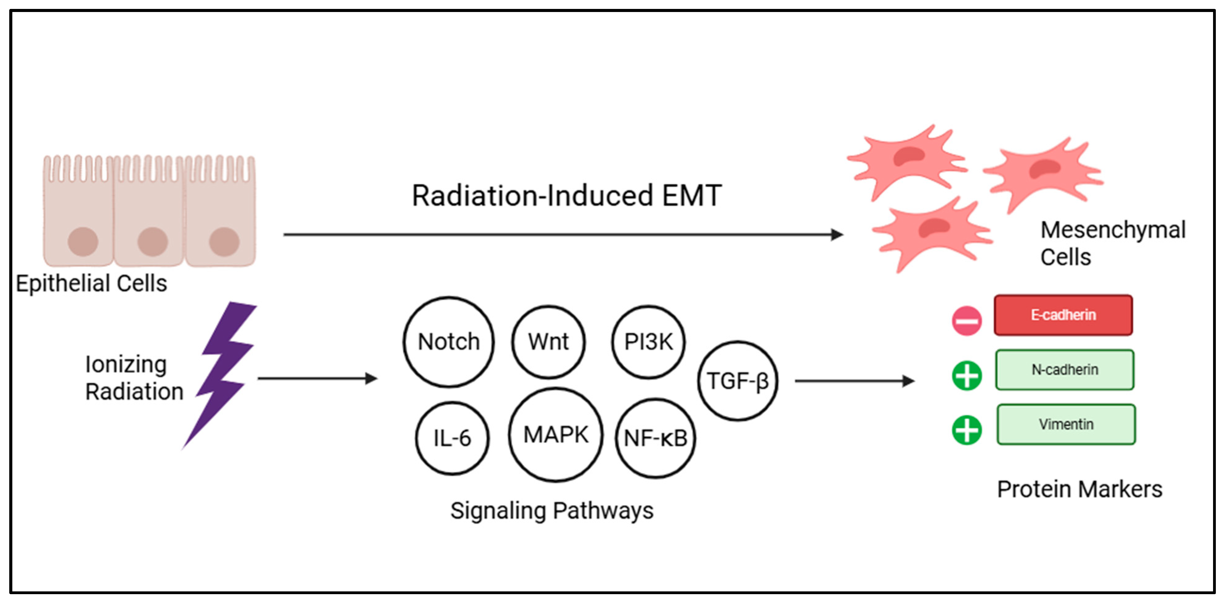
- Loss of Epithelial Markers: A defining hallmark of EMT is the downregulation or complete loss of key cell–cell adhesion molecules, most notably E-cadherin. This loss of E-cadherin is highly implicated in carcinoma progression, as it allows tumor cells to detach from the primary tumor mass and become more migratory and invasive [30,31]. Concurrently, there is a downregulation of other epithelial proteins such as cytokeratin and claudin.
- Gain of Mesenchymal Markers: Simultaneously, cells undergoing EMT upregulate the expression of mesenchymal proteins, including N-cadherin, Vimentin, and Fibronectin. The shift from E-cadherin to N-cadherin (cadherin switch) is a common feature of EMT, promoting cell motility and invasiveness [30,32].
- Cytoskeletal Rearrangement: The internal actin cytoskeleton undergoes significant reorganization, which facilitates the increased cell motility and invasive capabilities characteristic of mesenchymal cells.
3.2. The Central Role of EMT in Acquiring Radioresistance
- Increased Drug Efflux: A frequently discussed mechanism of drug resistance, which can overlap with radioresistance, is the excessive efflux of therapeutic agents from cells. EMT cells are known to frequently overexpress ATP-binding cassette (ABC) transporters, which are membrane proteins responsible for actively pumping drugs out of the cell. Critically, the promoters of these ABC transporters contain binding sites for EMT-TFs, establishing a direct molecular link between the EMT program and enhanced drug efflux, thereby contributing to multidrug resistance [38,39,40].
- Acquisition of Cancer Stem Cell (CSC) Properties: EMT is a key driver in the acquisition of stem cell properties by cancer cells, leading to the generation of tumor-initiating CSCs [21,22,23,24,41]. These CSCs are inherently more resistant to radiation therapy, possess enhanced DNA repair capabilities, and are a significant cause of tumor recurrence and metastasis following treatment. The close association between EMT and stemness means that EMT can drive radioresistance by promoting the transition of non-CSCs to a CSC-like phenotype.
- Modulation of DNA Damage Repair and Cell Cycle: EMT can influence cellular processes critical for cell survival following radiation exposure, including the efficiency of DNA damage repair and alterations in cell cycle progression. For example, EMT-TFs can affect the expression of genes involved in DNA repair pathways [17].
3.3. Radiation-Induced EMT: Molecular Events and Cellular Changes
4. The Role of MicroRNAs in Radiation-Induced EMT and Radioresistance
4.1. MicroRNA Biogenesis and Mechanisms of Gene Regulation
4.2. MiRNAs Modulating EMT-Related Pathways in Radioresistance
4.3. MiRNAs Directly Influencing Cellular Radiosensitivity
4.3.1. Upstream Regulation by Radiation and Signaling
4.3.2. Downstream Modulation of DNA Damage Response
- Regulation of Cell Cycle, Apoptosis, and Signaling: Beyond DNA repair, miRNAs dictate cell fate by modulating apoptosis and pro-survival signaling pathways.
- Apoptotic Regulators: The impact of miRNAs on apoptosis is often context dependent. For example, miR-181a can sensitize malignant glioma cells by targeting the anti-apoptotic protein Bcl-2 [71]. However, in cervical cancer, its upregulation inhibits radiation-induced apoptosis, conferring resistance [16,18]. Conversely, miR-25 and miR-29 facilitate apoptosis by targeting BIM and MCL1, respectively [5,16,72,73].
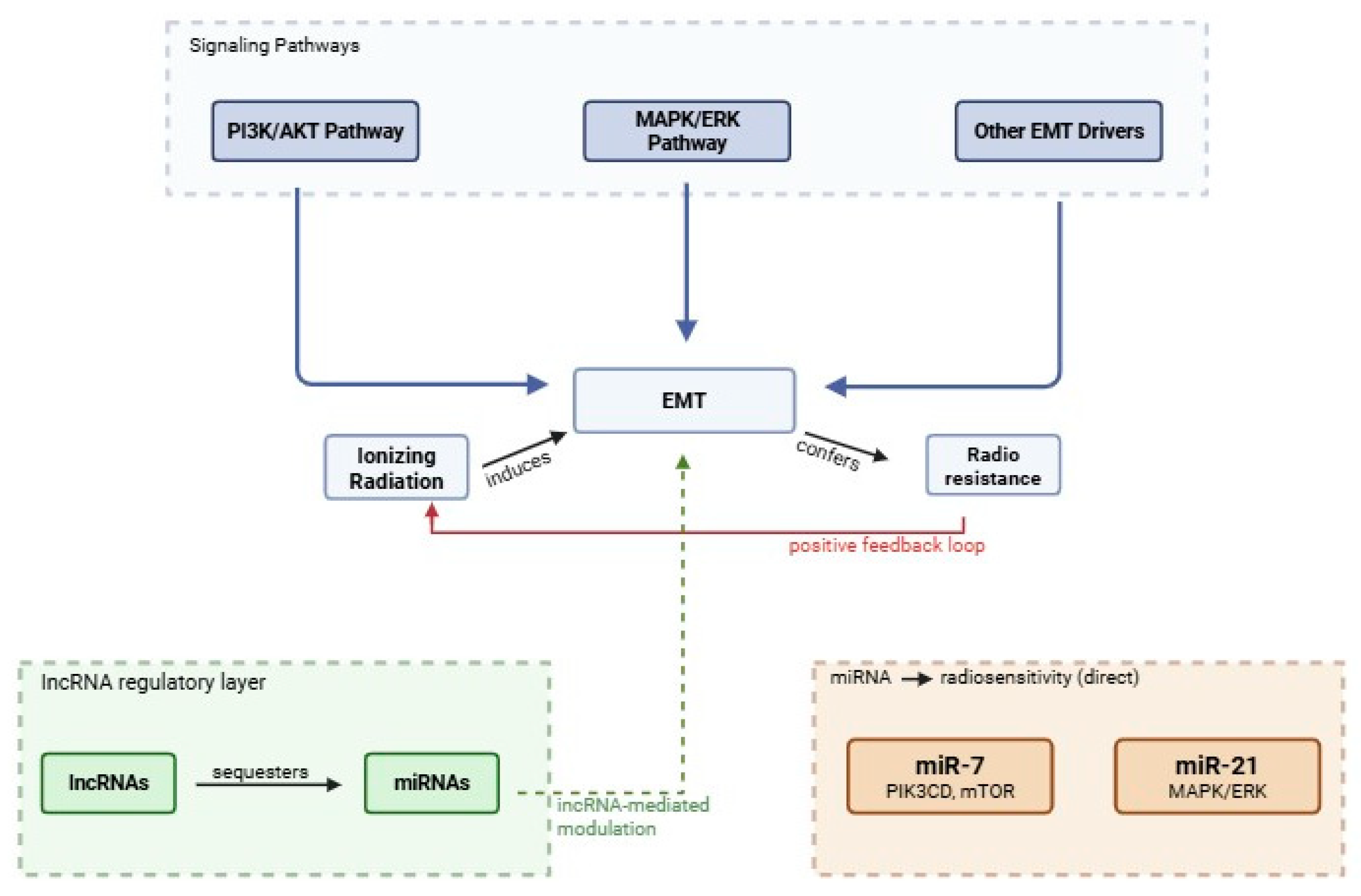
- Signaling Pathways: MiRNAs also function as “fine tuners” of major survival cascades. miR-21 reinforces radioresistance by activating the ERK/NF-κB pathway [5,16,74]. In contrast, miR-7 inhibits the PI3K/AKT pathway by targeting upstream receptors like IGFR, IRS1, and IRS2, effectively cutting off survival signals that would otherwise protect the cell from radiation toxicity [16,75,76].
5. Crosstalk Between MicroRNAs and EMT in Radioresistance
5.1. Synergistic Mechanisms and Regulatory Loops
5.2. Complex Interactions Within the Tumor Microenvironment
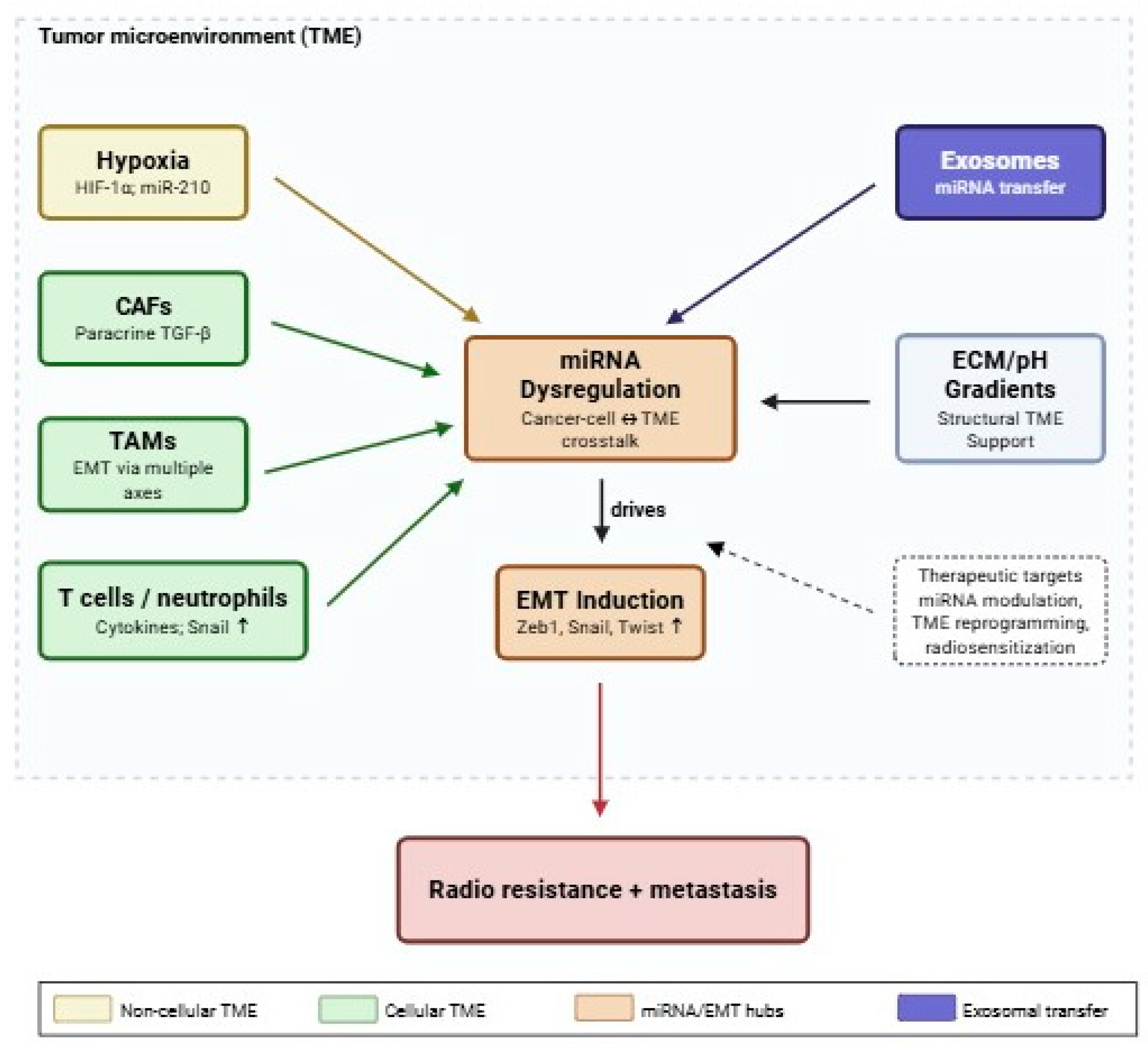
- Hypoxia: A hostile microenvironment, hypoxia, is a significant factor in eliciting EMT. It stabilizes HIFs, which activate key EMT-driving pathways such as TGF-β, NF-κB, and Notch, promoting the expression of EMT-TFs like ZEB1, Snail, and Twist. As shown in Figure 4, hypoxia also shares signal pathways with EMT and can confer stem cell-like properties to tumor cells, further contributing to radioresistance [24,25,26,27,28].
- Cellular Components:
- ○
- ○
- Immune Cells: TAMs induce EMT through multiple signaling pathways (e.g., PI3K/AKT-ERK1/2, COX-2, HIF-1α, EGFR/ERK1/2, Smad/Snail, TGF-β, JAK2/STAT3/miR-506-3p/FoxQ1 axis) [24,26,84]. Activated T cells can release soluble factors like TNF-α, IL-6, and TGF-β that facilitate EMT-related gene expression [85]. Even neutrophils can exacerbate hypoxia and stabilize Snail, eliciting partial EMT [26].
- ○
- Exosomes: Extracellular vesicles, such as exosomes derived from irradiated T cells, can promote metastasis by inducing EMT through increased β-catenin expression and activation of the NF-κB/Snail pathway. Exosomes can also transfer miRNAs, mediating EMT-induced drug resistance. For example, exosomes containing miR-155 from paclitaxel-resistant gastric cancer cells can induce EMT and chemoresistance in sensitive cells [16,26,86,87,88].
- MiRNAs and TME: MiRNAs are intimately involved in mediating the crosstalk between cancer cells and the TME. For instance, miR-210 is a hypoxia-induced miRNA that is an independent prognostic marker in lung cancer [89]. The dysregulation of miRNAs can transform an unreceptive cancer microenvironment into a cancer-friendly microenvironment, as seen in Figure 4 [16,26].
6. Therapeutic Strategies to Overcome Radioresistance
6.1. Targeting EMT Pathways for Radiosensitization
- Inhibiting EMT-Related Signaling Pathways: Disrupting the key signaling cascades that drive EMT can effectively block its progression [26].
- ○
- Wnt/β-catenin Pathway: Therapeutic agents targeting the Wnt-signaling pathway, such as the porcupine (PORCN) inhibitor LGK-974 (WNT-974), have been evaluated preclinically and in early-phase clinical trials. In preclinical models, LGK-974 inhibits WNT-related gene expression and WNT-dependent phosphorylation of LRP6, demonstrating tumor growth inhibition in pancreatic cancer [90]. However, direct radiosensitization has not yet been established in clinical settings, and Phase I trial data remain preliminary.
- ○
- TGF-β Pathway: Inhibitors of the TGF-β pathway represent another strategy for radiosensitization. Preclinically, the TGF-β receptor I kinase inhibitor SB-431542 has been shown to block EMT and cancer stem cell programs, increasing radiosensitivity in breast cancer models [91]. At the clinical level, galunisertib (LY2157299), a small-molecule TGF-βRI inhibitor, has been evaluated in Phase I and II trials across multiple solid tumors, with manageable toxicity reported [92]. Fresolimumab, a pan-TGF-β neutralizing antibody, has been combined with radiotherapy in a Phase I/II study, where it demonstrated the ability to augment radiation-induced abscopal responses in metastatic breast cancer [93]. These clinical results support further investigation of TGF-β inhibition as a radiosensitization strategy, though definitive Phase III evidence is lacking.
- ○
- NF-κB Pathway: Blocking NF-κB activity can counteract EMT formation and decrease radioresistance [94]. Denosumab, a monoclonal antibody targeting RANKL, which can activate NF-κB signaling, has been evaluated clinically; however, the ABCSG-18 trial demonstrated that denosumab did not improve disease-free survival outcomes in early breast cancer beyond its established bone-protective effects [95]. Direct radiosensitization through NF-κB inhibition therefore remains an area requiring more targeted clinical investigation.
- ○
- PI3K/Akt/mTOR Pathway: Inhibitors of the PI3K/AKT/mTOR axis have shown promise in reducing EMT and CSC markers, though evidence varies considerably by agent and context. Preclinically, the dual PI3K/mTOR inhibitor BEZ235 (dactolisib) decreases EMT marker expression and promotes radiosensitivity in cancer cell lines [96]; however, its clinical development has been substantially limited by toxicity. Simvastatin, a statin repurposed preclinically, has been shown to sensitize radioresistant esophageal cancer cells and reverse EMT via the PTEN-PI3K/AKT axis in preclinical models [97], though clinical radiosensitization data remain limited. PF-05212384 and everolimus have been evaluated in clinical trials across multiple solid tumor types, with everolimus demonstrating efficacy in hormone receptor-positive breast cancer [98]; their specific contribution to radiosensitization through EMT modulation remains under investigation.
- ○
- Notch Pathway: γ-secretase inhibitors (GSIs) suppress Notch signaling and have been explored as a means of reversing EMT and improving radiosensitivity [43,44]. Among these, RO4929097 has been evaluated in Phase Ib clinical trials, where it demonstrated limited single-agent efficacy, underscoring the need for combination approaches [99]. Preclinically, natural compounds with GSI activity including tangeretin, rhamnetin, and cirsiliol have been shown to suppress Notch signaling and reverse EMT in cancer cell models, though these agents have not yet been evaluated in clinical radiosensitization studies.
- ○
- HIF-1-Targeting Agents: Given hypoxia’s role in inducing EMT and radioresistance, agents that suppress HIF-1α represent a rational radiosensitization strategy [28]. Sunitinib and sorafenib, multi-kinase inhibitors that target VEGFR and reduce HIF-1α-driven angiogenesis, have been evaluated preclinically in combination with radiotherapy with evidence of radiosensitization through tumor vasculature normalization and reduction in hypoxic regions [100]. Bortezomib, a proteasome inhibitor that indirectly suppresses HIF-1α, has also demonstrated preclinical radiosensitizing activity. It should be noted that paclitaxel, while used clinically as a concurrent radiosensitizer, acts primarily through mitotic arrest rather than HIF-1α suppression and is more accurately categorized as a cytotoxic radiosensitizer [101]. Clinical evidence for HIF-1α-targeted radiosensitization specifically remains limited, and further trials are needed.
- Reversing the EMT Phenotype: Certain compounds can induce MET, thereby re-sensitizing cells. Metformin, an anti-diabetic drug, has demonstrated preclinical activity against EMT and cancer stemness through multiple mechanisms. It has been shown to suppress EMT-transcription factors including ZEB1, TWIST1, and SLUG in cancer cell models, thereby reducing invasiveness and stem-like properties [102,103]. Preclinical studies have additionally demonstrated radiosensitizing effects of metformin in several cancer types [104]. Clinical evidence for metformin as a radiosensitizer remains limited, and its role in this context is currently under investigation. Berberine can perturb TGF-β-induced EMT and sensitize colon epithelial cancer cells to radiation [105]. The histone deacetylase inhibitor vorinostat has also shown promise in reversing EMT [26,106].
6.2. MicroRNA-Based Therapeutic Approaches
- MiRNA Mimics: This strategy involves re-introducing tumor-suppressive miRNAs that are downregulated in resistant cells. For example, miR-34a re-expression has been shown preclinically to induce radiosensitization through suppression of Notch-1 signaling in cancer cell models [108]. Clinically, MRX34, a liposomal miR-34a mimic, was advanced into a Phase I trial in patients with advanced solid tumors [109]; however, the trial was terminated early due to severe immune-related adverse events, including five treatment-related deaths, highlighting that immunogenicity and systemic toxicity remain critical unresolved barriers to clinical translation of miRNA mimic therapies. Preclinically, overexpression of miR-100 has been shown to suppress ATM expression in a human glioma cell line (M059J), thereby impairing DNA damage repair and enhancing sensitivity to both chemotherapy and radiation in that model [110]. These findings are currently limited to in vitro evidence and have not been validated in clinical settings.
- Anti-miRNAs (Antagomirs): Inhibiting oncogenic miRNAs that are upregulated in resistant cells. For instance, inhibiting the oncogenic miR-21 using antisense antagomirs has been shown preclinically to enhance apoptosis and reduce radioresistance in glioblastoma cell lines [111]. Clinical translation of anti-miR-21 strategies has not yet been achieved, and evidence remains confined to preclinical models.
- Combination with Conventional Therapies: MiRNA-based therapies are particularly attractive in combination with other anti-cancer therapies due to their ability to target multiple genes associated with resistance-mediating signaling pathways. Curcumin, a natural polyphenol, has been shown preclinically to induce cancer cell death and chemosensitization through modulation of Notch signaling pathways in esophageal cancer models [112]. While curcumin has been reported to influence miRNA expression profiles in some contexts, its extremely poor oral bioavailability represents a significant barrier to clinical translation, and direct evidence of miRNA-mediated EMT suppression as its primary radiosensitizing mechanism remains to be established.
6.3. Emerging Combination Therapies
- Targeted Therapy Combined with Radiotherapy: Combining RT with agents that target specific pro-survival pathways has shown clinical promise. Cetuximab, an anti-EGFR monoclonal antibody, demonstrated significantly improved locoregional control and overall survival when combined with radiotherapy in a landmark Phase III trial in locally advanced head and neck squamous cell carcinoma, establishing it as a clinically validated radiosensitizer [115]. Preclinically, the dual EGFR/HER2 inhibitor lapatinib has also demonstrated radiosensitizing effects through MEK/ERK pathway inhibition and cell cycle arrest, though its clinical radiosensitization data are less mature than those for cetuximab [5]. Similarly, inhibiting the VEGF/VEGFR pathway has been proposed as a means of normalizing tumor vasculature, reducing local hypoxia, and thereby increasing radiosensitivity [116]. Regorafenib, a multi-kinase inhibitor targeting VEGFR among other pathways, has established clinical utility in colorectal and gastrointestinal stromal tumors; however, its specific combination with radiotherapy for radiosensitization purposes has limited clinical evidence and warrants dedicated investigation.
- EMT-Targeting Agents with RT: Combining agents that inhibit or reverse EMT with radiation therapy is a logical strategy to prevent or overcome acquired radioresistance [10,26]. Metformin, an EMT-targeting agent, has been shown to improve progression-free survival when combined with EGFR tyrosine kinase inhibitors in advanced lung adenocarcinoma in a clinical study, suggesting that concurrent EMT suppression may augment the efficacy of targeted therapies [102,117].
- MiRNA-Based Combinatorial Therapy: MiRNAs’ ability to regulate multiple resistance-mediating pathways by targeting multiple genes makes them ideal candidates for combinatorial approaches. Modulating dysregulated miRNA levels (via mimics or inhibitors) can sensitize cancer cells to other anti-cancer therapies, offering a powerful synergistic effect [5,16].
- Targeting CSCs: Strategies that target CSC phenotypes represent another avenue for overcoming radioresistance. The ionophore salinomycin was shown in a foundational preclinical study to selectively eliminate breast cancer stem cells with greater potency than conventional chemotherapy [118]. It has subsequently demonstrated the ability to reduce tumor-initiating cell populations in preclinical models [21,22]. Its potential to enhance radiosensitivity when combined with RT remains largely unexplored clinically and dedicated in vivo and clinical studies are needed.
7. Conclusions and Future Perspectives
7.1. Summary of Key Findings and Clinical Implications
7.2. Challenges and Future Directions in Research
- Complexity of Molecular Mechanisms: The sheer complexity and redundancy of the signaling pathways involved in interactions between EMT and miRNA regulation pose a considerable challenge. Targeting a single pathway may lead to compensatory activation of alternative routes, limiting therapeutic efficacy [5].
- Tumor Heterogeneity and Plasticity: Tumors are highly heterogeneous, containing diverse cell populations, including CSCs and cells in various EMT states. This plasticity allows cancer cells to adapt rapidly to therapeutic pressures, making durable responses difficult to achieve.
- Drug Delivery and Off-Target Effects: For miRNA-based therapies and many targeted agents, developing efficient and safe delivery systems that ensure specific targeting of cancer cells while minimizing off-target effects remains a significant hurdle [16]. Challenges include stable nanoconstructs, appropriate delivery methods, rapid excretion, incorrect intracellular release, poor biostability, endosomal escape, and immunogenicity.
- Integrated Multi-Omics Approaches: Utilizing advanced multi-omics technologies (genomics, transcriptomics, proteomics, metabolomics) to comprehensively map the dynamic changes in EMT and miRNA profiles during and after radiation therapy. This could reveal novel, interconnected targets [5].
- Development of Novel Combination Therapies: Designing rational combination therapies that simultaneously target multiple, synergistic resistance pathways. This includes combining radiation with EMT inhibitors and miRNA mimics/antagomirs. The goal is to achieve synergistic radiosensitization while minimizing toxicity [5,16].
Author Contributions
Funding
Institutional Review Board Statement
Informed Consent Statement
Data Availability Statement
Acknowledgments
Conflicts of Interest
References
- Zhu, H.; Chua, M.L.K.; Chitapanarux, I.; Kaidar-Person, O.; Mwaba, C.; Alghamdi, M.; Rodríguez Mignola, A.; Amrogowicz, N.; Yazici, G.; Bourhaleb, Z.; et al. Global radiotherapy demands and corresponding radiotherapy-professional workforce requirements in 2022 and predicted to 2050: A population-based study. Lancet Glob. Health 2024, 12, e1945–e1953. [Google Scholar] [CrossRef] [Scilit] [PubMed]
- Siegel, R.L.; Miller, K.D.; Jemal, A. Cancer statistics, 2020. CA Cancer J. Clin. 2020, 70, 7–30. [Google Scholar] [CrossRef] [Scilit] [PubMed]
- Bray, F.; Ferlay, J.; Soerjomataram, I.; Siegel, R.L.; Torre, L.A.; Jemal, A. Global cancer statistics 2018: GLOBOCAN estimates of incidence and mortality worldwide for 36 cancers in 185 countries. CA Cancer J. Clin. 2018, 68, 394–424, Erratum in CA Cancer J. Clin. 2020, 70, 313. [Google Scholar] [CrossRef] [Scilit] [PubMed]
- Huang, R.X.; Zhou, P.K. DNA damage response signaling pathways and targets for radiotherapy sensitization in cancer. Signal Transduct. Target. Ther. 2020, 5, 60. [Google Scholar] [CrossRef] [Scilit] [PubMed]
- Wu, Y.; Song, Y.; Wang, R.; Wang, T. Molecular mechanisms of tumor resistance to radiotherapy. Mol. Cancer 2023, 22, 96. [Google Scholar] [CrossRef] [Scilit] [PubMed]
- Zhou, S.; Zhang, M.; Zhou, C.; Wang, W.; Yang, H.; Ye, W. The role of epithelial-mesenchymal transition in regulating radioresistance. Crit. Rev. Oncol. Hematol. 2020, 150, 102961. [Google Scholar] [CrossRef] [Scilit] [PubMed]
- Hanahan, D. Hallmarks of Cancer: New Dimensions. Cancer Discov. 2022, 12, 31–46. [Google Scholar] [CrossRef] [Scilit] [PubMed]
- Drápela, S.; Bouchal, J.; Jolly, M.K.; Culig, Z.; Souček, K. ZEB1: A Critical Regulator of Cell Plasticity, DNA Damage Response, and Therapy Resistance. Front. Mol. Biosci. 2020, 7, 36. [Google Scholar] [CrossRef] [Scilit] [PubMed]
- Nieto, M.A.; Huang, R.Y.-J.; Jackson, R.A.; Thiery, J.P. EMT: 2016. Cell 2016, 166, 21–45. [Google Scholar] [CrossRef] [Scilit] [PubMed]
- Dudás, J.; Ladányi, A.; Ingruber, J.; Steinbichler, T.B.; Riechelmann, H. Epithelial to Mesenchymal Transition: A Mechanism that Fuels Cancer Radio/Chemoresistance. Cells 2020, 9, 428. [Google Scholar] [CrossRef] [Scilit] [PubMed]
- Bakir, B.; Chiarella, A.M.; Pitarresi, J.R.; Rustgi, A.K. EMT, MET, Plasticity, and Tumor Metastasis. Trends Cell Biol. 2020, 30, 764–776. [Google Scholar] [CrossRef] [Scilit] [PubMed]
- Yang, J.; Antin, P.; Berx, G.; Blanpain, C.; Brabletz, T.; Bronner, M.; Campbell, K.; Cano, A.; Casanova, J.; Christofori, G.; et al. Guidelines and definitions for research on epithelial-mesenchymal transition. Nat. Rev. Mol. Cell Biol. 2020, 21, 341–352, Correction in Nat. Rev. Mol. Cell Biol. 2021, 22, 843. [Google Scholar] [CrossRef] [Scilit] [PubMed]
- Brabletz, T.; Kalluri, R.; Nieto, M.A.; Weinberg, R.A. EMT in cancer. Nat. Rev. Cancer 2018, 18, 128–134. [Google Scholar] [CrossRef] [Scilit] [PubMed]
- Qiao, L.; Chen, Y.; Liang, N.; Xie, J.; Deng, G.; Chen, F.; Wang, X.; Liu, F.; Li, Y.; Zhang, J. Targeting epithelial-to-mesenchymal transition in radioresistance: Crosslinked mechanisms and strategies. Front. Oncol. 2022, 12, 775238. [Google Scholar] [CrossRef] [Scilit] [PubMed]
- Ribatti, D.; Tamma, R.; Annese, T. Epithelial-Mesenchymal Transition in Cancer: A Historical Overview. Transl. Oncol. 2020, 13, 100773. [Google Scholar] [CrossRef] [Scilit] [PubMed]
- De Craene, B.; Berx, G. Regulatory networks defining EMT during cancer initiation and progression. Nat. Rev. Cancer 2013, 13, 97–110. [Google Scholar] [CrossRef] [Scilit] [PubMed]
- Tan, J.; Sun, X.; Zhao, H.; Guan, H.; Gao, S.; Zhou, P.K. Double-strand DNA break repair: Molecular mechanisms and therapeutic targets. MedComm 2023, 4, e388. [Google Scholar] [CrossRef] [Scilit] [PubMed]
- Ke, G.; Liang, L.; Yang, J.M.; Huang, X.; Han, D.; Huang, S.; Zhao, Y.; Zha, R.; He, X.; Wu, X. MiR-181a confers resistance of cervical cancer to radiation therapy through targeting the pro-apoptotic PRKCD gene. Oncogene 2013, 32, 3019–3027. [Google Scholar] [CrossRef] [Scilit] [PubMed]
- Mongroo, P.S.; Rustgi, A.K. The role of the miR-200 family in epithelial-mesenchymal transition. Cancer Biol. Ther. 2010, 10, 219–222. [Google Scholar] [CrossRef] [PubMed]
- Warrier, N.M.; Kelkar, N.; Johnson, C.T.; Govindarajan, T.; Prabhu, V.; Kumar, P. Understanding cancer stem cells and plasticity: Towards better therapeutics. Eur. J. Cell Biol. 2023, 102, 151321. [Google Scholar] [CrossRef] [Scilit] [PubMed]
- Schulz, A.; Meyer, F.; Dubrovska, A.; Borgmann, K. Cancer Stem Cells and Radioresistance: DNA Repair and Beyond. Cancers 2019, 11, 862. [Google Scholar] [CrossRef] [Scilit] [PubMed]
- Arnold, C.R.; Mangesius, J.; Skvortsova, I.I.; Ganswindt, U. The Role of Cancer Stem Cells in Radiation Resistance. Front. Oncol. 2020, 10, 164. [Google Scholar] [CrossRef] [Scilit] [PubMed]
- Gillespie, M.S.; Ward, C.M.; Davies, C.C. DNA Repair and Therapeutic Strategies in Cancer Stem Cells. Cancers 2023, 15, 1897. [Google Scholar] [CrossRef] [Scilit] [PubMed]
- Lytle, N.K.; Barber, A.G.; Reya, T. Stem cell fate in cancer growth, progression and therapy resistance. Nat. Rev. Cancer 2018, 18, 669–680. [Google Scholar] [CrossRef] [Scilit] [PubMed]
- Bouleftour, W.; Rowinski, E.; Louati, S.; Sotton, S.; Wozny, A.S.; Moreno-Acosta, P.; Mery, B.; Rodriguez-Lafrasse, C.; Magne, N. A Review of the Role of Hypoxia in Radioresistance in Cancer Therapy. Med. Sci. Monit. 2021, 27, e934116. [Google Scholar] [CrossRef] [Scilit] [PubMed]
- Roche, J.; Gemmill, R.M.; Drabkin, H.A. Epigenetic Regulation of the Epithelial to Mesenchymal Transition in Lung Cancer. Cancers 2017, 9, 72. [Google Scholar] [CrossRef] [Scilit] [PubMed]
- Tam, S.Y.; Wu, V.W.C.; Law, H.K.W. Hypoxia-Induced Epithelial-Mesenchymal Transition in Cancers: HIF-1α and Beyond. Front. Oncol. 2020, 10, 486. [Google Scholar] [CrossRef] [Scilit] [PubMed]
- Kabakov, A.E.; Yakimova, A.O.; Matchuk, O.N. Hypoxia-Induced Cancer Cell Responses Driving Radioresistance of Hypoxic Tumors: Approaches to Targeting and Radiosensitizing. Cancers 2021, 13, 1102. [Google Scholar] [CrossRef] [Scilit] [PubMed]
- Pastushenko, I.; Blanpain, C. EMT Transition States during Tumor Progression and Metastasis. Trends Cell Biol. 2019, 29, 212–226. [Google Scholar] [CrossRef] [Scilit] [PubMed]
- Hakim, S.G.; Taubitz, C.; Hoppe, S.; Steller, D.; Rades, D.; Ribbat-Idel, J.; Alsharif, U.; Falougy, M. Prognostic impact of the loss of E-cadherin and de novo expression of N-cadherin at the invasive front of primary and recurrent oral squamous cell carcinoma. Front. Oncol. 2023, 13, 1151879. [Google Scholar] [CrossRef] [Scilit] [PubMed]
- Niknami, Z.; Muhammadnejad, A.; Ebrahimi, A.; Harsani, Z.; Shirkoohi, R. Significance of E-cadherin and Vimentin as epithelial-mesenchymal transition markers in colorectal carcinoma prognosis. EXCLI J. 2020, 19, 889–901. [Google Scholar] [CrossRef] [Scilit] [PubMed]
- Usman, S.; Waseem, N.H.; Nguyen, T.K.N.; Mohsin, S.; Jamal, A.; Teh, M.T.; Waseem, A. Vimentin Is at the Heart of Epithelial Mesenchymal Transition (EMT) Mediated Metastasis. Cancers 2021, 13, 4985. [Google Scholar] [CrossRef] [Scilit] [PubMed]
- Moreno, C.S. SOX4: The unappreciated oncogene. Semin. Cancer Biol. 2020, 67, 57–64. [Google Scholar] [CrossRef] [Scilit] [PubMed]
- Seo, J.; Ha, J.; Kang, E.; Cho, S. The role of epithelial-mesenchymal transition-regulating transcription factors in anti-cancer drug resistance. Arch. Pharmacal Res. 2021, 44, 281–292. [Google Scholar] [CrossRef] [Scilit] [PubMed]
- Pawlicka, M.; Gumbarewicz, E.; Błaszczak, E.; Stepulak, A. Transcription Factors and Markers Related to Epithelial-Mesenchymal Transition and Their Role in Resistance to Therapies in Head and Neck Cancers. Cancers 2024, 16, 1354. [Google Scholar] [CrossRef] [Scilit] [PubMed]
- Fischer, K.R.; Durrans, A.; Lee, S.; Sheng, J.; Li, F.; Wong, S.T.; Choi, H.; El Rayes, T.; Ryu, S.; Troeger, J.; et al. Epithelial-to-mesenchymal transition is not required for lung metastasis but contributes to chemoresistance. Nature 2015, 527, 472–476. [Google Scholar] [CrossRef] [Scilit] [PubMed]
- Viswanathan, V.S.; Ryan, M.J.; Dhruv, H.D.; Gill, S.; Eichhoff, O.M.; Seashore-Ludlow, B.; Kaffenberger, S.D.; Eaton, J.K.; Shimada, K.; Aguirre, A.J.; et al. Dependency of a therapy-resistant state of cancer cells on a lipid peroxidase pathway. Nature 2017, 547, 453–457. [Google Scholar] [CrossRef] [Scilit] [PubMed]
- Muriithi, W.; Macharia, L.W.; Heming, C.P.; Echevarria, J.L.; Nyachieo, A.; Filho, P.N.; Moura Neto, V. ABC transporters and the hallmarks of cancer: Roles in cancer aggressiveness beyond multidrug resistance. Cancer Biol. Med. 2020, 17, 253–269. [Google Scholar] [CrossRef] [Scilit] [PubMed]
- Xiao, H.; Zheng, Y.; Ma, L.; Tian, L.; Sun, Q. Clinically-Relevant ABC Transporter for Anti-Cancer Drug Resistance. Front. Pharmacol. 2021, 12, 648407. [Google Scholar] [CrossRef] [Scilit] [PubMed]
- Fletcher, J.I.; Haber, M.; Henderson, M.J.; Norris, M.D. ABC transporters in cancer: More than just drug efflux pumps. Nat. Rev. Cancer 2010, 10, 147–156. [Google Scholar] [CrossRef] [Scilit] [PubMed]
- Chaves, L.P.; Melo, C.M.; Saggioro, F.P.; Reis, R.B.d.; Squire, J.A. Epithelial-mesenchymal transition signaling and prostate cancer stem cells: Emerging biomarkers and opportunities for precision therapeutics. Genes 2021, 12, 1900. [Google Scholar] [CrossRef] [Scilit] [PubMed]
- Batlle, E.; Massagué, J. Transforming Growth Factor-β Signaling in Immunity and Cancer. Immunity 2019, 50, 924–940. [Google Scholar] [CrossRef] [Scilit] [PubMed]
- Giuli, M.V.; Mancusi, A.; Giuliani, E.; Screpanti, I.; Checquolo, S. Notch signaling in female cancers: A multifaceted node to overcome drug resistance. Cancer Drug Resist. 2021, 4, 805–836. [Google Scholar] [CrossRef] [Scilit] [PubMed]
- Xiu, M.; Wang, Y.; Li, B.; Wang, X.; Xiao, F.; Chen, S.; Zhang, L.; Zhou, B.; Hua, F. The Role of Notch3 Signaling in Cancer Stemness and Chemoresistance: Molecular Mechanisms and Targeting Strategies. Front. Mol. Biosci. 2021, 8, 694141. [Google Scholar] [CrossRef] [Scilit] [PubMed]
- Gomez-Casal, R.; Bhattacharya, C.; Ganesh, N.; Bailey, L.; Basse, P.; Gibson, M.; Epperly, M.; Levina, V. Non-small cell lung cancer cells survived ionizing radiation treatment display cancer stem cell and epithelial-mesenchymal transition phenotypes. Mol. Cancer 2013, 12, 94. [Google Scholar] [CrossRef] [Scilit] [PubMed]
- Peng, Y.; Wang, Y.; Zhou, C.; Mei, W.; Zeng, C. PI3K/Akt/mTOR Pathway and Its Role in Cancer Therapeutics: Are We Making Headway? Front. Oncol. 2022, 12, 819128. [Google Scholar] [CrossRef] [Scilit] [PubMed]
- Kong, D.; Banerjee, S.; Ahmad, A.; Li, Y.; Wang, Z.; Sethi, S.; Sarkar, F.H. Epithelial to mesenchymal transition is mechanistically linked with stem cell signatures in prostate cancer cells. PLoS ONE 2010, 5, e12445. [Google Scholar] [CrossRef] [Scilit] [PubMed]
- Zhang, Y.; Huo, L.; Wei, Z.; Tang, Q.; Sui, H. Hotspots and frontiers in inflammatory tumor microenvironment research: A scientometric and visualization analysis. Front. Pharmacol. 2022, 13, 862585. [Google Scholar] [CrossRef] [Scilit] [PubMed]
- Zhang, T.; Ma, C.; Zhang, Z.; Zhang, H.; Hu, H. NF-κB signaling in inflammation and cancer. MedComm 2021, 2, 618–653. [Google Scholar] [CrossRef] [Scilit] [PubMed]
- Bartel, D.P. Metazoan microRNAs. Cell 2018, 173, 20–51. [Google Scholar] [CrossRef] [Scilit] [PubMed]
- Cavallari, I.; Ciccarese, F.; Sharova, E.; Urso, L.; Raimondi, V.; Silic-Benussi, M.; D’Agostino, D.M.; Ciminale, V. The miR-200 Family of microRNAs: Fine Tuners of Epithelial-Mesenchymal Transition and Circulating Cancer Biomarkers. Cancers 2021, 13, 5874. [Google Scholar] [CrossRef] [Scilit] [PubMed]
- Gregory, P.A.; Bert, A.G.; Paterson, E.L.; Barry, S.C.; Tsykin, A.; Farshid, G.; Vadas, M.A.; Khew-Goodall, Y.; Goodall, G.J. The miR-200 family and miR-205 regulate epithelial to mesenchymal transition by targeting ZEB1 and SIP1. Nat. Cell Biol. 2008, 10, 593–601. [Google Scholar] [CrossRef] [Scilit] [PubMed]
- Bracken, C.P.; Gregory, P.A.; Kolesnikoff, N.; Bert, A.G.; Wang, J.; Shannon, M.F.; Goodall, G.J. A double-negative feedback loop between ZEB1-SIP1 and the microRNA-200 family regulates epithelial-mesenchymal transition. Cancer Res. 2008, 68, 7846–7854. [Google Scholar] [CrossRef] [Scilit] [PubMed]
- Nishijima, N.; Seike, M.; Soeno, C.; Chiba, M.; Miyanaga, A.; Noro, R.; Sugano, T.; Matsumoto, M.; Kubota, K.; Gemma, A. miR-200/ZEB axis regulates sensitivity to nintedanib in non-small cell lung cancer cells. Int. J. Oncol. 2016, 48, 937–944. [Google Scholar] [CrossRef] [Scilit] [PubMed]
- Cortez, M.A.; Valdecanas, D.; Zhang, X.; Zhan, Y.; Bhardwaj, V.; Calin, G.A.; Komaki, R.; Giri, D.K.; Quini, C.C.; Wolfe, T.; et al. Therapeutic delivery of miR-200c enhances radiosensitivity in lung cancer. Mol. Ther. J. Am. Soc. Gene Ther. 2014, 22, 1494–1503. [Google Scholar] [CrossRef] [Scilit] [PubMed]
- Slabáková, E.; Culig, Z.; Remšík, J.; Souček, K. Alternative mechanisms of miR-34a regulation in cancer. Cell Death Dis. 2017, 8, e3100, Correction in Cell Death Dis. 2018, 9, 783. [Google Scholar] [CrossRef] [Scilit] [PubMed]
- Cortez, M.A.; Ivan, C.; Valdecanas, D.; Wang, X.; Peltier, H.J.; Ye, Y.; Araujo, L.; Carbone, D.P.; Shilo, K.; Giri, D.K.; et al. PDL1 regulation by p53 via miR-34. J. Natl. Cancer Inst. 2016, 108, djv303. [Google Scholar] [CrossRef] [Scilit] [PubMed]
- Kim, N.H.; Kim, H.S.; Li, X.Y.; Lee, I.; Choi, H.S.; Kang, S.E.; Cha, S.Y.; Ryu, J.K.; Yoon, D.; Fearon, E.R.; et al. A p53/miRNA-34 axis regulates Snail1-dependent cancer cell epithelial-mesenchymal transition. J. Cell Biol. 2011, 195, 417–433. [Google Scholar] [CrossRef] [Scilit] [PubMed]
- Ji, Q.; Hao, X.; Zhang, M.; Tang, W.; Yang, M.; Li, L.; Xiang, D.; Desano, J.T.; Bommer, G.T.; Fan, D.; et al. MicroRNA miR-34 inhibits human pancreatic cancer tumor-initiating cells. PLoS ONE 2009, 4, e6816. [Google Scholar] [CrossRef] [Scilit] [PubMed]
- Cheng, X.; Shen, T.; Liu, P.; Fang, S.; Yang, Z.; Li, Y.; Dong, J. mir-145-5p is a suppressor of colorectal cancer at early stage, while promotes colorectal cancer metastasis at late stage through regulating AKT signaling evoked EMT-mediated anoikis. BMC Cancer 2022, 22, 1151. [Google Scholar] [CrossRef] [Scilit] [PubMed]
- Conde-Muíño, R.; Cuadros, M.; Zambudio, N.; Segura-Jiménez, I.; Cano, C.; Palma, P. Predictive Biomarkers to Chemoradiation in Locally Advanced Rectal Cancer. BioMed Res. Int. 2015, 2015, 921435. [Google Scholar] [CrossRef] [Scilit] [PubMed]
- Zhang, Y.; Zheng, L.; Huang, J.; Gao, F.; Lin, X.; He, L.; Li, D.; Li, Z.; Ding, Y.; Chen, L. MiR-124 Radiosensitizes human colorectal cancer cells by targeting PRRX1. PLoS ONE 2014, 9, e93917. [Google Scholar] [CrossRef] [Scilit] [PubMed]
- Pan, F.; Mao, H.; Bu, F.; Tong, X.; Li, J.; Zhang, S.; Liu, X.; Wang, L.; Wu, L.; Chen, R.; et al. Sp1-mediated transcriptional activation of miR-205 promotes radioresistance in esophageal squamous cell carcinoma. Oncotarget 2017, 8, 5735–5752. [Google Scholar] [CrossRef] [Scilit] [PubMed]
- Niemoeller, O.M.; Niyazi, M.; Corradini, S.; Zehentmayr, F.; Li, M.; Lauber, K.; Belka, C. MicroRNA expression profiles in human cancer cells after ionizing radiation. Radiat. Oncol. 2011, 6, 29. [Google Scholar] [CrossRef] [Scilit] [PubMed]
- Chang, C.J.; Chao, C.H.; Xia, W.; Yang, J.Y.; Xiong, Y.; Li, C.W.; Yu, W.H.; Rehman, S.K.; Hsu, J.L.; Lee, H.H.; et al. p53 regulates epithelial-mesenchymal transition and stem cell properties through modulating miRNAs. Nat. Cell Biol. 2011, 13, 317–323, Correction in Nat. Cell Biol. 2011, 13, 1467. [Google Scholar] [CrossRef] [Scilit] [PubMed]
- Davis, B.N.; Hilyard, A.C.; Lagna, G.; Hata, A. SMAD proteins control DROSHA-mediated microRNA maturation. Nature 2008, 454, 56–61. [Google Scholar] [CrossRef] [Scilit] [PubMed]
- Tsai, K.W.; Wu, C.W.; Hu, L.Y.; Li, S.C.; Liao, Y.L.; Lai, C.H.; Kao, H.W.; Fang, W.L.; Huang, K.H.; Chan, W.C.; et al. Epigenetic regulation of miR-34b and miR-129 expression in gastric cancer. Int. J. Cancer 2011, 129, 2600–2610. [Google Scholar] [CrossRef] [Scilit] [PubMed]
- Yan, D.; Ng, W.L.; Zhang, X.; Wang, P.; Zhang, Z.; Mo, Y.Y.; Mao, H.; Hao, C.; Olson, J.J.; Curran, W.J.; et al. Targeting DNA-PKcs and ATM with miR-101 sensitizes tumors to radiation. PLoS ONE 2010, 5, e11397. [Google Scholar] [CrossRef] [Scilit] [PubMed]
- Lee, K.M.; Choi, E.J.; Kim, I.A. microRNA-7 increases radiosensitivity of human cancer cells with activated EGFR-associated signaling. Radiother. Oncol. J. Eur. Soc. Ther. Radiol. Oncol. 2011, 101, 171–176. [Google Scholar] [CrossRef] [Scilit] [PubMed]
- Song, L.; Lin, C.; Wu, Z.; Gong, H.; Zeng, Y.; Wu, J.; Li, M.; Li, J. miR-18a impairs DNA damage response through downregulation of ataxia telangiectasia mutated (ATM) kinase. PLoS ONE 2011, 6, e25454. [Google Scholar] [CrossRef] [Scilit] [PubMed]
- Chen, G.; Zhu, W.; Shi, D.; Lv, L.; Zhang, C.; Liu, P.; Hu, W. MicroRNA-181a sensitizes human malignant glioma U87MG cells to radiation by targeting Bcl-2. Oncol. Rep. 2010, 23, 997–1003. [Google Scholar] [CrossRef] [Scilit]
- Mott, J.L.; Kobayashi, S.; Bronk, S.F.; Gores, G.J. mir-29 regulates Mcl-1 protein expression and apoptosis. Oncogene 2007, 26, 6133–6140. [Google Scholar] [CrossRef] [Scilit] [PubMed]
- Zhang, H.; Zuo, Z.; Lu, X.; Wang, L.; Wang, H.; Zhu, Z. MiR-25 regulates apoptosis by targeting Bim in human ovarian cancer. Oncol. Rep. 2012, 27, 594–598. [Google Scholar] [CrossRef] [Scilit] [PubMed]
- Peng, J.; Lv, Y.; Wu, C. Radiation-resistance increased by overexpression of microRNA-21 and inhibition of its target PTEN in esophageal squamous cell carcinoma. J. Int. Med. Res. 2020, 48, 300060519882543. [Google Scholar] [CrossRef] [Scilit] [PubMed]
- Kefas, B.; Godlewski, J.; Comeau, L.; Li, Y.; Abounader, R.; Hawkinson, M.; Lee, J.; Fine, H.; Chiocca, E.A.; Lawler, S.; et al. microRNA-7 inhibits the epidermal growth factor receptor and the Akt pathway and is down-regulated in glioblastoma. Cancer Res. 2008, 68, 3566–3572. [Google Scholar] [CrossRef] [Scilit] [PubMed]
- Webster, R.J.; Giles, K.M.; Price, K.J.; Zhang, P.M.; Mattick, J.S.; Leedman, P.J. Regulation of epidermal growth factor receptor signaling in human cancer cells by microRNA-7. J. Biol. Chem. 2009, 284, 5731–5741. [Google Scholar] [CrossRef] [Scilit] [PubMed]
- Li, L.; Lin, X.; Zhao, C.; Yang, Z.; Qiu, C. The Mechanism of Long Non-coding RNA in Cancer Radioresistance/Radiosensitivity: A Systematic Review. Front. Pharmacol. 2022, 13, 879704. [Google Scholar] [CrossRef] [Scilit] [PubMed]
- Salmena, L.; Poliseno, L.; Tay, Y.; Kats, L.; Pandolfi, P.P. A ceRNA Hypothesis: The Rosetta Stone of a Hidden RNA Language? Cell 2011, 146, 353–358. [Google Scholar] [CrossRef] [Scilit] [PubMed]
- Korkaya, H.; Orsulic, S. The tumor microenvironment: Recent advances and novel therapeutic approaches. Front. Cell Dev. Biol. 2020, 8, 586176. [Google Scholar] [CrossRef] [Scilit] [PubMed]
- Bukhari, I.; Zhang, Y.; Thorne, R.F.; Mi, Y. Complexity of tumor microenvironment: A major culprit in cancer development. Front. Endocrinol. 2022, 13, 1059885. [Google Scholar] [CrossRef] [Scilit] [PubMed]
- Faisal, S.M.; Comba, A.; Varela, M.L.; Argento, A.E.; Garcia, C. The complex interactions between the cellular and non-cellular components of the brain tumor microenvironmental landscape and their therapeutic implications. Front. Oncol. 2022, 12, 1005069. [Google Scholar] [CrossRef] [Scilit] [PubMed]
- Genova, C.; Dellepiane, C.; Carrega, P.; Sommariva, S.; Ferlazzo, G.; Pronzato, P.; Gangemi, R.; Filaci, G.; Coco, S.; Croce, M. Therapeutic implications of tumor microenvironment in lung cancer: Focus on immune checkpoint blockade. Front. Immunol. 2022, 12, 799455. [Google Scholar] [CrossRef] [Scilit] [PubMed]
- Guo, Z.; Zhang, H.; Fu, Y.; Kuang, J.; Zhao, B.; Zhang, L.F.; Lin, J.; Lin, S.; Wu, D.; Xie, G. Cancer-associated fibroblasts induce growth and radioresistance of breast cancer cells through paracrine IL-6. Cell Death Discov. 2023, 9, 6. [Google Scholar] [CrossRef] [Scilit] [PubMed]
- Akinsipe, T.; Mohamedelhassan, R.; Akinpelu, A.; Fagboyegun, S.; Adeyemo, B. Cellular interactions in tumor microenvironment during breast cancer progression: New frontiers and implications for novel therapeutics. Front. Immunol. 2024, 15, 1302587. [Google Scholar] [CrossRef] [Scilit] [PubMed]
- Melo, C.M.; Vidotto, T.; Chaves, L.P.; Lautert-Dutra, W.; Reis, R.B.d.; Squire, J.A. The role of somatic mutations on the immune response of the tumor microenvironment in prostate cancer. Int. J. Mol. Sci. 2021, 22, 9550. [Google Scholar] [CrossRef] [Scilit] [PubMed]
- Santos, P.; Almeida, F. Role of Exosomal miRNAs and the Tumor Microenvironment in Drug Resistance. Cells 2020, 9, 1450. [Google Scholar] [CrossRef] [Scilit] [PubMed]
- Yang, Q.; Xu, J.; Gu, J.; Shi, H.; Zhang, J.; Zhang, J.; Chen, Z.; Fang, X.; Zhu, T.; Zhang, X. Extracellular Vesicles in Cancer Drug Resistance: Roles, Mechanisms, and Implications. Adv. Sci. 2022, 9, e2201609. [Google Scholar] [CrossRef] [Scilit] [PubMed]
- Wang, M.; Qiu, R.; Yu, S.; Xu, X.; Li, G.; Gu, R.; Tan, C.; Zhu, W.; Shen, B. Paclitaxel-resistant gastric cancer MGC-803 cells promote epithelial-to-mesenchymal transition and chemoresistance in paclitaxel-sensitive cells via exosomal delivery of miR-155-5p. Int. J. Oncol. 2019, 54, 326–338. [Google Scholar] [CrossRef] [Scilit] [PubMed]
- Grosso, S.; Doyen, J.; Parks, S.K.; Bertero, T.; Paye, A.; Cardinaud, B.; Gounon, P.; Lacas-Gervais, S.; Noel, A.; Pouyssegur, J.; et al. MiR-210 promotes a hypoxic phenotype and increases radioresistance in human lung cancer cell lines. Cell Death Dis. 2013, 4, e544. [Google Scholar] [CrossRef] [Scilit] [PubMed]
- Madan, B.; Ke, Z.; Harmston, N.; Ho, S.Y.; Frois, A.O.; Alam, J.; Jeyaraj, D.A.; Pendharkar, V.; Ghosh, K.; Virshup, I.H.; et al. Wnt addiction of genetically defined cancers reversed by PORCN inhibition. Oncogene 2016, 35, 2197–2207. [Google Scholar] [CrossRef] [Scilit] [PubMed]
- Bhola, N.E.; Balko, J.M.; Dugger, T.C.; Kuba, M.G.; Sánchez, V.; Sanders, M.; Stanford, J.; Cook, R.S.; Arteaga, C.L. TGF-β inhibition enhances chemotherapy action against triple-negative breast cancer. J. Clin. Investig. 2013, 123, 1348–1358. [Google Scholar] [CrossRef] [Scilit] [PubMed]
- Melisi, D.; Oh, D.Y.; Hollebecque, A.; Calvo, E.; Varghese, A.; Borazanci, E.; Macarulla, T.; Merz, V.; Zecchetto, C.; Zhao, Y.; et al. Safety and activity of the TGFβ receptor I kinase inhibitor galunisertib plus the anti-PD-L1 antibody durvalumab in metastatic pancreatic cancer. J. Immunother. Cancer 2021, 9, e002068. [Google Scholar] [CrossRef] [Scilit] [PubMed]
- Formenti, S.C.; Rudqvist, N.P.; Golden, E.; Cooper, B.; Wennerberg, E.; Lhuillier, C.; Vanpouille-Box, C.; Friedman, K.; Ferrari de Andrade, L.; Wucherpfennig, K.W.; et al. Radiotherapy induces responses of lung cancer to CTLA-4 blockade. Nat. Med. 2018, 24, 1845–1851. [Google Scholar] [CrossRef] [Scilit] [PubMed]
- Barker, H.E.; Paget, J.T.; Khan, A.A.; Harrington, K.J. The tumour microenvironment after radiotherapy: Mechanisms of resistance and recurrence. Nat. Rev. Cancer 2015, 15, 409–425, Erratum in Nat. Rev. Cancer 2015, 15, 509. [Google Scholar] [CrossRef] [Scilit] [PubMed]
- Gnant, M.; Pfeiler, G.; Dubsky, P.C.; Hubalek, M.; Greil, R.; Jakesz, R.; Wette, V.; Balic, M.; Haslbauer, F.; Melbinger, E.; et al. Austrian Breast and Colorectal Cancer Study Group Adjuvant denosumab in breast cancer (ABCSG-18): A multicentre, randomised, double-blind, placebo-controlled trial. Lancet 2015, 386, 433–443. [Google Scholar] [CrossRef] [Scilit] [PubMed]
- Fokas, E.; Im, J.H.; Hill, S.; Yameen, S.; Stratford, M.; Beech, J.; Hackl, W.; Maira, S.M.; Bernhard, E.J.; McKenna, W.G.; et al. Dual inhibition of the PI3K/mTOR pathway increases tumor radiosensitivity by normalizing tumor vasculature. Cancer Res. 2012, 72, 239–248. [Google Scholar] [CrossRef] [Scilit] [PubMed]
- Li, C.; Li, W.; Mohanraj, L.; Cai, Q.; Ansher, M.S.; Oh, Y. Multiple mechanisms for anti-fibrotic functions of statins on radiotherapy induced fibrosis. J. Cancer Res. Updates 2014, 3, 73–80. [Google Scholar] [CrossRef] [Scilit]
- Baselga, J.; Campone, M.; Piccart, M.; Burris, H.A., 3rd; Rugo, H.S.; Sahmoud, T.; Noguchi, S.; Gnant, M.; Pritchard, K.I.; Lebrun, F.; et al. Everolimus in postmenopausal hormone-receptor-positive advanced breast cancer. N. Engl. J. Med. 2012, 366, 520–529. [Google Scholar] [CrossRef] [Scilit] [PubMed]
- Diaz-Padilla, I.; Hirte, H.; Oza, A.M.; Clarke, B.A.; Cohen, B.; Reedjik, M.; Zhang, T.; Kamel-Reid, S.; Ivy, S.P.; Hotte, S.J.; et al. A phase Ib combination study of RO4929097, a gamma-secretase inhibitor, and temsirolimus in patients with advanced solid tumors. Investig. New Drugs 2013, 31, 1182–1191. [Google Scholar] [CrossRef] [Scilit] [PubMed]
- Semenza, G.L. Hypoxia-inducible factors in physiology and medicine. Cell 2012, 148, 399–408. [Google Scholar] [CrossRef] [Scilit] [PubMed]
- Overgaard, J. Hypoxic radiosensitization: Adored and ignored. J. Clin. Oncol. Off. J. Am. Soc. Clin. Oncol. 2007, 25, 4066–4074. [Google Scholar] [CrossRef] [Scilit] [PubMed]
- Hirsch, H.A.; Iliopoulos, D.; Tsichlis, P.N.; Struhl, K. Metformin selectively targets cancer stem cells, and acts together with chemotherapy to block tumor growth and prolong remission. Cancer Res. 2009, 69, 7507–7511, Correction in Cancer Res. 2009, 69, 8832–8833. [Google Scholar] [CrossRef] [Scilit] [PubMed]
- Song, C.W.; Lee, H.; Dings, R.P.; Williams, B.; Powers, J.; Santos, T.D.; Choi, B.H.; Park, H.J. Metformin kills and radiosensitizes cancer cells and preferentially kills cancer stem cells. Sci. Rep. 2012, 2, 362. [Google Scholar] [CrossRef] [Scilit] [PubMed]
- Skinner, H.D.; McCurdy, M.R.; Echeverria, A.E.; Lin, S.H.; Welsh, J.W.; O’Reilly, M.S.; Hofstetter, W.L.; Ajani, J.A.; Komaki, R.; Cox, J.D.; et al. Metformin use and improved response to therapy in esophageal adenocarcinoma. Acta Oncol. 2013, 52, 1002–1009. [Google Scholar] [CrossRef] [Scilit] [PubMed]
- Huang, C.; Liu, H.; Yang, Y.; He, Y.; Shen, W. Berberine suppressed the epithelial-mesenchymal transition (EMT) of colon epithelial cells through the TGF-β1/Smad and NF-κB pathways associated with miRNA-1269a. Heliyon 2024, 10, e36059. [Google Scholar] [CrossRef] [Scilit] [PubMed]
- Baschnagel, A.; Russo, A.; Burgan, W.E.; Carter, D.; Beam, K.; Palmieri, D.; Steeg, P.S.; Tofilon, P.; Camphausen, K. Vorinostat enhances the radiosensitivity of a breast cancer brain metastatic cell line grown in vitro and as intracranial xenografts. Mol. Cancer Ther. 2009, 8, 1589–1595. [Google Scholar] [CrossRef] [Scilit] [PubMed]
- Rupaimoole, R.; Slack, F.J. MicroRNA therapeutics: Towards a new era for the management of cancer and other diseases. Nat. Rev. Drug Discov. 2017, 16, 203–222. [Google Scholar] [CrossRef] [Scilit] [PubMed]
- Kasinski, A.L.; Slack, F.J. Epigenetics and genetics. MicroRNAs en route to the clinic: Progress in validating and targeting microRNAs for cancer therapy. Nat. Rev. Cancer 2011, 11, 849–864. [Google Scholar] [CrossRef] [Scilit] [PubMed]
- Beg, M.S.; Brenner, A.J.; Sachdev, J.; Borad, M.; Kang, Y.K.; Stoudemire, J.; Smith, S.; Bader, A.G.; Kim, S.; Hong, D.S. Phase I study of MRX34, a liposomal miR-34a mimic, administered twice weekly in patients with advanced solid tumors. Investig. New Drugs 2017, 35, 180–188. [Google Scholar] [CrossRef] [Scilit] [PubMed]
- Ng, W.L.; Yan, D.; Zhang, X.; Mo, Y.Y.; Wang, Y. Over-expression of miR-100 is responsible for the low-expression of ATM in the human glioma cell line: M059J. DNA Repair 2010, 9, 1170–1175, Correction in DNA Repair 2024, 134, 103631. [Google Scholar] [CrossRef] [Scilit] [PubMed]
- Li, Y.; Zhao, S.; Zhen, Y.; Li, Q.; Teng, L.; Asai, A.; Kawamoto, K. A miR-21 inhibitor enhances apoptosis and reduces G(2)-M accumulation induced by ionizing radiation in human glioblastoma U251 cells. Brain Tumor Pathol. 2011, 28, 209–214. [Google Scholar] [CrossRef] [Scilit] [PubMed]
- Subramaniam, D.; Ponnurangam, S.; Ramamoorthy, P.; Standing, D.; Battafarano, R.J.; Anant, S.; Sharma, P. Curcumin induces cell death in esophageal cancer cells through modulating Notch signaling. PLoS ONE 2012, 7, e30590. [Google Scholar] [CrossRef] [Scilit] [PubMed]
- O’Neill, C.P.; Dwyer, R.M. Nanoparticle-Based Delivery of Tumor Suppressor microRNA for Cancer Therapy. Cells 2020, 9, 521. [Google Scholar] [CrossRef] [Scilit] [PubMed]
- Bravo-Vázquez, L.A.; Méndez-García, A.; Rodríguez, A.L.; Sahare, P.; Pathak, S.; Banerjee, A.; Duttaroy, A.K.; Paul, S. Applications of nanotechnologies for miRNA-based cancer therapeutics: Current advances and future perspectives. Front. Bioeng. Biotechnol. 2023, 11, 1208547. [Google Scholar] [CrossRef] [Scilit] [PubMed]
- Bonner, J.A.; Harari, P.M.; Giralt, J.; Azarnia, N.; Shin, D.M.; Cohen, R.B.; Jones, C.U.; Sur, R.; Raben, D.; Jassem, J.; et al. Radiotherapy plus cetuximab for squamous-cell carcinoma of the head and neck. N. Engl. J. Med. 2006, 354, 567–578. [Google Scholar] [CrossRef] [Scilit] [PubMed]
- Sennino, B.; McDonald, D.M. Controlling escape from angiogenesis inhibitors. Nat. Rev. Cancer 2012, 12, 699–709. [Google Scholar] [CrossRef] [Scilit] [PubMed]
- Morgillo, F.; Sasso, F.C.; Della Corte, C.M.; Vitagliano, D.; D’Aiuto, E.; Troiani, T.; Martinelli, E.; De Vita, F.; Orditura, M.; De Palma, R.; et al. Synergistic effects of metformin treatment in combination with gefitinib, a selective EGFR tyrosine kinase inhibitor, in LKB1 wild-type NSCLC cell lines. Clin. Cancer Res. Off. J. Am. Assoc. Cancer Res. 2013, 19, 3508–3519. [Google Scholar] [CrossRef] [Scilit] [PubMed]
- Gupta, P.B.; Onder, T.T.; Jiang, G.; Tao, K.; Kuperwasser, C.; Weinberg, R.A.; Lander, E.S. Identification of selective inhibitors of cancer stem cells by high-throughput screening. Cell 2009, 138, 645–659. [Google Scholar] [CrossRef] [Scilit] [PubMed]
- Tsimberidou, A.M.; Fountzilas, E.; Nikanjam, M.; Kurzrock, R. Review of precision cancer medicine: Evolution of the treatment paradigm. Cancer Treat. Rev. 2020, 86, 102019. [Google Scholar] [CrossRef] [Scilit] [PubMed]
- Roi, A.; Boia, S.; Rusu, L.C.; Roi, C.I.; Boia, E.R.; Riviș, M. Circulating miRNA as a Biomarker in Oral Cancer Liquid Biopsy. Biomedicines 2023, 11, 965. [Google Scholar] [CrossRef] [Scilit] [PubMed]



| Signaling Pathway | Targeted Molecule/Key Receptor | Function in EMT & Radioresistance |
|---|---|---|
| TGF-β Pathway | TGF-β Receptor | Induces EMT, promotes the CSC program, regulates EMT markers |
| Wnt/β-catenin Pathway | β-catenin, LRP6, Snail | Modulates EMT-related gene expression, increases ALDH activity, and strengthens Snail expression |
| PI3K/AKT Pathway | PI3K, AKT, PTEN | Regulates Snail, Twist, and EMT markers; activated by PTEN inhibition |
| Notch Pathway | Notch Receptors | Promotes expression of ZEB1, Slug, Snail, NF-κB, Vimentin; highly activated by IR |
| NF-κB Pathway | NF-κB, IκB | Regulates Twist, Snail, SIP1; involved in EMT-mediated radioresistance; activated by IR |
| IL-6/STAT3 Pathway | IL-6R, STAT3 | Regulates expression of ZEB1 and mesenchymal markers; mediates progression and resistance |
| MAPK/ERK Pathway | MEK, ERK | Promotes cell survival, proliferation, and differentiation; upregulates Snail; protects from IR cytotoxicity |
| miRNA | Expression in Radioresistance | Key Target Genes/Pathways | Functional Impact on EMT & Radiosensitivity | Reported Cancer Type(s) |
|---|---|---|---|---|
| miR-200 family | Downregulated in EMT/Resistance | ZEB1, ZEB2 | Suppresses EMT; Enhances radiosensitivity | NSCLC; Lung cancer |
| miR-34 family | Downregulated in EMT/Resistance | SNAI1 (Snail), IL-6R, ZNF281 | Suppresses EMT; Enhances radiosensitivity; Reverses resistance-induced EMT/stemness | Pancreatic cancer; General p53-dependent contexts |
| miR-21 | Upregulated in EMT/Resistance | PTEN, EGFR/STAT3, HBP1, ERK/NF-κB | Promotes radioresistance; Induces tumor angiogenesis & metastasis; Reinforces invasiveness | Esophageal squamous cell carcinoma; Glioblastoma |
| miR-145 | Low expression in radioresistance | ZEB2, PRRX1, Snail | Suppresses EMT; Enhances radiosensitivity; Associated with poor response to chemoradiation | Rectal cancer; Colorectal cancer |
| miR-124 | Enhances sensitivity | PRRX1 | Enhances radiosensitivity by inhibiting the EMT regulator and stemness inducer | Colorectal cancer |
| miR-205 | Context-dependent (promotes/suppresses) | PI3K/AKT, ZEB1, ZEB2, SRC | Promotes radioresistance (via PI3K/AKT); Suppresses EMT (via ZEB1/2); Facilitates invasion (via PTEN inhibition) | Esophageal squamous cell carcinoma |
| miR-7 | Tumor suppressor | EGFR, IGFR, IRS1, IRS2, PIK3CD, mTOR, p70S6K | Increases radiosensitivity; Regulates tumor cell survival and proliferation | Glioblastoma |
| miR-101 | Tumor suppressor | DNA-PK, ATM, ZEB1 | Sensitizes tumor cells to radiation by influencing DDR; Inhibits TGF-β1-induced EMT | General tumor cell models |
| miR-181a | Context-dependent (sensitizes/confers resistance) | Bcl-2, PRKCD, ATM | Sensitizes glioma cells (via Bcl-2); Confers radioresistance in cervical cancer (via PRKCD); Regulates CSC properties (via ATM) | Malignant glioma; Cervical cancer |
| miR-10b | Promotes EMT | Twist, RBICC1, PTEN/PI3K/AKT | Promotes CSC features and invasiveness; Regulated by TWIST and TGF-β | General/breast cancer contexts |
| miR-210 | Upregulated by hypoxia | Snail | Promotes EMT in hypoxic TME | Lung cancer |
Disclaimer/Publisher’s Note: The statements, opinions and data contained in all publications are solely those of the individual author(s) and contributor(s) and not of MDPI and/or the editor(s). MDPI and/or the editor(s) disclaim responsibility for any injury to people or property resulting from any ideas, methods, instructions or products referred to in the content. |
© 2026 by the authors. Licensee MDPI, Basel, Switzerland. This article is an open access article distributed under the terms and conditions of the Creative Commons Attribution (CC BY) license.
Share and Cite
Rajakumar, A.; Cai, Q.; Oh, Y. Comprehensive Review of the Interplay of MicroRNA and Epithelial–Mesenchymal Transition in Radiation Resistance of Cancer. Int. J. Mol. Sci. 2026, 27, 5781. https://doi.org/10.3390/ijms27135781
Rajakumar A, Cai Q, Oh Y. Comprehensive Review of the Interplay of MicroRNA and Epithelial–Mesenchymal Transition in Radiation Resistance of Cancer. International Journal of Molecular Sciences. 2026; 27(13):5781. https://doi.org/10.3390/ijms27135781
Chicago/Turabian StyleRajakumar, Anshu, Qing Cai, and Youngman Oh. 2026. "Comprehensive Review of the Interplay of MicroRNA and Epithelial–Mesenchymal Transition in Radiation Resistance of Cancer" International Journal of Molecular Sciences 27, no. 13: 5781. https://doi.org/10.3390/ijms27135781
APA StyleRajakumar, A., Cai, Q., & Oh, Y. (2026). Comprehensive Review of the Interplay of MicroRNA and Epithelial–Mesenchymal Transition in Radiation Resistance of Cancer. International Journal of Molecular Sciences, 27(13), 5781. https://doi.org/10.3390/ijms27135781