Application and Research Progress of Self-Assembling Protein Nanoparticles in Vaccine Development

 ,
,
Abstract
1. Introduction
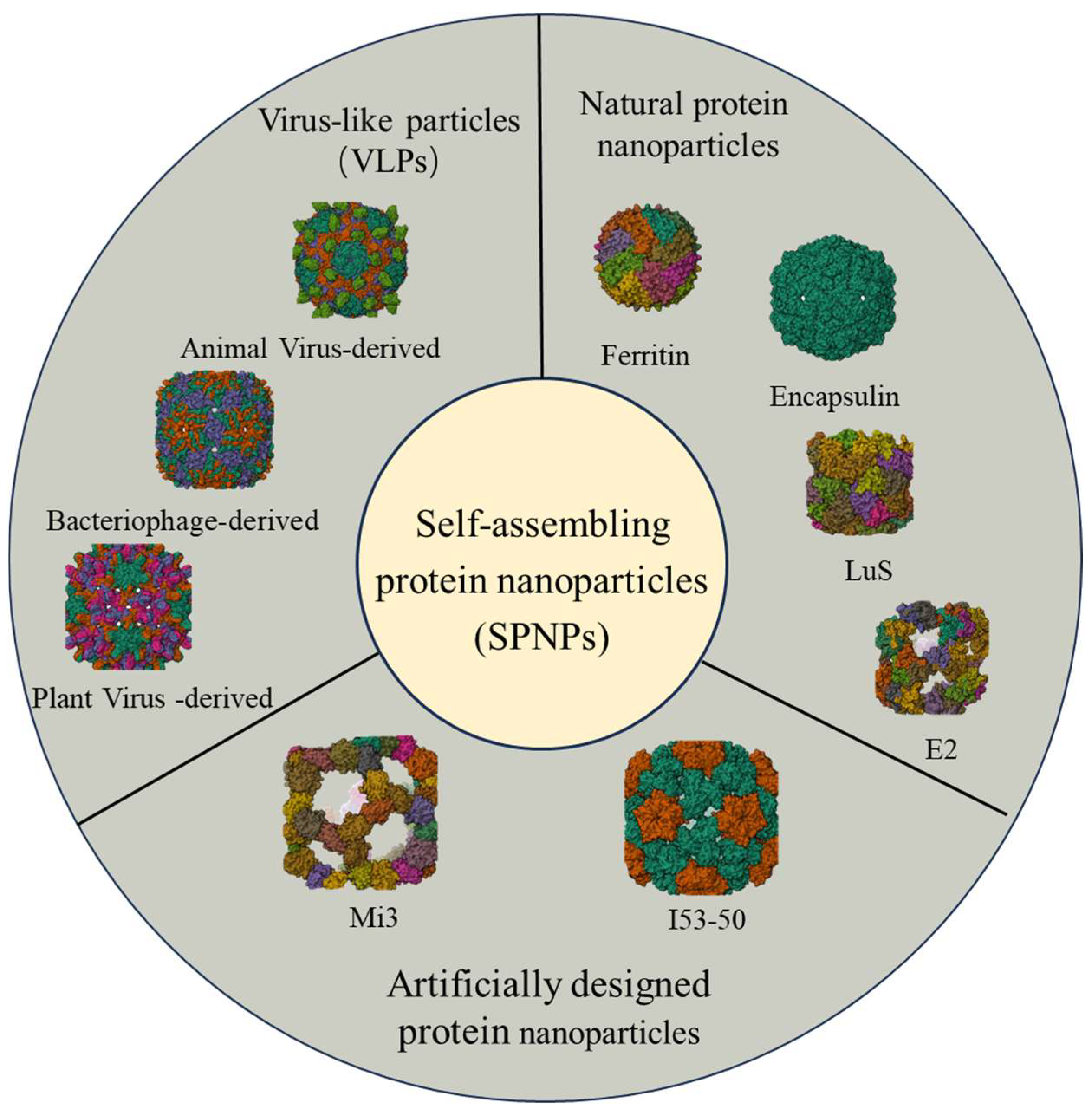
2. VLPs in Vaccine Research
2.1. Animal Virus-Derived VLPs
2.2. Bacteriophage-Derived VLPs
2.3. Plant Virus-Derived VLPs

| VLPs Platform | Target Pathogen/Disease | Vaccine Antigen/Epitope | Expression System | Reference |
|---|---|---|---|---|
| Alfalfa mosaic virus (AWV) | Plasmodium spp. | Pfs25 | Eukaryotic (Tobacco plant) | [36,37] |
| Papaya mosaic virus (PapMV) | Hepatitis C virus (HCV) | E2 antigenic epitope | Prokaryotic (E. coli) | [47] |
| Cowpea mosaic virus (CMV) | Dengue virus | Domain III of envelope protein type 1 (DV1-EDIII) | Prokaryotic (E. coli) | [39] |
| Physalis mosaic virus (PhMV) | Influenza A virus | M2e peptide | Prokaryotic (E. coli) | [41] |
| Maize mosaic virus (MaMV) | H3N8 influenza virus | M2e peptide | Prokaryotic (E. coli) | [40] |
| Cowpea chlorotic mottle virus (CCMV) | Clostridium tetani | Tetanus toxin epitope (TT830–843) | Prokaryotic (E. coli) | [48] |
| Group B Streptococcus type III | Capsular polysaccharide S9 peptide | Eukaryotic (Pichia pastoris) | [44] | |
| Peanut yellow mosaic virus (PYV) | Hepatitis B virus (HBV) | preS1 domain | Prokaryotic (E. coli) | [38] |
| Felis catus mite | Fel d 1 allergen | Prokaryotic (E. coli) | [49] | |
| Cowpea mosaic virus (CMV) | Psoriasis/Alzheimer’s disease/Cat allergy | IL-17/β-amyloid peptide/Fel d 1 | Prokaryotic (E. coli) | [44] |
| Tobacco mosaic virus (TMV) | Rabbit papillomavirus (CRPV and ROPV) | L2 capsid epitope | Prokaryotic (E. coli) | [50] |
| Physalis mosaic virus (PhMV) | Breast cancer | CH401 peptide derived from HER2 | Prokaryotic (E. coli) | [51] |
| Turnip mosaic virus (TuMV) | Allergy | Prup3 allergen | Eukaryotic (Tobacco plant) | [52] |
| Cowpea mosaic virus (CMV) | Allergy | IgE-Fc fragment | Eukaryotic (HEK293F cells) | [53] |
3. Natural Protein Nanoparticles in Vaccine Research
3.1. Ferritin in Vaccine Research [63]
3.2. Encapsulin in Vaccine Research
3.3. Lumazine Synthase in Vaccine Research
3.4. Pyruvate Dehydrogenase E2 in Vaccine Research
4. Artificially Designed Protein Nanoparticles in Vaccine Research
| Nanoparticle Platform | Target Pathogen/ Antigen | Immunogenicity or Detection Performance | References |
|---|---|---|---|
| Mi3 | SARS-CoV-2 (RBD) | Induced broad-spectrum neutralizing antibodies effective against multiple variants | [130,131] |
| CSFV (E2) | Increased antibody titers tenfold and enhanced cross-genotype protection. | [132] | |
| Influenza A (NA) | Elicited higher antibody titers and protection against H1N1 and H3N2 | [133] | |
| H9N2 (HA1) | Provided effective protection against lethal challenges from multiple H9N2 strains | [134] | |
| SARS-CoV-2 | Enhanced neutralization against wild-type and variant strains | [135] | |
| PEDV (S1) | Promoted stronger humoral and cellular immune responses | [136] | |
| Aflatoxin B1/Ochratoxin A nanobodies | Enabled highly sensitive detection of both mycotoxins | [137] | |
| I53-50 | Respiratory syncytial virus (DS-Cav1/Sc9-10) | Conjugating RSV pre-fusion F to NPM significantly enhanced its immunogenicity, stability, and bioactivity compared to preF displayed on the I53-50 carrier | [138] |
| SARS-CoV-2 (RBD) | Elicited strong neutralizing antibodies and effective protection | [122,139] | |
| RSV (F glycoprotein) | Induced tenfold higher neutralizing antibody levels than DS-Cav1 | [123] | |
| EBV (gB) | Induced potent and durable neutralizing antibodies. | [140] | |
| Epstein–Barr (gb + gHgL) | Enhanced vaccine immunogenicity and neutralizing antibody induction | [141] | |
| Influenza (HA) | Generated broad cross-protective neutralizing antibodies | [142] | |
| EBOV and SUDV (GP antigens) | Provides broad protection against both Zaire and Sudan strains in mouse and guinea pig models | [143] | |
| P. falciparum (HLA class I-restricted epitopes) | elicited increased IFN-γ T-cell Responses against the inserted epitopes and significantly higher antibody | [144] | |
| I3-01 | Filovirus (glycoproteins) | Antibody responses induced by filovirus GP trimers and SApNPs bearing wildtype or modified glycans are assessed in mice | [125] |
| EBV (gp350) | Elicited higher titers of total IgG and neutralizing antibodies | [126] | |
| SARS-CoV-2 (RBD) | Enhanced SARS-CoV-2 clearance from the nose and lungs of Syrian hamsters | [127] |
5. Summary and Outlook
- (1)
- Mechanistic elucidation—systematically clarifying the molecular interactions between different protein nanoparticles and the immune system;
- (2)
- Structural optimization—leveraging structural biology and computational design for multivalent antigen co-loading, precise targeting, and controlled release;
- (3)
- Safety and protective efficacy—investigating biosafety and the mechanisms of durable protection;
- (4)
- Broad-spectrum applications—extending the platform use to emerging pathogens, drug-resistant bacteria, and tumor immunotherapy [153].
Author Contributions
Funding
Data Availability Statement
Conflicts of Interest
Abbreviations
| SPNPs | Self-assembling protein nanoparticles |
| SARS-CoV-2 | Severe Acute Respiratory Syndrome Coronavirus 2 |
| RBD | Receptor-binding domain |
| VLPs | Virus-like particles |
| RSV | Respiratory syncytial virus |
| EBV | Epstein–Barr Virus |
References
- Bi, J.; Mo, C.; Li, S.; Huang, M.; Lin, Y.; Yuan, P.; Liu, Z.; Jia, B.; Xu, S. Immunotoxicity of metal and metal oxide nanoparticles: From toxic mechanisms to metabolism and outcomes. Biomater. Sci. 2023, 11, 4151–4183. [Google Scholar] [CrossRef]
- Lehman, S.E.; Benkstein, K.D.; Cleveland, T.E.; Anderson, K.W.; Carrier, M.J.; Vreeland, W.N. Particle Metrology Approach to Understanding How Storage Conditions Affect Long-Term Liposome Stability. Langmuir 2023, 39, 12313–12323. [Google Scholar] [CrossRef]
- Grego, E.A.; Siddoway, A.C.; Uz, M.; Liu, L.; Christiansen, J.C.; Ross, K.A.; Kelly, S.M.; Mallapragada, S.K.; Wannemuehler, M.J.; Narasimhan, B. Polymeric Nanoparticle-Based Vaccine Adjuvants and Delivery Vehicles. Curr. Top. Microbiol. Immunol. 2020, 433, 29–76. [Google Scholar] [CrossRef]
- Bhaskar, S.; Lim, S. Engineering protein nanocages as carriers for biomedical applications. NPG Asia Mater. 2017, 9, e371. [Google Scholar] [CrossRef]
- João, J.; Prazeres, D.M.F. Manufacturing of non-viral protein nanocages for biotechnological and biomedical applications. Front. Bioeng. Biotechnol. 2023, 11. [Google Scholar] [CrossRef] [PubMed]
- Lutz, I.D.; Wang, S.; Norn, C.; Courbet, A.; Borst, A.J.; Zhao, Y.T.; Dosey, A.; Cao, L.; Xu, J.; Leaf, E.M.; et al. Top-down design of protein architectures with reinforcement learning. Science 2023, 380, 266–273. [Google Scholar] [CrossRef]
- Berreiros-Hortala, H.; Vilchez-Pinto, G.; Diaz-Perales, A.; Garrido-Arandia, M.; Tome-Amat, J. Virus-like Particles as Vaccines for Allergen-Specific Therapy: An Overview of Current Developments. Int. J. Mol. Sci. 2024, 25, 7429. [Google Scholar] [CrossRef]
- Gomes, A.C.; Mohsen, M.; Bachmann, M.F. Harnessing Nanoparticles for Immunomodulation and Vaccines. Vaccines 2017, 5, 6. [Google Scholar] [CrossRef]
- Zhang, Y.; Ardejani, M.S.; Orner, B.P. Design and Applications of Protein-Cage-Based Nanomaterials. Chem. Asian J. 2016, 11, 2814–2828. [Google Scholar] [CrossRef]
- Tariq, H.; Batool, S.; Asif, S.; Ali, M.; Abbasi, B.H. Virus-Like Particles: Revolutionary Platforms for Developing Vaccines Against Emerging Infectious Diseases. Front. Microbiol. 2022, 12, 790121. [Google Scholar] [CrossRef]
- Bachmann, M.F.; Jennings, G.T. Vaccine delivery: A matter of size, geometry, kinetics and molecular patterns. Nat. Rev. Immunol. 2010, 10, 787–796. [Google Scholar] [CrossRef]
- Sun, H.Y.; Acharya, D.; Paul, A.M.; Lai, H.F.; He, J.Y.; Bai, F.W.; Chen, Q. Antibody-Dependent Enhancement Activity of a Plant-Made Vaccine against West Nile Virus. Vaccines 2023, 11, 197. [Google Scholar] [CrossRef]
- Hager, K.J.; Marc, G.P.; Gobeil, P.; Diaz, R.S.; Heizer, G.; Llapur, C.; Makarkov, A.I.; Vasconcellos, E.; Pillet, S.; Riera, F.; et al. Efficacy and Safety of a Recombinant Plant-Based Adjuvanted Covid-19 Vaccine. N. Engl. J. Med. 2022, 386, 2084–2096. [Google Scholar] [CrossRef]
- Chappuis, F.; Farinelli, T.; Deckx, H.; Sarnecki, M.; Go, O.; Salzgeber, Y.; Stals, C. Immunogenicity and estimation of antibody persistence following vaccination with an inactivated virosomal hepatitis A vaccine in adults: A 20-year follow-up study. Vaccine 2017, 35, 1448–1454. [Google Scholar] [CrossRef]
- Keating, G.M.; Noble, S. Recombinant Hepatitis B Vaccine (Engerix-B®). Drugs 2003, 63, 1021–1051. [Google Scholar] [CrossRef]
- Etzion, O.; Novack, V.; Perl, Y.; Abel, O.; Schwartz, D.; Munteanu, D.; Abufreha, N.; Ben-Yaakov, G.; Maoz, E.D.; Moshaklo, A.; et al. Sci-B-VacTMVs ENGERIX-B Vaccines for Hepatitis B Virus in Patients with Inflammatory Bowel Diseases: A Randomised Controlled Trial. J. Crohn’s Colitis 2016, 10, 905–912. [Google Scholar] [CrossRef][Green Version]
- Abravanel, F.; Lhomme, S. Hecolin vaccine: Long-term efficacy against HEV for a three-dose regimen. Lancet 2024, 403, 782–783. [Google Scholar] [CrossRef]
- Termrungruanglert, W.; Khemapech, N.; Vasuratna, A.; Havanond, P.; Tantitamit, T. Cost-effectiveness analysis of single-dose or 2-dose of bivalent, quadrivalent, or nonavalent HPV vaccine in a low/middle-income country setting. J. Gynecol. Oncol. 2024, 35, e85. [Google Scholar] [CrossRef]
- Weber, W.C.; Streblow, D.N.; Coffey, L.L. Chikungunya Virus Vaccines: A Review of IXCHIQ and PXVX0317 from Pre-Clinical Evaluation to Licensure. Biodrugs 2024, 38, 727–742. [Google Scholar] [CrossRef]
- Kekarainen, T.; Montoya, M.; Dominguez, J.; Mateu, E.; Segalés, J. Porcine circovirus type 2 (PCV2) viral components immunomodulate recall antigen responses. Vet. Immunol. Immunopathol. 2008, 124, 41–49. [Google Scholar] [CrossRef]
- Wu, P.C.; Lin, W.L.; Wu, C.M.; Chi, J.N.; Chien, M.S.; Huang, C.J. Characterization of porcine circovirus type 2 (PCV2) capsid particle assembly and its application to virus-like particle vaccine development. Appl. Microbiol. Biotechnol. 2012, 95, 1501–1507. [Google Scholar] [CrossRef]
- Li, H.Z.; Liu, P.; Dong, H.; Dekker, A.; Harmsen, M.M.; Guo, H.C.; Wang, X.X.; Sun, S.Q. Foot-and-mouth disease virus antigenic landscape and reduced immunogenicity elucidated in atomic detail. Nat. Commun. 2024, 15, 8774. [Google Scholar] [CrossRef]
- Kheirvari, M.; Liu, H.; Tumban, E. Virus-like Particle Vaccines and Platforms for Vaccine Development. Viruses 2023, 15, 1109. [Google Scholar] [CrossRef]
- Yang, L.; Xiao, A.; Wang, H.; Zhang, X.; Zhang, Y.; Li, Y.; Wei, Y.; Liu, W.; Chen, C. A VLP-Based Vaccine Candidate Protects Mice against Japanese Encephalitis Virus Infection. Vaccines 2022, 10, 197. [Google Scholar] [CrossRef]
- Garg, H.; Mehmetoglu-Gurbuz, T.; Joshi, A. Virus Like Particles (VLP) as multivalent vaccine candidate against Chikungunya, Japanese Encephalitis, Yellow Fever and Zika Virus. Sci. Rep. 2020, 10, 4017. [Google Scholar] [CrossRef]
- Dang, M.; Wu, L.J.; Zhang, S.R.; Zhu, J.R.; Hu, Y.Z.; Yang, C.X.; Zhang, X.Y. MS2 Virus-like Particles as a Versatile Peptide Presentation Platform: Insights into the Deterministic Abilities for Accommodating Heterologous Peptide Lengths. ACS Synth. Biol. 2023, 12, 3704–3715. [Google Scholar] [CrossRef]
- Zhou, G.; Luo, Z.; Zhang, Z.; Cao, S.; Li, Y. MS2 virus-like particles as a versatile platform for multi-disease vaccines: A review. Front. Immunol. 2025, 16. [Google Scholar] [CrossRef]
- Essus, V.A.; Souza, G.S.E.; Nunes, G.H.P.; Oliveira, J.D.; de Faria, B.M.; Romao, L.F.; Cortines, J.R. Bacteriophage P22 Capsid as a Pluripotent Nanotechnology Tool. Viruses 2023, 15, 516. [Google Scholar] [CrossRef]
- Yenkoidiok-Douti, L.; Williams, A.E.; Canepa, G.E.; Molina-Cruz, A.; Barillas-Mury, C. Engineering a Virus-Like Particle as an Antigenic Platform for a Pfs47-Targeted Malaria Transmission-Blocking Vaccine. Sci. Rep. 2019, 9, 16833. [Google Scholar] [CrossRef]
- Ho, P.L.; Tissot, A.C.; Renhofa, R.; Schmitz, N.; Cielens, I.; Meijerink, E.; Ose, V.; Jennings, G.T.; Saudan, P.; Pumpens, P.; et al. Versatile Virus-Like Particle Carrier for Epitope Based Vaccines. PLoS ONE 2010, 5, e9809. [Google Scholar] [CrossRef]
- Ximba, P.; Chapman, R.; Meyers, A.; Margolin, E.; van Diepen, M.T.; Sander, A.F.; Woodward, J.; Moore, P.L.; Williamson, A.-L.; Rybicki, E.P. Development of a synthetic nanoparticle vaccine presenting the HIV-1 envelope glycoprotein. Nanotechnology 2022, 33, 485102. [Google Scholar] [CrossRef]
- Fougeroux, C.; Goksøyr, L.; Idorn, M.; Soroka, V.; Myeni, S.K.; Dagil, R.; Janitzek, C.M.; Søgaard, M.; Aves, K.-L.; Horsted, E.W.; et al. Capsid-like particles decorated with the SARS-CoV-2 receptor-binding domain elicit strong virus neutralization activity. Nat. Commun. 2021, 12, 324. [Google Scholar] [CrossRef]
- Hills, R.A.; Howarth, M. Virus-like particles against infectious disease and cancer: Guidance for the nano-architect. Curr. Opin. Biotechnol. 2022, 73, 346–354. [Google Scholar] [CrossRef]
- Fiedler, J.D.; Higginson, C.; Hovlid, M.L.; Kislukhin, A.A.; Castillejos, A.; Manzenrieder, F.; Campbell, M.G.; Voss, N.R.; Potter, C.S.; Carragher, B.; et al. Engineered Mutations Change the Structure and Stability of a Virus-Like Particle. Biomacromolecules 2012, 13, 2339–2348. [Google Scholar] [CrossRef]
- Kant, R.; Llauró, A.; Rayaprolu, V.; Qazi, S.; de Pablo, P.J.; Douglas, T.; Bothner, B. Changes in the stability and biomechanics of P22 bacteriophage capsid during maturation. Biochim. Biophys. Acta Gen. Subj. 2018, 1862, 1492–1504. [Google Scholar] [CrossRef]
- Carvalho, L.H.; Jones, R.M.; Chichester, J.A.; Mett, V.; Jaje, J.; Tottey, S.; Manceva, S.; Casta, L.J.; Gibbs, S.K.; Musiychuk, K.; et al. A Plant-Produced Pfs25 VLP Malaria Vaccine Candidate Induces Persistent Transmission Blocking Antibodies against Plasmodium falciparum in Immunized Mice. PLoS ONE 2013, 8, e79538. [Google Scholar] [CrossRef]
- Chichester, J.A.; Green, B.J.; Jones, R.M.; Shoji, Y.; Miura, K.; Long, C.A.; Lee, C.K.; Ockenhouse, C.F.; Morin, M.J.; Streatfield, S.J.; et al. Safety and immunogenicity of a plant-produced Pfs25 virus-like particle as a transmission blocking vaccine against malaria: A Phase 1 dose-escalation study in healthy adults. Vaccine 2018, 36, 5865–5871. [Google Scholar] [CrossRef]
- Kalnciema, I.; Skrastina, D.; Ose, V.; Pumpens, P.; Zeltins, A. Potato Virus Y-Like Particles as a New Carrier for the Presentation of Foreign Protein Stretches. Mol. Biotechnol. 2011, 52, 129–139. [Google Scholar] [CrossRef]
- Sobczak, J.M.; Barkovska, I.; Balke, I.; Rothen, D.A.; Mohsen, M.O.; Skrastina, D.; Ogrina, A.; Martina, B.; Jansons, J.; Bogans, J.; et al. Identifying Key Drivers of Efficient B Cell Responses: On the Role of T Help, Antigen-Organization, and Toll-like Receptor Stimulation for Generating a Neutralizing Anti-Dengue Virus Response. Vaccines 2024, 12, 661. [Google Scholar] [CrossRef]
- Alsharifi, M.; Leclerc, D.; Rivest, M.; Babin, C.; López-Macias, C.; Savard, P. A Novel M2e Based Flu Vaccine Formulation for Dogs. PLoS ONE 2013, 8, e77084. [Google Scholar] [CrossRef]
- Blokhina, E.A.; Mardanova, E.S.; Zykova, A.A.; Shuklina, M.A.; Stepanova, L.A.; Tsybalova, L.M.; Ravin, N.V. Chimeric Virus-like Particles of Physalis Mottle Virus as Carriers of M2e Peptides of Influenza a Virus. Viruses 2024, 16, 1802. [Google Scholar] [CrossRef]
- McCormick, A.A.; Corbo, T.A.; Wykoff-Clary, S.; Palmer, K.E.; Pogue, G.P. Chemical Conjugate TMV−Peptide Bivalent Fusion Vaccines Improve Cellular Immunity and Tumor Protection. Bioconjugate Chem. 2006, 17, 1330–1338. [Google Scholar] [CrossRef]
- McCormick, A.A.; Corbo, T.A.; Wykoff-Clary, S.; Nguyen, L.V.; Smith, M.L.; Palmer, K.E.; Pogue, G.P. TMV-peptide fusion vaccines induce cell-mediated immune responses and tumor protection in two murine models. Vaccine 2006, 24, 6414–6423. [Google Scholar] [CrossRef]
- Zeltins, A.; West, J.; Zabel, F.; El Turabi, A.; Balke, I.; Haas, S.; Maudrich, M.; Storni, F.; Engeroff, P.; Jennings, G.T.; et al. Incorporation of tetanus-epitope into virus-like particles achieves vaccine responses even in older recipients in models of psoriasis, Alzheimer’s and cat allergy. npj Vaccines 2017, 2, 3096. [Google Scholar] [CrossRef]
- Fettelschoss-Gabriel, A.; Fettelschoss, V.; Thoms, F.; Giese, C.; Daniel, M.; Olomski, F.; Kamarachev, J.; Birkmann, K.; Bühler, M.; Kummer, M.; et al. Treating insect-bite hypersensitivity in horses with active vaccination against IL-5. J. Allergy Clin. Immunol. 2018, 142, 1194–1205. [Google Scholar] [CrossRef]
- von Loga, I.S.; El-Turabi, A.; Jostins, L.; Miotla-Zarebska, J.; Mackay-Alderson, J.; Zeltins, A.; Parisi, I.; Bachmann, M.F.; Vincent, T.L. Active immunisation targeting nerve growth factor attenuates chronic pain behaviour in murine osteoarthritis. Ann. Rheum. Dis. 2019, 78, 672–675. [Google Scholar] [CrossRef]
- Denis, J.; Majeau, N.; Acosta-Ramirez, E.; Savard, C.; Bedard, M.C.; Simard, S.; Lecours, K.; Bolduc, M.; Pare, C.; Willems, B.; et al. Immunogenicity of papaya mosaic virus-like particles fused to a hepatitis C virus epitope: Evidence for the critical function of multimerization. Virology 2007, 363, 59–68. [Google Scholar] [CrossRef]
- Zinkhan, S.; Ogrina, A.; Balke, I.; Reseviča, G.; Zeltins, A.; de Brot, S.; Lipp, C.; Chang, X.; Zha, L.; Vogel, M.; et al. The impact of size on particle drainage dynamics and antibody response. J. Control. Release 2021, 331, 296–308. [Google Scholar] [CrossRef]
- Ogrina, A.; Skrastina, D.; Balke, I.; Kalnciema, I.; Jansons, J.; Bachmann, M.F.; Zeltins, A. Comparison of Bacterial Expression Systems Based on Potato Virus Y-like Particles for Vaccine Generation. Vaccines 2022, 10, 485. [Google Scholar] [CrossRef]
- Palmer, K.E.; Benko, A.; Doucette, S.A.; Cameron, T.I.; Foster, T.; Hanley, K.M.; McCormick, A.A.; McCulloch, M.; Pogue, G.P.; Smith, M.L.; et al. Protection of rabbits against cutaneous papillomavirus infection using recombinant tobacco mosaic virus containing L2 capsid epitopes. Vaccine 2006, 24, 5516–5525. [Google Scholar] [CrossRef]
- Hu, H.; Steinmetz, N.F. Development of a Virus-Like Particle-Based Anti-HER2 Breast Cancer Vaccine. Cancers 2021, 13, 2909. [Google Scholar] [CrossRef]
- Pazos-Castro, D.; Margain, C.; Gonzalez-Klein, Z.; Yuste-Calvo, C.; Garrido-Arandia, M.; Zurita, L.; Esteban, V.; Tome-Amat, J.; Diaz-Perales, A.; Ponz, F. Suitability of potyviral recombinant virus-like particles bearing a complete food allergen for immunotherapy vaccines. Front. Immunol. Erratum in Front. Immunol. 2023, 14, 1133046. https://doi.org/10.3389/fimmu.2023.1133046. 2022, 13, 986823. [Google Scholar] [CrossRef]
- Gharailoo, Z.; Plattner, K.; Augusto, G.; Engeroff, P.; Vogel, M.; Bachmann, M.F. Generation of a virus-like particles based vaccine against IgE. Allergy 2024, 79, 2207–2221. [Google Scholar] [CrossRef]
- González-Domínguez, I.; Lorenzo, E.; Bernier, A.; Cervera, L.; Gòdia, F.; Kamen, A. A Four-Step Purification Process for Gag VLPs: From Culture Supernatant to High-Purity Lyophilized Particles. Vaccines 2021, 9, 1154. [Google Scholar] [CrossRef]
- Wong, Z.W.; Yang, D. Disassembly and reassembly of AP205 virus-like particles and the removal of bound RNA for cargo encapsulation. Int. J. Biol. Macromol. 2025, 315, 144641. [Google Scholar] [CrossRef]
- Kawana, K.; Yoshikawa, H.; Taketani, Y.; Yoshiike, K.; Kanda, T. In vitro construction of pseudovirions of human papillomavirus type 16: Incorporation of plasmid DNA into reassembled L1/L2 capsids. J. Virol. 1998, 72, 10298–10300. [Google Scholar] [CrossRef]
- Schoonen, L.; Maassen, S.; Nolte, R.J.M.; van Hest, J.C.M. Stabilization of a Virus-Like Particle and Its Application as a Nanoreactor at Physiological Conditions. Biomacromolecules 2017, 18, 3492–3497. [Google Scholar] [CrossRef]
- Park, E.Y.; Minkner, R. A systematic approach for scalable purification of virus-like particles. Protein Expr. Purif. 2025, 228, 106664. [Google Scholar] [CrossRef]
- Gupta, R.; Arora, K.; Roy, S.S.; Joseph, A.; Rastogi, R.; Arora, N.M.; Kundu, P.K. Platforms, advances, and technical challenges in virus-like particles-based vaccines. Front. Immunol. 2023, 14, 1123805. [Google Scholar] [CrossRef]
- Zhang, L.; Xu, W.; Ma, X.; Sun, X.; Fan, J.; Wang, Y. Virus-like Particles as Antiviral Vaccine: Mechanism, Design, and Application. Biotechnol. Bioprocess. Eng. 2023, 28, 1–16. [Google Scholar] [CrossRef]
- Gallet, S.; Hannani, D.; Dergan-Dylon, S.; Vassal-Stermann, E.; Bally, I.; Chevillard, C.; Fenel, D.; Epaulard, O.; Poignard, P.; Fender, P. Pre-Existing Anti-Vector Immunity to Adenovirus-Inspired VLP Vaccines Shows an Adjuvant-Dependent Antagonism. Vaccines 2025, 13, 238. [Google Scholar] [CrossRef]
- Ra, J.-S.; Shin, H.-H.; Kang, S.; Do, Y. Lumazine synthase protein cage nanoparticles as antigen delivery nanoplatforms for dendritic cell-based vaccine development. Clin. Exp. Vaccine Res. 2014, 3, 227–234. [Google Scholar] [CrossRef]
- Zhang, Y.; Ru, Y.; Hao, R.; Li, Y.; Zhao, L.; Yang, Y.; Lu, B.; Liu, H.; Zheng, H. Applications of ferritin nanoparticles in biological fields. Chin. J. Biotechnol. 2025, 41, 2501–2518. [Google Scholar] [CrossRef]
- Srivastava, A.K.; Scalcione, L.J.; Arosio, P.; Bou-Abdallah, F. Hyperthermostable recombinant human heteropolymer ferritin derived from a novel plasmid design. Protein Sci. 2023, 32, e4543. [Google Scholar] [CrossRef]
- Perotti, M.; Marcandalli, J.; Demurtas, D.; Sallusto, F.; Perez, L. Rationally designed Human Cytomegalovirus gB nanoparticle vaccine with improved immunogenicity. PLoS Pathog. 2020, 16, e1009169. [Google Scholar] [CrossRef]
- Fan, K.; Jiang, B.; Guan, Z.; He, J.; Yang, D.; Xie, N.; Nie, G.; Xie, C.; Yan, X. Fenobody: A Ferritin-Displayed Nanobody with High Apparent Affinity and Half-Life Extension. Anal. Chem. 2018, 90, 5671–5677. [Google Scholar] [CrossRef]
- Song, N.N.; Zhang, J.L.; Zhai, J.; Hong, J.J.; Yuan, C.; Liang, M.M. Ferritin: A Multifunctional Nanoplatform for Biological Detection, Imaging Diagnosis, and Drug Delivery. Acc. Chem. Res. 2021, 54, 3313–3325. [Google Scholar] [CrossRef]
- Fu, D.; Wang, W.M.; Zhang, Y.; Zhang, F.; Yang, P.Y.; Yang, C.; Tian, Y.F.; Yao, R.Q.; Jian, J.W.; Sun, Z.X.; et al. Self-assembling nanoparticle engineered from the ferritinophagy complex as a rabies virus vaccine candidate. Nat. Commun. 2024, 15, 8601. [Google Scholar] [CrossRef]
- Liang, C.; Li, R.; Pu, Z.; Chen, R.; Li, Y.; Chen, S.; Feng, J.; Liu, J.; Bai, Y.; Qin, X.; et al. Targeting heptad repeats and fusion peptide: Nanoparticle vaccine elicits mucosal immune response against SARS-CoV-2 variants. J. Nanobiotechnology 2025, 23, 483. [Google Scholar] [CrossRef]
- Sliepen, K.; Ozorowski, G.; Burger, J.A.; van Montfort, T.; Stunnenberg, M.; LaBranche, C.; Montefiori, D.C.; Moore, J.P.; Ward, A.B.; Sanders, R.W. Presenting native-like HIV-1 envelope trimers on ferritin nanoparticles improves their immunogenicity. Retrovirology 2015, 12, 82. [Google Scholar] [CrossRef]
- Wang, W.J.; Zhou, X.X.; Bian, Y.J.; Wang, S.; Chai, Q.; Guo, Z.Q.; Wang, Z.N.; Zhu, P.; Peng, H.; Yan, X.Y.; et al. Dual-targeting nanoparticle vaccine elicits a therapeutic antibody response against chronic hepatitis B. Nat. Nanotechnol. 2020, 15, 406–416. [Google Scholar] [CrossRef]
- Yin, C.H.; Yao, Y.F.; Yang, P.P.; Liu, H.; Gao, G.; Peng, Y.; Chen, M.Y.; Lu, M.Q.; Zhang, X.K.; Guo, W.W.; et al. A highly effective ferritin-based divalent nanoparticle vaccine shields Syrian hamsters against lethal Nipah virus. Front. Immunol. 2024, 15, 1387811. [Google Scholar] [CrossRef]
- Li, Y.Y.; Tian, S.Y.; Ai, Y.B.; Hu, Z.L.; Ma, C.; Fu, M.J.; Xu, Z.Q.; Li, Y.; Liu, S.Y.; Zou, Y.J.; et al. A nanoparticle vaccine displaying varicella-zoster virus gE antigen induces a superior cellular immune response than a licensed vaccine in mice and non-human primates. Front. Immunol. 2024, 15, 1419634. [Google Scholar] [CrossRef]
- Yang, D.; Su, M.; Guo, D.; Zhao, F.; Wang, M.; Liu, J.; Zhou, J.; Sun, Y.; Yang, X.; Qi, S.; et al. Combination of S1–N–Terminal and S1–C–Terminal Domain Antigens Targeting Double Receptor-Binding Domains Bolsters Protective Immunity of a Nanoparticle Vaccine against Porcine Epidemic Diarrhea Virus. ACS Nano 2024, 18, 12235–12260. [Google Scholar] [CrossRef]
- Sun, Y.Y.; Gao, Y.N.; Su, T.J.; Zhang, L.J.; Zhou, H.R.; Zhang, J.; Sun, H.F.; Bai, J.; Jiang, P. Nanoparticle Vaccine Triggers Interferon-Gamma Production and Confers Protective Immunity against Porcine Reproductive and Respiratory Syndrome Virus. ACS Nano 2025, 19, 852–870. [Google Scholar] [CrossRef]
- Chen, Y.B.; Hu, Y.; Chen, H.C.; Li, X.M.; Qian, P. A ferritin nanoparticle vaccine for foot-and-mouth disease virus elicited partial protection in mice. Vaccine 2020, 38, 5647–5652. [Google Scholar] [CrossRef]
- Song, J.X.; Wang, M.X.; Zhou, L.; Tian, P.P.; Sun, Z.Y.; Sun, J.R.; Wang, X.N.; Zhuang, G.Q.; Jiang, D.W.; Wu, Y.A.; et al. A candidate nanoparticle vaccine comprised of multiple epitopes of the African swine fever virus elicits a robust immune response. J. Nanobiotechnol. 2023, 21, 424. [Google Scholar] [CrossRef]
- Wang, W.J.; Liu, Z.D.; Zhou, X.X.; Guo, Z.Q.; Zhang, J.; Zhu, P.; Yao, S.; Zhu, M.Z. Ferritin nanoparticle-based SpyTag/SpyCatcher-enabled click vaccine for tumor immunotherapy. Nanomed. Nanotechnol. Biol. Med. 2019, 16, 69–78. [Google Scholar] [CrossRef]
- Salinas, N.D.; Ma, R.; Mcaleese, H.; Ouahes, T.; Long, C.A.; Miura, K.; Lambert, L.E.; Tolia, N.H. A Self-Assembling Pfs230D1-Ferritin Nanoparticle Vaccine Has Potent and Durable Malaria Transmission-Reducing Activity. Vaccines 2024, 12, 546. [Google Scholar] [CrossRef]
- Wei, Y.J.; Cheng, X.; Liao, Y.L.; Zeng, S.; Li, Y.H.; Zhang, Y.W.; Gao, C.; Zhang, Y.; Wan, J.Q.; Gu, J.; et al. Recombinant Pseudomonas aeruginosa flagellin delivered using ferritin nanoparticles provides enhanced cross-protection against lung infection in mice. Mol. Immunol. 2023, 163, 235–242. [Google Scholar] [CrossRef]
- Fang, Q.N.; Lu, X.Y.; Zhu, Y.Q.; Lv, X.; Yu, F.; Ma, X.C.; Liu, B.F.; Zhang, H. Development of a PCSK9-targeted nanoparticle vaccine to effectively decrease the hypercholesterolemia. Cell Rep. Med. 2024, 5, 101614. [Google Scholar] [CrossRef]
- Ober Shepherd, B.L.; Scott, P.T.; Hutter, J.N.; Lee, C.; McCauley, M.D.; Guzman, I.; Bryant, C.; McGuire, S.; Kennedy, J.; Chen, W.-H.; et al. SARS-CoV-2 recombinant spike ferritin nanoparticle vaccine adjuvanted with Army Liposome Formulation containing monophosphoryl lipid A and QS-21: A phase 1, randomised, double-blind, placebo-controlled, first-in-human clinical trial. Lancet Microbe 2024, 5, e581–e593. [Google Scholar] [CrossRef]
- Sutter, M.; Boehringer, D.; Gutmann, S.; Guenther, S.; Prangishvili, D.; Loessner, M.J.; Stetter, K.O.; Weber-Ban, E.; Ban, N. Structural basis of enzyme encapsulation into a bacterial nanocompartment. Nat. Struct. Mol. Biol. 2008, 15, 939–947. [Google Scholar] [CrossRef]
- Zhang, Y.; Ru, Y.; Zhao, L.; Hao, R.; Yang, Y.; Shen, C.; Shi, Z.; Zheng, H. Encapsulin nanoparticle-conjugated p54 protein boosts immune responses against African swine fever virus. Int. J. Biol. Macromol. 2025, 311, 143912. [Google Scholar] [CrossRef]
- Valdés-Stauber, N.; Scherer, S. Isolation and characterization of Linocin M18, a bacteriocin produced by Brevibacterium linens. Appl. Environ. Microbiol. 1994, 60, 3809–3814. [Google Scholar] [CrossRef]
- Almeida, A.V.; Carvalho, A.J.; Pereira, A.S. Encapsulin nanocages: Protein encapsulation and iron sequestration. Coord. Chem. Rev. 2021, 448, 214188. [Google Scholar] [CrossRef]
- Rahmanpour, R.; Bugg, T.D.H. Assembly in vitro of Rhodococcus jostii RHA1 encapsulin and peroxidase DypB to form a nanocompartment. FEBS J. 2013, 280, 2097–2104. [Google Scholar] [CrossRef]
- Jones, J.A.; Benisch, R.; Giessen, T.W. Encapsulin cargo loading: Progress and potential. J. Mater. Chem. B 2023, 11, 4377–4388. [Google Scholar] [CrossRef]
- Kanekiyo, M.; Bu, W.; Joyce, M.G.; Meng, G.; Whittle, J.R.; Baxa, U.; Yamamoto, T.; Narpala, S.; Todd, J.P.; Rao, S.S.; et al. Rational Design of an Epstein-Barr Virus Vaccine Targeting the Receptor-Binding Site. Cell 2015, 162, 1090–1100. [Google Scholar] [CrossRef]
- Khaleeq, S.; Sengupta, N.; Kumar, S.; Patel, U.R.; Rajmani, R.S.; Reddy, P.; Pandey, S.; Singh, R.; Dutta, S.; Ringe, R.P.; et al. Neutralizing Efficacy of Encapsulin Nanoparticles against SARS-CoV2 Variants of Concern. Viruses 2023, 15, 346. [Google Scholar] [CrossRef]
- Zhang, Y.; Ru, Y.; Zhao, L.; Hao, R.; Yang, Y.; Li, Y.; Zhang, R.; Jiang, C.; Zheng, H. Ferritin and Encapsulin Nanoparticles as Effective Vaccine Delivery Systems: Boosting the Immunogenicity of the African Swine Fever Virus C129R Protein. Viruses 2025, 17, 556. [Google Scholar] [CrossRef]
- Zhang, X.F.; Meining, W.; Fischer, M.; Bacher, A.; Ladenstein, R. X-ray structure analysis and crystallographic refinement of lumazine synthase from the hyperthermophile Aquifex aeolicus at 1.6 A resolution: Determinants of thermostability revealed from structural comparisons. J. Mol. Biol. 2001, 306, 1099–1114. [Google Scholar] [CrossRef]
- Azuma, Y.; Edwardson, T.G.W.; Hilvert, D. Tailoring lumazine synthase assemblies for bionanotechnology. Chem. Soc. Rev. 2018, 47, 3543–3557. [Google Scholar] [CrossRef]
- Zhang, X.F.; Konarev, P.V.; Petoukhov, M.V.; Svergun, D.I.; Xing, L.; Cheng, R.H.; Haase, I.; Fischer, M.; Bacher, A.; Ladenstein, R.; et al. Multiple assembly states of lumazine synthase: A model relating catalytic function and molecular assembly. J. Mol. Biol. 2006, 362, 753–770. [Google Scholar] [CrossRef]
- Azuma, Y.; Herger, M.; Hilvert, D. Diversification of Protein Cage Structure Using Circularly Permuted Subunits. J. Am. Chem. Soc. 2018, 140, 558–561. [Google Scholar] [CrossRef]
- Ladenstein, R.; Morgunova, E. Second career of a biosynthetic enzyme: Lumazine synthase as a virus-like nanoparticle in vaccine development. Biotechnol. Rep. 2020, 27, e00494. [Google Scholar] [CrossRef]
- Li, Q.L.; Zhang, J.S.; Deng, Q.; Liao, C.H.; Qian, J.; Chen, Z.L.; Lu, J.H. A Divalent Chikungunya and Zika Nanovaccine with Thermostable Self-Assembly Multivalent Scaffold LS-SUMO. Adv. Healthc. Mater. 2024, 13, e2303619. [Google Scholar] [CrossRef]
- Zhu, H.C.; Li, X.X.; Li, X.M.; Chen, H.C.; Qian, P. Protection against the H1N1 influenza virus using self-assembled nanoparticles formed by lumazine synthase and bearing the M2e peptide. Virology 2024, 597, 110162. [Google Scholar] [CrossRef]
- Cohen, K.W.; De Rosa, S.C.; Fulp, W.J.; deCamp, A.C.; Fiore-Gartland, A.; Mahoney, C.R.; Furth, S.; Donahue, J.; Whaley, R.E.; Ballweber-Fleming, L.; et al. A first-in-human germline-targeting HIV nanoparticle vaccine induced broad and publicly targeted helper T cell responses. Sci. Transl. Med. 2023, 15, eadf3309. [Google Scholar] [CrossRef]
- Peng, T.; Lee, H.; Lim, S. Isolating a Trimer Intermediate in the Self-Assembly of E2 Protein Cage. Biomacromolecules 2012, 13, 699–705. [Google Scholar] [CrossRef]
- Dalmau, M.; Lim, S.; Chen, H.C.; Ruiz, C.; Wang, S.W. Thermostability and Molecular Encapsulation Within an Engineered Caged Protein Scaffold. Biotechnol. Bioeng. 2008, 101, 654–664. [Google Scholar] [CrossRef]
- He, L.; de Val, N.; Morris, C.D.; Vora, N.; Thinnes, T.C.; Kong, L.; Azadnia, P.; Sok, D.; Zhou, B.; Burton, D.R.; et al. Presenting native-like trimeric HIV-1 antigens with self-assembling nanoparticles. Nat. Commun. 2016, 7. [Google Scholar] [CrossRef]
- Klatzkow, H.; Bhavsar-Burke, I.; Pearson, M.; Wentworth, B.J. Primary Biliary Cholangitis in 2025: A New Frontier. Am. J. Gastroenterol. 2025, 120, 2746–2749. [Google Scholar] [CrossRef] [PubMed]
- Yang, J.; Srinivasan, A.; Sun, Y.; Mrazek, J.; Shu, Z.; Kickhoefer, V.A.; Rome, L.H. Vault nanoparticles engineered with the protein transduction domain, TAT48, enhances cellular uptake. Integr. Biol. 2013, 5, 151–158. [Google Scholar] [CrossRef]
- Iravani, S.; Varma, R.S. Vault, viral, and virus-like nanoparticles for targeted cancer therapy. Mater. Adv. 2023, 4, 2909–2917. [Google Scholar] [CrossRef]
- Fang, T.; Zhu, W.; Li, C.; Zhang, F.; Gao, D.; Zhang, Z.P.; Liang, A.; Zhang, X.E.; Li, F. Role of Surface RGD Patterns on Protein Nanocages in Tumor Targeting Revealed Using Precise Discrete Models. Small 2019, 15, e1904838. [Google Scholar] [CrossRef]
- Guan, X.; Chang, Y.; Sun, J.; Song, J.; Xie, Y. Engineered Hsp Protein Nanocages for siRNA Delivery. Macromol. Biosci. 2018, 18, e1800013. [Google Scholar] [CrossRef]
- Uchida, M.; Maier, B.; Waghwani, H.K.; Selivanovitch, E.; Pay, S.L.; Avera, J.; Yun, E.; Sandoval, R.M.; Molitoris, B.A.; Zollman, A.; et al. The archaeal Dps nanocage targets kidney proximal tubules via glomerular filtration. J. Clin. Investig. 2019, 129, 3941–3951. [Google Scholar] [CrossRef]
- Lamontagne, F.; Arpin, D.; Cote-Cyr, M.; Khatri, V.; St-Louis, P.; Gauthier, L.; Archambault, D.; Bourgault, S. Engineered Curli Nanofilaments as a Self-Adjuvanted Antigen Delivery Platform. Adv. Healthc. Mater. 2023, 12, e2300224. [Google Scholar] [CrossRef] [PubMed]
- St-Louis, P.; Martin, C.; Khatri, V.; Bourgault, S.; Archambault, D. Intranasal delivery of a self-adjuvanted nanovaccine composed of the curli filaments and the highly conserved M2e epitope confers protection against influenza a virus in mice. Vaccine 2024, 42, 2144–2149. [Google Scholar] [CrossRef]
- Jegerlehner, A.; Wiesel, M.; Dietmeier, K.; Zabel, F.; Gatto, D.; Saudan, P.; Bachmann, M.F. Carrier induced epitopic suppression of antibody responses induced by virus-like particles is a dynamic phenomenon caused by carrier-specific antibodies. Vaccine 2010, 28, 5503–5512. [Google Scholar] [CrossRef]
- Kraft, J.C.; Pham, M.N.; Shehata, L.; Brinkkemper, M.; Boyoglu-Barnum, S.; Sprouse, K.R.; Walls, A.C.; Cheng, S.; Murphy, M.; Pettie, D.; et al. Antigen- and scaffold-specific antibody responses to protein nanoparticle immunogens. Cell Rep. Med. 2022, 3, 100780. [Google Scholar] [CrossRef]
- Wei, Y.; Wahome, N.; VanSlyke, G.; Whitaker, N.; Kumar, P.; Barta, M.L.; Picking, W.L.; Volkin, D.B.; Mantis, N.J.; Middaugh, C.R. Evaluation of lumazine synthase from Bacillus anthracis as a presentation platform for polyvalent antigen display. Protein Sci. 2017, 26, 2059–2072. [Google Scholar] [CrossRef]
- Joseph, J.; Modenkattil Sethumadhavan, K.; Ahlawat, P.; Prakash, M.; Kandpal, G.; Raj, G.; Srivastava, H.; Charulekha, P.; Dev, A.K.; Radhakrishnan, A.; et al. Lumazine Synthase Nanoparticles as a Versatile Platform for Multivalent Antigen Presentation and Cross-Protective Coronavirus Vaccines. ACS Nano 2025, 19, 28295–28314. [Google Scholar] [CrossRef]
- Ferapontov, A.; Omer, M.; Baudrexel, I.; Nielsen, J.S.; Dupont, D.M.; Juul-Madsen, K.; Steen, P.; Eklund, A.S.; Thiel, S.; Vorup-Jensen, T.; et al. Antigen footprint governs activation of the B cell receptor. Nat. Commun. 2023, 14, 976. [Google Scholar] [CrossRef]
- Padilla, J.E.; Colovos, C.; Yeates, T.O. Nanohedra: Using symmetry to design self assembling protein cages, layers, crystals, and filaments. Proc. Natl. Acad. Sci. 2001, 98, 2217–2221. [Google Scholar] [CrossRef]
- King, N.P.; Sheffler, W.; Sawaya, M.R.; Vollmar, B.S.; Sumida, J.P.; André, I.; Gonen, T.; Yeates, T.O.; Baker, D. Computational Design of Self-Assembling Protein Nanomaterials with Atomic Level Accuracy. Science 2012, 336, 1171–1174. [Google Scholar] [CrossRef]
- Zhang, S.L.; Zang, J.C.; Wang, W.M.; Chen, H.; Zhang, X.R.; Wang, F.D.; Wang, H.F.; Zhao, G.H. Conversion of the Native 24-mer Ferritin Nanocage into Its Non-Native 16-mer Analogue by Insertion of Extra Amino Acid Residues. Angew. Chem. Int. Ed. 2016, 55, 16064–16070. [Google Scholar] [CrossRef]
- Stupka, I.; Biela, A.P.; Piette, B.; Kowalczyk, A.; Majsterkiewicz, K.; Borzęcka-Solarz, K.; Naskalska, A.; Heddle, J.G. An artificial protein cage made from a 12-membered ring. J. Mater. Chem. B 2024, 12, 436–447. [Google Scholar] [CrossRef]
- Hsia, Y.; Bale, J.B.; Gonen, S.; Shi, D.; Sheffler, W.; Fong, K.K.; Nattermann, U.; Xu, C.; Huang, P.-S.; Ravichandran, R.; et al. Design of a hyperstable 60-subunit protein icosahedron. Nature Erratum in Nature 2016, 540, 150. https://doi.org/10.1038/nature20108.. 2016, 535, 136–139. [Google Scholar] [CrossRef]
- Bruun, T.U.J.; Andersson, A.M.C.; Draper, S.J.; Howarth, M. Engineering a Rugged Nanoscaffold To Enhance Plug-and-Display Vaccination. ACS Nano 2018, 12, 8855–8866. [Google Scholar] [CrossRef]
- Kang, Y.F.; Sun, C.; Sun, J.; Xie, C.; Zhuang, Z.; Xu, H.Q.; Liu, Z.; Liu, Y.H.; Peng, S.; Yuan, R.Y.; et al. Quadrivalent mosaic HexaPro-bearing nanoparticle vaccine protects against infection of SARS-CoV-2 variants. Nat. Commun. 2022, 13, 2674. [Google Scholar] [CrossRef]
- Marcandalli, J.; Fiala, B.; Ols, S.; Perotti, M.; de van der Schueren, W.D.; Snijder, J.; Hodge, E.; Benhaim, M.; Ravichandran, R.; Carter, L.; et al. Induction of Potent Neutralizing Antibody Responses by a Designed Protein Nanoparticle Vaccine for Respiratory Syncytial Virus. Cell 2019, 176, 1420–1431. [Google Scholar] [CrossRef]
- Walls, A.C.; Fiala, B.; Schäfer, A.; Wrenn, S.; Pham, M.N.; Murphy, M.; Tse, L.; Shehata, L.; O’Connor, M.A.; Chen, C.B.; et al. Elicitation of Potent Neutralizing Antibody Responses by Designed Protein Nanoparticle Vaccines for SARS-CoV-2. Cell 2020, 183, 1367–1382. [Google Scholar] [CrossRef]
- Lee, Y.Z.; Zhang, Y.N.; Newby, M.L.; Ward, G.; Braz Gomes, K.; Auclair, S.; DesRoberts, C.; Allen, J.D.; Ward, A.B.; Stanfield, R.L.; et al. Rational design of next-generation filovirus vaccines combining glycoprotein stabilization and nanoparticle display with glycan modification. Nat. Commun. 2025, 16, 11368. [Google Scholar] [CrossRef]
- Kang, Y.F.; Zhang, X.; Yu, X.H.; Zheng, Q.; Liu, Z.; Li, J.P.; Sun, C.; Kong, X.W.; Zhu, Q.Y.; Chen, H.W.; et al. Immunization with a Self-Assembled Nanoparticle Vaccine Elicits Potent Neutralizing Antibody Responses against EBV Infection. Nano Lett. 2021, 21, 2476–2486. [Google Scholar] [CrossRef]
- Patel, D.R.; Minns, A.M.; Sim, D.G.; Field, C.J.; Kerr, A.E.; Heinly, T.A.; Luley, E.H.; Rossi, R.M.; Bator, C.M.; Moustafa, I.M.; et al. Intranasal SARS-CoV-2 RBD decorated nanoparticle vaccine enhances viral clearance in the Syrian hamster model. Microbiol. Spectr. 2024, 12, e0499822. [Google Scholar] [CrossRef]
- Rankovic, S.; Carr, K.D.; Decarreau, J.; Skotheim, R.; Kibler, R.D.; Ols, S.; Lee, S.; Chun, J.-H.; Tooley, M.R.; Dauparas, J.; et al. Computational design of bifaceted protein nanomaterials. Nat. Mater. 2025, 24, 1635–1643. [Google Scholar] [CrossRef]
- Eweje, F.; Ibrahim, V.; Shajii, A.; Walsh, M.L.; Ahmad, K.; Alrefai, A.; Miyasato, D.; Davis, J.R.; Ham, H.; Li, K.; et al. Self-assembling protein nanoparticles for cytosolic delivery of nucleic acids and proteins. Nat. Biotechnol. 2025, 44, 602–615. [Google Scholar] [CrossRef]
- Kang, Y.F.; Sun, C.; Zhuang, Z.; Yuan, R.Y.; Zheng, Q.B.; Li, J.P.; Zhou, P.P.; Chen, X.C.; Liu, Z.; Zhang, X.; et al. Rapid Development of SARS-CoV-2 Spike Protein Receptor-Binding Domain Self-Assembled Nanoparticle Vaccine Candidates. ACS Nano 2021, 15, 2738–2752. [Google Scholar] [CrossRef]
- Tan, T.K.; Rijal, P.; Rahikainen, R.; Keeble, A.H.; Schimanski, L.; Hussain, S.; Harvey, R.; Hayes, J.W.P.; Edwards, J.C.; McLean, R.K.; et al. A COVID-19 vaccine candidate using SpyCatcher multimerization of the SARS-CoV-2 spike protein receptor-binding domain induces potent neutralising antibody responses. Nat. Commun. 2021, 12, 1403. [Google Scholar] [CrossRef]
- Liu, Z.H.; Xu, H.L.; Han, G.W.; Tao, L.N.; Lu, Y.; Zheng, S.Y.; Fang, W.H.; He, F. A self-assembling nanoparticle: Implications for the development of thermostable vaccine candidates. Int. J. Biol. Macromol. Erratum in Int. J. Biol. Macromol. 2024, 282, 136973. https://doi.org/10.1016/j.ijbiomac.2024.136973.. 2021, 183, 2162–2173. [Google Scholar] [CrossRef]
- Pascha, M.N.; Ballegeer, M.; Roelofs, M.C.; Meuris, L.; Albulescu, I.C.; van Kuppeveld, F.J.M.; Hurdiss, D.L.; Bosch, B.J.; Zeev-Ben-Mordehai, T.; Saelens, X.; et al. Nanoparticle display of neuraminidase elicits enhanced antibody responses and protection against influenza A virus challenge. npj Vaccines 2024, 9, 97. [Google Scholar] [CrossRef]
- Hao, M.; Wang, Y.; Yang, W.; Xu, M.; Guan, Y.; Zhang, Y.; Chen, J. Epitope-Optimized Influenza Hemagglutinin Nanoparticle Vaccine Provides Broad Cross-Reactive Immunity against H9N2 Influenza Virus. ACS Nano 2025, 19, 20824–20840. [Google Scholar] [CrossRef]
- Wang, Y.Z.; Song, F.B.; Wang, S.L.; Lan, M.L.; Li, T.D.; Guo, H.L.; Zhang, Y.L.; Ge, S.X.; Zheng, Z.Z.; Xia, N.S. Nanoparticle-based multivalent human antibodies offer potent and broad neutralization against Omicron sublineages. Signal Transduct. Target. Ther. 2023, 8, 284. [Google Scholar] [CrossRef]
- Li, M.H.; Sun, X.K.; Chen, Y.L.; Wang, S.Q.; Li, Q.; Wang, Y.N.; Wang, Y.; Li, R.Q.; Ding, P.Y.; Zhang, G.P. Enhancing humoral and mucosal immune response of PED vaccine candidate by fusing S1 protein to nanoparticle multimerization. Vet. Microbiol. 2024, 290, 110003. [Google Scholar] [CrossRef]
- Wu, S.; Xu, J.; Chen, W.; Wang, F.; Tan, X.; Zou, X.; Zhou, W.; Huang, W.; Zheng, Y.; Wang, S.; et al. Protein nanoscaffold enables programmable nanobody-luciferase immunoassembly for sensitive and simultaneous detection of aflatoxin B1 and ochratoxin A. J. Hazard. Mater. 2024, 462, 132701. [Google Scholar] [CrossRef]
- Hu, Z.; Tian, S.; Zhou, Y.; Wang, Y.; Li, Y.; Zhang, S.; Wei, P.; Zhuang, Z.; Ren, L.; Liu, J.; et al. Nanoparticle vaccine based on the pre-fusion F glycoprotein of respiratory syncytial virus elicits robust protective immune responses. J. Virol. 2025, 99, e0090325. [Google Scholar] [CrossRef]
- Brouwer, P.J.M.; Brinkkemper, M.; Maisonnasse, P.; Dereuddre-Bosquet, N.; Grobben, M.; Claireaux, M.; de Gast, M.; Marlin, R.; Chesnais, V.; Diry, S.; et al. Two-component spike nanoparticle vaccine protects macaques from SARS-CoV-2 infection. Cell 2021, 184, 1188–1200. [Google Scholar] [CrossRef]
- Sun, C.; Kang, Y.F.; Fang, X.Y.; Liu, Y.N.; Bu, G.L.; Wang, A.J.; Li, Y.; Zhu, Q.Y.; Zhang, H.; Xie, C.; et al. A gB nanoparticle vaccine elicits a protective neutralizing antibody response against EBV. Cell Host Microbe 2023, 31, 1882–1897.e1810. [Google Scholar] [CrossRef]
- Sun, C.; Xie, C.; Fang, X.Y.; Hong, D.C.; Zhang, H.; Wu, P.H.; Liu, Y.N.; Bu, G.L.; Cao, D.H.; Si-Tu, M.Y.; et al. Chimeric Glycoprotein Nanoparticles Elicit Robust Neutralizing Antibodies Against Epstein-Barr Virus. Adv. Mater. 2025, 38, e07012. [Google Scholar] [CrossRef]
- Boyoglu-Barnum, S.; Ellis, D.; Gillespie, R.A.; Hutchinson, G.B.; Park, Y.J.; Moin, S.M.; Acton, O.J.; Ravichandran, R.; Murphy, M.; Pettie, D.; et al. Quadrivalent influenza nanoparticle vaccines induce broad protection. Nature 2021, 592, 623–628. [Google Scholar] [CrossRef]
- Brunette, N.; Weidle, C.; Wrenn, S.P.; Fiala, B.; Ravichandran, R.; Carr, K.D.; Zak, S.E.; Zumbrun, E.E.; Bakken, R.R.; Murphy, M. A Multivalent Pan-Ebolavirus Nanoparticle Vaccine Provides Protection in Rodents from Lethal Infection by Adapted Zaire and Sudan Viruses. bioRxiv 2025. [Google Scholar] [CrossRef]
- Belmonte, M.; Kusi, K.A.; Kyei-Baafour, E.; Ofori, E.A.; Burkhard, P.; Angov, E.; Matyas, G.R.; Beck, Z.; Pfeffer-Kleemann, C.; Storey, L.; et al. Immunogenicity of HLA-restricted peptide vaccine candidates delivered by self-assembling protein nanoparticle (SAPN) technology. Vaccine 2025, 66, 127832. [Google Scholar] [CrossRef]
- Pandey, K.K.; Sahoo, B.R.; Pattnaik, A.K. Protein Nanoparticles as Vaccine Platforms for Human and Zoonotic Viruses. Viruses 2024, 16, 936. [Google Scholar] [CrossRef]
- Dowling, Q.M.; Park, Y.-J.; Fries, C.N.; Gerstenmaier, N.C.; Ols, S.; Yang, E.C.; Wargacki, A.J.; Dosey, A.; Hsia, Y.; Ravichandran, R.; et al. Hierarchical design of pseudosymmetric protein nanocages. Nature 2024, 638, 553–561. [Google Scholar] [CrossRef]
- de Haas, R.J.; Brunette, N.; Goodson, A.; Dauparas, J.; Yi, S.Y.; Yang, E.C.; Dowling, Q.; Nguyen, H.; Kang, A.; Bera, A.K.; et al. Rapid and automated design of two-component protein nanomaterials using ProteinMPNN. Proc. Natl. Acad. Sci. USA 2024, 121, e2314646121. [Google Scholar] [CrossRef]
- Belghith, A.A.; Cotter, C.A.; Ignacio, M.A.; Earl, P.L.; Hills, R.A.; Howarth, M.R.; Yee, D.S.; Brenchley, J.M.; Moss, B. Mpox multiprotein virus-like nanoparticle vaccine induces neutralizing and protective antibodies in mice and non-human primates. Nat. Commun. 2025, 16, 4726. [Google Scholar] [CrossRef]
- Xiao, L.; Yu, W.; Shen, L.; Yan, W.; Qi, J.; Hu, T. Mucosal SARS-CoV-2 Nanoparticle Vaccine Based on Mucosal Adjuvants and Its Immune Effectiveness by Intranasal Administration. ACS Appl. Mater. Interfaces 2023, 15, 35895–35905. [Google Scholar] [CrossRef]
- Lynn, G.M.; Sedlik, C.; Baharom, F.; Zhu, Y.; Ramirez-Valdez, R.A.; Coble, V.L.; Tobin, K.; Nichols, S.R.; Itzkowitz, Y.; Zaidi, N.; et al. Peptide-TLR-7/8a conjugate vaccines chemically programmed for nanoparticle self-assembly enhance CD8 T-cell immunity to tumor antigens. Nat. Biotechnol. 2020, 38, 320–332. [Google Scholar] [CrossRef]
- He, L.; Pan, R.; Liang, R.; Li, B.; Zhang, P.; He, S.; Li, B.; Li, Y. Nanomaterial Adjuvants for Veterinary Vaccines: Mechanisms and Applications. Research 2025, 8, 0761. [Google Scholar] [CrossRef]
- Hendricks, G.G.; Grigoryan, L.; Navarro, M.J.; Catanzaro, N.J.; Hubbard, M.L.; Powers, J.M.; Mattocks, M.; Treichel, C.; Walls, A.C.; Lee, J.; et al. Computationally designed mRNA-launched protein nanoparticle immunogens elicit protective antibody and T cell responses in mice. Sci. Transl. Med. 2025, 17, eadu2085. [Google Scholar] [CrossRef]
- Cao, M.; Li, Y.; Song, X.; Lu, Z.; Zhai, H.; Qiu, H.-J.; Sun, Y.; Favoreel, H.W. Broad-spectrum vaccines against various and evolving viruses: From antigen design to nanoparticle delivery. J. Virol. 2025, 99, e0099725. [Google Scholar] [CrossRef]

Disclaimer/Publisher’s Note: The statements, opinions and data contained in all publications are solely those of the individual author(s) and contributor(s) and not of MDPI and/or the editor(s). MDPI and/or the editor(s) disclaim responsibility for any injury to people or property resulting from any ideas, methods, instructions or products referred to in the content. |
© 2026 by the authors. Licensee MDPI, Basel, Switzerland. This article is an open access article distributed under the terms and conditions of the Creative Commons Attribution (CC BY) license.
Share and Cite
Zhang, Y.; Ru, Y.; Li, X.; Wang, G.; Hu, Y.; Jian, Y.; Ma, L. Application and Research Progress of Self-Assembling Protein Nanoparticles in Vaccine Development. Int. J. Mol. Sci. 2026, 27, 4503. https://doi.org/10.3390/ijms27104503
Zhang Y, Ru Y, Li X, Wang G, Hu Y, Jian Y, Ma L. Application and Research Progress of Self-Assembling Protein Nanoparticles in Vaccine Development. International Journal of Molecular Sciences. 2026; 27(10):4503. https://doi.org/10.3390/ijms27104503
Chicago/Turabian StyleZhang, Yue, Yi Ru, Xiuping Li, Guanghua Wang, Yong Hu, Yingna Jian, and Liqing Ma. 2026. "Application and Research Progress of Self-Assembling Protein Nanoparticles in Vaccine Development" International Journal of Molecular Sciences 27, no. 10: 4503. https://doi.org/10.3390/ijms27104503
APA StyleZhang, Y., Ru, Y., Li, X., Wang, G., Hu, Y., Jian, Y., & Ma, L. (2026). Application and Research Progress of Self-Assembling Protein Nanoparticles in Vaccine Development. International Journal of Molecular Sciences, 27(10), 4503. https://doi.org/10.3390/ijms27104503