Article

2-{N-[ω-(1-Benzylpiperidin-4-yl)alkyl]amino}-6-[(prop-2-yn-1-yl)amino]pyridine-3,5-dicarbonitriles Showing High Affinity for σ1/2 Receptors

by
Winnie Deuther-Conrad
1,
Dirk Schepmann
2,
Isabel Iriepa
3,4,
Francisco López-Muñoz
5,6,7,
Mourad Chioua
8,
Bernhard Wünsch
2,
Abdelouahid Samadi
9,* and
José Marco-Contelles
8,10,*
1
Helmholtz-Zentrum Dresden-Rossendorf (HZDR), Department of Neuroradiopharmaceuticals, Institute of Radiopharmaceutical Cancer Research, D-04318 Leipzig, Germany
2
Institut für Pharmazeutische und Medizinische Chemie, Universität Münster, Corrensstraße 48, D-48149 Münster, Germany
3
Departamento de Química Orgánica y Química Inorgánica, Instituto de Investigación Química “Andrés M. del Río” (IQAR), Universidad de Alcalá, Alcalá de Henares, 28805 Madrid, Spain
4
Grupo DISCOBAC, Instituto de Investigación Sanitaria de Castilla-La Mancha (IDISCAM), 28805 Madrid, Spain
5
Faculty of Health Sciences–HM Hospitals, University Camilo José Cela, 28692 Madrid, Spain
6
HM Hospitals Health Research Institute, 28015 Madrid, Spain
7
Neuropsychopharmacology Unit, “Hospital 12 de Octubre” Research Institute, 28041 Madrid, Spain
8
Institute of General Organic Chemistry (CSIC), C/Juan de la Cierva 3, 28006 Madrid, Spain
9
Department of Chemistry, College of Science, United Arab Emirates University, Al Ain 15551, United Arab Emirates
10
Centre for Biomedical Network Research on Rare Diseases (CIBERER), CIBER, ISCIII, 46010 Madrid, Spain
*
Authors to whom correspondence should be addressed.
Int. J. Mol. Sci. 2025, 26(3), 1266; https://doi.org/10.3390/ijms26031266
Submission received: 19 December 2024 / Revised: 29 January 2025 / Accepted: 30 January 2025 / Published: 31 January 2025

Abstract

Sigma receptors (σRs) represent very attractive biological targets for the development of potential agents for the treatment of several neurological disorders. In the search for new small molecule drugs against neuropathic pain, we identified 2-{[2-(1-benzylpiperidin-4-yl)ethyl]amino}-6-[methyl(prop-2-yn-1-yl)amino]pyridine-3,5-dicarbonitrile (5) as a polyfunctionalized small pyridine with potent dual-target activities against acetylcholinesterase (AChE) (IC50 = 13 nM) and butyrylcholinesterase (BuChE) (IC50 = 3.1 µM), exhibiting high σ1R affinity (Ki(hσ1R) = 1.45 nM) and 290-fold selectivity over the σ2R subtype. These results are in good agreement with those found in the molecular modeling of compound 5. This is possibly due to the preferred combination in this molecule of a linker n = 2 connecting the N-Bn-piperidine motif to the C2 pyridine, without a phenyl group at C4, and a N-Me-substituted propargyl amine in the chain located at C6.

1. Introduction

Sigma receptors (σRs) are a type of singular receptor implemented in diverse biological facts and events. Two σRs subtypes are known: the σ1 receptor (σ1R) and σ2 receptor (σ2R). σRs have been involved in diverse human conditions, such as Alzheimer’s disease (AD), neuropathic pain, and cancer [1,2]. In spite of the fact that from a structural point of view σ1R and σ2R are distinct proteins, it has been possible to identify different ligands showing affinity for σ1R and/or σ2R [3], conceive new compounds that modulate these proteins, [4] and develop molecules for PET techniques [5].
σ1Rs play a key role in Ca2+ and other ion channel signaling processes in the regulation of the endoplasmic reticulum, as well as in mitochondrial activity [6]. σ1Rs are also involved in cognition and pain [7] and are affected by trophic factors [1,6]. Finally, it is well known that σ1R agonists have proved successful in the recovery of cognitive impairment in suitable animal models, mostly due to enhanced activity in the cholinergic and glutamatergic systems [8]. For appropriate models of pain, σ1R antagonists cancel sensory hypersensitivity, with σ1R agonists showing the reverse effect of σ1R antagonists [9,10]. These results have been applied with success to design new σ1R modulators [11,12]. The reduction in the central sensitization due to σ1R antagonists is of paramount importance for the treatment of pain in humans, particularly neuropathic pain [13,14], and for advanced clinical studies [15,16].
Regarding the structure–activity connections, some general trends have been observed [1]. Thus, ligands bearing a basic amino group in piperidines [17], such as haloperidol and (+)-MR200, [18] in ethylenediamines, [19,20] pyrimidines, [21] or in polyfunctionalized 1,3-dioxanes [22] and isoxazoles [23] are typical σ1R antagonists, although some exceptions have been reported [24]. Of particular interest, the σ1R antagonist S1RA [25,26] is a new chemical entity in phase II clinical tests for the therapy of neuropathic pain [27]. In this context, ligands bearing the pyridine motif have been barely analyzed as σ12R activators [28], and only two reports [29,30] have been communicated.
In our current research project, which aims to identify multitarget directed ligands showing σ12R affinity for AD and neuropathic pain [31], we have now considered polyfunctional pyridines 112 of type I (Table 1). Compounds of type I (Table 1) are polyfunctionalized pyridines substituted with cyano groups at 3 and 5 positions and bearing a proton (Ia,b) or a phenyl moiety (Ic,d) at 4 positions. In addition, compounds of type I (Table 1) are substituted at 2 positions with a 1-benzylpiperidin-4-yl moiety linked to the pyridine ring by a spacer (n = 0–4), which is responsible for the observed cholinesterase (ChE) inhibition properties of these ligands [32,33]. Furthermore, compounds of type I (Table 1) are substituted at 6 positions with a N-(prop-2-yn-1-yl)amino (Ia,c) or a N-methyl-N-(prop-2-yn-1-yl)amino (Ib,d) moiety, which is responsible for the presumed and observed neuroprotection and monoamine oxidase (MAO) A/B inhibition properties of these molecules [32,33]. As shown in Table S1 (Supporting Information), compounds 4, 7, and 10 were able to selectively and significantly inhibit AChE in the 1.1–1.7 nM range vs. BuChE, in the 530−840 nM range and among all the ligands, only compound 7 showed the capacity to inhibit MAO-A selectively (3950 nM).
Thus, we were curious to analyze whether ligands of type I (Table 1) incorporating N-benzyl-substituted piperidines linked to polyfunctionalized pyridines are able to modulate functional and biological responses in σ1/2R. It is important to highlight that, in agreement with all the pharmacophore models, which have the presence of one positively charged group with hydrophobic features in common, and almost all include a polar group [34,35], pyridines of type I (Table 1) possess a basic amino moiety as a positively ionizable group flanked by two hydrophobic regions. Consequently, in this work, we investigated the modulation of σ1/2Rs by pyridines 112 (Table 1).
Table 1. Binding affinities of pyridines 112 and reference compounds vs. σ1R and σ2R [36,37].
Ijms 26 01266 i001
Comp.Ki Human σ1R a
(nM)
Ki Rat σ2R b
(nM)
Selectivity Index of
Rat σ2R/Human σ1R
129.2 (23.9; 34.5)1735.9
27.57 ± 0.5942 ± 85.6
32.97 ± 0.2237 ± 513
43.97 ± 0.66163 ± 5041
51.45 ± 0.43389270
63.05 ± 1.27129 ± 2642
73.09 ± 1.2528893
827.3 (21.4; 33.2)1415.2
97.45 ± 4.32104 ± 2214
1015.4 (12.3; 18.5)392.5
11119 (157; 81.4)550.46
1210.9 ± 1.939336
NE-1002.00 (1.9; 2.1)n.d. cn.d. c
PB281.9n.d. cn.d. c
a Ki values for hσ1R were measured on membrane preparations of HEK-293 cells stably transfected with hσ1R using (+)-[3H]pentazocine as the radioligand. Non-specific binding of the radioligand was determined with 10 µM haloperidol. In each experiment, compounds were tested in technical triplicate in the range of 10−11–10−5 M. Ki values were calculated according to Cheng and Prusoff [36]. A single Ki value was obtained from a single experiment. The mean of the Ki values of two independent experiments is given, with the individual values of each experiment in parentheses. The mean of Ki values from ≥ three independent experiments is reported with the standard deviation (mean ± SD). b The σ2R affinity was determined using rat liver membrane preparations with the radioligand [3H]ditolylguani-dine in the presence of 100 nM (+)-pentazocine to mask σ1R binding sites. Non-specific binding was determined with 10 μM ditolylguanidine [37]. Ki values were calculated according to Cheng and Prusoff [36], and data from at least three independent experiments are represented, each performed in triplicate. The results are given as the mean (standard error of the mean (SEM)). c n.d. = not determined.

2. Results and Discussion

2.1. In Vitro Modulation of σ1R and σ2R by Pyridines of Type I

We investigated the interaction of pyridines 112 (Table 1) and reference standards NE-100 and PB28 with σ1R and σ2R (Experimental Section 4.1.), obtaining the results shown in Table 1 [36,37] using the established experimental protocols (see Experimental) [38].
Regarding the Ki values of the human σ1R (hσ1R), eleven out of the twelve compounds tested were bound towards hσ1R with high affinities, as reflected by Ki values below 30 nM (Table 1). However, the following relationships between the structural features of this particular family of compounds and the σ1R affinity can be identified as follows:
  • The linker between the 1-benzylpiperidine moiety and the pyridine ring plays a crucial role in σ1R affinity, as increasing the length from the amino group in compound 1 (n = 0, Ki = 29.2 nM) to an ethylamino (2: n= 2, Ki = 7.57 ± 0.59 nM), a propylamino (3: n = 3, Ki = 2.97 ± 0.22 nM) and to a butylamino group (4: n = 4, Ki = 3.97 ± 0.66 nM), resulted in increased hσ1R affinity.
  • The introduction of a methyl moiety at the N-(prop-2-yn-1-yl)amino substituent at C6 of ligands bearing R1 as H, such as 2, 3, and 4, led to pyridines 5, 6, and 7, significantly increasing their affinity from ligand 2 (Ki = 7.57 ± 0.59 nM) to pyridine 5 (Ki = 1.45 nM) with n = 2 as the linker, but had no remarkable effect on the transition from the ligands 3 and 4 to the pyridines 6 and 7 with n = 3 as the linker. However, the introduction of a methyl substituent for the R2 group in compound 9 (Ki = 7.45 nM) bearing R1 as Ph to yield compound 12 (Ki = 10.9 nM) significantly decreased the affinity.
  • The insertion of a phenyl group at C4 of the pyridine ring had no impact on the σ1R affinity for n = 0 as the linker, 1 vs. 8, but significantly decreased the affinity from ligand 3 to compound 9 (n = 3), and from ligand 4 to compound 10 (n = 4). A similar decreasing σ1R affinity effect was induced by the insertion of a phenyl group at C4 on the N-methyl-propynylamine-substituted pyridine 6 (Ki = 3.05 nM) compared to compound 12 (Ki = 10.9 nM) for n = 3 as the linker.
  • Finally, compound 11 showed the lowest hσ1R affinity, most likely due to the combination of the sub-optimal distance of the two essential hydrophobic regions (n = 1) and the effects of the N-methyl-propynylamine and the phenyl-substituted pyridine moiety serving as the aromatic hydrophobic region.
As shown in Table 1, the ligand with the highest affinity for binding to hσ1R was 2-{[2-(1-benzylpiperidin-4-yl)ethyl]amino}-6-[methyl(prop-2-yn-1-yl)amino]pyridine-3,5-dicarbonitrile (5) (Table 1) with a Ki value of 1.45 ± 0.43 nM, which is in the range of the standard compounds NE-100 (Ki = 2.00 nM) and PB28 (Ki = 1.87 nM), followed by very potent ligands 3, 6 and 7 with Ki values in the 3 nM range.
To determine the selectivity, the Ki values of rat σ2Rs (rσ2Rs) were determined, and the selectivity ratio (Ki rat σ2R/Ki hσ1R) was calculated. As shown in Table 1, ligands 5 and 7 are highly selective for hσ1R vs rσ2R, with compound 5 showing a selectivity of 270-fold, whereas compound 7 was only 93-fold more selective for hσ1R vs. rσ2R. In conclusion, ligand 5 showed high σ1R affinity and selectivity over the σ2R subtype. Very interestingly, among all the investigated compounds, only ligand 2-{[(1-benzylpiperidin-4-yl)methyl]amino}-6-[methyl(prop-2-yn-1-yl)amino]-4-phenylpyridine-3,5-dicarbonitrile (11) (Table 1) was 2-fold more affine for rat σ2R than for hσ1R (Table 1). This is possibly due to the combination of a linker n = 1 connecting the N-Bn-piperidine motif to the C2 pyridine in this molecule alone, with a phenyl group at C4, and a N-Me substituted propargyl amine in the chain located at C6.
Consequently, the docking analysis on compounds 3, 5, 6, 9, 11 and 12 (Table 1) was performed to determine and justify its binding affinity in silico.

2.2. Molecular Docking of Pyridines 3, 5, 6, 9, 11 and 12 with σ1R and σ2R

AutoDock Vina [39] and Discovery Studio were selected to perform the docking simulations and visualizations.
We used a three-dimensional model of hσ1Rs [38] based on the crystal structure of the hσ1R model bound to the antagonist PD144418 (PDB ID: 5HK1). In our model, each ligand adapted to the receptor active site by rearranging the side chains of residues Tyr103, Glu172, Phe107, Asp126, Val152, Phe146, Gln135, His154, Glu158, Ser117, Tyr120, and Tyr206. The binding pocket comprised hydrophobic residues Val84, Trp89, Met93, Leu95, Leu105, Leu182, Phe107, Ile124, Trp164, and Tyr103, except for two acidic residues: Glu172 and Asp126. Glu172 and Tyr103 could be considered forms of the “binding dyad” that anchor the ligand to the receptor. The most active ligand, compound 5, resembles the known binding pose of the antagonist PD144418, fully occupies the active site, and establishes several interactions with σ1R (Figure 1).
As shown in Figure 2, the propargylamine group is perfectly encased in the cavity lined by residues Tyr206, Ala98, Leu95, and Thr181. On the other hand, the pyridine ring establishes π-alkyl interactions with Leu105, Met93, and Ala185. Glu172 forms a hydrogen bond with the NH group, and carbon–hydrogen interactions are established with the CH2 group of the alkyl chain (Figure 2). The piperidinium moiety was found to involve salt bridge interactions with Asp126 and Glu172, as well as carbon–hydrogen interactions with Ser117. Finally, the phenyl ring interacted with Val152 via π–alkyl interactions.
The residue interactions of compounds 3, 6, 9 and 12 against human σ1Rs were also investigated to identify key residues crucial for ligand binding (Supporting Information).
The docking results show that compounds 9 and 12 occupy q similar binding site as compound 5 at σ1R [Figure 1 and Figure S1 (Supporting Information)] by displaying matching contacts between piperidinium nitrogen and Glu172 through a salt bridge. In addition, the hydrophobic contacts observed include those between the pyridine ring and Met93 (Figure S2, Supporting Information).
Meanwhile, prominent differences between the binding of compounds 3, 6, and compound 5 were also noted. Ligands 3 and 6 and compound 5 bind to this receptor in opposite directions. That is, compounds 3 and 6 bind to the receptor with the benzyl group proximal to the membrane, while compounds 5, 9, and 12 bind with the pyridine ring near the membrane (Figure S1, Supporting Information). Both compounds 3 and 6 show good docking scores (−12 kcal/mol and −11.7 kcal/mol, respectively).
Compounds 3 and 6 shared a common attractive charge and π–anion interactions with Glu172, as well as π–cation interactions with Tyr103 (Figure S2, Supporting Information).
The two most active ligands, 5 and 3, also interacted directly through hydrogen bonds with Glu172 and Asp126, respectively, whereas compounds 6, 9, and 12 did not form any direct hydrogen bond with these key amino acids. These results show that NH is crucial to stabilizing the compounds in the active site. Compound 5, with a linker of n = 2, interacts with the receptor through the NH group that connects the Bn–piperidine motif to the C2 position of the pyridine ring. In contrast, compound 3, with a linker of n = 3, interacts via the NH group of the propargylamine moiety (Figure S2, Supporting Information). Additionally, compounds 9 (binding energy = −11.1 kcal/mol) and 12 (binding energy = −10.4 kcal/mol), bearing a phenyl group at the C4 position of the pyridine ring, showed a decrease in docking scores, and the Me-substitution on the propargylamine moiety had limited influence on their mechanism of interaction.
The molecular docking results showed that compounds 5, 6, 9, 11, and 12 adopted a similar orientation within the active site of the σ2R (Figure S3, Supporting Information). It was observed that the N-benzyl-piperidinium system of these compounds binds to the same pocket formed by His21, Met59, Phe66, Leu70, Leu111, Ileu114, Tyr147, Tyr150, Asp29, and Glu73. These two last key amino acids were involved in stabilizing the compounds in the active site. Glu73 was found to form π–anion interactions with the benzyl moiety, except for compound 5. Asp29 interacts with the piperidinium moiety through salt bridges or attractive charge interactions (Figure S4, Supporting Information). Furthermore, for compounds 5, 6, 9, and 12, the alkyl-linker occupied the adjacent small site formed by Ile24, Ile28, Tyr50, and Asp56 (Figure S4, Supporting Information). In the case of compound 11, which has the shortest alkyl linker, phenyl-substituted pyridine occupies the aforementioned site (Figure S4, Supporting Information). Finally, the substituted-pyridine moiety is located in the pocket formed by Leu46, Trp49, Phe54, and Val146.
Interestingly, compound 3, the most active compound against σ2R, adopted an opposite orientation to that of the other compounds (Figure S3, Supporting Information) while still interacting with the same amino acids. In this situation, the substituted-pyridine moiety established π–anion interactions with Asp29, and the NH of the propargylamine group formed a hydrogen bond with Glu73. This network of key interactions may be responsible for the greater activity of compound 3 compared to the other ligands.
Although there is not much difference, the docking binding energy scores of the compounds 5 (−7.9 kcal/mol), 6 (−8.1 kcal/mol), 9 (−8.2 kcal/mol), 11 (−8.4 kcal/mol), and 12 (−7.8 kcal/mol) are consistent with the binding affinities against the σ2R.

2.3. Virtual ADME of Pyridines 3, 5, 6, 9, 11, and 12

The ADME (Absorption, Distribution, Metabolism, and Excretion) properties of compounds 3, 5, 6, 9, 11, and 12 were theoretically calculated using the QikProp module of the Schrödinger suite (QikProp, Schrödinger, LLC, New York, NY, USA, 2024) in normal mode to evaluate their druggability. A wide range of physically significant descriptors and pharmacologically relevant properties were predicted and analyzed (Table S2, Supporting Information).
These findings indicate that compounds 3, 5, 6, and 11 adhere to Lipinski’s rule of five [40]. Compounds 9 and 12 violate this rule once and twice, respectively. All compounds had most of the calculated descriptors and properties within the expected QikProp thresholds, except for the estimated number of hydrogen bonds that the solute could accept (accept HB). Aqueous solubility (QPlogS) is crucial for many ADME-related properties. Compounds 3, 5 and 6 exhibited solubility values within the acceptable range (QPlogS = −5.437 to −6.156; limits −6.5 to 0.5; S, in mol/dm3). The partition coefficient (QPlogPo/w) for all compounds, which is essential for predicting absorption in the body, fell within the recommended range (QPlogPo/w = 3.666 to 6.125; limits −2.0 to 6.5) (Table S2, Supporting Information). Among the various properties, the predicted Blood–Brain Barrier (BBB) penetration value (QPlogBB: acceptable range −3.0 to 1.2) was particularly important, as it indicates the molecule’s ability to cross the BBB. The predicted QPlogBB value for compounds 3, 5, 6, 9, 11, and 12 (QPlogBB = −0.887 to −1.626; see Table S2, Supporting Information) fell within the optimal penetration range. The Polar Surface Area (PSA) measures the molecule’s hydrogen bonding capacity, and its value should be below a certain threshold for central nervous system activity. Molecules with a PSA <100 Ų are more likely to penetrate the BBB. All compounds had PSA values within this range. They also failed the rule of three [41] once for the QPlogS limit. The compounds also had a high percentage of human oral absorption (78.86 to 96.23%) (Table S2, Supporting Information). Other physicochemical descriptors predicted by QikProp (Table S2, Supporting Information) were found within acceptable limits for human use.
Consequently, this study demonstrates that the designed compounds 3, 5, 6, 9, 11, and 12 have suitable pharmacokinetic properties, making them viable candidates for drug development. However, it is important to keep in mind that the presence of the phenyl ring considerably decreases the solubility of the compounds bearing it.

3. Conclusions

Sigma receptors (σRs) are appropriate biological targets for the therapy of neuropathic pain. We have recently embarked on a research program targeting the research of new small molecules for the treatment of neuropathic pain [31]. Furthermore, in this work we identified that 2-{[2-(1-benzylpiperidin-4-yl)ethyl]amino}-6-[methyl(prop-2-yn-1-yl)amino]pyridine-3,5-dicarbonitrile (5) is an easily available polyfunctionalized small pyridine exhibiting potent dual-target activities against AChE (13 nM), BuChE (3.1 μM), and σ1/2R affinity, behaving as a σ1R agent, showing Ki values of 1.45 nM for hσ1R and 2.9 nM for gpσ1R. As shown in Table 1, compound 5 has around a 1.3-fold higher affinity for hσ1R than the standards NE-100 and PB2. These results are in good agreement with those found in the molecular modeling of compound 5. This is possibly due to the preferred combination in this molecule of a linker n = 2 connecting the N-Bn-piperidine motif to the C2 pyridine, without a phenyl group at C4, and a N-Me-substituted propargyl amine in the chain located at C6.
Based on the promising in silico ADME analysis confirming its potential druggability, work is now in progress in our laboratory to investigate the functional analysis of compound 5 in-depth for its potential use in therapy to treat neuropathic pain.

4. Materials and Methods

4.1. Biological Assays

4.1.1. In Vitro σ1R Competitive Binding Assay

The hσ1R competitive binding assay was performed as previously reported [38]. In brief, radioligand competition experiments were performed using preparations from HEK293 cells stably transfected with human σ1R cells (provided by Olivier Soriani, Université de Nice Sophia-Antipolis, Nice, France) [42]. The binding of the σ1R specific radioligand (+)-[3H]pentazocine (1.051 TBq/mmol; PerkinElmer LAS GmbH, Rodgay, Germany) was measured at equilibrium in the presence of the test compounds at different concentrations (10 mM stock solutions in DMSO; final concentrations 10−5–10−11 M, 0.1 % DMSO) in TRIS buffer at pH 7.4 (50 mM TRIS-HCl, 120 mM NaCl, 5 mM KCl, 2 mM CaCl2, 1 mM MgCl2). Total binding was measured without the addition of a competitor, and non-specific binding was measured with 10 µM haloperidol as the competitor. The vials were incubated at room temperature (rt) (~21 °C) at 300 rpm. The incubation was terminated after 60 min by vacuum filtration using Whatman® GF/B glass-fiber filtermats (#FPD-100, Brandel Inc., Gaithersburg, MD, USA), soaked with freshly prepared 0.3% polyethyleneimine, using a 48-well harvester (Brandel Inc., Gaithersburg, MD, USA). Filters were washed four times with cold buffer (50 mM TRIS-HCl, pH 7.4 at 4 °C) and placed in scintillation vials. In total, 3 mL of a liquid scintillation cocktail (Ultima Gold TM; PerkinElmer, Waltham, MA, USA) was filled in each vial, and the vials were shaken at rt at 375 rpm for 120 min. Filter-bound radioactivity was measured in a Hidex 600 SL liquid scintillation counter (Hidex, Turku, Finnland). By performing nonlinear regression analysis of the binding curves (GraphPad Prism 3.0, GraphPad Software, Inc., La Jolla, CA, USA), the IC50 values were obtained, and the equilibrium inhibition constants Ki were calculated according to the Cheng–Prusoff equation [36], using the in-house determined equilibrium dissociation constant KD of 33 nM (+)-[3H]pentazocine. Each test compound was measured in 2–3 independent experiments, each performed in triplicate.

4.1.2. In Vitro σ2R Competitive Binding Assay [38]

Two rat livers were cut into small pieces and homogenized using a potter [Elvehjem Potter (B. Braun Biotech International, Melsungen, Germany); 500–800 rpm, 10 up-and-down strokes] in 6 volumes of cold 0.32 M sucrose. The suspension was centrifuged at 1200× g for 10 min at 4 °C. The supernatant was separated and centrifuged at 31,000× g for 20 min at 4 °C. The pellet was resuspended in 5–6 volumes of buffer (50 mM TRIS, pH 8.0) and incubated at rt for 30 min. After the incubation, the suspension was centrifuged again at 31,000× g for 20 min at 4 °C. The final pellet was resuspended in 5–6 volumes of buffer and stored at −80 °C in 1.5 mL portions containing about 2 mg of protein/mL.
The stock solution of the respective test compound (10 mM in DMSO) was diluted with the assay buffer to obtain the required test solutions for the assay. All binding experiments were carried out in duplicates in 96-well multiplates. The concentrations given are the final concentrations in the assay. The assays were performed with the radioligand [3H]-DTG (specific activity 50 Ci/mmol; ARC, St. Louis, MO, USA). The thawed membrane preparation of rat liver (about 100 µg of protein) was incubated with various concentrations of the test compound, 3 nM [3H]-DTG, and a buffer containing (+)-pentazocine (500 nM (+)-pentazocine in 50 mM TRIS, pH 8.0) at rt. The non-specific binding was determined with 10 μM non-labeled DTG. The Kd value of [3H]-DTG is 17.9 nM [43].
Generally, the assays were performed by adding 50 µL of the respective assay buffer and 50 µL of the test compound solution in various concentrations (10−5, 10−6, 10−7, 10−8, 10−9, and 10−10 mol/L), 50 µL of the corresponding radioligand solution, and 50 µL of the respective receptor preparation into each well of the multiplate (total volume 200 µL). The receptor preparation was added last. During the incubation, the multiplates were shaken at a speed of 500−600 rpm at rt. The assays were terminated after 120 min by rapid filtration using the harvester (MicroBeta FilterMate 96; PerkinElmer LAS, Rodgau, Germany) equipped with filtermats presoaked in 0.5% aqueous polyethylenimine solution for 2 h at rt before use. During the filtration, each well was washed five times with 300 µL of water. Subsequently, the filtermats were dried at 95 °C. The solid scintillator (Meltilex; PerkinElmer LAS, Rodgau, Germany) was melted on the dried filtermats at a temperature of 95 °C for 5 min. After solidifying the scintillator at rt, the trapped radioactivity in the filtermats was measured with the scintillation analyzer (MicroBeta Trilux; PerkinElmer LAS, Rodgau, Germany). Each position on the filtermat corresponds to one well of the multiplate and was measured for 5 min with the [3H]-counting protocol. The overall counting efficiency was 20%. The IC50 values were calculated with the program GraphPad Prism® 3.0 (GraphPad Software, San Diego, CA, USA) by nonlinear regression analysis. Subsequently, the IC50 values were transformed into Ki values using the equation of Cheng and Prusoff [36]. The Ki values are given as the mean value ± SEM from three independent experiments.

4.2. Molecular Simulations

Molecular Modeling

Compound 5 as protonated amine was prepared with Discovery Studio (DS), 2022, software package, using standard bond lengths and bond angles. The molecular geometry of the compound was energy-minimized using the adopted-based Newton–Rapson algorithm with the CHARMm force field [44] until the RMS gradient was below 0.01 kcal/mol.Å. The ligand was set up for docking with the help of AutoDockTools (ADT; version 1.5.6), and all the rotatable bonds were allowed to rotate freely. The three-dimensional crystal structure of hσ1R, which is bound to the antagonist PD144418 (PDB ID: 5HK1; chain B), was obtained from the Protein Data Bank. In the case of σ2R, the rat σ2R model was retrieved from the SWISS-MODEL Repository [45]. A putative three-dimensional structure of rat σ2R was created based on the crystal structure of bovine σ2R (PDB ID: 7M93; chain A). Next, the receptor structures were prepared for docking. First, in the PDB crystallographic structure, water molecules, any co-crystallized solvent, and the ligand were removed. Then, proper bonds, bond orders, hybridization, and charges were assigned using the protein model tool in the DS software package. ADT was used to add hydrogen and partial charges using Gasteiger charges and to generate the docking input files. The docking approach included protein flexibility through a set of different conformations of selected side chains into the σ1R macromolecule. Using the AutoTors module, to give flexibility to the σ1R binding site, side chains of twelve residues lining the site were allowed to move as follows: Tyr103, Glu172, Phe107, Asp126, Val152, Phe146, Gln135, His154, Glu158, Ser117, Tyr120, and Tyr206. The docking box was positioned in the middle of the protein (x = −6.978; y = 20.413; z = −27.539). A grid box of 28 × 22 × 34 with a grid point spacing of 1 Ǻ was used. For σ2R, the grid box was built with a resolution of 1 Å and 40 × 58 × 34 points, and it was positioned in the middle of the protein (x = 9.476; y = 29.642; z = −8.526). AutoDock Vina software (version 1.2.5) [39] was employed for the protein–ligand docking calculations with the default settings except for num_modes, which was set to 40. The more energetically favorable conformation was selected as the best pose. DS software was also used to process the docking results.

Supplementary Materials

The following supporting information can be downloaded at: https://www.mdpi.com/article/10.3390/ijms26031266/s1.

Author Contributions

J.M.-C. conceived the project and wrote the manuscript. M.C. and A.S. carried out the synthesis of the ligands. I.I. carried out the docking and ADME analysis. W.D.-C., D.S. and B.W. carried out and supervised the in vitro radioliogand, σ1R, and σ2R affinity analyses. F.L.-M. provided financial support. All authors have read and agreed to the published version of the manuscript.

Funding

This research was funded by Camilo José Cela University (UCJC) [(Grants: In vivo analysis of new analogues of Contilisant “MITOPI”-2022, and Neuroactive Steroid Nitrones “NSN”-2022)], by the Spanish State Research Agency (10.13039/501100011033) under the auspices of MINECO (grant number PID2019-108691RB-I00), and by the UAEU through an internal start-up grant (Grant Code G00004400)].

Data Availability Statement

The data presented in this study are included in the article/Supplementary Material. Further inquiries can be directed to the corresponding authors.

Acknowledgments

J.M.-C., W.D.-C. and B.W. thank EU COST ACTION SIGMA-1 EUROPE (CA23156) for the support. WDC would like to thank Tina Spalholz, Helmholtz-Zentrum Dresden-Rossendorf (HZDR) for carrying out the binding experiments.

Conflicts of Interest

The authors declare no conflicts of interest.

References

  1. Arena, E.; Dichiara, M.; Floresta, G.; Parenti, C.; Marrazzo, A.; Pittalà, V.; Amata, E.; Prezzavento, O. Novel Sigma-1 Receptor Antagonists: From Opioids to Small Molecules: What is New? Futur. Med. Chem. 2017, 10, 231–256. [Google Scholar] [CrossRef] [PubMed]
  2. Abate, C.; Niso, M.; Berardi, F. Sigma-2 Receptor: Past, Present and Perspectives on Multiple Therapeutic Exploitations. Futur. Med. Chem. 2018, 10, 1997–2018. [Google Scholar] [CrossRef]
  3. Brune, S.; Pricl, S.; Wünsch, B. Structure of the σ1 receptor and its ligand binding site. J. Med. Chem. 2013, 56, 9809–9819. [Google Scholar] [CrossRef]
  4. Amata, E.; Dichiara, M.; Gentile, D.; Marrazzo, A.; Turnaturi, R.; Arena, E.; La Mantia, A.; Tomasello, B.R.; Acquaviva, R.; Di Giacomo, C.; et al. Sigma Receptor Ligands Carrying a Nitric Oxide Donor Nitrate Moiety: Synthesis, In Silico, and Biological Evaluation. ACS Med. Chem. Lett. 2020, 11, 889–894. [Google Scholar] [CrossRef] [PubMed]
  5. Jia, H.; Zhang, Y.; Huang, Y. Imaging sigma receptors in the brain: New opportunities for diagnosis of Alzheimer’s disease and therapeutic development. Neurosci. Lett. 2018, 691, 3–10. [Google Scholar] [CrossRef] [PubMed]
  6. Nguyen, L.; Lucke-Wold, B.P.; Mookerjee, S.A.; Cavendish, J.Z.; Robson, M.J.; Scandinaro, A.L.; Matsumoto, R.R. Role of sigma-1 receptors in neurodegenerative diseases. J. Pharmacol. Sci. 2015, 127, 17–29. [Google Scholar] [CrossRef] [PubMed]
  7. Maurice, T.; Goguadze, N. Role of σ1 Receptors in Learning and Memory and Alzheimer’s Disease-Type Dementia. In Advances in Experimental Medicine and Biology; Smith, S., Su, T.P., Eds.; Springer: Cham, Switzerland, 2017; Volume 964, pp. 213–233. [Google Scholar]
  8. Meunier, J.; Ieni, J.; Maurice, T. The anti-amnesic and neuroprotective effects of donepezil against amyloid b25-35 peptide-induced toxicity in mice involve an interaction with σ1 receptor, Br. J. Pharmacol. 2006, 149, 998–1012. [Google Scholar]
  9. Entrena, J.M.; Cobos, E.J.; Nieto, F.R.; Cendán, C.M.; Gris, G.; Del Pozo, E.; Zamanillo, D.; Baeyens, J.M. Sigma-1 receptors are essential for capsaicin-induced mechanical hypersensitivity: Studies with selective sigma-1 ligands and sigma-1 knockout mice. Pain 2009, 143, 252–261. [Google Scholar] [CrossRef]
  10. Entrena, J.M.; Sánchez-Fernández, C.; Nieto, F.R.; González-Cano, R.; Yeste, S.; Cobos, E.J.; Baeyens, J.M. Sigma-1 Receptor Agonism Promotes Mechanical Allodynia After Priming the Nociceptive System with Capsaicin. Sci. Rep. 2016, 6, 37835. [Google Scholar] [CrossRef]
  11. Szczepańska, K.; Podlewska, S.; Dichiara, M.; Gentile, D.; Patamia, V.; Rosier, N.; Mönnich, D.; Cantero, M.C.R.; Karcz, T.; Łażewska, D.; et al. Structural and Molecular Insight into Piperazine and Piperidine Derivatives as Histamine H3 and Sigma-1 Receptor Antagonists with Promising Antinociceptive Properties. ACS Chem. Neurosci. 2021, 13, 1–15. [Google Scholar] [CrossRef]
  12. Dichiara, M.; Artacho-Cordón, A.; Turnaturi, R.; Santos-Caballero, M.; González-Cano, R.; Pasquinucci, L.; Barbaraci, C.; Rodríguez-Gómez, I.; Gómez-Guzmán, M.; Marrazzo, A.; et al. Dual Sigma-1 receptor antagonists and hydrogen sulfide-releasing compounds for pain treatment: Design, synthesis, and pharmacological evaluation. Eur. J. Med. Chem. 2022, 230, 114091. [Google Scholar] [CrossRef] [PubMed]
  13. Woolf, C.J. Central sensitization: Implications for the diagnosis and treatment of pain. Pain 2011, 152, S2–S15. [Google Scholar] [CrossRef] [PubMed]
  14. Almansa, C.; Vela, J.M. Selective Sigma-1 Receptor Antagonists for The Treatment of Pain. Futur. Med. Chem. 2014, 6, 1179–1199. [Google Scholar] [CrossRef] [PubMed]
  15. Nieto, F.R.; Cendán, C.M.; Sánchez-Fernández, C.; Cobos, E.J.; Entrena, J.M.; Tejada, M.A.; Zamanillo, D.; Vela, J.M.; Baeyens, J.M. Role of Sigma-1 Receptors in Paclitaxel-Induced Neuropathic Pain in Mice. Pain 2012, 13, 1107–1121. [Google Scholar] [CrossRef] [PubMed]
  16. Bruna, J.; Velasco, R. Sigma-1 receptor: A new player in neuroprotection against chemotherapy-induced peripheral neuropathy. Neural Regen. Res. 2018, 13, 775–778. [Google Scholar] [CrossRef]
  17. Prezzavento, O.; Campisi, A.; Parenti, C.; Ronsisvalle, S.; Aricò, G.; Arena, E.; Pistolozzi, M.; Scoto, G.M.; Bertucci, C.; Vanella, A.; et al. Synthesis and Resolution of cis-(±)-Methyl (1R,2S/1S,2R)-2-[(4-Hydroxy-4-phenylpiperidin-1-yl)methyl]-1-(4-methylphenyl)cyclopropanecarboxylate [(±)-PPCC)]: New σ Receptor Ligands with Neuroprotective Effect. J. Med. Chem. 2010, 53, 5881–5885. [Google Scholar] [CrossRef] [PubMed]
  18. Marrazzo, A.; Parenti, C.; Scavo, V.; Ronsisvalle, S.; Scoto, G.M.; Ronsisvalle, G. In vivo evaluation of (+)-MR200 as a new selective sigma ligand modulating MOP, DOP and KOP supraspinal analgesia. Life Sci. 2006, 78, 2449–2453. [Google Scholar] [CrossRef]
  19. McCracken, K.A.; Bowen, W.D.; Matsumoto, R.R. Novel σ receptor ligands attenuate the locomotor stimulatory effects of cocaine. Eur. J. Pharmacol. 1999, 365, 35–38. [Google Scholar] [CrossRef] [PubMed]
  20. Mésangeau, C.; Narayanan, S.; Green, A.M.; Shaikh, J.; Kaushal, N.; Viard, E.; Xu, Y.-T.; Fishback, J.A.; Poupaert, J.H.; Matsumoto, R.R.; et al. Conversion of a Highly Selective Sigma-1 Receptor–Ligand to Sigma-2 Receptor Preferring Ligands with Anticocaine Activity. J. Med. Chem. 2008, 51, 1482–1486. [Google Scholar] [CrossRef]
  21. Lan, Y.; Chen, Y.; Cao, X.; Zhang, J.; Wang, J.; Xu, X.; Qiu, Y.; Zhang, T.; Liu, X.; Liu, B.-F.; et al. Synthesis and Biological Evaluation of Novel Sigma-1 Receptor Antagonists Based on Pyrimidine Scaffold as Agents for Treating Neuropathic Pain. J. Med. Chem. 2014, 57, 10404–10423. [Google Scholar] [CrossRef] [PubMed]
  22. Utech, T.; Köhler, J.; Wünsch, B. Synthesis of 4-(aminoalkyl) substituted 1,3-dioxanes as potent NMDA and σ receptor antagonists. Eur. J. Med. Chem. 2011, 46, 2157–2169. [Google Scholar] [CrossRef] [PubMed]
  23. Sun, H.; Shi, M.; Zhang, W.; Zheng, Y.-M.; Xu, Y.-Z.; Shi, J.-J.; Liu, T.; Gunosewoyo, H.; Pang, T.; Gao, Z.-B.; et al. Development of Novel Alkoxyisoxazoles as Sigma-1 Receptor Antagonists with Antinociceptive Efficacy. J. Med. Chem. 2016, 59, 6329–6343. [Google Scholar] [CrossRef] [PubMed]
  24. Díaz, J.L.; Christmann, U.; Fernández, A.; Luengo, M.; Bordas, M.; Enrech, R.; Carro, M.; Pascual, R.; Burgueño, J.; Merlos, M.; et al. Synthesis and Biological Evaluation of a New Series of Hexahydro-2H-pyrano[3,2-c]quinolines as Novel Selective σ1 Receptor Ligands. J. Med. Chem. 2013, 56, 3656–3665. [Google Scholar] [CrossRef]
  25. Díaz, J.L.; Cuberes, R.; Berrocal, J.; Contijoch, M.; Christmann, U.; Fernández, A.; Port, A.; Holenz, J.; Buschmann, H.; Laggner, C.; et al. Synthesis and Biological Evaluation of the 1-Arylpyrazole Class of σ1 Receptor Antagonists: Identification of 4-{2-[5-Methyl-1-(naphthalen-2-yl)-1H-pyrazol-3-yloxy]ethyl}morpholine (S1RA, E-52862). J. Med. Chem. 2012, 55, 8211–8224. [Google Scholar] [CrossRef]
  26. Lalut, J.; Santoni, G.; Karila, D.; Lecoutey, C.; Davis, A.; Nachon, F.; Silman, I.; Sussman, J.; Weik, M.; Maurice, T.; et al. Novel multitarget-directed ligands targeting acetylcholinesterase and σ1 receptors as lead compounds for treatment of Alzheimer’s disease: Synthesis, evaluation, and structural characterization of their complexes with acetylcholin-esterase. Eur. J. Med. Chem. 2019, 162, 234–248. [Google Scholar] [CrossRef]
  27. Vela, J.M.; Merlos, M.; Almansa, C. Investigational sigma-1 receptor antagonists for the treatment of pain. Expert Opin. Investig. Drugs 2015, 24, 883–896. [Google Scholar] [CrossRef] [PubMed]
  28. Bach, P.; Boström, J.; Brickmann, K.; van Giezen, J.; Hovland, R.; Petersson, A.U.; Ray, A.; Zetterberg, F. A novel series of piperazinyl-pyridine ureas as antagonists of the purinergic P2Y12 receptor. Bioorganic Med. Chem. Lett. 2011, 21, 2877–2881. [Google Scholar] [CrossRef] [PubMed]
  29. Miyata, K.; Möller, G.; Schepmann, D.; Wünsch, B. Pyridine analogues of spirocyclic σ1 receptor ligands. Bioorganic Med. Chem. 2014, 22, 4277–4284. [Google Scholar] [CrossRef] [PubMed]
  30. Abate, A.; Ferorelli, S.; Niso, M.; Lovicario, C.; Infantino, V.; Convertini, P.; Perrone, R.; Berardi, F. 2-Aminopyridine Deriva-tives as Potential σ2 Receptor Antagonists. ChemMedChem 2012, 7, 1847–1857. [Google Scholar] [CrossRef] [PubMed]
  31. Entrena, J.M.; Artacho-Cordón, A.; Ravez, S.; Liberelle, M.; Melnyk, P.; Toledano-Pinedo, M.; Almendros, P.; Cobos, E.J.; Marco-Contelles, J. The proof of concept of 2-{3-[N-(1-benzylpiperidin-4-yl)propyl]amino}-6-[N-methyl-N-(prop-2-yn-1-yl)amino]-4-phenylpyridine-3,5-dicarbonitrile for the therapy of neuropathic pain. Bioorg. Chem. 2024, 150, 107537. [Google Scholar] [CrossRef]
  32. Samadi, A.; Chioua, M.; Bolea, I.; de los Ríos, C.; Iriepa, I.; Moraleda, I.; Bastida, A.; Esteban, G.; Unzeta, M.; Gálvez, E.; et al. Synthesis, biological assessment and molecular modeling of new multipotent MAO and cholinesterase in-hibitors as potential drugs for the treatment of Alzheimer’s disease. Eur. J. Med. Chem. 2011, 46, 4665–4668. [Google Scholar] [CrossRef] [PubMed]
  33. Bautista-Aguilera, O.M.; Esteban, G.; Chioua, M.; Nikolic, K.; Agbaba, D.; Moraleda, I.; Iriepa, I.; Soriano, E.; Samadi, A.; Unzeta, M.; et al. Multipotent cholinesterase/monoamine oxidase inhibitors for the treatment of Alzheimer’s disease: Design, synthesis, biochemical evaluation, ADMET, molecular modeling, and QSAR analysis of novel donepezil-pyridyl hybrids. Drug Des. Devel. Ther. 2014, 8, 1893–1910. [Google Scholar] [PubMed]
  34. Pascual, R.; Almansa, C.; Plata-Salamán, C.; Vela, J.M. A New Pharmacophore Model for the Design of Sigma-1 Ligands Validated on a Large Experimental Dataset. Front. Pharmacol. 2019, 10, 519. [Google Scholar] [CrossRef] [PubMed]
  35. Schmidt, H.R.; Zheng, S.; Gurpinar, E.; Koehl, A.; Manglik, A.; Kruse, A.C. Crystal structure of the human σ1 receptor. Nature 2016, 532, 527–530. [Google Scholar] [CrossRef]
  36. Cheng, Y.; Prusoff, W.H. Relationship between the inhibition constant (KI) and the concentration of inhibitor which causes 50 per cent inhibition (I50) of an enzymatic reaction. Biochem Pharmacol. 1973, 22, 3099–3108. [Google Scholar] [PubMed]
  37. Maier, C.A.; Wünsch, B. Novel σ Receptor Ligands. Part 2. SAR of Spiro[[2]benzopyran-1,4‘-piperidines] and Spi-ro[[2]benzofuran-1,4‘-piperidines] with Carbon Substituents in Position 3. J. Med. Chem. 2002, 45, 4923–4930. [Google Scholar] [CrossRef] [PubMed]
  38. Bautista-Aguilera, O.M.; Budni, J.; Mina, F.; Medeiros, E.B.; Deuther-Conrad, W.; Entrena, J.M.; Moraleda, I.; Iriepa, I.; López-Muñoz, F.; Marco-Contelles, J. Contilisant, a tetratarget small molecule for Alzheimer’s disease therapy combining cholinesterase, monoamine oxidase inhibition, and H3R antagonism with S1R agonism profile. J. Med. Chem. 2018, 61, 6937–6943. [Google Scholar] [CrossRef] [PubMed]
  39. Trott, O.; Olson, A.J. AutoDock Vina: Improving the speed and accuracy of docking with a new scoring function, efficient optimization, and multithreading. J. Comput. Chem. 2010, 31, 455–461. [Google Scholar] [CrossRef] [PubMed]
  40. Lipinski, C.A.; Lombardo, F.; Dominy, B.W.; Feeney, P.J. Experimental and computational approaches to estimate solubility and permeability in drug discovery and development settings. Adv. Drug Deliv. Rev. 1997, 23, 3–25. [Google Scholar] [CrossRef]
  41. Jorgensen, W. Efficient Drug Lead Discovery and Optimization. Acc. Chem. Res. 2009, 42, 724–733. [Google Scholar] [CrossRef] [PubMed]
  42. Renaudo, A.; L’Hoste, S.; Guizouarn, H.; Borgèse, F.; Soriani, O. Cancer Cell Cycle Modulated by a Functional Coupling between Sigma-1 Receptors and Cl– Channels. J. Biol. Chem. 2007, 282, 2259–2267. [Google Scholar] [CrossRef] [PubMed]
  43. Mach, R.H.; Smith, C.R.; Childers, S.R. Ibogaine possesses a selective affinity for σ2 receptors. Life Sci. 1995, 57, PL57–PL62. [Google Scholar] [CrossRef]
  44. Brooks, B.R.; Brooks, C.L., III; MacKerell, A.D., Jr.; Nilsson, L.; Petrella, R.J.; Roux, B.; Won, Y.; Archontis, G.; Bartels, C.; Boresch, S.; et al. CHARMM: The biomolecular simulation program. J. Comput. Chem. 2009, 30, 1545–1614. [Google Scholar] [CrossRef]
  45. Bienert, S.; Waterhouse, A.; de Beer, T.A.P.; Tauriello, G.; Studer, G.; Bordoli, L.; Schwede, T. The SWISS-MODEL Repository—New features and functionality. Nucleic Acids Res. 2017, 45, D313–D319. [Google Scholar] [CrossRef] [PubMed] [PubMed Central]
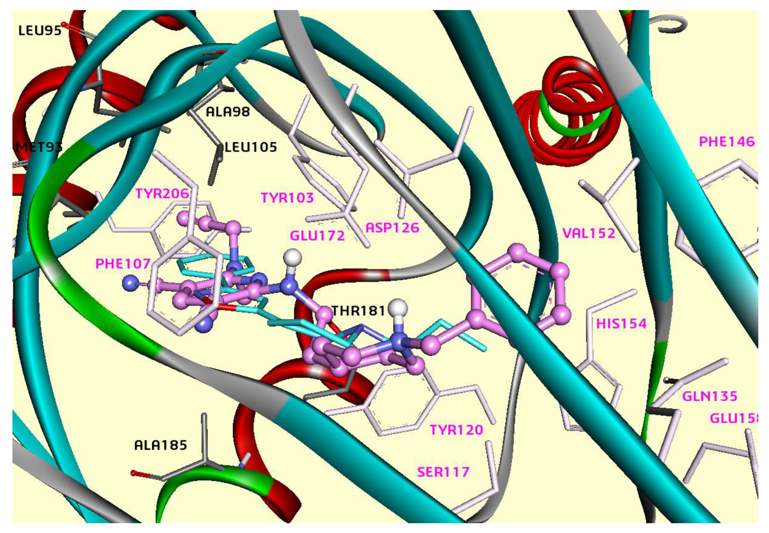
Figure 1. The modeled complex of the hσ1R with ligand 5 (pink). Binding energy: −11.2 kcal/mol. Flexible residues are shown by pink sticks. Compound PD144418 from the σ1R crystal structure (PDB: 5HK1) is shown by blue sticks.
Ijms 26 01266 g001
Figure 2. Two-dimensional representation of the interactions of ligand 5 (pink) in the active site of σ1Rs.
Ijms 26 01266 g002
Disclaimer/Publisher’s Note: The statements, opinions and data contained in all publications are solely those of the individual author(s) and contributor(s) and not of MDPI and/or the editor(s). MDPI and/or the editor(s) disclaim responsibility for any injury to people or property resulting from any ideas, methods, instructions or products referred to in the content.