Review

Twin Small RNAs and Divergent Fates: The Expansive Regulatory Networks of OmrA and OmrB

University of Warsaw, Faculty of Biology, Institute of Microbiology, Department of Molecular Microbiology, Miecznikowa 1, 02-096 Warsaw, Poland
*
Author to whom correspondence should be addressed.
Int. J. Mol. Sci. 2025, 26(23), 11713; https://doi.org/10.3390/ijms262311713
Submission received: 9 October 2025 / Revised: 21 November 2025 / Accepted: 1 December 2025 / Published: 3 December 2025
(This article belongs to the Section Molecular Microbiology)

Abstract

Small RNAs (sRNAs) have emerged as key regulators of bacterial physiology, enabling rapid adaptation to environmental changes through post-transcriptional control. The homologous sRNAs OmrA and OmrB, conserved in Escherichia coli and other Enterobacterales, share sequence similarity yet differ in expression dynamics and functional outputs. Both act by base-pairing with target mRNAs, a process facilitated by the RNA chaperone Hfq, which promotes duplex formation and influences RNA stability. In some contexts, regulation also depends on RelA, which stabilizes Hfq-RNA complexes and links OmrA/B activity to the stringent response. Together, OmrA and OmrB modulate outer membrane protein synthesis, motility, biofilm formation, iron uptake, and virulence. OmrA is preferentially induced under nutrient limitation and integrates into the σS stress regulon, whereas OmrB is more broadly expressed and can engage in context-dependent mechanisms such as target sequestration. This review highlights the molecular mechanisms of OmrA/B regulation and their contribution to global sRNA-mediated regulatory networks that coordinate bacterial adaptation and pathogenicity.

1. Introduction

Small noncoding RNAs (sRNAs), usually between ~50 and 200 nucleotides long, have become key players in bacterial post-transcriptional regulation. Bacterial sRNAs are generally classified into two groups: cis-encoded antisense RNAs, which are transcribed from the DNA strand opposite their target gene and show strong complementarity, and trans-encoded sRNAs, which are encoded on separate genomic loci and bind imperfectly to multiple mRNAs through short seed regions [1,2]. Trans-acting sRNAs commonly regulate gene expression by blocking or exposing ribosome-binding sites to repress or activate translation, by promoting or preventing RNase E-mediated decay, or by influencing transcription termination and mRNA processing [3,4,5]. Functionally, sRNAs are involved in a variety of processes, such as maintaining envelope integrity and regulating metabolic homeostasis, quorum sensing, virulence and host interaction, stress adaptation, motility and biofilm formation [6,7,8,9,10,11].
Computational analyses have identified hundreds of these noncoding regulatory RNAs in Escherichia coli, but only around 80 have been confirmed through experimental validation [12]. OmrA and OmrB are two trans-acting homologous small RNAs (sRNAs) originally discovered in non-pathogenic E. coli, encoded within an approximately 600-base pair intergenic region located between the aas and galR genes. These two sRNAs are the product of an ancestral gene duplication event and share a high degree of sequence similarity. OmrA is 88 nucleotides long, while OmrB is slightly shorter at 82 nucleotides. They are nearly identical at their 5′ and 3′ terminal regions, with divergence occurring after the 21st nucleotide; the middle sections of OmrA/B are different—32 nucleotides long in OmrA and 26 in OmrB. The conserved terminal sequences are essential for target recognition and binding, and their conservation across other bacterial species suggests that these regions fulfill important and evolutionarily preserved functions [13,14,15]. This arrangement is preserved in most, though not all, Enterobacterales. While this structural similarity implies functional overlap, differences in expression dynamics have been observed. OmrA expression peaks during the late stationary phase, whereas OmrB shows more variable expression patterns [16]. Additionally, environmental factors such as temperature or osmolarity further influence their differential expression [17]. OmrA/B typically use their 5′ seed region to pair near the Shine–Dalgarno/start codon of target mRNAs, therefore preventing ribosome binding, a classical mechanism for translational repression [18]. For many bacterial trans-encoded sRNAs, including OmrA/B, effective function depends on their association with RNA chaperone Hfq, which not only protects them from ribonuclease-mediated degradation but also promotes their interaction with target mRNAs. By binding to both the sRNA and the target transcript, Hfq supports imperfect base-pairing that would otherwise be inefficient or unstable. In this way, Hfq ensures that sRNAs effectively repress or activate gene expression after transcription [19,20,21].
Comparative genomic studies have confirmed the presence of OmrA/B across many Enterobacterales, although the conservation of OmrB is more variable. Skippington and Ragan (2012) [22] investigate the evolutionary origin, regulatory functions, conservation, and variability of OmrA/B in E. coli and Shigella. Both sRNAs have been shown to regulate nearly identical targets, a redundancy that may give an evolutionary advantage by enabling tighter control of regulator abundance and mRNA targets. OmrA is conserved across all 27 E. coli and Shigella genomes analyzed, and the intergenic region encoding omrA and omrB is preserved in many Enterobacterales genomes. By contrast, OmrB is absent from both strains of S. boydii examined, demonstrating that this redundancy is dispensable in certain lineages. Phylogenetically, OmrB is classified as a variable sRNA and inferred to be paraphyletic, consistent with gene loss during E. coli-Shigella evolution. Given its substantial overlap in regulatory targets with OmrA, OmrB represents an unusual case of a variable sRNA modulating core genes. Whereas most variable sRNAs interact with only one or two targets, OmrA/B is exceptional in acting on multiple targets, a feature more typical of broadly conserved sRNAs [2,22].

2. Regulation of omrA/B Expression

2.1. EnvZ/OmpR Two-Component System

Expression of OmrA/B is induced by environmental signals through the EnvZ/OmpR two-component system, which directly activates their transcription. Two-component systems are widespread in bacteria and mediate adaptation to environmental changes by a sensor kinase and a response regulator. When the sensor detects a stimulus, it undergoes autophosphorylation and then transfers the phosphate group to the regulator protein, which in turn controls the expression of target genes. EnvZ, an inner membrane-associated sensor kinase, phosphorylates or dephosphorylates the response regulator OmpR, thereby modulating its activity. OmpR is a global transcriptional regulator that controls a wide range of genes involved in outer membrane permeability (e.g., ompF and ompC, encoding major porins), stress adaptation and biofilm formation (e.g., csgD, the master regulator of curli and extracellular matrix production), motility (e.g., flhDC, the flagellar master regulator), and metabolic/transport processes (e.g., narU, a nitrate/nitrite transporter, and tppB, a tripeptide permease), thereby coordinating bacterial responses to osmotic, environmental, and host-associated signals. Genome-wide analyses in E. coli have identified at least 37 genes within 24 transcriptional units as part of the OmpR regulon under osmotic stress conditions [23,24].
EnvZ becomes activated under high-osmolarity conditions. At low osmolarity, when phosphorylated OmpR (OmpR-P) levels are low, the ompF gene is transcribed, producing a porin with a relatively large pore size. Conversely, at high osmolarity, elevated OmpR-P represses ompF and activates transcription of ompC [25,26,27]. Upon phosphorylation by EnvZ in a high osmolarity environment, OmpR also directly induces transcription of both small RNAs OmrA and OmrB. Notably, OmrB can be activated at lower OmpR-P levels than OmrA, which is likely explained by its higher basal expression [18]. OmrA/B transcription can also be modulated by unphosphorylated OmpR. Overproduction of this form of OmpR still enhanced omrA/B expression, suggesting that both OmpR forms may interact with their promoters [28].
Further research showed that OmrA/B sRNAs negatively autoregulate their transcription by directly repressing the ompR-envZ operon that encodes their transcriptional activator. This creates a negative feedback loop in which OmpR activates omrA/B transcription, and OmrA/B repress ompR-envZ mRNA, thereby limiting their own expression. Unlike other OmpR targets such as ompC and ompF, OmrA/B expression is sensitive to the overall OmpR protein concentration rather than exclusively to OmpR-P levels. Experimental evidence shows that deletion of omrA/B increases transcription from the ompR-envZ promoter, while OmrA/B overexpression represses their promoter without significantly affecting ompC or ompF expression. Thus, OmrA/B specifically fine-tune their own expression without disturbing the balance of the major porins [18,28,29].

2.2. Alternative Sigma Factor by RpoS (σS)

RpoS, also known as σS, is an alternative sigma factor in E. coli (and related bacteria) that plays a central role in orchestrating the general stress response. It enables survival under diverse stress conditions such as stationary-phase entry, nutrient deprivation, oxidative stress, acidic environments, and osmotic stress. RpoS accomplishes this by directing RNA polymerase to transcribe a large set of stress-protective genes, the RpoS regulon, with over 300 promoters under its control [30].
In Salmonella enterica, OmrA/B are tightly integrated into regulatory networks that control virulence and stress adaptation. OmrA belongs to the group of sRNAs whose promoters show hallmarks of RpoS-dependent regulation, specifically containing conserved features such as a cytosine at the -13 position that distinguish them from promoters recognized by the housekeeping sigma factor RpoD [31].
OmrA/B expression are not regulated identically. In the Northern blot experiments, OmrA was detected in the wild type but its signal was lost or strongly reduced in the ΔrpoS strain, showing that transcription of omrA is increased in the presence of RpoS. By contrast, omrB is not significantly affected, indicating that it is controlled by other regulatory pathways rather than RpoS. This suggests that OmrA integrates input from RpoS together with other regulators such as OmpR, allowing it to remain active in stationary phase where it can downregulate a wide range of genes involved in processes like outer membrane protein synthesis, metabolism, motility, and biofilm formation. Whereas, OmrB is expressed at very low levels under the same stationary phase conditions, despite its similarity to OmrA and shared regulation by OmpR, demonstrating that the two sRNAs are differentially regulated in late stationary phase. This regulatory difference may help explain conservation of both sRNAs: OmrA, tied directly to σS, under nutrient limitation or stationary-phase conditions, while OmrB responds independently to other signals. Such diversification provides flexibility: although OmrA/B share targets, their distinct expression controls may enable them to act in different contexts [32].
Another study identified a clear RpoS-binding site upstream of omrA, but not omrB in E. coli. This further supports that omrA is directly regulated by σS, while omrB is not. Analyses showed that inactivation of rpoS almost abolished omrA transcription, while omrB expression was unaffected and even slightly increased in the rpoS mutant background. Thus, omrA is almost completely σS-dependent, whereas omrB is σS-independent. The basis for this regulatory divergence lies in their promoter sequences. The omrA promoter contains a cytosine residue at position -12 within its -10 region, a determinant that favors recognition and promoter opening by σS but not by the housekeeping σ70. In contrast, the omrB promoter carries the canonical -12T, which is optimal for σ70 recognition, explaining its independence from σS. Mutagenesis experiments confirmed this: substituting the -12C of the omrA promoter with -12T drastically increased promoter strength and abolished its dependence on σS. These results demonstrate that the -12C is a key specificity element for σS regulation of omrA. Thus, OmrA/B have diverged in promoter architecture and regulatory logic: omrA is integrated into the σS regulon as part of the stationary-phase stress response, while omrB is expressed independently, likely responding to distinct environmental signals [17].

3. Target Specificity of OmrA/B

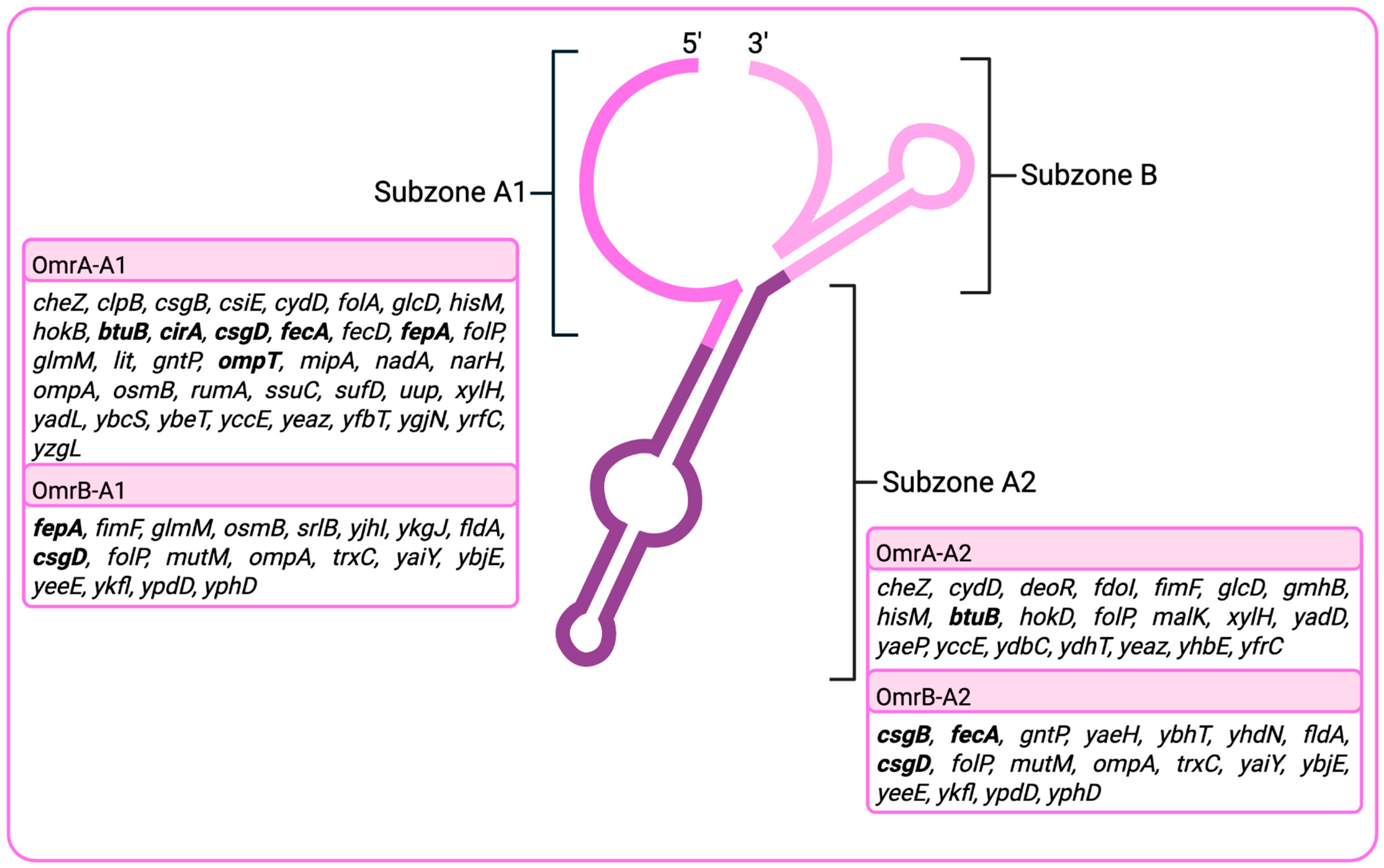
In E. coli, OmrA/B functions through a modular interaction structure with its diverse mRNA targets. Identified as an sRNA with an unusually large interaction set, OmrA regulates approximately 50 mRNAs, with distinct sequence subzones specialized for binding different transcript regions. The study by Tello et al. (2018) [33] divided the OmrA sequence into three subzones, A1, A2 and B. Subzone A1 (Figure 1), located near the 5′ end, interacts with 39 mRNAs, ~89% of which occur within the middle of coding sequences (CDS-M). In contrast, subzone A2, positioned closer to the 3′ end, associates with 21 mRNAs, ~60% of which map to 5′ untranslated regions (5′ UTRs). Furthermore, it was shown that none of the selected mRNA targets interacted with subzone B. This division suggests a modular specialization: A1 primarily engages coding regions, whereas A2 preferentially targets upstream regulatory regions, enabling OmrA to fine-tune translation at multiple stages. Conserved sequence motifs were also identified in target mRNAs. A1 recognizes a six-base motif (CUCUGG) present in 21 of its 39 targets, including gntP, ompT, fecA, fepA, and glmM. Similarly, A2 binds a six-base consensus motif (GCGUAC) found in all 20 of its targets, including malK, btuB, glcD, and fimF. Only xylH contained motifs for both subzones, indicating dual regulation through independent interaction sites. These conserved binding motifs demonstrate that OmrA acts not through random interactions, but by recognizing recurrent sequence patterns across families of mRNAs. Notably, most conserved interactions occur within coding sequences rather than untranslated regions, underscoring the CDS as a critical platform for sRNA-mediated regulation. OmrB demonstrates a similar pattern, there were 19 identified mRNA targets for subzone A1 and 18 for A2 subzone [33].

4. Iron Homeostasis and Regulation of Siderophore Uptake

OmrA/B may play a key role in relieving pressure on the machinery responsible for targeting and properly inserting proteins into the outer membrane (OM). Several essential proteins involved in this process have been identified [18,34], and overproduction of porins is known to trigger the “membrane stress response” [35]. Physiologically, the genes they regulate, including iron acquisition systems, fimbriae, adhesins, proteases, and therefore also crucial for pathogen-host interactions [29].
CirA, FecA, and FepA are outer membrane receptors specialized for the uptake of iron–siderophore complexes, and their genes are part of the Fur regulon. The Fur-Fe2+ protein represses expression of the iron regulon, including cirA, fecA, and fepA. Under low-iron conditions, when Fur is in its inactive state, expression of these iron-uptake genes is induced [36,37]. Enterobactin is the prototypical and one of the most efficient siderophores. The iron–enterobactin complex (Fe-Ent) is recognized by the outer membrane receptor FepA, transported across the outer membrane by a TonB-dependent mechanism, and subsequently translocated across the inner membrane via the FepBCG ATP-binding cassette system. Once inside the cell, the Fes esterase cleaves the enterobactin molecule to release iron, ensuring effective acquisition. Together, these features underscore the central role of enterobactin and FepA in microbial iron homeostasis [38].
Yersinia enterocolitica carries a functional OmrA homolog with conserved sequence and structural features similar to those of E. coli, while OmrB is absent, consistent with genomic searches showing no second homolog. In Y. enterocolitica, OmrA plays a key role as a post-transcriptional regulator of iron homeostasis, acting together with OmpR. It downregulates expression of fecA and fepA. Computational predictions indicated that OmrA base-pairs with regions in the early coding sequences of these mRNAs rather than in the 5′ UTR. Deletion of omrA resulted in significantly increased transcript levels of both fecA and fepA, with effects particularly increased under iron-limited conditions. These findings demonstrate that OmrA normally functions to silence these iron uptake systems [39].
In Y. enterocolitica, OmrA is also under the transcriptional control of OmpR, a component of the EnvZ/OmpR two-component system. OmpR binds directly to the omrA promoter, and omrA expression was strongly reduced in a ΔompR mutant. Environmental factors such as osmolarity, pH, and temperature further influenced omrA expression, showing that OmrA connects environmental sensing to iron homeostasis. OmrA also responds back to regulate ompR-envZ itself. Predicted base-pairing between OmrA and ompR-envZ mRNA overlaps the ribosome-binding site, and experiments confirmed that OmrA reduces ompR-envZ transcript. This establishes a regulatory loop: OmpR activates OmrA transcription, while OmrA represses OmpR post-transcriptionally. Together, these findings establish OmrA as a key regulator in Y. enterocolitica iron homeostasis, acting post-transcriptionally to silence siderophore transporter genes while also engaging in feedback regulation with OmpR. RNA chaperone Hfq and the small RNA OmrA cooperate to regulate key pathways involved in iron homeostasis, and outer membrane composition. Our recent work has shown that Hfq silences several iron-related genes, including fur, fecA, and fepA, by reducing protein synthesis, while OmrA represses the iron uptake genes fecA and fepA. This regulation ensures tight control of iron acquisition, which is critical for bacterial adaptation and virulence in host environments. This suggests that Hfq participates in this process, likely stabilizing OmrA and promoting its base-pairing with target transcripts [39].
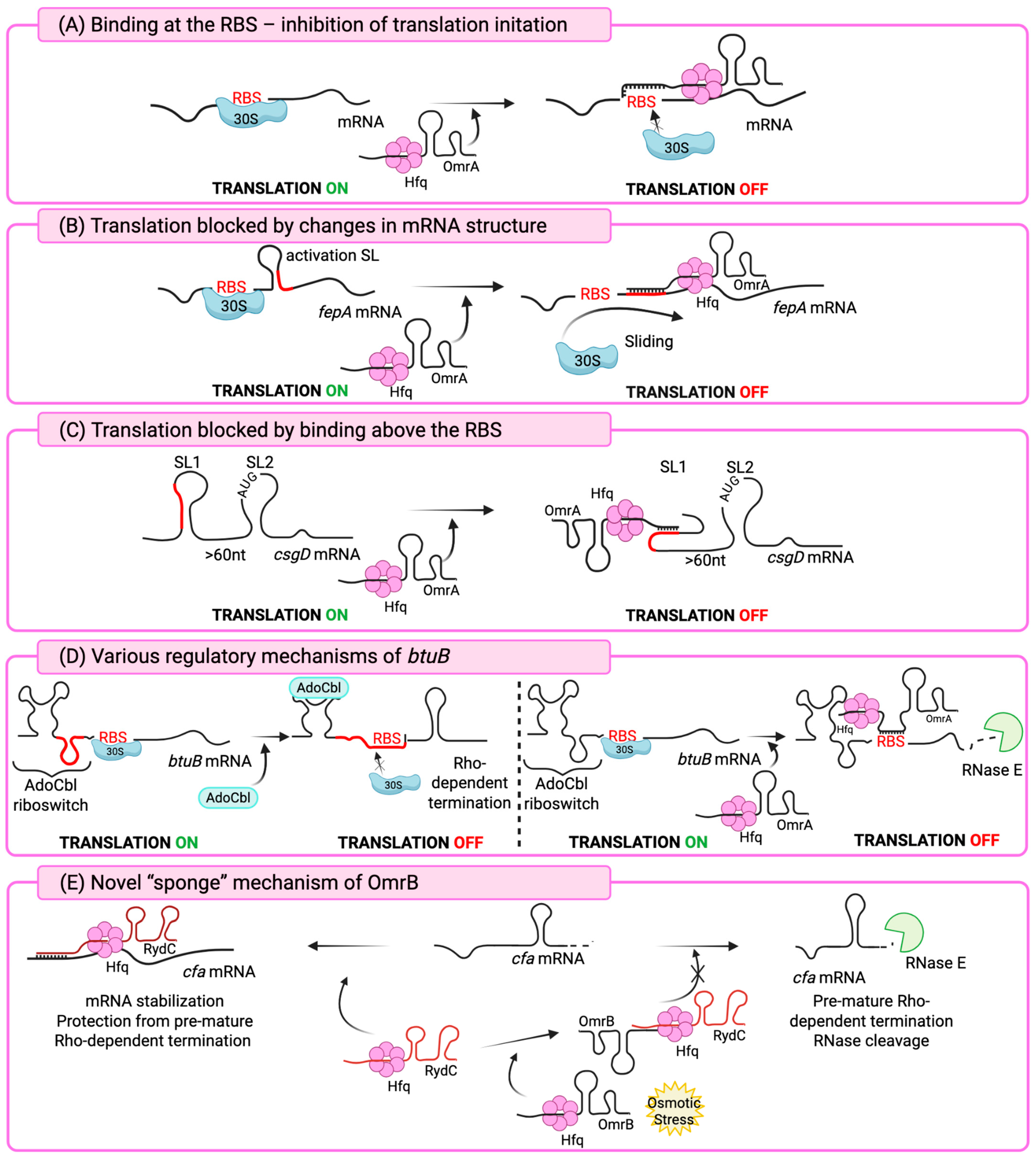
A particularly interesting example of OmrA/B regulatory function is the unique mechanism by which they control expression of the fepA gene in E. coli. Unlike the classical mode of repression, in which sRNAs base-pair with the translation initiation region to block ribosome access (Figure 2A), both OmrA and OmrB instead use their conserved 5′ end sequences to directly pair with a short region of the fepA coding sequence (Figure 2B), located at positions +14 to +22 downstream of the start codon. The fepA transcript contains an activating stem-loop (SL) structure around nucleotide +20 of the open reading frame, which normally enhances translation initiation by serving as a “starting block” for the ribosome. Since OmrA/B pairing with fepA does not overlap the ribosome-binding site, repression occurs through disruption of this stem-loop, thereby preventing efficient ribosome binding. This represents a novel model of sRNA-mediated gene regulation. The study shows that translation activation by SL is not limited to fepA but can also occur in other mRNAs, exemplified by bamA, which encodes an essential outer membrane protein assembly factor [40].
Experimental evidence from toeprinting assays confirmed that OmrA and OmrB binding to the stem-loop interferes with formation of the translation initiation complex, blocking ribosome access to fepA mRNA. The requirement for complementary pairing was demonstrated through mutational analysis: disrupting base-pairing sites in either OmrA/B or fepA abolished repression, while compensatory mutations restoring complementarity re-established regulation. These results underscore that direct base-pairing between OmrA/B and fepA is essential for blocking translation. Importantly, repression persisted even in the absence of RNase E. Using an RNase E mutant strain with reduced activity, the authors showed that fepA repression still occurred, indicating that OmrA/B act primarily by blocking translation rather than by initiating mRNA cleavage. Although both sRNAs use the same complementary 5′ sequence to pair with the +14 to +22 region of mRNA fepA, the study reported differences in efficiency. OmrA repressed translation of fepA more strongly, likely due to forming a more stable interaction with this region or engaging more effectively with the surrounding RNA structure. OmrB also repressed fepA expression but less efficiently, making its effect weaker and more condition-dependent. This functional difference helps explain the retention of both sRNAs in E. coli: OmrA may act as the primary repressor of fepA, while OmrB plays a secondary role, providing fine-tuning or redundancy. This post-transcriptional regulation is integrated into the broader EnvZ/OmpR regulatory network, which induces OmrA/B expression in response to various stress environments. By repressing fepA, OmrA/B help limit iron uptake under stress conditions, acting in cooperation with transcriptional regulators such as Fur to coordinate iron homeostasis. OmrA/B therefore help restrict not only the production of FepA but also other iron siderophore receptors, such as FecA and CirA, in conditions where siderophore-mediated iron uptake is unnecessary or could even be toxic. Moreover, downregulation of immunogenic outer membrane proteins such as FepA, which has been shown to induce protective antibody responses in mice, may also support bacterial evasion of host immune defenses [40,41].

5. Regulation of Motility and Biofilm Formation

Enterobacteria, including E. coli and S. enterica, adjust their lifestyle according to environmental cues, altering between a sessile biofilm state and a motile, flagellated form. These two states are generally viewed as incompatible, and this balance is maintained through complex transcriptional networks that promote one lifestyle while simultaneously repressing the other [42,43].
FlhDC is the master regulator of flagellar biosynthesis in motile bacteria such as E. coli and S. enterica. The flhD and flhC gene products form a heterohexameric FlhD4FlhC2 complex that binds upstream of flagellar operons, bends DNA, recruits RNA polymerase, and activates basal body gene transcription, initiating the flagellar assembly hierarchy [44]. FlhDC drives σ70-dependent transcription of class II genes [45], which build the hook-basal body (HBB). Class III gene expression requires σ28 (FliA), produced from class II, but initially inhibited by FlgM [46,47]. The flhDC operon, transcribed by σ70 RNA polymerase, is tightly regulated: activated by CRP and H-NS, and repressed by LrhA, HdfR, and two-component systems such as EnvZ/OmpR [44].
The flhDC mRNA contains a conserved 198-nucleotide 5′ untranslated region (5′ UTR), and the global regulator CsrA has been shown to bind this leader sequence, enhancing translation [48]. Analyses suggest that OmrA forms extensive predicted base-pairing with the flhDC leader sequence, whereas OmrB exhibits weaker complementarity, consistent with its reduced ability to regulate flhDC expression. In motility assays, OmrA overexpression strongly reduced motility, nearly eliminating it, while OmrB produced a milder but still significant reduction. At the molecular level, OmrA and OmrB reduced expression of a translational flhD′–′lacZ fusion. Mutations introduced into OmrA and OmrB disrupted their ability to regulate this fusion, whereas compensatory mutations in the flhD leader restored repression. This demonstrated that regulation occurs through direct base-pairing with the 5′ UTR of flhDC mRNA, near the ribosome-binding site, thereby likely blocking ribosome access and translation initiation (Figure 2A). It was also suggested that alternative interaction patterns may exist, since disrupting a single predicted pairing site did not fully abolish repression, consistent with the extensive potential complementarity between the sRNAs and the flhDC leader sequence [49].
OmrA/B also directly repress synthesis of the anti-sigma factor FlgM, which normally inhibits the sigma factor σ28 (FliA). Both sRNAs bind through complementary base-pairing to the early coding region of flgM mRNA, close to the start codon, thereby blocking ribosome binding. Computational predictions (IntaRNA) identified base-pairing interactions between the conserved 5′ seed sequences of OmrA and OmrB and the early coding region of flgM. Disrupting these base-pairing sites reduced OmrA/B-mediated repression of flgM, while compensatory mutations that restored complementarity re-established regulation. OmrA/B prevent formation of the 30S initiation complex on flgM mRNA, and both sRNAs, particularly OmrA, strongly decrease FlgM protein synthesis. OmrA/B lower FlgM levels, releasing σ28 to activate class III flagellar genes like fliC, which encodes flagellin. Deletion of flgM increases class III expression without affecting class II, and OmrA/B-mediated FlgM repression produces a similar, though weaker effect. Engineered mutants showed that OmrA’s control of flhDC and flgM can be separated, and that targeting flgM alone is enough to increase fliC expression [50]. Hfq plays a central role in enabling OmrA/B to regulate the flgM gene expression in Enterobacterales. Hfq stabilizes OmrA/B against ribonuclease degradation and promotes their imperfect base-pairing with the flgM transcript, ensuring efficient translational inhibition. Experimental evidence shows that deletion of hfq abolishes OmrA/B repression of flgM, even if the sRNAs are expressed, demonstrating that Hfq is essential for the interaction. In this regulatory circuit, Hfq is not only a stabilizer but also a catalyst of post-transcriptional control, ensuring that OmrA/B repression of flgM effectively links sRNA regulation to flagellar gene expression and motility outcomes [50].
CsgD (encoded by csgD) is a transcriptional regulator of the LuxR family that functions as the central “master regulator” of biofilm formation in E. coli. It activates expression of the curli genes, particularly the csgBAC operon, and also promotes cellulose synthesis, another major component of the biofilm extracellular matrix [51]. By positively regulating csgBAC operon, CsgD drives curli fiber assembly [52]. Multiple environmental stimuli, including temperature, osmolarity, nutrient availability, and oxygen levels, modulate curli gene expression [53].
Both OmrA and OmrB in E. coli function in parallel to repress expression of the transcriptional regulator CsgD. Either sRNA alone is sufficient for repression, and overexpression of OmrA/B abolishes curli fiber formation and reduces cellulose production, demonstrating their broad influence on extracellular matrix components and biofilm traits. OmrA/B repress csgD by direct antisense base-pairing with the 5′ untranslated region of the mRNA, as with previously described targets. However, their binding site lies far upstream of the ribosome-binding site (RBS), distinguishing this regulation from most antisense RNAs, which act by directly blocking ribosomal access [54].
The csgD leader is unusually long (~150 nucleotides) and contains two structured modules, termed SL1 (stem-loop 1) and SL2 (stem-loop 2). OmrA/B base-pair with ~18–19 nucleotides in the SL1 region, located ~60–80 bases upstream of the start codon (Figure 2C). This interaction occurs in a bulge that makes the target accessible, and binding remodels the SL1 structure by partially opening its stem. SL2, located closer to the Shine–Dalgarno sequence, normally sequesters it in a hairpin, reducing translation efficiency. Mutational analyses demonstrated that SL1 is essential for OmrA/B regulation, while SL2 primarily tunes basal expression. Despite the distant binding site, OmrA/B pairing prevents initiating ribosome binding at the translation start region, making this an unusual example of long-distance sRNA-mediated repression. Unlike other sRNAs such as McaS, RprA, and GcvB, OmrA/B do not promote endonucleolytic cleavage or degradation of csgD mRNA. Instead, they inhibit translation in a cleavage-independent manner by restricting ribosomal access. Functionally, repression of csgD by OmrA/B consequently suppresses the csgBA operon, thereby preventing biofilm-associated traits under conditions that favor motility. OmrA/B may also act as “scavengers,” eliminating misregulated csgD transcripts and fine-tuning cell surface remodeling and group behavior in bacterial populations [54].
The RNA chaperone Hfq is indispensable in the regulatory pathway of controlling csgD. Deletion of the hfq gene leads to drastically reduced levels of csgD mRNA and of OmrA/B themselves, showing that Hfq is required for the stability of both the sRNAs and their target mRNA even before any sRNA-mediated inhibition occurs. Moreover, when hfq is deleted, sRNA-dependent repression is effectively abolished, even when OmrA/B are overexpressed [54]. Hfq promotes pairing between sRNAs and mRNAs, and regulatory models suggest that an Hfq-binding site within the csgD leader not only stabilizes sRNA binding but may also recruit RNase E, promoting degradation of the csgD transcript [55,56]. This places them alongside sRNAs such as RydC and RybB, which inhibit translation by base-pairing near or upstream of the RBS without triggering transcript degradation, representing a distinctive mode of post-transcriptional regulation [54,55].
DgcM is one of several GGDEF-domain diguanylate cyclases in E. coli that generate c-di-GMP, a key regulator of biofilms, motility, and the transition between sessile and motile lifestyles [57,58]. Within a local signaling module, DgcM interacts with PdeR and MlrA to control csgD and curli expression [59]. OmrA/B repress DgcM by base-pairing near or overlapping its ribosome-binding site. Mutational analysis confirmed this interaction: disrupting pairing eliminated regulation, and compensatory mutations restored OmrB’s but not OmrA’s repression, implying that OmrA may additionally bind to a second, less functional site. In strains with csgD and ompR mutation, OmrA, but not OmrB, still repressed biofilm formation, likely via dgcM. Two adjacent binding sites were identified on the dgcM mRNA. Both OmrA and OmrB recognize site 1, but site 2 is bound differently: OmrA can extend its binding into an additional region that OmrB cannot reach. The differential ability to engage site 2 is key to explaining why OmrA retains repression in the csgD/ompR mutant backgrounds while OmrB does not. Moreover, gel-shift assays demonstrated that OmrA binds more efficiently than OmrB and can simultaneously occupy both sites on a single mRNA molecule, suggesting cooperative or extended base-pairing that strengthens binding and regulatory efficacy. Repression of dgcM translation critically depends on the RNA chaperone Hfq. Unlike its classical role as a passive matchmaker, Hfq plays an active remodeling role in this case. Specifically, Hfq’s distal face is required to unfold a structured RNA hairpin within the dgcM 5′ region, exposing the otherwise inaccessible OmrA/B binding site. A distal-face mutant abolishes regulation in vitro and in vivo, while proximal/rim mutations have little effect. This mechanism demonstrates that Hfq is not merely a stabilizer of OmrA/B but under certain circumstances is essential for granting them access to their target sequence, highlighting its dual role as both RNA chaperone and RNA remodeler [60].
RelA is a stringent response regulator in E. coli and many other bacteria that produces the signaling molecules ppGpp and pppGpp, which are central to the stringent response, which is triggered during stress [61,62]. RelA recognizes GGAG or GGAGA motifs [63] and facilitates the regulation of mRNAs carrying such motifs by sRNAs that themselves lack GGAG. DgcT, formerly known as YcdT, is a diguanylate cyclase, which is associated with repression of swimming motility in E. coli [64]. The predicted binding site of OmrB on the dgcT mRNA spans nucleotides −12 to +2 relative to the AUG start codon, while the GGAG site is located much further downstream, at +294 nt relative to AUG. Thus, the OmrB binding site does not overlap with the RelA-recognized GGAG motif. It was proved that in the wild-type strain RelA facilitated regulation of dgcT′-′lacZ fusions by OmrB, regulation was lost in the ΔrelA mutant and in the RelA:C289Y mutant (defective in GGAG recognition), which confirms that RelA protein is required for said regulation. (p)ppGpp synthesis and RNA binding are mutually exclusive activities of RelA. Furthermore, amino acid starvation is an inducer of (p)ppGpp synthesis and reduces RelA’s ability to promote base-pairing regulation. Amino acid starvation decreased dgcT fusion protein expression in relA mutants relative to non-starved controls, under conditions of OmrB overexpression, which indicates that dgcT regulation by OmrB depends specifically on RelA binding to the GGAG motif. Overall, the findings suggest that RelA promotes sRNA-mRNA pairing by binding to GGAG-containing mRNAs and, much like in RelA-sRNA-Hfq interactions, stabilizes otherwise unstable mRNA-Hfq monomer complexes, enabling their assembly into an Hfq hexamer that supports efficient sRNA-mRNA duplex formation [63,65].
The article by Zeng and Sundin (2014) [66] reports that the single small RNA named OmrAB acts as a regulator in Erwinia amylovora, the causative agent of fire blight disease in apple and pear trees. The group identified OmrAB among the sRNAs with important roles during infection. Functional assays showed that OmrAB promotes motility while repressing production of amylovoran, an exopolysaccharide that is a major component of biofilms [67]. These activities suggest that OmrAB coordinates the pathogen’s transition between infection stages. Motility is important at early stages for bacterial entry into plant tissues, while amylovoran and biofilm formation are critical at later stages for establishing systemic infection. Notably, this regulatory pattern differs from E. coli, where OmrA/B act as negative regulators of motility. In E. amylovora, by contrast, OmrAB functions as a positive regulator of swimming motility, indicating that while their role as motility regulators is conserved, the precise mechanisms and outcomes are species-specific [66]. This finding was later re-examined, and functional experiments revealed that OmrAB acts as a positive regulator of flagellar motility. The authors proposed that OmrA/B must also target additional motility-related mRNAs (beyond flhDC). Candidate targets include FlgD (a flagellar assembly protein), a methyl-accepting chemotaxis protein, and potentially the global regulator H-NS. Moreover, when E. amylovora omrAB was expressed in E. coli, it behaved like its E. coli counterpart and repressed motility, which underscores that the regulatory pathways differ between the two bacteria Assays confirmed that strains with ΔomrAB mutation exhibited reduction in swarming motility, and it was restored after introduction of omrAB with a native promoter on a complementation plasmid [68].
OmrAB influences flagellar regulation by acting on the flhDC transcript at the post-transcriptional level. To test this, Schachterle et al. (2019) [68] introduced an flhDC translational fusion into E. coli strains engineered to produce E. amylovora OmrAB. Reporter signal was strongly reduced, indicating that OmrAB directly repressed translation of flhDC. This regulatory effect was specific to translation rather than transcript stability, as the half-life of flhDC mRNA was unchanged in the ΔomrAB mutant compared to wild-type or other sRNA mutants. Since OmrA and OmrB are known in E. coli to directly bind the flhDC mRNA, the conservation of these interactions was evaluated in E. amylovora. Sequence alignments across Enterobacterales showed moderate conservation of OmrAB itself and of the corresponding OmrAB-interacting region in the flhDC 5′ UTR, supporting the idea that this regulatory interaction is evolutionarily preserved. Taken together, these findings demonstrate that OmrAB acts as a conserved post-transcriptional repressor of flhDC in E. amylovora, influencing flagellar regulation by reducing translational efficiency rather than altering mRNA stability [68].

6. Virulence Regulation

Beyond their roles in metabolic regulation and cell envelope remodeling, OmrA and OmrB also modulate virulence-related phenotypes in diverse bacterial pathogens. In Klebsiella pneumoniae, the capsule (CPS) is a critical virulence factor that protects the bacterium from host immunity by preventing phagocytosis and complement system activation. Hypervirulent strains (hvKp) frequently produce unusually large amounts of capsule, resulting in a hypermucoviscous (HMV) phenotype. This trait is associated with enhanced survival in host tissues and increased transmission [69]. KvrA, a LysR-type transcriptional regulator, positively regulates capsule gene expression. Deletion of kvrA results in decreased capsule production and reduced hypermucoviscosity, leading to attenuated virulence in animal infection models. KvrA likely functions by directly or indirectly activating transcription of capsule biosynthesis genes and possibly interacting with other global regulators. These findings place KvrA as a central regulator linking capsule synthesis to the overall virulence of K. pneumoniae [70].
OmrB was found to base-pair with kvrA mRNA, and GFP reporter assays confirmed that OmrB binding suppressed kvrA translation. OmrB overexpression also decreased KvrA protein levels under different growth conditions. Mutational analysis, in which compensatory mutations were introduced into both omrB and kvrA, demonstrated that repression depends on direct base-pairing between the two RNAs. Functionally, OmrB overexpression reduced both capsule production and HMV phenotype, as shown by uronic acid quantification and sedimentation assays. However, deletion of omrA/B did not result in substantial changes in capsule production or HMV compared to the wild type, even under conditions known to induce OmrB expression in E. coli (nutrient broth with 20% sucrose). This suggests the presence of additional sRNAs that also contribute to capsule regulation. Like OmrB, OmrA overexpression also decreased capsule production and HMV, indicating that both sRNAs can act as negative regulators of these key virulence traits. Interestingly, OmrB’s role in capsule regulation appears complex: while it repressed kvrA translation, capsule phenotypes in different experimental setups did not always perfectly align with kvrA expression inhibition. This suggests that OmrB may influence capsule regulation through additional targets or indirect pathways. In conclusion, OmrA/B act as repressors of capsule production and hypermucoviscosity in K. pneumoniae. OmrB directly downregulates kvrA through base-pairing and OmrA produces similar phenotypes while overexpressed [71].
Enteropathogenic E. coli (EPEC) and enterohemorrhagic E. coli (EHEC) are closely related but differ in disease patterns and host impact. EPEC mainly affects children under two in developing countries, causing severe watery diarrhea. EHEC, more common in industrialized nations, affects both children and adults, leading to bloody or non-bloody diarrhea and sometimes hemolytic uremic syndrome (HUS)—a serious complication that can cause lasting kidney damage and require dialysis or transplantation. Unlike EPEC, EHEC produces Shiga toxins that damage renal endothelial cells and trigger HUS [72]. The LEE is a pathogenicity island (PAI) of ~35–40 kilobases present in EPEC and EHEC, and some related pathogens (like Citrobacter rodentium). These bacteria rely on a type III secretion system (T3SS) to translocate effector proteins from the bacterial cytosol into host cells [73]. LEE is essential for “attaching-and-effacing” (A/E) lesions formation. It contains ~41 genes, organized in operons named LEE1-LEE7 that encode a T3SS, its chaperones, secreted effectors, adhesins, and regulators [74,75]. LEE1, LEE2, and LEE3 contain the esc and sep genes, which encode the major components of the T3SS, while LEE1 also carries the ler gene encoding Ler, a DNA-binding protein that functions as the primary transcriptional regulator of the LEE region and modulates expression of the other LEE operons [76]. Attaching and effacing lesions are signature features of infection, characterized by tight bacterial adhesion to the host plasma membrane, destruction of enterocyte microvilli, and cytoskeletal rearrangements beneath the adherent bacteria [73].
OmrA/B are identified as important virulence factors in enteropathogenic E. coli (EPEC), since both sRNAs globally silence genes of the LEE. Overexpression of OmrA/B strongly reduced the protein levels of LEE-encoded effectors, including EspA (encodes a secreted filamentous protein that forms a filament connecting the bacterial T3SS to the host cell membrane), EspB (translocator protein, required for creating a pore in the host cell membrane), and Tir (principal effector that is translocated into the host cell and then inserted into the host plasma membrane, acting as a receptor for the bacterial outer membrane adhesin intimin) [77,78]. This correlated with a strong decrease in the mRNA abundance of LEE operons genes espA and tir, showing that repression occurred at the transcript level. Overall, deletion of omrA/B alone had little effect, but overexpression remarkably decreased LEE gene expression, indicating that these sRNAs act as effective repressors when present at high levels. Mechanistically, OmrA/B do not directly base-pair with LEE transcripts. Instead, they indirectly repress transcription of the LEE master regulator Ler, which normally activates all other LEE operons. Reporter fusion assays, under LEE1 promoter (which controls ler) showed that OmrA and OmrB reduce its expression but not by direct interaction with ler mRNA. Instead, the effect seems to be mediated by an unidentified transcription factor that binds within 200 base pairs upstream of the transcription start site of the LEE1 promoter. Since OmrA/B reduced ler expression without a base-pairing mechanism, the authors concluded that repression is indirect, likely mediated through another transcription factor or regulator that acts upstream of ler [78].

7. Riboswitch and Small RNA: Regulation of btuB

The btuB riboswitch is a cis-regulatory RNA structural element embedded in the 5′ UTR of the btuB mRNA, which encodes an outer membrane transporter responsible for importing vitamin B12 (cobalamin) into the cell. Its expression is regulated in response to intracellular levels of adenosyl-cobalamin (AdoCbl), the biologically active form of vitamin B12, through alterations in mRNA folding [79,80]. The riboswitch depends on a “kissing loop,” a specific interaction in which two RNA hairpin loops pair with each other. This interaction is essential both for ligand recognition and for stabilizing the RNA in its repressive conformation that prevents gene expression [81]. Importantly, folding is coordinated with transcriptional pausing by RNA polymerase, which allows the aptamer to adopt the correct structure while the mRNA is still being synthesized [79].
OmrA and OmrB both participate in repressing btuB expression in E. coli, but they do so with different efficiencies and by partially distinct mechanisms. As previously described, OmrA/B typically regulate their targets by base-pairing through a conserved sequence at their 5′ end. However, in the case of btuB, OmrA uses its central region, which is not conserved in OmrB, to recognize and pair with the mRNA. Sequence analyses revealed that this central portion of OmrA is complementary to the translation initiation region of btuB, overlapping the ribosome-binding site, which explains its strong repressive effect. By contrast, OmrB shows only partial complementarity to this region, and its ability to base-pair with btuB mRNA is much weaker. Thus, while OmrA can form extensive and stable interactions that effectively block translation, OmrB’s binding is less precise and less efficient, so it contributes only weakly and primarily in the absence of OmrA, only by interacting with 5′ UTR of btuB [82].
Experiments with btuB′-′lacZ fusions demonstrated that OmrA and OmrB regulate btuB primarily at the translational level, with OmrA exerting stronger effects than OmrB. Translational fusions carrying the btuB 5′ UTR and increasing lengths of the coding sequence (CDS) revealed that OmrA reduced expression by at least 50% when 81 nt of the CDS were present, and repression became much stronger as the CDS length increased. OmrB showed a similar trend but consistently lower efficiency, with maximal repression plateauing at about 50% for the longer constructs. In contrast, transcriptional btuB::lacZ fusions containing the same CDS regions, but fused to lacZ with its own initiation signals, were mostly unaffected by OmrA/B. These results indicate that OmrA/B do not strongly influence transcriptional output but act primarily at the translational level, with decreased mRNA levels likely being a downstream consequence of translational inhibition, most plausibly through RNase E-mediated cleavage of ribosome-free transcripts beyond nucleotide 210 of the btuB CDS. Mutational analyses confirmed that altering either the central region of OmrA or the complementary sequence in btuB strongly impaired repression. Introducing mutation in 5′UTR of OmrB abolished repression. These findings indicate that btuB is a preferential target of OmrA, with OmrB playing at most a minor or conditional role [82].
Physiological assays further confirmed OmrA’s dominance under acidic conditions, when its expression is induced by OmpR activation. In this environment, deletion of omrA caused a clear increase in btuB expression, while deletion of omrB alone had little effect. Only when both sRNAs were deleted repression was fully lost, highlighting OmrA’s primary role in controlling btuB. The study demonstrated that OmrA can regulate specific targets independently of OmrB, while OmrB’s contribution is probably context-dependent. This differential regulation likely explains why both sRNAs have been conserved in many enterobacteria despite their high sequence similarity. These findings establish btuB as an unusual case in which OmrA acts through its central region rather than its conserved 5′ end, making it a specific OmrA target. OmrB can contribute weakly, but only in the absence of OmrA. The regulation of btuB by OmrA strictly requires Hfq, but through a mechanism distinct from canonical OmrA/B targets like ompR. Using a translational fusion and hfq point mutants, it was shown that OmrA repressed btuB expression in the wild type, while OmrB had only a weak effect. This repression was lost in Δhfq, as well as in the proximal face and rim of Hfq mutants, largely due to reduced OmrA/B accumulation in these strains. However, in the distal face mutant, OmrA and OmrB accumulated to high levels but no longer repressed btuB, while still strongly repressed ompR. This indicates that OmrA/B use the canonical 5′-end mechanism for ompR regulation but require Hfq binding at the distal face and recruitment to the mRNA for btuB regulation. Thus, OmrA-mediated repression of btuB relies on a Hfq-dependent mechanism distinct from its regulation of other targets [82].
This represents the first example of gene regulation in E. coli where a riboswitch and sRNAs act on the same mRNA through completely different molecular pathways. The riboswitch acts in cis, sensing adenosyl-cobalamin and altering its structure to block ribosome binding and trigger Rho-dependent transcription termination. In contrast, OmrA (and weakly OmrB) act in trans by base-pairing with btuB mRNA near the ribosome-binding site, which promotes degradosome activity and leads to mRNA decay. Thus, the riboswitch regulates btuB co-transcriptionally, while OmrA/B regulate it post-transcriptionally through targeted RNA degradation (Figure 2D) [82].

8. Novel “Sponge” Mechanism of OmrB

Regulation of the cfa gene expression, which encodes cyclopropane fatty acid (CFA) synthase, involves both transcriptional and post-transcriptional mechanisms. At the transcriptional level, RpoS plays a key role: osmotic stress activates RpoS, which promotes cfa transcription from an RpoS-dependent promoter. This ensures CFA synthase production under stress, enabling modification of membrane lipids for increased resilience [83,84]. Beyond transcription, sRNA RydC fine-tunes cfa expression post-transcriptionally by binding to its mRNA, protecting it from premature Rho-dependent termination and stabilizing the transcript [85]. OmrB was shown to have a novel role as a negative regulator of RydC. When OmrB was expressed in a ΔyieP mutant, an E. coli strain lacking yieP, which encodes a minor global transcriptional regulator [86], cfa expression was decreased. YieP normally represses rydC, so its deletion results in increased RydC levels, creating a background where OmrB’s repressive effect on cfa could be observed. This repression disappeared when rydC was deleted, demonstrating that OmrB does not act directly on cfa mRNA but instead interferes with RydC itself [87].
Experimental evidence supports a “sponge” model in which OmrB base-pairs with RydC, preventing it from activating cfa (Figure 2E). Mutational analysis confirmed this: introducing a point mutation into OmrB that weakened its complementarity to RydC eliminated its ability to repress cfa. These findings indicate that OmrB regulates cfa indirectly by controlling the availability and activity of RydC. Physiologically, this mechanism provides an additional layer of control over cfa expression during stress. Because OmrB is induced by regulators such as EnvZ/OmpR in response to environmental cues, it can fine-tune the balance between RydC-mediated activation and RpoS-driven transcription of cfa. This expands OmrB’s functional repertoire, showing that it can regulate activity of other sRNAs as well [87].

9. OmrA/B as Potential Synthetic RNAs

Synthetic small RNAs (sRNAs) serve as adaptable tools for post-transcriptional gene regulation in bacteria, enabling targeted repression through the combination of a customizable antisense seed region with a defined structural scaffold. In the modular plasmid system developed by Köbel et al. (2022) [88], this design principle is implemented through a Golden Gate based assembly strategy that allows rapid construction and benchmarking of synthetic sRNAs in E. coli. Their study focused on regulating acrA, a component of the AcrAB-TolC efflux pump implicated in β-lactam susceptibility, using oxacillin-based assays to evaluate regulatory efficiency [88].
In this system, the scaffold provides the RNA architecture that supports the function of the engineered sRNA, including stem-loops, an intrinsic terminator, and structural elements that facilitate interaction with the RNA chaperone Hfq, which stabilizes sRNAs and promotes sRNA-mRNA pairing. While the seed sequence determines target specificity, the scaffold dictates whether the seed is presented in an accessible conformation and whether the chaperone can effectively associate with the synthetic construct. These features are essential for achieving robust repression in vivo. The OmrB scaffold, tested alongside RybB, MicA, and MicF, retains natural structural motifs that support stability and Hfq-dependent activity, making it a relevant candidate for synthetic designs [88].
A central question in the study was whether validated antisense seed sequences maintain their regulatory potential when transplanted onto different scaffolds. To address this, the authors evaluated the widely used s8 seed, which originates from the 5′ target-recognition region of the outer membrane protein regulating sRNA RybB and was previously shown to act as an effective antisense module [89]. When fused to the native RybB scaffold (RybB-s8), the construct produced strong repression of acrA and a pronounced susceptibility phenotype in the oxacillin assays performed in their study. In contrast, when the same seed was fused to the OmrB scaffold (OmrB-s8), the regulatory effect was substantially weaker: only a slight inhibitory phenotype was detectable in their oxacillin plating assay, and overall activity was clearly reduced compared with RybB-s8. Importantly the functional data with RNAfold and Mfold predictions showing that the s8 seed becomes largely sequestered within a stable 5′ stem-loop in the OmrB context, provides an explanation for the reduced activity by indicating that the seed is likely less accessible for base-pairing with the acrA mRNA.
These scaffold-dependent differences are consistent with broader principles emerging from recent synthetic sRNA design research. A high-throughput library analysis demonstrated that scaffold architecture strongly influences several aspects of synthetic sRNA behavior, including seed-region accessibility, Hfq dependence, RNase E processing, and overall regulatory output, which highlights that seed sequences optimized in one backbone do not necessarily retain full activity in another. Their findings reinforce that scaffold-specific structural constraints must be considered when designing synthetic sRNAs [90].
Taken together, these results show that OmrB can function as a synthetic scaffold, but its regulatory efficacy is highly sensitive to structural compatibility between the scaffold’s 5′ conformation and the inserted seed. The weak activity of OmrB-s8 in the study illustrates that ensuring appropriate seed exposure is a critical design criterion. More generally, the combined insights emphasize that effective synthetic sRNA engineering requires integrating modular design with careful assessment of RNA folding, scaffold-specific structural features, and protein interactions to achieve predictable regulatory performance.

10. Conclusions

OmrA/B represent a typical example of how small RNAs integrate environmental sensing, stress adaptation, and virulence regulation within Enterobacterales. Emerging from an ancestral gene duplication, they have retained highly conserved terminal regions that act as structural domains for base-pairing with a wide range of target transcripts (Table 1), while their divergent central regions enable functional asymmetry. Their integration into the EnvZ/OmpR regulon ensures that their expression is responsive to environmental changes such as osmolarity and temperature, while feedback repression of ompR-envZ further integrates them within global signaling loops. By controlling outer membrane proteins, siderophore receptors, and envelope-associated enzymes, OmrA/B fine-tune membrane permeability and nutrient acquisition, functions that are essential for both survival in fluctuating niches and pathogenic success in host environments.
The regulatory scope of OmrA/B extends well beyond nutrient uptake. Through repression of flhDC, flgM, csgD, and dgcM, they directly link motility to biofilm formation, balancing mutually opposing lifestyles. Their interaction with riboswitch-controlled genes such as btuB illustrates how cis- and trans-regulatory mechanisms can converge on the same transcript, providing multilayered control of key metabolic pathways. In pathogenic species, including K. pneumoniae, E. amylovora, and S. enterica, OmrA/B contribute to virulence by modulating capsule synthesis, motility, and stress responses, while their target repertoires are shaped by species-specific regulatory contexts. These examples underscore their evolutionary flexibility: while they preserve a conserved regulatory core, their physiological outcomes vary across different bacterial hosts.
Central to all of these activities is the RNA chaperone Hfq, which is indispensable for OmrA/B stability, base-pairing efficiency, and access to structured target regions. In some cases, such as regulation of dgcM or csgD, Hfq acts not only as a stabilizer but also as an RNA remodeler, actively reshaping target structures to permit effective pairing. In this way, Hfq amplifies the regulatory reach of OmrA/B, ensuring that their imperfect base-pairing interactions can exert strong biological effects. Additional factors, such as RelA, further enhance their efficiency by modifying target RNA accessibility, thereby linking sRNA function to global stress pathways such as the stringent response.
One potential use of many bacterial sRNAs, including OmrA and OmrB, is to inspire the design of programmable antisense oligonucleotides (ASOs) for species-specific RNA antibiotics. Much like trans-acting sRNAs, ASOs silence gene expression through short, sequence-specific base pairing, typically targeting 5′ mRNA regions to block translation. Advances in modified nucleic acid chemistries and peptide-based delivery now allow ASOs to selectively inhibit essential genes across diverse bacteria, offering a level of precision far beyond broad-spectrum antibiotics. Importantly, mechanistic insights from natural sRNAs, including seed-based recognition, pairing within extended mRNA regions, and multiple modes of translational repression provide a conceptual framework for improving ASO design and specificity. As bacterial RNA biology continues to advance, sRNAs such as OmrA/B highlight how endogenous regulatory strategies can guide the development of next-generation RNA-based antimicrobials for species-level microbiome editing [91,92].
Together, OmrA/B illustrate the versatility of bacterial small RNAs as post-transcriptional regulators. They are not limited to the repression of single transcripts but act as central nodes that coordinate multiple cellular processes. Future research should continue to explore their species-specific functions, their interplay with global regulators, and their potential as scaffolds in synthetic biology. Ultimately, OmrA/B exemplify how sRNAs enable bacteria to fine-tune gene expression with remarkable precision, ensuring adaptability in both environmental and host-associated contexts.

Author Contributions

J.K. and K.J. researched the literature and wrote the manuscript; P.L. prepared the graphics; A.R. revised the manuscript and supervised its accuracy. All authors have read and agreed to the published version of the manuscript.

Funding

This research was supported by the National Science Center, Poland, PRELUDIUM BIS 3 grant no. 2021/43/O/NZ6/00008, and co-financed by resources from University of Warsaw, “Excellence Initiative—Research University”, Action 1.2.4.

Institutional Review Board Statement

Not applicable.

Data Availability Statement

No new data were created or analyzed in this study. Data sharing is not applicable to this article.

Conflicts of Interest

The authors declare no conflicts of interest.

References

  1. Gottesman, S.; Storz, G. Bacterial Small RNA Regulators: Versatile Roles and Rapidly Evolving Variations. Cold Spring Harb Perspect Biol. 2011, 3, a003798. [Google Scholar] [CrossRef]
  2. Papenfort, K.; Vogel, J. Multiple target regulation by small noncoding RNAs rewires gene expression at the post-transcriptional level. Res. Microbiol. 2009, 160, 278–287. [Google Scholar] [CrossRef] [PubMed]
  3. Wagner, E.G.H.; Romby, P. Chapter Three—Small RNAs in Bacteria and Archaea: Who They Are, What They Do, and How They Do It. Adv. Genet. 2015, 90, 133–208. [Google Scholar] [CrossRef]
  4. Bandyra, K.J.; Said, N.; Pfeiffer, V.; Górna, M.W.; Vogel, J.; Luisi, B.F. The Seed Region of a Small RNA Drives the Controlled Destruction of the Target mRNA by the Endoribonuclease RNase E. Mol. Cell 2012, 47, 943–953. [Google Scholar] [CrossRef]
  5. Sedlyarova, N.; Shamovsky, I.; Bharati, B.K.; Epshtein, V.; Chen, J.; Gottesman, S.; Schroeder, R.; Nudler, E. sRNA-Mediated Control of Transcription Termination in Escherichia coli. Cell 2016, 167, 111–121.e13. [Google Scholar] [CrossRef]
  6. Zhang, A.; Wassarman, K.M.; Rosenow, C.; Tjaden, B.C.; Storz, G.; Gottesman, S. Global analysis of small RNA and mRNA targets of Hfq. Mol. Microbiol. 2003, 50, 1111–1124. [Google Scholar] [CrossRef]
  7. Chao, Y.; Papenfort, K.; Reinhardt, R.; Sharma, C.M.; Vogel, J. An atlas of Hfq-bound transcripts reveals 3′ UTRs as a genomic reservoir of regulatory small RNAs. EMBO J. 2012, 31, 4005–4019. [Google Scholar] [CrossRef] [PubMed]
  8. Feng, L.; Rutherford, S.T.; Papenfort, K.; Bagert, J.D.; van Kessel, J.C.; Tirrell, D.A.; Wingreen, N.S.; Bassler, B.L. A Qrr Noncoding RNA Deploys Four Different Regulatory Mechanisms to Optimize Quorum-Sensing Dynamics. Cell 2015, 160, 228–240. [Google Scholar] [CrossRef]
  9. Banerjee, R. Tiny but Mighty: Small RNAs-The Micromanagers of Bacterial Survival, Virulence, and Host-Pathogen Interactions. Non-Coding RNA 2025, 11, 36. [Google Scholar] [CrossRef] [PubMed]
  10. Fröhlich, K.S.; Gottesman, S. Small Regulatory RNAs in the Enterobacterial Response to Envelope Damage and Oxidative Stress. Regul. RNA Bact. Archaea 2018, 17, 211–228. [Google Scholar] [CrossRef]
  11. Thomason, M.K.; Fontaine, F.; De Lay, N.; Storz, G. A small RNA that regulates motility and biofilm formation in response to changes in nutrient availability in Escherichia coli. Mol. Microbiol. 2012, 84, 17–35. [Google Scholar] [CrossRef] [PubMed]
  12. Raghavan, R.; Groisman, E.A.; Ochman, H. Genome-wide detection of novel regulatory RNAs in Escherichia coli. Genome Res. 2011, 21, 1487–1497. [Google Scholar] [CrossRef] [PubMed]
  13. Argaman, L.; Hershberg, R.; Vogel, J.; Bejerano, G.; Wagner, E.G.H.; Margalit, H.; Altuvia, S. Novel small RNA-encoding genes in the intergenic regions of Escherichia coli. Curr. Biol. 2001, 11, 941–950. [Google Scholar] [CrossRef]
  14. Wassarman, K.M.; Repoila, F.; Rosenow, C.; Storz, G.; Gottesman, S. Identification of novel small RNAs using comparative genomics and microarrays. Genes Dev. 2001, 15, 1637–1651. [Google Scholar] [CrossRef]
  15. De Lay, N.; Schu, D.J.; Gottesman, S. Bacterial small RNA-based negative regulation: Hfq and its accomplices. J. Biol. Chem. 2013, 288, 7996–8003. [Google Scholar] [CrossRef]
  16. Vogel, J.; Bartels, V.; Tang, T.H.; Churakov, G.; Slagter-Jäger, J.G.; Hüttenhofer, A.; Wagner, E.G.H. RNomics in Escherichia coli detects new sRNA species and indicates parallel transcriptional output in bacteria. Nucleic Acids Res. 2003, 31, 6435–6443. [Google Scholar] [CrossRef]
  17. Peano, C.; Wolf, J.; Demol, J.; Rossi, E.; Petiti, L.; De Bellis, G.; Geiselmann, J.; Egli, T.; Lacour, S.; Landini, P. Characterization of the Escherichia coli σS core regulon by Chromatin Immunoprecipitation-sequencing (ChIP-seq) analysis. Sci. Rep. 2015, 5, 10469. [Google Scholar] [CrossRef] [PubMed]
  18. Guillier, M.; Gottesman, S.; Storz, G. Modulating the outer membrane with small RNAs. Genes Dev. 2006, 20, 2338–2348. [Google Scholar] [CrossRef]
  19. Faner, M.A.; Feig, A.L. Identifying and characterizing Hfq-RNA interactions. Methods 2013, 63, 144–159. [Google Scholar] [CrossRef]
  20. Updegrove, T.B.; Shabalina, S.A.; Storz, G. How do base-pairing small RNAs evolve? FEMS Microbiol. Rev. 2015, 39, 379–391. [Google Scholar] [CrossRef]
  21. Bobrovskyy, M.; Vanderpool, C.K.; Richards, G.R. Small RNAs Regulate Primary and Secondary Metabolism in Gram-negative Bacteria. Metab. Bact. Pathog. 2015, 4, 59–94. [Google Scholar] [CrossRef]
  22. Skippington, E.; Ragan, M.A. Evolutionary Dynamics of Small RNAs in 27 Escherichia coli and Shigella Genomes. Genome Biol. Evol. 2012, 4, 330–345. [Google Scholar] [CrossRef]
  23. Seo, S.W.; Gao, Y.; Kim, D.; Szubin, R.; Yang, J.; Cho, B.-K.; Palsson, B.O. Revealing genome-scale transcriptional regulatory landscape of OmpR highlights its expanded regulatory roles under osmotic stress in Escherichia coli K-12 MG1655. Sci. Rep. 2017, 7, 2181. [Google Scholar] [CrossRef]
  24. Ogasawara, H.; Yamada, K.; Kori, A.; Yamamoto, K.; Ishihama, A. Regulation of the Escherichia coli csgD promoter: Interplay between five transcription factors. Microbiology. 2010, 156, 2470–2483. [Google Scholar] [CrossRef]
  25. Pratt, L.A.; Silhavy, T.J. Porin regulon of Escherichia coli. In Two-Component Signal Transduct; Wiley Online Library: Hoboken, NJ, USA, 1995; pp. 105–127. [Google Scholar] [CrossRef]
  26. Pratt, L.A.; Silhavy, T.J. Identification of base pairs important for OmpR-DNA interaction. Mol. Microbiol. 1995, 17, 565–573. [Google Scholar] [CrossRef] [PubMed]
  27. Kenney Linda, J.; Anand Ganesh, S. EnvZ/OmpR Two-Component Signaling: An Archetype System That Can Function Noncanonically. EcoSal Plus. 2020, 9, 1–30. [Google Scholar] [CrossRef]
  28. Brosse, A.; Korobeinikova, A.; Gottesman, S.; Guillier, M. Unexpected properties of sRNA promoters allow feedback control via regulation of a two-component system. Nucleic Acids Res. 2016, 44, 9650–9666. [Google Scholar] [CrossRef]
  29. Guillier, M.; Gottesman, S. The 5′ end of two redundant sRNAs is involved in the regulation of multiple targets, including their own regulator. Nucleic Acids Res. 2008, 36, 6781–6794. [Google Scholar] [CrossRef]
  30. Sophie, B.; Taran, S.B.; Susan, G. RpoS and the bacterial general stress response. Microbiol. Mol. Biol. Rev. 2024, 88, e00151-22. [Google Scholar] [CrossRef]
  31. Colgan, A.M.; Kröger, C.; Diard, M.; Hardt, W.-D.; Puente, J.L.; Sivasankaran, S.K.; Hokamp, K.; Hinton, J.C.D. The impact of 18 ancestral and horizontally-acquired regulatory proteins upon the transcriptome and sRNA landscape of Salmonella enterica serovar Typhimurium. PLoS Genet. 2016, 12, e1006258. [Google Scholar] [CrossRef] [PubMed]
  32. Levi-Meyrueis, C.; Monteil, V.; Sismeiro, O.; Dillies, M.-A.; Monot, M.; Jagla, B.; Coppée, J.-Y.; Dupuy, B.; Norel, F. Expanding the RpoS/σS-network by RNA sequencing and identification of σS-controlled small RNAs in Salmonella. PLoS ONE 2014, 9, e96918. [Google Scholar] [CrossRef]
  33. Tello, M.; Avalos, F.; Orellana, O. Codon usage and modular interactions between messenger RNA coding regions and small RNAs in Escherichia coli. BMC Genom. 2018, 19, 657. [Google Scholar] [CrossRef]
  34. Ruiz, N.; Falcone, B.; Kahne, D.; Silhavy, T.J. Chemical Conditionality: A Genetic Strategy to Probe Organelle Assembly. Cell 2005, 121, 307–317. [Google Scholar] [CrossRef]
  35. Ruiz, N.; Silhavy, T.J. Sensing external stress: Watchdogs of the Escherichia coli cell envelope. Curr. Opin. Microbiol. 2005, 8, 122–126. [Google Scholar] [CrossRef]
  36. McHugh, J.P.; Rodríguez-Quiñones, F.; Abdul-Tehrani, H.; Svistunenko, D.A.; Poole, R.K.; Cooper, C.E.; Andrews, S.C. Global Iron-dependent Gene Regulation in Escherichia coli: A New Mechanism for Iron Homeostasis. J. Biol. Chem. 2003, 278, 29478–29486. [Google Scholar] [CrossRef]
  37. Zhang, Z.; Gosset, G.; Barabote, R.; Gonzalez, C.S.; Cuevas, W.A.; Saier, M.H., Jr. Functional Interactions between the Carbon and Iron Utilization Regulators, Crp and Fur, in Escherichia coli. J. Bacteriol. 2005, 187, 980–990. [Google Scholar] [CrossRef]
  38. Raymond, K.N.; Dertz, E.A.; Kim, S.S. Enterobactin: An archetype for microbial iron transport. Proc. Natl. Acad. Sci. USA 2003, 100, 3584–3588. [Google Scholar] [CrossRef]
  39. Jaworska, K.; Konarska, J.; Gomza, P.; Rożen, P.; Nieckarz, M.; Krawczyk-Balska, A.; Brzostek, K.; Raczkowska, A. Interplay between the RNA Chaperone Hfq, Small RNAs and Transcriptional Regulator OmpR Modulates Iron Homeostasis in the Enteropathogen Yersinia enterocolitica. Int. J. Mol. Sci. 2023, 24, 11157. [Google Scholar] [CrossRef] [PubMed]
  40. Jagodnik, J.; Chiaruttini, C.; Guillier, M. Stem-Loop Structures Within mRNA Coding Sequences Activate Translation Initiation and Mediate Control by Small Regulatory RNAs. Mol. Cell 2017, 68, 158–170.e3. [Google Scholar] [CrossRef] [PubMed]
  41. Baghal, S.M.L.; Gargari, S.L.M.; Rasooli, I. Production and immunogenicity of recombinant ferric enterobactin protein (FepA). Int. J. Infect. Dis. 2010, 14, e166–e170. [Google Scholar] [CrossRef] [PubMed][Green Version]
  42. Guttenplan, S.B.; Kearns, D.B. Regulation of flagellar motility during biofilm formation. FEMS Microbiol. Rev. 2013, 37, 849–871. [Google Scholar] [CrossRef]
  43. Pesavento, C.; Becker, G.; Sommerfeldt, N.; Possling, A.; Tschowri, N.; Mehlis, A.; Hengge, R. Inverse regulatory coordination of motility and curli-mediated adhesion in Escherichia coli. Genes Dev. 2008, 22, 2434–2446. [Google Scholar] [CrossRef] [PubMed]
  44. Patrick, J.E.; Kearns, D.B. Swarming motility and the control of master regulators of flagellar biosynthesis. Mol. Microbiol. 2012, 83, 14–23. [Google Scholar] [CrossRef]
  45. Wang, S.; Fleming, R.T.; Westbrook, E.M.; Matsumura, P.; McKay, D.B. Structure of the Escherichia coli FlhDC Complex, a Prokaryotic Heteromeric Regulator of Transcription. J. Mol. Biol. 2006, 355, 798–808. [Google Scholar] [CrossRef]
  46. Hughes, K.T.; Mathee, K. The anti-sigma factors. Annu. Rev. Microbiol. 1998, 52, 231–286. [Google Scholar] [CrossRef]
  47. Rudenko, I.; Ni, B.; Glatter, T.; Sourjik, V. Inefficient Secretion of Anti-sigma Factor FlgM Inhibits Bacterial Motility at High Temperature. iScience 2019, 16, 145–154. [Google Scholar] [CrossRef]
  48. Wei, B.L.; Brun-Zinkernagel, A.-M.; Simecka, J.W.; Prüß, B.M.; Babitzke, P.; Romeo, T. Positive regulation of motility and flhDC expression by the RNA-binding protein CsrA of Escherichia coli. Mol. Microbiol. 2001, 40, 245–256. [Google Scholar] [CrossRef]
  49. De Lay, N.; Gottesman, S. A complex network of small non-coding RNAs regulate motility in Escherichia coli. Mol. Microbiol. 2012, 86, 524–538. [Google Scholar] [CrossRef] [PubMed]
  50. Romilly, C.; Hoekzema, M.; Holmqvist, E.; Wagner, E.G.H. Small RNAs OmrA and OmrB promote class III flagellar gene expression by inhibiting the synthesis of anti-Sigma factor FlgM. RNA Biol. 2020, 17, 872–880. [Google Scholar] [CrossRef]
  51. Hiroshi, O.; Kaneyoshi, Y.; Akira, I. Role of the Biofilm Master Regulator CsgD in Cross-Regulation between Biofilm Formation and Flagellar Synthesis. J. Bacteriol. 2011, 193, 2587–2597. [Google Scholar] [CrossRef]
  52. Eva, B.; Andrea, B.; Corinne, D.; Paolo, L. Gene Expression Regulation by the Curli Activator CsgD Protein: Modulation of Cellulose Biosynthesis and Control of Negative Determinants for Microbial Adhesion. J. Bacteriol. 2006, 188, 2027–2037. [Google Scholar] [CrossRef]
  53. Ou, C.; Dozois, C.M.; Daigle, F. Differential regulatory control of curli (csg) gene expression in Salmonella enterica serovar Typhi requires more than a functional CsgD regulator. Sci. Rep. 2023, 13, 14905. [Google Scholar] [CrossRef]
  54. Holmqvist, E.; Reimegård, J.; Sterk, M.; Grantcharova, N.; Römling, U.; Wagner, E.G.H. Two antisense RNAs target the transcriptional regulator CsgD to inhibit curli synthesis. EMBO J. 2010, 29, 1840–1850. [Google Scholar] [CrossRef] [PubMed]
  55. Andreassen, P.R.; Pettersen, J.S.; Szczerba, M.; Valentin-Hansen, P.; Møller-Jensen, J.; Jørgensen, M.G. sRNA-dependent control of curli biosynthesis in Escherichia coli: McaS directs endonucleolytic cleavage of csgD mRNA. Nucleic Acids Res. 2018, 46, 6746–6760. [Google Scholar] [CrossRef]
  56. Schu, D.J.; Zhang, A.; Gottesman, S.; Storz, G. Alternative Hfq-sRNA interaction modes dictate alternative mRNA recognition. EMBO J. 2015, 34, 2557–2573. [Google Scholar] [CrossRef]
  57. Sarenko, O.; Klauck, G.; Wilke, F.M.; Pfiffer, V.; Richter, A.M.; Herbst, S.; Kaever, V.; Hengge, R. More than Enzymes That Make or Break Cyclic Di-GMP—Local Signaling in the Interactome of GGDEF/EAL Domain Proteins of Escherichia coli. mBio 2017, 8, e01639-17. [Google Scholar] [CrossRef] [PubMed]
  58. Lamprecht, O.; Ratnikava, M.; Jacek, P.; Kaganovitch, E.; Buettner, N.; Fritz, K.; Biazruchka, I.; Köhler, R.; Pietsch, J.; Sourjik, V. Regulation by cyclic di-GMP attenuates dynamics and enhances robustness of bimodal curli gene activation in Escherichia coli. PLoS Genet. 2023, 19, e1010750. [Google Scholar] [CrossRef] [PubMed]
  59. Serra, D.O.; Hengge, R. A c-di-GMP-Based Switch Controls Local Heterogeneity of Extracellular Matrix Synthesis Which Is Crucial for Integrity and Morphogenesis of Escherichia coli Macrocolony Biofilms. J. Mol. Biol. 2019, 431, 4775–4793. [Google Scholar] [CrossRef]
  60. Hoekzema, M.; Romilly, C.; Holmqvist, E.; Wagner, E.G.H. Hfq-dependent mRNA unfolding promotes sRNA-based inhibition of translation. EMBO J. 2019, 38, e101199. [Google Scholar] [CrossRef]
  61. Jonathan, D. Understanding the Stringent Response: Experimental Context Matters. mBio 2023, 14, e03404-22. [Google Scholar] [CrossRef]
  62. Zhu, M.; Dai, X. Stringent response ensures the timely adaptation of bacterial growth to nutrient downshift. Nat. Commun. 2023, 14, 467. [Google Scholar] [CrossRef]
  63. Basu, P.; Elgrably-Weiss, M.; Hassouna, F.; Kumar, M.; Wiener, R.; Altuvia, S. RNA binding of Hfq monomers promotes RelA-mediated hexamerization in a limiting Hfq environment. Nat. Commun. 2021, 12, 2249. [Google Scholar] [CrossRef] [PubMed]
  64. Jonas, K.; Edwards, A.N.; Simm, R.; Romeo, T.; Römling, U.; Melefors, Ö. The RNA binding protein CsrA controls cyclic di-GMP metabolism by directly regulating the expression of GGDEF proteins. Mol. Microbiol. 2008, 70, 236–257. [Google Scholar] [CrossRef] [PubMed]
  65. Basu, P.; Altuvia, S. RelA binding of mRNAs modulates translation or sRNA-mRNA basepairing depending on the position of the GGAG site. Mol. Microbiol. 2022, 117, 143–159. [Google Scholar] [CrossRef] [PubMed]
  66. Zeng, Q.; Sundin, G.W. Genome-wide identification of Hfq-regulated small RNAs in the fire blight pathogen Erwinia amylovora discovered small RNAs with virulence regulatory function. BMC Genom. 2014, 15, 414. [Google Scholar] [CrossRef]
  67. Koczan, J.M.; McGrath, M.J.; Zhao, Y.; Sundin, G.W. Contribution of Erwinia amylovora Exopolysaccharides Amylovoran and Levan to Biofilm Formation: Implications in Pathogenicity. Phytopathology 2009, 99, 1237–1244. [Google Scholar] [CrossRef]
  68. Schachterle, J.K.; Zeng, Q.; Sundin, G.W. Three Hfq-dependent small RNAs regulate flagellar motility in the fire blight pathogen Erwinia amylovora. Mol. Microbiol. 2019, 111, 1476–1492. [Google Scholar] [CrossRef]
  69. Walker, K.A.; Miller, V.L. The intersection of capsule gene expression, hypermucoviscosity and hypervirulence in Klebsiella pneumoniae. Curr. Opin. Microbiol. 2020, 54, 95–102. [Google Scholar] [CrossRef]
  70. Palacios, M.; Miner, T.A.; Frederick, D.R.; Sepulveda, V.E.; Quinn, J.D.; Walker, K.A.; Miller, V.L. Identification of Two Regulators of Virulence That Are Conserved in Klebsiella pneumoniae Classical and Hypervirulent Strains. mBio 2018, 9, e01443-18. [Google Scholar] [CrossRef]
  71. Goh, K.J.; Altuvia, Y.; Argaman, L.; Raz, Y.; Bar, A.; Lithgow, T.; Margalit, H.; Gan, Y.-H. RIL-seq reveals extensive involvement of small RNAs in virulence and capsule regulation in hypervirulent Klebsiella pneumoniae. Nucleic Acids Res. 2024, 52, 9119–9138. [Google Scholar] [CrossRef]
  72. Mellies Jay, L.; Barron Alex, M.S.; Carmona Anna, M. Enteropathogenic and Enterohemorrhagic Escherichia coli Virulence Gene Regulation. Infect. Immun. 2007, 75, 4199–4210. [Google Scholar] [CrossRef]
  73. Cepeda-Molero, M.; Berger, C.N.; Walsham, A.D.; Ellis, S.J.; Wemyss-Holden, S.; Schüller, S.; Frankel, G.; Fernández, L.Á. Attaching and effacing (A/E) lesion formation by enteropathogenic Escherichia coli on human intestinal mucosa is dependent on non-LEE effectors. PLoS Pathog. 2017, 13, e1006706. [Google Scholar] [CrossRef]
  74. Mellies, J.L.; Elliott, S.J.; Sperandio, V.; Donnenberg, M.S.; Kaper, J.B. The Per regulon of enteropathogenic Escherichia coli: Identification of a regulatory cascade and a novel transcriptional activator, the locus of enterocyte effacement (LEE)-encoded regulator (Ler). Mol Microbiol. 1999, 33, 296–306. [Google Scholar] [CrossRef]
  75. Croxen, M.A.; Finlay, B.B. Molecular mechanisms of Escherichia coli pathogenicity. Nat. Rev. Microbiol. 2010, 8, 26–38. [Google Scholar] [CrossRef]
  76. Elliott, S.J.; Sperandio, V.; Girón, J.A.; Shin, S.; Mellies, J.L.; Wainwright, L.; Hutcheson, S.W.; McDaniel, T.K.; Kaper, J.B. The Locus of Enterocyte Effacement (LEE)-Encoded Regulator Controls Expression of Both LEE- and Non-LEE-Encoded Virulence Factors in Enteropathogenic and Enterohemorrhagic Escherichia coli. Infect. Immun. 2000, 68, 6115–6126. [Google Scholar] [CrossRef]
  77. Gaytán, M.O.; Martínez-Santos, V.I.; Soto, E.; González-Pedrajo, B. Type Three Secretion System in Attaching and Effacing Pathogens. Front. Cell. Infect. Microbiol. 2016, 6, 129. [Google Scholar] [CrossRef] [PubMed]
  78. Muche, S.; El-Fenej, J.; Mihaita, A.; Mrozek, Z.; Cleary, S.; Critelli, B.; Marino, M.; Yu, W.; Amos, B.; Hunter, T.; et al. The two sRNAs OmrA and OmrB indirectly repress transcription from the LEE1 promoter of enteropathogenic Escherichia coli. Folia Microbiol. 2023, 68, 415–430. [Google Scholar] [CrossRef]
  79. Perdrizet, G.A.; Artsimovitch, I.; Furman, R.; Sosnick, T.R.; Pan, T. Transcriptional pausing coordinates folding of the aptamer domain and the expression platform of a riboswitch. Proc. Natl. Acad. Sci. USA 2012, 109, 3323–3328. [Google Scholar] [CrossRef] [PubMed]
  80. Palou-Mir, J.; Musiari, A.; Sigel, R.K.O.; Barceló-Oliver, M. Characterization of the full-length btuB riboswitch from Klebsiella pneumoniae. J. Inorg. Biochem. 2016, 160, 106–113. [Google Scholar] [CrossRef] [PubMed]
  81. Lussier, A.; Bastet, L.; Chauvier, A.; Lafontaine, D.A. A Kissing Loop Is Important for btuB Riboswitch Ligand Sensing and Regulatory Control. J. Biol. Chem. 2015, 290, 26739–26751. [Google Scholar] [CrossRef]
  82. Bastet, L.; Korepanov, A.P.; Jagodnik, J.; Grondin, J.P.; Lamontagne, A.-M.; Guillier, M.; Lafontaine, D.A. Riboswitch and small RNAs modulate btuB translation initiation in Escherichia coli and trigger distinct mRNA regulatory mechanisms. Nucleic Acids Res. 2024, 52, 5852–5865. [Google Scholar] [CrossRef] [PubMed]
  83. Wang, A.-Y.; Cronan, J.E., Jr. The growth phase-dependent synthesis of cyclopropane fatty acids in Escherichia coli is the result of an RpoS(KatF)-dependent promoter plus enzyme instability. Mol. Microbiol. 1994, 11, 1009–1017. [Google Scholar] [CrossRef]
  84. Bianco, C.M.; Fröhlich, K.S.; Vanderpool, C.K. Bacterial Cyclopropane Fatty Acid Synthase mRNA Is Targeted by Activating and Repressing Small RNAs. J. Bacteriol. 2019, 201, e00461-19. [Google Scholar] [CrossRef]
  85. Fröhlich, K.S.; Papenfort, K.; Fekete, A.; Vogel, J. A small RNA activates CFA synthase by isoform-specific mRNA stabilization. EMBO J. 2013, 32, 2963–2979. [Google Scholar] [CrossRef]
  86. Nguyen-Vo, T.P.; Ko, S.; Ryu, H.; Kim, J.R.; Kim, D.; Park, S. Systems evaluation reveals novel transporter YohJK renders 3-hydroxypropionate tolerance in Escherichia coli. Sci. Rep. 2020, 10, 19064. [Google Scholar] [CrossRef] [PubMed]
  87. Bianco, C.M.; Caballero-Rothar, N.N.; Ma, X.; Farley, K.R.; Vanderpool, C.K. Transcriptional and post-transcriptional mechanisms modulate cyclopropane fatty acid synthase through small RNAs in Escherichia coli. J. Bacteriol. 2024, 206, e00049-24. [Google Scholar] [CrossRef]
  88. Köbel, T.S.; Melo Palhares, R.; Fromm, C.; Szymanski, W.; Angelidou, G.; Glatter, T.; Georg, J.; Berghoff, B.A.; Schindler, D. An Easy-to-Use Plasmid Toolset for Efficient Generation and Benchmarking of Synthetic Small RNAs in Bacteria. ACS Synth. Biol. 2022, 11, 2989–3003. [Google Scholar] [CrossRef] [PubMed]
  89. Papenfort, K.; Bouvier, M.; Mika, F.; Sharma, C.M.; Vogel, J. Evidence for an autonomous 5′ target recognition domain in an Hfq-associated small RNA. Proc. Natl. Acad. Sci. USA 2010, 107, 20435–20440. [Google Scholar] [CrossRef]
  90. Brück, M.; Köbel, T.S.; Dittmar, S.; Ramírez Rojas, A.A.; Georg, J.; Berghoff, B.A.; Schindler, D. A library-based approach allows systematic and rapid evaluation of seed region length and reveals design rules for synthetic bacterial small RNAs. iScience 2024, 17, 27. [Google Scholar] [CrossRef]
  91. Vogel, J.; Faber, F.; Barquist, L.; Sparmann, A.; Popella, L.; Ghosh, C. ASOBIOTICS 2024: An interdisciplinary symposium on antisense-based programmable RNA antibiotics. RNA 2025, 31, 465–474. [Google Scholar] [CrossRef]
  92. Vogel, J. An RNA biology perspective on species-specific programmable RNA antibiotics. Mol. Microbiol. 2020, 113, 550–559. [Google Scholar] [CrossRef] [PubMed]
Figure 1. Interaction subzones located in the predicted secondary structure of the OmrA/B, based on Tello et al. (2018) [33]. Boxes indicate individual mRNA targets, with interaction subzones mapped relative to the sRNA-binding sites determined in the study. Genes shown in bold correspond to those highlighted in this review article as experimentally validated OmrA/OmrB targets. Created in BioRender [https://BioRender.com/hfis3cu (accessed on 20 September 2025)].
Ijms 26 11713 g001
Figure 2. Schematic representation of the regulatory mechanisms of sRNAs OmrA/B. Different modes of OmrA/OmrB regulatory activity are shown: inhibition of translation via RBS binding (A), alteration of mRNA structures (B,C), cis-regulation of btuB through riboswitch and RBS binding by OmrA (D), and the novel “sponge” function of OmrB (E). See text for details. Created in BioRender [https://BioRender.com/7gccqgi (accessed on 20 September 2025)].
Ijms 26 11713 g002
Table 1. Experimentally supported OmrA/B targets.
TargetGene FunctionRegulatory MechanismRegulated BySpeciesSource
ompR-envZTwo-component regulator of porin expressionPost-transcriptional repression,
negative feedback loop
OmrA/BE. coli
Y. enterocolitica
[18,28,29,39]
ompTOuter-membrane proteasePost-transcriptional repressionOmrA/BE. coli[18,29]
cirASiderophore receptorPost-transcriptional repressionOmrA/BE. coli[18,29]
fecAFerric-citrate receptorPost-transcriptional repressionOmrA/BE. coli,
Y. enterocolitica
[18,29,39,40]
fepAEnterobactin receptorDirect CDS pairing, blocks the SL of the fepA transcript → translation inhibitionOmrA/BE. coli,
Y. enterocolitica
[18,29,39,40]
fimA/
fimI
Fimbriae assemblyPost-transcriptional repressionOmrA/BE. coli[18]
flu
(Ag43)
AdhesinPost-transcriptional repressionOmrA/BE. coli[18]
glmMCell-wall synthesisPost-transcriptional repressionOmrA/BE. coli[18]
folPFolate biosynthesisPost-transcriptional repressionOmrA/BE. coli[18]
btuBVitamin B12 receptorRBS pairing → translation inhibitionOmrA strong, OmrB weakE. coli[82]
flhDCFlagellar master regulator5′UTR pairing → translation blockOmrA/BE. coli,
E. amylovora
[49,50,68]
flgMAnti-sigma28CDS pairing → translation inhibitionOmrA/BE. coli[50]
fliCFlagellinIndirect via flgM repressionOmrA/B
indirect
E. coli[50]
csgDCurli/biofilm regulatorUpstream RBS pairing, remodels SL1 of csgD leader → translation inhibitionOmrA/BE. coli[54,55]
dgcMDiguanylate cyclaseRBS pairing, OmrA binds extra site → translation inhibitionOmrA/BE. coli[60]
dgcTDiguanylate cyclaseCodon pairing near AUG, RelA-dependent → translation inhibitionOmrBE. coli[63,65]
rydCsRNA regulatorOmrB “sponge” actionOmrBE. coli[87]
cfaFA synthaseIndirect via RydC sequestration, OmrB “sponge” actionOmrB indirectE. coli[87]
acrAEfflux pumpSynthetic OmrB scaffold → translation inhibitionSyntheticE. coli[88]
kvrACapsule regulatorPost-transcriptional repressionOmrB direct; possibly OmrA K. pneumoniae[71]
LEE genesT3SS effectorsIndirect via LEE1OmrA/B indirectEHEC/EPEC[78]
lerLEE master regulatorIndirect (no base pairing)OmrA/BEHEC/EPEC[78]
Disclaimer/Publisher’s Note: The statements, opinions and data contained in all publications are solely those of the individual author(s) and contributor(s) and not of MDPI and/or the editor(s). MDPI and/or the editor(s) disclaim responsibility for any injury to people or property resulting from any ideas, methods, instructions or products referred to in the content.