Article

Unraveling the Role of BAG3 in Hepatic Fibrosis: Genetic and Biomarker Insights in Metabolic Dysfunction-Associated Steatotic Liver Disease (MASLD)

1
Department of Medicine, Surgery and Dentistry, “Scuola Medica Salernitana”, University of Salerno, 84081 Baronissi, Italy
2
FIBROSYS s.r.l. Academic Spin-off, University of Salerno, 84081 Baronissi, Italy
*
Author to whom correspondence should be addressed.
Int. J. Mol. Sci. 2025, 26(23), 11286; https://doi.org/10.3390/ijms262311286 (registering DOI)
Submission received: 22 October 2025 / Revised: 19 November 2025 / Accepted: 20 November 2025 / Published: 22 November 2025

Abstract

Metabolic dysfunction-associated steatotic liver disease (MASLD) represents a major global health burden, with hepatic fibrosis being the strongest determinant of clinical outcomes. While genetic variants such as PNPLA3 and TM6SF2 are recognized drivers of disease progression, the downstream molecular mediators linking genetic susceptibility to fibrogenesis remain unclear. This study investigated the role of the stress-response co-chaperone BAG3 as a circulating biomarker of hepatic fibrosis and its association with PNPLA3 and TM6SF2 genotypes. In a well-characterized Southern European cohort of 146 MASLD patients, liver fibrosis was assessed using the FIB-4 index, serum BAG3 levels were quantified by ELISA, and genotyping for PNPLA3 and TM6SF2 variants was performed. BAG3 concentrations were significantly higher in patients with advanced disease and correlated positively with FIB-4 values. Carriers of the TM6SF2 risk allele exhibited increased BAG3 levels, and a cumulative increase was observed with multiple PNPLA3 and TM6SF2 risk alleles. These findings suggest that BAG3 may represent a potential, non-invasive biomarker reflecting fibrogenic burden and a mechanistic link between genetic risk and hepatic fibrosis in MASLD.

Graphical Abstract

1. Introduction

Metabolic dysfunction-associated steatotic liver disease (MASLD) has emerged as the most common cause of chronic liver disease worldwide, affecting approximately 25% of the global population [1]. It is characterized by excessive hepatic fat accumulation in the presence of cardiometabolic risk factors—such as obesity, dyslipidemia, insulin resistance, and hypertension—and may progress to hepatic inflammation, fibrosis, cirrhosis, and hepatocellular carcinoma (HCC) [2]. Among these, hepatic fibrosis is the strongest predictor of liver-related morbidity and mortality in MASLD [3].
In addition, the socioeconomic impact of MASLD and its related cardiometabolic comorbidities represents a growing global challenge in terms of healthcare expenditures [4].
Early diagnosis of liver fibrosis and appropriate clinical management are essential to prevent progression to cirrhosis and its complications and may justify screening in populations with cardiometabolic risk factors. The 2024 EASL–EASD–EASO guidelines advocate a stepwise diagnostic approach in at-risk individuals. Initially, a validated, non-proprietary blood-based score—such as the Fibrosis-4 index (FIB-4)—should be employed [5]. Subsequently, if fibrosis is suspected or patients belong to high-risk categories, non-invasive imaging techniques such as transient elastography are recommended to further assess fibrosis severity.
Although liver biopsy is no longer required for the diagnosis of MASLD, it remains essential for grading hepatic inflammation and excluding alternative causes of liver disease. Consequently, a major future challenge lies in the development of non-invasive, blood-based diagnostic tools capable of simultaneously identifying liver fibrosis and quantifying hepatic inflammation. Polygenic risk factors play a crucial role in MASLD, influencing both disease susceptibility and progression. Polygenic risk score (PRS) models, incorporating multiple genetic variants, enhance the prediction of MASLD risk and offer new avenues for precision medicine [6,7,8]. In this context, several SNPs have been robustly associated with MASLD.
The PNPLA3 gene encodes a lipase involved in triglyceride hydrolysis in hepatocytes. A single nucleotide polymorphism (SNP), resulting in an isoleucine-to-methionine substitution at residue 148 (I148M), impairs PNPLA3’s lipolytic activity, leading to hepatic triglyceride accumulation, lipotoxicity, and fibrogenesis [9,10]. This variant is strongly associated with increased susceptibility to steatosis, steatohepatitis, advanced fibrosis, and HCC across diverse populations [11]. Importantly, PNPLA3’s pathogenic role extends beyond steatosis, influencing stellate cell activation and fibrotic responses [12].
Similarly, TM6SF2 encodes a transmembrane protein that regulates hepatic very-low-density lipoprotein (VLDL) secretion. The E167K variant impairs lipid export from hepatocytes, promoting intracellular lipid accumulation [13,14]. While this defect paradoxically reduces circulating lipids, it exacerbates hepatic steatosis and fibrosis, thus contributing to progressive liver disease [15]. Both PNPLA3 and TM6SF2 variants highlight the critical role of hepatocellular lipid dysregulation in the pathogenesis of MASLD. Beyond lipid metabolism, recent attention has focused on cellular stress response proteins in the progression of liver disease.
Bcl-2-associated athanogene 3 (BAG3) is a stress-inducible co-chaperone that modulates proteostasis, autophagy, apoptosis, and cytoskeletal integrity by interacting with heat shock protein 70 (Hsp70) [16]. In physiological conditions, BAG3 supports cell survival under mechanical or metabolic stress by facilitating the clearance of damaged proteins and organelles [17]. In pathological contexts, aberrant BAG3 expression has been linked to fibrosis in tumors and cardiac tissues through modulation of fibroblast and macrophages activation, apoptosis resistance, and extracellular matrix remodeling [18,19,20,21,22].
Although the role of BAG3 in hepatic physiology is less well characterized, emerging evidence suggests that it may influence hepatocyte survival, autophagic flux, and hepatic stellate cell (HSC) activation—all critical processes in liver fibrosis development [23,24]. Stress conditions typical of MASLD, such as lipid accumulation, oxidative stress, and inflammation, may upregulate BAG3 expression, thereby impacting the fibrogenic microenvironment.
In particular, its induction in response to ER stress and unfolded protein response supports a model in which BAG3 functions both as a marker of hepatocellular stress and a participant in maladaptive fibrogenic signaling [25]. The chronic activation of such stress response programs may facilitate HSC activation and resistance to apoptosis, promoting extracellular matrix deposition and progression of fibrosis [23].
Given that PNPLA3 and TM6SF2 promote lipid retention and cellular stress in hepatocytes, BAG3 may represent a downstream effector that connects metabolic and genetic insults to fibrosis development. The possible interaction between PNPLA3, TM6SF2, and BAG3 pathways in the liver represents a promising yet still underexplored area of research. Both PNPLA3 and TM6SF2 variants contribute to hepatic lipid accumulation, promoting a lipotoxic environment characterized by endoplasmic reticulum (ER) stress, mitochondrial dysfunction, and reactive oxygen species (ROS) production [26,27,28]. These cellular stressors may upregulate BAG3 in hepatocytes in response to lipotoxic stress. BAG3 might play a dual role, initially preserving hepatocyte viability, but potentially contributing to fibrosis by promoting survival of activated HSCs and increasing extracellular matrix deposition [19,29].
Moreover, the dysregulated lipid handling caused by PNPLA3 and TM6SF2 mutations might potentiate BAG3’s stress-response pathways, creating a vicious cycle where impaired lipid clearance, hepatocellular injury, and fibrotic signaling reinforce one another. Understanding how BAG3 expression and function intersect with genetic determinants of MASLD could thus uncover additional biomarkers and therapeutic targets useful in improving disease stratification and intervention.
In this study, we explore the significance of BAG3 detection in patients’ sera in hepatic fibrosis within the context of MASLD and its interaction with key genetic variants PNPLA3 and TM6SF2, aiming to unravel potential new insights into the molecular mechanisms driving disease progression.

2. Results

2.1. Clinical and Biochemical Characteristics

The study cohort included 146 patients with MASLD, of whom 121 (82.9%) presented without advanced complications, 23 (15.8%) had cirrhosis, and 2 (1.4%) were diagnosed with MASLD-related HCC. Given the very small sample size and the biological continuum between cirrhosis and HCC in MASLD progression, they were grouped together to highlight the trend toward advanced disease. The mean age was 62.9 ± 14.0 years, and 56.2% were male. As expected, patients with cirrhosis or HCC were significantly older and exhibited higher AST levels and FIB-4 scores, along with increased liver stiffness (LS) values. These patients presented elevated serum BAG3 concentrations, compared to those with uncomplicated MASLD (Table 1).
A complete version of the clinical and biochemical characteristics for the MASLD, cirrhosis, and HCC subgroups is available in Supplementary Table S1.
Indeed, notably, serum BAG3 levels were more than two-fold higher in the cirrhosis/HCC subgroup (143.8 ± 245.1 pg/mL) than in the uncomplicated MASLD group (61.8 ± 220.9 pg/mL; p = 0.011), indicating a potential link between BAG3 concentrations in the sera and disease severity.

2.2. Association Between Serum BAG3 and Fibrosis

We observed a significant positive correlation between serum BAG3 and FIB-4 scores (Spearman’s ρ = 0.158, p = 0.012, Figure 1), suggesting that higher BAG3 concentrations are associated with advanced liver fibrosis.
Stratified analyses confirmed this relationship across multiple BAG3 thresholds: individuals with levels above 23.13 pg/mL (mean level observed in healthy controls) had significantly higher FIB-4 values (p = 0.023, Figure 2A), with this effect becoming more pronounced at levels above 30.35 pg/mL (95th percentile, p = 0.008, Figure 2B) and 100 pg/mL (an arbitrary cut- off chosen among the highest values observed in healthy subjects, p = 0.004, Figure 2C), indicating a dose–response relationship between BAG3 expression and fibrosis severity. These thresholds were derived from previously published healthy control distribution [30] and are proposed as exploratory reference ranges for future validation studies.
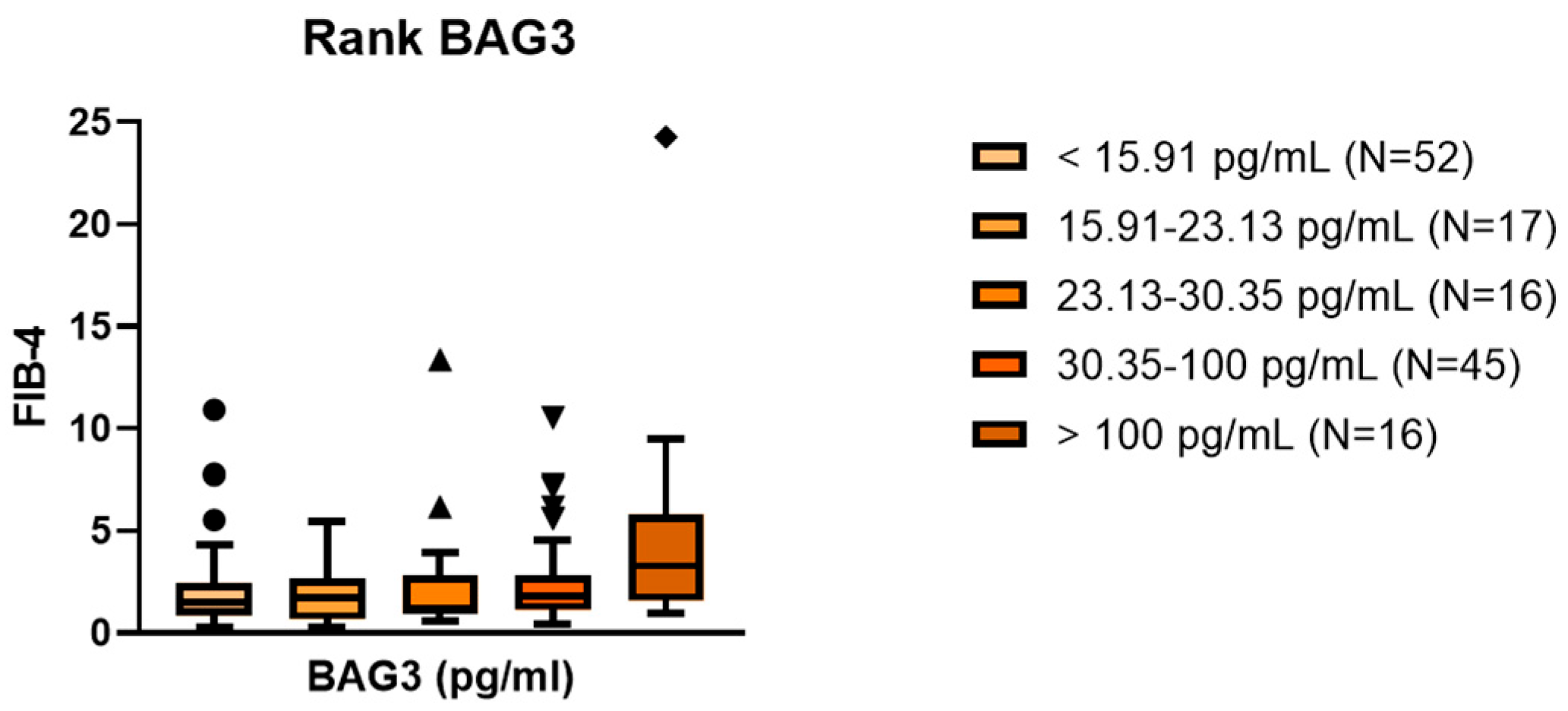
Furthermore, FIB-4 values demonstrated a progressive increase across five BAG3 categories, defined according to healthy control distribution (Kruskal–Wallis p = 0.027; Figure 3).

2.3. PNPLA3 and TM6F2 Genotypes Influences BAG3 Sera Levels

While BAG3 levels did not significantly differ by PNPLA3 rs738409 genotype (ANOVA p = 0.361), a significant association was observed with TM6SF2 rs58542926: indeed, T-allele carriers had higher BAG3 concentrations (ANOVA p = 0.028). This observation suggests a stronger mechanistic link between TM6SF2-mediated lipid retention and ER stress, which may trigger BAG3 upregulation. Importantly, BAG3 levels increased incrementally with the combined number of PNPLA3 and TM6SF2 risk alleles, indicating a cumulative genetic effect (ANOVA p = 0.003; Figure 4).

3. Discussion

EASL’s adoption of the MASLD nomenclature and diagnostic framework represents a paradigm shift in the diagnosis of liver disease, with a focus on early identification of fibrotic risk in metabolically predisposed individuals.
The liver is a stress-sensitive organ, and chronic metabolic insults in MASLD—such as lipotoxicity, oxidative stress, and ER dysfunction—are known inducers of BAG3 expression [17,31]. In this study, we demonstrate for the first time that serum BAG3 concentrations are positively associated with hepatic fibrosis severity in patients with MASLD. These findings add to the growing evidence implicating stress response pathways in fibrogenesis and indicate that BAG3 may serve as a potential non-invasive biomarker candidate for fibrosis assessment in MASLD, pending validation in larger cohorts.
In addition, we propose BAG3 as a possible mediator of proteostasis and fibrogenic progression in the disease. Indeed, the observed positive correlation between serum BAG3 levels and fibrosis severity is consistent with previous observations highlighting BAG3’s role in modulating fibroblast survival and extracellular matrix deposition under stress conditions [18,19,20], and suggests that BAG3 up-regulation might be a mechanistically relevant component of the fibrogenic process.
The progressive increase in BAG3 concentrations across fibrosis stages and genetic risk burden suggests its induction reflects a stress-adaptive yet pro-fibrotic milieu. TM6SF2-associated lipid retention, and to a lesser extent PNPLA3-driven lipotoxicity, may activate endoplasmic reticulum stress pathways, with BAG3 acting as a downstream effector that enables cellular survival at the cost of promoting hepatic stellate cell activation and matrix deposition. Intriguingly, BAG3 levels were associated with TM6SF2 but not PNPLA3 genotype, reinforcing a model where TM6SF2-driven hepatic lipotoxicity directly engages ER stress and stress-inducible chaperone systems. TM6SF2’s role in reducing VLDL secretion contributes to hepatocyte lipid accumulation, a recognized trigger of unfolded protein response pathways in MASLD pathogenesis [14,15]. This supports a plausible mechanistic link between TM6SF2-mediated lipid retention and BAG3 induction.
The additive effect of PNPLA3 and TM6SF2 risk alleles on BAG3 levels further highlights the intersection of metabolic genetics and stress-response biology. This suggests that BAG3 may integrate upstream genetic insults with cellular stress responses and fibrogenic outputs, functioning both as a biomarker and a potential therapeutic checkpoint. Overall, our results support the hypothesis that BAG3 could act as a stress-induced biomarker likely linking genetic susceptibility and fibrotic remodeling in MASLD, although longitudinal studies are necessary to confirm its prognostic role.

4. Materials and Methods

4.1. Study Population

This cross-sectional study included 146 adults with a clinical diagnosis of metabolic dysfunction-associated steatotic liver disease (MASLD), enrolled at a tertiary care center. All participants self-reported Southern European ancestry.
Inclusion criteria were:
  • Evidence of hepatic steatosis on imaging (ultrasound or controlled attenuation parameter [CAP]) or liver histology;
  • ≥1 cardiometabolic risk factor, including overweight/obesity, dyslipidaemia, dysglycaemia, or hypertension.
Participants were phenotyped into three groups:
  • Uncomplicated MASLD (n = 121);
  • Cirrhotic MASLD (n = 23);
  • MASLD-related hepatocellular carcinoma (HCC) (n = 2).
The study was conducted according to standard clinical practice protocols and was approved by the local Ethics Committee (CEI Campania Sud IRB n.8/2018). Informed consent was obtained from all participants prior to enrollment.

4.2. Fibrosis Assessment

Liver fibrosis was assessed non-invasively using FIB-4 index, calculated from age, AST, ALT, and platelet count [32]. FIB-4 is the most widely established and available tool [33]. However, its ability to detect fibrosis is limited in the intermediate range (1.3–2.67). Notably, a different lower FIB-4 cut-off of 2.0 applies in individuals over 65 years of age.
Risk categories were defined as:
  • Low risk: <1.30 (or <2.00 if age > 65 years).
  • Intermediate risk: 1.30–2.67.
  • High risk: >2.67.

4.3. Serum BAG3 Quantification

BAG3 (Bcl-2-associated athanogene 3) concentrations were measured in serum samples using a validated in-house ELISA, performed in duplicate. Methodological details were previously published [30].
Serum BAG3 levels in healthy controls were obtained from a previously published study [30]. Data were used solely as a reference range for BAG3 values. Mean serum BAG3 concentration in healthy subjects was 23.13 ± 10.4 pg/mL, with a 95th percentile of 30.35 pg/mL.

4.4. Genotyping

DNA was isolated from peripheral blood using QiAmp® DNA Mini kit (Qiagen, Hilden, Germany), in accordance with the user manual protocol. Quality and quantity were determined with the spectrophotometer Implen NP80 Touch (Implen, München, Germany).
The following SNPs were genotyped using TaqMan® 5′-nuclease SNP assays (ThermoFisher, Waltham, MA, USA):
  • PNPLA3 rs738409 (C>G, p.I148M).
  • TM6SF2 rs58542926 (C>T, p.E167K).
Genotype frequencies were in Hardy–Weinberg equilibrium (χ2 p > 0.05).

4.5. Statistical Analysis

Continuous variables are reported as mean ± standard deviation (SD); categorical data as n (%).
Comparisons between groups were performed using ANOVA or Kruskal–Wallis tests.
Spearman’s correlation was used for association testing between BAG3 and FIB-4.
A p-value < 0.05 was considered significant. All analyses were conducted using IBM SPSS Statistics v26.

5. Limitations and Future Directions

This study’s cross-sectional nature precludes assessment of causal relationships. Liver fibrosis was assessed using the FIB-4 index rather than histopathology, although FIB-4 is well-validated in clinical settings [32]. Additionally, the cohort’s relatively small size and ethnic homogeneity (all participants were from Southern European ancestry) may limit generalizability. While we included two individuals with HCC to illustrate trends at the highest fibrosis stages, the small number prevents any definitive conclusions regarding HCC-specific associations. Future studies should examine longitudinal trajectories of BAG3 expression and assess its utility in predicting fibrosis progression or treatment response. Additionally, mechanistic studies should assess whether BAG3 knockdown or pharmacological modulation alters HSC activation or hepatocyte stress tolerance under lipotoxic conditions, especially in genetic models of MASLD. Targeting BAG3, either to enhance its adaptive roles or block maladaptive signaling, may offer new therapeutic strategies in MASLD.

6. Conclusions

The presence and severity of hepatic steatosis and fibrosis should be assessed in all individuals with at least one cardiometabolic risk factor using validated non-invasive blood-based tests, despite their limited accuracy in distinguishing intermediate-risk categories. Most importantly, they do not provide information on the degree of hepatic inflammation. Determining fibrosis risk is essential to identify individuals at an increased risk of developing liver-related complications during early follow-up [34].
This study identifies BAG3 as potentially informative biomarker of hepatic fibrosis in patients with MASLD. Serum BAG3 levels not only correlated with non-invasive fibrosis scores but also increased with the burden of TM6SF2 and PNPLA3 risk alleles—highlighting their relevance at the intersection of genetic susceptibility and stress-responsive fibrogenic pathways. These findings suggest that BAG3 implication in MASLD may reflect a previously underappreciated mechanism of fibrotic remodeling linked to hepatocellular stress and metabolic dysfunction.
Importantly, the integration of BAG3 measurements with established genetic and clinical parameters could improve risk stratification and disease monitoring in MASLD. Further longitudinal and mechanistic studies are needed to validate BAG3 as a prognostic biomarker and to clarify whether it might represent a therapeutic target in liver fibrosis.

Supplementary Materials

The following supporting information can be downloaded at: https://www.mdpi.com/article/10.3390/ijms262311286/s1.

Author Contributions

Conceptualization, B.M.M., M.C.T. and M.P.; resources, M.P.; formal analysis, B.M.M. and A.R.; investigation, B.M.M., A.R. and A.B.; data curation, M.F. and T.S.; writing (original draft preparation), B.M.M., A.R., M.F., M.C.T. and M.P.; writing (review and editing), B.M.M., A.R., M.C.T. and M.P.; supervision, M.C.T. and M.P. All authors have read and agreed to the published version of the manuscript.

Funding

This research received no external funding.

Institutional Review Board Statement

The study was conducted according to standard clinical practice protocols and was approved by the local Ethics Committee (CEI Campania Sud IRB n.8/2018 on 30 January 2018).

Informed Consent Statement

Informed consent was obtained from all subjects involved in the study.

Data Availability Statement

The raw data supporting the conclusions of this article will be made available by the authors on request.

Conflicts of Interest

Authors Alessandra Rosati and Maria Caterina Turco are from FIBROSYS s.r.l. The authors declare no conflicts of interest.

Abbreviations

The following abbreviations are used in this manuscript:
ALTAlanine Aminotransferase
ASTAspartate Aminotransferase
BAG3Bcl-2-associated Athanogene 3
BMIBody Mass Index
CAPControlled Attenuation Parameter
CEIComitato Etico Interaziendale (Ethics Committee)
EASDEuropean Association for the Study of Diabetes
EASLEuropean Association for the Study of the Liver
EASOEuropean Association for the Study of Obesity
ELISAEnzyme-Linked Immunosorbent Assay
EREndoplasmic Reticulum
FIB-4Fibrosis-4 Index
HCCHepatocellular Carcinoma
HSCHepatic Stellate Cell
Hsp70Heat Shock Protein 70
IRBInstitutional Review Board
LSLiver Stiffness
MASLDMetabolic Dysfunction-Associated Steatotic Liver Disease
PNPLA3Patatin-like Phospholipase Domain-containing Protein 3
PRSPolygenic Risk Score
ROSReactive Oxygen Species
SNPSingle Nucleotide Polymorphism
TM6SF2Transmembrane 6 Superfamily Member 2
VLDLVery-Low-Density Lipoprotein

References

  1. Younossi, Z.M.; Koenig, A.B.; Abdelatif, D.; Fazel, Y.; Henry, L.; Wymer, M. Global epidemiology of nonalcoholic fatty liver disease-Meta-analytic assessment of prevalence, incidence, and outcomes. Hepatology 2016, 64, 73–84. [Google Scholar] [CrossRef]
  2. Eslam, M.; Sanyal, A.J.; George, J.; Panel, I.C. MAFLD: A Consensus-Driven Proposed Nomenclature for Metabolic Associated Fatty Liver Disease. Gastroenterology 2020, 158, 1999–2014.e1991. [Google Scholar] [CrossRef]
  3. Dulai, P.S.; Singh, S.; Patel, J.; Soni, M.; Prokop, L.J.; Younossi, Z.; Sebastiani, G.; Ekstedt, M.; Hagstrom, H.; Nasr, P.; et al. Increased risk of mortality by fibrosis stage in nonalcoholic fatty liver disease: Systematic review and meta-analysis. Hepatology 2017, 65, 1557–1565. [Google Scholar] [CrossRef]
  4. Allen, A.M.; Lazarus, J.V.; Younossi, Z.M. Healthcare and socioeconomic costs of NAFLD: A global framework to navigate the uncertainties. J. Hepatol. 2023, 79, 209–217. [Google Scholar] [CrossRef]
  5. European Association for the Study of the Liver (EASL); European Association for the Study of Diabetes (EASD); European Association for the Study of Obesity (EASO). EASL-EASD-EASO Clinical Practice Guidelines on the management of metabolic dysfunction-associated steatotic liver disease (MASLD). J. Hepatol. 2024, 81, 492–542. [Google Scholar] [CrossRef]
  6. Jamialahmadi, O.; De Vincentis, A.; Tavaglione, F.; Malvestiti, F.; Li-Gao, R.; Mancina, R.M.; Alvarez, M.; Gelev, K.; Maurotti, S.; Vespasiani-Gentilucci, U.; et al. Partitioned polygenic risk scores identify distinct types of metabolic dysfunction-associated steatotic liver disease. Nat. Med. 2024, 30, 3614–3623, Erratum in Nat. Med. 2025, 31, 700. [Google Scholar] [CrossRef] [PubMed]
  7. Xiao, L.; Li, Y.; Hong, C.; Ma, P.; Zhu, H.; Cui, H.; Zou, X.; Wang, J.; Li, R.; He, J.; et al. Polygenic risk score of metabolic dysfunction-associated steatotic liver disease amplifies the health impact on severe liver disease and metabolism-related outcomes. J. Transl. Med. 2024, 22, 650. [Google Scholar] [CrossRef]
  8. Kanda, T.; Sasaki-Tanaka, R.; Abe, H.; Kimura, N.; Yoshida, T.; Hayashi, K.; Sakamaki, A.; Yokoo, T.; Kamimura, H.; Tsuchiya, A.; et al. Polygenic Risk Score for Metabolic Dysfunction-Associated Steatotic Liver Disease and Steatohepatitis: A Narrative Review. Int. J. Mol. Sci. 2025, 26, 5164. [Google Scholar] [CrossRef] [PubMed]
  9. He, S.; McPhaul, C.; Li, J.Z.; Garuti, R.; Kinch, L.; Grishin, N.V.; Cohen, J.C.; Hobbs, H.H. A sequence variation (I148M) in PNPLA3 associated with nonalcoholic fatty liver disease disrupts triglyceride hydrolysis. J. Biol. Chem. 2010, 285, 6706–6715. [Google Scholar] [CrossRef]
  10. Romeo, S.; Kozlitina, J.; Xing, C.; Pertsemlidis, A.; Cox, D.; Pennacchio, L.A.; Boerwinkle, E.; Cohen, J.C.; Hobbs, H.H. Genetic variation in PNPLA3 confers susceptibility to nonalcoholic fatty liver disease. Nat. Genet. 2008, 40, 1461–1465. [Google Scholar] [CrossRef] [PubMed]
  11. Sookoian, S.; Pirola, C.J. Meta-analysis of the influence of I148M variant of patatin-like phospholipase domain containing 3 gene (PNPLA3) on the susceptibility and histological severity of nonalcoholic fatty liver disease. Hepatology 2011, 53, 1883–1894. [Google Scholar] [CrossRef] [PubMed]
  12. Pingitore, P.; Dongiovanni, P.; Motta, B.M.; Meroni, M.; Lepore, S.M.; Mancina, R.M.; Pelusi, S.; Russo, C.; Caddeo, A.; Rossi, G.; et al. PNPLA3 overexpression results in reduction of proteins predisposing to fibrosis. Hum. Mol. Genet. 2016, 25, 5212–5222. [Google Scholar] [CrossRef]
  13. Kozlitina, J.; Smagris, E.; Stender, S.; Nordestgaard, B.G.; Zhou, H.H.; Tybjærg-Hansen, A.; Vogt, T.F.; Hobbs, H.H.; Cohen, J.C. Exome-wide association study identifies a TM6SF2 variant that confers susceptibility to nonalcoholic fatty liver disease. Nat. Genet. 2014, 46, 352–356. [Google Scholar] [CrossRef] [PubMed]
  14. Mahdessian, H.; Taxiarchis, A.; Popov, S.; Silveira, A.; Franco-Cereceda, A.; Hamsten, A.; Eriksson, P.; van’t Hooft, F. TM6SF2 is a regulator of liver fat metabolism influencing triglyceride secretion and hepatic lipid droplet content. Proc. Natl. Acad. Sci. USA 2014, 111, 8913–8918. [Google Scholar] [CrossRef]
  15. Liu, Y.L.; Reeves, H.L.; Burt, A.D.; Tiniakos, D.; McPherson, S.; Leathart, J.B.; Allison, M.E.; Alexander, G.J.; Piguet, A.C.; Anty, R.; et al. TM6SF2 rs58542926 influences hepatic fibrosis progression in patients with non-alcoholic fatty liver disease. Nat. Commun. 2014, 5, 4309. [Google Scholar] [CrossRef]
  16. Doong, H.; Vrailas, A.; Kohn, E.C. What’s in the ‘BAG’?—A functional domain analysis of the BAG-family proteins. Cancer Lett. 2002, 188, 25–32. [Google Scholar] [CrossRef]
  17. Rosati, A.; Graziano, V.; De Laurenzi, V.; Pascale, M.; Turco, M.C. BAG3: A multifaceted protein that regulates major cell pathways. Cell Death Dis. 2011, 2, e141. [Google Scholar] [CrossRef] [PubMed]
  18. Fang, X.; Bogomolovas, J.; Wu, T.; Zhang, W.; Liu, C.; Veevers, J.; Stroud, M.J.; Zhang, Z.; Ma, X.; Mu, Y.; et al. Loss-of-function mutations in co-chaperone BAG3 destabilize small HSPs and cause cardiomyopathy. J. Clin. Investig. 2017, 127, 3189–3200. [Google Scholar] [CrossRef]
  19. Behl, C. Breaking BAG: The Co-Chaperone BAG3 in Health and Disease. Trends Pharmacol. Sci. 2016, 37, 672–688. [Google Scholar] [CrossRef]
  20. De Marco, M.; Del Papa, N.; Reppucci, F.; Iorio, V.; Basile, A.; Falco, A.; Iaccarino, R.; Brongo, S.; De Caro, F.; Capunzo, M.; et al. BAG3 induces α-SMA expression in human fibroblasts and its over-expression correlates with poorer survival in fibrotic cancer patients. J. Cell Biochem. 2022, 123, 91–101. [Google Scholar] [CrossRef]
  21. Rosati, A.; Basile, A.; D’Auria, R.; d’Avenia, M.; De Marco, M.; Falco, A.; Festa, M.; Guerriero, L.; Iorio, V.; Parente, R.; et al. BAG3 promotes pancreatic ductal adenocarcinoma growth by activating stromal macrophages. Nat. Commun. 2015, 6, 8695. [Google Scholar] [CrossRef]
  22. De Marco, M.; Gauttier, V.; Pengam, S.; Mary, C.; Ranieri, B.; Basile, A.; Festa, M.; Falco, A.; Reppucci, F.; Cammarota, A.L.; et al. Concerted BAG3 and SIRPα blockade impairs pancreatic tumor growth. Cell Death Discov. 2022, 8, 94. [Google Scholar] [CrossRef]
  23. Ji, J.; Yu, F.; Ji, Q.; Li, Z.; Wang, K.; Zhang, J.; Lu, J.; Chen, L.; E, Q.; Zeng, Y.; et al. Comparative proteomic analysis of rat hepatic stellate cell activation: A comprehensive view and suppressed immune response. Hepatology 2012, 56, 332–349. [Google Scholar] [CrossRef] [PubMed]
  24. Jung, S.Y.; Yun, H.H.; Lim, J.H.; Lee, D.H.; Seo, S.B.; Baek, J.Y.; Lee, J.; Yoo, K.; Kim, H.; Kim, H.L.; et al. Hepatocyte-specific deletion of Bis causes senescence in the liver without deteriorating hepatic function. Biochem. Biophys. Res. Commun. 2022, 619, 42–48. [Google Scholar] [CrossRef]
  25. Damiani, V.; Lamolinara, A.; Cicalini, I.; Cufaro, M.C.; Del Pizzo, F.; Di Marco, F.; Del Boccio, P.; Dufrusine, B.; Hahne, M.; Lattanzio, R.; et al. Pancreatic beta-cell specific BAG3 knockout results in chronic hyperinsulinemia inducing insulin resistance. Mol. Metab. 2023, 74, 101752. [Google Scholar] [CrossRef]
  26. Longo, M.; Meroni, M.; Paolini, E.; Erconi, V.; Carli, F.; Fortunato, F.; Ronchi, D.; Piciotti, R.; Sabatini, S.; Macchi, C.; et al. TM6SF2/PNPLA3/MBOAT7 Loss-of-Function Genetic Variants Impact on NAFLD Development and Progression Both in Patients and in In Vitro Models. Cell Mol. Gastroenterol. Hepatol. 2022, 13, 759–788. [Google Scholar] [CrossRef]
  27. Puri, P.; Mirshahi, F.; Cheung, O.; Natarajan, R.; Maher, J.W.; Kellum, J.M.; Sanyal, A.J. Activation and dysregulation of the unfolded protein response in nonalcoholic fatty liver disease. Gastroenterology 2008, 134, 568–576. [Google Scholar] [CrossRef] [PubMed]
  28. Sanyal, A.J. Past, present and future perspectives in nonalcoholic fatty liver disease. Nat. Rev. Gastroenterol. Hepatol. 2019, 16, 377–386. [Google Scholar] [CrossRef]
  29. Schwabe, R.F.; Tabas, I.; Pajvani, U.B. Mechanisms of Fibrosis Development in Nonalcoholic Steatohepatitis. Gastroenterology 2020, 158, 1913–1928. [Google Scholar] [CrossRef] [PubMed]
  30. De Marco, M.; Armentaro, G.; Falco, A.; Minniti, A.; Cammarota, A.L.; Iannone, C.; Basile, A.; D’Ardia, A.; Zeppa, P.; Marzullo, L.; et al. Overexpression of BAG3 (Bcl2-associated athanogene 3) in serum and skin of patients with systemic sclerosis. Clin. Exp. Rheumatol. 2024, 42, 1623–1628. [Google Scholar] [CrossRef]
  31. Venkatesan, N.; Doskey, L.C.; Malhi, H. The Role of Endoplasmic Reticulum in Lipotoxicity during Metabolic Dysfunction-Associated Steatotic Liver Disease (MASLD) Pathogenesis. Am. J. Pathol. 2023, 193, 1887–1899. [Google Scholar] [CrossRef] [PubMed]
  32. Sterling, R.K.; Lissen, E.; Clumeck, N.; Sola, R.; Correa, M.C.; Montaner, J.; Sulkowski, M.S.; Torriani, F.J.; Dieterich, D.T.; Thomas, D.L.; et al. Development of a simple noninvasive index to predict significant fibrosis in patients with HIV/HCV coinfection. Hepatology 2006, 43, 1317–1325. [Google Scholar] [CrossRef] [PubMed]
  33. European Association for the Study of the Liver. EASL Clinical Practice Guidelines on non-invasive tests for evaluation of liver disease severity and prognosis—2021 update. J. Hepatol. 2021, 75, 659–689. [Google Scholar] [CrossRef]
  34. Boursier, J.; Hagström, H.; Ekstedt, M.; Moreau, C.; Bonacci, M.; Cure, S.; Ampuero, J.; Nasr, P.; Tallab, L.; Canivet, C.M.; et al. Non-invasive tests accurately stratify patients with NAFLD based on their risk of liver-related events. J. Hepatol. 2022, 76, 1013–1020. [Google Scholar] [CrossRef] [PubMed]
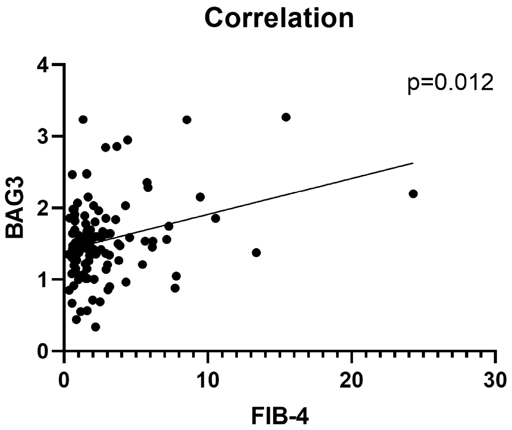
Figure 1. Correlation between serum BAG3 levels and liver fibrosis severity. The scatter plot shows the correlation between log serum BAG3 concentrations and FIB-4 index values in 146 patients with MASLD (Spearman’s ρ = 0.158, p = 0.012).
Ijms 26 11286 g001
Figure 2. Stepwise increase in FIB-4 scores across stratified BAG3 serum concentration thresholds. Boxplots illustrate FIB-4 index values across increasing serum BAG3 concentration thresholds and categories, benchmarked against reference values from healthy controls. (A) BAG3 level cut-off of 23.13 pg/mL (mean in healthy controls, p = 0.023). (B) BAG3 concentration cut-off of 30.35 pg/mL (95th percentile in healthy controls, p = 0.008). (C) BAG3 concentration cut-off of 100 pg/mL (p = 0.004).
Ijms 26 11286 g002
Figure 3. Stepwise association between BAG3 categories and FIB-4. The boxplot illustrates progressive FIB-4 increase across BAG3 categories (<15.91, 15.91–23.13, 23.13–30.35, 30.35–100, >100 pg/mL; p = 0.006).
Ijms 26 11286 g003
Figure 4. Serum BAG3 levels according to number of risk alleles. Additive effect of combined PNPLA3 and TM6SF2 risk alleles on BAG3 levels, showing stepwise increases. The bar chart shows mean ± SD of BAG3 concentrations for 0, 1, 2, and ≥3 risk alleles.
Ijms 26 11286 g004
Table 1. Characteristics of study cohort.
VariableTotal (n = 146)MASLD (n = 121)Cirrhosis + HCC (n = 25)p-Value
Age (years)62.9 ± 14.061.8 ± 14.867.9 ± 8.40.020
Male sex82 (56.2%)64 (52.9%)18 (72.0%)
BMI (kg/m2)32.3 ± 6.832.0 ± 6.834.2 ± 6.5
AST (U/L)35.5 ± 23.535.6 ± 21.144.7 ± 31.60.040
ALT (U/L)40.8 ± 33.042.9 ± 35.030.3 ± 18.1
LS (kPa)9.36 ± 9.37.24 ± 4.0219.2 ± 17.52.98 × 10−8
FIB-42.51 ± 2.891.74 ± 1.356.25 ± 4.892.11 × 10−10
BAG3 (pg/mL)75.9 ± 226.561.8 ± 220.9143.8 ± 245.10.011
Abbreviations: BMI: Body Mass Index; AST: Aspartate Aminotransferase; ALT: Alanine Aminotransferase; LS: Liver Stiffness; FIB-4: Fibrosis-4 Index; BAG3: BCL2-Associated Athanogene 3. Bold = significantly higher in that group.
Disclaimer/Publisher’s Note: The statements, opinions and data contained in all publications are solely those of the individual author(s) and contributor(s) and not of MDPI and/or the editor(s). MDPI and/or the editor(s) disclaim responsibility for any injury to people or property resulting from any ideas, methods, instructions or products referred to in the content.