Article

Transient Receptor Potential Vanilloid 4 (TRPV4) in the Human Carotid Body

by
Yolanda García-Mesa
1,2,
Elda Alba
3,4,
Graciela Martínez-Barbero
1,
Iván Suazo
5,
Patricia Cuendias
1,2,
José Martín-Cruces
1,6,
Mirian Teulé-Trull
1,6,
José A. Vega
1,5,*,
Olivia García-Suárez
1,2,† and
Teresa Cobo
7,8,†
1
Grupo SINPOS, Departamento de Morfología y Biología Celular, Universidad de Oviedo, 33003 Oviedo, Spain
2
Instituto de Investigación Sanitaria del Principado de Asturias, ISPA, 33011 Oviedo, Spain
3
Instituto de Neurociencias Vithas, 28010 Madrid, Spain
4
Servicio de Neurología, Hospital Clínico San Carlos, 28040 Madrid, Spain
5
Facultad de Ciencias de la Salud, Universidad Autónoma de Chile, Santiago 8330015, Chile
6
Master ENDORE, Universidad de Santiago de Compostela, 15705 Santiago de Compostela, Spain
7
Departamento de Cirugía y Especialidades Médico-Quirúrgicas, Universidad de Oviedo, 33003 Oviedo, Spain
8
Instituto Asturiano de Odontología, 33006 Oviedo, Spain
*
Author to whom correspondence should be addressed.
These authors contributed equally to this work.
Int. J. Mol. Sci. 2025, 26(21), 10689; https://doi.org/10.3390/ijms262110689
Submission received: 12 September 2025 / Revised: 29 October 2025 / Accepted: 30 October 2025 / Published: 3 November 2025

Abstract

TRPV4 (transient receptor potential vanilloid 4) is a non-selective, multifunctional cationic channel that is expressed in numerous cells in the body. It can be activated by temperature, mechanical forces, and chemical and biochemical molecules. Functionally, TRPV4 participates in maintaining osmotic homeostasis, blood pressure, and hypoxic preconditioning. As far as we know, the presence of TRPV4 has never been reported in the carotid body despite the overlap that exists between some biological functions of TRPV4 and the physiology of the carotid body. In the present work, immunofluorescence associated with confocal laser microscopy, associated with quantitative analysis (area occupied by immunofluorescence), has been used to examine the occurrence of TRPV4 in the human carotid body. The results demonstrated the presence of TRPV4 in a subpopulation of chemoreceptor type I cells (approximately 65–68%), a subpopulation of type II supporting cells, and in nerve terminals in the human carotid body. Its function, if any, in this multisensory organ must be demonstrated, but it is in line with the functions attributed to the carotid body.

1. Introduction

The carotid body (glomus caroticum) is regarded as the most important peripheral chemoreceptor, and it is particularly sensitive to variations in blood levels of oxygen (hypoxemia), carbon dioxide (hypercapnia), and pH (acidosis) [1,2]. However, it should be considered as a polymodal sensor since it also detects temperature, participates in homeostatic control of arterial pressure [3,4], glycemia [5,6,7], in metabolic and hormonal regulation [8,9,10], as well as in the control of sympathetic and parasympathetic activity [11,12]. In addition, some studies indicate that the carotid body senses inflammatory molecules [12,13,14] and therefore could be involved in the inflammatory processes that accompany the physiopathological processes in which the carotid body participate [2,15,16,17].
Structurally the carotid body consists of clusters of chemosensory, or type I, cells, surrounded by supporting, or type II, cells that form the so-called glomic glomeruli, which in turn are grouped into glomic lobules; between the glomerulus and lobules are nerve bundles, arteries, and veins embedded in fibrous tissue that at the periphery form a capsule. Type I cells are a heterogenous population and receives afferent innervation from neurons whose bodies are localized in the petrosal ganglion of the glossopharyngeal nerve. The type II form networks surrounding type I cells and normally lack innervation. On the other hand, nerve fibres that originated in the post-ganglionic neurons of the superior sympathetic ganglion give efferent innervation to the glomic arteries and type I cells. In addition to type I and type II the carotid body contains variable amounts of progenitor cells and neuroblasts (Scheme 1) [1].
The physiological and physiopathological ways in which the carotid body participates involve type I cells, type II cells, and afferent and efferent nerves [18,19,20] in the so-called tripartite synapse [21,22,23] all orchestrated by neurotransmitters [2,24,25] and ion channels [2,26,27,28,29,30]. Of particular interest in the physiology of the carotid body are members of the transient receptor potential (TRP) superfamily of ion channels acting alone [26,27,28] or mediating the actions of cytokines [12,13] or leptin [3].
TRPV4 (transient receptor potential vanilloid transient 4) is a non-selective cation channel permeable to Na+, Ca2+, and Mg2+ ions. It consists of four monomers, and each monomer comprises six transmembrane domains with a pore loop in between the fifth and the sixth transmembrane domains [31,32]. TRPV4 is widely expressed in systemic tissues (see [33] for references) and can be activated by temperature [34], mechanical forces [35,36,37,38,39,40], and chemical stimuli (arachidonic acid, 4α-phorbol esters, endocannabinoids, and others) [41,42,43].
TRPV4 plays an important role in maintaining cellular osmotic homeostasis [44], regulating blood pressure to produce both vasodilation and vasoconstriction, and acting as hypoxic preconditioning [45,46,47,48]. Altogether, these data suggest that TRPV4 could be involved in several of the functions of the carotid body [49]. On the other hand, and consistently with the proposed roles for TRPV4, emerging evidence indicates that it is involved in various pathophysiological states, including hypertension, diabetes, and obesity [50,51,52].
Considering the overlapping between the biological functions attributed to TRPV4 and the physiology of the carotid body, we have conducted research to analyze whether TRPV4 is present in this sensory organ. As far as we know, this has never been explored, although TRPV4 has been detected in the petrosal ganglion of the glossopharyngeal nerve that is responsible for the afferent innervation of the carotid body [53]. The present study has been carried out on the human carotid body using immunofluorescence techniques and aims to contribute to the knowledge of the functions of the carotid body.

2. Results

The carotid body is structured into glomeruli formed by type I cells, type II cells, blood vessels, and nerves (afferent and efferent). Type II cells are immunofluorescent for the S100 protein (Figure 1a), while type I cells display neuronal-specific enolase (Figure 1b) and synaptophysin (Figure 2b) immunofluorescence. Type II cells are arranged around type I cells, and they are two completely segregated populations for the antigens used as markers (Figure 1c,d).

Immunolocalization of TRPV4 in the Human Carotid Body

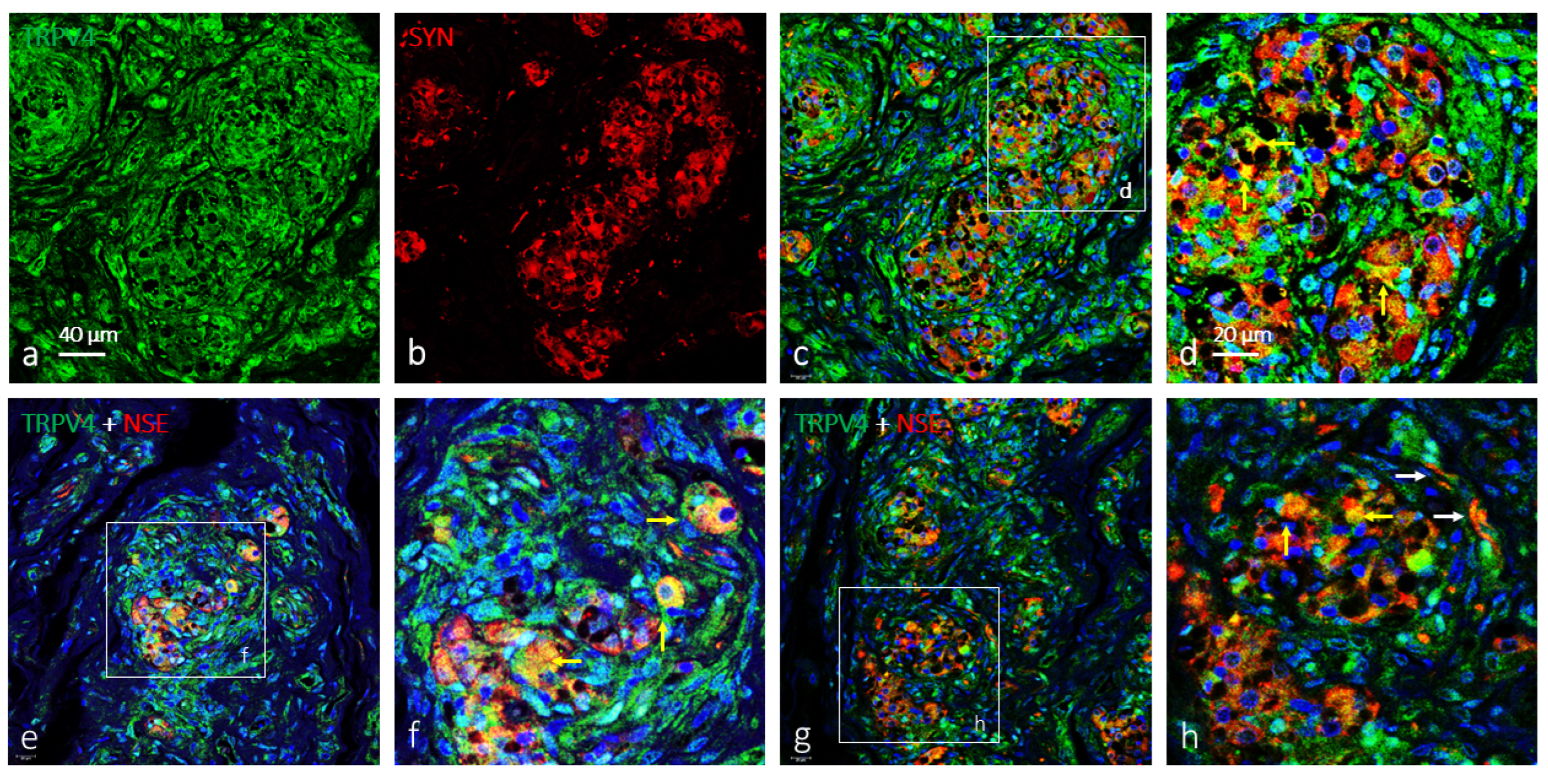
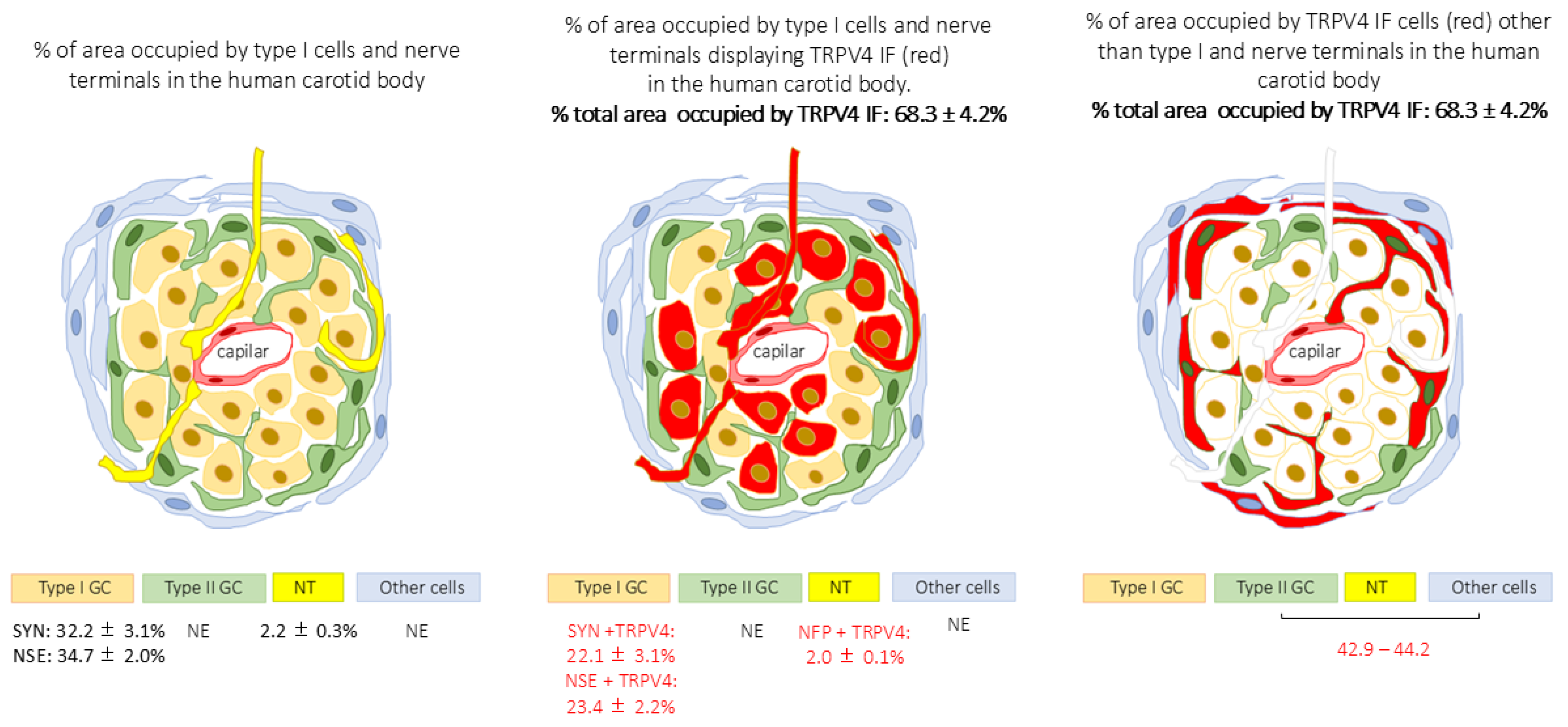
Within the carotid body the glomeruli and lobule are very well defined, and the immunofluorescence for TRPV4 was extremely broad and apparently included all cell types. (Figure 2a). In the co-localization studies, it was observed that the area occupied by type I cells (synaptophysin positive, Figure 2b, and neuron-specific enolase positive, Figure 2e–h) is smaller than that of TRPV4 cells, and in the merge images it was observed that only a subpopulation of type I cells also expresses TRPV4 (Figure 2c–h). The remaining TRPV4 immunofluorescent cells correspond to type II glomic cells, and other cells present in the carotid body such as endothelial cells, cells of the connective septa, progenitor cells, and neuroblasts. The quantitative analysis showed that type I-SYN positive cells or type I-NSE positive cells occupied in the carotid body correspond to an area of approximately 32% and 35%, respectively, whereas that occupied by TRPV4 positive cells correspond to about 68%. Therefore, the percentage of area occupied by TRPV4-positive cells exceeds that of type I cells, suggesting that other cell types of the carotid body, especially type II cells, show immunofluorescence for TRPV4 (occupied approximately 50% of the TRPV4 immunofluorescent area). The results of the quantitative analysis of TRPV4-SYN or TRPV4-NSE mergence show that approximately 65–70% of type I cells express TRPV4 (Table 1 and Scheme 2). The small differences in the areas of co-localization of TRPV4 with SYN and NSE may be due to the fact that NSE also marks the nerve fibre terminals.
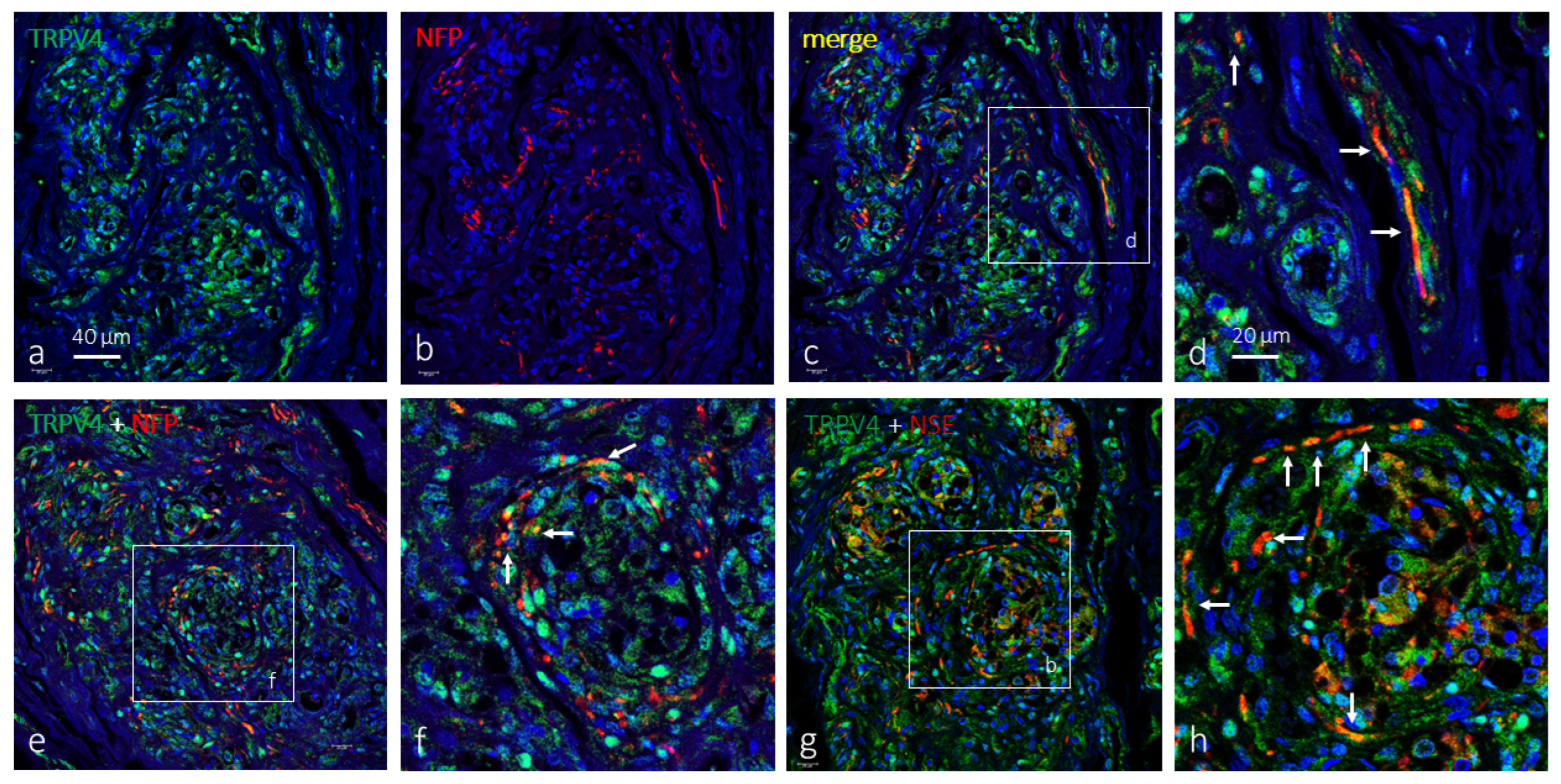
On the other hand, the presence of TRPV4 in the nerve fibre terminals of the carotid body was investigated. Nerve terminals immunofluorescence for TRPV4 were detected by co-localization immunofluorescence with NSE (Figure 3a–e; it is likely that drop profiles of TRPV4-NSE immunofluorescence merge correspond to transverse sections of nerve terminals) and neurofilament proteins (NFP; Figure 3g,h). Based on the quantitative analysis, about 90% of the nerve fibre terminals in the human carotid body are TRPV4 positive (Table 1 and Scheme 2).
Therefore, in the human carotid body, TRPV4 is present in a subpopulation of type I cells and in most of the nerves that supply it, but also in uncharacterized cells that mainly correspond to type II cells.

3. Discussion

TRPV4 is a multifunctional non-selective cation channel belonging to the vanilloid family of the transient receptor potential (TRP) ion channels superfamily (see [41,53]). This ion channel has been detected with different techniques in most tissues, such as the smooth muscle cells, vasculature, lungs, brain, heart, kidneys, salivary glands, liver, bladder, trachea, skin, bone, spleen, testicle, dorsal root, and cranial nerve sensory ganglia [33,46,53,54,55,56,57,58,59]. As far as we know, the presence of TRPV4 in the normal human carotid body is demonstrated now for the first time. No data is available about TRPV4 in other peripheral chemoreceptors, whereas it has been found in peripheral baroreceptors [45]. However, other members of the TRP ion channel superfamily have been detected in the carotid body. Buniel and co-workers [26] demonstrated the presence of members of the canonical family of TRPs (TRPC3, TRPC4, TRPC5, TRPC6, and TRPC7) in different neuronal subpopulations of the rat petrosal ganglion that gives afferent innervation to the carotid body, although only TRPC1 and TRPC4 were localized in the afferent nerve terminals of the carotid body. The role of these channels in the chemosensory pathway of the carotid body is yet to be established. One member of the vanilloid TRP family, TRPV1, mediates the sensing of the carotid body to Th2 cytokines and lysophosphatidic acid [12,13], and a member of the melastatin TRP family, TRPM7, is involved in the regulation of obesity-induced hypertension mediated by leptin [3,8]. However, the role of the channels of the different families of TRP channels is far from being fully known [27].
In our hands, specific immunofluorescence for TRPV4, was detected in a subpopulation of type I and in nerve terminals, and presumably also in other cells including type II cells, blood vessel cells, progenitor cells, neuroblasts, and cells of the connective septa. There is evidence that it is present in the satellite glial cells [60] and neurons [61] of the spinal ganglia, as well as in the complex peripheral sensory ganglia that innervates different organs of the carotid body area [54]. So, the occurrence of TRPV4 in the nerve terminals within the carotid body is consistent with those data. The type I carotid body cells and the peripheral sensory ganglia are both derivatives of the neural crest, and type I cells and neurons as well as type II cells and satellite glial cells share common antigens [62]. Experiments are in progress in our laboratory to determine which TRP, ASIC, and PIEZO ion channels are present in type II cells of the human carotid body, and which of them are present in cells of the carotid body other than type I and type II.
Because of the wide spectrum of functions of TRPV4 it is difficult to assign it a single role in the carotid body. However, TRPV4 presumably is related to the functions of this multisensor. The carotid body detects variations in acidosis and TRPV4 is an acidosis detector [63], most likely in association with ASIC channels [29,64]. TRPV4 is also involved in the control of glycemia [50,65], metabolic, and hormonal regulation [66,67], such as the carotid body [4,5,68].
But surely the most obvious role of TRPV4 in the carotid body is as mechanosensor and osmosensor [53,69,70]. In the carotid body, a subpopulation of type I cells, and surely most of type II cells express TRPV4. Interestingly, type I cells express PIEZO2 via mechanosensor–mechanotransducer ion channels, and type II cells express both PIEZO1 and PIEZO2 [30]. Also acid-sensing ion channels which participate in mechano detection were found in type I and type II cells in the human carotid body [29,71].
In view of the present results, it can be hypothesized that the carotid body presumably plays a role in regulating various physiological processes in both normal and pathological conditions, such as many metabolic or cardiovascular diseases and hypertension [48,72,73,74,75,76,77]. However, many more studies are needed to definitively establish the role of TRPV4 in the carotid body.

4. Materials and Methods

Tissue samples of the carotid body were obtained during removal of organs for transplantation in subjects who died in traffic accidents (Hospital Universitario Central de Asturias, Oviedo, Spain). There were 8 pieces (from 5 males and 3 females), with ages ranging between 38 and 68 years. The petrosal ganglia (n = 4) and the sympathetic superior ganglion (n = 4) were dissected out and included in the study. The pieces were cleaned in 4 °C saline solution; fixed in 10% formaldehyde in 0.1 M phosphate-buffered saline, pH 7.4, for 24 h at 4 °C; dehydrated; and routinely embedded in paraffin.
These materials were obtained in compliance with the Spanish Law and the guidelines of the Helsinki Declaration II. Sections of the same material had been used in previous studies on the carotid body [16,17]. This material, which was used for research purposes, is deposited in the Department of Morphology and Cell Biology of the University of Oviedo, National Registry of Biobanks (Collections Section, Ref. C-0001627), created and authorized by the Ministry of Economy and Competitiveness of the Government of Spain on 30 November 2012.

4.1. Double Immunofluorescence

Tissue samples were cut 10 μm thick and the sections were mounted on gelatin-coated slides. Sections were then indirectly immunohistochemically stained with peroxidase–anti-peroxidase, rehydrated, and rinsed in 0.05 M Tris-HCl buffer (pH 7.5) containing 0.1% bovine serum albumin and 0.1% Triton X-100. The sections were processed for the simultaneous detection of TRPV4 together with neuronal (neurofilament protein, NFP) type I cell (neuron-specific enolase, NSE; synaptophysin, SYN) and type II cell (S100 protein, S100P) markers [29,30,78,79,80] (Table 2. It should be noted that type I glomus cells also express NSE. The antibody against TRPV4 was raised in rabbit anti-human TRPV4 (manufacturer’s notice).
The sections were incubated overnight at 4 °C, in a humid chamber, with a 1:1 (v/v) mixture of polyclonal antibodies against TRPV4 and monoclonal antibodies against NFP, NSE, S100P, or SYN. After rinsing, the sections were sequentially treated, in a dark and humid chamber, for 1 h at room temperature, with a goat anti-rabbit antibody conjugated to Alexa Fluor 488 (Serotec™, Oxford, UK; 1:1000 dilution) and a donkey anti-mouse antibody conjugated to Cy3 (Jackson-ImmunoResearch™, Baltimore, MD, USA; 1:50 dilution). Finally, cell nuclei were counterstained with DAPI (10 ng/mL). Immunofluorescence was examined using a Leica DMR-XA automated fluorescence microscope equipped with Leica confocal software, version 2.5 (Leica Microsystems, Heidelberg GmbH, Heidelberg, Germany). For controls, some sections were processed following the same protocol, but the primary antibodies were either omitted or replaced with non-immune rabbit or mouse sera.

4.2. Quantitative Analysis

A quantitative image analysis was carried out in the carotid body which was processed for the immunodetection of TRPV4 using an automatic image analysis system (Quantimet 550, Leica, Wetzlar, Germany, QWIN Program).
Measurements were made in 10 randomly selected fields per section (5 mm2), with five sections for glomus 100 µm apart (total 400 fields). The immunoreactive area for SYN or NSE was considered to be 100% of the area of type I cells; the area occupied by the merging of SYN + TRPV4 immunoreactivities or NSE + TRPV4 were considered as the area of type I cells with immunoreaction for TRPV4. The results are expressed as the values of the mean ± standard error. Also, in 10 randomly selected fields per section (5 mm2), with five sections for glomus, 100 µm apart (total 400 fields), the density of nerve terminals innervating the carotid body was based on the detection of NFP immunofluorescence; the area of immunoreactive NFP positive nerve terminals was considered to be 100% of the area occupied by the nerve terminals; and the area occupied by the merging of NFP + TRPV4 was considered to be the area of nerves with immunoreaction for TRPV4. The results are expressed as the area occupied by nerve terminals/mm2.
All experiments were performed in duplicate, and the results in both cases were homogeneous.

Author Contributions

Conceptualization, J.A.V., T.C. and Y.G.-M.; methodology, E.A., G.M.-B., I.S. and P.C.; validation, J.A.V., T.C., Y.G.-M. and J.M.-C.; formal analysis, J.A.V.; investigation, G.M.-B., E.A., J.M.-C., M.T.-T. and O.G.-S.; data curation, Y.G.-M., O.G.-S. and P.C.; writing—original draft preparation, T.C., Y.G.-M., I.S. and J.A.V.; writing—review and editing, J.A.V., T.C. and Y.G.-M.; visualization, G.M.-B., E.A., J.A.V., M.T.-T. and J.M.-C.; supervision, J.A.V. and T.C.; project administration, J.A.V. and O.G.-S. All authors have read and agreed to the published version of the manuscript.

Funding

This research received no external funding.

Institutional Review Board Statement

The study was conducted in accordance with the Declaration of Helsinki and approved by the Ethical Committee for Biomedical Research of the Principality of Asturias, Spain (Cod. CElm, PAst: Proyecto 266/18, 19 November 2018).

Informed Consent Statement

The collection of the material used in this research, although it is of human origin, did not require informed consent since part of it was deposited in the Department of Morphology and Cell Biology of the University of Oviedo, as part of the National Registry of Biobanks (Collections Section, Ref. C-0001627), created and authorized by the Ministry of Economy and Competitiveness of the Government of Spain on 30 November 2012.

Data Availability Statement

The original contributions presented in this study are included in the article. Further inquiries can be directed to the corresponding author.

Acknowledgments

P.C. was supported by a grant, “Severo Ochoa Program,” from the Government of the Principality of Asturias (PA-21-PF-BP20-122). The authors thank Marta Alonso Guervós for her technical assistance with confocal microscopy.

Conflicts of Interest

The authors declare no conflicts of interest.

References

  1. Ortega-Sáenz, P.; López-Barneo, J. Physiology of the Carotid Body: From Molecules to Disease. Annu. Rev. Physiol. 2020, 82, 127–149. [Google Scholar] [CrossRef] [PubMed]
  2. Iturriaga, R.; Alcayaga, J.; Chapleau, M.W.; Somers, V.K. Carotid body chemoreceptors: Physiology, pathology, and implications for health and disease. Physiol. Rev. 2021, 101, 1177–1235. [Google Scholar] [CrossRef]
  3. Shin, M.K.; Mitrut, R.; Gu, C.; Kim, L.J.; Yeung, B.H.Y.; Lee, R.; Pham, L.; Tang, W.Y.; Sham, J.S.K.; Cui, H.; et al. Pharmacological and Genetic Blockade of TRPM7 in the Carotid Body Treats Obesity-Induced Hypertension. Hypertension 2021, 78, 104–114. [Google Scholar] [CrossRef] [PubMed]
  4. Prabhakar, N.R.; Peng, Y.J.; Nanduri, J. Carotid body hypersensitivity in intermittent hypoxia and obstructive sleep apnoea. J. Physiol. 2023, 601, 5481–5494. [Google Scholar] [CrossRef] [PubMed]
  5. Koyama, Y.; Coker, R.H.; Stone, E.E.; Lacy, D.B.; Jabbour, K.; Williams, P.E.; Wasserman, D.H. Evidence that carotid bodies play an important role in glucoregulation in vivo. Diabetes 2000, 49, 1434–1442. [Google Scholar] [CrossRef]
  6. Pardal, R.; López-Barneo, J. Low glucose-sensing cells in the carotid body. Nat. Neurosci. 2002, 5, 197–198. [Google Scholar] [CrossRef]
  7. Zhang, M.; Buttigieg, J.; Nurse, C.A. Neurotransmitter mechanisms mediating low-glucose signalling in cocultures and fresh tissue slices of rat carotid body. J. Physiol. 2007, 578 Pt 3, 735–750. [Google Scholar] [CrossRef]
  8. Shin, M.K.; Eraso, C.C.; Mu, Y.P.; Gu, C.; Yeung, B.H.Y.; Kim, L.J.; Liu, X.R.; Wu, Z.J.; Paudel, O.; Pichard, L.E.; et al. Leptin Induces Hypertension Acting on Transient Receptor Potential Melastatin 7 Channel in the Carotid Body. Circ. Res. 2019, 125, 989–1002. [Google Scholar] [CrossRef]
  9. Zheng, F.; Mu, S.; Rusch, N.J. Leptin Activates TRPM7 Channels in the Carotid Body As a Mechanism of Obesity-Related Hypertension. Circ. Res. 2019, 125, 1003–1005. [Google Scholar] [CrossRef]
  10. Thakkar, P.; Pauza, A.G.; Murphy, D.; Paton, J.F.R. Carotid body: An emerging target for cardiometabolic co-morbidities. Exp. Physiol. 2023, 108, 661–671. [Google Scholar] [CrossRef]
  11. Zera, T.; Moraes, D.J.A.; da Silva, M.P.; Fisher, J.P.; Paton, J.F.R. The Logic of Carotid Body Connectivity to the Brain. Physiology 2019, 34, 264–282. [Google Scholar] [CrossRef]
  12. Jendzjowsky, N.G.; Roy, A.; Barioni, N.O.; Kelly, M.M.; Green, F.H.Y.; Wyatt, C.N.; Pye, R.L.; Tenorio-Lopes, L.; Wilson, R.J.A. Preventing acute asthmatic symptoms by targeting a neuronal mechanism involving carotid body lysophosphatidic acid receptors. Nat. Commun. 2018, 9, 4030. [Google Scholar] [CrossRef]
  13. Jendzjowsky, N.G.; Roy, A.; Iftinca, M.; Barioni, N.O.; Kelly, M.M.; Herrington, B.A.; Visser, F.; Altier, C.; Wilson, R.J.A. PKCε stimulation of TRPV1 orchestrates carotid body responses to asthmakines. J. Physiol. 2021, 599, 1335–1354. [Google Scholar] [CrossRef]
  14. Katayama, P.L.; Leirão, I.P.; Kanashiro, A.; Menani, J.V.; Zoccal, D.B.; Colombari, D.S.A.; Colombari, E. The carotid body: A novel key player in neuroimmune interactions. Front. Immunol. 2022, 13, 1033774. [Google Scholar] [CrossRef]
  15. Del Rio, R.; Moya, E.A.; Parga, M.J.; Madrid, C.; Iturriaga, R. Carotid body inflammation and cardiorespiratory alterations in intermittent hypoxia. Eur. Respir. J. 2012, 39, 1492–1500. [Google Scholar] [CrossRef] [PubMed]
  16. Iturriaga, R.; Del Rio, R.; Alcayaga, J. Carotid Body Inflammation: Role in Hypoxia and in the Anti-inflammatory Reflex. Physiology 2022, 37, 128–140. [Google Scholar] [CrossRef]
  17. Iturriaga, R. Carotid body contribution to the physio-pathological consequences of intermittent hypoxia: Role of nitro-oxidative stress and inflammation. J. Physiol. 2023, 601, 5495–5507. [Google Scholar] [CrossRef]
  18. Eyzaguirre, C.; Abudara, V. Carotid body glomus cells: Chemical secretion and transmission (modulation?) across cell-nerve ending junctions. Respir. Physiol. 1999, 115, 135–149. [Google Scholar] [CrossRef] [PubMed]
  19. Eyzaguirre, C. Chemical and electric transmission in the carotid body chemoreceptor complex. Biol. Res. 2005, 38, 341–345. [Google Scholar] [CrossRef] [PubMed]
  20. Eyzaguirre, C. Electric synapses in the carotid body-nerve complex. Respir. Physiol. Neurobiol. 2007, 157, 116–122. [Google Scholar] [CrossRef]
  21. Nurse, C.A.; Piskuric, N.A. Signal processing at mammalian carotid body chemoreceptors. Semin. Cell Dev. Biol. 2013, 24, 22–30. [Google Scholar] [CrossRef]
  22. Leonard, E.M.; Salman, S.; Nurse, C.A. Sensory Processing and Integration at the Carotid Body Tripartite Synapse: Neurotransmitter Functions and Effects of Chronic Hypoxia. Front. Physiol. 2018, 9, 225. [Google Scholar] [CrossRef]
  23. Leonard, E.M.; Nurse, C.A. The Carotid Body “Tripartite Synapse”: Role of Gliotransmission. Adv. Exp. Med. Biol. 2023, 1427, 185–194. [Google Scholar] [CrossRef]
  24. Prabhakar, N.R. Neurotransmitters in the carotid body. Adv. Exp. Med. Biol. 1994, 360, 57–69. [Google Scholar] [CrossRef] [PubMed]
  25. Bairam, A.; Carroll, J.L. Neurotransmitters in carotid body development. Respir. Physiol. Neurobiol. 2005, 149, 217–232. [Google Scholar] [CrossRef]
  26. Buniel, M.C.; Schilling, W.P.; Kunze, D.L. Distribution of transient receptor potential channels in the rat carotid chemosensory pathway. J. Comp. Neurol. 2003, 464, 404–413. [Google Scholar] [CrossRef]
  27. Kim, I.; Fite, L.; Donnelly, D.F.; Kim, J.H.; Carroll, J.L. Possible Role of TRP Channels in Rat Glomus Cells. Adv. Exp. Med. Biol. 2015, 860, 227–232. [Google Scholar] [CrossRef] [PubMed]
  28. Zubieta-DeUrioste, N.; Wilson, R.J.A. Beyond arterial blood gas sensing: The carotid body’s TRP to broad physiological function. J. Physiol. 2022, 600, 4965–4966. [Google Scholar] [CrossRef] [PubMed]
  29. Martínez-Barbero, G.; García-Mesa, Y.; Cobo, R.; Cuendias, P.; Martín-Biedma, B.; García-Suárez, O.; Feito, J.; Cobo, T.; Vega, J.A. Acid-Sensing Ion Channels’ Immunoreactivity in Nerve Profiles and Glomus Cells of the Human Carotid Body. Int. J. Mol. Sci. 2023, 24, 17161. [Google Scholar] [CrossRef]
  30. Alba, E.; García-Mesa, Y.; Cobo, R.; Cuendias, P.; Martín-Cruces, J.; Suazo, I.; Martínez-Barbero, G.; Vega, J.A.; García-Suárez, O.; Cobo, T. Immunohistochemical Detection of PIEZO Ion Channels in the Human Carotid Sinus and Carotid Body. Biomolecules 2025, 15, 386. [Google Scholar] [CrossRef]
  31. Becker, D.; Bereiter-Hahn, J.; Jendrach, M. Functional interaction of the cation channel transient receptor potential vanilloid 4 (TRPV4) and actin in volume regulation. Eur. J. Cell Biol. 2009, 88, 141–152. [Google Scholar] [CrossRef]
  32. Toft-Bertelsen, T.; MacAulay, N. TRPing to the Point of Clarity: Understanding the Function of the Complex TRPV4 Ion Channel. Cells 2021, 10, 165. [Google Scholar] [CrossRef]
  33. Rosenbaum, T.; Benítez-Angeles, M.; Sánchez-Hernández, R.; Morales-Lázaro, S.L.; Hiriart, M.; Morales-Buenrostro, L.E.; Torres-Quiroz, F. TRPV4: A Physio and Pathophysiologically Significant Ion Channel. Int. J. Mol. Sci. 2020, 21, 3837. [Google Scholar] [CrossRef]
  34. Liedtke, W.B. TRPV channels’ function in osmo- and mechanotransduction. In TRP Ion Channel Function in Sensory Transduction and Cellular Signaling Cascades; Liedtke, W.B., Heller, S., Eds.; CRC Press: Boca Raton, FL, USA, 2007. [Google Scholar]
  35. Shibasaki, K. TRPV4 activation by thermal and mechanical stimuli in disease progression. Lab. Investig. 2020, 100, 218–223. [Google Scholar] [CrossRef] [PubMed]
  36. Liedtke, W.; Friedman, J.M. Abnormal osmotic regulation in trpv4-/- mice. Proc. Natl. Acad. Sci. USA 2003, 100, 13698–13703. [Google Scholar] [CrossRef] [PubMed]
  37. Loukin, S.H.; Su, Z.; Kung, C. Hypotonic shocks activate rat TRPV4 in yeast in the absence of polyunsaturated fatty acids. FEBS Lett. 2009, 583, 754–758. [Google Scholar] [CrossRef] [PubMed]
  38. Thodeti, C.K.; Matthews, B.; Ravi, A.; Mammoto, A.; Ghosh, K.; Bracha, A.L.; Ingber, D.E. TRPV4 Channels Mediate Cyclic Strain–Induced Endothelial Cell Reorientation Through Integrin-to-Integrin Signaling. Circ. Res. 2009, 104, 1123–1130. [Google Scholar] [CrossRef]
  39. Matthews, B.D.; Thodeti, C.K.; Tytell, J.; Mammoto, A.; Overby, D.; Ingber, D.E. Ultra-rapid activation of TRPV4 ion channels by mechanical forces applied to cell surface β1 integrins. Integr. Biol. 2010, 2, 435–442. [Google Scholar] [CrossRef]
  40. Adapala, R.K.; Katari, V.; Teegala, L.R.; Thodeti, S.; Paruchuri, S.; Thodeti, C.K. TRPV4 mechanotransduction in Fibrosis. Cells 2021, 10, 3053. [Google Scholar] [CrossRef]
  41. Plant, T.D.; Strotmann, R. TRPV4. Handb. Exp. Pharmacol. 2007, 179, 189–205. [Google Scholar] [CrossRef]
  42. Garcia-Elias, A.; Mrkonjić, S.; Jung, C.; Pardo-Pastor, C.; Vicente, R.; Valverde, M.A. The TRPV4 channel. Handb. Exp. Pharmacol. 2014, 222, 293–319. [Google Scholar] [CrossRef]
  43. Darby, W.G.; Grace, M.S.; Baratchi, S.; McIntyre, P. Modulation of TRPV4 by diverse mechanisms. Int. J. Biochem. Cell Biol. 2016, 78, 217–228. [Google Scholar] [CrossRef]
  44. Arniges, M.; Vázquez, E.; Fernández-Fernández, J.M.; Valverde, M.A. Swelling-activated Ca2+ entry via TRPV4 channel is defective in cystic fibrosis airway epithelia. J. Biol. Chem. 2004, 279, 54062–54068. [Google Scholar] [CrossRef]
  45. Hartmannsgruber, V.; Heyken, W.T.; Kacik, M.; Kaistha, A.; Grgic, I.; Harteneck, C.; Liedtke, W.; Hoyer, J.; Köhler, R. Arterial response to shear stress critically depends on endothelial TRPV4 expression. PLoS ONE 2007, 2, e827. [Google Scholar] [CrossRef]
  46. Randhawa, P.K.; Jaggi, A.S. TRPV4 channels: Physiological and pathological role in cardiovascular system. Basic Res. Cardiol. 2015, 110, 54. [Google Scholar] [CrossRef]
  47. Zhang, P.; Sun, C.; Li, H.; Tang, C.; Kan, H.; Yang, Z.; Mao, A.; Ma, X. TRPV4 (Transient Receptor Potential Vanilloid 4) Mediates Endothelium-Dependent Contractions in the Aortas of Hypertensive Mice. Hypertension 2018, 71, 134–142. [Google Scholar] [CrossRef]
  48. Liu, L.; Guo, M.; Lv, X.; Wang, Z.; Yang, J.; Li, Y.; Yu, F.; Wen, X.; Feng, L.; Zhou, T. Role of Transient Receptor Potential Vanilloid 4 in Vascular Function. Front. Mol. Biosci. 2021, 8, 677661. [Google Scholar] [CrossRef] [PubMed]
  49. White, J.P.; Cibelli, M.; Urban, L.; Nilius, B.; McGeown, J.G.; Nagy, I. TRPV4: Molecular Conductor of a Diverse Orchestra. Physiol. Rev. 2016, 96, 911–973. [Google Scholar] [CrossRef]
  50. Hu, W.; Ding, Y.; Li, Q.; Shi, R.; He, Y. Transient receptor potential vanilloid 4 channels as therapeutic targets in diabetes and diabetes-related complications. J. Diabetes Investig. 2020, 11, 757–769. [Google Scholar] [CrossRef] [PubMed]
  51. Matsumoto, T.; Taguchi, K.; Kobayashi, T. Role of TRPV4 on vascular tone regulation in pathophysiological states. Eur. J. Pharmacol. 2023, 959, 176104. [Google Scholar] [CrossRef] [PubMed]
  52. Hernández-Vega, A.M.; García-Villegas, R.; Rosenbaum, T. Roles for TRPV4 in disease: A discussion of possible mechanisms. Cell Calcium 2024, 124, 102972. [Google Scholar] [CrossRef]
  53. Jiang, D.; Guo, R.; Dai, R.; Knoedler, S.; Tao, J.; Machens, H.G.; Rinkevich, Y. The Multifaceted Functions of TRPV4 and Calcium Oscillations in Tissue Repair. Int. J. Mol. Sci. 2024, 25, 1179. [Google Scholar] [CrossRef]
  54. Hossain, M.Z.; Ando, H.; Unno, S.; Roy, R.R.; Kitagawa, J. Pharmacological activation of transient receptor potential vanilloid 4 promotes triggering of the swallowing reflex in rats. Front. Cell. Neurosci. 2023, 17, 1149793. [Google Scholar] [CrossRef]
  55. Goldenberg, N.M.; Ravindran, K.; Kuebler, W.M. TRPV4: Physiological role and therapeutic potential in respiratory diseases. Naunyn Schmiedebergs Arch. Pharmacol. 2015, 388, 421–436. [Google Scholar] [CrossRef]
  56. Filosa, J.A.; Yao, X.; Rath, G. TRPV4 and the regulation of vascular tone. J. Cardiovasc. Pharmacol. 2013, 61, 113–119. [Google Scholar] [CrossRef] [PubMed]
  57. Negri, S.; Faris, P.; Berra-Romani, R.; Guerra, G.; Moccia, F. Endothelial transient receptor potential channels and vascular remodeling: Extracellular Ca2+ entry for angiogenesis, arteriogenesis and vasculogenesis. Front. Physiol. 2019, 10, 1618. [Google Scholar] [CrossRef] [PubMed]
  58. Cui, Y.Y.; Li, M.Y.; Li, Y.T.; Ning, J.Y.; Gou, X.C.; Shi, J.; Li, Y.Q. Expression and functional characterization of transient receptor potential vanilloid 4 in the dorsal root ganglion and spinal cord of diabetic rats with mechanical allodynia. Brain Res. Bull. 2020, 162, 30–39. [Google Scholar] [CrossRef] [PubMed]
  59. Yang, H.; Tenorio Lopes, L.; Barioni, N.O.; Roeske, J.; Incognito, A.V.; Baker, J.; Raj, S.R.; Wilson, R.J.A. The molecular makeup of peripheral and central baroreceptors: Stretching a role for transient receptor potential (TRP), epithelial sodium channel (ENaC), acid sensing ion channel (ASIC), and Piezo channels. Cardiovasc. Res. 2022, 118, 3052–3070. [Google Scholar] [CrossRef]
  60. Rajasekhar, P.; Poole, D.P.; Liedtke, W.; Bunnett, N.W.; Veldhuis, N.A. P2Y1 receptor activation of the TRPV4 ion channel enhances purinergic signaling in satellite glial cells. J. Biol. Chem. 2015, 290, 29051–29062. [Google Scholar] [CrossRef]
  61. Fukazawa, A.; Hori, A.; Estrada, J.; Kim, H.K.; Hotta, N.; Iwamoto, G.A.; Smith, S.A.; Vongpatanasin, W.; Mizuno, M. Insulin potentiates mechanical responses in small dorsal root ganglion neurons by increasing the sensitization of TRPV4 channels. Am. J. Physiol. Cell Physiol. 2025, 328, C1982–C1994. [Google Scholar] [CrossRef]
  62. Kameda, Y. Molecular and cellular mechanisms of the organogenesis and development of the mammalian carotid body. Dev. Dyn. 2020, 249, 592–609. [Google Scholar] [CrossRef]
  63. Holzer, P. Acid-sensitive ion channels and receptors. Handb. Exp. Pharmacol. 2009, 194, 283–332. [Google Scholar] [CrossRef]
  64. Glitsch, M. Mechano- and pH-sensing convergence on Ca2+-mobilising proteins—A recipe for cancer? Cell Calcium. 2019, 80, 38–45. [Google Scholar] [CrossRef] [PubMed]
  65. Monaghan, K.; McNaughten, J.; McGahon, M.K.; Kelly, C.; Kyle, D.; Yong, P.H.; McGeown, J.G.; Curtis, T.M. Hyperglycemia and diabetes downregulate the functional expression of TRPV4 channels in retinal microvascular endothelium. PLoS ONE 2015, 10, e0128359. [Google Scholar] [CrossRef] [PubMed]
  66. Hayakawa, S.; Tanaka, T.; Ogawa, R.; Ito, S.; Ueno, S.; Koyama, H.; Tomotaka, O.; Sagawa, H.; Tanaka, T.; Iwakura, H.; et al. Potential role of TRPV4 in stretch-induced ghrelin secretion and obesity. Int. J. Endocrinol. 2022, 2022, 7241275. [Google Scholar] [CrossRef]
  67. Philip, N.; Yun, X.; Pi, H.; Murray, S.; Hill, Z.; Fonticella, J.; Perez, P.; Zhang, C.; Pathmasiri, W.; Sumner, S.; et al. Fatty acid metabolism promotes TRPV4 activity in lung microvascular endothelial cells in pulmonary arterial hypertension. Am. J. Physiol. Lung Cell Mol. Physiol. 2024, 326, L252–L265. [Google Scholar] [CrossRef]
  68. Holmes, A.P.; Hauton, D.; Kumar, P. The interaction between low glucose and hypoxia in the in vitro, rat carotid body. Adv. Exp. Med. Biol. 2012, 758, 123–127. [Google Scholar] [CrossRef]
  69. Shibasaki, K. TRPV4 ion channel as important cell sensors. J. Anesth. 2016, 30, 1014–1019. [Google Scholar] [CrossRef]
  70. Everaerts, W.; Nilius, B.; Owsianik, G. The vanilloid transient receptor potential channel TRPV4: From structure to disease. Prog. Biophys. Mol. Biol. 2010, 103, 2–17. [Google Scholar] [CrossRef]
  71. Cheng, Y.R.; Jiang, B.Y.; Chen, C.C. Acid-sensing ion channels: Dual function proteins for chemo-sensing and mechano-sensing. J. Biomed. Sci. 2018, 25, 46. [Google Scholar] [CrossRef]
  72. Wen, X.; Peng, Y.; Peng, Y.; Zhu, Y.; Yu, F.; Geng, L.; Zhou, T.; Wang, X.; Feng, L.; Meng, Q. Aortic smooth muscle TRPV4 channels regulate vasoconstriction in high salt-induced hypertension. Hypertens. Res. 2023, 46, 2356–2367. [Google Scholar] [CrossRef]
  73. Zhang, X.; Buckley, C.; Lee, M.D.; Salaun, C.; MacDonald, M.; Wilson, C.; McCarron, J.G. Increased TRPV4 channel expression enhances and impairs blood vessel function in hypertension. Hypertension 2025, 82, 57–68. [Google Scholar] [CrossRef]
  74. Iturriaga, R.; Oyarce, M.P.; Dias, A.C.R. Role of carotid body in intermittent hypoxia-related hypertension. Curr. Hypertens. Rep. 2017, 19, 38. [Google Scholar] [CrossRef]
  75. Iturriaga, R. Translating carotid body function into clinical medicine. J. Physiol. 2018, 596, 3067–3077. [Google Scholar] [CrossRef]
  76. Felippe, I.S.A.; Río, R.D.; Schultz, H.; Machado, B.H.; Paton, J.F.R. Commonalities and differences in carotid body dysfunction in hypertension and heart failure. J. Physiol. 2023, 601, 5527–5551. [Google Scholar] [CrossRef] [PubMed]
  77. Felippe, I.S.A.; Zera, T.; da Silva, M.P.; Moraes, D.J.A.; McBryde, F.; Paton, J.F.R. The sympathetic nervous system exacerbates carotid body sensitivity in hypertension. Cardiovasc. Res. 2023, 119, 316–331. [Google Scholar] [CrossRef] [PubMed]
  78. Buffa, R.; Rindi, G.; Sessa, F.; Gini, A.; Capella, C.; Jahn, R.; Navone, F.; De Camilli, P.; Solcia, E. Synaptophysin immunoreactivity and small clear vesicles in neuroendocrine cells and related tumours. Mol. Cell. Probes. 1987, 1, 367–381. [Google Scholar] [CrossRef]
  79. Kondo, H.; Iwanaga, T.; Nakajima, T. Immunocytochemical study on the localization of neuron-specific enolase and S-100 protein in the carotid body of rats. Cell. Tissue Res. 1982, 227, 291–295. [Google Scholar] [CrossRef] [PubMed]
  80. Otlyga, D.; Tsvetkova, E.; Junemann, O.; Saveliev, S. Immunohistochemical Characteristics of the Human Carotid Body in the Antenatal and Postnatal Periods of Development. Int. J. Mol. Sci. 2021, 225, 8222. [Google Scholar] [CrossRef]
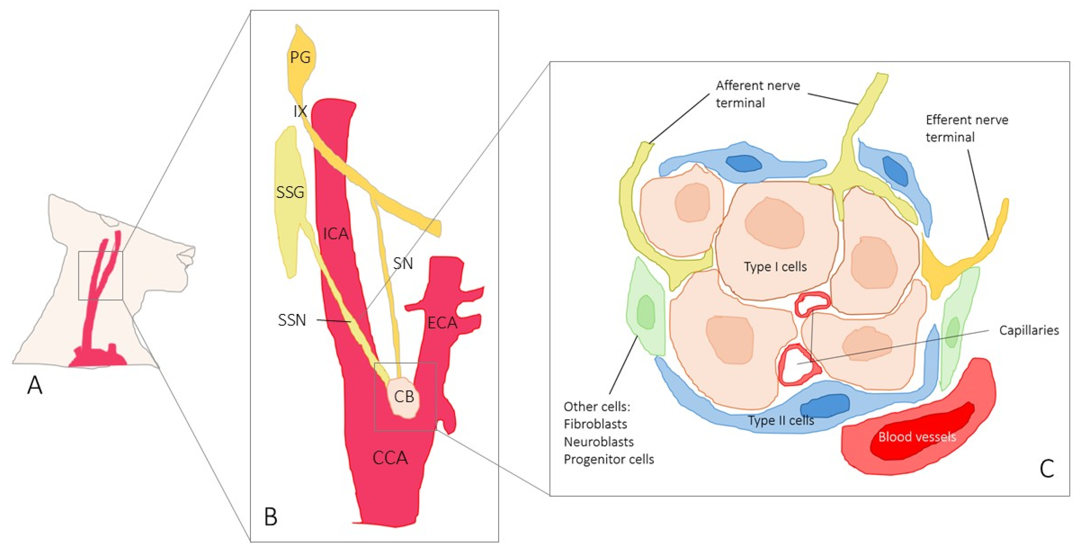
Scheme 1. Diagram of the location, innervation, and structure of the human carotid body. (A) Right-side view of the neck showing the carotid arterial system. (B) The carotid body (CB) is located at the carotid bifurcation, has ovoid morphology, and receives afferent innervation through a collateral nerve (de Casto’s nerve) of the carotid sinus nerve (sinus nerve or Hering’s nerve), branch of the glossopharyngeal nerve (IX). The somas of the afferent terminals are located in the petrosal ganglion (PG) of the IX. In addition, the BC receives efferent innervation through sympathetic sinus nerves (SSNs) that can be individual structures or form perivascular plexuses. SSNs are post-ganglionic sympathetic fibers whose somas are located in the superior sympathetic ganglion (SSG). CCA: common carotid artery, ACE: external carotid artery, ICA: internal carotid artery. (C) The structural unit of the carotid body is the glomic glomerulus that consists of type I cells arranged in contact with the capillaries, and type II cells that surround them. Type I cells are in contact with afferent and efferent nerve endings. In addition to the two main types of glomic cells, there are also precursor cells, neuroblasts, and fibroblasts in the glomeruli.
Ijms 26 10689 sch001
Figure 1. Immunofluorescence-based detection of S100 protein (S100P) and neuron-specific enolase (NSE) in the human carotid body. S100P (a) was found in type II cells (green fluorescence) and NSE (b) in type I cells (red fluorescence). Merge of (c) and (d) was not detected therefore demonstrating that S100P and NSE are segregated and specifically identify type II and type I carotid body cells, respectively. (d) is an enlargement of (c). Blue fluorescence corresponds to the staining of the nuclei with DAPI. Scale bar in a is identical for a–c. Objective: 20×/0.75 oil; pinhole: 1; XY resolution: 156 nm; and Z resolution: 334 nm.
Ijms 26 10689 g001
Figure 2. Immunofluorescence-based detection of TRPV4 in the human carotid body. TRPV4 was observed in some type I and type II cells throughout glomic glomeruli (yellow arrows). Merge (yellow in (c,d), (e,f), and (g,h)) of TRPV4 (green in (a) and (e)) with SYN (red in (bd)) or NSE ((e,f) and (g,h)) demonstrate localization of TRPV in type I cells (yellow arrows); (d), (f), and (h) are details of (c), (e), and (g), respectively. Images of TRPV4 immunofluorescence in nerve terminals were also found ((h), white arrows). Blue fluorescence corresponds to the staining of the nuclei with DAPI. SYN: synaptophysin; NSE: neuron-specific enolase. Scale bar in a is identical for a-c and e-g; scale bar in d is identical for d and h. Objective: 20×/0.75 oil; pinhole: 1; XY resolution: 156 nm; and Z resolution: 334 nm.
Ijms 26 10689 g002
Scheme 2. Schematic representation of a glomic glomerulus of the carotid body and the percentages of the area occupied by cells and nerves displaying TRPV4 immunofluorescence. Red color indicates positivity for TRPV4.
Ijms 26 10689 sch002
Figure 3. Immunofluorescence-based detection of TRPV4 in nerves of the human carotid body. TRPV4 (green in (a)) was detected in nerve fibres within the carotid body since merge of TRPV4 with NFP was found (red in (b)). Merge of TRPV4 with NFP ((c,d) and (e,f)) and NSE (g,h) is indicated with white arrows. (d), (f), and (h) are details of (c), (e), and (g), respectively. Blue fluorescence corresponds to the staining of the nuclei with DAPI. NFP: neurofilament proteins. NSE: neuron-specific enolase. Scale bar in a is identical for a-c and e-g; scale bar in d is identical for d and h. Objective: 20×/0.75 oil; pinhole: 1; XY resolution: 156 nm; and Z resolution: 334 nm.
Ijms 26 10689 g003
Table 1. Percentage of area occupied by TRPV4 in the human carotid body in type I glomus cells (merging NSE + TRPV4) and nerves (merging of NFP + TRPV4).
% are occupied by TRPV4 IF cells68.3 ± 4.2%
% area occupied by SYN IF cells (type I cells)32.2 ± 3.1%
% area occupied by NSE IF cells (type I cells)34.7 ± 2.0%
% area occupied by NFP IF (nerve fibre terminals)2.2 ± 0.3%
% mergence of TRPV4 + SYN
and
% mergence TRPV4 + NSE
(TRPV4 positive type I cells)
22.1 ± 3.1% (68.6% of total type I cells)
and
22.8 ± 2.6% (65.7% of total type I cells)
% mergence of TRPV4 + NFP (TRPV4 + nerve fibre terminals)2.0 ± 0.1% (90% of total nerve terminals)
Table 2. Primary antibodies used.
AntigenOriginDilutionSupplier
TRPV4 (LS-C401108)Rabbit1:200LifeSpan BioSciences 1
NSE (clone BBS/NC/VI-H14)Mouse1:500Dako 2
Synaptophysin (clone 27G12)MousePredilutedLeica Biosystems 3
NFP (clone 2F11)Mouse1:200Roche 4
1 Newark, NJ, USA; 2 Glostrup, Denmark; 3 Madrid, Spain; 4 Barcelona, Spain.
Disclaimer/Publisher’s Note: The statements, opinions and data contained in all publications are solely those of the individual author(s) and contributor(s) and not of MDPI and/or the editor(s). MDPI and/or the editor(s) disclaim responsibility for any injury to people or property resulting from any ideas, methods, instructions or products referred to in the content.