Article

Deletion of the Epidermal Protease KLK5 Aggravates the Symptoms of Congenital Ichthyosis CDSN-nEDD

1
Department of Pharmacy, School of Health Sciences, University of Patras, Rion, 26504 Patras, Greece
2
Toulouse Institute for Infectious and Inflammatory Diseases (INFINITy), CNRS, Toulouse University, Inserm, 31024 Toulouse, France
3
Laboratory of Pharmacology, School of Pharmacy, Aristotle University of Thessaloniki, 54124 Thessaloniki, Greece
4
Department of Cell Biology and Cytology, Federative Institute of Biology, Purpan University Hospital, 31300 Toulouse, France
*
Authors to whom correspondence should be addressed.
These authors contributed equally to this work.
Int. J. Mol. Sci. 2025, 26(17), 8605; https://doi.org/10.3390/ijms26178605
Submission received: 4 August 2025 / Revised: 28 August 2025 / Accepted: 2 September 2025 / Published: 4 September 2025

Abstract

Congenital ichthyoses, now grouped under the acronym EDD (Epidermal Differentiation Disorders), include nonsyndromic forms (nEDD) that may be caused by loss-of-function mutations in the CDSN gene encoding corneodesmosin (CDSN-nEDD, formerly Peeling skin syndrome type 1). It is characterized by skin peeling, inflammation, itching and food allergies, while no specific therapy is currently available. High levels of KLK5, the serine protease that initiates the desquamation cascade, are found in the epidermis of CDSN-nEDD patients. Thus, we hypothesized that KLK5 inhibition would alleviate the symptoms of CDSN-nEDD and could serve as a new pharmacological target. A human epidermal equivalent (HEE) model for CDSN-nEDD was developed using shRNA-mediated CDSN knockdown. This model was characterized and used to assess the role of KLK5 knockdown on CDSN-nEDD. Also, Klk5−/− mice were crossed with Cdsnepi−/− mice, the murine model of CDSN-nEDD, to examine in vivo the effect(s) of Klk5 deletion in CDSN-nEDD. Both models recapitulated the CDSN-nEDD desquamating phenotype. Elimination of KLK5 aggravated the CDSN-nEDD phenotype. Epidermal proteolysis was surprisingly elevated, while severe ultrastructural (corneo)desmosomal alterations increased epidermal barrier permeability and stratum corneum detachment was manifested. Based on these results, we concluded that targeting epidermal proteolysis with KLK5 ablation cannot compensate for the loss of corneodesmosin and rescue over-desquamation of the CDSN-nEDD. Possibly, in the absence of KLK5, other proteases take over which increases the severity of over-desquamation in CDSN-nEDD. The translational outcome is that over-desquamation may not always be rescued by eliminating epidermal proteolysis, but fine protease modulation is more likely required.

1. Introduction

Corneodesmosin-nonsyndromic Epidermal Differentiation Disorder [CDSN-nEDD (formerly Peeling skin syndrome type 1, OMIM270300)] [1] is a very rare autosomal recessive form of congenital ichthyosis. It was identified using a whole genome analysis approach in a large family of consanguineous Roma in Germany, where four members showed superficial skin peeling from birth [2]. Since then, only a few cases have been reported worldwide, with the incidence of the disease estimated at 0.11 per million in France [3]. It is caused by homozygous inactivating mutations in CDSN gene encoding corneodesmosin (CDSN). This protein forms specialized junctions known as corneodesmosomes, which hold together the corneocytes in the stratum corneum (SC). The disease is characterized by skin over-desquamation, inflammation, food allergies and itching that significantly affect their quality of life, albeit their life expectancy is normal [2,4]. These symptoms highly resemble the symptoms of SPINK5-sEDD (formerly Netherton syndrome, OMIM 256500). Even trichorrhexis invaginata (bamboo hair), a feature considered pathognomonic of SPINK5-sEDD, has been reported in a CDSN-nEDD patient [5]. As with other forms of ichthyoses, no effective treatment of CDSN-nEDD is currently available. Thus, patients receive only symptomatic therapy (e.g., emollients, antibiotics, steroids) to alleviate disease symptoms. Global or epidermal conditional Cdsn−/− mice recapitulate the disease phenotype and as in many cases of invalidation of genes expressed in late differentiation of epidermis, these mice die soon after birth [6,7].
Due to the phenotypic similarities between CDSN-nEDD and SPINK5-sEDD, diagnosis can only be succeeded with immunohistochemical staining demonstrating the absence of CDSN or LEKTI, the product of the SPINK5 gene, respectively. Definitive diagnosis requires DNA sequencing of the respective genes. The complexity for diagnosing ichthyoses was highlighted previously [8,9].
Epidermal desquamation depends on the tightly regulated activity of the epidermal kallikrein-related peptidases (KLKs). It is well-established that the KLK5, KLK7 and KLK14 members of the KLK family cleave the corneodesmosomal proteins, namely desmoglein 1, desmocollin 1, and CDSN. The process is controlled by the endogenous inhibitor LEKTI which quenches their activities through the formation of KLK-LEKTI complexes. These complexes dissociate only at the outermost SC layers that has acidic pH due to the acidic mantle. Therefore, only the outer layers of SC are shed [10,11,12]. KLK5 acts as the upstream activator of the epidermal proteolytic cascade, since it can autoactivate and, subsequently, activate the other KLK pro-enzymes, thus augmenting the proteolytic activities in the epidermis [10].
SPINK5-sEDD is due to inactivating mutations in the SPINK5 gene encoding for LEKTI. Thus, in the absence of LEKTI, the KLKs are activated at the stratum granulosum (SG)-SC boundary and allow for premature shedding of SC [12]. Accordingly, the elimination of the Klk5 gene in Spink5−/− mice, the SPINK5-sEDD murine model, rescued neonatal lethality [13]. To this end, it should be emphasized that inhibitors for KLK5 are under development [14,15,16,17,18,19]. A borolane-based inhibitor known as GSK951 exhibiting increased selectivity and low IC50 of 250 pM, showed therapeutic efficacy in the transgenic KLK5 mouse model of SPINK5-sEDD [20]. These inhibitors could also be important for the treatment of other more common diseases governed by upregulated KLK5, such as rosacea [21] and atopic dermatitis [22]. Finally, a bispecific KLK5/KLK7 antibody has been generated and tested in the mouse model of SPINK5-sEDD and atopic dermatitis [23]. If KLK5 proves to be the main player in other skin diseases as in CDSN-nEDD, this will expand the disease repertoire where these compounds can be administered, while exerting pressure for their clinical implementation.
In contrast, in CDSN-nEDD, over-desquamation is due to the defective structure of corneodesmosomes. Nevertheless, in CDSN-nEDD, a strong elevation of the KLK5 expression was found [2,24]. In addition, it has been suggested that CDSN protects corneodesmosomes from premature proteolysis [25]. Therefore, we hypothesized that this abnormally high KLK5 expression in combination with the absence of CDSN expression could lead to enhanced cleavage of corneodesmosomes, and to over-desquamation. Further support for the rationale of targeting KLK5 for the treatment of CDSN-nEDD comes from the observation that absence of Klk5 results in increased number and length of desmosomes [26]. To investigate whether KLK5 targeting could attenuate the symptoms of CDSN-nEDD, we employed two different models: (a) an in vitro organotypic culture and (b) an in vivo mouse model. The in vitro model consisted of a human epidermal equivalent (HEE) model where the genes KLK5 and CDSN were co-knocked down by shRNA interference, while the in vivo model involved the generation and study of double knockout Cdsnepi−/−Klk5−/− mice. Contrary to our original hypothesis, KLK5 exacerbated the CDSN-nEDD phenotype.

2. Results

2.1. Production and Validation of the HEE Model for CDSN-nEDD

Single and double gene expression knockdown was achieved using shRNA in normal human primary keratinocytes, which were secondarily used to generate HEE. Efficiency of single and double gene silencing was controlled at the RNA and protein level by RT-qPCR and Western blot, respectively (Figure S1). CDSN and/or KLK5 mRNA expressions were always decreased by 65% or more in comparison to the corresponding control (shCtrl puro, shCtrl neo or shCtrl puro-neo).

2.2. KLK5 Gene Silencing Exacerbates the Histological and Ultrastructural Abnormalities Observed in shCDSN HEE

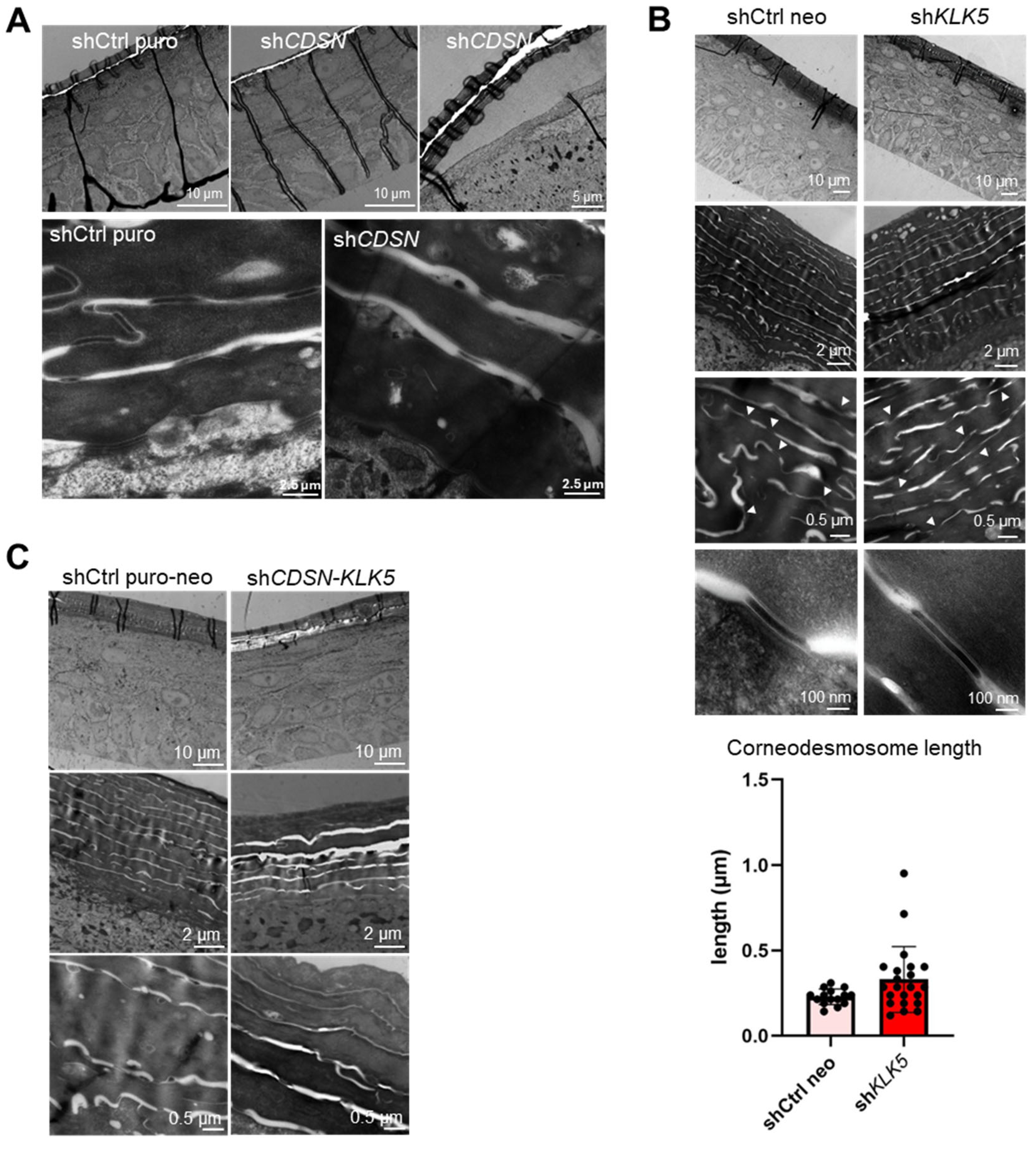
HEEs with single or double gene silencing were fully differentiated within 12 days of culture at the air–liquid interface, as it was the case with control shRNA HEEs. Control and shKLK5 HEE had a similar morphology, whereas the global thickness of shCDSN HEE (living layers and SC) was reduced. This effect appeared amplified in the case of the double shCDSN-KLK5 silencing (Figure 1). Observation of shCDSN HEE by TEM confirmed the histological observations and the detachment between the SG–SC boundary, which never occurred in the control HEE.
Moreover, CDSN depletion led to a slight widening of the intercorneocyte spaces and corneodesmosomes appeared smaller and less cohesive (Figure 2A). The general structure and the morphology of the SC of shKLK5 HEE was like control HEE. However, KLK5 silencing had an impact on the number of corneodesmosomes as well as their length which increases (although with high variability, p = 0.076) in comparison to shCtrl Neo-HEE (Figure 2B). This is in accordance with the significantly increased number of desmosomes in the epidermis of Klk5−/− mice relative to wt [26]. Finally, ultrastructural observation of shCDSN-KLK5 HEE revealed a phenotype similar to shCDSN but exacerbated. In particular, the SC was thinner, loosely cohesive, with enlarged intercorneocyte spaces and aberrant corneodesmosomes (Figure 2C).

2.3. KLK5 Gene Silencing Worsens the Impaired Permeability Barrier Observed in the shCDSN HEEs

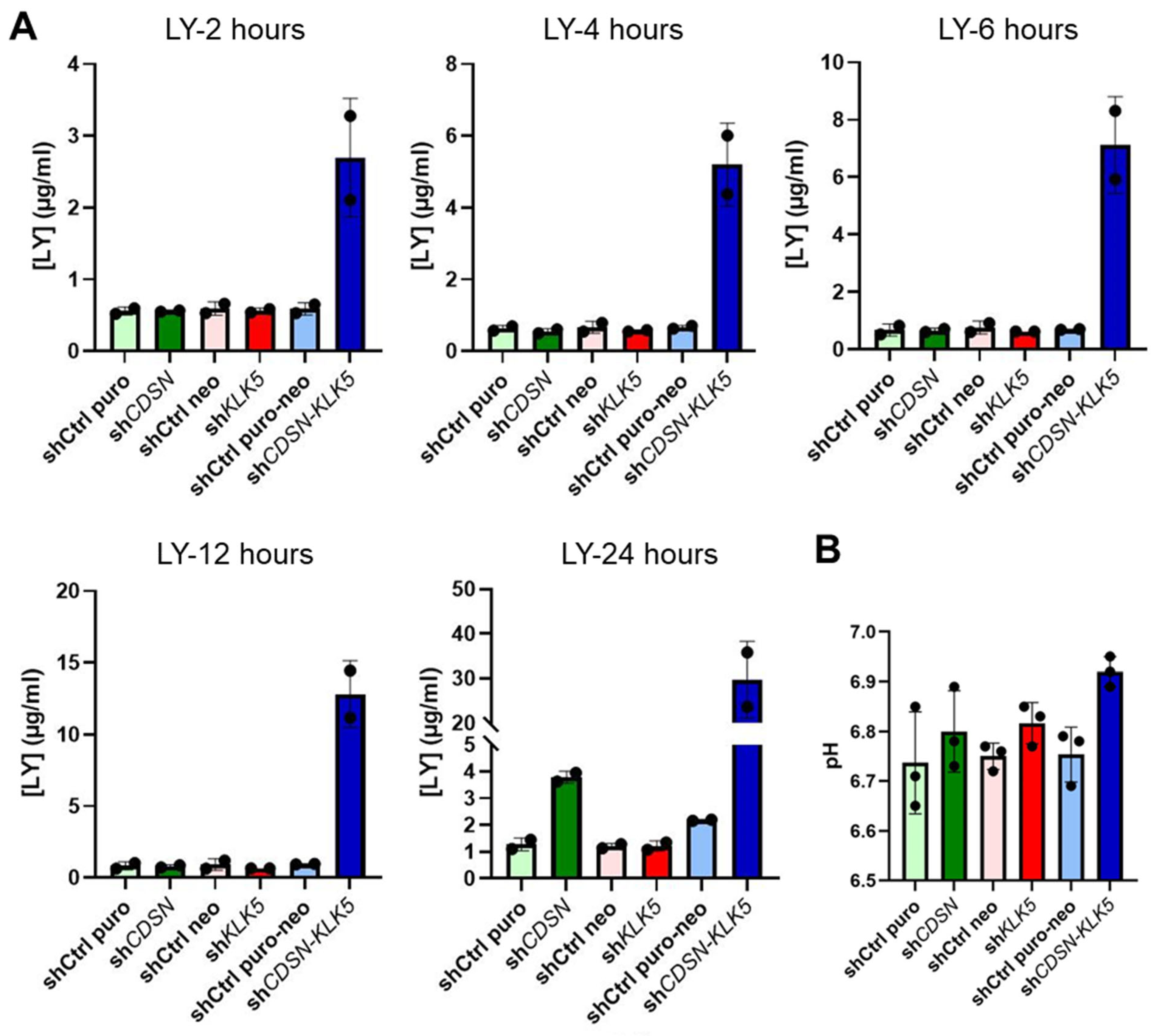
The permeability barrier, surface pH and the SC lipids were determined for HEEs. The effect of CDSN and/or KLK5 silencing on the permeability barrier function of HEE was assessed by dye exclusion (lucifer yellow) assay. After 24 h, shCDSN HEE showed an elevated permeability defect in comparison to control (shCtrl puro). KLK5 silencing alone had no effect on the outside–in permeability barrier. In contrast, when both CDSN and KLK5 gene expression were inhibited, a barrier defect was detected within the first hours of incubation and dramatically increased with time. Twenty-four hours after dye application, its concentration in the culture medium was around thirty-times higher in the case of shCDSN-KLK5 HEE than in controls or single gene knockdown HEE (Figure 3A). The pH at the surface of the SC of shCDSN-KLK5 HEE was not significantly different in comparison with shCtrl puro-neo HEE (6.92 vs. 6.75, mean values) (Figure 3B).
Since lipid organization in the SC is important for the normal function of the epidermal barrier, we analyzed the extracellular lipids in the SC of the various HEEs by oil red O staining. We did not observe any effect of CDSN and/or KLK5 gene silencing on the staining of neutral lipids in the SC (Figure S2A). During cornification, some lipids are covalently bound to the protein shell of the cornified envelopes to form mature hydrophobic envelopes. We assessed cornified envelope maturation by nile red/involucrin double staining of envelopes purified from control HEEs, CDSN and/or KLK5 knockdown HEEs. We found mature hydrophobic envelopes as well as immature involucrin-positive envelopes in similar proportions in each HEE condition (Figure S2B).

2.4. Deletion of Klk5 in Cdsnepi−/− Mice Does Not Improve the Phenotype of CSDN-nEDD

The Cdsnepi−/− died within 1 h from birth due to severe desquamation that led to extensive dehydration [6]. Here, the Cdsnepi−/−Klk5−/− (DKO) mice were generated according to the crossing scheme depicted in Figure S3.
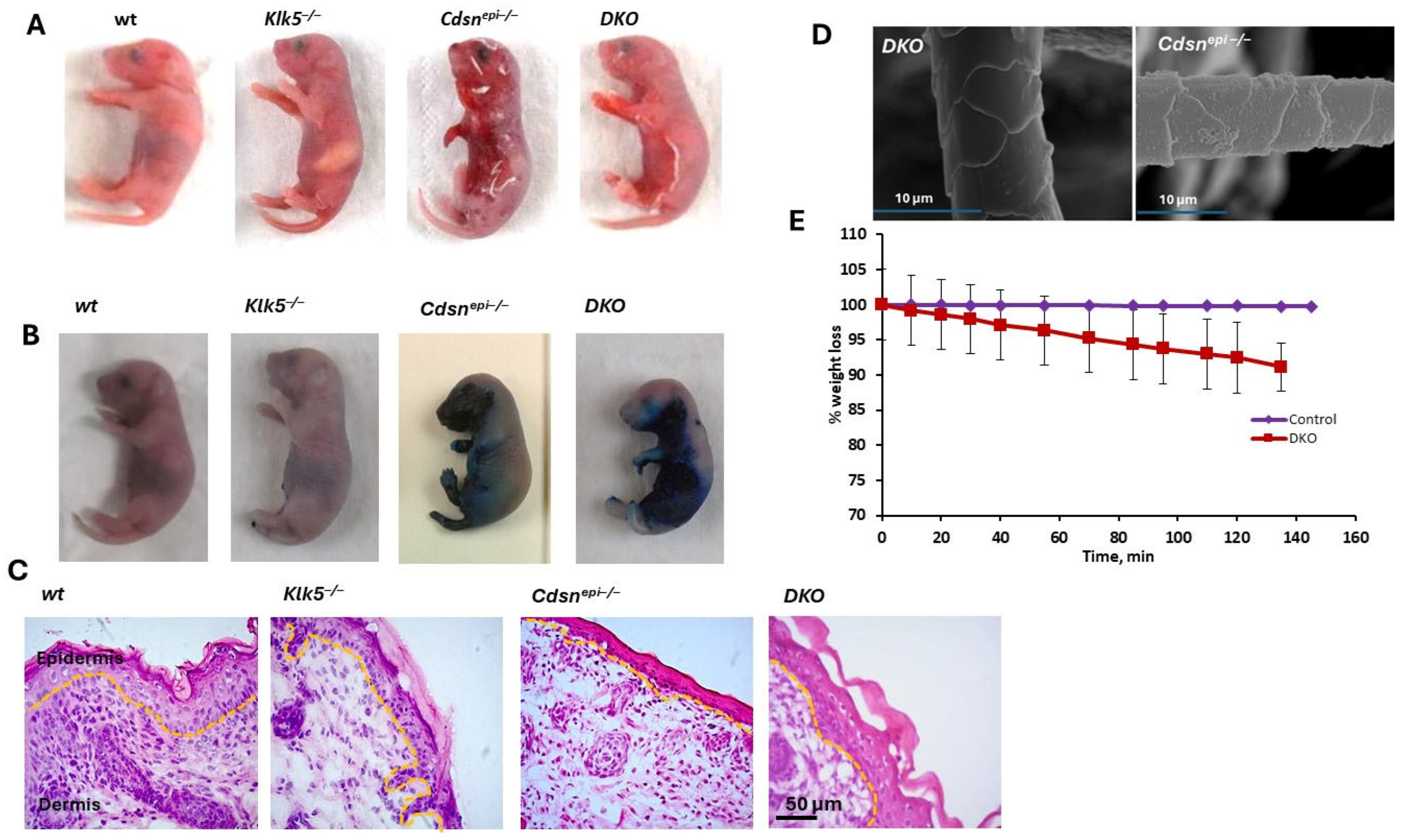
The DKOs appeared normal at birth, but within a few minutes from birth, DKOs developed a dry and wrinkled appearance accompanied with SC detachment and the development of a red, shiny appearance, uniformly resulting in mice death ~7 h after birth. This phenotype is reminiscent of Cdsnepi−/− neonates phenotype (Figure 4A). The deterioration of epidermal barrier was demonstrated with toluidine blue dye exclusion assay (Figure 4B). Histology examination of skin sections from neonatal Cdsn−/−Klk5−/− mice revealed persistent SC detachment suggesting that elimination of Klk5 is not sufficient to ameliorate the cutaneous hallmarks of CDSN-nEDD (Figure 4C). The weight of DKO is normal at birth but is rapidly reduced due to dehydration (Figure 4D). Examination of the whisker morphology with SEM did not show any abnormalities (Figure 4E). Therefore, deletion of Klk5−/− does not appear to rescue the lethal over-desquamating phenotype of Cdsn−/− mice.

2.5. DKO Mice Display Ultrastructural Alteration Similar to Those Observed in shCDSN-KLK5 HEEs

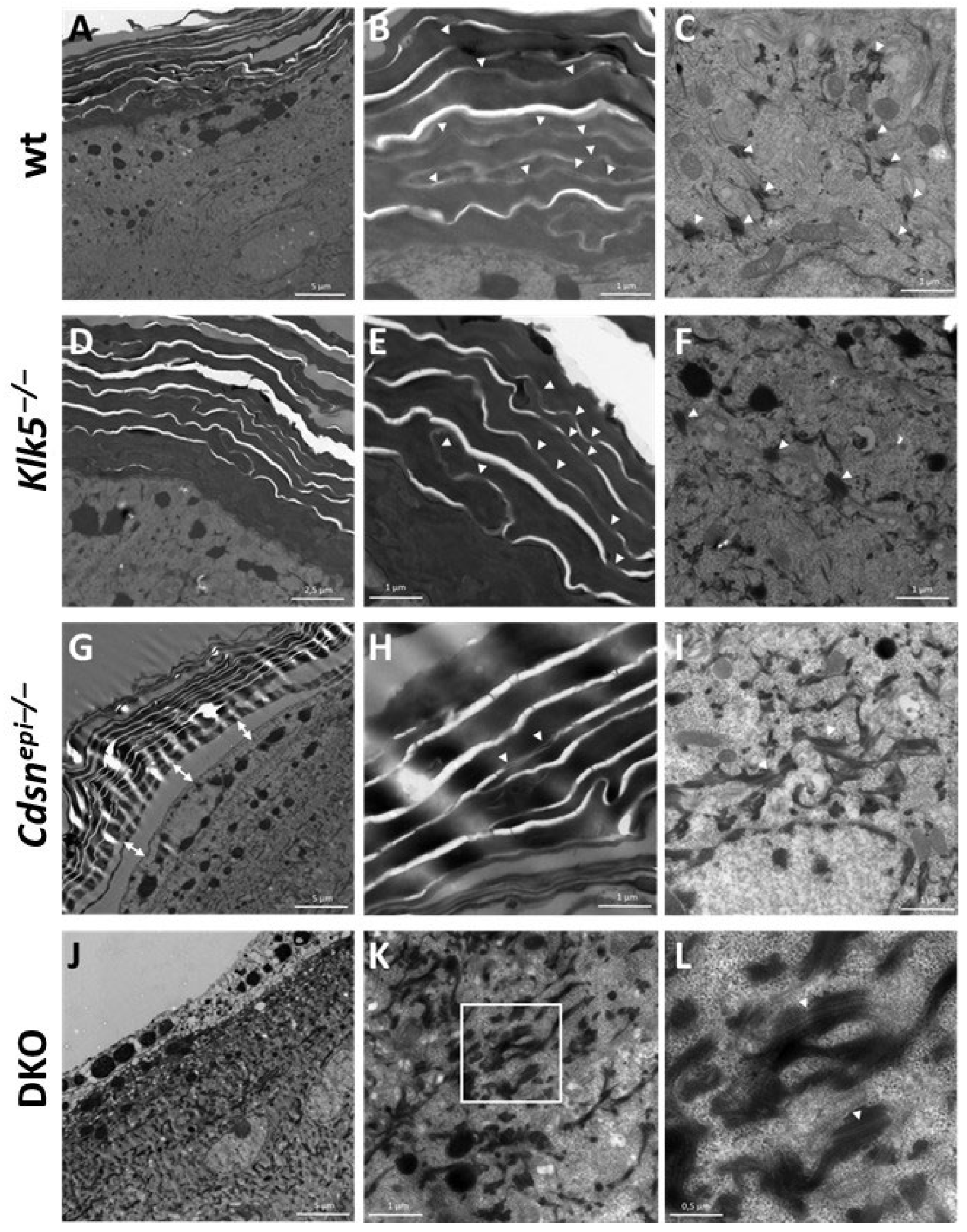
At the ultrastructural level, Klk5−/− epidermis appeared quite like the wt epidermis, with numerous desmosomes in the SG and corneodesmosomes in the SC (Figure 5A–F). In Cdsn−/− epidermis, the SC was loosely cohesive and abnormal widening of the SG/SC interface was often observed (Figure 5G). There were less numerous corneodesmosomes in the SC, whereas desmosomes with normal appearance were present in the SG (Figure 5G–I). The DKO epidermis showed a more drastic phenotype. The SC was totally absent from the observed samples, most probably it detached from the skin surface because of lack of cohesion. Consequently, the underlying SG appeared damaged, but a few desmosomes could be observed in this epidermal layer (Figure 5J–L).

2.6. Aberrant Overall Proteolytic Activities in DKO Epidermis

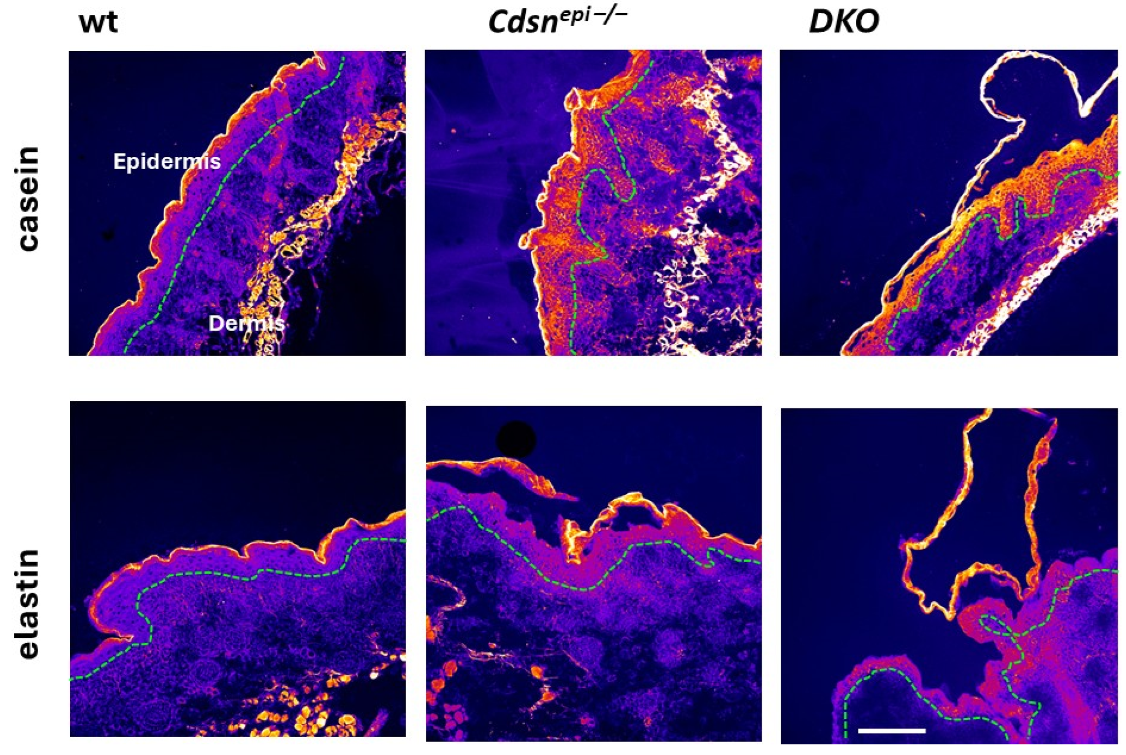
Since KLK5 is the initiator of the epidermal KLK cascade, we examined whether elimination of Klk5 could have a limiting effect on epidermal proteolytic activities in the DKO model. For this, in situ zymography using casein and elastin quenched fluorescent substrates was used. Interestingly, the epidermis of Cdsnepi−/− and DKO mice exhibited highly increased proteolytic activities. Therefore, deletion of Klk5 cannot reduce the increased overall epidermal proteolysis in Cdsn−/− epidermis (Figure 6).
Then, we studied the expression of characteristic molecules of epidermal differentiation in Cdsnepi−/− and DKO epidermis. As shown in Figure 7, no changes in the expression pattern relative to the controls were found. This indicates that epidermal differentiation is not affected by deletion of Cdsn or Cdsn and Klk5. The normal differentiation pattern in Cdsnepi−/− epidermis is in accordance with a previous study [6].

2.7. Lipid Characterization of the DKO Epidermis and Human SC Sample

The lipid profile was analyzed with 1H-NMR and the results are shown in Figure 8. Significant differences in the epidermal lipidome between control mice including wt and Klk5−/− and disease mouse models Cdsnepi−/− and DKO were observed. Total cholesterol is upregulated in Cdsnepi−/− epidermis while it is almost normalized in DKO epidermis. Quantification of the peaks corresponding to -N+(CH3)3, which is present in sphingomyelin (SM) or phosphatidylcholine (PC), and to vinyl group with unique resonance at 5.6 ppm, which is present in SM, shows that these lipids are increased in Cdsnepi−/− while they are normalized in DKO. In addition, the lipids in the Klk5−/− epidermal extracts are higher than in wt. The alteration of lipid profiling in Cdsnepi−/− upon deletion of KLK5 suggests that the production of epidermal lipids is regulated by KLK5 either directly or indirectly. Interestingly, the normalization of certain epidermal lipids by deletion of KLK5 does not suffice to restore the epidermal barrier.
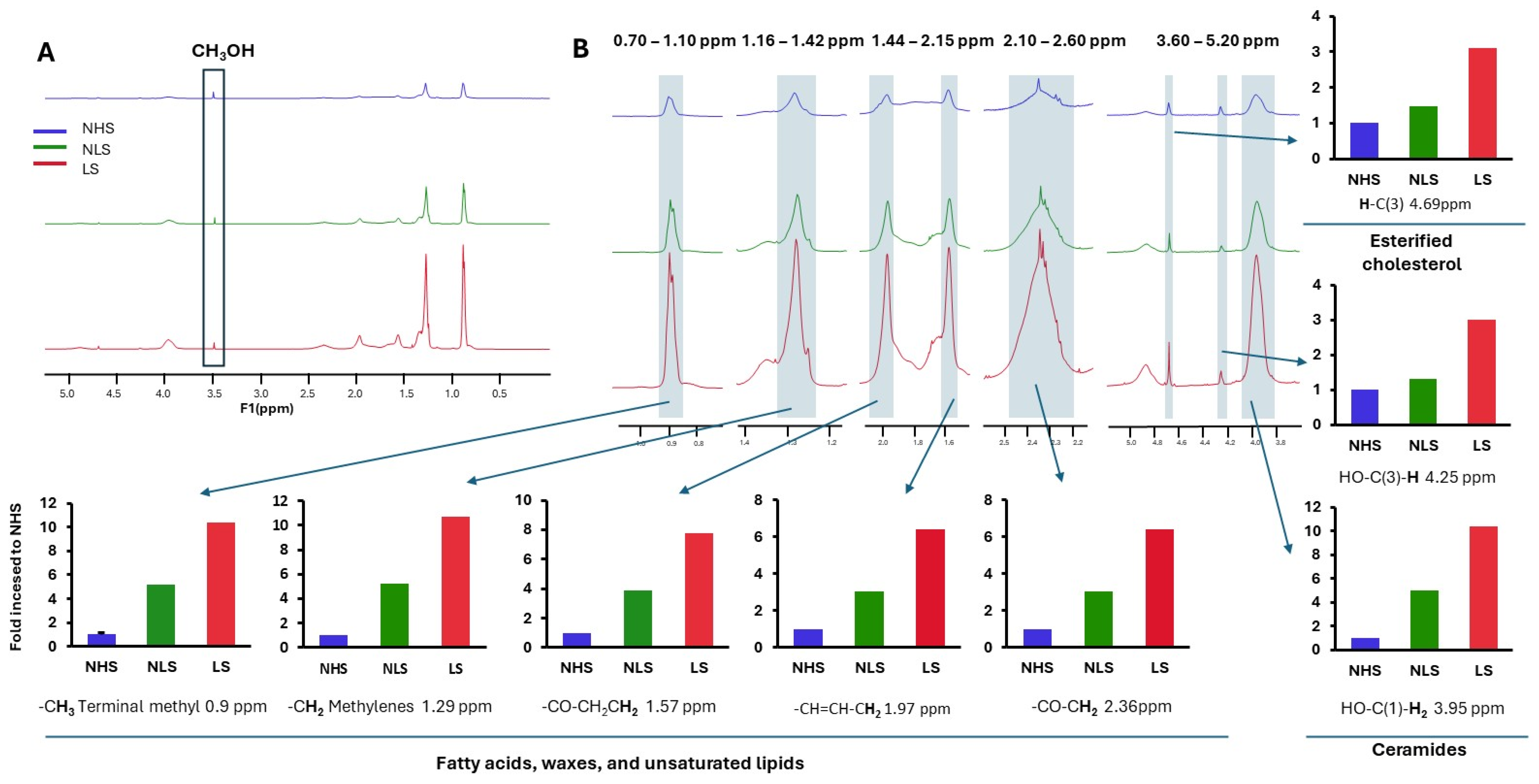
Then, we analyzed extracts from SC from two healthy donors and from a lesional and a non-lesional site obtained from a CDSN-nEDD patient. Quantification of the peaks corresponding to -CH3 terminal methyls, -CH2 methylenes, -CH=CH-CH2 (vinyl protons indicating high degree of unsaturation) and -CO-CH3, which are present in all fatty acyl chains, showed a remarkable increase in the lipid content of lesional CDSN-nEDD SC compared to non-lesional skin and normal SC (Figure 9). This resembles the Cdsnepi−/−. Further, high levels of ceramides and esterified cholesterol were found in the lesional skin compared to unlesional or normal skin.
Fatty acids are important for preserving the epidermal barrier [27]; thus, their increase in the patient sample and in the mouse model may reflect a compensatory mechanism. On the other hand, the increase in unsaturated fatty acid content may be associated with the defects in epidermal barrier. Previously, increased epidermal levels of unsaturated fatty acids as found in atopic dermatitis and SPINK5-sEDD were associated with defects in epidermal barrier due to reduction in packing density [28]. Another important factor that regulates the epidermal barrier is the length of carbon chain in fatty acids and ceramides. Decreased carbon length is associated with barrier defect [29]. Here, we have not studied this parameter since such information cannot be extracted from 1H-NMR data and will require future mass spectrometry-based approaches for lipid speciation.
No significant alterations in the lipid profile between the control and the CDSN-silenced HEEs were found (Figure S2). However, these analyses were conducted using semi-quantitative histological staining techniques, which are significantly less sensitive and precise than 1H-NMR. Moreover, oil red O only stains the neutral lipids, and the Nile red staining analysis [30] was not performed on HEE section but on cornified envelopes purified from HEE SC. In contrast, 1H-NMR provides analyses of all extractable lipids. Thus, these results cannot be directly compared.

3. Discussion

CDSN-nEDD is a rare skin peeling disease for which there is no specific therapy. KLK5 is the key enzyme that regulates skin desquamation due to its ability to autoactivate and then activate other KLKs. Further, KLK5 cleaves components of (corneo)desmosomes [10,11]. High levels of KLK5 expression were previously found in CDSN-nEDD [2,24]. Here, we used an in vitro HEE and an in vivo model to assess the role of KLK5 in CDSN-nEDD. HEEs are valuable models to study rare skin diseases and test potential new therapeutic strategies. In this direction, a skin equivalent (encompassing both epidermis and dermis) for SPINK5-sEDD (Netherton syndrome) was previously designed, to test the role of KLK7 and KLK5 gene silencing [31], or their chemical inhibition [32] in reversing the disease phenotype. Recently, a protein replacement therapy was suggested for CDSN-nEDD after demonstrating that application of recombinant CDSN in liposomes to a CDSN-nEDD HEE model improved barrier function and SC structure [33]. However, this strategy will take time to enter clinical practice. Thus, in an attempt to identify new pathways that mediate pathology and that can be targeted for pharmacological manipulation, we examined the role of KLK5 in CDSN-nEDD.
When the expression of both CDSN and KLK5 was downregulated in HEE, the cohesive defect at the SG/SC interface was no longer observed, whereas the SC still appeared loosely cohesive and the overall HEE thickness was more importantly reduced in comparison to shCDSN HEE. Moreover, shCDSN-KLK5 HEE presented an important permeability barrier defect, in comparison to control or single gene knockdown HEE. Thus, the downregulation of both CDSN and KLK5 expressions in HEE resulted in the more dramatic phenotype we observed. On the other hand, KLK5 silencing in HEE led to a phenotype very similar to that of control HEE, with no impact on either morphology or permeability barrier function. The number and length of (corneo)desmosomes in the shKLK5-HEE model were increased in comparison to control conditions, in accordance with the previously described phenotype of Klk5−/− mice [26].
Deletion of Klk5 did not rescue the lethal neonatal phenotype of Cdsnepi−/− mice. The DKO mice exhibited increased epidermal proteolytic activity and severe desquamation leading to dehydration and neonatal lethality. The detachment of SC from the underlying SG was so severe that the SC was lost during preparation for TEM study. Here, both the HEE model and the animals show the same phenotype and defects. Notably, this is not always the case. In the organotypic skin culture model for SPINK5-sEDD, silencing of KLK7 showed a more potent effect in alleviating the phenotype than the KLK5 silencing. However, later in vivo studies showed that targeting Klk5 can rescue the neonatal lethality of Spink5−/− mice, the murine SPINK5-sEDD model [13,34], while Klk7 cannot [34].
Our study suggests that KLK5 does not contribute to the overall epidermal proteolysis in CDSN-nEDD and the reported upregulated KLK5 expression may not correlate with activity. However, it should be mentioned that in another study the expression of KLK5 was found reduced in some CDSN-nEDD patients, indicating high variability in its expression [35]. Thus, other proteolytic activities could play a role in CDSN-nEDD. In this direction, Klk13 was found highly upregulated in the epidermis of Cdsniepi−/− mice [35]. It should be noted that KLK13, KLK6 and KLK14 are implicated in other over-desquamating diseases related to S. aureus infections [36]. Thus, KLK13 may be involved in CDSN-nEDD, and indeed KLK13, but also KLK7 were found highly upregulated in the skin extract from a CDSN-nEDD [35]. In addition, another study found very high levels of KLK6, KLK7, KLK8, KLK11, KLK13, and KLK14 in SC extracts from two CDSN-nEDD patients. These expressions were associated with increased SC trypsin-like activities, but not chymotrypsin-like activities (i.e., KLK7) [24]. Plasmin-like activities were also upregulated in the SC extracts [24]. Future studies using new proteomic or degradomic approaches [37] will assist in the delineation of the role of other proteases that take over in the absence of KLK5 in CDSN-nEDD. These omic approaches will also allow for the studying of the expression of the major epidermal protease inhibitors in CDSN-nEDD, such as SCALP, SLPI and LEKTI [38]. Finally, the potential participation of MMPs or elastase-2 in the phenotype of CDSN-nEDD cannot be excluded, especially due to their interplay with KLKs [39].
No change in the differentiation status was found in Cdsnepi−/− mice, as previously described [6]. However, it may require more time to develop alterations in the differentiation status. In this direction, when the skin of Cdsn−/− was grafted in nude mice, to study disease progression to adulthood, it was shown that Cdsn−/− had signs of acanthosis, hyperkeratosis, parakeratosis and Stat3 nuclear localization, resembling a psoriatic phenotype [6]. This observation is consistent with the fact that the gene for CDSN is located within the psoriasis susceptibility locus [4].
The Cdsnepi−/− mice appear to recapitulate the alterations in the lipid profile observed in patients. This should be confirmed in the future since here we only analyzed a SC from one CDSN-nEDD patient and compared it with the epidermal extract of lipids from the Cdsnepi−/−. The reason of this was two-fold; first, SC is much easier to be obtained from CDSN-nEDD patients (they desquamate every day) without any invasion, i.e., biopsy; and second, one patient sample was available due to the extreme rarity of the disease. Future studies aiming for the delineation of the epidermal or SC lipidome in a larger cohort of patients is required for more accurate conclusions. Analysis of KLKs expressions or activities in these samples may also aid in the stratification of samples (e.g., high activity vs. low activity) and their correlation with lipid profiling. KLK7 was previously linked with epidermal lipid processing since it was able to cleave β-glucocerebrosidase and acidic sphingomyelinase [40]. Thus, potentially high KLK7 expression, as observed in a patient with CDSN-nEDD [35], could lead to reduced sphingomyelinase that translates to increased sphingomyelin content as observed here.
Overall, both of our models, namely the HEE and the mice, provide mechanistic insights into the CDSN-nEDD. Nevertheless, grafting skin from Cdsnepi−/− mouse in wt mouse allows for the studying of the effects of the absence of Cdsn in the long-term. Such study has revealed the infiltration of inflammatory cells in the dermis [6]. In this direction, grafting of HEE in mice could also allow investigating the interplay between epidermis with the underlying dermis and with immune cells. Another potentially useful strategy to study the absence of Cdsn in the long-term, and at the same time allow testing of potential therapeutics (e.g., small molecules, protein replacement, etc.) could involve the generation of a mosaic Cdsn−/− mouse that could overcome the neonatal lethality as in the case with the SPINK5-sEDD [41].
In conclusion, the data presented here indicates that elimination of KLK5 does not rescue the CDSN-nEDD phenotype. On the contrary, KLK5 ablation likely aggravates over-desquamation associated with a severe skin barrier defect in this case. The study advances the understanding of the mechanisms that govern the CDSN-nEDD pathology.

4. Materials and Methods

4.1. Materials

All chemicals used were obtained from Sigma-Aldrich (Louis, MO, USA). The fluorescent substrates were obtained from Thermo Scientific, Waltham, MA, USA. The antibodies were obtained from commercial sources as follow: Keratin 5, Abcam (ab24647), Cambridge, UK; Dsg1, Santa Cruz (sc-20114), Santa Cruz, CA, USA; loricrin, Abcam (ab24722); involucrin, Santa Cruz (sc-15230) and Sigma (I908), St. Louis, MO, USA; Dsc1, Santa Cruz (sc-18115), KLK5 (R&D Systems, AF2008, Minneapolis, MN, USA), and actin (Chemicon, MAB1501, Tokyo, Japan). The anti-corneodesmosin monoclonal antibody G36-19 was produced and characterized in our laboratory [25].

4.2. Human Samples

Human abdominal skin samples from healthy donors undergoing plastic surgery were obtained from Genoskin (Toulouse, France), following their written informed consent, as authorized by the French Ministry of Research (#AC-2017-2897). Tape strippings from lesional and non-lesional skin area of a CDSN-mutated patient were collected for diagnosis and gathered in a biological collection (n°DC-2011-1388, French National Ethics Committees). Written informed consent was obtained from the patient’s legal representatives.

4.3. Mice

Klk5−/− mice generated previously are viable, fertile, and macroscopically identical to wild-type (wt) mice [13]. Cdsnepi−/− are epidermal conditional corneodesmosin knockout mice that have been generated by K14-promoter driven Cre-mediated deletion of Cdsn gene in the epidermis [6]. All mice are of the C57BL6 background. Genotyping was conducted as described previously [6,13]. All experiments with animals were approved by the local Ethics Committee and carried out according to our institutional guidelines and European Union legislation. Specifically, all experiments with animals were approved by the General Directorate of Rural Economy and Veterinary Matters (Region of Western Greece) with number 187537/627 on 26 June 2018.

4.4. Cell Culture and Generation of HEE

Primary normal human epidermal keratinocytes (NHEK) culture and production of HEE were performed as previously described [42]. NHEsK were established from human abdominal skin samples and grown in Dermalife culture medium (CellSystems, Troisdorf, Germany). 350,000 subconfluent keratinocytes in suspension in EpiLife medium containing 1.5 mmol/L calcium were seeded on polycarbonate culture inserts (area of 0.63 cm2 with pores 0.4 mm in diameter; Merck Millipore, Molsheim, France). After 48 hours of incubation at 37 °C in a humidified atmosphere containing 5% CO2, cells were exposed to the air–liquid interface (which corresponded to day zero of differentiation), and 50 mg/mL vitamin C (Sigma-Aldrich, Louis, MO, USA) and 10 ng/mL keratinocyte growth factor (Sigma-Aldrich) were added to the medium in the lower compartment. The medium was renewed every two days until harvesting on Day 12 of the air-exposed phase.

4.5. shRNA Lentiviral Particles and Keratinocyte Transduction

We proceeded as previously described [42]. In brief, we designed different shRNA sequences for each target gene (CDSN and KLK5) and the lentiviral particles were produced in pLKO.1 vectors (Table S1), with puromycin (in the case of shCDSN) or neomycin (in the case of shKLK5) antibiotic resistance gene. Gene silencing efficiency was assessed for each shRNA vs. control (nontarget shRNA) lentiviral particles and two shRNA per target gene were selected for further experiments. For the double silencing experiments, one pair of lentiviral particles targeting CDSN and KLK5, respectively, was chosen for co-transduction of keratinocytes. Each single- or double-silencing experiment was repeated using keratinocytes from two different donors.

4.6. In Situ Zymography

In situ zymography to detect epidermal proteolytic activities was conducted on 5 μm skin cryosections, as described previously [26]. Briefly, skin cryosections were incubated in 2% Tween-20 in PBS for 2 min followed by a wash in PBS and then, incubated with 10 μg/mL BODIPY FL casein, 10 μg/mL BODIPY FL elastin or 10 μg/mL BODIPY FL gelatin substrates at 37 °C. Then, the excess substrate was removed, and biopsies were covered with Mowiol medium and observed under a confocal laser scanning electron microscope (Leica TCS SP5, Leica Biosystems, Nussloch, Germany).

4.7. Western Blot

HEEs were lyzed in Laemmli sample buffer and processed for Western blot analysis using the primary antibodies, as previously described [42]. The detection was realized with the ECL Prime system (GE Healthcare, Chicago, IL, USA) and images were acquired with a G:BOX Chemi XT4 CCD camera (Syngene, Cambridge, UK) and GeneSys software (version 1.0.7.154, Genesys, Daly City, CA, USA). ImageJ software (version 1.52a, National Institutes of Health, Bethesda, MD, USA) was used to quantify immunoreactive bands. Signals were normalized to actin immunodetection.

4.8. Transmission Electron Microscopy (TEM)

Skin biopsies from newborn mice of all genotypes (i.e., wt, Cdsn−/−, Klk5−/− and DKO Cdsnepi−/−Klk5−/−) or from HEEs were collected, fixed in 2% glutaraldehyde and 4% formaldehyde in 0.1 M cacodylate buffer, then, washed, stained with 1% OsO4, dehydrated in a series of acetone baths and, finally, embedded in epoxy-resin (Sigma-Aldrich). Ultrathin sections were mounted on 100 mesh collodion-coated copper grids and post-stained with 3% uranyl acetate and 8.5% lead citrate before being examined with a Hitachi HT7700 electron microscope (Hitachi, Tokyo, Japan).

4.9. Scanning Electron Microscopy (SEM)

Neonates were decapitated and the heads were fixed in 4% formaldehyde in PBS for 24 h, and then, dehydrated in a series of ethanol solutions 25, 50 and 100% for 10 min. Ethanol was replaced with 100% acetone by two washes for 10 min each. The samples were allowed to dry completely, gold sputtered and observed in a field emission scanning electron microscope (JEOL, Pleasanton, CA, USA, 6300).

4.10. Toluidine Blue Penetration and Dehydration Assay

The staining and the loss of water of newborn mice were carried out as described previously [13].

4.11. Lucifer Yellow Permeability Assay

The assay was performed as previously described [42]. Dye concentration in the culture medium was measured fluorometrically in a Fluoroskan Ascent (Thermo Scientific, Waltham, MA, USA) with excitation and emission at 425 and 550 nm, respectively.

4.12. Histology and Immunohistochemistry (IHC)

Skin from mice or HEE were fixed with 4% paraformaldehyde in PBS for 24 h. Then, the tissues were embedded in paraffin, cut to 5 μm sections with a microtome and processed for hemalun-eosin staining.
IHC was performed in 5 μm skin cryosections which were fixed in acetone for 10 min and then, were air dried at room temperature for 5 min, followed by hydration in PBS for 5 min. Endogenous peroxidase activity was blocked by incubation in 3% H2O2 in PBS for 10 min and blocking of unspecific binding-sites was conducted by incubation with 0.3% BSA in 0.1% Triton X-100 in PBS for 5 min. Slides were then, incubated with the primary antibodies either for 1h at room temperature or overnight in a humidity chamber at 4 °C depending on the working conditions of each antibody. Then, secondary HRP-conjugated antibody in 0.3% BSA in PBS was applied in section for 30 min at room temperature. After this, slides were washed twice (5 min per step) with 0.3% BSA in PBS and 1 time with PBS for 5 min. Finally, the slides were incubated with DAB substrate for 10 min and counterstained with hematoxylin.

4.13. Lipid Extraction and 1H-NMR

Lipids were extracted from the epidermis of wt, Cdsn−/−, Klk5−/−, and Cdsn−/−Klk5−/− mice with chloroform/methanol/PBS (2:1:0.1). The lipids were normalized against the protein content. The solvents were evaporated, and the lipid extracts were dissolved in CDCl3 for 1H-NMR analysis. A known concentration of methanol was added as an internal standard to assist in quantification. From human samples, lipids were extracted with the same procedure but from the SC. 1H-NMR (available at the University of Patras, Center for Instrumental Analysis) spectra were recorded at 22 ± 1 °C on a Bruker AM-600 WB spectrometer (Billerica, MA, USA) operating at a field strength of 600 MHz for 1H. Peak assignment was based on Kriat et al., [43], Adosraku et al., [44], and Tynkkynen [45].

4.14. Stratum Corneum pH Measurement

The measurement of pH at the surface of the HEE was carried out using the pH meter HI 99181 (Hanna Instrument, Smithfield, RI, USA) equipped with the electrode HI 1414D/50.

4.15. Oil Red Staining of HEE Cryosections

Cryosections (5 µm-thick) from HEE previously embedded in Tissue-Tek OCT compound (VWR, Radnor, PA, USA) were fixed with 4% formaldehyde-containing buffer. After a brief rinse in phosphate-buffered saline and 30% isopropanol, sections were stained with freshly prepared oil red O (Sigma-Aldrich) working solution for 15 min at room temperature, rinsed in 30% isopropanol and distilled water, and mounted in ProLong Gold Antifade Mountant containing 4,6-diamidino-2-phenylindole (DAPI, Invitrogen Life Technologies, Carlsbad, CA, USA). Images were captured using a ×63 magnification oil immersion lens in a Zeiss Apotome microscope (Carl Zeiss, Jena, Germany).

4.16. Cornified Envelopes Maturation Assay

Cornified envelopes were isolated from HEE samples as previously described [46]. Appropriate concentrations of cornified envelope suspension were dropped onto a slide-glass and air-dried. They were fixed in acetone at −20 °C for 10 min, and hydrated in phosphate-buffered saline. The primary antibody anti-Involucrin (SY5) allowed a reaction for 1 h at room temperature. A secondary antibody was then applied for 1 h at room temperature. After washing, the cornified envelopes were stained with Nile red solution (1 mg/mL) for 30 min at room temperature. Slides were then mounted in Mowiol. Images were taken by fluorescence microscopy using a Nikon eclipse 80i microscope equipped with a Nikon DXM 1200C digital camera (Tokyo, Japan) and NIS image analysis software (v. 5.21.00).

4.17. RNA Extraction and RT-qPCR

Total RNA was extracted with the Nucleospin RNA kit (Macherey-Nagel, Düren, Germany). One μg of total RNA was reversed-transcribed with Superscript First-Strand Synthesis System for RT-PCR (Invitrogen). For quantitative PCR, primer sequences were designed using Primer3 software to generate amplicons of 150–200 bp. BLAST+ 2.8.1 analysis ensured the absence of similarity to any other human sequence. The primers used are listed in Table S2.

Supplementary Materials

The following supporting information can be downloaded at: https://www.mdpi.com/article/10.3390/ijms26178605/s1.

Author Contributions

Conceptualization, G.P., G.S. (Guy Serre), N.J. and G.S. (Georgia Sotiropoulou); methodology, E.Z., M.R., G.P., G.S. (Guy Serre), G.S. (Georgia Sotiropoulou) and N.J.; software, E.Z. and M.R.; validation, E.Z., M.R., G.S. (Georgia Sotiropoulou) and N.J.; formal analysis, E.Z. and M.R.; investigation, E.Z., M.R. and G.P.; resources, G.S. (Georgia Sotiropoulou) and N.J.; data curation, M.R., G.S. (Georgia Sotiropoulou) and N.J.; writing—original draft preparation, G.P., G.S. (Georgia Sotiropoulou) and N.J.; writing—review and editing, M.R., G.P., G.S. (Guy Serre), N.J. and G.S. (Georgia Sotiropoulou); visualization, M.R. and N.J.; supervision, G.S. (Georgia Sotiropoulou) and N.J.; project administration, G.S. (Georgia Sotiropoulou) and N.J.; funding acquisition, G.S. (Georgia Sotiropoulou), N.J. All authors have read and agreed to the published version of the manuscript.

Funding

This research was funded by ERA-NET/E-Rare-3: Joint Translational Call (2015) for “European Research Projects on Rare Diseases” (2016–2019), Project title: Tracing the untackled facets of Peeling Skin Disease-Targeting epidermal proteolysis for treatment with number T3EPA-00052.

Institutional Review Board Statement

Human abdominal skin samples from healthy donors undergoing plastic surgery were commercially obtained from Genoskin (Toulouse, France), following their written informed consent, as authorized by the French Ministry of Research and the Committee for the Protection of Persons of the French South-West and Overseas Regions II (#AC-2017-2897) in accordance with the Declaration of Helsinki. Tape strippings from lesional and non lesional skin area of a CDSN-mutated patient were collected for diagnosis and gathered in a biological collection (n°DC-2011-1388: 8 June 2012, French National Ethics Committees). The animal study was conducted according to National and EU regulations. All experiments with animals were approved by the General Directorate of Rural Economy and Veterinary Matters (Region of Western Greece) with number 187537/627 on 26 June 2018.

Informed Consent Statement

Informed consent was obtained from all subjects involved in the study.

Data Availability Statement

All data are included in the manuscript and the Supplementary Materials.

Acknowledgments

We wish to thank members of the histopathology facility (CREFRE, Inserm US006, Toulouse, France) and of the electron microscopy facility (CMEAB, Toulouse University, Toulouse, France). We would like to thank Golfo G. Kordopati and Eleni Tsiaousi for their assistance in the acquisition and analysis of the NMR spectra.

Conflicts of Interest

The authors declare no conflicts of interest.

Abbreviations

The following abbreviations are used in this manuscript:
CDSNcorneodesmosin
DKOdouble knockout
EDDepidermal differentiation disorder
HEEhuman epidermal equivalent
SCstratum corneum
SGstratum granulosum
TEMtransmission electron microscopy

References

  1. Hernández-Martín, A.; Paller, A.S.; Sprecher, E.; Akiyama, M.; Granier Tournier, C.; Aldwin-Easton, M.; Bodemer, C.; Choate, K.; Fischer, J.; Gostynski, A.; et al. Proposal for a new pathogenesis-guided classification for inherited epidermal differentiation disorders. Br. J. Dermatol. 2025, 193, 544–548. [Google Scholar] [CrossRef]
  2. Oji, V.; Eckl, K.M.; Aufenvenne, K.; Nätebus, M.; Tarinski, T.; Ackermann, K.; Seller, N.; Metzer, D.; Nürnberg, G.; Fölster-Holst, R.; et al. Loss of corneodesmosin leads to severe skin barrier defect, pruritis, and atopy: Unraveling the peeling skin disease. Am. J. Hum. Genet. 2010, 87, 274–281. [Google Scholar] [CrossRef]
  3. Gutiérrez-Cerrajero, C.; Sprecher, E.; Paller, A.S.; Akiyama, M.; Mazereeuw-Hautier, J.; Hernández-Martín, A.; González-Sarmiento, R. Ichthysosis. Nat. Rev. Dis. Primers 2023, 9, 2. [Google Scholar] [CrossRef]
  4. Jonca, N.; Leclerc, E.A.; Caubet, C.; Simon, M.; Guerrin, M.; Serre, G. Corneodesmosomes and corneodesmosin: From the stratum corneum cohesion to the pathophysiology of genodermatoses. Eur. J. Dermatol. 2011, 21 (Suppl. S2), 35–42. [Google Scholar] [CrossRef]
  5. Gordon, H.; Yap, P.; Hsiao, K.-C.; Watson, M.; Purvis, D. A novel pathogenic variant in the corneodesmosin gene causing generalized inflammatory peeling skin syndrome with marked eosinophilia and trichorrhexis invaginata. Pediatr. Dermatol. 2022, 39, 268–272. [Google Scholar] [CrossRef]
  6. Leclerc, E.A.; Huchenq, A.; Mattiuzzo, N.R.; Metzger, D.; Chambon, P.; Ghyselinck, N.B.; Serre, G.; Jonca, N.; Guerrin, M. Corneodesmosin gene ablation induces lethal skin-barrier disruption and hair-follicle degeneration related to desmosome dysfunction. J. Cell Sci. 2009, 122, 2699–2709. [Google Scholar] [CrossRef]
  7. Matsumoto, M.; Zhou, Y.; Matsuo, S.; Nakanishi, H.; Hirose, K.; Oura, H.; Arase, S.; Ishida-Yamamoto, A.; Bando, Y.; Izumi, K.; et al. Targeted deletion of the murine corneodesmosin gene delineates its essential role in skin and hair physiology. Proc. Natl. Acad. Sci. USA 2008, 105, 6720–6724. [Google Scholar] [CrossRef]
  8. Süβmuth, K.; Traupe, H.; Metze, D.; Oji, V. Ichthyoses in everyday practice: Management of a rare group of diseases. J. Dtsch. Dermatol. Ges. 2020, 18, 225–243. [Google Scholar] [CrossRef] [PubMed]
  9. Diociaiuti, A.; El Hachem, M.; Pisaneschi, E.; Giancristoforo, S.; Genovese, S.; Sirleto, P.; Boldrini, R.; Angioni, A. Role of molecular testing in the multidisciplinary diagnostic approach of ichthyosis. Orphanet J. Rare Dis. 2016, 11, 4. [Google Scholar] [CrossRef] [PubMed]
  10. Sotiropoulou, G.; Zingkou, E.; Pampalakis, G. Reconstructing the epidermal proteolytic cascades in health and disease. J. Pathol. 2022, 257, 545–560. [Google Scholar] [CrossRef]
  11. Nauroy, P.; Nyström, A. Kallikreins: Essential epidermal messengers for regulation of the skin microenvironment during homeostasis, repair and disease. Matrix Biol. Plus 2020, 6–7, 100019. [Google Scholar]
  12. Furio, L.; Hovnanian, A. Netherton syndrome: Defective kallikrein inhibition in the skin leads to skin inflammation and allergy. Biol. Chem. 2014, 395, 945–955. [Google Scholar] [CrossRef] [PubMed]
  13. Furio, L.; Pampalakis, G.; Michael, I.P.; Nagy, A.; Sotiropoulou, G.; Hovnanian, A. KLK5 inactivation reverses cutaneous hallmarks of Netherton syndrome. PLoS Genet. 2015, 11, e1005389. [Google Scholar] [CrossRef]
  14. White, G.V.; Edgar, E.V.; Holmes, D.S.; Lewell, X.Q.; Liddle, J.; Polyakova, O.; Smith, K.J.; Thorpe, J.H.; Walker, A.L.; Wang, Y.; et al. Kallikrein 5 inhibitors identified through structure based drug design in search for a treatment of Netherton syndrome. Bioorg. Med. Chem. Lett. 2019, 29, 821–825. [Google Scholar] [PubMed]
  15. Tan, X.; Soualmia, F.; Furio, L.; Renard, J.F.; Kempen, I.; Qin, L.; Pagano, M.; Pirotte, B.; El Amri, C.; Hovnanian, A.; et al. Toward the first class of suicide inhibitors of kallikreins involved in skin diseases. J. Med. Chem. 2015, 58, 598–612. [Google Scholar] [PubMed]
  16. Gonschorek, P.; Zorzi, A.; Maric, T.; Le Jeune, M.; Schüttel, M.; Montagnon, M.; Gómez-Ojea, R.; Vollmar, D.P.; Whitfield, C.; Reymond, L.; et al. Phage display selected cyclic peptide inhibitors of kallikrein-related peptidase 5 and 7 and their in vivo delivery to the skin. J. Med. Chem. 2022, 65, 9735–9749. [Google Scholar] [CrossRef]
  17. De Veer, S.J.; Swedberg, J.E.; Brattsand, M.; Clements, J.A.; Harris, J.M. Exploring the active site binding specificity of kallikrein-related peptidase 5 (KLK5) guides the design of new peptide substrates and inhibitors. Biol. Chem. 2016, 397, 1237–1249. [Google Scholar] [CrossRef]
  18. Walker, A.L.; Bingham, R.P.; Edgar, E.V.; Ferrie, A.; Holmes, D.S.; Liddle, J.; Polyakova, O.; Rella, M.; Smith, K.J.; Thorpe, J.H.; et al. Structure guided drug design to develop kallikrein 5 inhibitors to treat Netherton syndrome. Bioorg. Med. Chem. Lett. 2019, 29, 1454–1458. [Google Scholar]
  19. Walker, A.L.; Denis, A.; Bingham, R.P.; Bouillot, A.; Edgar, E.V.; Ferrie, A.; Holmes, D.S.; Laroze, A.; Liddle, J.; Fouchet, M.H.; et al. Design and development of a series of borocycles as selective, covalent kallikrein 5 inhibitors. Bioorg. Med. Chem. Lett. 2019, 29, 126675, Erratum in Bioorg Med Chem Lett. 2020. [Google Scholar]
  20. Liddle, J.; Beneton, V.; Benson, M.; Bingham, R.; Bouillot, A.; Boullay, A.B.; Brook, E.; Cryan, J.; Denis, A.; Edgar, E.; et al. A potent and selective kallikrein-5 inhibitor delivers high pharmacological activity in skin from patients with Netherton syndrome. J. Investig. Dermatol. 2021, 141, 2272–2279. [Google Scholar] [CrossRef] [PubMed]
  21. Yamasaki, K.; Di Nardo, A.; Bardan, A.; Murakami, M.; Ohtake, T.; Coda, A.; Dorschner, R.A.; Bonnart, C.; Descargues, P.; Hovnanian, A.; et al. Increased serine protease activity and cathelicidin promotes skin inflammation in rosacea. Nat. Med. 2007, 13, 975–980. [Google Scholar] [CrossRef]
  22. Canbolat, P.; Wilzopolski, J.; Kaessmeyer, S.; Filor, V.; Vidak, J.; Rüger, M.; Mägert, H.-J.; Forssmann, W.-G.; Bäumer, W. LEKTI domain 6 displays anti-inflammatory action in vitro and in a murine atopic dermatitis model. J. Dermatol. Sci. 2024, 115, 13–20. [Google Scholar] [CrossRef]
  23. Chavarria-Smith, J.; Chiu, C.P.C.; Jackman, J.K.; Yin, J.; Zhang, J.; Hackney, J.A.; Lin, W.-Y.; Tyagi, T.; Sun, Y.; Tao, J.; et al. Dual antibody inhibition of KLK5 and KLK7 for Netherton syndrome and atopic dermatitis. Sci. Transl. Med. 2022, 14, eabp9159. [Google Scholar] [CrossRef]
  24. Komatsu, N.; Suga, Y.; Saijoh, K.; Liu, A.C.; Khan, S.; Mizuno, Y.; Ikeda, S.; Wu, H.K.; Jayakumar, A.; Clayman, G.L.; et al. Elevated human tissue kallikrein levels in the stratum corneum and serum of peeling skin syndrome-type B patients suggests and over-desquamation of corneocytes. J. Investig. Dermatol. 2006, 126, 2338–2342. [Google Scholar] [CrossRef]
  25. Lundström, A.; Serre, G.; Haftek, M.; Egelrud, T. Evidence for a role of corneodesmosin, a protein which may serve to modify desmosomes during cornification, in stratum corneum cohesion and desquamation. Arch. Dermatol. Res. 1994, 286, 369–375. [Google Scholar] [CrossRef] [PubMed]
  26. Pampalakis, G.; Zingkou, E.; Kaklamanis, L.; Spella, M.; Stathopoulos, G.T.; Sotiropoulou, G. Elimination of KLK5 inhibits early skin tumorigenesis by reducing epidermal proteolysis and reinforcing epidermal microstructure. Biochim. Biophys. Acta Mol. Basis Dis. 2019, 1865, 165520. [Google Scholar] [CrossRef]
  27. Ananthapadmanabhan, K.P.; Mukherjee, S.; Chandar, P. Stratum corneum fatty acids: Their critical role in preserving barrier integrity during cleansing. Int. J. Cosmet. Sci. 2013, 35, 337–345. [Google Scholar] [CrossRef]
  28. Mojumdar, E.H.; Helder, R.W.J.; Gooris, G.S.; Bouwstra, J.A. Monounsaturated fatty acids reduce the barrier of stratum corneum lipid membranes by enhancing the formation of a hexagonal lateral packing. Langmuir 2014, 30, 6534–6543. [Google Scholar] [CrossRef] [PubMed]
  29. Janssens, M.; van Smeden, J.; Gooris, G.S.; Bras, W.; Portale, G.; Caspers, P.J.; Vreeken, R.J.; Hankemeier, T.; Kezic, S.; Wolterbeek, R.; et al. Increase in short-chain ceramides correlates with an altered lipid organization and decreased barrier function in atopic eczema patients. J. Lipid Res. 2012, 53, 2755–2766. [Google Scholar] [CrossRef]
  30. Greenspan, P.; Mayer, E.P.; Fowler, S.D. Nile red: A selective fluorescent stain for intracellular lipid droplets. J. Cell Biol. 1985, 100, 965–973. [Google Scholar] [CrossRef]
  31. Wang, S.; Olt, S.; Schoefmann, N.; Stuetz, A.; Winiski, A.; Wolff-Wininski, B. SPINK5 knockdown in organotypic human skin culture as a model system for Netherton syndrome: Effect of genetic inhibition of serine proteases kallikrein 5 and kallikrein 7. Exp. Dermatol. 2014, 23, 524–526. [Google Scholar] [CrossRef]
  32. Yeo, S.Y.; Low, T.; Fernandis, A.Z. Chemical suppression of KLK5 and KLK7 rescues barrier integrity in a Netherton syndrome model. Exp. Dermatol. 2025, 34, e70055. [Google Scholar] [CrossRef]
  33. Valentin, F.; Wiegmann, H.; Tarinski, T.; Nikolenko, H.; Traupe, H.; Liebau, E.; Dathe, M.; Oji, V. Development of a pathogenesis-based therapy for peeling skin syndrome type 1. Br. J. Dermatol. 2021, 184, 1123–1131. [Google Scholar] [CrossRef]
  34. Kasparek, P.; Ileninova, Z.; Zbodakova, O.; Kanchev, I.; Benada, O.; Chalupsky, K.; Brattsand, M.; Beck, I.M.; Sedlacek, R. KLK5 and KLK7 ablation fully rescues lethality of Netherton Syndrome-like phenotype. PLoS Genet. 2017, 13, e1006566. [Google Scholar] [CrossRef] [PubMed]
  35. Zaafouri, S.; Pichery, M.; Huchenq, A.; Valentin, F.; Oji, V.; Mazereeuw-Hautier, J.; Serre, J.; Jonca, N. Transcriptomic analysis of two Cdsn-deficient mice shows gene signatures biologically relevant for peeling skin disease. J. Investig. Dermatol. 2018, 138, 1431–1435. [Google Scholar] [CrossRef]
  36. Williams, M.R.; Nakatsuji, T.; Sanford, J.A.; Vrbanac, A.F.; Gallo, R.L. Staphylococcus aureus induces increased serine protease activity in keratinocytes. J. Investig. Dermatol. 2017, 137, 377–384. [Google Scholar] [CrossRef] [PubMed]
  37. Canbay, V.; Auf dem Keller, U. New strategies to identify protease substrates. Curr. Opin. Chem. Biol. 2021, 60, 89–96. [Google Scholar] [CrossRef]
  38. de Veer, S.J.; Furio, L.; Harris, J.M.; Hovnanian, A. Proteases: Common culprits in human skin disorders. Trends Mol. Med. 2014, 20, 166–178. [Google Scholar] [CrossRef]
  39. Yoon, H.; Blaber, S.I.; Li, W.; Scarisbrick, I.A.; Blaber, M. Activation profiles of human kallikrein-related peptidases by matric metalloproteinases. Biol. Chem. 2013, 394, 137–147. [Google Scholar] [CrossRef]
  40. Hachem, J.-P.; Man, M.-Q.; Crumrine, D.; Uchida, Y.; Brown, B.E.; Rogiers, V.; Roseeuw, D.; Feingold, K.R.; Elias, P.M. Sustained serine proteases activity by prolonged increase in pH leads to degradation of lipid processing enzymes and profound alterations of barrier function and stratum corneum integrity. J. Investig. Dermatol. 2005, 125, 510–520. [Google Scholar] [CrossRef]
  41. Kasparek, P.; Ileninova, Z.; Haneckova, R.; Kanchev, I.; Jenickova, I.; Sedlacek, R. A viable mouse model for Netherton syndrome based on mosaic inactivation of the Spink5 gene. Biol. Chem. 2016, 397, 1287–1292. [Google Scholar] [CrossRef]
  42. Reynier, M.; Allart, S.; Gaspard, E.; Moga, A.; Goudounèche, D.; Serre, G.; Simon, M.; Leprince, C. Rab11a is essential for lamellar body biogenesis in the human epidermis. J. Investig. Dermatol. 2016, 136, 1199–1209. [Google Scholar] [CrossRef]
  43. Kriat, M.; Vion-Dury, J.; Confort-Gouny, S.; Favre, R.; Viout, P.; Sciaky, M.; Sari, H.; Cozzone, P.J. Analysis of plasma lipids by NMR spectroscopy: Application to modifications induced by malignant tumors. J. Lipid Res. 1993, 34, 1009–1019. [Google Scholar] [CrossRef] [PubMed]
  44. Adosraku, R.K.; Choi, G.T.; Constantinou-Kokotos, V.; Anderson, M.M.; Gibbons, W.A. NMR lipid profiles of cells, tissues, and body fluids: Proton NMR analysis of human erythrocyte lipids. J. Lipid Res. 1994, 35, 1925–1931. [Google Scholar] [CrossRef] [PubMed]
  45. Tynkkynen, T. 1H NMR Analysis of Serum Lipids. Ph.D. Thesis, University of Eastern Finland, Kuopio, Finland, 2012. [Google Scholar]
  46. Pancarte, M.; Leignadier, J.; Courrech, S.; Serre, G.; Attia, J.; Jonca, N. Strengthening the skin barrier by using a late cornified envelope 6A-derived biomimetic. Exp. Dermatol. 2024, 33, e15191. [Google Scholar] [CrossRef] [PubMed]
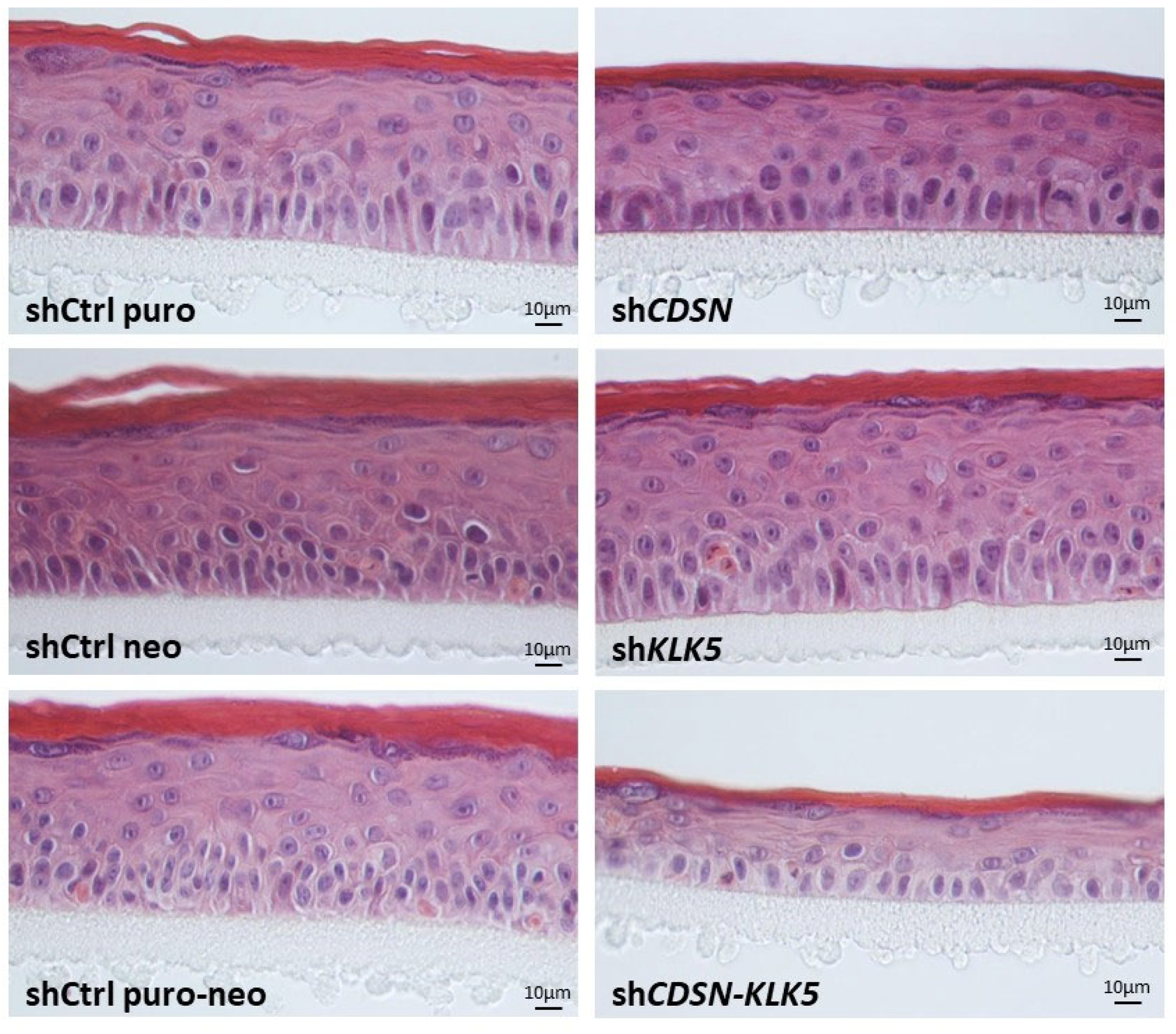
Figure 1. Histological analysis of HEEs produced with shCDSN and/or shKLK5 keratinocytes. The HEEs generated from shCDSN keratinocytes showed thinner epidermis compared to the controls or the shKLK5 keratinocytes-derived HEEs. Notably, co-silencing of CDSN and KLK5 further reduced the thickness of the HEEs. Representative images of paraffin-embedded HEE sections observed after hematoxylin-eosin staining are shown (n ≥ 4 HEE/condition).
Ijms 26 08605 g001
Figure 2. Ultrastructural analysis of the HEEs generated with shCDSN and/or shKLK5 keratinocytes. Representative images of HEE sections observed by transmission electron microscopy (TEM) are shown for shCDSN: (A) shKLK5 (B) and shCDSN-KLK5 (C). White arrowheads indicate the corneodesmosomes. In the SC of shCtrl neo and shKLK5 HEE, the length of corneodesmosomes was measured (n = 15 and 22 corneodesmosomes, respectively) and shown in (B). Importantly, the worsening of the HEE structural phenotype obtained from co-silencing CDSN and KLK5 in keratinocytes was observed. Specifically, the SC was thinner, loosely cohesive, with enlarged intercorneocyte spaces and aberrant corneodesmosomes. (n ≥ 2 HEEs/condition).
Ijms 26 08605 g002
Figure 3. Effect of CDSN and/or KLK5 gene silencing on the epidermal barrier function and the SC pH of HEEs. (A) Lucifer yellow (LY) dye penetration assay. A LY solution was applied on the surface of HEE and the dye concentration in the culture medium was measured fluorometrically in a fluoroscan at the indicated times (n = 2 independent experiments for each condition). (B) Measurement of pH of the external surface of the indicated HEEs (n = 3 independent experiments for each condition).
Ijms 26 08605 g003
Figure 4. Deletion of the Klk5 gene in Cdsnepi−/− background does not rescue neonatal lethality. (A) Macroscopic appearance of neonatal DKO mice shows severe signs of desquamation. (B) Toluidine blue dye penetration assay shows that DKO and Cdsnepi−/− mice exhibit severe epidermal barrier defect. (C) HE staining of skin sections reveal complete absence of SC in the Cdsnepi−/− sample and complete detachment in the DKO sample. (D) No morphological alterations in the whisker structure of DKO and Cdsnepi−/− are shown. (E) DKO mice exhibit rapid weight loss due to dehydration.
Ijms 26 08605 g004
Figure 5. Representative TEM images of the epidermis of wt (AC), Klk5−/− (DF), Cdsnepi−/− (GI) and DKO (JL) mice. (A,D,G,J): Upper epidermis; (B,E,H): higher magnification in the SG; (C,F,I,K,L): SG. White arrowheads show corneodesmosomes in (B,E,H) and desmosomes in (C,F,J,L). Double white arrows in (G) show abnormal spacing between the SG and the SC in Cdsn−/− epidermis. Note the total absence of SC in DKO epidermis. (L) corresponds to a magnification of the square in (K). Bar = 5 µm (A,G,J), 2.5 µm (D), 1 µm (B,C,E,F,H,I,K) and 0.5 µm (L).
Ijms 26 08605 g005
Figure 6. Elevated epidermal proteolytic activities in Cdsn−/− and DKO epidermis. In situ zymography using fluorescence-quenched elastin or casein. Skin tissue sections were prepared from wt, Cdsnepi−/− and Cdsnepi−/−Klk5−/− (DKO) mice. Fluorescence intensity data was transformed into a color gradient (as shown) using ImageJ software (1.52a). In situ zymography FL substrates (casein, and elastin) show elevated overall epidermal proteolysis in Cdsnepi−/− and DKO neonates. The green dashed line indicates the epidermis–dermis junction. Scale bar = 50 µm.
Ijms 26 08605 g006
Figure 7. Normal differentiation process and normal expression of Dsg1 and Dsc1 in DKO epidermis. The expression of keratin 5, Dsg1, loricrin, involucrin and Dsc1 was analyzed with immunohistochemistry (IHC). No significant changes were found in Cdsn-deficient and Cdsn−/−Klk5−/− epidermis. Scale bar = 50 µm.
Ijms 26 08605 g007
Figure 8. Quantification of various lipid classes in the epidermis of mouse models. The 1H-NMR peaks associated with different lipid structures at certain chemical shifts (in ppm) are shown at the right of each graph and under the same magnification. The area under the peak was integrated and compared using the MestReNova software (https://mestrelab.com/). The epidermal lipid profile appears to be normalized after deleting Klk5 in Cdsnepi−/−. The upper left picture shows the representative 1H-NMR spectra, while the other spectra used for quantification of certain lipid classes represent a magnification of the corresponding region (n = 4 for Cdsnepi−/−Klk5−/− and n = 3 for all other genotypes).
Ijms 26 08605 g008
Figure 9. Lipid profile of clinical specimens. (A) 1H-NMR spectra from a representative lipid extract obtained from the SC of healthy individual (NHS), a non-lesional (NLS) and a lesional site (LS) from a CDSN-nEDD patient. The methanol peak used as an internal control for quantification is boxed. (B) Magnification of certain regions of the 1H-NMR spectrum and their quantification. A strong increase in the amount of fatty acid acyl chains and ceramides is found in the lesional SC skin of CDSN-nEDD patient. Further, increased amount of unsaturated lipids is found in the SC of NLS and LS of the CDSN-nEDD patient.
Ijms 26 08605 g009
Disclaimer/Publisher’s Note: The statements, opinions and data contained in all publications are solely those of the individual author(s) and contributor(s) and not of MDPI and/or the editor(s). MDPI and/or the editor(s) disclaim responsibility for any injury to people or property resulting from any ideas, methods, instructions or products referred to in the content.