Article

Integrin β Regulates the Hepatopancreas Antiviral Innate Immune System by Affecting the Expression of Antimicrobial Peptides in Penaeus vannamei

1
State Key Laboratory of Mariculture Biobreeding and Sustainable Goods, Yellow Sea Fisheries Research Institute, Chinese Academy of Fishery Sciences, Qingdao 266071, China
2
College of Fisheries and Life Science, Shanghai Ocean University, Shanghai 201306, China
3
Laboratory for Marine Fisheries Science and Food Production Processes, Qingdao Marine Science and Technology Center, Qingdao 266237, China
*
Authors to whom correspondence should be addressed.
These authors contributed equally to this work.
Int. J. Mol. Sci. 2025, 26(17), 8478; https://doi.org/10.3390/ijms26178478 (registering DOI)
Submission received: 7 August 2025 / Revised: 28 August 2025 / Accepted: 29 August 2025 / Published: 31 August 2025
(This article belongs to the Special Issue Molecular Genetics and Genomics of Aquatic Crustaceans)

Abstract

Penaeus vannamei aquaculture production accounts for the majority of total shrimp aquaculture output, but it has suffered a severe decline in production and economic losses due to WSSV disease. Therefore, elucidating the relationship between the host immune system and pathogens is crucial for shrimp disease prevention and control. Integrins, as receptor-related molecules, have been shown to participate in various physiological functions, including cell migration, organismal development, and the pathogenesis of multiple diseases. However, the regulatory mechanisms of integrin genes in the shrimp immune system remain unclear. This study reports that integrins may regulate the Toll, IMD, and STAT signaling pathways in P. vannamei by influencing Spätzle, TLR, and Domeless, thereby affecting the shrimp’s innate immune system against diseases. Additionally, integrins can inhibit viral entry and replication. Through RNA interference (RNAi) experiments, it was found that knocking down Pv-Integrin β increases the viral load of white spot syndrome virus (WSSV), making shrimp more susceptible to WSSV and giving rise to increasing mortality. Further research indicates that Pv-Integrin β acts as an upstream recognition receptor in the disease resistance immune pathway, influencing other signaling pathway receptors to regulate the innate immune system. Importantly, knocking down Pv-Integrin β upregulates the expression of antimicrobial peptides such as ALF1 and ALF2, but reduces the expression of Crustin1, Crustin2 and prophenoloxidase. In conclusion, this study reveals that Pv-Integrin β regulates the disease resistance immune signaling pathways by affecting the related receptors.

Graphical Abstract

1. Introduction

White spot syndrome virus (WSSV) can infect not only shrimp but also crabs and other crustaceans [1]. The virus first emerged in the region of Asia in the 1990s and subsequently spread to shrimp farming regions worldwide, causing severe mortality rates and substantial economic losses. Despite extensive research over the past few decades, no effective treatment is currently available. WSSV belongs to the genus Whispovirus in the family Nimaviridae, and is a rod-shaped, double-stranded DNA (dsDNA) virus with a genome size of approximately 300 kbp [2,3]. Although a series of studies have been conducted, the molecular mechanisms by which the virus regulates the host cells remain incompletely understood. Further exploration of the molecular interactions between WSSV and its host may provide potential therapeutic targets for white spot syndrome.
According to research reports, the process of viral infection involves two distinct stages: attachment to the cell surface and entry into the cell [4]. In fact, viral infection of the host cells primarily occurs through the viral envelope proteins, which facilitate intracellular assembly and the budding process [5]. Taking WSSV as an example, multiple envelope protein structures have been identified. However, a recent study revealed that 58 structural proteins have been identified in WSSV, including nucleocapsid proteins and envelope proteins, with the VP series (such as VP 19, VP 24, VP 26, and VP 28) being the most predominant [6,7]. During viral infection, studies have shown that host cell membrane components can act as receptors by binding to viral envelope proteins, thereby promoting viral penetration. In crustaceans, known receptor components involved in viral recognition include Toll receptors, C-type lectins (CTL), scavenger receptors (SR), β-integrins, polymeric immunoglobulin receptors (pIgR), laminin receptors, globular C1q receptors (gC1qR), lipopolysaccharides (LPS), β-1,3-glucan binding proteins (LGBP), chitin binding proteins (CBP), Ras-associated binding proteins (Rab), and Down syndrome cell adhesion molecules (Dscam) [8]. Although the extent of research on these receptor molecules varies, they hold significant potential in pathogen blockade defense and target therapy, with receptor genes undoubtedly being the optimal choice.
Notably, integrins are a class of heterodimeric cell surface receptors composed of α and β subunits, playing a crucial role in mediating cell–matrix and cell–cell interactions, as well as bidirectional signal transduction [9]. Each subunit consists of three main regions: an extracellular domain, a transmembrane domain, and a small cytoplasmic domain [10]. Research has demonstrated that integrins are essential in various physiological processes, including cell migration, development, wound healing, immune system regulation, and the pathogenesis of multiple diseases [11]. Additionally, as cellular receptors, integrins have been shown to facilitate viral internalization into host cells, a function that is particularly critical in the early stages of viral infection [12,13]. It is noteworthy that integrins, as heterodimeric proteins, have been extensively studied in multiple species, including humans, zebrafish, and fruit flies [9]. However, despite an in-depth exploration of integrin functions in these species, the existence of their subunits in crustaceans has not yet been definitively confirmed [14]. This research gap highlights the importance of future studies to explore the functions and potential roles of integrins in crustaceans. In crustaceans, the Integrin β gene of Fenneropenaeus chinensis is distributed in the hemocytes, heart, gills, gonads, intestines, muscles, and hepatopancreas, with varying expression levels across these tissues. Notably, the Fc-Integrin β gene is significantly induced in gill tissues following viral infection [14]. Additionally, in a study on Penaeus vannamei, the Integrin β gene was successfully cloned from hemocytes. RNA interference (RNAi) experiments revealed that Integrin β plays a crucial role in regulating shrimp immune responses, including the modulation of the prophenoloxidase system, phagocytosis, and the antioxidant system [15]. However, these studies have yet to elucidate the specific disease resistance pathways through which the Integrin β gene participates in the innate immune response of shrimp.
Invertebrates lack the adaptive immune response system found in vertebrates and are, therefore, believed to rely entirely on their innate immune system to combat viral pathogens. When pattern recognition receptors (PRRs) are activated by viral components, the innate antiviral response is rapidly triggered. This response encompasses both humoral and cellular immunity. In invertebrates, the humoral immune response primarily involves the prophenoloxidase (proPO) system, the coagulation cascade, and the production of various antimicrobial peptides. Meanwhile, the cellular immune response includes mechanisms such as apoptosis and phagocytosis. In aquatic crustaceans, the innate immune system plays a crucial role as an essential defense mechanism against pathogens [16].
Through this study, it was found that the Integrin β molecule of P. vannamei can respond to an infection of WSSV. As an important economic aquaculture species, P. vannamei has long been affected by pathogens. By using RNAi technology to knock down the Integrin β gene, we aim to validate its role in the molecular regulation mechanism of the shrimp’s innate immune response against pathogens. The Integrin β gene plays a role in influencing the Toll, IMD, and STAT pathways, as shown during this study, providing new insights into resistance and immune mechanisms between pathogens and hosts.

2. Results

2.1. Pv-Integrin β Gene Sequence Information

In our previous study, we identified a gene sequence (mRNA sequence LOC113805717, amino acid sequence XM_027356770.1) from the transcriptome sequencing data of P. vannamei infected with WSSV. Subsequently, we amplified a 1254 bp sequence via PCR and sent it to Sangon Biotech (Shanghai) for sequencing. Based on the sequencing results, we performed basic bioinformatics analysis and aligned the sequence using the BLAST 2.17.0 software (http://blast.ncbi.nlm.nih.gov/Blast.cgi, accessed on 20 August 2024). According to the sequence’s characteristics, we designated it as the Pv-Integrin β gene. This gene contains an open reading frame (ORF) encoding 342 amino acids, with a predicted theoretical molecular weight of 37.6 kDa. Further analysis revealed that this gene contains a conserved “NPxY” motif (marked as a yellow area) and a transmembrane domain and cytoplasmic domain, indicated with solid underlines and dashed underlines, respectively (Figure 1).

2.2. Multiple Sequence Alignment and Phylogenetic Tree

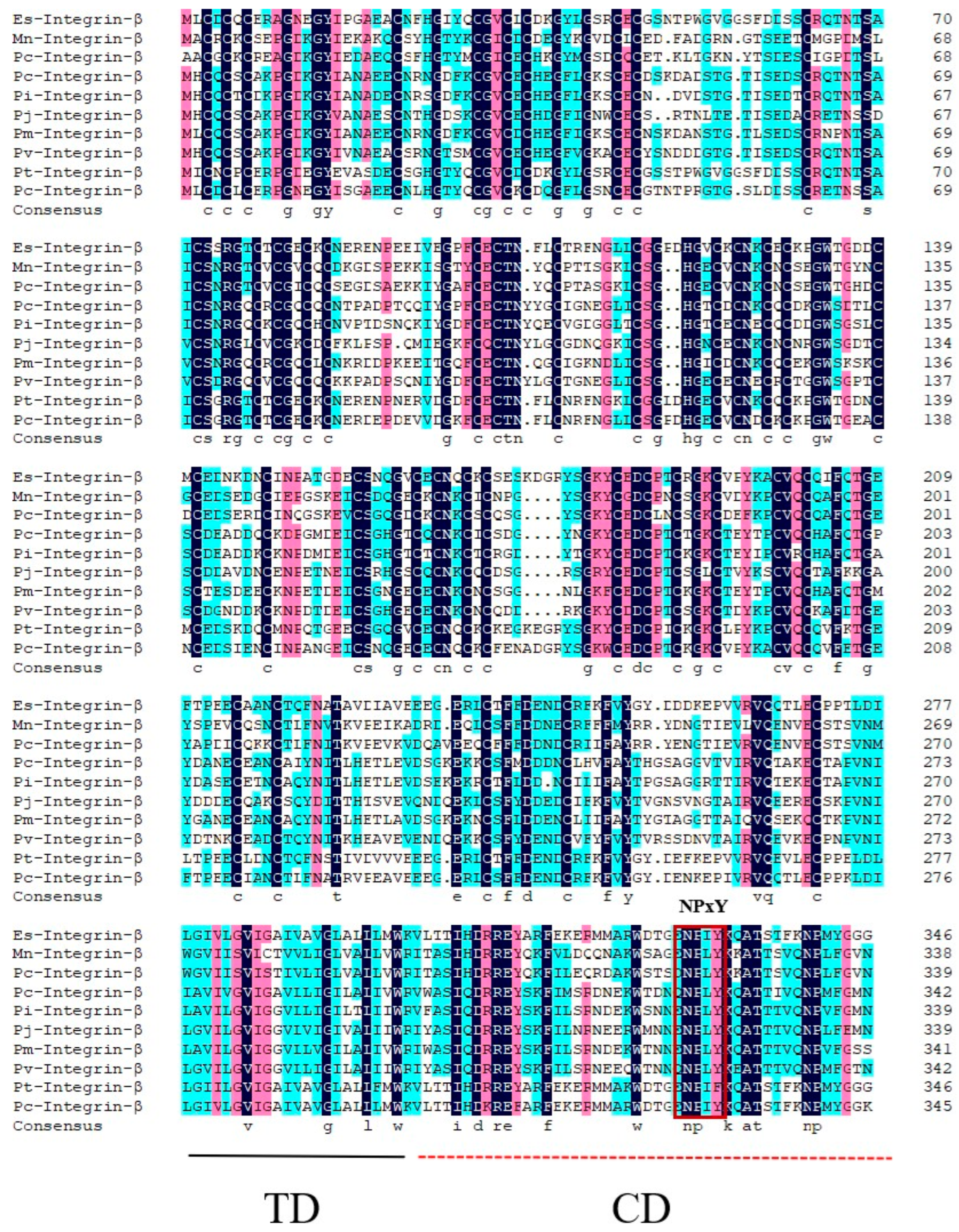
Using BLAST, we aligned the amino acid sequence of Pv-Integrin β and found that it shares high similarity (67.17%) with Integrin β sequences from several species, including Penaeus chinensis-Integrin β (XP_047479545.1), Penaeus monodon-Integrin β (XP_037801846.1), Penaeus japonicus-Integrin β (XP_042886327.1), Procambarus clarkii-Integrin β (XP_045617424.1), Eriocheir sinensis-Integrin β (XP_050729686.1), Macrobrachium nipponense-Integrin β (XP_064077230.1), Palaemon carinicauda-Integrin β (XP_068210289.1), Penaeus indicus-Integrin β (XP_063601209.1), and Portunus trituberculatus-Integrin β (XP_045106617.1). Further analysis revealed that Pv-Integrin β contains a conserved “NPxY” motif, as well as a transmembrane domain (TD) and a cytoplasmic domain (CD), which are also present in Integrin β sequences from other species (Figure 2).
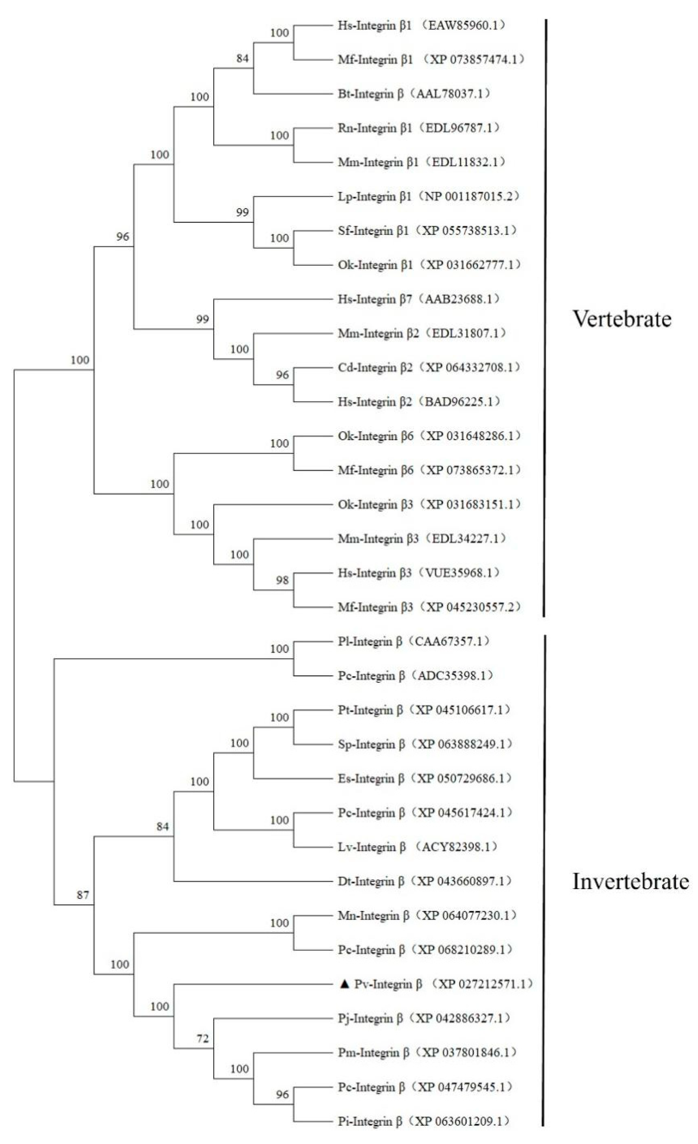
Based on the amino acid sequence alignment, we constructed a phylogenetic tree using MEGA11.0 software. Since P. vannamei is an invertebrate, we selected both invertebrate and vertebrate species for comparison. The invertebrate species included P. chinensis-Integrin β (XP_047479545.1), P. japonicus-Integrin β (XP_042886327.1), P. clarkii-Integrin β (XP_045617424.1), E. sinensis-Integrin β (XP_050729686.1), M. nipponense-Integrin β (XP_064077230.1), P. carinicauda-Integrin β (XP_068210289.1), P. indicus-Integrin β (XP_063601209.1), P. trituberculatus-Integrin β (XP_045106617.1), Scylla paramamosain-Integrin β (XP_063888249.1), Drosophila teissieri-Integrin β (XP_043660897.1), L. vannamei-Integrin β (ACY82398.1), P. chinensis-Integrin β (ADC35398.1), P. leniusculus-Integrin β (CAA67357.1), and P. monodon-Integrin β (XP_037801846.1). The vertebrate species included Rattus norvegicus-Integrin β1 (EDL96787.1), Bos taurus-Integrin β1 (AAL78037.1), Camelus dromedarius-Integrin β2 (XP_064332708.1), Salvelinus fontinalis-Integrin β1 (XP_055738513.1), Ictalurus punctatus-Integrin β1 (NP_001187015.2), Homo sapiens-Integrin β7 (AAB23688.1), Homo sapiens-Integrin β3 (VUE35968.1), Homo sapiens-Integrin β1 (EAW85960.1), Homo sapiens-Integrin β2 (BAD96225.1), Mus musculus-Integrin β1 (EDL11832.1), Mus musculus-Integrin β3 (EDL34227.1), Mus musculus-Integrin β2 (EDL31807.1), Oncorhynchus kisutch-Integrin β1 (XP_031662777.1), Oncorhynchus kisutch-Integrin β6 (XP_031648286.1), Oncorhynchus kisutch-Integrin β3 (XP_031683151.1), Macaca fascicularis-Integrin β1 (XP_073857474.1), Macaca fascicularis-Integrin β6 (XP_073865372.1), and Macaca fascicularis-Integrin β3 (XP_045230557.2). The phylogenetic tree revealed that Integrin β sequences from the vertebrates and invertebrates formed two distinct clades, with vertebrates grouped into one branch and invertebrates into another. Within the invertebrate clade, Pv-Integrin β clustered closely with M. nipponense-Integrin β (XP_064077230.1), P. japonicus-Integrin β (XP_042886327.1), and P. carinicauda-Integrin β (XP_068210289.1), indicating a close evolutionary relationship. The amino acid sequences of L. vannamei-Integrin β (ACY82398.1) and Pv-Integrin β form two independent branches in the phylogenetic tree, despite both sequences belonging to the invertebrate clade. However, L. vannamei-Integrin β (ACY82398.1) and P. clarkii-Integrin β (XP_045617424.1) are on the same branch, indicating a closer phylogenetic relationship. In contrast, Pv-Integrin β showed a lower level of evolution with H. sapiens-Integrin β1 (EAW85960.1) and M. fascicularis-Integrin β1 (XP_073857474.1) (Figure 3).

2.3. Analysis of Pv-Integrin β Expression Patterns Following WSSV Infection

We first analyzed the relative expression levels of the Pv-Integrin β gene in various tissues of healthy P. vannamei using qRT-PCR. The results showed that among the five tissues examined (hemocytes, hepatopancreas, the intestines, gills, and muscle), the Pv-Integrin β gene exhibited the highest expression level in hemocytes and the lowest in muscle tissue (Figure 4).
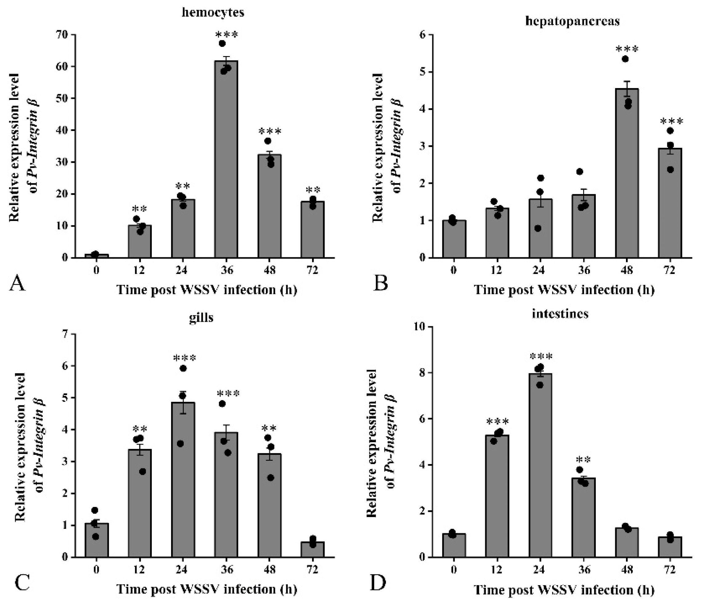
Furthermore, we examined the expression changes of the Pv-Integrin β gene in hemocytes, hepatopancreas, gills, and the intestine after P. vannamei was infected with WSSV. The results demonstrated that WSSV infection significantly upregulates the expression of the Pv-Integrin β gene in these four tissues. Specifically, in hemocytes, the relative expression level of Pv-Integrin β peaked at 36 hpi, increasing up to 60-fold (Figure 5A). In the hepatopancreas, the expression level rose to 4.5-fold at 48 hpi (Figure 5B). In the gills, the expression level increased to 4.8-fold at 24 hpi (Figure 5C), while in the intestines, the gene expression level reached 7.8-fold at 24 hpi (Figure 5D).
These experimental findings reveal that WSSV infection induces the significant upregulation of the Pv-Integrin β gene in multiple tissues of P. vannamei, particularly in hemocytes. This suggests that the Pv-Integrin β gene may play a role in the antiviral immune response of P. vannamei.

2.4. Analysis of dsRNA Interference Efficiency and Survival Rate of P. vannamei

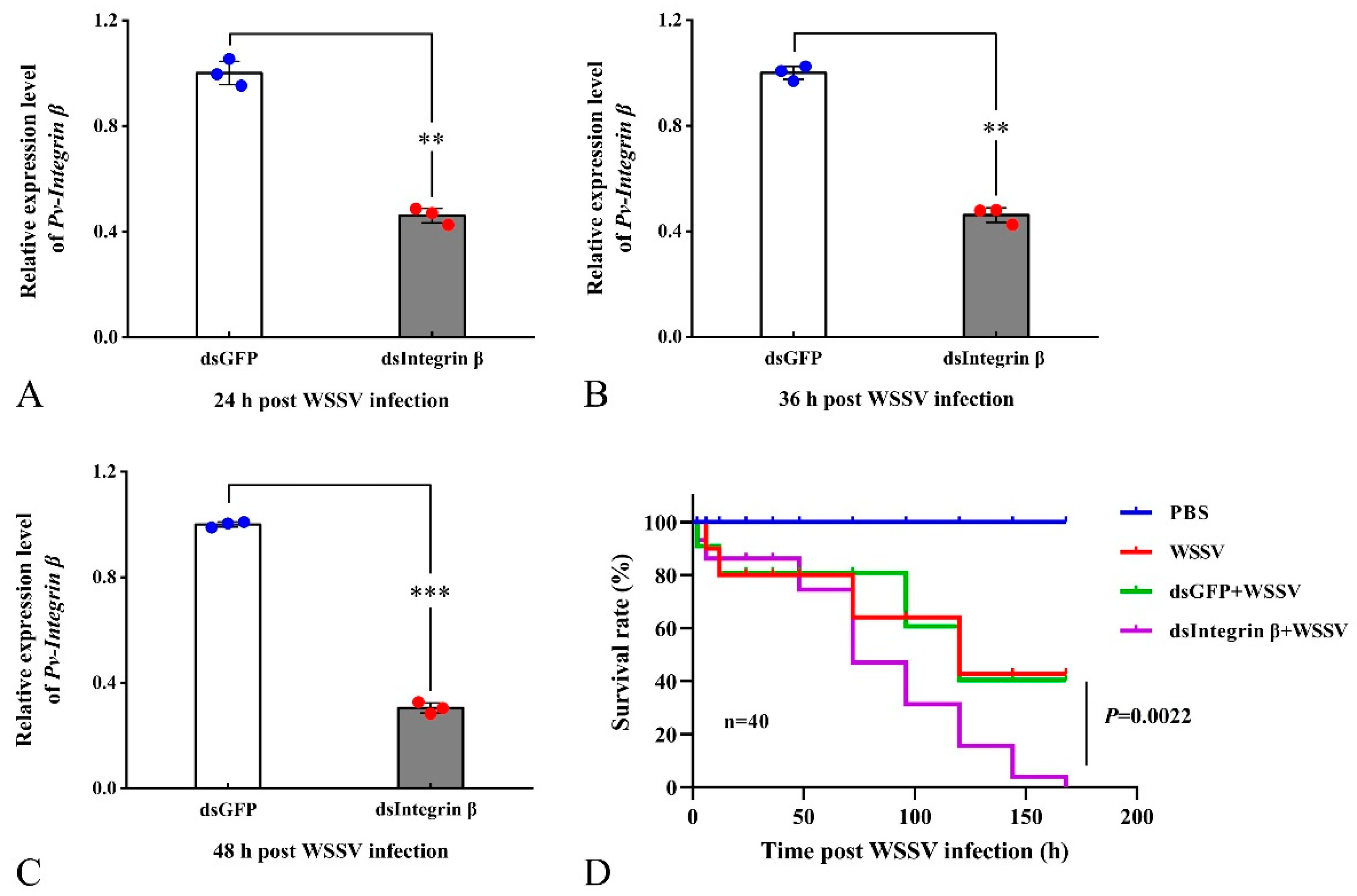
To verify the interference effect of dsRNA injection, we measured the relative expression level of the Pv-Integrin β gene after dsRNA injection using qRT-PCR. The experimental results showed that, compared to the control group (dsGFP + WSSV), the target gene was effectively knocked down at different time points after the injection of dsIntegrin β + WSSV. Specifically, the interference efficiency of Pv-Integrin β remained at 54%, 58%, and 70% at 24, 36, and 48 hpi, respectively (Figure 6A–C). Subsequently, we statistically analyzed the survival rates of P. vannamei in four groups: PBS, WSSV, dsGFP + WSSV, and dsIntegrin β + WSSV. The results revealed that, compared to the control group (dsGFP + WSSV), the survival rate of shrimp in the dsIntegrin β + WSSV group showed a significant downward trend (Figure 6D).

2.5. Knockdown of the Pv-Integrin β Gene Promotes WSSV replication

To further investigate the role of the Pv-Integrin β gene in antiviral immunity in shrimp, the study employed a knockdown approach targeting Pv-Integrin β to assess its impact on WSSV replication. Furthermore, the expression levels of the key WSSV proteins IE 1 and vp 28 were analyzed, revealing that the knockdown of Pv-Integrin β led to a significant upregulation of these viral proteins (Figure 7A,B). These findings suggest that Pv-Integrin β may play a critical role in shrimp antiviral immunity against WSSV. In order to more accurately detect the viral load, a probe-based method was used to quantify the WSSV copy numbers. Compared to the dsGFP + WSSV control group, the knockdown of Pv-Integrin β significantly enhanced WSSV replication in infected shrimp (the WSSV load significantly increased at 36 and 48 h; see Figure 7C).

2.6. Role of Pv-Integrin β in the Regulation of Innate Immune Pathways

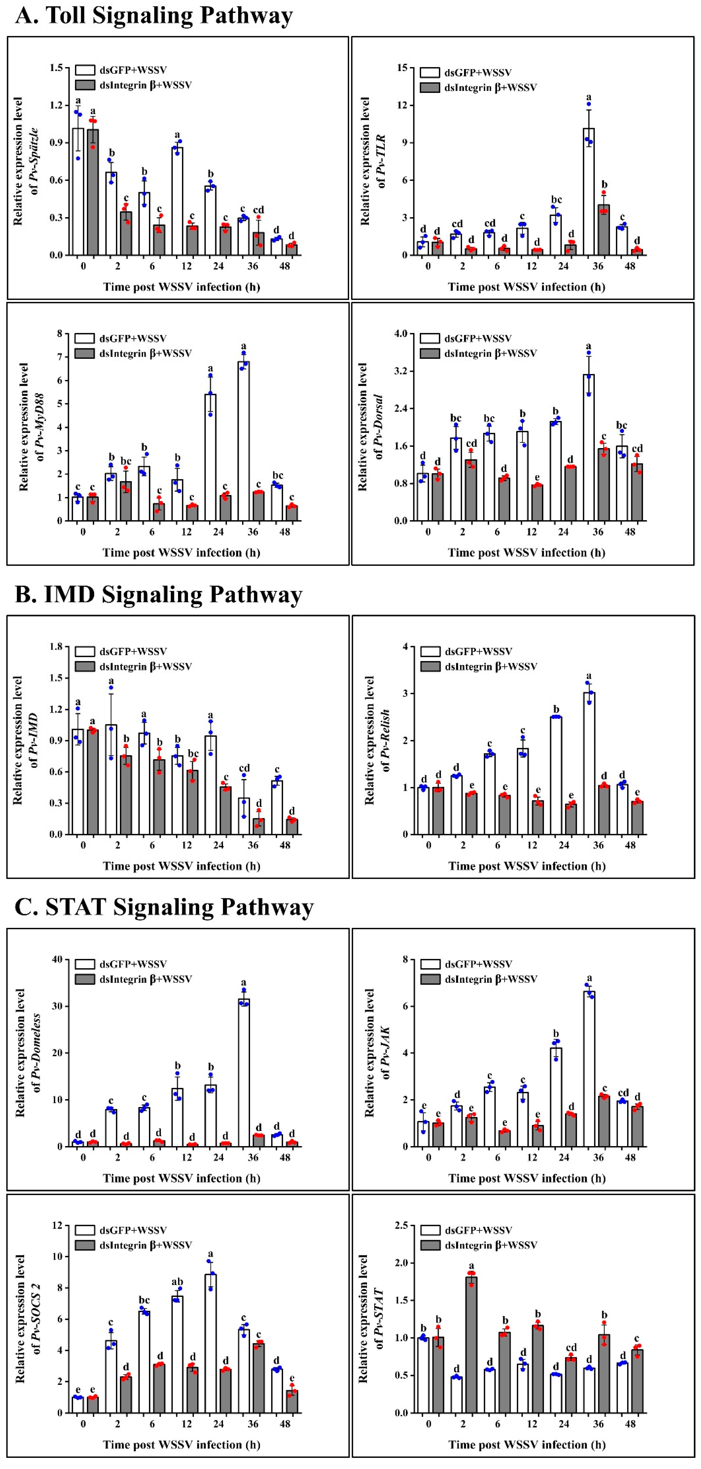
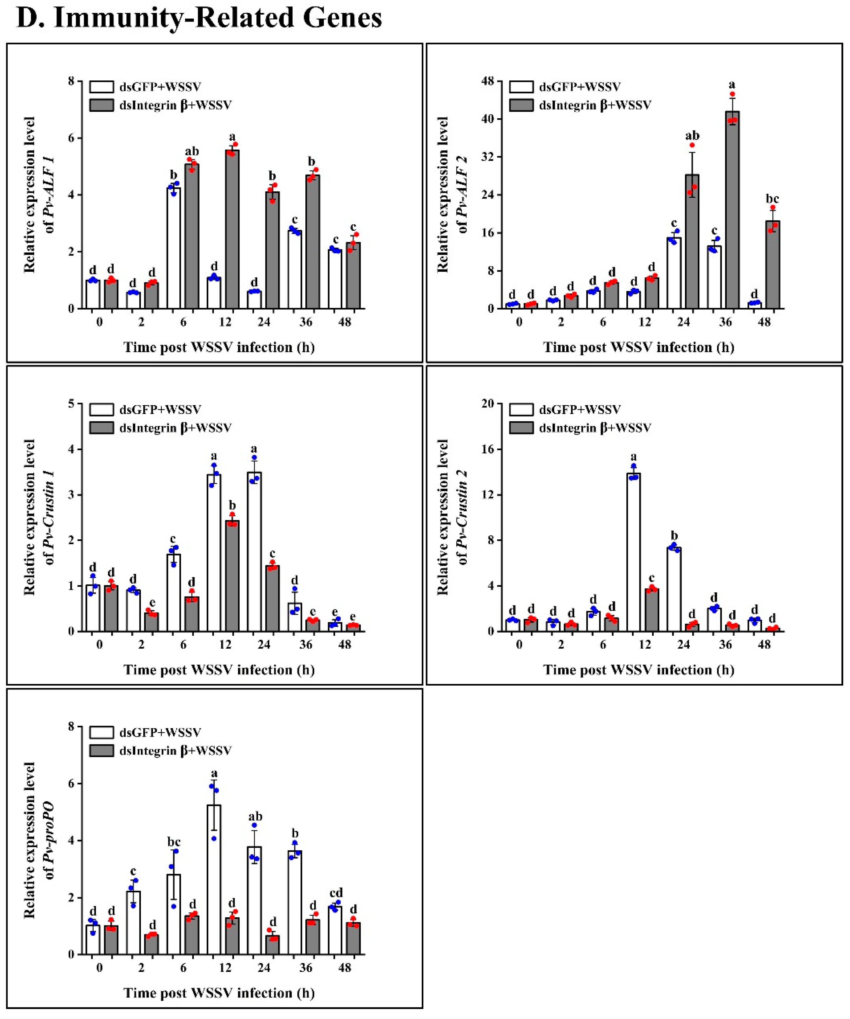
Pv-Integrin β, as an important membrane receptor gene, plays a crucial role in anti-disease innate immunity. To further elucidate its regulatory mechanism, we examined the expression levels of Toll, IMD, the STAT signaling pathways, and related effector molecules. The study found that in the Toll signaling pathway, knocking down the Pv-Integrin β gene resulted in a significant decrease in the expression levels of Spätzle, TLR, MyD88, and Dorsal (Figure 8A). Similarly, in the IMD signaling pathway, the expression levels of IMD and Relish also significantly decreased (Figure 8B). However, in the STAT signaling pathway, the expression levels of Domeless, JAK, and SOCS2 significantly decreased, while STAT expression levels significantly increased (Figure 8C). In the family of antimicrobial peptides (AMP), antilipopolysaccharide factors ALF1 and ALF2 are significantly upregulated, while Crustin1 and Crustin2 are significantly downregulated. Additionally, proPO also shows a significant downward trend (Figure 8D). These results suggest that Pv-Integrin β may regulate the expression of antiviral pathway genes in shrimp by modulating the Toll, IMD, and STAT signaling pathways, thereby playing a crucial role in innate immune responses.

3. Discussion

Integrins are adhesion molecules that are located on the cell surface, playing a significant role in an organism’s growth, development, and immune response. Additionally, they regulate various cellular processes, including proliferation, differentiation, adhesion, migration, and apoptosis [17]. In our study, Pv-Integrin β was identified from the transcriptome data of P. vannamei infected with WSSV. Subsequent PCR amplification and sequencing confirmed the ORF of Pv-Integrin β. Bioinformatics analysis revealed that Pv-Integrin β exhibits evolutionary conservation, particularly the “NPxY” motif, which is consistent across species. Most Integrin β subunits contain at least one conserved NPX [Y/F] motif in their cytoplasmic domains, which serves as a typical phosphotyrosine binding site. This motif is implicated in intracellular signaling and the activation of integrins, facilitating communication between the cell and its environment [18]. Although the Pv-Integrin β amino acid sequence used in this study consists of 342 amino acids, this is not the full-length integrin sequence. This may be due to incomplete annotations in the database. However, upon analysis, we found that this sequence covers the key functional domains of the protein, including the transmembrane domain (TD), the cytoplasmic domain (CD), and the “NPxY” motif. Although we attempted to obtain the full-length sequence using 5’-RACE, this was unsuccessful. Nevertheless, this sequence remains of significant research value for elucidating the disease resistance mechanisms in shrimp. However, the sequence analysis may have certain limitations. It is hoped that through further research, the complete integrin beta sequence can be obtained for use in functional comparative studies. Further analysis using qRT-PCR revealed the relative expression levels of the Pv-Integrin β gene in various tissues from P. vannamei. The results showed that Pv-Integrin β exhibited the highest relative expression levels in hemocytes, while the lowest expression levels were observed in muscle tissue. These findings are consistent with previous studies on integrin gene expression in F. chinensis, where Fc-Integrin was shown to be expressed in various tissues, including the hemocytes, heart, gills, gonads, intestines, muscle, and hepatopancreas, with distinct expression levels across tissues [14].
In the infection experiments, it was observed that WSSV infection induced the upregulation of Pv-Integrin β gene expression in the hemocytes, hepatopancreas, gills, and intestine. Interestingly, the upregulation of integrins under WSSV stimulation can activate integrin-associated signaling pathways, thereby promoting the spread of WSSV in the host [12]. In another study on shrimp, it was found that the expression levels of Integrin α5 and FAK mRNA were also significantly upregulated following WSSV infection. By analyzing the phenotypic changes in shrimp following the knockdown of the integrin α5 subunit and β subunit, we found that the magnitude of phenotypic changes induced by α5 knockdown differs from that of β subunit knockdown. Specifically, α5 knockdown has a more significant impact on cell migration and adhesion, while β subunit knockdown primarily affects shrimp survival and pathogen proliferation. These differences may be attributed to the distinct roles of the α5 and β subunits in integrin function [19].
To further investigate the functional roles of integrin genes, we conducted RNAi knockdown experiments targeting the Pv-Integrin β gene. To validate the interference efficiency, we confirmed that Pv-Integrin β was effectively downregulated at different time points after dsRNA injection. Following the knockdown of Pv-Integrin β, a significant decrease in the survival rate of shrimp was observed. Additionally, the knockdown of Pv-Integrin β resulted in a significant increase in WSSV infection efficiency and replication capacity. These findings are consistent with a previous study in whiteleg shrimp, where the knockdown of Pv-Integrin α5 also promoted WSSV replication and reduced shrimp survival [19]. Although both studies focused on Integrin genes, they targeted different subtypes. Sequence comparison revealed that these genes are distinct, with the integrins consisting of α and β subunits. However, they may share functional similarities in their roles as receptor genes, potentially mediating the related disease resistance signaling pathways.
The involvement of integrins in classical immune pathways, such as the Toll pathway, IMD pathway, and STAT pathway, remains unclear. To explore the role of Pv-Integrin β in disease resistance pathways, we analyzed the hepatopancreas tissue after knockdown of the target gene. In the Toll pathway, qRT-PCR analysis revealed the significant downregulation of Spätzle, TLR, MyD88, and Dorsal. The Toll pathway is known to play a critical role in antiviral immunity. Similarly, the IMD pathway also contributes to these defense mechanisms. Our qRT-PCR results showed that the knockdown of Pv-Integrin β led to the significant downregulation of IMD and Relish. However, in the antiviral STAT pathway, knockdown of Pv-Integrin β resulted in the significant downregulation of Domeless, JAK, and SOCS2, while STAT exhibited an upregulated expression trend. These findings suggest that Pv-Integrin β may influence multiple immune pathways, potentially modulating the shrimp’s antiviral response. The signaling pathways mediated by integrins primarily include the Jak/STAT, MAPK, and PI3K/AKT pathways [20]. However, in invertebrates, reports on the signaling pathways mediated by integrins are scarce, and research into their functional mechanisms is limited.
In mammals, Integrin αvβ3 has been identified as a crucial immune recognition receptor. It can act independently or synergize with Toll-like receptor 2 (TLR2) to recognize various pathogens, including bacteria and viruses, thereby triggering immune defense mechanisms [21]. Both human and mouse models have shown that Integrin αvβ3 plays a key role in combating invading pathogens. Recent studies have highlighted a significant synergistic effect between integrin αvβ3 and TLR2 in mediating immune responses to bacterial lipopeptides, lipopolysaccharides, and viruses. Specifically, in cells co-expressing Integrin αvβ3 and TLR2, the expression levels of interferon alpha and interferon beta are significantly upregulated, along with the enhanced expression of specific cytokines, such as interleukin-2 and interleukin-10. Additionally, nuclear factor κB is activated in response to herpes simplex virus infection or LPS stimulation. Notably, when the integrin β3 subunit is silenced in TLR2-positive cells using gene-silencing techniques, the production of these cytokines and NF-κB activation are significantly reduced [22]. This finding aligns with the conclusions of previous shrimp disease resistance studies, where the inhibition of Pv-Integrin β expression led to the significant downregulation of Dorsal and Relish. In shrimp and other crustaceans, Dorsal, a member of the NF-κB family, plays a role in regulating immune responses. NF-κB, comprising a family of evolutionarily conserved transcription factors, is central to various biological processes [23]. Relish, as a member of the NF-κB family of proteins, plays a crucial role in the immune response. Upon stimulation by pathogens, Relish is cleaved into two fragments within the cytoplasm: the N-terminal fragment containing the Rel homology domain (RHD) and the C-terminal fragment containing the Ankyrin repeat (ANK). The N-terminal fragment, which includes the RHD, is then transported into the nucleus [24]. Here, it binds to the corresponding κB sites in the promoters/enhancers of antimicrobial peptides and other target genes. This binding regulates the transcription of immune-related genes, enabling the organism to mount an effective immune response against the invading pathogens. In crustaceans, studies have shown that Relish is involved in immune responses against bacteria, fungi, and WSSV [25,26,27]. Previous studies have reported that the expression of the Relish gene is stimulated by Vibrio harveyi, WSSV, and yellow head virus [28]. In another study of hemocytes in Fenneropenaeus chinensis, Relish expression was significantly altered under bacterial and WSSV stimulation [29]. This indicates that Relish participates in the immune response of the organism during bacterial or viral infections.
While there is no direct evidence indicating that integrins regulate the IMD pathway, their involvement in cell adhesion and signal transduction may influence the activation and regulation of the IMD pathway. In shrimp, integrin signaling impacts NF-κB activity, thereby indirectly affecting the IMD pathway’s signaling output. The IMD pathway typically interacts with other immune pathways to regulate immune responses. For instance, the Toll and IMD pathways can synergize to activate innate immune responses [30]. Integrins may influence these interactions, indirectly affecting IMD pathway functionality. Indeed, when Pv-Integrin β is interfered with, it inhibits JAK expression, while STAT expression is upregulated, potentially reflecting regulatory mechanisms within the shrimp. As shown in our experimental results, the knockdown of Integrin β affects the Toll, IMD, and STAT transcription factors, regulates the AMPs, and participates in the anti-disease immunity of shrimp. We observed distinct expression profiles for the ALF and Crustin genes. This differential regulation may be attributed to differences in their upstream signaling pathways and transcriptional regulation. In our study, knocking down Integrin β led to significant downregulation trends in certain pathways, such as Spätzle, TLR, MyD88, IMD, Domeless, JAK and SOCS2. However, transcription factors such as Dorsal and Relish were significantly downregulated, while STAT showed an upregulation trend. This suggests that the regulation of ALF and Crustin genes may involve distinct transcriptional mechanisms, with Dorsal and Relish potentially regulating Crustin genes, and STAT influencing ALF gene expression. Currently, the main research into the pathways regulating antimicrobial peptide families focuses on Toll, IMD, and STAT. The TLR, IMD, and JNK pathways recognize pathogen molecules and activate transcription factors to regulate the expression of antimicrobial peptides. ALFs and Crustins exhibit significant differences in structure, function, and target points; ALFs primarily target Gram-negative bacteria and viruses, while Crustins demonstrate broader antimicrobial and antiviral activities. A study suggests that knocking down integrins may suppress JAK expression, but the upregulation of STAT could indicate the viral hijacking of the STAT pathway by WSSV to facilitate its replication. Lv-SOCS 2, a negative regulator of the JAK/STAT pathway, has been shown to enhance shrimp resistance to WSSV when knocked out [31]. Simultaneously, it affects the expression levels of proPO in the white shrimp P. vannamei. Research has confirmed that Pv-Integrin β is involved in the activation of proPO and the regulation of the shrimp immune system [15].
In this study, knocking down the Integrin β gene in P. vannamei resulted in decreased survival rates and increased WSSV copy numbers, primarily due to the attenuation of multiple functions of Integrin β in viral entry and immune regulation. The absence of Integrin β facilitated viral infection and replication in host cells, concomitantly inhibiting the activation of antiviral immune pathways, ultimately leading to increased mortality in shrimp. This study provides crucial insights into the role of Integrin β in antiviral immunity in shrimp and offers new perspectives for developing strategies to combat WSSV.

4. Materials and Methods

4.1. Experimental Samples Collection

The P. vannamei used in the experiment (about 3.6 ± 0.5 g each) were all sourced from BLUP Aquabreed Co., Ltd. (Weifang, China). To ensure the accuracy of the infection experiment, SPF-grade shrimp were selected from the stock of BLUP Aquabreed Co., Ltd. During the experiment, the shrimp were fed twice daily and cultured in sterile seawater for 7 days (salinity 29‰; temperature 25 ± 1 °C). The specific methods were as follows: for the infection experiment, 15 μL of WSSV (4.5 × 10⁶ copies) was injected into the third abdominal segment of each shrimp. Subsequently, the hemolymph, hepatopancreas, gills, and intestines were collected at 12, 24, 36, 48, and 72 h post-infection (hpi). The samples were centrifuged at 4 °C and 3000 rpm for 10 min, and then the hemolymph was separated from the hemocytes. The sampled tissues were stored at ultra-low temperatures for subsequent total RNA extraction experiments.

4.2. Total RNA Extraction and cDNA Synthesis

In this experiment, total RNA extraction was performed using the RNA extraction kit provided by Vazyme Biotech Co., Ltd. (Nanjing, China), strictly following the manufacturer’s instructions. For hemocyte tissue, a glass homogenizer was used for homogenization, while other tissues were homogenized mechanically. The extracted total RNA was verified by 1.5% agarose gel electrophoresis, and its concentration was measured. Subsequently, cDNA synthesis was carried out using the First-strand cDNA Synthesis Kit from Vazyme Biotech Co., Ltd. (Nanjing, China).

4.3. Gene Cloning and Bioinformatic Analysis

The amplification primers for the Pv-Integrin β gene (LOC113805717) were designed using online primer design software (https://primer3.ut.ee/, accessed on 15 August 2024), based on the sequence retrieved from the National Center for Biotechnology Information database. The primer sequences are listed in Table 1. The primers were synthesized by Sangon Biotech Co., Ltd. (Shanghai, China). The ORF sequence of the Pv-Integrin β gene was amplified using Quick Taq HS DyeMix (TOYOBO, Shanghai, China), with cDNA from the hepatopancreas of P. vannamei as the template. The PCR reaction was performed in a 50 μL system containing 2 μL of cDNA solution (0.1 mg/μL), 25 μL of 2xQuick Taq HS DyeMix (TOYOBO, Shanghai, China), and 1 μL of each primer F/R (10 μM), along with 21 μL of nuclease-free distilled water. The PCR program consisted of initial denaturation at 95 °C for 3 min, followed by 35 cycles of denaturation at 95 °C for 3 s, annealing at 56 °C for 45 s, and extension at 72 °C for 50 s, with a final extension at 72 °C for 5 min. The PCR products were analyzed by 1.0% agarose gel electrophoresis and sent to Sangon Biotech Co., Ltd. (Shanghai, China) for Sanger sequencing. The obtained ORF sequence of the Pv-Integrin β gene was translated into an amino acid sequence and used for protein prediction, using the ExPASy online tool (http://www.expasy.org, accessed on 18 August 2024). Subsequently, multiple sequence alignment analysis was performed using the BLAST 2.17.0 online tool (http://blast.ncbi.nlm.nih.gov/Blast.cgi, accessed on 20 August 2024). Finally, a phylogenetic tree was constructed using the DNAMAN 6.0 software (https://www.lynnon.com/, accessed on 10 September 2024).

4.4. Tissue Distribution and Expression Pattern Analysis of the Pv-Integrin β Gene

To analyze the relative expression levels of Pv-Integrin β in the various tissues of the shrimp, hemocytes, hepatopancreas, gills, intestines, and muscle tissues from healthy P. vannamei were selected for an analysis of Integrin β expression in these five tissues. In the pathogen infection experiment, 60 shrimp were divided into two groups. Among them, 15 shrimp from the uninfected group were used as the control before infection, and the remaining 45 shrimp would be immunologically infected through injection. The expression levels of the hemocytes, hepatopancreas, gills, intestines, and muscle tissues of the infected shrimp were analyzed. The spatial and temporal expression patterns of Pv-Integrin β in normal tissues and after WSSV infection were analyzed using quantitative real-time PCR (qRT-PCR, Applied Biosystems, Foster City, CA, USA) with the SYBR Green Real-time PCR Master Mix Kit (TOYOBO, Shanghai, China). The 18 S ribosomal RNA (18 S rRNA) was used as an internal reference control. The quantitative primers for Pv-Integrin β-qRT-F/R and 18 S-F/R are listed in Table 1. The total reaction volume for RT-PCR was 20 μL, containing 10 μL of SYBR PCR mix (TOYOBO, Shanghai, China), 0.8 μL of each primer (10 μM), 2 μL of cDNA solution (0.1 mg/μL), and 6.4 μL of nuclease-free distilled water. The PCR program consisted of initial denaturation at 95 °C for 60 s, followed by 40 cycles of denaturation at 60 °C for 15 s, annealing at 60 °C for 15 s, and extension at 72 °C for 45 s. The data were analyzed using the 2−ΔΔCT method, and the results were compared using an unpaired two-tailed t-test. The experiment was independently repeated three times, including three biological replicates. A value of p < 0.05 indicates a significant difference, p < 0.01 indicates an extremely significant difference, and p < 0.001 indicates an exceptionally significant difference.

4.5. Double-Stranded RNA Preparation

To perform the RNAi experiment, double-stranded RNA (dsRNA) targeting the Pv-Integrin β gene was synthesized using the T7 in vitro transcription kit (Takara, Shiga, Japan) according to the method described by Yang et al. [32]. A total reaction volume of 20 μL was prepared by mixing the following components in sequence: 2 μL of DNA template, 2 μL of 10 × transcription buffer, 2 μL of A/U/C/GTP mixture (10 μM each), 0.5 μL of RNase inhibitor, and 2 μL of T7 RNA polymerase, with RNase-free water added to bring the total volume to 20 μL. The reaction mixture was incubated at 42 °C for 2 h, followed by the addition of 2 μL of RNase-free DNase I, after which it was mixed thoroughly and further incubated at 37 °C for 30 min. To ensure the quality of the dsRNA, phenol/chloroform extraction was performed, followed by ethanol precipitation. Before use, the working concentration of the dsRNA was adjusted to 3 μg/μL, and the samples were stored in an ultra-low-temperature (−80 °C) freezer. Following the same procedure, control dsGFP RNA was prepared, and the interference primer information is listed in Table 1.

4.6. Survival Rate Analysis After Pv-Integrin β Knockdown

Before analyzing the survival rate of shrimp after the knockdown of the target gene, we first verified the efficiency of the RNAi. Sixty shrimp were randomly divided into two groups, with thirty shrimp in each group (dsGFP + WSSV and dsIntegrin β + WSSV). The prepared dsRNA (approximately 30 μg) was injected into the abdominal segment of each shrimp using a microinjector. To enhance the RNAi efficiency, a second injection of dsRNA was performed 24 h later, followed by infection with WSSV (4.5 × 10⁶ copies). Total RNA was extracted from the hepatopancreas of the shrimp at 24, 36, and 48 hpi, and the expression level of Pv-Integrin β was detected using qRT-PCR, with 18 S rRNA as the internal reference gene. The quantitative primer information is listed in Table 1.
In the shrimp survival rate analysis experiment, 160 shrimp were randomly divided into four groups, with 40 shrimp in each group: PBS, WSSV-infected, dsGFP + WSSV, and dsIntegrin β + WSSV. After the knockdown of Pv-Integrin β, the survival rate of the shrimp was statistically analyzed. To ensure the robustness of the experiment, the above procedures were performed in triplicate. The experimental data were analyzed using GraphPad Prism 8.0 software (https://www.graphpad.com/, accessed on 8 December 2024), and statistical significance was assessed using a Kaplan–Meier plot (log-rank χ2 test).

4.7. Pathogen Detection After Pv-Integrin β Knockdown

To detect the replication of WSSV in P. vannamei, we selected muscle tissue for the quantification of pathogen copy numbers. After infecting the host, WSSV typically begins by invading the epidermis and subcutaneous tissues of the shrimp, causing white spots to appear on the body surface. As the infection progresses, the virus also invades the muscle tissue, leading to necrosis and softening of the muscles, which subsequently affects the shrimp’s motor function and survival rate. In this experiment, DNA was first extracted from the muscle tissues of the dsGFP + WSSV group and the dsIntegrin β + WSSV group at different time points (using the TIANamp Marine Animals DNA Kit, Tiangen Biotech Co., Ltd., Beijing, China). The pathogen detection experiment involved 60 shrimp, which were divided into 2 groups with 30 shrimp in each group. The WSSV copy number was then detected using TaqMan real-time PCR technology [33]. The specific primers and probes for WSSV are detailed in Table 1. The detection was performed using the THUNDERBIRD™ Probe qPCR Mix Kit (TOYOBO) and quantified with the Applied BiosystemsTM QuantStudio 1 real-time PCR system. The reaction mixture (20 μL) consisted of 10 μL of THUNDERBIRD Probe qPCR Mix, 0.6 μL each of F/R primers (0.2 μM), 0.4 μL of the WSSV probe (10 μmol/L), 0.1 μL of ROX reference dye, 2 μL of the DNA template, and 6.3 μL of nuclease-free distilled water. The PCR protocol included an initial denaturation process at 95 °C for 30 s, followed by 40 cycles of 95 °C for 5 s and 60 °C for 34 s. This experiment was conducted in three independent and parallel trials. Using this method, we were able to accurately detect the replication of WSSV in muscle tissue, providing reliable data for further research into the infection mechanisms of WSSV. Meanwhile, the relative expression levels of the WSSV encoded envelope protein gene vp28 and the immediate-early gene (IE1) in the hepatopancreas were also detected by qRT-PCR after the knockdown of the target gene. The primer information is listed in Table 1.

4.8. Detection of Innate Immune Signaling Pathway Genes Following the Knockdown of the Pv-Integrin β Gene

To further investigate the changes in disease resistance pathway genes following the knockdown of the integrin receptor gene, qRT-PCR was employed to analyze the innate immune system. The experimental design was as follows: the shrimp were first subjected to a second injection of dsRNA (30 μg), followed by infection with WSSV (4.5 × 10⁶ copies). The relative expression levels of the genes were analyzed at 0, 2, 6, 12, 24, 36, and 48 hpi. A total of 90 shrimp were used in the experiment and divided into two groups: the experimental group (dsIntegrin β + WSSV) and the control group (dsGFP + WSSV). Hepatopancreas tissue was collected from the shrimp, and total RNA was extracted and reverse transcribed into cDNA for the detection of the mRNA expression levels of key genes; the genes analyzed included those related to the Toll, IMD, and STAT pathways. The primer information used for qRT-PCR is listed in Table 1. The experimental data were analyzed using the 2−ΔΔCT method, and the experiment was independently repeated three times.

5. Conclusions

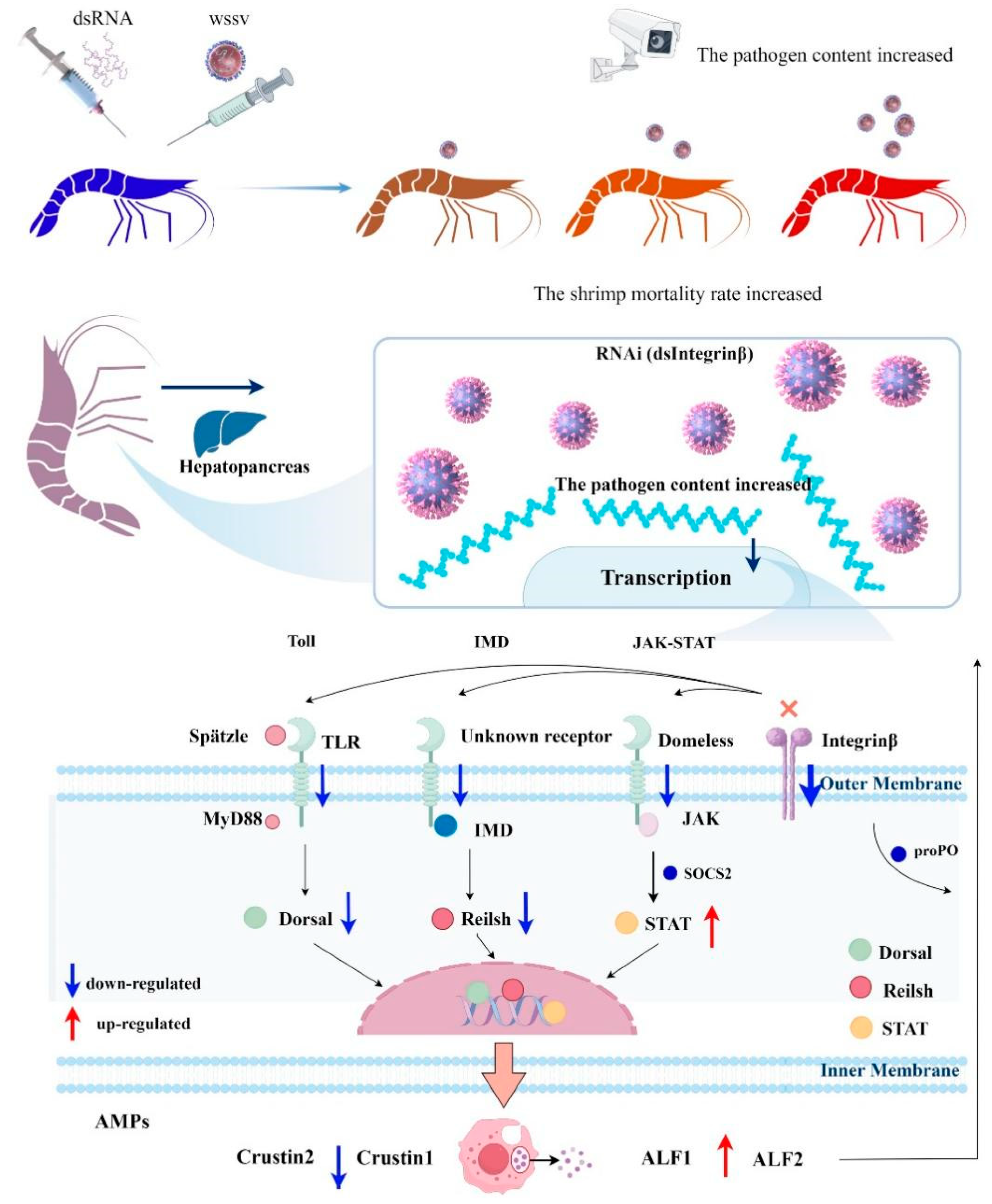
Through our research, we have proposed a molecular theoretical model where Integrin β regulates the antiviral innate immune system in the hepatopancreas of P. vannamei by influencing the expression of antimicrobial peptides. In fact, WSSV induces the expression of Pv-Integrin β. Knocking down Pv-Integrin β significantly increases the viral copy number of WSSV and also raises the cumulative mortality rate of the shrimp. Further analysis of the gene regulation mechanism reveals that the Pv-Integrin β protein may influence the expression of Spätzle, Toll-like receptors, and Domeless, thereby regulating the Toll, IMD, and STAT signaling pathways. This, in turn, modulates the expression of ALF1, ALF2, Crustin1, Crustin2, and proPO in the hepatopancreas, playing a crucial role in regulating the antiviral innate immune system in the hepatopancreas. The SOCS2 gene, as a negative regulatory factor of STAT, may be involved in regulating the upregulation of STAT expression, thereby regulating the upregulation of ALF1 and ALF2 in order to participate in the antiviral immune response of shrimp. Dorsal and Relish regulate the downregulation of Crustin1 and Crustin2 expression (Figure 9).

Author Contributions

B.Y.: Original draft, methodology, formal analysis, data curation, and conceptualization. L.Z.: Visualization, validation, software, and investigation. K.L.: Investigation. S.L.: Software and conceptualization. J.K.: Supervision and methodology. Q.F.: Conceptualization. J.C.: Software. B.C.: Methodology. P.D.: Investigation. X.L.: Supervision, methodology, and conceptualization. X.M.: Writing—review and editing, resources, and funding acquisition. All authors have read and agreed to the published version of the manuscript.

Funding

This work was financially supported by the National Key R&D Program of China (2022YFF1000304), the National Natural Science Foundation of China (32172960), the China Agriculture Research System (CARS-48), and the Central Public-interest Scientific Institution Basal Research Fund, CAFS (2020TD26).

Institutional Review Board Statement

The research object used in this study was Penaeus vannamei, which was used for experiments as a non-endangered invertebrate. According to the Fisheries Law of the People’s Republic of China, no permission is required to collect these experimental animals, so this study did not require formal ethical approval.

Informed Consent Statement

Not applicable.

Data Availability Statement

The data presented in this study are available in the article.

Conflicts of Interest

The authors declare no conflicts of interest.

References

  1. Desrina; Slamet, B.P.; Verdegem, M.C.J.; Verreth, J.A.J.; Vlak, J.M. White spot syndrome virus host range and impact on transmission. Rev. Aquac. 2022, 14, 1843–1860. [Google Scholar] [CrossRef]
  2. Wang, H.-C.; Hirono, I.; Maningas, M.B.B.; Somboonwiwat, K.; Stentiford, G.; Consortium, I.R. ICTV virus taxonomy profile: Nimaviridae. J. Gen. Virol. 2019, 100, 1053–1054. [Google Scholar] [CrossRef] [PubMed]
  3. Leu, J.H.; Yang, F.; Zhang, X.; Xu, X.; Kou, G.H.; Lo, C.F. Whispovirus. Curr. Top. Microbiol. Immunol. 2009, 328, 197–227. [Google Scholar] [PubMed]
  4. Wißing, M.H.; Brüggemann, Y.; Steinmann, E.; Todt, D. Virus-Host Cell Interplay during Hepatitis E Virus Infection. Trends Microbiol. 2021, 29, 309–319. [Google Scholar] [CrossRef]
  5. Xie, X.; Xu, L.; Yang, F. Proteomic analysis of the major envelope and nucleocapsid proteins of white spot syndrome virus. J. Virol. 2006, 80, 10615–10623. [Google Scholar] [CrossRef]
  6. van Hulten, M.C.W.; Goldbach, R.W.; Vlak, J.M. Three functionally diverged major structural proteins of white spot syndrome virus evolved by gene duplication. J. Gen. Virol. 2000, 81, 2525–2529. [Google Scholar] [CrossRef]
  7. Zhao, Z.Y.; Yin, Z.X.; Xu, X.P.; Weng, S.P.; Rao, X.Y.; Dai, Z.X.; Luo, Y.W.; Yang, G.; Li, Z.S.; Guan, H.J. A novel C-type lectin from the shrimp Litopenaeus vannamei possesses anti-white spot syndrome virus activity. J. Virol. 2009, 83, 347–356. [Google Scholar]
  8. Tran, N.T.; Liang, H.; Zhang, M.; Bakky, M.A.H.; Zhang, Y.; Li, S. Role of Cellular Receptors in the Innate Immune System of Crustaceans in Response to White Spot Syndrome Virus. Viruses 2022, 14, 743. [Google Scholar] [CrossRef]
  9. Takada, Y.; Ye, X.; Simon, S. The integrins. Genome Biol. 2007, 8, 215. [Google Scholar] [CrossRef]
  10. Akiyama, S.K. Integrins in cell adhesion and signaling. Hum. Cell 1996, 9, 181–186. [Google Scholar]
  11. Shekhar, M.S.; Ponniah, A.G. Recent insights into host-pathogen interaction in white spot syndrome virus infected penaeid shrimp. J. Fish Dis. 2015, 38, 599–612. [Google Scholar] [CrossRef]
  12. Tang, X.; Zhai, F.; Sheng, X.; Xing, J.; Zhan, W. The Roles of β-Integrin of Chinese Shrimp (Fenneropenaeus chinensis) in WSSV Infection. Int. J. Mol. Sci. 2017, 18, 1465. [Google Scholar] [CrossRef]
  13. Feire, A.L.; Koss, H.; Compton, T. Cellular integrins function as entry receptors for human cytomegalovirus via a highly conserved disintegrin-like domain. Proc. Natl. Acad. Sci. USA 2004, 101, 15470–15475. [Google Scholar] [CrossRef] [PubMed]
  14. Tang, X.Q.; Wang, X.L.; Zhan, W. An integrin β subunit of Chinese shrimp Fenneropenaeus chinensis involved in WSSV infection. Aquaculture. 2012, 368–369, 1–9. [Google Scholar] [CrossRef]
  15. Lin, Y.C.; Chen, J.C.; Chen, Y.Y.; Liu, C.H.; Cheng, W.; Hsu, C.H.; Tsui, W.C. Characterization of white shrimp Litopenaeus vannamei integrin β and its role in immunomodulation by dsRNA-mediated gene silencing. Dev. Comp. Immunol. 2013, 40, 167–179. [Google Scholar] [CrossRef]
  16. Zhang, X.; Tang, X.; Tran, N.T.; Huang, Y.; Gong, Y.; Zhang, Y.; Zheng, H.; Ma, H.; Li, S. Innate immune responses and metabolic alterations of mud crab (Scylla paramamosain) in response to Vibrio parahaemolyticus infection. Fish Shellfish Immunol. 2019, 87, 166–177. [Google Scholar] [CrossRef] [PubMed]
  17. Hynes, R.O. Integrins: Versatility, modulation, and signaling in cell adhesion. Cell 1992, 69, 11–25. [Google Scholar] [CrossRef]
  18. Geiger, B.; Spatz, J.P.; Bershadsky, A.D. Environmental sensing through focal adhesions. Nat. Rev. Mol. Cell Biol. 2009, 10, 21–33. [Google Scholar] [CrossRef]
  19. Lu, K.; Chen, X.; Zhang, H.; Zhu, J.; Zhao, Y.; Chen, X.; Zhang, Y.; Yao, D. White spot syndrome virus IE1 protein blocks the integrin-FAK signaling to enhance viral infection in shrimp. Fish Shellfish Immunol. 2025, 157, 110073. [Google Scholar] [CrossRef]
  20. Giancotti, F.G.; Ruoslahti, E. Integrin signaling. Science 1999, 285, 1028–1032. [Google Scholar] [CrossRef]
  21. Gianni, T.; Leoni, V.; Campadelli-Fiume, G. Type I interferon and NF-κB activation elicited by herpes simplex virus gH/gL via αvβ3 integrin in epithelial and neuronal cell lines. J Virol. 2013, 87, 13911–13916. [Google Scholar] [CrossRef]
  22. Gianni, T.; Campadelli-Fiume, G. The epithelial αvβ3-integrin boosts the MYD88-dependent TLR2 signaling in response to viral and bacterial components. PLoS Pathog. 2014, 10, e1004477. [Google Scholar] [CrossRef] [PubMed]
  23. Deng, H.; Hu, L.; Li, J.; Yan, W.; Song, E.; Kuang, M.; Liu, S.; He, J.; Weng, S. The NF-κB family member dorsal plays a role in immune response against Gram-positive bacterial infection in mud crab (Scylla paramamosain). Dev. Comp. Immunol. 2020, 106, 103581. [Google Scholar] [CrossRef] [PubMed]
  24. Cornwell, W.D.; Kirkpatrick, R.B. Cactus-independent nuclear translocation of Drosophila RELISH. J. Cell. Biochem. 2001, 82, 22–37. [Google Scholar] [CrossRef] [PubMed]
  25. Li, F.; Wang, L.; Zhang, H.; Zheng, P.; Zhao, J.; Qiu, L.; Zhang, Y.; Song, L. Molecular cloning and expression of a Relish gene in Chinese mitten crab Eriocheir sinensis. Int. J. Immunogenet. 2010, 37, 499–508. [Google Scholar] [CrossRef] [PubMed]
  26. Li, F.; Yan, H.; Wang, D.; Priya, T.A.; Li, S.; Wang, B.; Zhang, J.; Xiang, J. Identification of a novel relish homolog in Chinese shrimp Fenneropenaeus chinensis and its function in regulating the transcription of antimicrobial peptides. Dev. Comp. Immunol. 2009, 33, 1093–1101. [Google Scholar] [CrossRef]
  27. Qiu, W.; Zhang, S.; Chen, Y.G.; Wang, P.H.; Xu, X.P.; Li, C.Z.; Chen, Y.H.; Fan, W.Z.; Yan, H.; Weng, S.P. Litopenaeus vannamei NF-κB is required for WSSV replication. Dev. Comp. Immunol. 2014, 45, 156–162. [Google Scholar] [CrossRef]
  28. Visetnan, S.; Supungul, P.; Hirono, I.; Tassanakajon, A.; Rimphanitchayakit, V. Activation of PmRelish from Penaeus monodon by yellow head virus. Fish Shellfish Immunol. 2015, 42, 335–344. [Google Scholar] [CrossRef]
  29. Li, F.; Wang, D.; Li, S.; Yan, H.; Zhang, J.; Wang, B.; Zhang, J.; Xiang, J. A Dorsal homolog (FcDorsal) in the Chinese shrimp Fenneropenaeus chinensis is responsive to both bacteria and WSSV challenge. Dev. Comp. Immunol. 2010, 34, 874–883. [Google Scholar] [CrossRef]
  30. Tanji, T.; Hu, X.; Weber, A.N.; Ip, Y.T. Toll and IMD pathways synergistically activate an innate immune response in Drosophila melanogaster. Mol. Cell. Biol. 2007, 27, 4578–4588. [Google Scholar] [CrossRef]
  31. Wang, S.; Song, X.; Zhang, Z.; Li, H.; Lǚ, K.; Yin, B.; He, J.; Li, C. Shrimp with knockdown of LvSOCS2, a negative feedback loop regulator of JAK/STAT pathway in Litopenaeus vannamei, exhibit enhanced resistance against WSSV. Dev. Comp. Immunol. 2016, 65, 289–298. [Google Scholar] [CrossRef]
  32. Yang, B.; Li, Q.; Zhang, M.; Lin, S.; Shen, X.; Du, Z. Molecular cloning and functional characterization of peroxiredoxin 4 (prx 4) in freshwater crayfish, Procambarus clarkii. Fish Shellfish Immunol. 2023, 137, 108781. [Google Scholar] [CrossRef]
  33. Durand, S.V.; Lightner, D.V. Quantitative real time PCR for the measurement of white spot syndrome virus in shrimp. J. Fish Dis. 2002, 25, 381–389. [Google Scholar] [CrossRef]
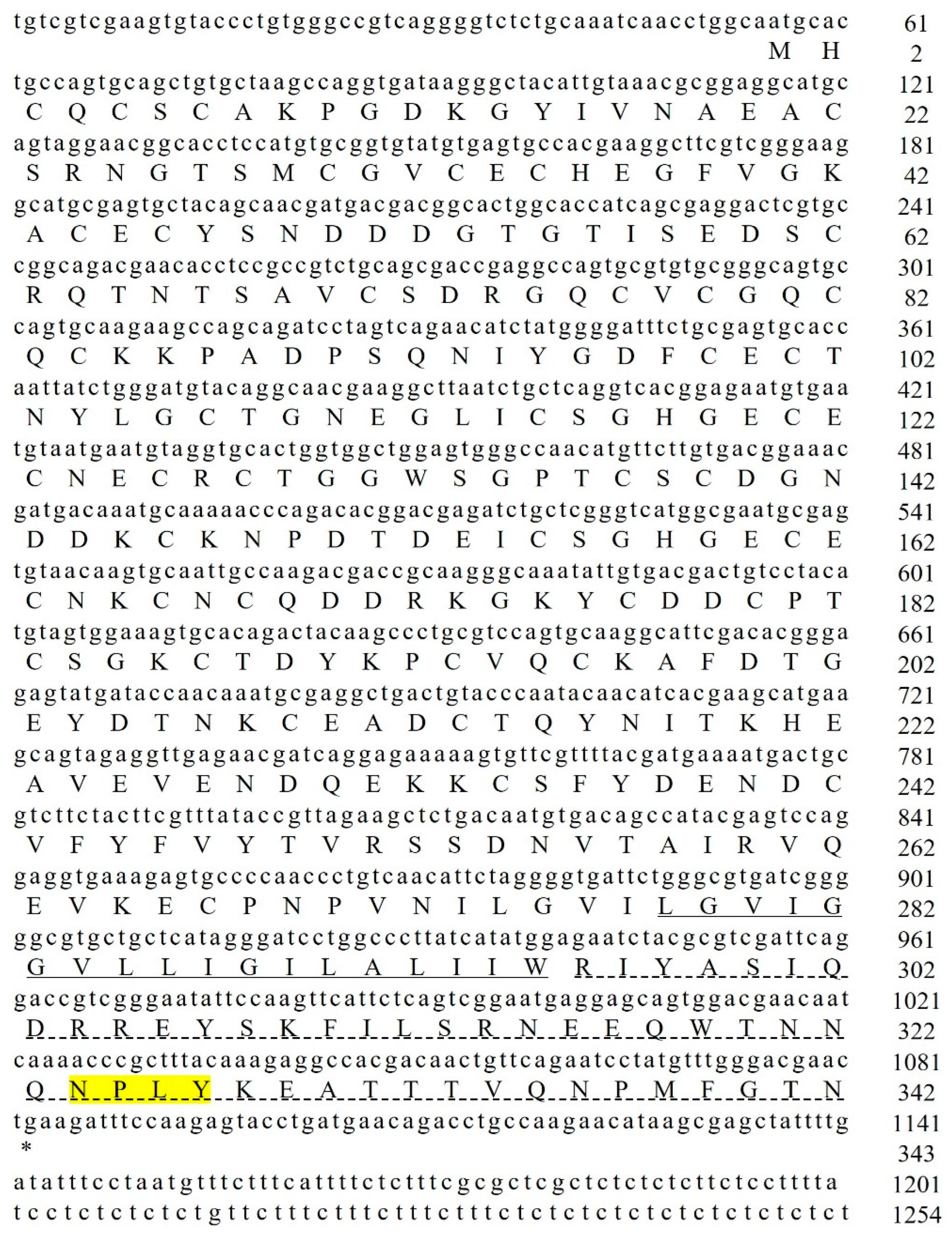
Figure 1. The cDNA sequence of Pv-Integrin β is shown in the figure, with lowercase letters representing the gene sequence and uppercase letters representing the amino acid sequence. It contains a conserved “NPxY” motif (yellow area), a transmembrane domain, and a cytoplasmic domain.
Ijms 26 08478 g001
Figure 2. The multiple sequence alignment analysis of the Pv-Integrin β molecule. The numbers on the right indicate the amino acid positions. The analysis reveals that this molecule contains a transmembrane domain and a cytoplasmic domain, with a conserved “NPxY” motif highlighted in a red box. In the alignment results, the different colors represent the conservation levels of amino acids: black indicates complete conservation, red indicates high conservation, and blue indicates moderate conservation.
Ijms 26 08478 g002
Figure 3. The phylogenetic tree of Pv-Integrin β with Integrin β molecules from both vertebrates and invertebrates. The Δ symbol represents the Integrin β molecule of P. vannamei. The neighbor-joining tree was constructed using MEGA 11.0 software, and the results were validated with 1000 bootstrap replicates to ensure reliability.
Ijms 26 08478 g003
Figure 4. The tissue distribution of the Pv-Integrin β gene in normal P. vannamei was detected using qRT-PCR technology, using 18 S rRNA as an internal control. Three parallel experiments were performed to ensure the reliability of the results (including three biological replicates). A histogram was used to display the results of the statistical analysis. The data were analyzed using the 2−ΔΔCT method.
Ijms 26 08478 g004
Figure 5. After infection with WSSV, the expression pattern of the Pv-Integrin β gene is as follows: (A) shows the expression level of the Pv-Integrin β gene in hemocytes, (B) shows the relative expression level in hepatopancreas tissue, (C) shows the relative expression level in gill tissue, and (D) shows the relative expression level in intestine tissue. The experiment was independently repeated three times and included three biological replicates. The data were analyzed using the 2−ΔΔCT method, and the results were compared using an unpaired two-tailed t-test. Statistical analysis of the data was conducted using an ANOVA (IBM SPSS Statistics 22, accessed on 27 September 2024). The results are expressed as mean ± standard deviation. Significant differences compared to the control group are indicated by asterisks; ** p < 0.01 indicates an extremely significant difference, and *** p < 0.001 indicates an exceptionally significant difference.
Ijms 26 08478 g005
Figure 6. The effective knockdown of the target gene and the increased mortality of shrimp after an injection of dsIntegrin β RNA: (AC) demonstrates the interference effect validation at 24, 36, and 48 h, while (D) shows a statistical analysis of shrimp survival rates. Compared to the control group (dsGFP + WSSV), the survival rate of the dsIntegrin β + WSSV group showed a significant decreasing trend. Statistical analysis of the data was conducted using an ANOVA. This survival rate-related statistical experiment was independently repeated three times. Significant differences compared to the control group are indicated by asterisks (** p < 0.01, *** p < 0.001).
Ijms 26 08478 g006
Figure 7. Graphs demonstrating that the knockdown of the target gene promotes WSSV replication. The injection of dsIntegrin β RNA, combined with WSSV, was used to validate its antiviral activity in vivo. The qRT-PCR results showed that compared to the control group (dsGFP + WSSV), knockdown of the target gene significantly upregulated the expression levels of IE 1 and vp 28 (A,B). The detection of WSSV viral load revealed that the WSSV load in the dsIntegrin β + WSSV group was significantly increased at 36 and 48 h (C). All experiments were independently repeated three times. The data were analyzed using the 2−ΔΔCT method, and the results were compared using an unpaired two-tailed t-test. Statistical analysis of the data was conducted using an ANOVA. Significant differences compared to the control group are indicated by asterisks; * p < 0.05 indicates a significant difference, ** p < 0.01 indicates an extremely significant difference, and *** p < 0.001 indicates an exceptionally significant difference.
Ijms 26 08478 g007
Figure 8. Integrin β regulates immune genes through the Toll, IMD, and STAT signaling pathways: (A) shows the Toll signaling pathway; (B) illustrates the IMD signaling pathway; (C) demonstrates the STAT signaling pathway; (D) presents the immune-related genes. The experiment was independently repeated three times and included three biological replicates. The data were analyzed using the 2−ΔΔCT method, and the results were compared using an unpaired two-tailed t-test. Statistical analysis of the data was conducted using an ANOVA. Compared with the control group, identical letters indicate no significant difference (p > 0.05), while different letters signify statistically significant differences between groups (p < 0.05).
Ijms 26 08478 g008aIjms 26 08478 g008b
Figure 9. This figure is a schematic diagram illustrating the immune pathway of P. vannamei in response to certain diseases regulated by Integrin β. After the knockdown of Integrin β, it was observed that viral replication increased, and the survival rate of the shrimp decreased. Further exploration of its molecular mechanism revealed that Integrin β might affect other receptors and, through regulating the Toll, IMD, and STAT signaling pathways, regulate the expression of antimicrobial peptides, thereby influencing the innate immune system of P. vannamei against diseases. This schematic diagram was drawn using the Figdraw 2.0 software (https://www.figdraw.com/static/index.html#/, accessed on 10 May 2025). Explanation: The cross mark in the pattern diagram represents knockdown.
Ijms 26 08478 g009
Table 1. The sequences of primers and probes used in this study.
Primer NameSequence (5′~3′)
Amplified sequence
Pv-Integrin-β-FGTCAGGGGTCTCTGCAAATCA
Pv-Integrin-β-RCAGGTCTGTTCATCAGGTACT
For the qRT-PCR assay
Integrin-β-FTGCAAGAAGCCAGCAGATCC
Integrin-β-RGAACATGTTGGCCCACTCCA
18 S-FTATACGCTAGTGGAGCTGGAA
18 S-RGGGGAGGTAGTGACGAAAAAT
VP 28-FTTCTTTCACTCTTTCGGTCGT
VP 28-RGCCAACTTCATCCTCATCAAT
IE 1-FTGGCACAACAACAGACCCTA
IE 1-RCTTTCCTTGAAGTACGAGAC
SOCS 2-FCTGCCAAACGCCCACTTC
SOCS 2-RCGTGGCAGGCATTGTGTG
MyD88-FGGCAAAGGGCTATTGGAACTAT
MyD88-RATGATCCAGACACCTCTCGTATTC
proPO-FTACATGCACCAGCAAATTATCG
proPO-RAGTTTGGGGAAGTAGCCGTC
ALF1-FTTACTTCAATGGCAGGATGTGG
ALF1-RGTCCTCCGTGATGAGATTACTCTG
ALF2-FGGCCATTGCGAACAAACTCAC
ALF2-RGTCCATCCTGGGCACCACAT
Crustin1-FGTAGGTGTTGGTGGTGGTTTC
Crustin1-RCTCGCAGCAGTAGGCTTGAC
Crustin2-FGGTACGTCTGCTGCAAGCC
Crustin2-RCTGAGAACCTGCCACGATGG
Domeless-FTCAGACAGGAGGTCTCATAC
Domeless-RGTACCAGTGTGAAGCCTTAC
JAK-FTACCCTGGTCTACGCTATAC
JAK-RTGAGACGGTAGTACCCATTC
Spätzle-FTGGGGACTCTCCTTACGATG
Spätzle-RGGGAACAGACAGGTCTCCAA
Dorsal-FGATGGAATGATAGAATGGGAAGC
Dorsal-RCACTGGTACTCTTGTCTGGTGGTC
Relish-FCTACATTCTGCCCTTGACTCTGG
Relish-RGGCTGGCAAGTCGTTCTCG
IMD-FCGGCTCTGCGGTTCACAT
IMD-RCCTCGACCTTGTCTCGTTCCT
STAT-FAGCCCCTGTCTGAGCGAAA
STAT-RGGTGTTCTCTTGTGACCTTCATCA
TLR-FGGGGTACCATGGTGTCAGGACTGGAGTCAGGCAACC
TLR-RTGCTCTAGATACACTTTTCGAGTTTGATTTAACAAG
Pathogen detection primers
WSSV-FTGGTCCCGTCCTCATCTCAG
WSSV-RGCTGCCTTGCCGGAAATTA
WSSV-probeAGCCATGAAGAATGCCGTCTATCACACA
For dsRNA synthesis
dsGFP-FiGCGTAATACGACTCACTATAGGCATCTTCTTCAAGGACGACGG
dsGFP-RiGCGTAATACGACTCACTATAGGAGTTCACCTTGATGCCGTTCT
Pv-Integrin-β-FiGCGTAATACGACTCACTATAGGACGACCGCAAGGGCAAAT
Pv-Integrin-β-RiGCGTAATACGACTCACTATAGGGACGCAGGGCTTGTAGTCTGT
Disclaimer/Publisher’s Note: The statements, opinions and data contained in all publications are solely those of the individual author(s) and contributor(s) and not of MDPI and/or the editor(s). MDPI and/or the editor(s) disclaim responsibility for any injury to people or property resulting from any ideas, methods, instructions or products referred to in the content.