Abstract
The exhaled fraction of nitric oxide (FeNO) is a biomarker of type 2 inflammation, reflecting the activity of inducible nitric oxide synthase (iNOS) in the bronchial epithelium in response to IL-4 and IL-13. Elevated FeNO levels support asthma diagnosis; however, it is unclear whether active helminth infections and rural environments influence this biomarker. The aim of this study was to compare FeNO levels among subjects naturally infected with helminth parasites and to evaluate their correlation with eosinophil counts and other inflammatory mediators. A total of 275 adult asthmatic patients and 161 healthy controls were involved; also, 223 asthmatic children and 114 healthy controls from the urban area of Cartagena were compared to 90 healthy children from a rural area. We found significant differences in FeNO levels between asthmatic patients and healthy controls in both adult and children’s cohorts (p < 0.0001). There was no difference in FeNO levels between Ascaris-positive and Ascaris-negative adults nor between subjects with active helminth infection and the non-infected. However, FeNO levels were significantly lower in rural healthy children (median 7.50 ppb, [IQR 4–14 ppb]) compared to urban healthy children (median 13.5 ppb, [IQR 10–18.5 ppb], p < 0.0001) and asthmatic children (median 20 ppb, [IQR 11–51 ppb], p < 0.0001). Rural healthy children had the highest total IgE levels (median 508 kU/L, [IQR 168–1020 kU/L]), high eosinophil counts (median 550 eos/μL, [IQR 360–800 eos/μL]) and plasma IL-5 levels (median 0.276 pg/mL, [IQR 0.19–0.53 pg/mL]). In conclusion, FeNO levels are not influenced by either natural exposure to helminth parasites or active infection, which supports its usefulness as a robust asthma biomarker in the tropics. Rural children have the lowest FeNO levels together with the highest total IgE levels, IL-5, and eosinophil counts, suggesting that lung-specific mechanisms are in place controlling iNOS expression during type 2 responses in healthy children.
Keywords:
exhaled fraction of nitric oxide; FeNO; helminth; asthma; infection; biomarkers; eosinophils; ascaris; rural; urban; IgE; CXCL1; CXCL9; SCF; type 2 inflammation 1. Introduction
Helminth infections, mainly by Ascaris lumbricoides, are endemic in tropical developing countries and induce an intense type 2 immune response with high production of immunoglobulin (IgE) antibodies, which resembles in several molecular and physiological aspects the type 2 inflammation involved in the mechanisms of allergy and asthma [1,2]. Type 2 response is implicated in the defence against helminths [3], and alarmins, type 2 innate lymphoid cells (ILC2) [4], T helper 2 (Th2) cells, mast cells, and eosinophils help to expel worms and promote strong immunity to avoid reinfection [5,6]. Pulmonary type 2 response has also been shown to decrease trafficking of Ascaris suum larvae in lung tissue [7]. However, chronic and intense helminth infections induce host immunoregulation, suppressing the capacity to react to antigens/allergens and reducing the susceptibility to chronic inflammatory diseases including asthma [8,9,10,11,12,13]. This immunosuppression due to helminth infection has a gradient, and is not as strong in light infections, normally found in urban populations of underdeveloped areas [12,14]. Indeed, a number of studies have demonstrated that mild helminth infections can increase the predisposition to wheezing [15], bronchial hyperreactivity [16], asthma [17,18], and IgE sensitisation to invertebrate pan allergens [14,19,20].
To date, it is unclear if helminth infections could influence the levels and the cut-offs for the interpretation of type 2 biomarkers in tropical regions. We previously found that active Ascaris lumbricoides infection (ascariasis) is associated with decreased H3 and H4 histone acetylation at the promoter of the IL-13 encoding gene (IL13) poising its expression [21]. The presence of specific IgE antibodies to this helminth was also associated with H4 acetylation levels at the promoter of the IL4 gene, encoding for IL-4 [21]. These cytokines are, however, difficult to measure in plasma, limiting their use as biomarkers. Instead, the exhaled fraction of nitric oxide (FeNO), IgE levels, and blood eosinophil counts are the most commonly used biomarkers of type 2 response and helpful to identify type 2 inflammation [22].
FeNO is a biomarker of type 2 inflammation that reflects the activity of the enzyme nitric oxide synthase (iNOS) in the bronchial epithelium in response to IL-4 and IL-13 [23]. FeNO levels above 25 parts per billion (ppb) in adults and 20 (ppb) in children are indicators of asthma [24] and have been included in recent guidelines for asthma diagnosis, at levels above 40 ppb in the GEMA guideline [25] and above 50 ppb in the GINA guideline (www.ginaasthma.org (accessed on 23 July 2025)). A previous study suggested that ascariasis was associated with elevated FeNO [26]. However, it is still unclear whether active helminth infection and rural environments influence FeNO levels in asthmatic children and adults. Helminth infections such as A. lumbricoides, which have a pulmonary phase in their life cycle [27], can cause a dry cough, wheezing, dyspnea, pulmonary infiltrates, and eosinophilia [28], and cause pneumonia and asthma-like symptoms in Loeffler syndrome [29]. Also, A. lumbricoides infection can modify plasma immune mediators and type 2 proteins [30] and cytokine levels [31]. For instance, periostin and chitinase-like protein YLK-40, the latter also known as chitinase 3 like 1 (CHI3L1), are recognised as asthma biomarkers in developed regions [32,33], but few studies have evaluated their levels in populations exposed to tropical parasites. As shown in a meta-analysis performed by Izuhara et al. [34], serum periostin levels correlate with blood eosinophil counts, FeNO, serum total IgE, and the response to anti IL-4/IL-13 therapies [33]. Moreover, an association between FeNO and periostin has been described [35,36]. YLK-40 is an acute phase protein associated with increased risk of asthma [32,37]. Mammalian chitinases and chitinase-like proteins are produced by neutrophils, monocytes, and macrophages in response to parasitic or fungal infections. Some studies have reported increased YKL-40 in children with Schistosoma haematobium infection [38] and changes in histone acetylation at the promoter of the YLK-40-encoding gene (CHI3L1) have been associated with specific IgE levels to Ascaris [21]. It is possible that helminth infections may affect the levels of these type 2 biomarkers or others plasma mediators.
The aims of this study were (1) to evaluate the relationship between FeNO and the presence of IgE antibodies against Ascaris, (2) to evaluate the effect of ascariasis on the levels of type 2 inflammation biomarkers in asthmatic patients, and (3) to evaluate the relationship of FeNO and eosinophil counts in asthmatic patients and healthy controls.
2. Results
2.1. FeNO Levels in Asthmatics with Positive Specific IgE to Ascaris
In an exploratory phase, FeNO levels were comparatively analysed in a total of 102 adult subjects, including 54 asthmatics and 48 healthy controls. FeNO levels were significantly higher in asthmatic patients compared to healthy controls (p < 0.0001), with a mean of 30 ppb [95%CI 22–38 ppb] in asthmatics and 14 ppb [95%CI, 12–16 ppb] in healthy subjects (Figure 1A). Specific IgE levels to Ascaris (p1 antigen, ImmunoCAP™) were also significantly higher in asthmatics (mean 1.8 kU/L [95%CI, 0.19–3.4 kU/L]) compared to controls (mean 0.13 kU/L [95%CI, 0.06–0.21 kU/L]) (Mann–Whitney p = 0.0005) (Figure 1B). There were 26 individuals with positive IgE to Ascaris (≥0.35 kU/L), and the frequency of positive individuals was higher in the asthma group (20.3%) compared to controls (n = 6, 12.5%), (p = 0.005). However, there was no difference in FeNO levels between asthmatics with positive IgE to Ascaris compared to those IgE-negative to Ascaris (p = 0.8; Figure 1C), even after adjusting for age and gender; also, there was no correlation between specific IgE levels and FeNO levels.

Figure 1.
Comparative FeNO levels between adult asthmatic patients and healthy controls (HC) depending on the positive IgE sensitisation to the nematode Ascaris (p1 antigen, ImmunoCAP™). (A) FeNO levels between asthma patients and healthy controls. (B) Specific IgE levels to Ascaris between asthmatic patients and healthy controls. (C) FeNO levels according to the presence of positive IgE sensitisation to Ascaris in adult asthmatics. ppb: parts per billion. p-values are shown for significant differences. **** p < 0.0001, *** p = 0.003.
2.2. The Relationship of FeNO Levels and Specific IgE to Ascaris with Plasma Mediators in Asthmatics
In the subgroup of 54 adult asthmatics, we analysed the correlation between FeNO levels and plasma levels of immune mediators (including periostin, YKL-40, and 67 other inflammatory proteins). We found that FeNO levels were inversely correlated with levels of caspase 8, extracellular newly identified receptor for advanced glycation end-products binding protein (EN-RAGE), and sirtuin 2 (Supplementary file S1, Table S1). Also, there was a direct association between FeNO levels and periostin levels that was significant after adjusting by age and gender (Supplementary file S1, Table S2).
We then analysed the levels of immune mediators between Ascaris-positive and Ascaris-negative patients, and we found no differences in periostin (Figure 2A) or YKL-40 (Figure 2B) levels. Interestingly, two plasma proteins, stem cell factor (SCF) and CXC motif chemokine ligand 1 (CXCL1), were found significantly decreased in Ascaris-positive asthmatics (Figure 2C,D) and these differences remained significant after adjustment by age and gender. Overall, there was no relationship between plasma mediators associated with FeNO levels and those associated with specific IgE to Ascaris (Supplementary file S1, Table S3).
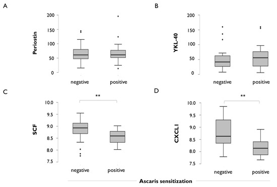
Figure 2.
Plasma levels of inflammatory mediators in adult asthma patients according to the positive IgE sensitisation to the nematode Ascaris. (A) Periostin, (B) chitinase-3-like protein 1 (YKL-40), (C) stem cell factor (SCF), (D) CXC motif chemokine ligand 1 (CXCL1). Protein levels are expressed in Normalised Protein eXpression values (NPX); error bars indicate mean and 95% confidence interval. ** p = 0.01. p-values are shown for significant differences.
2.3. FeNO Levels in Adult Asthmatics with Positive Specific IgE to Ascaris lumbricoides and in the Context of Active Infection
To further compare FeNO levels according to the presence of specific IgE to Ascaris lumbricoides, an independent cohort of 221 adult asthmatics and 113 healthy controls were analysed. FeNO levels were higher in asthmatic patients (mean, 52.8 ppb [95%CI, 46.7–58.9 ppb]) compared to healthy controls (mean, 17.3 ppb [95%CI 15.6–19.0 ppb]; Figure 3A), and there was no difference in FeNO levels between individuals with positive specific IgE to Ascaris lumbricoides in either the group of asthmatics or in healthy controls (Figure 3B). Since specific IgE reflects past exposure but not active infection, we then analysed the relationship between the presence of active Ascaris lumbricoides infection, as detected by stool analysis and FeNO levels. We only found 14 cases of active Ascaris lumbricoides infection in adults, specifically in seven asthmatics and seven controls, and we found that FeNO levels were not altered by active infection of light intensity (Figure 3C).
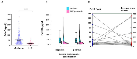
Figure 3.
FeNO levels in a replication cohort of adult asthmatic patients and healthy controls (HC). (A) FeNO levels between asthma patients and healthy controls, **** p < 0.0001. (B) FeNO levels according to the presence of positive IgE sensitisation to Ascaris lumbricoides stratifying by disease group. (C) FeNO levels in 14 subjects with active Ascaris lumbricoides infection. The line connects paired values for FeNO and eggs per gram (epg) of faeces in the same subject.
2.4. FeNO Levels in Asthmatics and Healthy Children with Active Ascaris lumbricoides Infection
We then compared FeNO levels in a paediatric cohort of asthmatic children and controls, and we observed significant differences in FeNO levels between asthmatic children (n = 223; median 20 ppb [IQR 11–51 ppb]) and healthy urban controls (n = 103; median 13.5 ppb [IQR 10–18.5 ppb]; Figure 4A). We also included a subset of rural healthy children to explore FeNO levels in this environment, in which helminth infections are more prevalent than in urban children. We found that FeNO levels were significantly lower in healthy rural children (n = 90; median 7.50 ppb [95%CI, IQR 4–14 ppb]) compared to urban healthy children or asthmatics (Figure 4A). In the asthmatic group, there were six children with active A. lumbricoides infection as diagnosed by stool test, and one of them was coinfected with Trichuris trichiura (138 epg of faeces). In the group of urban healthy children, we found nine infected with A. lumbricoides. Their FeNO values are presented in Table 1.
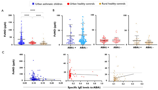
Figure 4.
FeNO levels in asthmatic children and healthy controls. (A) FeNO levels between asthmatic children and healthy controls from urban and rural environments, **** p < 0.0001. (B) FeNO levels according to the positive IgE sensitisation to the Ascaris specific antigen ABA-1. Error bars indicate mean and standard deviation (C) Correlation between FeNO levels and IgE levels to the Ascaris specific antigen ABA-1 (in optical density); The black straight line indicates the line of best fit for the linear regression of FeNO and specific IgE levels to ABA-1. The dotted gray line indicates the cutoff of normal FeNO values.

Table 1.
FeNO levels in urban children with active Ascaris infection.
In the rural children, we found 17 children infected by Trichuris trichiura, 2 coinfected with Ancylostoma duodenale, 2 with Hymenolepis nana, and 2 with Ascaris lumbricoides (Table 2). The low number of children diagnosed with an active infection precluded statistical comparison of FeNO levels between infected and non-infected subjects. However, we compared FeNO levels according to the presence of positive IgE to the nematode-specific antigen ABA-1. We found no differences in FeNO levels between ABA-1-positive and ABA-1-negative children (Figure 4B). Moreover, we found no significant correlation between FeNO and IgE levels to ABA-1 in any of the groups analysed (Figure 4C).

Table 2.
FeNO levels in rural children with active helminth infection.
2.5. Other Type 2 Biomarkers and Chemokines in Healthy Rural Children with Low FeNO Levels
We then compared total IgE levels and the numbers of peripheral blood eosinophil counts between groups. There was no significant difference in total IgE levels between asthmatic children (median, 245 kU/L IQR, 86–443 kU/L]) and urban healthy children (median, 292 kU/L [ IQR, 94–729 kU/L]; p = 0.35), but we found that total IgE was significantly higher in rural healthy children (median, 508 kU/L [IQR 168–1020 kU/L]) compared to asthmatics (p = 0.003) or urban healthy children (p = 0.03; Figure 5A). Regarding eosinophil cells counts, there was no difference between asthmatic children (median, 340 cells/μL [IQR 160–580 cells/μL]) and urban healthy children (median, 370 cells/μL [IQR 210–670 cells/μl]; p = 0.34), but those were significantly higher in rural healthy children (median, 600 cells/μL [IQR 410–897 cells/μL] compared to any of the other two groups (p < 0.0001), as shown in Figure 5B. Moreover, we found that plasma levels of IL-5 were significantly higher in urban healthy children (median, 0.24 pg/mL [IQR, 0.15–0.38 pg/mL]) compared to asthmatic children (median, 0.16 pg/mL [IQR 0.10–0.28 pg/mL]; p = 0.006), and even the highest in rural healthy children (median, 0.27 pg/mL [IQR 0.19–0.53 pg/mL]) compared to asthmatic children (p < 0.0001; Figure 5C), but there was no difference in IL-5 levels between healthy children from urban and rural environments as shown in Figure 5C. When we analysed chemokine levels, we found increased levels of CXCL9/MIG, a bioactive form of interferon gamma, in rural healthy children (median, 165 pg/mL [IQR, 111–262 pg/mL]) compared to asthmatic children (median, 100 pg/mL [IQR 69–166 pg/mL]; p = 0.0001; Figure 5D). CXCL9/MIG levels were also higher in healthy urban children than in asthmatic children, but this difference was not statistically significant (p = 0.062).
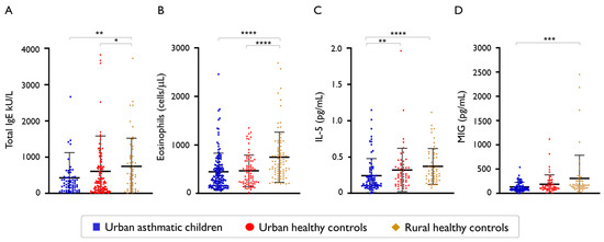
Figure 5.
Type 2 inflammation biomarkers between asthmatic children and healthy controls from urban and rural environments. (A) Total IgE levels * p = 0.0361, ** p = 0.0032, (B) peripheral blood eosinophil counts **** p < 0.0001, (C) plasma IL-5 levels ** p = 0.0060, **** p < 0.0001, (D) Monokine Induced by Interferon-gamma (MIG) levels *** p = 0.0001. Error bars indicate mean and standard deviation.
2.6. The Relationship of FeNO Levels with Blood Eosinophil Counts
We found significant positive correlation between FeNO levels and peripheral blood eosinophil counts in asthmatic children (Spearman rho = 0.49, p < 0.0001). However, FeNO levels were not correlated with blood eosinophil counts in healthy children from urban or rural environments (Spearman rho = −0.02, p = 0.80 in urban children and Spearman rho = −0–04, p = 0.69 in rural children) (Figure 6).
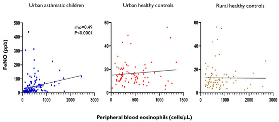
Figure 6.
Spearman correlation between FeNO levels and blood eosinophil counts in asthmatic children and healthy controls from urban and rural environments. Each dot represents a child, coefficients and p-values are only shown for significant correlations. The black straight line indicates the line of best fit for the linear regression of FeNO levels and blood eosinophil counts.
In adult asthmatic patients, we also confirmed significant correlation between FeNO levels and blood eosinophil counts in both cohorts (Figure 7), and there was no correlation between FeNO levels and blood eosinophil counts in adult healthy controls.
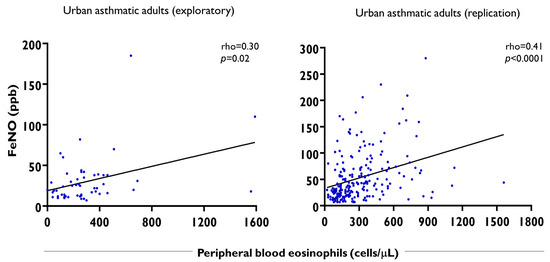
Figure 7.
Spearman correlation between FeNO levels and peripheral blood eosinophil counts in adult asthmatics from two cohorts. Each dot represents an asthmatic patient. Eos/μL: peripheral blood eosinophil counts per microliter of blood. The black straight line indicates the line of best fit for the linear regression of FeNO levels and blood eosinophil counts.
To investigate further the correlation between FeNO and blood eosinophils only in asthmatic subjects and not in healthy subjects, we analysed IL-5 levels in induced sputum samples of adult asthmatics and healthy controls. We found that asthmatics have significant increased levels of IL-5 in sputum (mean 2.3 [95%CI 0.37–8.6]) compared to healthy subjects (mean 0.02, [95%CI 0.0–0.28], p = 0.004). IL-5 levels in plasma were also higher in adult asthmatic patients (mean 0.11 [95%CI 0.04–0.19]) compared to healthy controls (mean 0.012, [95%CI 0.0- 0.06], p = 0.039) (Figure 8); remarkably, IL-5 levels in sputum were higher than those detected in plasma.
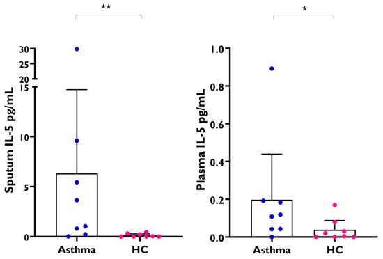
Figure 8.
IL-5 levels in induced sputum samples and paired plasma samples from adult asthmatic patients (n = 8) and healthy controls (HC, n = 8). Each dot represents an individual, error bars indicate mean and 95% confidence interval. (Mann–Whitney test sputum IL-5, asthma vs. HC, ** p = 0.0042) (Mann–Whitney test plasma IL-5, asthma vs. HC, * p = 0.039).
3. Discussion
In this study, we report the levels of FeNO in adult asthmatic patients from an urban tropical area, and in a paediatric cohort of asthmatics and healthy children living in an urban or rural environment. We show that FeNO levels are significantly increased in asthmatic patients living in tropical areas and can be used as a robust asthma biomarker in our region, and that FeNO levels are not affected by past or active helminth infection by Ascaris lumbricoides. We also confirmed positive correlations of FeNO with blood eosinophil counts and FeNO with periostin levels in asthmatic patients [35,36].
Based on previous studies suggesting that ascariasis can influence asthma [26,39], we analysed the relationship between A. lumbricoides exposure, FeNO levels, and plasma immune mediators, finding that FeNO did not differ between Ascaris-positive and Ascaris-negative asthmatics, suggesting that it is not influenced by Ascaris sensitisation in adult patients. The levels of other type 2 biomarkers, such as periostin and YKL-40, also did not differ according to the presence of Ascaris sensitisation. However, lower plasma levels of SCF and CXCL1 were observed in Ascaris-positive asthmatics, suggesting that previous infection with this nematode can influence plasma levels of some immune mediators. These differences were still significant after adjusting for covariates.
SCF is a cytokine produced by several cell types and triggers its biological effects by binding to its receptor c-Kit, a member of the tyrosine kinase receptor family [40]. It plays a crucial role in allergic inflammation, particularly in the context of airway hyperreactivity and eosinophil recruitment [41,42]. It acts as a cytokine that promotes mast cell survival, activation, and differentiation, which in turn can lead to increased inflammation and allergic symptoms [43]. SCF also directly interacts with eosinophils, causing their activation and degranulation, leading to the release of inflammatory mediators and chemokines [44]. SCF is also implicated in the protective immune response against intestinal helminth infections [45]. SCF contributes to mucosal mast cell hyperplasia during nematode infection [40,46]. It has also been shown that administration of an anti-SCF polyclonal antibody completely eliminated the mucosal mast cell population in a rat model [46], impairing worm expulsion in animals infected with T. spiralis [47,48]. Other reports suggest that SCF can reduce type 2 inflammation [49]. Further studies are needed to elucidate the mechanisms of how Ascaris exposure reduces SCF levels in human plasma and if this is a consequence of host immunomodulation by this helminth.
We also observed reduced levels of CXCL1 in Ascaris-sensitised asthmatics. Chemokines are involved in type 2 inflammation in allergic diseases [50]. Indeed, during allergy and anti-helminth immunity, epithelial cells of damaged barriers release alarmins including IL-25, IL-33, and thymic stromal lymphopoietin (TSLP), but also chemokines such as CXCL1 and CCL11, to promote cell recruitment and inflammation [51]. CXCL1 is involved in airway inflammation, and its presence in the airways of asthmatic patients and animal models of allergy has been linked to increased levels of IL-17 and neutrophil activation [52,53]. In the context of infection, CXCL1 acts as a chemotactic signal, attracting neutrophils to the area where parasites are present [54]. The decrease in CXCL1 in asthma leads to the increased recruitment of mast cells to the airways [55]. In this study, we found decreased levels of SCF and CXCL1 in asthma patients with positive IgE to Ascaris. More studies are needed to understand how the reduction in SCF and CXCL1 observed in our patients occurred by natural exposure to Ascaris and its implications in their immune response [56].
We then confirmed that FeNO levels are higher in asthmatics than in healthy controls in an independent adult sample set. In addition, we replicated our observation made in the exploratory cohort that positive IgE to Ascaris lumbricoides does not influence FeNO levels. Since positive IgE reflects parasite exposure but not necessarily active infection, we also analysed FeNO levels in subjects with worm eggs in faeces. The presence of active infection by the nematode Ascaris lumbricoides did not increase FeNO levels (Figure 3C and Table 1 and Table 2).
We then analysed FeNO levels and type 2 biomarkers in a paediatric cohort, in which stool samples were collected and analyses for parasitic infection were performed. We confirmed that FeNO levels were higher in asthmatic children and that FeNO levels were not influenced by natural infection by helminth parasites in children, which supports its usefulness as a robust asthma biomarker in the tropics. There were also other types of parasites detected, such as Trichuris trichiura, which also did not affect FeNO levels. Collectively, it might suggest that even though these nematodes induce a systemic type 2 immune response, those are not sufficient to generate a local response in the lung capable of altering FeNO levels. We focused mainly on Ascaris lumbricoides because it undergoes a migratory phase through the lungs in its life cycle [27], during which it induces strong tissue eosinophilic inflammation. This infection may cause cough and wheezing and is known as Loeffler syndrome [29]. In the rural cohort, the infections with Trichuris trichiura were more common than those with Ascaris lumbricoides, and we could observe that FeNO levels were not affected. Unlike Ascaris lumbricoides, Trichuris trichiura does not undergo a pulmonary phase [57]. Moreover, it should be considered that the numbers of eggs per gram of faeces in our samples were low, indicating infections of light intensity. We cannot rule out that, in the case of heavy intensity infection (>50,000 eggs), the response may differ, as was found in a work performed in a small village of Colombia [39].
Depending on intensity of the infection and type of helminth [58], some helminthiasis can downregulate different immune pathways [59], being helminths with life stages in different tissues and/or solid organs (i.e., Schistosoma ssp., Ancylostoma duodenale, Trichinella ssp.) very suppressive [60,61]. Ascaris lumbricoides also have several immunomodulatory molecules in their body fluids [62,63,64] and can modulate the immune responses [8,30,39,65]. Also, early heavy infections with T. trichiura may protect against the development of allergen skin test reactivity in later childhood [66].
We also confirmed no difference in FeNO levels between children with positive IgE levels to ABA-1 and the negative controls in any of the analysed groups. Interestingly, rural children had the lowest FeNO levels while, at the same time, they had the highest total IgE levels, blood eosinophil counts, and plasma IL-5 levels, suggesting that lung-specific mechanisms are in place that control lung iNOS expression during systemic type 2 response in healthy children. The elevated production of MIG/CXCL9, a bioactive form of interferon gamma, in rural children may play a role in their immune homeostasis. The increased levels of IL-5 in rural children, in whom helminth infections are more frequent, reflects the importance of this cytokine in helminth response. However, in the context of a respiratory disease such as asthma, bronchial IL-5 levels increase and blood levels do not reflect that found in the airways. Although several factors may influence immune responses in rural children, the apparent dissociation of high systemic type 2 markers (IgE, eosinophils, IL-5) but the lowest FeNO can be explained; first by helminth infections that favour local lung regulatory mechanisms [67] and downregulation of the IL-4/IL-13 pathway [68,69] and thereby iNOS, together with protozoa coinfections that boost type 1 and type 17 responses. Moreover, helminth infections are known to modify arginase activity in peripheral blood mononuclear cells and plasma [70] and may affect availability of L-arginine needed by iNOS to produce nitric oxide.
The comparison of IL-5 levels in plasma and induced sputum samples revealed that the degree and intensity of IL-5 response differs between plasma and the airways, being below 1 pg/mL in plasma, in the setting of natural exposure to helminths but much higher in the airway as measured in the induced sputum of adult asthmatic patients. Although type 2 responses, as reflected by higher IL-5 in plasma, were even stronger in healthy rural children compared to urban healthy or asthmatic children, the measured plasma IL-5 levels were much lower than those observed in the sputum of adult asthmatics. Positive correlations between blood eosinophils counts and FeNO levels, observed in all cohorts of asthmatics but not in healthy controls, illustrates the dysregulation of IL-5 in asthma [71] and the relevance of this pathway in asthma therapy [72]. These findings also indicate that, in addition to analysing peripheral blood eosinophils counts, future studies are needed to elucidate their cell surface markers and subtypes, identifying those that are relevant in helminth infections [73] or in disease states like asthma. The magnitude of correlation between FeNO levels and blood eosinophil counts also reflects that these two markers although directly related, are providing information on two different type 2 inflammation pathways.
It also important to consider the species-specific effects that each helminth may exert on asthma. It has been reported that Trichuris trichiura infections during the first 5 years of life were associated with reduced wheeze, while Ascaris lumbricoides infestations at 5 years of age were related to increased wheeze and asthma [26]. Recent meta-analyses suggest that Ascaris can increase the risk of bronchial hyperreactivity [16], whereas other studies suggest that helminth effects on asthma and wheeze can be protective [74]. Thus, the data are conflicting. In this study, we confirmed that natural exposure to Ascaris lumbricoides, even though it undergoes a pulmonary migration phase, does not affect FeNO levels in humans, and that there is a dissociation between type 2 responses occurring during natural helminth exposure not accompanied by asthma and type 2 inflammation during asthma.
4. Materials and Methods
4.1. Adult Cohorts
We included two adult cohorts of asthmatic patients and healthy controls. The first and exploratory dataset comprised 102 individuals from Cartagena (Colombia), including 54 adult asthma patients and 48 healthy controls. This subset of patients was recruited in a specialised pulmonary clinic and diagnosed by a pulmonologist [75] following the GEMA criteria and pulmonary function tests [25]. The second and independent dataset consisting of 221 adult asthmatics and 113 healthy controls from the “Study on the Pathogenesis of Asthma in the Tropics (EPAT)”, with data on stool analysis and IgE levels to A. lumbricoides, was used as a replication cohort. All participants signed a written informed consent to participate in the study. This study was approved by the Ethics Committee of the University of Cartagena (No. 416-9722017 and No. 128-14112019).
4.2. Children Cohort
4.2.1. Urban Children
A total of 338 urban children were included, including 223 asthmatics and 114 healthy controls. Asthmatic children were recruited in a specialised local clinic in which the diagnosis of asthma was performed by an allergologist or pulmonologist. Children and their parents were reinterviewed and examined by a physician of the research staff. Demographic and medical information was documented by questionaries. The healthy children were recruited in local schools and neighbourhoods. Mothers were questioned about health conditions, familiar history of disease, and exposures. Parents or legal guardians signed a written informed consent to participate.
4.2.2. Rural Children
Samples from rural children were collected by random sampling in a rural area of Santa Catalina, Colorado, and Loma Arena, three small villages of the department of Bolívar, contacting approximately 100 children. Of these, 90 children were eligible with stool sample to detect active parasite infections and assess FeNO levels. A subset of 55 children with available blood samples were included for protein plasma measurements. The children were 6–14 years old, so they were capable of performing the FeNO measurement. This group was classified into rural children with a parasitic infection diagnosed by stool examination (infected group) and rural children with a negative stool examination and no clinical signs of intestinal parasitosis (non-infected group). Parents or legal guardians signed a written informed consent to participate.
4.3. Fractional Exhaled Nitric Oxide (FeNO)
FeNO measurement was performed by a researcher trained in this procedure, using the portable NObreath® device v2. (Bedfont Scientific Ltd, Kent, UK). Three measurements were taken per patient at a flow rate of 50 mL/s, monitored by a visible airflow metre and following the recommendations of the American Thoracic Society [24]. Results were expressed in parts per billion (ppb). Prior to measuring FeNO, each individual was questioned about recent cigarette smoking, complete fasting, and whether they had had respiratory infections in the past 2 months. FeNO measurements for this study were recorded for those not having smoked cigarettes in the previous 48 h, having fasted for at least 12 h, not having consumed alcohol, or reported no symptoms of respiratory infections in the past 2 months. FeNO levels are considered normal below <25 ppb in adults and <20 ppb in children [24]. According to the GINA guideline, FeNO values above 50 ppb in adults and 35 ppb in children support the diagnosis of type 2 asthma (Global Initiative for Asthma. Global Strategy for Asthma Management and Prevention 2025. Updated May 2025, available from: www.ginaasthma.org (accessed on 23 July 2025)).
4.4. Blood Sample Collection
Blood samples were collected in EDTA tubes by antecubital venipuncture according to standardised protocols. A tube was used to measure white blood cell counts (WBC) including eosinophils in an automated haematology analyser (Advia 2120i, Siemens Healthcare Diagnostics Inc., Tarrytown, NY, USA), and another tube was centrifuged at 1000 rpm and 4 °C to obtain plasma. This was carefully separated with sterile micropipette tips, aliquoted into sterile tubes, and stored at −70 °C until use.
4.5. Stool Samples
Stool samples were collected by children’s mothers in a sterile plastic provided by the researchers and analysed using 0.85% saline solution and Lugol’s stain. If parasites were found, egg quantification was performed using the Kato–Katz method using a commercial kit (Copro Kit, C&M Medical, Campinas, Brazil). The helminth load was expressed as eggs per gram (epg) of faeces. The presence of protozoa, helminth eggs, or adult parasites was diagnostic of active infection (positive/negative). A. lumbricoides infection was classified as light intensity (1–4999 eggs), moderate intensity (5000–49,999), and heavy intensity (>50,000 eggs), following criteria of the World Health organisation (WHO) [76].
4.6. Induced Sputum Samples
Sputum was collected from 16 subjects (8 asthmatics and 8 healthy controls) using nebulised 4% saline solution and dissolved using 10% DTT (SPUTOLYSIN®®, EMD Millipore Corp, Burlington, MA, USA). After homogenisation for 15 min at 37 °C samples were centrifuged at 400× g for 10 min at 4 °C, and supernatant was collected in sterile tubes and stored at −70 °C until cytokine measurement, as described previously [77,78].
4.7. IgE Measurements
Total IgE levels were measured by ImmunoCAP™ (Thermo Fisher, Uppsala, Sweden) in all the participants; a calibration curve was used with a range of 2 to 5000 kU/L (2, 10, 50, 200, 1000, 5000 kU/L). Specific IgE levels to Ascaris spp. (p1) were measured in the adult cohort by ImmunoCAP™, a calibration curve of 0.35 to 100 kUA/L was used (0.35, 0.7, 3.5, 17.5, 50, 100 kUA/L). A cut-off of 0.35 kUA/L was used to consider a positive IgE sensitisation to Ascaris. Specific IgE levels to A. lumbricoides and the nematode specific marker ABA-1 were quantified using enzyme-linked immunosorbent assay (ELISA) in the EPAT cohort and the children cohort. Antigens were diluted in PBS and 64 mM NaHCO3-Na2CO3 buffer (pH 9.6). Each well was coated with 1μg of antigen, incubated overnight at 4 °C, washed five times with 0.1% PBS-Tween 20, and then blocked for 3 h using 1% Bovine Serum Albumin (BSA) in humid chamber at room temperature. After five washes, serum samples (diluted 1:5) were added to each well and incubated overnight at 4 °C in humid chamber. After five washes, plates were incubated for 2 h with antihuman IgE–alkaline phosphatase conjugate (Sigma-Aldrich Co., St. Louis, MO, USA) at room temperature. The assay was developed with para-nitrophenyl phosphate (1 mg/mL, Sigma-Aldrich, Co., St. Louis, MO, USA) diluted in 10% diethanolamine (pH 9.8) and the plate incubated in the dark at room temperature for 30 min. The reaction was stopped with 100 μl of 3 N NaOH per well and absorbance measured at 405 nm using a spectrophotometer (MultiScan Go, Thermo Fisher Scientific, Vantaa, Finland). Determinations were made in duplicate, and IgE levels above 0.113 optical density (mean OD of 6 negative, non-allergic, non-parasite-exposed controls + 3 SD) were considered positive for the presence of specific IgE to Ascaris or ABA-1.
4.8. Periostin and YKL-40 Measurements
Plasma periostin/OSF-2 levels were measured using a sandwich enzyme-linked immunosorbent assay (ELISA) with a capture antibody in the solid phase (Cat. 844441, R&D Systems, Inc, Minneapolis, MN, USA) and detection antibodies (Cat. 844442, R&D Systems, Inc, Minneapolis, MN, USA), using a DuoSet®® 2 kit (R&D Systems, Inc, Minneapolis, MN, USA, Cat. DY008) as previously described [75]. YKL-40 measurement was performed with a commercially ELISA Duoset kit (Cat. DY2599 R&D Systems, Minneapolis, MN, USA) following the manufacturer’s instructions and the levels were expressed in pg/mL.
4.9. Proximity Extension Assay for Plasma Protein Levels
For plasma profiling in the adult cohort, samples were randomly distributed in 96-well plates, and protein levels were measured by Proximity Extension Assay (PEA) using the Target 96 Inflammation Panel (Olink Proteomics, Analysis Service Facility, Boston, USA), which includes a broad selection of proteins established as inflammatory signatures in diverse inflammatory diseases. A total of 67 out of 92 plasma molecules were detected in heparinized plasma (73%). Normalised Protein Levels are expressed as NPX units (log2 scale). The intra- and inter-assay average coefficients of variation (%CV) were 6% and 11%, respectively.
4.10. Cytokine Bead Array
In the children cohort, plasma IL-5 and chemokine levels (IL-8, MCP-1, IP-10, MIG, RANTES) were measured by flow cytometry using the Cytokine Bead Array (CBA). IL-5 measurements were performed with the BDTM CBA Human IL-5 Flex Set (BD Biosciences, San Diego, CA, USA). In brief, mixed capture beads were incubated with human plasma and standards for 1 h at room temperature, then the human IL-5 PE detection reagent was added and incubated for 2 h at room temperature in the dark, beads were washed and acquired. For chemokines, the BDTM CBA Human Chemokine Kit (BD Biosciences, San Diego, CA, USA) was used. In brief, five bead populations were mixed together to form an array that is resolved in a red channel (FL3-FL4) with different fluorescence intensities ranging from the dimmest to the brightest. Recombinant standards or human plasma samples were incubated with capture beads and the PE-conjugated detection antibodies for 3 h at room temperature in the dark. Wash buffer was added and samples centrifuged at 200× g for 5 min; afterwards, supernatant was carefully removed and the beads resuspended in wash buffer. Samples were acquired in a BD FACSAriaTM III cell sorter (BD Biosciences, San Jose, CA, USA), with reporter channel in PE and bead channels in APC. The intensity of PE fluorescence of each sandwich complex was compared to a standard curve from 0 to 2500 pg/mL, with the four-parameter curve fit option to extrapolate values with the concentration of each chemokine. Theoretical limits of detection were 0.2 pg/mL for IL-8, 1 pg/mL for RANTES, 2.5 pg/mL for MIG/CXCL9, 2.7 pg/mL for MCP-1 and 2.8 pg/mL for IP-10, and 1.1 pg/mL for IL-5. The FCAP ArrayTM software (v3.0.1) was used to generate results in concentrations derived from the standard curve.
4.11. Statistical Analysis
Qualitative variables were compared using chi2 test. Normality of the distribution of the qualitative variables was tested the Shapiro–Wilk method. Quantitative variables are given as means with 95% confidence interval (95%CI) or medians with interquartile ranges, depending on the distribution observed. Comparisons of qualitative variables between two groups were performed using the Mann–Whitney U-test. Correlations were analysed using the Spearman coefficient. Linear regression was used to model the relationship between FeNO levels, mediator levels, and specific IgE levels, adjusting by age and gender. A p value ≤ 0.05 was considered statistically significant. Statistical analysis was performed using GraphPad Prism (v8.0.1) and JASP (v0.19.3).
5. Conclusions
In conclusion, FeNO levels were significantly higher in asthmatic patients compared to healthy controls, and there were no differences in FeNO levels between Ascaris-positive and Ascaris-negative controls. FeNO levels were not increased in adults or children with active infection by Ascaris lumbricoides. In this study, we observed for the first time lower levels of SCF and CXCL1 in asthmatic patients with positive IgE levels to Ascaris. Under type 2 response, plasma IL-5 levels, and eosinophils were significantly higher in rural children than in asthmatic children, showing a coexistence of increased type 2 biomarkers in peripheral blood circulation with the lowest FeNO levels. Rural controls also showed the highest total IgE levels. The increase in circulating type 2 biomarkers in rural children coincided with increased CXCL9 levels. Considering high FeNO levels and the fact that IL-5 is detectable at much high concentration in the induced sputum of asthmatics, one might hypothesize that divergent control mechanisms of the type 2 responses in health and disease exists, with very strong type 2 responses in the lung of allergic asthmatics, and only a subtle fine-tuning of this local, pulmonary production of type 2 mediators by helminth infections otherwise associated with systemically enhanced type 2 responses. FeNO levels and peripheral blood eosinophils are positively correlated biomarkers, and both important as indicators of type 2 inflammation, however, FeNO is more robust to confirm lung type 2 inflammation in asthma, because blood eosinophil counts can be found increased in healthy children of the tropics (and especially those in rural environments).
Supplementary Materials
The following supporting information can be downloaded at: https://www.mdpi.com/article/10.3390/ijms26178344/s1.
Author Contributions
Conceptualisation, M.M.D.V. and N.A.; methodology, M.M.D.V., R.R., J.R., J.Z., J.M.E.G., B.Z., L.T.F.d.A., D.P.P. and N.A.; software, M.M.D.V., J.Z. and N.A.; validation, M.M.D.V., R.R., J.R., J.M.E.G., L.T.F.d.A. and J.Z.; formal analysis, M.M.D.V., R.R., J.R., J.Z. and N.A.; investigation, M.M.D.V., R.R., J.R., J.Z., J.M.E.G., B.Z. and L.T.F.d.A.; resources, J.Z., D.P.P., L.C. and N.A.; data curation, M.M.D.V., R.R., J.R. and J.M.E.G.; writing—original draft preparation, M.M.D.V.; writing—review and editing, M.M.D.V., J.Z., D.P.P., N.A. and L.C., all authors.; visualisation, M.M.D.V., R.R. and N.A.; supervision, N.A. and L.C.; project administration, N.A. and L.C.; funding acquisition, N.A. and L.C. All authors have read and agreed to the published version of the manuscript.
Funding
This study was funded by the Ministry of Science of the Republic of Colombia (Grant 756-2017), Sistema General de Regalías (BPIN20200000100405) and the University of Cartagena. María De Vivero received funding as doctoral student from the Ministry of Science (Programa de Becas de Excelencia Doctoral del Bicentenario BB 2019-01, Res. 00699 de 2020 de la UdC).
Institutional Review Board Statement
The study was conducted in accordance with the Declaration of Helsinki, and approved by the Institutional Ethics Committee of the University of Cartagena (No. 416-9722017 2 June 2017 and No. 128-14112019 14 November 2019).
Informed Consent Statement
Written informed consent was obtained from all subjects involved in the study.
Data Availability Statement
The raw data supporting the conclusions of this article will be made available by the authors on request.
Acknowledgments
We thank Mileidis Avila for her support during blood sampling, Karen Donado and Sandra Correa for their support with stool examinations, and Victoria Marrugo and Dilia Mercado for their support with ELISA measurements for Ascaris specific IgE.
Conflicts of Interest
The authors declare that they have no conflicts of interest with relation to this work.
Abbreviations
The following abbreviations are used in this manuscript:
| CI | Confidence interval |
| CXCL1 | C-X-C motif chemokine ligand 1 |
| CXCL9 | C-X-C motif chemokine ligand 9 |
| epg | Eggs per gram |
| FeNO | Exhaled fraction of nitric oxide |
| GINA | Global Initiative for Asthma |
| HC | Healthy control |
| IL-5 | Interleukin 5 |
| iNOS | Inducible nitric oxide synthase |
| IQR | Interquartile range |
| ppb | Parts per billion |
| SCF | Stem cell factor |
References
- Vacca, F.; Le Gros, G. Tissue-specific immunity in helminth infections. Mucosal Immunol. 2022, 15, 1212–1223. [Google Scholar] [CrossRef] [PubMed]
- Gao, Y.D.; Wang, Z.J.; Ogulur, I.; Li, S.J.; Yazici, D.; Li, X.H.; Pat, Y.; Zheng, Y.; Babayev, H.; Zeyneloglu, C.; et al. Type 2 Immunity and Its Role in Allergic Disorders. Allergy 2025, 80, 1848–1877. [Google Scholar] [CrossRef]
- Maizels, R.M.; Gause, W.C. Targeting helminths: The expanding world of type 2 immune effector mechanisms. J. Exp. Med. 2023, 220, e20221381. [Google Scholar] [CrossRef]
- Can, U.I.; Stenske, S.E.; Rosenbaum, M.D.; Reinhardt, R.L. Rapid group-2 innate lymphoid cell mobilization from the intestine aids in early lung defense and repair. Cell Rep. 2025, 44, 115868. [Google Scholar] [CrossRef] [PubMed]
- Henry, E.K.; Inclan-Rico, J.M.; Siracusa, M.C. Type 2 cytokine responses: Regulating immunity to helminth parasites and allergic inflammation. Curr. Pharmacol. Rep. 2017, 3, 346–359. [Google Scholar] [CrossRef]
- Loser, S.; Smith, K.A.; Maizels, R.M. Innate Lymphoid Cells in Helminth Infections-Obligatory or Accessory? Front. Immunol. 2019, 10, 620. [Google Scholar] [CrossRef]
- Gazzinelli-Guimaraes, P.H.; Golec, D.P.; Karmele, E.P.; Sciurba, J.; Bara-Garcia, P.; Hill, T.; Kang, B.; Bennuru, S.; Schwartzberg, P.L.; Nutman, T.B. Eosinophil trafficking in allergen-mediated pulmonary inflammation relies on IL-13-driven CCL-11 and CCL-24 production by tissue fibroblasts and myeloid cells. J. Allergy Clin. Immunol. Glob. 2023, 2, 100131. [Google Scholar] [CrossRef]
- Zakzuk, J.; Lopez, J.F.; Akdis, C.; Caraballo, L.; Akdis, M.; van de Veen, W. Human Ascaris infection is associated with higher frequencies of IL-10 producing B cells. PLoS Neglected Trop. Dis. 2024, 18, e0012520. [Google Scholar] [CrossRef]
- Cooper, P.J. Can intestinal helminth infections (geohelminths) affect the development and expression of asthma and allergic disease? Clin. Exp. Immunol. 2002, 128, 398–404. [Google Scholar] [CrossRef] [PubMed]
- Bohnacker, S.; Troisi, F.; de Los Reyes Jimenez, M.; Esser-von Bieren, J. What Can Parasites Tell Us About the Pathogenesis and Treatment of Asthma and Allergic Diseases. Front. Immunol. 2020, 11, 2106. [Google Scholar] [CrossRef] [PubMed]
- Maizels, R.M. Infections and allergy-helminths, hygiene and host immune regulation. Curr. Opin. Immunol. 2005, 17, 656–661. [Google Scholar] [CrossRef]
- Smits, H.H.; Everts, B.; Hartgers, F.C.; Yazdanbakhsh, M. Chronic helminth infections protect against allergic diseases by active regulatory processes. Curr. Allergy Asthma Rep. 2010, 10, 3–12. [Google Scholar] [CrossRef]
- Danilowicz-Luebert, E.; O’Regan, N.L.; Steinfelder, S.; Hartmann, S. Modulation of specific and allergy-related immune responses by helminths. J. Biomed. Biotechnol. 2011, 2011, 821578. [Google Scholar] [CrossRef]
- Caraballo, L.; Llinas-Caballero, K. The Relationship of Parasite Allergens to Allergic Diseases. Curr. Allergy Asthma Rep. 2023, 23, 363–373. [Google Scholar] [CrossRef]
- Avokpaho, E.; Gineau, L.; Sabbagh, A.; Atindegla, E.; Fiogbe, A.; Galagan, S.; Ibikounle, M.; Massougbodji, A.; Walson, J.L.; Luty, A.J.F.; et al. Multiple overlapping risk factors for childhood wheeze among children in Benin. Eur. J. Med. Res. 2022, 27, 304. [Google Scholar] [CrossRef]
- Arrais, M.; Maricoto, T.; Nwaru, B.I.; Cooper, P.J.; Gama, J.M.R.; Brito, M.; Taborda-Barata, L. Helminth infections and allergic diseases: Systematic review and meta-analysis of the global literature. J. Allergy Clin. Immunol. 2022, 149, 2139–2152. [Google Scholar] [CrossRef]
- Mkhize-Kwitshana, Z.L.; Naidoo, P.; Nkwanyana, N.M.; Mabaso, M.L.H. Concurrent allergy and helminthiasis in underprivileged urban South African adults previously residing in rural areas. Parasite Immunol. 2022, 44, e12913. [Google Scholar] [CrossRef]
- Takeuchi, H.; Takanashi, S.; Hasan, S.M.T.; Hore, S.K.; Yeasmin, S.; Ahmad, S.M.; Alam, M.J.; Jimba, M.; Khan, M.A.; Iwata, T. Anti-Ascaris IgE as a Risk Factor for Asthma Symptoms among 5-Year-Old Children in Rural Bangladesh with Even Decreased Ascaris Infection Prevalence. Int. Arch. Allergy Immunol. 2022, 183, 662–672. [Google Scholar] [CrossRef] [PubMed]
- Santiago Hda, C.; Ribeiro-Gomes, F.L.; Bennuru, S.; Nutman, T.B. Helminth infection alters IgE responses to allergens structurally related to parasite proteins. J. Immunol. 2015, 194, 93–100. [Google Scholar] [CrossRef] [PubMed]
- Santiago Hda, C.; Nutman, T.B. Role in Allergic Diseases of Immunological Cross-Reactivity between Allergens and Homologues of Parasite Proteins. Crit. Rev. Immunol. 2016, 36, 1–11. [Google Scholar] [CrossRef] [PubMed]
- Zakzuk, J.; Acevedo, N.; Harb, H.; Eick, L.; Renz, H.; Potaczek, D.P.; Caraballo, L. IgE Levels to Ascaris and House Dust Mite Allergens Are Associated With Increased Histone Acetylation at Key Type-2 Immune Genes. Front. Immunol. 2020, 11, 756. [Google Scholar] [CrossRef]
- Matsunaga, K.; Koarai, A.; Koto, H.; Shirai, T.; Muraki, M.; Yamaguchi, M.; Hanaoka, M.; Japanese Respiratory Society Assembly on Pulmonary, P. Guidance for type 2 inflammatory biomarkers. Respir. Investig. 2025, 63, 273–288. [Google Scholar] [CrossRef]
- Escamilla-Gil, J.M.; Fernandez-Nieto, M.; Acevedo, N. Understanding the Cellular Sources of the Fractional Exhaled Nitric Oxide (FeNO) and Its Role as a Biomarker of Type 2 Inflammation in Asthma. Biomed. Res. Int. 2022, 2022, 5753524. [Google Scholar] [CrossRef]
- Dweik, R.A.; Boggs, P.B.; Erzurum, S.C.; Irvin, C.G.; Leigh, M.W.; Lundberg, J.O.; Olin, A.C.; Plummer, A.L.; Taylor, D.R.; American Thoracic Society Committee on Interpretation of Exhaled Nitric Oxide Levels for Clinical Applications. An official ATS clinical practice guideline: Interpretation of exhaled nitric oxide levels (FENO) for clinical applications. Am. J. Respir. Crit. Care Med. 2011, 184, 602–615. [Google Scholar] [CrossRef]
- Plaza Moral, V.; Alobid, I.; Alvarez Rodriguez, C.; Blanco Aparicio, M.; Ferreira, J.; Garcia, G.; Gomez-Outes, A.; Garin Escriva, N.; Gomez Ruiz, F.; Hidalgo Requena, A.; et al. GEMA 5.3. Spanish Guideline on the Management of Asthma. Open Respir. Arch. 2023, 5, 100277. [Google Scholar] [CrossRef]
- Cooper, P.J.; Chis Ster, I.; Chico, M.E.; Vaca, M.; Oviedo, Y.; Maldonado, A.; Barreto, M.L.; Platts-Mills, T.A.E.; Strachan, D.P. Impact of early life geohelminths on wheeze, asthma and atopy in Ecuadorian children at 8 years. Allergy 2021, 76, 2765–2775. [Google Scholar] [CrossRef] [PubMed]
- Hon, K.L.; Leung, A.K.C. An update on the current and emerging pharmacotherapy for the treatment of human ascariasis. Expert Opin. Pharmacother. 2024. [Google Scholar] [CrossRef] [PubMed]
- Weatherhead, J.E.; Gazzinelli-Guimaraes, P.; Knight, J.M.; Fujiwara, R.; Hotez, P.J.; Bottazzi, M.E.; Corry, D.B. Host Immunity and Inflammation to Pulmonary Helminth Infections. Front. Immunol. 2020, 11, 594520. [Google Scholar] [CrossRef] [PubMed]
- Ozdemir, O. Loeffler’s syndrome: A type of eosinophilic pneumonia mimicking community-acquired pneumonia and asthma that arises from Ascaris lumbricoides in a child. North Clin. Istanb. 2020, 7, 506–507. [Google Scholar] [CrossRef]
- Lopez, J.F.; Zakzuk, J.; Satitsuksanoa, P.; Lozano, A.; Buergi, L.; Heider, A.; Alvarado-Gonzalez, J.C.; Babayev, H.; Akdis, C.; van de Veen, W.; et al. Elevated circulating group-2 innate lymphoid cells expressing activation markers and correlated tryptase AB1 levels in active ascariasis. Front. Immunol. 2024, 15, 1459961. [Google Scholar] [CrossRef]
- Mugob, B.B.; Ngum, N.H.; Boubga, C.; Ngwenah, F.E.; Mahamat, O. Analysis of oxidative status, inflammatory cytokines, and Ascaris lumbricoides infection in women at a health district in Bamenda, Northwest, Cameroon. Egypt. J. Intern. Med. 2024, 36, 42. [Google Scholar] [CrossRef]
- Specjalski, K.; Chelminska, M.; Jassem, E. YKL-40 protein correlates with the phenotype of asthma. Lung 2015, 193, 189–194. [Google Scholar] [CrossRef] [PubMed]
- Matsumoto, H. Role of serum periostin in the management of asthma and its comorbidities. Respir. Investig. 2020, 58, 144–154. [Google Scholar] [CrossRef]
- Izuhara, K.; Nunomura, S.; Nanri, Y.; Ono, J.; Takai, M.; Kawaguchi, A. Periostin: An emerging biomarker for allergic diseases. Allergy 2019, 74, 2116–2128. [Google Scholar] [CrossRef] [PubMed]
- Hanibuchi, M.; Mitsuhashi, A.; Kajimoto, T.; Saijo, A.; Sato, S.; Kitagawa, T.; Nishioka, Y. Clinical significance of fractional exhaled nitric oxide and periostin as potential markers to assess therapeutic efficacy in patients with cough variant asthma. Respir. Investig. 2023, 61, 16–22. [Google Scholar] [CrossRef]
- Isomura, Y.; Hanibuchi, M.; Sato, S.; Mitsuhashi, A.; Kajimoto, T.; Saijo, A.; Nishioka, Y. Serial Changes of Fractional Exhaled Nitric Oxide and Periostin in the Treatment Course of Cough Variant Asthma. Am. J. Respir. Crit. Care Med. 2025, 211, A3460. [Google Scholar] [CrossRef]
- Liu, L.; Zhang, X.; Liu, Y.; Zhang, L.; Zheng, J.; Wang, J.; Hansbro, P.M.; Wang, L.; Wang, G.; Hsu, A.C. Chitinase-like protein YKL-40 correlates with inflammatory phenotypes, anti-asthma responsiveness and future exacerbations. Respir. Res. 2019, 20, 95. [Google Scholar] [CrossRef]
- Chimponda, T.N.; Mduluza, T. Inflammation during Schistosoma haematobium infection and anti-allergy in pre-school-aged children living in a rural endemic area in Zimbabwe. Trop. Med. Int. Health 2020, 25, 618–623. [Google Scholar] [CrossRef]
- Zakzuk, J.; Casadiego, S.; Mercado, A.; Alvis-Guzman, N.; Caraballo, L. Ascaris lumbricoides infection induces both, reduction and increase of asthma symptoms in a rural community. Acta Trop. 2018, 187, 1–4. [Google Scholar] [CrossRef]
- Tsai, M.; Valent, P.; Galli, S.J. KIT as a master regulator of the mast cell lineage. J. Allergy Clin. Immunol. 2022, 149, 1845–1854. [Google Scholar] [CrossRef] [PubMed]
- Al-Muhsen, S.; Shablovsky, G.; Olivenstein, R.; Mazer, B.; Hamid, Q. The expression of stem cell factor and c-kit receptor in human asthmatic airways. Clin. Exp. Allergy 2004, 34, 911–916. [Google Scholar] [CrossRef] [PubMed]
- Makowska, J.S.; Cieslak, M.; Kowalski, M.L. Stem cell factor and its soluble receptor (c-kit) in serum of asthmatic patients-correlation with disease severity. BMC Pulm. Med. 2009, 9, 27. [Google Scholar] [CrossRef]
- Da Silva, C.A.; Reber, L.; Frossard, N. Stem cell factor expression, mast cells and inflammation in asthma. Fundam. Clin. Pharmacol. 2006, 20, 21–39. [Google Scholar] [CrossRef]
- Oliveira, S.H.; Taub, D.D.; Nagel, J.; Smith, R.; Hogaboam, C.M.; Berlin, A.; Lukacs, N.W. Stem cell factor induces eosinophil activation and degranulation: Mediator release and gene array analysis. Blood 2002, 100, 4291–4297. [Google Scholar] [CrossRef]
- Donaldson, L.E.; Schmitt, E.; Huntley, J.F.; Newlands, G.F.; Grencis, R.K. A critical role for stem cell factor and c-kit in host protective immunity to an intestinal helminth. Int. Immunol. 1996, 8, 559–567. [Google Scholar] [CrossRef]
- Newlands, G.F.; Miller, H.R.; MacKellar, A.; Galli, S.J. Stem cell factor contributes to intestinal mucosal mast cell hyperplasia in rats infected with Nippostrongylus brasiliensis or Trichinella spiralis, but anti-stem cell factor treatment decreases parasite egg production during N brasiliensis infection. Blood 1995, 86, 1968–1976. [Google Scholar] [CrossRef]
- Grencis, R.K.; Else, K.J.; Huntley, J.F.; Nishikawa, S.I. The in vivo role of stem cell factor (c-kit ligand) on mastocytosis and host protective immunity to the intestinal nematode Trichinella spiralis in mice. Parasite Immunol. 1993, 15, 55–59. [Google Scholar] [CrossRef]
- Murakami, M.; Austen, K.F.; Arm, J.P. The immediate phase of c-kit ligand stimulation of mouse bone marrow-derived mast cells elicits rapid leukotriene C4 generation through posttranslational activation of cytosolic phospholipase A2 and 5-lipoxygenase. J. Exp. Med. 1995, 182, 197–206. [Google Scholar] [CrossRef]
- Ptaschinski, C.; Zhu, D.; Fonseca, W.; Lukacs, N.W. Stem cell factor inhibition reduces Th2 inflammation and cellular infiltration in a mouse model of eosinophilic esophagitis. Mucosal Immunol. 2023, 16, 727–739. [Google Scholar] [CrossRef] [PubMed]
- Ogulur, I.; Mitamura, Y.; Yazici, D.; Pat, Y.; Ardicli, S.; Li, M.; D’Avino, P.; Beha, C.; Babayev, H.; Zhao, B.; et al. Type 2 immunity in allergic diseases. Cell. Mol. Immunol. 2025, 22, 211–242. [Google Scholar] [CrossRef] [PubMed]
- Radtke, D.; Voehringer, D. Granulocyte development, tissue recruitment, and function during allergic inflammation. Eur. J. Immunol. 2023, 53, e2249977. [Google Scholar] [CrossRef] [PubMed]
- Willis, C.R.; Siegel, L.; Leith, A.; Mohn, D.; Escobar, S.; Wannberg, S.; Misura, K.; Rickel, E.; Rottman, J.B.; Comeau, M.R.; et al. IL-17RA Signaling in Airway Inflammation and Bronchial Hyperreactivity in Allergic Asthma. Am. J. Respir. Cell Mol. Biol. 2015, 53, 810–821. [Google Scholar] [CrossRef]
- Xu, L.; Wei, Z.; Wu, R.; Kong, S.; Bin, J.; Gao, Y.; Fang, L. Acute airway eosinophilic inflammation model in mice induced by ovalbumin, house dust mite, or shrimp tropomyosin: A comparative study. Front. Allergy 2025, 6, 1594028. [Google Scholar] [CrossRef]
- Sawant, K.V.; Poluri, K.M.; Dutta, A.K.; Sepuru, K.M.; Troshkina, A.; Garofalo, R.P.; Rajarathnam, K. Chemokine CXCL1 mediated neutrophil recruitment: Role of glycosaminoglycan interactions. Sci. Rep. 2016, 6, 33123. [Google Scholar] [CrossRef]
- Alkhouri, H.; Moir, L.M.; Armour, C.L.; Hughes, J.M. CXCL1 is a negative regulator of mast cell chemotaxis to airway smooth muscle cell products in vitro. Clin. Exp. Allergy 2014, 44, 381–392. [Google Scholar] [CrossRef] [PubMed]
- Rahmawati, S.F.; Te Velde, M.; Kerstjens, H.A.M.; Domling, A.S.S.; Groves, M.R.; Gosens, R. Pharmacological Rationale for Targeting IL-17 in Asthma. Front. Allergy 2021, 2, 694514. [Google Scholar] [CrossRef]
- Else, K.J.; Keiser, J.; Holland, C.V.; Grencis, R.K.; Sattelle, D.B.; Fujiwara, R.T.; Bueno, L.L.; Asaolu, S.O.; Sowemimo, O.A.; Cooper, P.J. Whipworm and roundworm infections. Nat. Rev. Dis. Primers 2020, 6, 44. [Google Scholar] [CrossRef] [PubMed]
- Kiflie, A.; Bewket, G.; Tajebe, F.; Abate, E.; Schön, T.; Blomgran, R. Helminth species-specific effects on IFN-gamma producing T cells during active and latent tuberculosis. PLoS Neglected Trop. Dis. 2023, 17, e0011094. [Google Scholar] [CrossRef]
- Sharma, M.; Khurana, S. Immunomodulation by helminthic parasites and worm therapy. Trop. Parasitol. 2025, 15, 2–7. [Google Scholar] [CrossRef]
- Maizels, R.M.; McSorley, H.J.; Smits, H.H.; Ten Dijke, P.; Hinck, A.P. Cytokines from parasites: Manipulating host responses by molecular mimicry. Biochem. J. 2025, 482, 433–449. [Google Scholar] [CrossRef]
- Stear, M.; Maruszewska-Cheruiyot, M.; Donskow-Lysoniewska, K. Modulation of the Immune Response by Nematode Derived Molecules. Int. J. Mol. Sci. 2025, 26, 5600. [Google Scholar] [CrossRef]
- Arora, P.; Moll, J.M.; Andersen, D.; Workman, C.T.; Williams, A.R.; Kristiansen, K.; Brix, S. Body fluid from the parasitic worm Ascaris suum inhibits broad-acting pro-inflammatory programs in dendritic cells. Immunology 2020, 159, 322–334. [Google Scholar] [CrossRef] [PubMed]
- Antunes, M.; Titz, T.D.O.; Batista, I.D.F.C.; Marques-Porto, R.; Oliveira, C.F.D.; De Araujo, C.A.; Macedo-Soares, M.F.D. Immunosuppressive PAS-1 is an excretory/secretory protein released by larval and adult worms of the ascarid nematode Ascaris suum. J. Helminthol. 2015, 89, 367–374. [Google Scholar] [CrossRef]
- Caraballo, L.; Zakzuk, J.; Acevedo, N. Helminth-derived cystatins: The immunomodulatory properties of an Ascaris lumbricoides cystatin. Parasitology 2021, 148, 1744–1756. [Google Scholar] [CrossRef] [PubMed]
- Acevedo, N.; Lozano, A.; Zakzuk, J.; Llinas-Caballero, K.; Brodin, D.; Nejsum, P.; Williams, A.R.; Caraballo, L. Cystatin from the helminth Ascaris lumbricoides upregulates mevalonate and cholesterol biosynthesis pathways and immunomodulatory genes in human monocyte-derived dendritic cells. Front. Immunol. 2024, 15, 1328401. [Google Scholar] [CrossRef] [PubMed]
- Rodrigues, L.C.; Newcombe, P.J.; Cunha, S.S.; Alcantara-Neves, N.M.; Genser, B.; Cruz, A.A.; Simoes, S.M.; Fiaccone, R.; Amorim, L.; Cooper, P.J.; et al. Early infection with Trichuris trichiura and allergen skin test reactivity in later childhood. Clin. Exp. Allergy 2008, 38, 1769–1777. [Google Scholar] [CrossRef]
- Kang, S.A.; Park, M.K.; Cho, M.K.; Park, S.K.; Jang, M.S.; Yang, B.G.; Jang, M.H.; Kim, D.H.; Yu, H.S. Parasitic nematode-induced CD4+Foxp3+T cells can ameliorate allergic airway inflammation. PLoS Neglected Trop. Dis. 2014, 8, e3410. [Google Scholar] [CrossRef]
- Araujo, C.A.; Perini, A.; Martins, M.A.; Macedo, M.S.; Macedo-Soares, M.F. PAS-1, a protein from Ascaris suum, modulates allergic inflammation via IL-10 and IFN-gamma, but not IL-12. Cytokine 2008, 44, 335–341. [Google Scholar] [CrossRef]
- Perez, M.G.; Gillan, V.; Anderson, W.M.; Gerbe, F.; Herbert, F.; McNeilly, T.N.; Maizels, R.M.; Jay, P.; Devaney, E.; Britton, C. A secreted helminth microRNA suppresses gastrointestinal cell differentiation required for innate immunity. Front. Immunol. 2025, 16, 1558132. [Google Scholar] [CrossRef]
- Getaneh, A.; Tamrat, A.; Tadesse, K. Arginase activity in peripheral blood of patients with intestinal schistosomiasis, Wonji, Central Ethiopia. Parasite Immunol. 2015, 37, 380–383. [Google Scholar] [CrossRef]
- Kouro, T.; Takatsu, K. IL-5- and eosinophil-mediated inflammation: From discovery to therapy. Int. Immunol. 2009, 21, 1303–1309. [Google Scholar] [CrossRef]
- Jackson, D.J.; Wechsler, M.E.; Brusselle, G.; Buhl, R. Targeting the IL-5 pathway in eosinophilic asthma: A comparison of anti-IL-5 versus anti-IL-5 receptor agents. Allergy 2024, 79, 2943–2952. [Google Scholar] [CrossRef]
- Noble, S.L.; Vacca, F.; Hilligan, K.L.; Mules, T.C.; Le Gros, G.; Inns, S. Helminth infection induces a distinct subset of CD101(hi) lung tissue-infiltrating eosinophils that are differentially regulated by type 2 cytokines. Immunol. Cell Biol. 2024, 102, 734–746. [Google Scholar] [CrossRef]
- Agache, I.; Salazar, J.; Rodriguez-Tanta, Y.; Saenz, F.K.F.; Haahtela, T.; Traidl-Hoffmann, C.; Damialis, A.; Vecillas, L.; Giovannini, M.; Nadeau, K.C.; et al. The Impact of Rhinovirus, Syncytial Respiratory Virus and Helminth Infection on the Risk of New-Onset Asthma and Other Allergic Conditions-A Systematic Review for the EAACI Guidelines on Environmental Science for Allergic Diseases and Asthma. Allergy 2025, 80, 1878–1898. [Google Scholar] [CrossRef] [PubMed]
- Escamilla-Gil, J.M.; Torres-Duque, C.A.; Llinas-Caballero, K.; Proanos-Jurado, N.J.; De Vivero, M.M.; Ramirez, J.C.; Regino, R.; Florez de Arco, L.T.; Dennis, R.; Gonzalez-Garcia, M.; et al. Plasma Levels of CXCL9 and MCP-3 are Increased in Asthma-COPD Overlap (ACO) Patients. Int. J. Chron. Obs. Pulmon Dis. 2025, 20, 1161–1174. [Google Scholar] [CrossRef] [PubMed]
- World Health Organization. Helminth control in school-age children: A guide for managers of control programmes. In Helminth Control in School-Age Children: A Guide for Managers of Control Programmes; World Health Organization: Geneva, Switzerland, 2011; p. 87. [Google Scholar]
- Dragonieri, S.; Bikov, A.; Capuano, A.; Scarlata, S.; Carpagnano, G.E. Methodological Aspects of Induced Sputum. Adv. Respir. Med. 2023, 91, 397–406. [Google Scholar] [CrossRef] [PubMed]
- Guiot, J.; Demarche, S.; Henket, M.; Paulus, V.; Graff, S.; Schleich, F.; Corhay, J.L.; Louis, R.; Moermans, C. Methodology for Sputum Induction and Laboratory Processing. J. Vis. Exp. 2017, 130, e56612. [Google Scholar] [CrossRef] [PubMed]
Disclaimer/Publisher’s Note: The statements, opinions and data contained in all publications are solely those of the individual author(s) and contributor(s) and not of MDPI and/or the editor(s). MDPI and/or the editor(s) disclaim responsibility for any injury to people or property resulting from any ideas, methods, instructions or products referred to in the content. |
© 2025 by the authors. Licensee MDPI, Basel, Switzerland. This article is an open access article distributed under the terms and conditions of the Creative Commons Attribution (CC BY) license (https://creativecommons.org/licenses/by/4.0/).