Salivary Lactate Dehydrogenase, Matrix Metalloproteinase-9, and Chemerin—The Most Promising Biomarkers for Oral Cancer? A Systematic Review with Meta-Analysis
Abstract
1. Introduction
2. Results
2.1. Study Characteristics
2.2. Quality Assessment
2.3. Meta-Analysis
2.3.1. Lactate Dehydrogenase (LDH)
2.3.2. Matrix Metalloproteinase-9 (MMP-9)
2.3.3. Chemerin
3. Discussion
Study Limitations and Future Directions
4. Materials and Methods
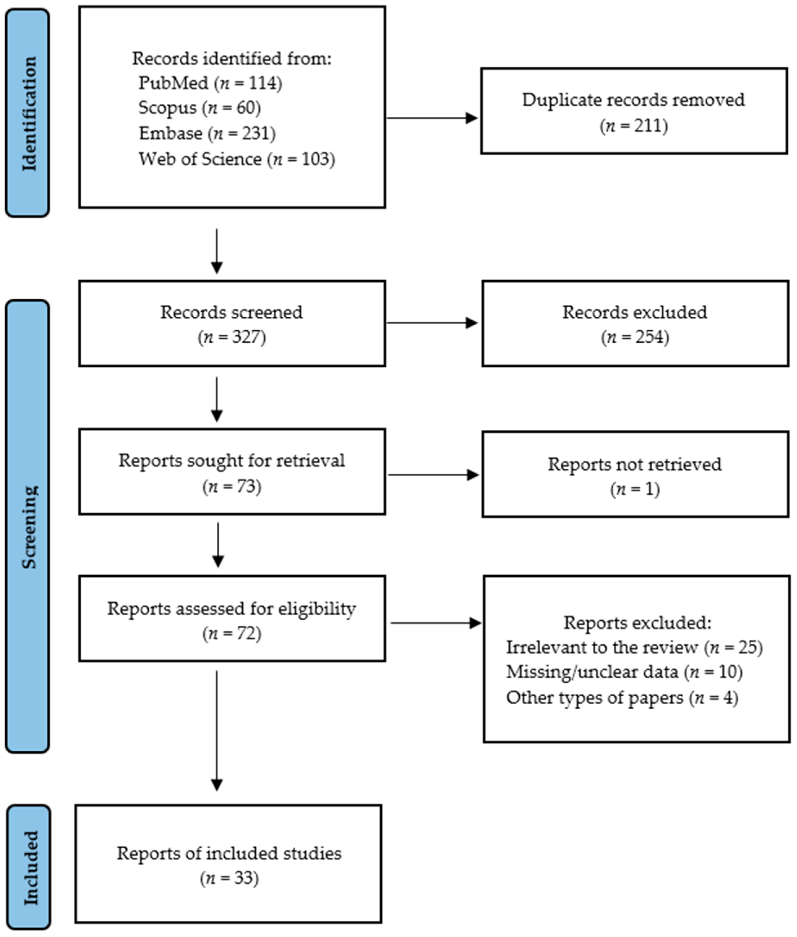
4.1. Search Strategy and Data Extraction
- -
- for PubMed: ((LDH OR Lactate dehydrogenase) OR (chemerin) OR (MMP-9 OR matrix metalloproteinase-9)) AND saliva* AND (oral cancer OR oral carcinoma OR oral squamous cell carcinoma OR oscc);
- -
- for Embase: ((LDH OR Lactate dehydrogenase) OR (chemerin) OR (MMP-9 OR matrix metalloproteinase-9)) AND saliva* AND (oral cancer OR oral carcinoma OR oral squamous cell carcinoma OR oscc);
- -
- for Scopus: TITLE-ABS-KEY ((LDH OR Lactate dehydrogenase) OR (chemerin) OR (MMP-9 OR matrix metalloproteinase-9)) AND saliva* AND (oral cancer OR oral carcinoma OR oral squamous cell carcinoma OR oscc);
- -
- for Web of Science: TS = ((LDH OR Lactate dehydrogenase) OR (chemerin) OR (MMP-9 OR matrix metalloproteinase-9)) AND saliva* AND (oral cancer OR oral carcinoma OR oral squamous cell carcinoma OR oscc).
4.2. Quality Assessment of Included Studies
5. Conclusions
Supplementary Materials
Author Contributions
Funding
Institutional Review Board Statement
Informed Consent Statement
Data Availability Statement
Conflicts of Interest
References
- Montero, P.H.; Patel, S.G. Cancer of the Oral Cavity. Surg. Oncol. Clin. N. Am. 2015, 24, 491–508. [Google Scholar] [CrossRef]
- Siegel, R.L.; Kratzer, T.B.; Giaquinto, A.N.; Sung, H.; Jemal, A. Cancer Statistics, 2025. CA Cancer J. Clin. 2025, 75, 10–45. [Google Scholar] [CrossRef]
- Oral Cancer 5-Year Survival Rates|Data & Statistics|National Institute of Dental and Craniofacial Research. Available online: https://www.nidcr.nih.gov/research/data-statistics/oral-cancer/survival-rates (accessed on 22 April 2025).
- Gormley, M.; Gray, E.; Richards, C.; Gormley, A.; Richmond, R.C.; Vincent, E.E.; Dudding, T.; Ness, A.R.; Thomas, S.J. An Update on Oral Cavity Cancer: Epidemiological Trends, Prevention Strategies and Novel Approaches in Diagnosis and Prognosis. Community Dent. Health 2022, 39, 197–205. [Google Scholar] [CrossRef]
- Yuwanati, M.; Gondivkar, S.; Sarode, S.C.; Gadbail, A.; Desai, A.; Mhaske, S.; Pathak, S.K.; N Khatib, M. Oral Health-Related Quality of Life in Oral Cancer Patients: Systematic Review and Meta-Analysis. Future Oncol. Lond. Engl. 2021, 17, 979–990. [Google Scholar] [CrossRef]
- D’souza, S.; Addepalli, V. Preventive Measures in Oral Cancer: An Overview. Biomed. Pharmacother. 2018, 107, 72–80. [Google Scholar] [CrossRef] [PubMed]
- Yete, S.; Saranath, D. MicroRNAs in Oral Cancer: Biomarkers with Clinical Potential. Oral Oncol. 2020, 110, 105002. [Google Scholar] [CrossRef]
- Warnakulasuriya, S.; Kujan, O.; Aguirre-Urizar, J.M.; Bagan, J.V.; González-Moles, M.Á.; Kerr, A.R.; Lodi, G.; Mello, F.W.; Monteiro, L.; Ogden, G.R.; et al. Oral Potentially Malignant Disorders: A Consensus Report from an International Seminar on Nomenclature and Classification, Convened by the WHO Collaborating Centre for Oral Cancer. Oral Dis. 2021, 27, 1862–1880. [Google Scholar] [CrossRef] [PubMed]
- Goldoni, R.; Scolaro, A.; Boccalari, E.; Dolci, C.; Scarano, A.; Inchingolo, F.; Ravazzani, P.; Muti, P.; Tartaglia, G. Malignancies and Biosensors: A Focus on Oral Cancer Detection through Salivary Biomarkers. Biosensors 2021, 11, 396. [Google Scholar] [CrossRef]
- Khurshid, Z.; Zafar, M.S.; Khan, R.S.; Najeeb, S.; Slowey, P.D.; Rehman, I.U. Role of Salivary Biomarkers in Oral Cancer Detection. Adv. Clin. Chem. 2018, 86, 23–70. [Google Scholar] [CrossRef]
- Chojnowska, S.; Baran, T.; Wilińska, I.; Sienicka, P.; Cabaj-Wiater, I.; Knaś, M. Human Saliva as a Diagnostic Material. Adv. Med. Sci. 2018, 63, 185–191. [Google Scholar] [CrossRef]
- Surdu, A.; Foia, L.G.; Luchian, I.; Trifan, D.; Tatarciuc, M.S.; Scutariu, M.M.; Ciupilan, C.; Budala, D.G. Saliva as a Diagnostic Tool for Systemic Diseases-A Narrative Review. Medicina 2025, 61, 243. [Google Scholar] [CrossRef]
- Nijakowski, K.; Owecki, W.; Jankowski, J.; Surdacka, A. Salivary Biomarkers for Parkinson’s Disease: A Systematic Review with Meta-Analysis. Cells 2024, 13, 340. [Google Scholar] [CrossRef]
- Owecki, W.; Wojtowicz, K.; Nijakowski, K. Salivary Extracellular Vesicles in Detection of Cancers Other than Head and Neck: A Systematic Review. Cells 2025, 14, 411. [Google Scholar] [CrossRef] [PubMed]
- Nijakowski, K.; Owecki, W.; Jankowski, J.; Surdacka, A. Salivary Biomarkers for Alzheimer’s Disease: A Systematic Review with Meta-Analysis. Int. J. Mol. Sci. 2024, 25, 1168. [Google Scholar] [CrossRef] [PubMed]
- Nijakowski, K.; Zdrojewski, J.; Nowak, M.; Gruszczyński, D.; Knoll, F.; Surdacka, A. Salivary Metabolomics for Systemic Cancer Diagnosis: A Systematic Review. Metabolites 2022, 13, 28. [Google Scholar] [CrossRef]
- Nijakowski, K.; Jankowski, J.; Gruszczyński, D.; Surdacka, A. Salivary Alterations of Myeloperoxidase in Patients with Systemic Diseases: A Systematic Review. Int. J. Mol. Sci. 2023, 24, 12078. [Google Scholar] [CrossRef] [PubMed]
- Nijakowski, K.; Surdacka, A. Salivary Biomarkers for Diagnosis of Inflammatory Bowel Diseases: A Systematic Review. Int. J. Mol. Sci. 2020, 21, 7477. [Google Scholar] [CrossRef]
- Ortarzewska, M.; Nijakowski, K.; Kolasińska, J.; Gruszczyński, D.; Ruchała, M.A.; Lehmann, A.; Surdacka, A. Salivary Alterations in Autoimmune Thyroid Diseases: A Systematic Review. Int. J. Environ. Res. Public. Health 2023, 20, 4849. [Google Scholar] [CrossRef]
- Zhang, C.-Z.; Cheng, X.-Q.; Li, J.-Y.; Zhang, P.; Yi, P.; Xu, X.; Zhou, X.-D. Saliva in the Diagnosis of Diseases. Int. J. Oral Sci. 2016, 8, 133–137. [Google Scholar] [CrossRef]
- Bahbah, E.I.; Noehammer, C.; Pulverer, W.; Jung, M.; Weinhaeusel, A. Salivary Biomarkers in Cardiovascular Disease: An Insight into the Current Evidence. FEBS J. 2021, 288, 6392–6405. [Google Scholar] [CrossRef]
- Owecki, W.; Wojtowicz, K.; Nijakowski, K. Salivary Extracellular Vesicles in Detection of Head and Neck Cancers: A Systematic Review. Int. J. Nanomed. 2025, 20, 6757–6775. [Google Scholar] [CrossRef]
- Bastías, D.; Maturana, A.; Marín, C.; Martínez, R.; Niklander, S.E. Salivary Biomarkers for Oral Cancer Detection: An Exploratory Systematic Review. Int. J. Mol. Sci. 2024, 25, 2634. [Google Scholar] [CrossRef]
- Khijmatgar, S.; Yong, J.; Rübsamen, N.; Lorusso, F.; Rai, P.; Cenzato, N.; Gaffuri, F.; Del Fabbro, M.; Tartaglia, G.M. Salivary Biomarkers for Early Detection of Oral Squamous Cell Carcinoma (OSCC) and Head/Neck Squamous Cell Carcinoma (HNSCC): A Systematic Review and Network Meta-Analysis. Jpn. Dent. Sci. Rev. 2024, 60, 32–39. [Google Scholar] [CrossRef]
- Chen, J.; Huang, Z.; Chen, Y.; Tian, H.; Chai, P.; Shen, Y.; Yao, Y.; Xu, S.; Ge, S.; Jia, R. Lactate and Lactylation in Cancer. Signal Transduct. Target. Ther. 2025, 10, 38. [Google Scholar] [CrossRef] [PubMed]
- Mafessoni, T.P.; Mazur, C.E.; Amenábar, J.M. Salivary Lactate Dehydrogenase (LDH) as a Tool for Early Diagnosis of Oral Cancer in Individuals with Fanconi Anemia. Med. Hypotheses 2018, 119, 29–31. [Google Scholar] [CrossRef]
- Mondal, S.; Adhikari, N.; Banerjee, S.; Amin, S.A.; Jha, T. Matrix Metalloproteinase-9 (MMP-9) and Its Inhibitors in Cancer: A Minireview. Eur. J. Med. Chem. 2020, 194, 112260. [Google Scholar] [CrossRef] [PubMed]
- Huang, H. Matrix Metalloproteinase-9 (MMP-9) as a Cancer Biomarker and MMP-9 Biosensors: Recent Advances. Sensors 2018, 18, 3249. [Google Scholar] [CrossRef] [PubMed]
- St-Pierre, Y. Towards a Better Understanding of the Relationships between Galectin-7, P53 and MMP-9 during Cancer Progression. Biomolecules 2021, 11, 879. [Google Scholar] [CrossRef]
- Treeck, O.; Buechler, C.; Ortmann, O. Chemerin and Cancer. Int. J. Mol. Sci. 2019, 20, 3750. [Google Scholar] [CrossRef]
- Qi, X.; Fan, J.; Zhu, J.; Ling, Y.; Mi, S.; Chen, H.; Fan, C.; Li, Y. Circulating Chemerin Level and Risk of Cancer: A Systematic Review and Meta-Analysis. Biomark. Med. 2020, 14, 919–928. [Google Scholar] [CrossRef]
- Shin, W.J.; Pachynski, R.K. Chemerin Modulation of Tumor Growth: Potential Clinical Applications in Cancer. Discov. Med. 2018, 26, 31–37. [Google Scholar] [PubMed]
- Al Shaar, A.; Hamadeh, O.; Ali, A. Saliva and Serum Biomarkers in Oral Diseases: A Case-Control Study. Medicine 2024, 103, e41072. [Google Scholar] [CrossRef]
- Anitha, G.; Kumar, K.V.; Deshpande, G.; Nagaraj, M.; Kalyani, V. Utility of Serum and Salivary Lactate Dehydrogenase and Uric Acid Levels as a Diagnostic Profile in Oral Squamous Cell Carcinoma Patients. J. Oral Maxillofac. Pathol. 2022, 26, 218–227. [Google Scholar] [CrossRef]
- Awasthi, N. Role of Salivary Biomarkers in Early Detection of Oral Squamous Cell Carcinoma. Indian. J. Pathol. Microbiol. 2017, 60, 464–468. [Google Scholar] [CrossRef]
- Bel’skaya, L.; Sarf, E.; Solomatin, D.; Kosenok, V. Diagnostic and Prognostic Value of Salivary Biochemical Markers in Oral Squamous Cell Carcinoma. Diagnostics 2020, 10, 818. [Google Scholar] [CrossRef]
- Bhuvaneswari, M.; Prasad, H.; Rajmohan, M.; Sri Chinthu, K.K.; Prema, P.; Mahalakshmi, L.; Kumar, G.S. Estimation of Salivary Lactate Dehydrogenase in Oral Squamous Cell Carcinoma, Oral Leukoplakia, and Smokers. J. Cancer Res. Ther. 2022, 18, S215–S218. [Google Scholar] [CrossRef]
- D’Cruz, A.M.; Pathiyil, V. Histopathological Differentiation of Oral Squamous Cell Carcinoma and Salivary Lactate Dehydrogenase: A Biochemical Study. South Asian J. Cancer 2015, 4, 58–60. [Google Scholar] [CrossRef]
- Dhivyalakshmi, M.; Uma Maheswari, T.N. Expression of Salivary Biomarkers-Alkaline Phosphatase & Lactate Dehydrogenase in Oral Leukoplakia. Int. J. ChemTech Res. 2014, 6, 2755–2759. [Google Scholar]
- Gholizadeh, N.; Alipanahi Ramandi, M.; Motiee-Langroudi, M.; Jafari, M.; Sharouny, H.; Sheykhbahaei, N. Serum and Salivary Levels of Lactate Dehydrogenase in Oral Squamous Cell Carcinoma, Oral Lichen Planus and Oral Lichenoid Reaction. BMC Oral Health 2020, 20, 314. [Google Scholar] [CrossRef] [PubMed]
- Goyal, G. Comparison of Salivary and Serum Alkaline Phosphates Level and Lactate Dehydrogenase Levels in Patients with Tobacco Related Oral Lesions with Healthy Subjects-A Step Towards Early Diagnosis. Asian Pac. J. Cancer Prev. APJCP 2020, 21, 983–991. [Google Scholar] [CrossRef] [PubMed]
- Honarmand, M.; Saravani, R.; Farhad-Mollashahi, L.; Smailpoor, A. Salivary Lactate Dehydrogenase, c-Reactive Protein, and Cancer Antigen 125 Levels in Patients with Oral Lichen Planus and Oral Squamous Cell Carcinoma. Int. J. Cancer Manag. 2021, 14, e108344. [Google Scholar] [CrossRef]
- Joshi, P.S.; Golgire, S. A Study of Salivary Lactate Dehydrogenase Isoenzyme Levels in Patients with Oral Leukoplakia and Squamous Cell Carcinoma by Gel Electrophoresis Method. J. Oral Maxillofac. Pathol. 2014, 18, S39–S44. [Google Scholar] [CrossRef]
- Kadiyala, S.V. A Study of Salivary Lactate Dehydrogenase (LDH) Levels in Oral Cancer and Oral Submucosal Fibrosis Patients Amoung the Normal Individulas. J. Pharm. Sci. Res. 2015, 7, 455–457. [Google Scholar]
- Kallalli, B.N.; Rawson, K.; Muzammil; Singh, A.; Awati, M.A.; Shivhare, P. Lactate Dehydrogenase as a Biomarker in Oral Cancer and Oral Submucous Fibrosis. J. Oral Pathol. Med. 2016, 45, 687–690. [Google Scholar] [CrossRef]
- Lokesh, K.; Kannabiran, J.; Rao, M.D. Salivary Lactate Dehydrogenase (LDH)—A Novel Technique in Oral Cancer Detection and Diagnosis. J. Clin. Diagn. Res. JCDR 2016, 10, ZC34–ZC37. [Google Scholar] [CrossRef] [PubMed]
- López-Pintor, R.M.; González-Serrano, J.; Vallina, C.; Ivaylova Serkedzhieva, K.; Virto, L.; Nuevo, P.; Caponio, V.C.A.; Iniesta, M.; Rodríguez Santamarta, T.; Lequerica Fernández, P.; et al. Factors Influencing Salivary Lactate Dehydrogenase Levels in Oral Squamous Cell Carcinoma and Oral Potentially Malignant Disorders. Front. Oral Health 2024, 5, 1525936. [Google Scholar] [CrossRef] [PubMed]
- Mantri, T.; Thete, S.G.; Male, V.; Yadav, R.; Grover, I.; Adsure, G.R.; Kulkarni, D. Study of the Role of Salivary Lactate Dehydrogenase in Habitual Tobacco Chewers, Oral Submucous Fibrosis and Oral Cancer as a Biomarker. J. Contemp. Dent. Pract. 2019, 20, 970–973. [Google Scholar] [CrossRef]
- Nandakumar, E.; Savitha, G. A Study of Salivary Lactate Dehydragenase (LDH) Level in Normal Individuals and the Oral Cancer Patients. Res. J. Pharm. Technol. 2015, 8, 932–934. [Google Scholar] [CrossRef]
- Patel, S.; Metgud, R. Estimation of Salivary Lactate Dehydrogenase in Oral Leukoplakia and Oral Squamous Cell Carcinoma: A Biochemical Study. J. Cancer Res. Ther. 2015, 11, 119–123. [Google Scholar] [CrossRef]
- Pathiyil, V.; D’Cruz, A.M. Salivary Lactate Dehydrogenase as a Prognostic Marker in Oral Squamous Cell Carcinoma Patients Following Surgical Therapy. J. Exp. Ther. Oncol. 2017, 11, 133–137. [Google Scholar]
- Rathore, B.S. Assessment of Salivary Biomarkers in Patients with Squamous Cell Carcinoma. Int. J. Life Sci. Biotechnol. Pharma Res. 2024, 13, 569–570. [Google Scholar] [CrossRef]
- Shetty, S.R.; Chadha, R.; Babu, S.; Kumari, S.; Bhat, S.; Achalli, S. Salivary Lactate Dehydrogenase Levels in Oral Leukoplakia and Oral Squamous Cell Carcinoma: A Biochemical and Clinicopathological Study. J. Cancer Res. Ther. 2012, 8 (Suppl. 1), S123–S125. [Google Scholar] [CrossRef]
- Subramanian, M.; Fenn, S.; Reddy, G.; Rajarammohan, K.; Thangavelu, R. Estimation of Salivary and Serum Lactate Dehydrogenase (LDH) Levels in Individuals with Oral Cancer, Oral Potentially Malignant Disorders, Tobacco Users, and Healthy Subjects-A Pilot Study. J. Indian Acad. Oral Med. Radiol. 2024, 36, 417–421. [Google Scholar] [CrossRef]
- Yu, J.-S.; Chen, Y.-T.; Chiang, W.-F.; Hsiao, Y.-C.; Chu, L.J.; Seei, L.-C.; Wu, C.-S.; Tu, H.-T.; Chen, H.-W.; Chen, C.-C.; et al. Saliva Protein Biomarkers To Detect Oral Squamous Cell Carcinoma in a High-Risk Population in Taiwan. Proc. Natl. Acad. Sci. USA 2016, 113, 11549–11554. [Google Scholar] [CrossRef]
- Feng, Y.; Li, Q.; Chen, J.; Yi, P.; Xu, X.; Fan, Y.; Cui, B.; Yu, Y.; Li, X.; Du, Y.; et al. Salivary Protease Spectrum Biomarkers of Oral Cancer. Int. J. Oral Sci. 2019, 11, 7. [Google Scholar] [CrossRef]
- Ghallab, N.A.; Shaker, O.G. Serum and Salivary Levels of Chemerin and MMP-9 in Oral Squamous Cell Carcinoma and Oral Premalignant Lesions. Clin. Oral Investig. 2017, 21, 937–947. [Google Scholar] [CrossRef]
- Krishnasree, R.; Jayanthi, P.; Varun, B.; Ramani, P.; Rathy, R. Evaluation of Salivary MMP-9 in Oral Squamous Cell Carcinoma Using Enzyme Linked Immunosorbent Assay-A Pilot Study. Oral Maxillofac. Pathol. J. 2023, 14, 190–193. [Google Scholar]
- Nasir, F.H.; Mohamed, R.J.; Helmi, R.M. Metalloproteinase Matrix of Level Saliva and Serum Carcinoma Squamous Cell. Biochem. Cell. Arch. 2020, 20, 3609–3612. [Google Scholar]
- Nisa, W.-U.; Khan, M.A.; Agha, F.; Khan, S.; Fatima, S.; Rathore, P. Salivary Biomarkers CYFRA 21-1 and MMP9: Predictive Indicators of Disease Progression in Oral Squamous Cell Carcinoma. Pak. J. Med. Health Sci. 2023, 17, 179–182. [Google Scholar] [CrossRef]
- Pazhani, J.; Chanthu, K.; Jayaraman, S.; Varun, B.R. Evaluation of Salivary MMP-9 in Oral Squamous Cell Carcinoma and Oral Leukoplakia Using ELISA. J. Oral Maxillofac. Pathol. 2023, 27, 649–654. [Google Scholar] [CrossRef]
- Peisker, A.; Raschke, G.-F.; Fahmy, M.-D.; Guentsch, A.; Roshanghias, K.; Hennings, J.; Schultze-Mosgau, S. Salivary MMP-9 in the Detection of Oral Squamous Cell Carcinoma. Med. Oral Patol. Oral Cir. Bucal 2017, 22, e270–e275. [Google Scholar] [CrossRef]
- Radulescu, R.; Totan, A.; Calenic, B.; Totan, C.; Greabu, M. Biomarkers of Oxidative Stress, Proliferation, Inflammation and Invasivity in Saliva from Oral Cancer Patients. J. Anal. Oncol. 2015, 4, 52–57. [Google Scholar] [CrossRef]
- Shin, Y.-J.; Vu, H.; Lee, J.-H.; Kim, H.-D. Diagnostic and Prognostic Ability of Salivary MMP-9 for Oral Squamous Cell Carcinoma: A Pre-/Post-Surgery Case and Matched Control Study. PLoS ONE 2021, 16, e0248167. [Google Scholar] [CrossRef]
- Smriti, K.; Ray, M.; Chatterjee, T.; Shenoy, R.-P.; Gadicherla, S.; Pentapati, K.-C.; Rustaqi, N. Salivary MMP-9 as a Biomarker for the Diagnosis of Oral Potentially Malignant Disorders and Oral Squamous Cell Carcinoma. Asian Pac. J. Cancer Prev. APJCP 2020, 21, 233–238. [Google Scholar] [CrossRef]
- Susha, K.P.; Ravindran, R. Evaluation of Salivary Chemerin in Oral Leukoplakia, Oral Squamous Cell Carcinoma and Healthy Controls. J. Orofac. Sci. 2023, 15, 141–146. [Google Scholar] [CrossRef]
- Akashanand; Zahiruddin, Q.S.; Jena, D.; Ballal, S.; Kumar, S.; Bhat, M.; Sharma, S.; Kumar, M.R.; Rustagi, S.; Gaidhane, A.M.; et al. Burden of Oral Cancer and Associated Risk Factors at National and State Levels: A Systematic Analysis from the Global Burden of Disease in India, 1990–2021. Oral Oncol. 2024, 159, 107063. [Google Scholar] [CrossRef]
- OCEBM Levels of Evidence. Available online: https://www.cebm.ox.ac.uk/resources/levels-of-evidence/ocebm-levels-of-evidence (accessed on 16 November 2024).
- Huang, L.; Luo, F.; Deng, M.; Zhang, J. The Relationship between Salivary Cytokines and Oral Cancer and Their Diagnostic Capability for Oral Cancer: A Systematic Review and Network Meta-Analysis. BMC Oral Health 2024, 24, 1044. [Google Scholar] [CrossRef]
- Chiamulera, M.M.A.; Zancan, C.B.; Remor, A.P.; Cordeiro, M.F.; Gleber-Netto, F.O.; Baptistella, A.R. Salivary Cytokines as Biomarkers of Oral Cancer: A Systematic Review and Meta-Analysis. BMC Cancer 2021, 21, 205. [Google Scholar] [CrossRef]
- Gallo, M.; Sapio, L.; Spina, A.; Naviglio, D.; Calogero, A.; Naviglio, S. Lactic Dehydrogenase and Cancer: An Overview. Front. Biosci. Landmark Ed. 2015, 20, 1234–1249. [Google Scholar] [CrossRef]
- Augoff, K.; Hryniewicz-Jankowska, A.; Tabola, R. Lactate Dehydrogenase 5: An Old Friend and a New Hope in the War on Cancer. Cancer Lett. 2015, 358, 1–7. [Google Scholar] [CrossRef]
- de la Cruz-López, K.G.; Castro-Muñoz, L.J.; Reyes-Hernández, D.O.; García-Carrancá, A.; Manzo-Merino, J. Lactate in the Regulation of Tumor Microenvironment and Therapeutic Approaches. Front. Oncol. 2019, 9, 1143. [Google Scholar] [CrossRef]
- Comandatore, A.; Franczak, M.; Smolenski, R.T.; Morelli, L.; Peters, G.J.; Giovannetti, E. Lactate Dehydrogenase and Its Clinical Significance in Pancreatic and Thoracic Cancers. Semin. Cancer Biol. 2022, 86, 93–100. [Google Scholar] [CrossRef]
- Sharma, D.; Singh, M.; Rani, R. Role of LDH in Tumor Glycolysis: Regulation of LDHA by Small Molecules for Cancer Therapeutics. Semin. Cancer Biol. 2022, 87, 184–195. [Google Scholar] [CrossRef]
- Urbańska, K.; Orzechowski, A. Unappreciated Role of LDHA and LDHB to Control Apoptosis and Autophagy in Tumor Cells. Int. J. Mol. Sci. 2019, 20, 2085. [Google Scholar] [CrossRef]
- Dong, T.; Liu, Z.; Xuan, Q.; Wang, Z.; Ma, W.; Zhang, Q. Tumor LDH-A Expression and Serum LDH Status Are Two Metabolic Predictors for Triple Negative Breast Cancer Brain Metastasis. Sci. Rep. 2017, 7, 6069. [Google Scholar] [CrossRef]
- Wei, Y.; Xu, H.; Dai, J.; Peng, J.; Wang, W.; Xia, L.; Zhou, F. Prognostic Significance of Serum Lactic Acid, Lactate Dehydrogenase, and Albumin Levels in Patients with Metastatic Colorectal Cancer. BioMed Res. Int. 2018, 2018, 1804086. [Google Scholar] [CrossRef]
- Zhang, X.; Guo, M.; Fan, J.; Lv, Z.; Huang, Q.; Han, J.; Wu, F.; Hu, G.; Xu, J.; Jin, Y. Prognostic Significance of Serum LDH in Small Cell Lung Cancer: A Systematic Review with Meta-Analysis. Cancer Biomark. Sect. Dis. Markers 2016, 16, 415–423. [Google Scholar] [CrossRef]
- Cai, H.; Li, J.; Zhang, Y.; Liao, Y.; Zhu, Y.; Wang, C.; Hou, J. LDHA Promotes Oral Squamous Cell Carcinoma Progression Through Facilitating Glycolysis and Epithelial-Mesenchymal Transition. Front. Oncol. 2019, 9, 1446. [Google Scholar] [CrossRef]
- Barbi, W.; Purohit, B.M. Serum Lactate Dehydrogenase Enzyme as a Tumor Marker in Potentially Malignant Disorders: A Systematic Review and Meta-Analysis. Asian Pac. J. Cancer Prev. APJCP 2022, 23, 2553–2559. [Google Scholar] [CrossRef]
- Iglesias-Velázquez, Ó.; López-Pintor, R.M.; González-Serrano, J.; Casañas, E.; Torres, J.; Hernández, G. Salivary LDH in Oral Cancer and Potentially Malignant Disorders: A Systematic Review and Meta-Analysis. Oral Dis. 2022, 28, 44–56. [Google Scholar] [CrossRef]
- Quintero-Fabián, S.; Arreola, R.; Becerril-Villanueva, E.; Torres-Romero, J.C.; Arana-Argáez, V.; Lara-Riegos, J.; Ramírez-Camacho, M.A.; Alvarez-Sánchez, M.E. Role of Matrix Metalloproteinases in Angiogenesis and Cancer. Front. Oncol. 2019, 9, 1370. [Google Scholar] [CrossRef] [PubMed]
- Choi, E.K.; Kim, H.D.; Park, E.J.; Song, S.Y.; Phan, T.T.; Nam, M.; Kim, M.; Kim, D.-U.; Hoe, K.-L. 8-Methoxypsoralen Induces Apoptosis by Upregulating P53 and Inhibits Metastasis by Downregulating MMP-2 and MMP-9 in Human Gastric Cancer Cells. Biomol. Ther. 2023, 31, 219–226. [Google Scholar] [CrossRef]
- Yin, P.; Su, Y.; Chen, S.; Wen, J.; Gao, F.; Wu, Y.; Zhang, X. MMP-9 Knockdown Inhibits Oral Squamous Cell Carcinoma Lymph Node Metastasis in the Nude Mouse Tongue-Xenografted Model through the RhoC/Src Pathway. Anal. Cell. Pathol. Amst. 2021, 2021, 6683391. [Google Scholar] [CrossRef]
- Li, Y.; He, J.; Wang, F.; Wang, X.; Yang, F.; Zhao, C.; Feng, C.; Li, T. Role of MMP-9 in Epithelial-Mesenchymal Transition of Thyroid Cancer. World J. Surg. Oncol. 2020, 18, 181. [Google Scholar] [CrossRef]
- Patterson, B.C.; Sang, Q.A. Angiostatin-Converting Enzyme Activities of Human Matrilysin (MMP-7) and Gelatinase B/Type IV Collagenase (MMP-9). J. Biol. Chem. 1997, 272, 28823–28825. [Google Scholar] [CrossRef]
- Vilen, S.-T.; Salo, T.; Sorsa, T.; Nyberg, P. Fluctuating Roles of Matrix Metalloproteinase-9 in Oral Squamous Cell Carcinoma. Sci. World J. 2013, 2013, 920595. [Google Scholar] [CrossRef]
- Zheng, W.-Y.; Zhang, D.-T.; Yang, S.-Y.; Li, H. Elevated Matrix Metalloproteinase-9 Expression Correlates With Advanced Stages of Oral Cancer and Is Linked to Poor Clinical Outcomes. J. Oral Maxillofac. Surg. 2015, 73, 2334–2342. [Google Scholar] [CrossRef]
- Deng, W.; Peng, W.; Wang, T.; Chen, J.; Zhu, S. Overexpression of MMPs Functions as a Prognostic Biomarker for Oral Cancer Patients: A Systematic Review and Meta-Analysis. Oral Health Prev. Dent. 2019, 17, 505–514. [Google Scholar] [CrossRef]
- Bozaoglu, K.; Bolton, K.; McMillan, J.; Zimmet, P.; Jowett, J.; Collier, G.; Walder, K.; Segal, D. Chemerin Is a Novel Adipokine Associated with Obesity and Metabolic Syndrome. Endocrinology 2007, 148, 4687–4694. [Google Scholar] [CrossRef]
- Goralski, K.B.; Jackson, A.E.; McKeown, B.T.; Sinal, C.J. More Than an Adipokine: The Complex Roles of Chemerin Signaling in Cancer. Int. J. Mol. Sci. 2019, 20, 4778. [Google Scholar] [CrossRef]
- Buechler, C.; Feder, S.; Haberl, E.M.; Aslanidis, C. Chemerin Isoforms and Activity in Obesity. Int. J. Mol. Sci. 2019, 20, 1128. [Google Scholar] [CrossRef]
- Treeck, O.; Buechler, C. Chemerin Signaling in Cancer. Cancers 2020, 12, 3085. [Google Scholar] [CrossRef] [PubMed]
- Wang, N.; Wang, Q.-J.; Feng, Y.-Y.; Shang, W.; Cai, M. Overexpression of Chemerin Was Associated with Tumor Angiogenesis and Poor Clinical Outcome in Squamous Cell Carcinoma of the Oral Tongue. Clin. Oral Investig. 2014, 18, 997–1004. [Google Scholar] [CrossRef] [PubMed]
- Lu, Z.; Liang, J.; He, Q.; Wan, Q.; Hou, J.; Lian, K.; Wang, A. The Serum Biomarker Chemerin Promotes Tumorigenesis and Metastasis in Oral Squamous Cell Carcinoma. Clin. Sci. 2019, 133, 681–695. [Google Scholar] [CrossRef]
- Gao, F.; Feng, Y.; Hu, X.; Zhang, X.; Li, T.; Wang, Y.; Ge, S.; Wang, C.; Chi, J.; Tan, X.; et al. Neutrophils Regulate Tumor Angiogenesis in Oral Squamous Cell Carcinoma and the Role of Chemerin. Int. Immunopharmacol. 2023, 121, 110540. [Google Scholar] [CrossRef] [PubMed]
- Hu, X.; Wang, N.; Gao, F.; Ge, S.; Lin, M.; Zhang, X.; Li, T.; Li, T.; Xu, C.; Huang, C.; et al. Prognostic Significance of Serum Chemerin and Neutrophils Levels in Patients with Oral Squamous Cell Carcinoma. Heliyon 2024, 10, e32393. [Google Scholar] [CrossRef]
- Lu, Z.; Liu, J.; Wan, Q.; Wu, Y.; Wu, W.; Chen, Y. Chemerin Promotes Invasion of Oral Squamous Cell Carcinoma by Stimulating IL-6 and TNF-α Production via STAT3 Activation. Mol. Biol. Rep. 2024, 51, 436. [Google Scholar] [CrossRef]
- Page, M.J.; McKenzie, J.E.; Bossuyt, P.M.; Boutron, I.; Hoffmann, T.C.; Mulrow, C.D.; Shamseer, L.; Tetzlaff, J.M.; Akl, E.A.; Brennan, S.E.; et al. The PRISMA 2020 Statement: An Updated Guideline for Reporting Systematic Reviews. BMJ 2021, 372, n71. [Google Scholar] [CrossRef]
- Study Quality Assessment Tools|NHLBI, NIH. Available online: https://www.nhlbi.nih.gov/health-topics/study-quality-assessment-tools (accessed on 7 November 2024).


| Author, Year | Setting | Study Group—OC; (F/M), Age | Study Group—OPMD; (F/M), Age | Control Group; (F/M), Age | Diagnosis | Histological Grading | Type of Saliva | Centrifugation and Storing | Method of Marker Determination |
|---|---|---|---|---|---|---|---|---|---|
| LDH | |||||||||
| Al Shaar et al., 2024 [33] | Syria | 12; (6/60), 57.67 ± 13.98 | LP: 15; (8/7), 46.13 ± 14.08 | 15; (7/8), 24.4 ± 2.95 | OC/OSCC | NR | unstimulated | centrifuged at 3000 rpm for 3 min; NR | Hitachi 911 automated clinical chemistry analyzer |
| Anitha et al., 2022 [34] | India | 18; (2/16), 44.67 | - | 18; (5/13), 34.56 | OSCC | MD: 13 WD: 5 | unstimulated | centrifuged at 2000 rpm for 10 min, stored at −20 °C | ErbaCHEM 5× semi-automatic analyzer machine, LDH-P reagent kit |
| Awasthi et al., 2017 [35] | India | 30; (2/28), 49.6 (25–70) | 9; (1/8), 34.2 (25–40) | 25; (3/22), 48.1 (25–68) | OSCC | PD: 1 MD: 20 WD: 9 | unstimulated | centrifuged at 3000 rpm for 15 min, stored at −80 °C | standard kit method |
| Bel’skaya et al., 2020 [36] | Russia | 68; NR, NR | - | 114; NR, NR | OSCC | NR | NR | centrifuged at 10,000× g for 10 min, no storage | kinetic ultraviolet method according to the NADH (Nicotinamide Adenine Dinucleotide) oxidation rate |
| Bhuvaneswari et al., 2022 [37] | India | 21; NR, NR | OL: 20; NR, NR | 20; NR, NR | OSCC | NR | unstimulated | centrifuged at “1000 rotations” at 4 °C for 10 min, stored at −80 °C | LDH enzyme kit, ultraviolet-visible spectrophotometer |
| D’Cruz et al., 2015 [38] | India | 30; NR, NR | - | 30; NR, NR | OSCC | PD: 10 MD: 10 WD: 10 | unstimulated | NR | standard kit, measured spectrophotometrically at 340 nm |
| Dhivyalakshmi et al., 2014 [39] | India | 14; NR, NR | OL: 14; NR, NR | 14; NR, NR | OSCC | NR | unstimulated | centrifuged at 2500 rpm for 15 min, NR | standard kit, measured using autoanalyzer |
| Gholizadeh et al., 2020 [40] | Iran | 25; (15/10), 61.00 ± 3.23 | LP: 15; (17/8), 49.73 ± 3.19; LR: 25; (17/8), 52.73 ± 2.78 | 25; (17/8), 42.73 ± 2.38 | OSCC | NR | unstimulated and stimulated | centrifuged at 2000 rpm for 10 min, stored at −20 °C | spectrophotometrically measured within 24 h, standard LDH kits |
| Goyal et al., 2020 [41] | India | 100; NR, NR | 100; NR, NR | 100; NR, NR | OSCC | NR | unstimulated | centrifuged at 2500 rpm for 15 min, NR | standard kit method |
| Honarmand et al., 2021 [42] | Iran | 15; NR, 50.4 ± 8.37 | LP: 20; NR, 45.4 ± 10.08 | 20; NR, 45.6 ± 9.77 | OSCC | NR | unstimulated | centrifuged at 3500 rpm for 20 min, stored at −70 °C | ELISA |
| Joshi et al., 2014 [43] | India | 30; (10/20), 47.96 | OL: 30; (1/29), 41.06 | 30; NR, NR | OSCC | PD: 1 MD: 7 WD: 22 | unstimulated | centrifuged at 1000 rpm for 10 min, NR | agarose gel electrophoresis method (SEBIA-HYDRAGEL ISO-LDH K-20 kit) |
| Kadiyala et al., 2015 [44] | India | 20; NR, NR | OSMF: 20; NR, NR | 20; NR, NR | OC | NR | unstimulated | centrifuged at 2500 rpm for 15 min, NR | ERBA CHEM 5 semi-automatic analyzer |
| Kallalli et al., 2016 [45] | India | 25; NR, NR | OSMF: 25; NR, NR | 10; NR, NR | OC | NR | unstimulated | centrifuged NR, NR | ERBA-CHEM 5 semi-automatic analyzer |
| Lokesh et al., 2016 [46] | India | 30; NR, 35–65 | - | 20; NR, NR | OSCC | PD: 5 MD: 10 WD: 15 | unstimulated | centrifuged NR, no storage | automated method using autoanalyzer readings, spectrophotometer at a wavelength of 340 nm (UV kinetic method) |
| López-Pintor et al., 2024 [47] | Spain | 12; (8/4), 69 ± 12.87 | 51; (35/16), 64.65 ± 10.39 | 29; (17/12), 59.83 ± 13.82 | OSCC | PD: 1 MD: 3 WD: 8 | unstimulated | centrifuged at 1160× g for 20 min, stored at −80 °C | LDH Assay Kit Colorimetric analyzed spectrophotometrically at a wavelength of 450 nm |
| Mantri et al., 2019 [48] | India | 30; NR, NR | OSMF: 30; NR, NR | 30; NR, NR | OSCC | NR | unstimulated | centrifuged at 5000 rpm for 5 min, stored at 4 °C | LDH-P kit within 24 h, analyzed by an Erba Chem UV semi-automated spectrophotometer |
| Nandakumar et al., 2015 [49] | India | 20; (8/12), female: 37.50 ± 5.01; male: 40.83 ± 4.35 | - | 20; (2/18), female: 41.00 ± 2.82; male: 39.56 ± 4.50 | OC | NR | unstimulated | centrifuged at 2500 rpm for 15 min, NR | ERBA CHEM 5 semi-automatic analyzer |
| Patel et al., 2015 [50] | India | 25; NR, NR | OL: 25; NR, NR | 25; NR, NR | OSCC | PD: 4 MD: 8 WD: 13 | unstimulated | NR, stored in an ice box | Semi-automatic Analyzer by using Biovision LDH Activity Colorimetric Assay Kit |
| Pathiyil et al., 2017 [51] | India | 20; NR, NR | - | 20; NR, NR | OSCC | NR | unstimulated | centrifuged at 3000 rpm for 10 min, NR | standard kit, measured spectrophotometrically at 340 nm |
| Rathore et al., 2024 [52] | India | 54; (16/38) NR | - | 54; NR, NR | OSCC | NR | unstimulated | centrifuged at 3000 rpm for 15 min, NR | standard kit method |
| Shetty et al., 2012 [53] | India | 25; NR, NR | OL: 25; NR, NR | 25; NR, NR | OSCC | NR | unstimulated | NR | standard kit, measured sphectrophotometrically at 340 nm |
| Subramanian et al., 2024 [54] | India | 30; (14/16), NR | 30; (6/24), NR | 30; (18/12), NR | OSCC | NR | unstimulated | centrifuged at 900 rpm for 12 min, stored at −20°C | LDH kit (Liquizyme), semi-automatic analyzer (spectrophotometer) |
| Yu et al., 2016 [55] | Taiwan | 131; (2/129), 52.5 ± 9.7; detectable: 129 | 103; (1/102), 49.5 ± 10.7 | 96; (0/96), 48.8 ± 11.8; detectable: 93 | OSCC | NR | unstimulated | centrifuged at 3000× g for 15 min at 4 °C, stored at −80°C | Liquid Chromatography-multiple reaction monitoring-Mass Spectrometry |
| MMP-9 | |||||||||
| Feng et al., 2019 [56] | China | 20; NR, NR | - | 20; NR, NR | OSCC | NR | stimulated | centrifuged at 10,000× g for 10 min at 4°C, stored at −80 °C | Human Protease Array Kit, human protease ELISA kits |
| Ghallab et al., 2017 [57] | Egypt | 15; (9/6), 47.66 ± 14.07 | 15; (8/7), 42.33 ± 10.99 | 15; (9/6), 43.26 ± 11.82 | OSCC | NR | unstimulated | centrifuged at 10,000× g for 2 min, stored at −80 °C | Quantikine ELISA kit |
| Krishnasree et al., 2023 [58] | India | 15; (NR), 64 ± 4 | - | 15; (NR), 60 ± 3.5 | OSCC | PD: 2 MD: 4 WD: 9 | unstimulated | centrifuged NR, stored at −80 °C | MMP-9 ELISA kit |
| Nasir et al., 2020 [59] | Iraq | Before treatment: 20; (NR), NR After treatment: 20; (NR), NR | - | 20; (NR), NR | OSCC | NR | unstimulated | NR | MMP-9 ELISA kit |
| Nisa et al., 2023 [60] | Pakistan | 45; (10/35), 18–70 | - | 45; (18/27), NR | OSCC | PD: 15 MD: 15 WD: 15 | NR | centrifuged at 8000 rpm for 15 min at 4 °C, stored at −80 °C | ELISA Bioassay Technology kit |
| Pazhani et al., 2023 [61] | India | 34; (6/28), 62.8 ± 12.9 | OL: 34; (11/23), 60.1 ± 11.5 | 34; (22/12), 52.4 ± 9.7 | OSCC | PD: 5 MD: 9 WD: 20 | unstimulated | centrifuged NR, stored at −80 °C | MMP-9 ELISA kit |
| Peisker et al., 2017 [62] | Germany | 30; (16/14), 65.0 ± 10.9 | - | 30; (12/18), 60.7 ± 12.3 | OSCC | UD: 1 PD: 8 MD: 20 WD: 1 | stimulated | centrifuged at 1000× g for 2 min at 20 °C, NR | ELISA |
| Radulescu et al., 2015 [63] | Romania | 30; (16/14), 45–60 | - | 14; (NR), 40–60 | OSCC | NR | unstimulated | centrifuged at 3000 rpm for 10 min, stored at −80 °C | MMP-9 ELISA kit |
| Shin et al., 2021 [64] | South Korea | 106; (44/62), 63.14 ± 9.7 | - | 212; (88/124), 63.09 ± 9.7 | OSCC | NR | unstimulated | centrifuged at 2600 rpm for 15 min at 4 °C, stored at −80 °C | Quantikine1 human MMP-9 immunoassay ELISA kit |
| Smriti et al., 2020 [65] | India | 24; (10/14), 58.63 ± 14.79 | 20; (6/14), 44 ± 14.19 | 22; (7/15), 48.09 ± 11.73 | OSCC/verrucous OC (2 patients) | PD: 6 MD: 9 WD: 7 | unstimulated | centrifuged at 4000× g for 10 min at 4°C, NR | Human MMP-9 PicokineTM ELISA kit |
| Yu et al., 2016 [55] | Taiwan | 131; (2/129), 52.5 ± 9.7; detectable: 126 | 103; (1/102), 49.5 ± 10.7 | 96; (0/96), 48.8 ± 11.8; detectable: 94 | OSCC | NR | unstimulated | centrifuged at 3000× g for 15 min at 4 °C, stored at −80 °C | Liquid Chromatography-multiple reaction monitoring-Mass Spectrometry |
| CHEMERIN | |||||||||
| Susha et al., 2023 [66] | India | 32; (6/28), 31–40: 3; 41–50: 3; 51–60: 8; 61–70: 9; >70: 9 | OL: 32; (NR), NR | 32; (NR), NR | OSCC | PD: 4 MD: 8 WD: 20 | unstimulated | centrifuged at 3000 rpm for 10 min, stored at −80 °C | ab155430 Chemerin Human ELISA kit |
| Ghallab et al., 2017 [57] | Egypt | 15; (9/6), 47.66 ± 14.07 | 15; (8/7), 42.33 ± 10.99 | 15; (9/6), 43.26 ± 11.82 | OSCC | NR | unstimulated | centrifuged at 10,000× g for 2 min, stored at −80 °C | RD191136200R Human Chemerin ELISA |
| Study | SMD | SE | 95% CI | p-Value | Weight% |
|---|---|---|---|---|---|
| OC Patients vs. Healthy Controls | |||||
| LDH | |||||
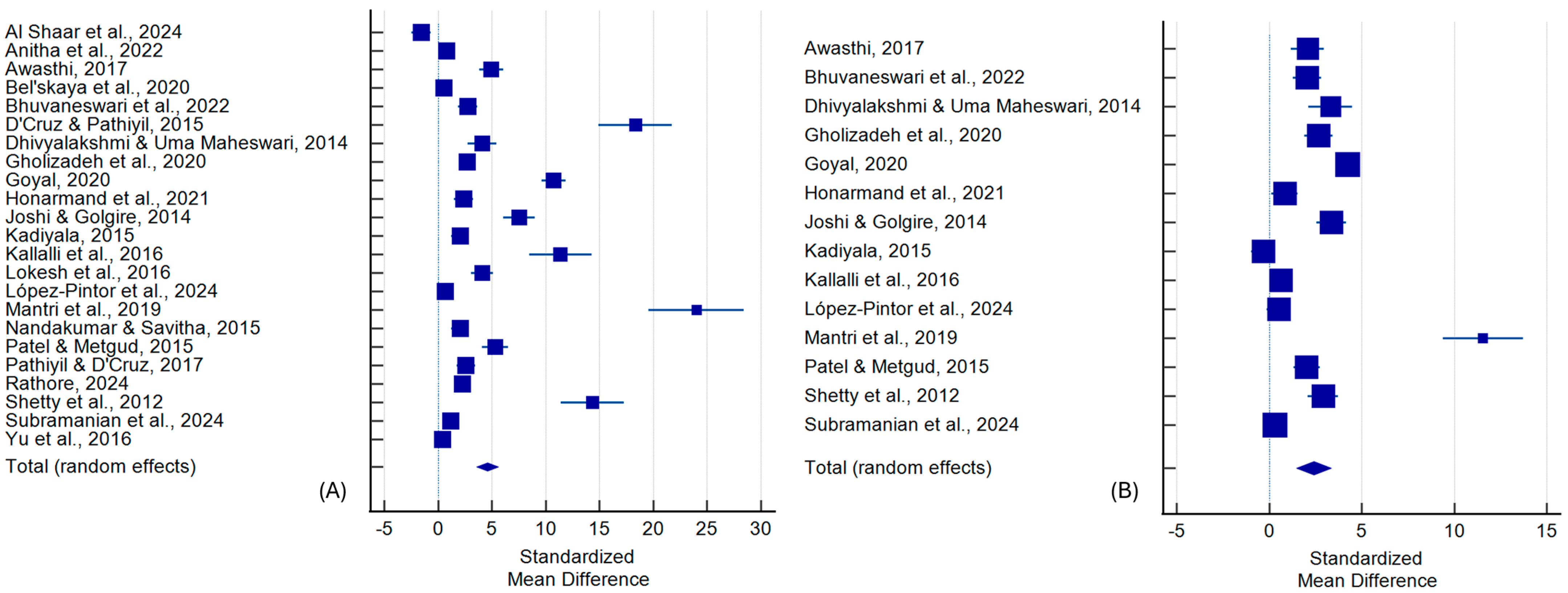
| Al Shaar et al., 2024 [33] | −1.559 | 0.431 | −2.447 to −0.671 | 4.61 | |
| Anitha et al., 2022 [34] | 0.811 | 0.340 | 0.121 to 1.501 | 4.66 | |
| Awasthi et al., 2017 [35] | 4.958 | 0.543 | 3.869 to 6.047 | 4.52 | |
| Bel’skaya et al., 2020 [36] | 0.520 | 0.155 | 0.214 to 0.825 | 4.74 | |
| Bhuvaneswari et al., 2022 [37] | 2.742 | 0.436 | 1.859 to 3.625 | 4.60 | |
| D’Cruz & Pathiyil, 2015 [38] | 18.323 | 1.692 | 14.936 to 21.710 | 3.15 | |
| Dhivyalakshmi & Uma Maheswari, 2014 [39] | 4.093 | 0.659 | 2.739 to 5.447 | 4.42 | |
| Gholizadeh et al., 2020 [40] | 2.708 | 0.388 | 1.927 to 3.489 | 4.63 | |
| Goyal et al., 2020 [41] | 10.747 | 0.556 | 9.652 to 11.843 | 4.51 | |
| Honarmand et al., 2021 [42] | 2.364 | 0.437 | 1.474 to 3.253 | 4.60 | |
| Joshi & Golgire, 2014 [43] | 7.531 | 0.733 | 6.063 to 8.999 | 4.34 | |
| Kadiyala et al., 2015 [44] | 2.040 | 0.385 | 1.261 to 2.819 | 4.64 | |
| Kallalli et al., 2016 [45] | 11.385 | 1.409 | 8.518 to 14.251 | 3.51 | |
| Lokesh et al., 2016 [46] | 4.087 | 0.498 | 3.086 to 5.087 | 4.56 | |
| López-Pintor et al., 2024 [47] | 0.646 | 0.344 | −0.050 to 1.342 | 4.66 | |
| Mantri et al., 2019 [48] | 24.000 | 2.206 | 19.585 to 28.415 | 2.55 | |
| Nandakumar & Savitha, 2015 [49] | 2.040 | 0.385 | 1.261 to 2.819 | 4.64 | |
| Patel & Metgud, 2015 [50] | 5.308 | 0.599 | 4.103 to 6.513 | 4.47 | |
| Pathiyil & D’Cruz, 2017 [51] | 2.579 | 0.423 | 1.722 to 3.436 | 4.61 | |
| Rathore et al., 2024 [52] | 2.255 | 0.245 | 1.769 to 2.741 | 4.71 | |
| Shetty et al., 2012 [53] | 14.352 | 1.462 | 11.412 to 17.291 | 3.44 | |
| Subramanian et al., 2024 [54] | 1.180 | 0.277 | 0.626 to 1.734 | 4.69 | |
| Yu et al., 2016 [55] | 0.395 | 0.137 | 0.125 to 0.665 | 4.74 | |
| Total (random effects) | 4.592 | 0.516 | 3.580 to 5.605 | <0.001 | |
| Egger’s test | <0.001 | ||||
| Begg’s test | <0.001 | ||||
| MMP-9 | |||||
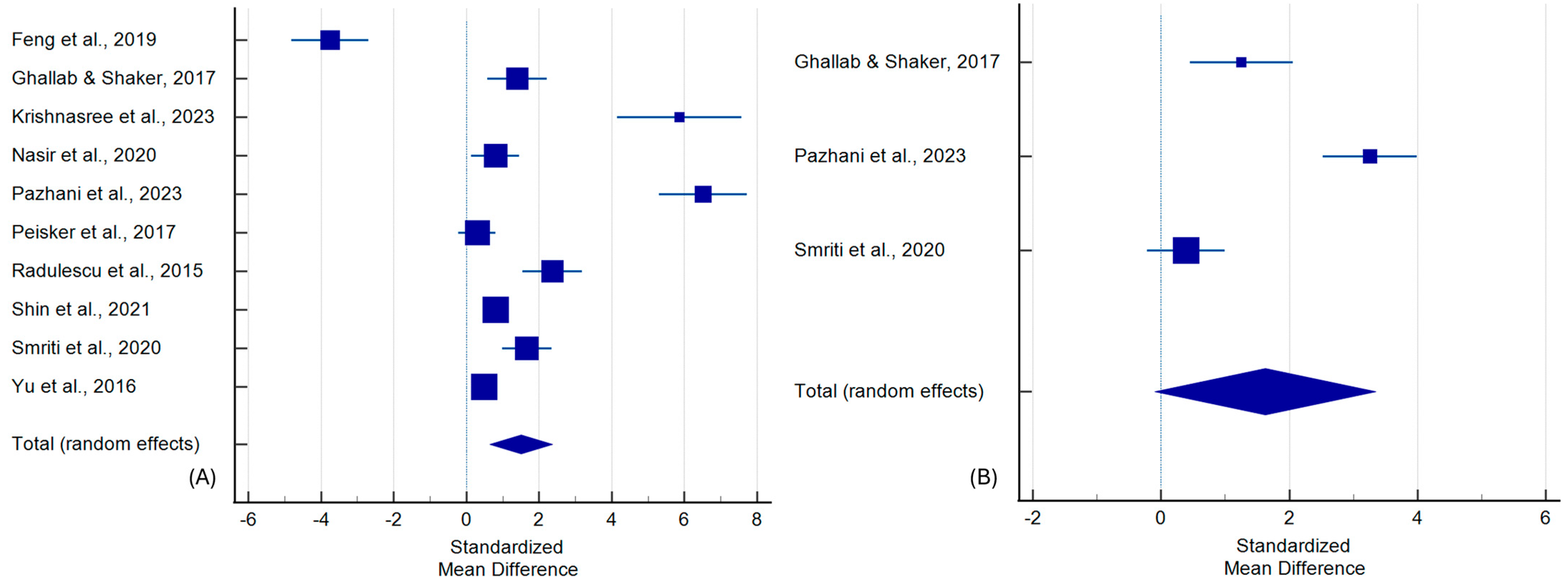
| Feng et al., 2019 [56] | −3.754 | 0.522 | −4.810 to −2.698 | 9.55 | |
| Ghallab & Shaker, 2017 [57] | 1.408 | 0.399 | 0.590 to 2.225 | 10.11 | |
| Krishnasree et al., 2023 [58] | 5.853 | 0.835 | 4.143 to 7.564 | 7.89 | |
| Nasir et al., 2020 [59] | 0.796 | 0.322 | 0.144 to 1.449 | 10.41 | |
| Pazhani et al., 2023 [61] | 6.510 | 0.608 | 5.297 to 7.723 | 9.11 | |
| Peisker et al., 2017 [62] | 0.300 | 0.256 | −0.213 to 0.813 | 10.63 | |
| Radulescu et al., 2015 [63] | 2.363 | 0.406 | 1.544 to 3.181 | 10.08 | |
| Shin et al., 2021 [64] | 0.802 | 0.123 | 0.561 to 1.044 | 10.94 | |
| Smriti et al., 2020 [65] | 1.667 | 0.338 | 0.986 to 2.349 | 10.36 | |
| Yu et al., 2016 [55] | 0.496 | 0.138 | 0.224 to 0.768 | 10.91 | |
| Total (random effects) | 1.507 | 0.439 | 0.644 to 2.369 | 0.001 | |
| Egger’s test | 0.279 | ||||
| Begg’s test | 0.040 | ||||
| Chemerin | |||||
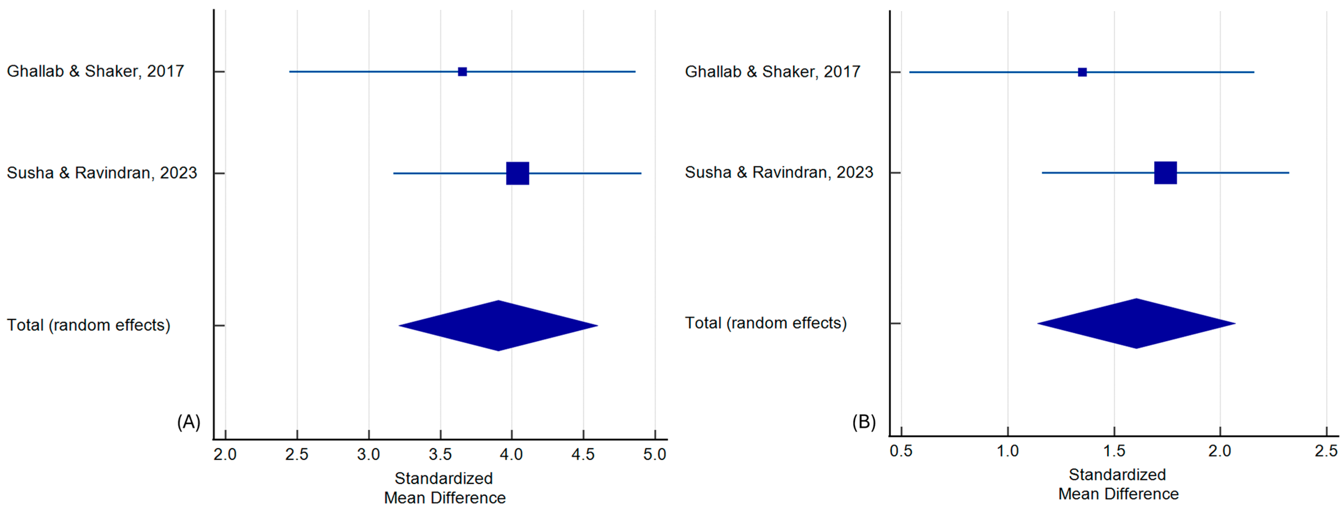
| Ghallab & Shaker, 2017 [57] | 3.655 | 0.591 | 2.446 to 4.865 | 35.07 | |
| Susha & Ravindran, 2023 [66] | 4.040 | 0.434 | 3.172 to 4.908 | 64.93 | |
| Total (random effects) | 3.905 | 0.350 | 3.210 to 4.600 | <0.001 | |
| Egger’s test | <0.001 | ||||
| Begg’s test | 0.317 | ||||
| OC vs. OPMD patients | |||||
| LDH | |||||
| Awasthi, 2017 [35] | 2.074 | 0.440 | 1.182 to 2.966 | 7.14 | |
| Bhuvaneswari et al., 2022 [37] | 2.058 | 0.381 | 1.286 to 2.830 | 7.25 | |
| Dhivyalakshmi & Uma Maheswari, 2014 [39] | 3.313 | 0.575 | 2.131 to 4.495 | 6.85 | |
| Gholizadeh et al., 2020 [40] | 2.678 | 0.386 | 1.901 to 3.454 | 7.24 | |
| Goyal et al., 2020 [41] | 4.253 | 0.255 | 3.750 to 4.756 | 7.44 | |
| Honarmand et al., 2021 [42] | 0.836 | 0.348 | 0.128 to 1.545 | 7.31 | |
| Joshi & Golgire, 2014 [43] | 3.368 | 0.399 | 2.569 to 4.167 | 7.22 | |
| Kadiyala et al., 2015 [44] | −0.320 | 0.312 | −0.952 to 0.312 | 7.36 | |
| Kallalli et al., 2016 [45] | 0.632 | 0.285 | 0.058 to 1.206 | 7.40 | |
| López-Pintor et al., 2024 [47] | 0.530 | 0.320 | −0.110 to 1.171 | 7.35 | |
| Mantri et al., 2019 [48] | 11.563 | 1.086 | 9.389 to 13.736 | 5.47 | |
| Patel & Metgud, 2015 [50] | 2.036 | 0.345 | 1.343 to 2.730 | 7.31 | |
| Shetty et al., 2012 [53] | 2.912 | 0.403 | 2.102 to 3.722 | 7.21 | |
| Subramanian et al., 2024 [54] | 0.319 | 0.256 | −0.195 to 0.832 | 7.44 | |
| Total (random effects) | 2.416 | 0.480 | 1.474 to 3.358 | <0.001 | |
| Egger’s test | 0.076 | ||||
| Begg’s test | 0.025 | ||||
| MMP-9 | |||||
| Ghallab & Shaker, 2017 [57] | 1.254 | 0.390 | 0.454 to 2.054 | 32.95 | |
| Pazhani et al., 2023 [61] | 3.260 | 0.368 | 2.525 to 3.996 | 33.19 | |
| Smriti et al., 2020 [65] | 0.387 | 0.300 | −0.218 to 0.993 | 33.86 | |
| Total (random effects) | 1.626 | 0.872 | −0.097 to 3.350 | 0.064 | |
| Egger’s test | 0.554 | ||||
| Begg’s test | 0.602 | ||||
| Chemerin | |||||
| Ghallab & Shaker, 2017 [57] | 1.350 | 0.396 | 0.539 to 2.160 | 35.11 | |
| Susha & Ravindran, 2023 [66] | 1.743 | 0.291 | 1.161 to 2.325 | 64.89 | |
| Total (random effects) | 1.605 | 0.234 | 1.139 to 2.071 | <0.001 | |
| Egger’s test | <0.001 | ||||
| Begg’s test | 0.317 | ||||
| Poorly and well-differentiated OC patients | |||||
| LDH | |||||
| D’Cruz & Pathiyil, 2015 [38] | 14.282 | 2.298 | 9.453 to 19.110 | 28.93 | |
| Lokesh et al., 2016 [46] | 4.930 | 0.923 | 2.991 to 6.870 | 35.07 | |
| Patel & Metgud, 2015 [50] | 0.827 | 0.561 | −0.369 to 2.022 | 36.00 | |
| Total (random effects) | 6.158 | 2.704 | 0.739 to 11.576 | 0.027 | |
| Egger’s test | 0.113 | ||||
| Begg’s test | 0.117 | ||||
| MMP-9 | |||||
| Krishnasree et al., 2023 [58] | 1.264 | 0.637 | −0.205 to 2.733 | 23.82 | |
| Nisa et al., 2023 [60] | 1.137 | 0.384 | 0.350 to 1.925 | 29.23 | |
| Pazhani et al., 2023 [61] | 3.972 | 0.741 | 2.439 to 5.506 | 21.59 | |
| Smriti et al., 2020 [65] | 1.178 | 0.567 | −0.070 to 2.425 | 25.36 | |
| Total (random effects) | 1.790 | 0.576 | 0.643 to 2.937 | 0.003 | |
| Egger’s test | 0.316 | ||||
| Begg’s test | 0.042 | ||||
| Parameter | Inclusion Criteria | Exclusion Criteria |
|---|---|---|
| Population | Patients aged 0–99 years, both genders | - |
| Exposure | Oral cancer | Cancers other than oral cancer, head and neck cancer without precise localization |
| Comparison | Healthy subjects | - |
| Outcomes | Salivary LDH, MMP-9, chemerin | Other salivary alterations |
| Study design | Case–control, cohort, and cross-sectional studies | Literature reviews, case reports, expert opinion, letters to the editor, conference reports |
| Indexed to 18 April 2025 | Not published in English |
Disclaimer/Publisher’s Note: The statements, opinions and data contained in all publications are solely those of the individual author(s) and contributor(s) and not of MDPI and/or the editor(s). MDPI and/or the editor(s) disclaim responsibility for any injury to people or property resulting from any ideas, methods, instructions or products referred to in the content. |
© 2025 by the authors. Licensee MDPI, Basel, Switzerland. This article is an open access article distributed under the terms and conditions of the Creative Commons Attribution (CC BY) license (https://creativecommons.org/licenses/by/4.0/).
Share and Cite
Owecki, W.; Nijakowski, K. Salivary Lactate Dehydrogenase, Matrix Metalloproteinase-9, and Chemerin—The Most Promising Biomarkers for Oral Cancer? A Systematic Review with Meta-Analysis. Int. J. Mol. Sci. 2025, 26, 7947. https://doi.org/10.3390/ijms26167947
Owecki W, Nijakowski K. Salivary Lactate Dehydrogenase, Matrix Metalloproteinase-9, and Chemerin—The Most Promising Biomarkers for Oral Cancer? A Systematic Review with Meta-Analysis. International Journal of Molecular Sciences. 2025; 26(16):7947. https://doi.org/10.3390/ijms26167947
Chicago/Turabian StyleOwecki, Wojciech, and Kacper Nijakowski. 2025. "Salivary Lactate Dehydrogenase, Matrix Metalloproteinase-9, and Chemerin—The Most Promising Biomarkers for Oral Cancer? A Systematic Review with Meta-Analysis" International Journal of Molecular Sciences 26, no. 16: 7947. https://doi.org/10.3390/ijms26167947
APA StyleOwecki, W., & Nijakowski, K. (2025). Salivary Lactate Dehydrogenase, Matrix Metalloproteinase-9, and Chemerin—The Most Promising Biomarkers for Oral Cancer? A Systematic Review with Meta-Analysis. International Journal of Molecular Sciences, 26(16), 7947. https://doi.org/10.3390/ijms26167947


