Review

Factors Contributing to Resistance to Ischemia-Reperfusion Injury in Olfactory Mitral Cells

by
Choong-Hyun Lee
1,
Ji Hyeon Ahn
2 and
Moo-Ho Won
3,4,*
1
Department of Pharmacy, College of Pharmacy, Dankook University, Cheonan 31116, Republic of Korea
2
Department of Physical Therapy, College of Health Science, Youngsan University, Yangsan 50510, Republic of Korea
3
Department of Emergency Medicine, Kangwon National University Hospital, Chuncheon 24289, Republic of Korea
4
Department of Emergency Medicine, School of Medicine, Kangwon National University, Chuncheon 24341, Republic of Korea
*
Author to whom correspondence should be addressed.
Int. J. Mol. Sci. 2025, 26(11), 5079; https://doi.org/10.3390/ijms26115079
Submission received: 21 April 2025 / Revised: 15 May 2025 / Accepted: 23 May 2025 / Published: 25 May 2025
(This article belongs to the Special Issue New Molecular Insights into Ischemia/Reperfusion: 2nd Edition)

Abstract

Brain ischemia-reperfusion (IR) injury is a critical pathological process that leads to extensive neuronal death, with hippocampal pyramidal cells, particularly those in the cornu Ammonis 1 (CA1) subfield, being highly vulnerable. Until now, human olfactory mitral cell resistance to IR injury has not been directly studied, but olfactory dysfunction in humans is frequently reported in systemic vascular conditions such as ischemic heart failure and may serve as an early clinical marker of neurological or cardiovascular disease. Mitral cells, the principal neurons of the olfactory bulb (OB), exhibit remarkable resistance to IR injury, suggesting the presence of unique molecular adaptations that support their survival under ischemic stress. Several factors may contribute to the resilience of mitral cells. They have a lower susceptibility to excitotoxicity, mitigating the harmful effects of excessive glutamate signaling. Additionally, they maintain efficient calcium homeostasis, preventing calcium overload—a major trigger for cell death in vulnerable neurons. Mitral cells may also express high baseline levels of antioxidant enzymes and their activities, counteracting oxidative stress. Their robust mitochondrial function enhances energy production and reduces susceptibility to metabolic failure. Furthermore, neuroprotective signaling pathways, including phosphatidylinositol-3-kinase (PI3K)/Akt, mitogen-activated protein kinase/extracellular signal-regulated kinase (MAPK/ERK), and nuclear factor erythroid-2-related factor 2 (Nrf2)-mediated antioxidative responses, further bolster their resistance. In addition to these intrinsic mechanisms, the unique microvascular architecture and metabolic support within the olfactory bulb provide an extra layer of protection. By comparing mitral cells to ischemia-sensitive neurons, key vulnerabilities—such as oxidative stress, excitotoxicity, calcium dysregulation, and mitochondrial dysfunction—can be identified and potentially mitigated in other brain regions. Understanding these molecular determinants of neuronal survival may offer valuable insights for developing novel neuroprotective strategies to combat IR injury in highly vulnerable areas, such as the hippocampus and cortex.

1. Introduction

The olfactory system is responsible for detecting and processing odor information and plays a vital role in sensory perception [1,2]. The system consists of peripheral components (the olfactory epithelium and olfactory nerve), central components (the olfactory bulb and olfactory tract), and higher-order processing areas (the olfactory cortex, amygdala, entorhinal cortex, hippocampus, and orbitofrontal cortex [1]. It begins with olfactory receptor neurons in the nasal epithelium, which transmit signals to the olfactory bulb (OB) [1]. Within the OB, mitral cells, as the principal output neurons, integrate sensory signals and refine odor information through interactions with tufted cells, granule cells, and periglomerular cells, which provide inhibitory modulation [2,3].
Brain or cerebral ischemia, which is characterized by reduced blood flow to neural tissue, leads to oxygen and nutrient deprivation and causes cellular dysfunction and death [4,5]. Reperfusion injury occurs when blood flow is restored, which contributes to secondary brain damage after ischemic stroke or cardiac arrest, often exacerbating damage through excitotoxicity, oxidative stress, and inflammation [4,5]. Most central neurons are highly vulnerable to these conditions, resulting in severe neurological impairments [6].
IR injury typically results in extensive neuronal damage in brain regions such as the hippocampus, but emerging studies in animal models (e.g., gerbils, rats, dogs) suggest that mitral cells in the olfactory bulb may exhibit resistance [7,8,9,10,11,12] (Figure 1). Investigating the mechanisms underlying their resilience could provide critical insights into neuroprotection, potentially guiding therapeutic approaches for ischemic stroke and other ischemic brain disorders. In addition, while mitral cell resistance to IR injury has been demonstrated in rodent and canine models [7,8,10,13,14], extrapolation to the human brain must be performed cautiously due to known species-specific differences in olfactory bulb structure, vascularization, and neural connectivity [1,13]. Although mitral cell morphology differs across species, the evolutionary conservation of redox homeostatic mechanisms [14] and the identification of a rodent-like neurogenic migratory system in primates [15] support the translational relevance of rodent models. This review aims to synthesize current evidence from these animal studies and explore molecular and anatomical factors contributing to this resilience, while acknowledging that extrapolation to human biology must be performed cautiously due to species-specific differences in olfactory system anatomy and function.
For the literature search strategy, we conducted a focused literature search using PubMed, Scopus, and Google Scholar databases. The keywords included: olfactory mitral cells, ischemia-reperfusion injury, neuronal resistance, oxidative stress, mitochondrial function, antioxidant enzymes, and neuroprotection. Emphasis was placed on original experimental research, reviews, and histological reports that compared IR-resistant and IR-vulnerable neurons.

2. Structure and Function of Olfactory Mitral Cells

In the OB, mitral cells are a principal type of excitatory neuron and play a crucial role in processing olfactory information. They receive direct input from olfactory receptor neurons, which are located in the nasal cavity, via synapses in the glomeruli of the OB, where each mitral cell connects to a specific set of sensory inputs [1,3] (Figure 2). Anatomically, mitral cells have large, apical dendrites that extend into the glomerular layer and basal dendrites that interact with local interneurons, such as granule cells, in the external plexiform layer [2] (Figure 2). Their axons project to higher-order brain regions (the piriform cortex, amygdala, entorhinal cortex, etc.), forming the olfactory pathway that links sensory perception to cognitive and emotional responses [2] (Figure 2).
Mitral cells are essential for olfactory signal processing as the primary relay between sensory input and higher cortical areas [1,2,3] as follows. The mitral cells receive excitatory input from olfactory sensory neurons and refine the signal through interactions with inhibitory interneurons, particularly periglomerular and granule cells. They exhibit synchronized oscillatory activity, which is crucial for encoding odor identity and intensity. Finally, they ensure, through their widespread projections, that olfactory information is integrated with memory and emotional processing centers, allowing for the perception and interpretation of complex odor stimuli. In addition, ultrastructural studies of olfactory mitral cells have revealed several unique morphological features that may support their functional resilience. Electron microscopy shows that mitral cells contain abundant, elongated mitochondria clustered near dendritic and axonal synapses, suggesting a high capacity for localized energy production and calcium buffering [17,18]. These mitochondria are often tightly associated with smooth and rough endoplasmic reticulum, forming mitochondrial-associated membranes (MAMs) that facilitate lipid transfer and calcium signaling—both of which are critical for stress responses [19,20]. The nuclei of mitral cells are typically large and euchromatic, reflecting high transcriptional activity, while their somatic cytoplasm is rich in ribosomes and Golgi apparatus, indicating robust protein synthesis [21]. In contrast to ischemia-sensitive neurons such as hippocampal CA1 pyramidal cells—which show early mitochondrial swelling and fragmentation following IR injury—mitral cell mitochondria appear more structurally stable, even under ischemic stress [14]. These ultrastructural attributes may underlie their superior bioenergetic function and lower susceptibility to ischemic damage.
When compared to other central nervous system neurons, mitral cells possess several unique properties that contribute to their specialized function. One of their remarkable characteristics is their resistance to ischemia [8,9,10,11], a feature that distinguishes them from vulnerable neurons in regions such as the hippocampus [22] and cortex [23]. In addition, the OB may possess evolutionary adaptations that confer enhanced resistance to IR injury, particularly in rodents. In many rodent species, the OB plays a central role in environmental navigation, food foraging, predator detection, and social communication, making olfaction their dominant sensory modality [24,25]. This reliance on olfaction likely exerted evolutionary pressure to preserve OB function under a variety of stress conditions, including transient metabolic deprivation. Supporting this idea, olfactory structures in rodents exhibit high vascular density, robust metabolic support, and relatively consistent perfusion even under systemic stress [26,27]. Moreover, rodents typically show a lower incidence of cerebrovascular events compared to humans, possibly reflecting species-specific cerebrovascular patterns and adaptive resistance mechanisms in critical brain regions like the OB [28,29]. These considerations suggest that the IR resistance observed in mitral cells may be the result of evolutionary optimization to protect a functionally dominant brain structure in species highly dependent on olfaction. Taken together, the resilience may be attributed to differences in metabolic demand, ion channel composition, or neuroprotective mechanisms, which are discussed in this review.

3. Mechanisms of IR Injury in the Brain

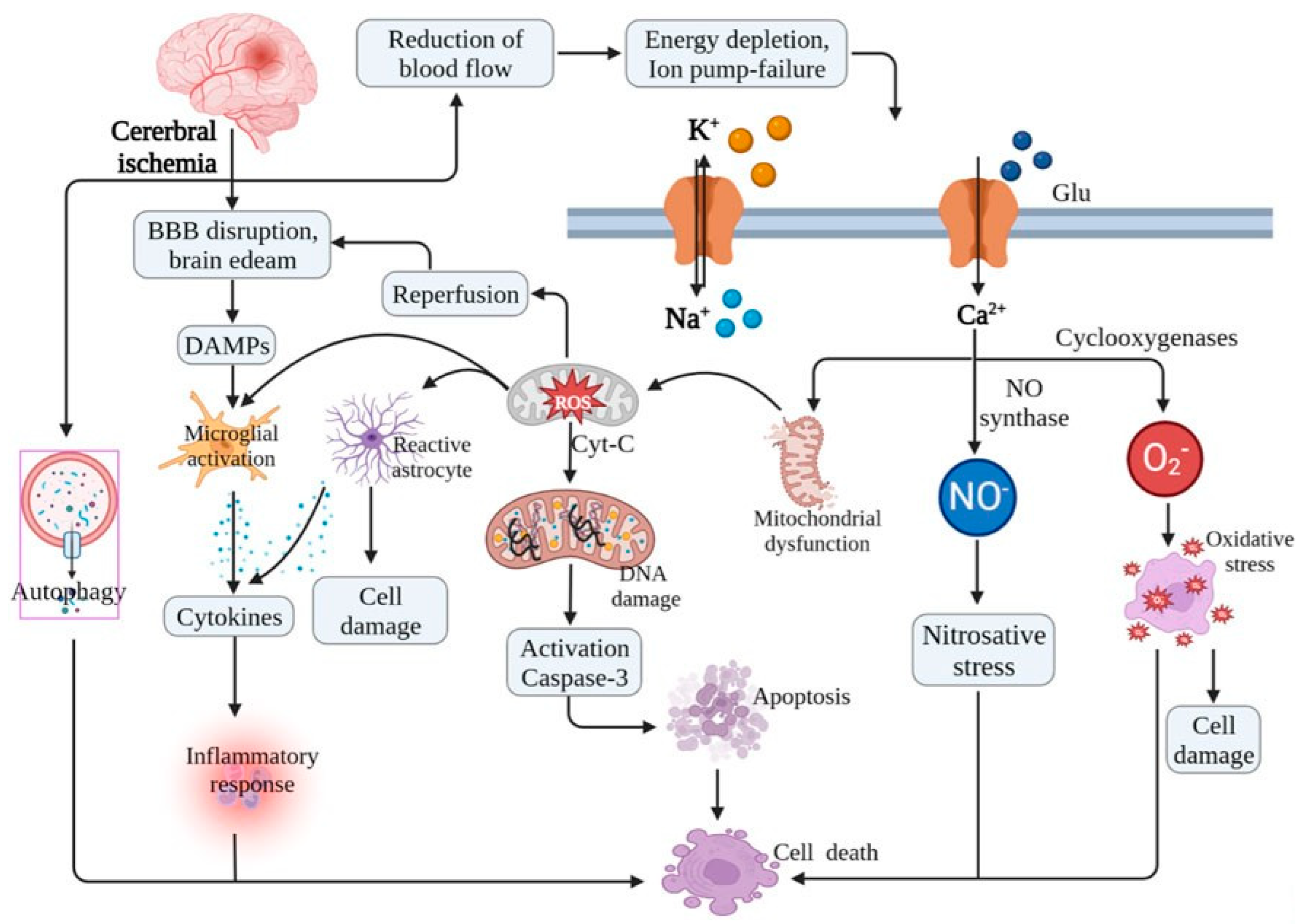
IR injury in the brain is a complex pathological process involving a cascade of biochemical and molecular events. Ischemic stroke occurs when cerebral blood flow is reduced due to an obstruction in the arteries and leads to oxygen and glucose deprivation [30]. When blood flow is restored, reperfusion can paradoxically exacerbate neuronal damage through excitotoxicity, oxidative stress, inflammation, and mitochondrial dysfunction [31] (Figure 3). Excitotoxicity occurs during ischemia as follows [32,33]. ATP production is reduced due to mitochondrial dysfunction leading to intracellular acidosis, ion pump failure, and the accumulation of intracellular sodium and calcium. To compensate for acidosis, the Na+/H+ exchanger is activated, resulting in Na+ influx and increased intracellular Na+ levels [34]. Simultaneously, impaired Na+/K+-ATPase function further exacerbates intracellular Na+ accumulation. These changes trigger the reverse operation of the Na+/Ca2+ exchanger, leading to Ca2+ overload. This loss of ionic homeostasis results in excessive glutamate release and sustained membrane depolarization, thereby initiating excitotoxicity. The overactivation of N-methyl-D-aspartate (NMDA) and alpha-amino-3-hydroxy-5-methyl-4-isooxazole-propionic acid (AMPA) receptors facilitates further calcium influx, which activates calpain, phospholipases, and other proteases that degrade cellular components and contribute to neuronal death.
Oxidative stress occurs following reperfusion as follows. Reperfusion introduces a sudden influx of oxygen, which promotes the generation of reactive oxygen species (ROS), including superoxide anions and hydroxyl radicals. Excessive ROS production damages lipids, proteins, and DNA, leading to apoptosis and necrosis [30,35].
Inflammation and blood-brain barrier (BBB) disruption after/during reperfusion occur as follows [36,37]. Reperfusion activates microglia and astrocytes, which leads to the release of pro-inflammatory cytokines such as tumor necrosis factor-alpha (TNF-α) and interleukins (IL-1β, IL-6). In addition, infiltrating neutrophils exacerbate inflammation by producing matrix metalloproteinases (MMPs), which degrade tight junction proteins, leading to BBB disruption, vasogenic edema, and hemorrhagic transformation.
During reperfusion, mitochondrial permeability transition pore (mPTP) opening leads to the release of cytochrome c and caspase activation, which promotes apoptosis. Additionally, excessive calcium and ROS contribute to mitochondrial collapse and neuronal death [31,38].

4. Types of Neurons Vulnerable and Resistant to IR Injury

Neurons exhibit varying degrees of vulnerability to IR injury. As described above, the vulnerability is influenced by factors such as metabolic demand, cellular structure, and blood supply. Neurons in the hippocampus, such as CA1 pyramidal cells, are highly susceptible due to their complex dendritic arborizations and dependence on high metabolic activity [39]. Purkinje cells, as principal neurons in the cerebellum, are highly vulnerable to IR injury, likely due to their high metabolic demand [40]. Conversely, certain neuron types display resistance to IR injury, notably olfactory mitral cells, which are protected by unique cellular features, such as lower excitability, specialized vascularization, and metabolic support. Overall, the response of neurons to IR injury is determined by a combination of anatomical, physiological, and molecular factors, with some cells exhibiting remarkable resistance to damage. As shown in Table 1, according to the types of neurons, they are known to be vulnerable or resistant to IR injury.

5. Factors of Mitral Cell Resistance to IR Injury

Olfactory mitral cells exhibit remarkable resistance to IR injury compared to other central nervous system (CNS) neurons, as described above. The following unique biochemical adaptation may contribute to mitral cells’ survival under ischemic stress, offering insights into potential neuroprotective strategies for vulnerable neuronal populations, such as hippocampal pyramidal cells, in ischemic brain injury.

5.1. Low Excitotoxicity Susceptibility and Efficient Calcium Homeostasis

In brain IR injury, excitotoxicity, as a major mechanism of neuronal death/loss, is primarily driven by excessive glutamate release and overstimulation of ionotropic glutamate receptors [46]. However, not all neurons exhibit the same degree of susceptibility to excitotoxic damage [47,48]. Upon reviewing the available literature, studies directly linking the low excitotoxicity susceptibility of olfactory mitral cells to their resistance against IR injury are limited or not readily accessible [49,50]. Nevertheless, one potential factor contributing to the resilience of mitral cells to IR injury is their relatively low NMDA receptor (NMDAR) density compared to highly vulnerable neuronal populations, such as hippocampal CA1 pyramidal neurons [51]. In addition, mitral cells receive extensive inhibitory input from granule and periglomerular cells, which regulate excitatory signaling and may help prevent sustained glutamate receptor activation and calcium overload [52,53,54]. Another important factor is the intrinsic calcium homeostasis of mitral cells. When compared to highly vulnerable neurons, mitral cells may have more efficient calcium buffering mechanisms, including enhanced mitochondrial calcium uptake or increased expression of calcium-binding proteins, both of which can limit excitotoxic damage [55,56]. Taken together, these findings suggest that mitral cells possess multiple intrinsic and network-level features that reduce their susceptibility to excitotoxic injury. However, direct experimental evidence confirming these mechanisms remains limited. Furthermore, although NMDA receptors are well-known mediators of excitotoxicity during IR injury due to their high calcium permeability, they are also essential for physiological processes such as learning and memory through their role in synaptic plasticity and long-term potentiation [57]. Therefore, broad inhibition of NMDA receptors—while neuroprotective in the short term—can impair cognitive function if used indiscriminately [58]. This dual role underscores the complexity of targeting NMDA signaling therapeutically. In olfactory mitral cells, lower baseline NMDA receptor density may offer a protective advantage during IR injury without entirely abolishing their synaptic plasticity or integrative function. Therefore, neuroprotective approaches may benefit from selectively targeting receptor subtypes or modulating temporal activation windows to reduce excitotoxicity while preserving memory-related functions [59]. Taken together, future studies should focus on electrophysiological and molecular analyses of glutamate receptor activity, inhibitory synaptic regulation, and calcium homeostasis in mitral cells following IR injury. Understanding these properties may provide valuable insights into neuronal resilience and potential neuroprotective strategies.

5.2. High Antioxidant Levels or Activities

Both the levels and activities of antioxidant enzymes play critical roles in neuronal resistance to IR injury, but enzyme activity is generally more critical for neuroprotection [60,61]. Upon reviewing the available literature, it appears that specific studies directly linking the high antioxidant enzyme levels of olfactory mitral cells to their resistance against IR injury are limited or not readily accessible [62]. While general mechanisms of neuronal resistance to oxidative stress have been documented, direct evidence pertaining to olfactory mitral cells is scarce. One potential factor contributing to this resilience is the high baseline expression of antioxidant enzymes, such as superoxide dismutase (SOD), catalase, and glutathione peroxidase, which neutralize reactive oxygen species (ROS) and mitigate oxidative stress [63]. Oxidative stress plays a critical role in IR-induced neuronal death, particularly in hippocampal CA1 pyramidal neurons, which have lower endogenous antioxidant defenses and are highly susceptible to excitotoxicity and mitochondrial dysfunction [61,64,65]. Taken together, it is suggested that antioxidant-rich environments protect neurons from ischemic damage, and mitral cells may inherently maintain a more robust antioxidant system, reduce ROS accumulation, and preserve cellular integrity. In the future, we need studies on changes in levels of antioxidant enzymes or their activities in the olfactory mitral cells following IR injury.

5.3. Robust Mitochondrial Function

Hippocampal CA1 pyramidal neurons, which are highly vulnerable to IR injury, display significant mitochondrial dysfunction, including impaired calcium handling and increased susceptibility to oxidative damage [66,67]. Mitochondria play a crucial role in neuronal survival and function, particularly during IR injury, where energy failure and oxidative stress contribute to neuronal damage. One of the key determinants of neuronal resistance to IR injury is mitochondrial integrity and efficiency in energy production, calcium buffering, and ROS regulation [68,69]. While direct studies on mitochondrial function in olfactory mitral cells following IR injury remain limited, evidence suggests that mitral cells may possess a robust mitochondrial network that enhances their resilience. One potential factor contributing to this resistance is the high density of mitochondria within mitral cells, which can support sustained synaptic activity and ATP production under metabolic stress [70,71]. Additionally, mitral cells may exhibit a more efficient oxidative phosphorylation system, reducing the likelihood of ATP depletion and mitochondrial depolarization, which are critical events in IR-induced neuronal death [72,73]. Finally, Mitochondrial uncoupling proteins (UCPs), such as UCP2, UCP4, and UCP5, are known to play neuroprotective roles by mitigating ROS generation and preserving mitochondrial membrane potential [74,75,76,77]. Although specific studies in mitral cells are lacking, the possible involvement of UCPs may contribute to the robust mitochondrial function observed in these cells under ischemic conditions. Taken together, these findings suggest that robust mitochondrial function, efficient ATP production, and enhanced mitochondrial resilience may underlie the lower susceptibility of mitral cells to IR-induced damage. Future studies should investigate mitochondrial dynamics, bioenergetic profiles, and oxidative stress responses in olfactory mitral cells following ischemic events to better understand their unique neuroprotective mechanisms.

5.4. Neuroprotective Signaling Pathways

Neuronal survival following IR injury is highly dependent on the activation of protective intracellular signaling pathways that regulate oxidative stress, inflammation, and apoptosis [78,79]. Studies have identified key signaling cascades, including the PI3K/Akt pathway, MAPK/ERK signaling, and Nrf2-mediated antioxidative responses, as critical determinants of neuronal resistance to ischemic damage [79,80,81]. However, direct evidence regarding the activation of these pathways in olfactory mitral cells remains limited. The PI3K/Akt pathway is a well-documented prosurvival mechanism that promotes neuronal resilience by inhibiting apoptosis and enhancing metabolic stability under stress conditions [79]. Given the relative resistance of mitral cells to IR-induced apoptosis, it is plausible that these neurons exhibit sustained Akt activation during ischemic stress. Another pathway is MAPK/ERK signaling, which plays a dual role in neuronal survival and plasticity. ERK1/2 activation is associated with neuroprotection by modulating gene expression, increasing antioxidant defenses, and reducing excitotoxic damage [80,82]. Since mitral cells demonstrate lower susceptibility to excitotoxicity compared to hippocampal CA1 pyramidal neurons, persistent ERK activity may underlie their enhanced ischemic tolerance. In addition, the Nrf2/ARE pathway is a crucial regulator of antioxidant gene expression and is essential for mitigating oxidative stress during IR injury [81]. Activation of Nrf2 enhances the transcription of antioxidant enzymes such as heme oxygenase-1 (HO-1), NAD(P)H quinone oxidoreductase-1 (NQO1), and glutathione S-transferase, which collectively reduce ROS accumulation and cellular damage [83,84]. While Nrf2 signaling is well-characterized in other neuronal populations, its specific role in mitral cell resilience warrants further investigation. Based on those findings, future studies should focus on characterizing the activation profiles of PI3K/Akt, MAPK/ERK, and Nrf2 pathways in mitral cells following IR injury, as these could provide insight into novel therapeutic targets for neuroprotection.

5.5. Unique Microvascular and Metabolic Support

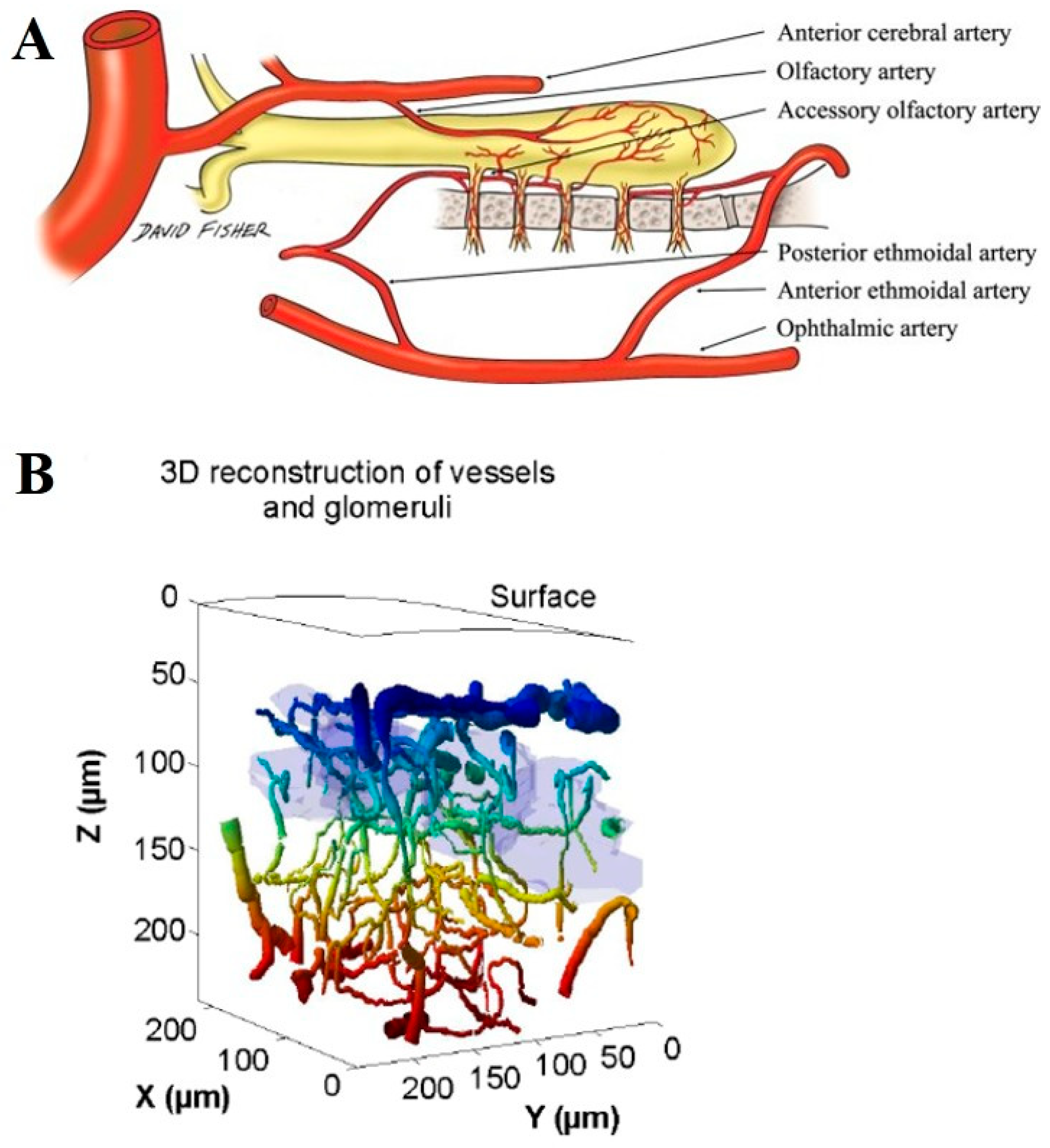
OB exhibits a distinct vascular architecture and metabolic profile that may contribute to the resilience of mitral cells against IR injury. Compared to other brain regions, the OB has a relatively high capillary density and unique patterns of blood flow regulation, which may enhance oxygen and nutrient delivery even under conditions of transient ischemia [26,27,85,86] (Figure 4). This well-developed microvascular network could facilitate more efficient metabolic support and reduce the severity of ischemic damage. One key factor in the metabolic resilience of mitral cells is their ability to efficiently utilize aerobic metabolism and maintain mitochondrial integrity. Studies have shown that neurons with robust mitochondrial function are better equipped to handle oxidative stress and energy depletion following IR injury [87,88]. The OB also possesses a high metabolic rate, which may be supported by enhanced glucose uptake and lactate metabolism from surrounding glial cells, particularly astrocytes [89,90]. Astrocyte-derived lactate can serve as an alternative energy substrate for neurons under metabolic stress, thereby preserving mitral cell function during IR episodes. Furthermore, the OB is uniquely equipped with high levels of neurovascular coupling and ensures rapid adjustments in blood flow in response to metabolic demands [91,92]. This mechanism may provide mitral cells with a more stable energy supply and reduce the impact of ischemic insults. Additionally, the presence of specialized pericytes and endothelial cells in olfactory bulb capillaries, which can modulate cerebral blood flow by changing capillary diameter, may enhance BBB integrity and facilitate neuroprotective signaling [91,93]. Overall, the unique microvascular and metabolic adaptations of OB likely play a crucial role in protecting mitral cells from IR-induced damage. Future studies should investigate the specific contributions of these adaptations to mitral cell survival and determine whether they can be leveraged for therapeutic strategies against cerebral ischemic insults.

6. Implications for Neuroprotection and Therapeutic Potential

The unique resilience of olfactory mitral cells to IR injury offers valuable insights into potential neuroprotective strategies for vulnerable brain regions. When we understand the molecular adaptations that confer resistance to mitral cells, we can explore targeted interventions to enhance neuronal survival following ischemic events. Several key mechanisms may serve as therapeutic targets as follows. The reduced susceptibility of mitral cells to glutamate-induced excitotoxicity suggests that therapies aimed at modulating glutamate receptors or enhancing glutamate clearance may help protect vulnerable neurons, such as hippocampal CA1 pyramidal cells, from IR injury. Maintaining calcium balance is crucial for neuronal survival. Strategies that enhance calcium buffering capacity, such as upregulating calcium-binding proteins or modulating ion channels, could mitigate calcium overload and associated cytotoxic effects in ischemia-sensitive neurons, such as hippocampal CA1 pyramidal neurons. The high baseline expression of antioxidant enzymes in mitral cells underscores the potential benefits of antioxidant-based therapies. Upregulating endogenous antioxidant responses through Nrf2 activators or administering exogenous antioxidants may reduce oxidative damage and improve neuronal resilience. Given the robust mitochondrial function of mitral cells, interventions that enhance mitochondrial bioenergetics—such as mitochondrial-targeted antioxidants, metabolic substrates, or pharmacological agents that stabilize mitochondrial integrity—could support neuronal survival in ischemic conditions. Targeting key protective signaling cascades, including PI3K/Akt and MAPK/ERK, could provide broad-spectrum neuroprotection. Pharmacological agents that activate these pathways may help sustain cell survival and function in ischemia-prone regions. The OB’s specialized vascular and metabolic environment highlights the importance of optimizing cerebral blood flow and energy supply. Strategies such as promoting angiogenesis, enhancing cerebral perfusion, and modulating metabolic substrates may improve neuronal survival after IR injury.

7. Future Directions and Open Questions

Several critical questions remain unanswered despite the understanding of mitral cell resilience to IR injury. Addressing these knowledge gaps may further enhance our ability to develop effective neuroprotective strategies: Identifying key genes, transcriptional regulators, and epigenetic modifications that contribute to their resilience could provide new therapeutic targets. Investigating the role of specialized vascular structures, BBB integrity, and metabolic coupling in sustaining mitral cells could inform strategies to enhance perfusion and nutrient supply in ischemia-sensitive regions. Determining whether ischemia-prone neurons can be reprogrammed or conditioned to adopt similar resistance mechanisms could open new avenues for neuroprotection. Understanding whether mitral cells exhibit unique interactions with glial cells or inflammatory mediators may reveal novel anti-inflammatory strategies for mitigating IR injury. Investigating whether mitral cells sustain subtle impairments post-ischemia and how they recover functionally over time may provide insights into long-term neuronal resilience. Furthermore, despite evidence from animal models indicating that mitral cells are structurally resistant to IR injury, olfactory dysfunction is widely recognized as an early biomarker of neurodegenerative diseases, including Parkinson’s and Alzheimer’s disease [13,94]. In addition, although human olfactory mitral cell resistance has not been directly studied, olfactory dysfunction in humans is frequently reported in systemic vascular conditions such as ischemic heart failure and may serve as an early clinical marker of neurological or cardiovascular disease [95,96]. This apparent discrepancy may reflect the fact that olfactory deficits can arise from functional alterations—such as synaptic changes, glial activation, and axonal degeneration—rather than overt neuronal loss [97], which deserves future investigation. Thus, mitral cells may survive IR insults structurally while experiencing sublethal dysfunction that contributes to clinical olfactory decline. These findings suggest that resilience to IR injury does not exclude the possibility of early olfactory impairment in neurodegenerative conditions and highlight the need to distinguish between neuronal viability and function in disease models. Taken together, future research should focus on mechanistic studies, in vivo models, and translational approaches to apply these findings to clinical settings. Addressing these open questions will be essential for advancing neuroprotective strategies against ischemic brain injury.

8. Conclusions

Olfactory mitral cells exhibit strong resistance to IR injury, which distinguishes them from other neuronal populations that are highly vulnerable to ischemic damage. Their resilience might be attributed to multiple intrinsic and extrinsic factors, including reduced excitotoxicity susceptibility, efficient calcium homeostasis, robust antioxidant defenses, strong mitochondrial function, and activation of neuroprotective signaling pathways. Additionally, the specialized vascular and metabolic environment of the OB provides further support for mitral cell survival. Understanding these protective mechanisms not only deepens our knowledge of neuronal ischemia resistance, but also holds significant therapeutic potential for treating IR injury in vulnerable brain regions. By leveraging insights from mitral cells, novel neuroprotective strategies—ranging from pharmacological interventions to gene therapy—could be developed to enhance neuronal survival following ischemic events. However, several critical questions remain, necessitating further research to fully elucidate the molecular and physiological basis of mitral cell resilience. By translating these findings into clinical applications, the field of neuroprotection may advance toward more effective therapies for ischemic stroke and other conditions characterized by neuronal vulnerability to ischemic stress.

Author Contributions

Conceptualization, M.-H.W., C.-H.L. and J.H.A.; data curation, M.-H.W., C.-H.L. and J.H.A.; writing—original draft preparation, M.-H.W., C.-H.L. and J.H.A.; writing—review and editing, M.-H.W.; supervision, M.-H.W. All authors have read and agreed to the published version of the manuscript.

Funding

This research received no external funding.

Institutional Review Board Statement

Not applicable.

Informed Consent Statement

Not applicable.

Data Availability Statement

Data sharing not applicable.

Conflicts of Interest

The authors declare no conflicts of interest.

Abbreviations

alpha-amino-3-hydroxy-5-methyl-4-isooxazole-propionic acid receptor (AMPAR), blood-brain barrier (BBB), central nervous system (CNS), cornu Ammonis (CA), interleukin (IL), Fluoro Jade B (F-J B), heme oxygenase-1 (HO-1), ischemia-reperfusion (IR), mitochondrial-associated membranes (MAMs), matrix metalloproteinases (MMPs), mitochondrial permeability transition pore (mPTP), mitogen-activated protein kinase/extracellular signal-regulated kinase (MAPK/ERK), NAD(P)H quinone oxidoreductase-1 (NQO1), N-methyl-D-aspartate receptor (NMDAR), nuclear factor erythroid-2-related factor 2 (Nrf2); olfactory bulb (OB), phosphatidylinositol-3-kinase (PI3K), reactive oxygen species (ROS); superoxide dismutase (SOD), terminal deoxynucleotidyl transferase dUTP nick end labeling (TUNEL), tumor necrosis factor-alpha (TNF-α).

References

  1. Smith, T.D.; Bhatnagar, K.P. Anatomy of the olfactory system. Handb. Clin. Neurol. 2019, 164, 17–28. [Google Scholar] [PubMed]
  2. Han, S.A.; Kim, J.K.; Cho, D.-Y.; Patel, Z.M.; Rhee, C.-S. The olfactory system: Basic anatomy and physiology for general otorhinolaryngologists. Clin. Exp. Otorhinolaryngol. 2023, 16, 308–316. [Google Scholar] [CrossRef]
  3. Olivares, J.; Schmachtenberg, O. An update on anatomy and function of the teleost olfactory system. PeerJ 2019, 7, e7808. [Google Scholar] [CrossRef]
  4. Chen, Z.; Wang, S.; Shu, T.; Xia, S.; He, Y.; Yang, Y. Progress in Research on Regulated Cell Death in Cerebral Ischaemic Injury After Cardiac Arrest. J. Cell. Mol. Med. 2025, 29, e70404. [Google Scholar] [CrossRef]
  5. Ren, Y.; Chen, G.; Hong, Y.; Wang, Q.; Lan, B.; Huang, Z. Novel Insight into the Modulatory Effect of Traditional Chinese Medicine on Cerebral Ischemia-Reperfusion Injury by Targeting Gut Microbiota: A Review. Drug Des. Dev. Ther. 2025, 19, 185–200. [Google Scholar] [CrossRef] [PubMed]
  6. Zheng, T.; Jiang, T.; Huang, Z.; Ma, H.; Wang, M. Role of traditional Chinese medicine monomers in cerebral ischemia/reperfusion injury: A review of the mechanism. Front. Pharmacol. 2023, 14, 1220862. [Google Scholar] [CrossRef] [PubMed]
  7. Zachariás, L.; Vanický, I.; Mechírová, E.; Marsala, M.; Marsala, J. Selective vulnerability of neuronal injury after experimental heart arrest. Bratisl. Lek. Listy 1995, 96, 661–665. [Google Scholar]
  8. Koh, U.; Hwang, I.; Lee, J.; Lee, H.; Seong, N.; Chung, H.; Kim, J.; Lee, H.; Choi, G.; Kang, T.C.; et al. Histochemical study on neurodegeneration in the olfactory bulb after transient forebrain ischaemia in the Mongolian gerbil. Anat. Histol. Embryol. 2004, 33, 208–211. [Google Scholar] [CrossRef]
  9. Hwang, I.K.; Koh, U.-S.; Lee, J.C.; Yoo, K.-Y.; Song, J.-H.; Jung, J.-Y.; Nam, Y.S.; Lee, I.S.; Kang, T.-C.; Won, M.H. Transient ischemia-induced changes of neurofilament 200 kDa immunoreactivity and protein content in the main olfactory bulb in gerbils. J. Neurol. Sci. 2005, 239, 59–66. [Google Scholar] [CrossRef]
  10. Zhan, X.; Wei, Y.; Miao, X.; Zhang, C.; Han, D. Effects of ischemia on olfactory bulb in rats. Lin Chuang Er Bi Yan Hou Tou Jing Wai Ke Za Zhi = J. Clin. Otorhinolaryngol. Head Neck Surg. 2007, 21, 219–221. [Google Scholar]
  11. Hwang, I.K.; Yoo, K.-Y.; Kim, D.W.; Li, H.; Park, O.K.; Lee, C.H.; Choi, J.H.; Won, M.-H. αII-Spectrin breakdown product increases in principal cells in the gerbil main olfactory bulb following transient ischemia. Neurosci. Lett. 2008, 435, 251–256. [Google Scholar] [CrossRef] [PubMed]
  12. Choi, J.H.; Yoo, K.Y.; Park, O.K.; Lee, C.H.; Kim, S.K.; Hwang, I.K.; Lee, Y.L.; Shin, H.C.; Won, M.H. Relation among neuronal death, cell proliferation and neuronal differentiation in the gerbil main olfactory bulb after transient cerebral ischemia. Cell Mol. Neurobiol. 2010, 30, 929–938. [Google Scholar] [CrossRef]
  13. Attems, J.; Walker, L.; Jellinger, K.A. Olfactory bulb involvement in neurodegenerative diseases. Acta Neuropathol. 2014, 127, 459–475. [Google Scholar] [CrossRef] [PubMed]
  14. Loboda, A.; Damulewicz, M.; Pyza, E.; Jozkowicz, A.; Dulak, J. Role of Nrf2/HO-1 system in development, oxidative stress response and diseases: An evolutionarily conserved mechanism. Cell. Mol. Life Sci. 2016, 73, 3221–3247. [Google Scholar] [CrossRef]
  15. Kornack, D.R.; Rakic, P. The generation, migration, and differentiation of olfactory neurons in the adult primate brain. Proc. Natl. Acad. Sci. USA 2001, 98, 4752–4757. [Google Scholar] [CrossRef]
  16. Cavarretta, F.; Burton, S.D.; Igarashi, K.M.; Shepherd, G.M.; Hines, M.L.; Migliore, M. Parallel odor processing by mitral and middle tufted cells in the olfactory bulb. Sci. Rep. 2018, 8, 7625. [Google Scholar] [CrossRef] [PubMed]
  17. Price, J.L.; Powell, T.P. The mitral and short axon cells of the olfactory bulb. J. Cell Sci. 1970, 7, 631–651. [Google Scholar] [CrossRef]
  18. Kosaka, K.; Kosaka, T. Chemical properties of type 1 and type 2 periglomerular cells in the mouse olfactory bulb are different from those in the rat olfactory bulb. Brain Res. 2007, 1167, 42–55. [Google Scholar] [CrossRef]
  19. Area-Gomez, E.; Schon, E.A. Mitochondria-associated ER membranes and Alzheimer disease. Curr. Opin. Genet. Dev. 2016, 38, 90–96. [Google Scholar] [CrossRef]
  20. Rowland, A.A.; Voeltz, G.K. Endoplasmic reticulum-mitochondria contacts: Function of the junction. Nat. Rev. Mol. Cell Biol. 2012, 13, 607–625. [Google Scholar] [CrossRef]
  21. Berkowicz, D.A.; Trombley, P.Q.; Shepherd, G.M. Evidence for glutamate as the olfactory receptor cell neurotransmitter. J. Neurophysiol. 1994, 71, 2557–2561. [Google Scholar] [CrossRef]
  22. McEwen, B.S. Plasticity of the hippocampus: Adaptation to chronic stress and allostatic load. Ann. N. Y. Acad. Sci. 2001, 933, 265–277. [Google Scholar] [CrossRef] [PubMed]
  23. Lee, C.-H.; Lee, T.-K.; Kim, D.W.; Lim, S.S.; Kang, I.J.; Ahn, J.H.; Park, J.H.; Lee, J.-C.; Kim, C.-H.; Park, Y.; et al. Relationship between neuronal damage/death and astrogliosis in the cerebral motor cortex of gerbil models of mild and severe ischemia and reperfusion injury. Int. J. Mol. Sci. 2022, 23, 5096. [Google Scholar] [CrossRef]
  24. Lledo, P.M.; Gheusi, G.; Vincent, J.D. Information processing in the mammalian olfactory system. Physiol. Rev. 2005, 85, 281–317. [Google Scholar] [CrossRef]
  25. Shepherd, G.M. The human sense of smell: Are we better than we think? PLoS Biol. 2004, 2, E146. [Google Scholar] [CrossRef] [PubMed]
  26. Lecoq, J.; Tiret, P.; Najac, M.; Shepherd, G.M.; Greer, C.A.; Charpak, S. Odor-evoked oxygen consumption by action potential and synaptic transmission in the olfactory bulb. J. Neurosci. 2009, 29, 1424–1433. [Google Scholar] [CrossRef]
  27. Uchida, S.; Kagitani, F. Effect of basal forebrain stimulation on extracellular acetylcholine release and blood flow in the olfactory bulb. J. Physiol. Sci. 2018, 68, 415–423. [Google Scholar] [CrossRef] [PubMed]
  28. Macrae, I.M. Preclinical stroke research—Advantages and disadvantages of the most common rodent models of focal ischaemia. Br. J. Pharmacol. 2011, 164, 1062–1078. [Google Scholar] [CrossRef]
  29. Zhang, S.; Boyd, J.; Delaney, K.; Murphy, T.H. Rapid reversible changes in dendritic spine structure in vivo gated by the degree of ischemia. J. Neurosci. 2005, 25, 5333–5338. [Google Scholar] [CrossRef]
  30. Chamorro, Á.; Dirnagl, U.; Urra, X.; Planas, A.M. Neuroprotection in acute stroke: Targeting excitotoxicity, oxidative and nitrosative stress, and inflammation. Lancet Neurol. 2016, 15, 869–881. [Google Scholar] [CrossRef]
  31. Kalogeris, T.; Baines, C.P.; Krenz, M.; Korthuis, R.J. Cell biology of ischemia/reperfusion injury. Int. Rev. Cell Mol. Biol. 2012, 298, 229–317. [Google Scholar] [PubMed]
  32. Dirnagl, U.; Iadecola, C.; Moskowitz, M.A. Pathobiology of ischaemic stroke: An integrated view. Trends Neurosci. 1999, 22, 391–397. [Google Scholar] [CrossRef]
  33. Zhang, C.; Ma, Y.; Zhao, Y.; Guo, N.; Han, C.; Wu, Q.; Mu, C.; Zhang, Y.; Tan, S.; Zhang, J.; et al. Systematic review of melatonin in cerebral ischemia-reperfusion injury: Critical role and therapeutic opportunities. Front. Pharmacol. 2024, 15, 1356112. [Google Scholar] [CrossRef] [PubMed]
  34. Verma, V.; Bali, A.; Singh, N.; Jaggi, A.S. Implications of sodium hydrogen exchangers in various brain diseases. J. Basic Clin. Physiol. Pharmacol. 2015, 26, 417–426. [Google Scholar] [CrossRef] [PubMed]
  35. Zhang, M.; Liu, Q.; Meng, H.; Duan, H.; Liu, X.; Wu, J.; Gao, F.; Wang, S.; Tan, R.; Yuan, J. Ischemia-reperfusion injury: Molecular mechanisms and therapeutic targets. Signal Transduct. Target. Ther. 2024, 9, 12. [Google Scholar] [CrossRef] [PubMed]
  36. Iadecola, C.; Anrather, J. The immunology of stroke: From mechanisms to translation. Nat. Med. 2011, 17, 796–808. [Google Scholar] [CrossRef]
  37. Yang, J.; Wang, Z.; Liu, X.; Lu, P. Modulation of vascular integrity and neuroinflammation by peroxiredoxin 4 following cerebral ischemia-reperfusion injury. Microvasc. Res. 2021, 135, 104144. [Google Scholar] [CrossRef]
  38. Zheng, J.; Chen, P.; Zhong, J.; Cheng, Y.; Chen, H.; He, Y.; Chen, C. HIF-1α in myocardial ischemia-reperfusion injury. Mol. Med. Rep. 2021, 23, 352. [Google Scholar] [CrossRef]
  39. Hsu, M.; Buzsaki, G. Vulnerability of mossy fiber targets in the rat hippocampus to forebrain ischemia. J. Neurosci. 1993, 13, 3964–3979. [Google Scholar] [CrossRef]
  40. Kim, Y.H.; Lee, T.-K.; Lee, J.-C.; Kim, D.W.; Hong, S.; Cho, J.H.; Shin, M.C.; Choi, S.Y.; Won, M.-H.; Kang, I.J. Therapeutic administration of oxcarbazepine saves cerebellar purkinje cells from ischemia and reperfusion injury induced by cardiac arrest through attenuation of oxidative stress. Antioxidants 2022, 11, 2450. [Google Scholar] [CrossRef]
  41. Lee, T.-K.; Lee, J.-C.; Kim, D.W.; Kim, B.; Sim, H.; Kim, J.D.; Ahn, J.H.; Park, J.H.; Lee, C.-H.; Won, M.-H.; et al. Ischemia-reperfusion under hyperthermia increases heme oxygenase-1 in pyramidal neurons and astrocytes with accelerating neuronal loss in gerbil hippocampus. Int. J. Mol. Sci. 2021, 22, 3963. [Google Scholar] [CrossRef]
  42. Volpe, B.T.; Blau, A.D.; Wessel, T.C.; Saji, M. Delayed histopathological neuronal damage in the substantia nigra compacta (nucleus A9) after transient forebrain ischaemia. Neurobiol. Dis. 1995, 2, 119–127. [Google Scholar] [CrossRef] [PubMed]
  43. Ahn, J.H.; Lee, T.-K.; Kim, D.W.; Shin, M.C.; Cho, J.H.; Lee, J.-C.; Tae, H.-J.; Park, J.H.; Hong, S.; Lee, C.-H. Therapeutic hypothermia after cardiac arrest attenuates hindlimb paralysis and damage of spinal motor neurons and astrocytes through modulating Nrf2/HO-1 signaling pathway in rats. Cells 2023, 12, 414. [Google Scholar] [CrossRef] [PubMed]
  44. Araki, T.; Kato, H.; Liu, X.-H.; Itoyama, Y.; Kogure, K.; Kato, K. Delayed damage of striatal interneurons after cerebral ischemia in the gerbil. Neurosci. Lett. 1994, 176, 17–20. [Google Scholar] [CrossRef] [PubMed]
  45. Mody, I.; Otis, T.; Bragin, A.; Hsu, M.; Buzsaki, G. GABAergic inhibition of granule cells and hilar neuronal synchrony following ischemia-induced hilar neuronal loss. Neuroscience 1995, 69, 139–150. [Google Scholar] [CrossRef]
  46. Mao, R.; Zong, N.; Hu, Y.; Chen, Y.; Xu, Y. Neuronal death mechanisms and therapeutic strategy in ischemic stroke. Neurosci. Bull. 2022, 38, 1229–1247. [Google Scholar] [CrossRef]
  47. Choi, D.W. Excitotoxic cell death. J. Neurobiol. 1992, 23, 1261–1276. [Google Scholar] [CrossRef]
  48. Liu, Y.; Wong, T.P.; Aarts, M.; Rooyakkers, A.; Liu, L.; Lai, T.W.; Wu, D.C.; Lu, J.; Tymianski, M.; Craig, A.M.; et al. NMDA receptor subunits have differential roles in mediating excitotoxic neuronal death both in vitro and in vivo. J. Neurosci. 2007, 27, 2846–2857. [Google Scholar] [CrossRef]
  49. Ennis, M.; Zhou, F.-M.; Ciombor, K.J.; Aroniadou-Anderjaska, V.; Hayar, A.; Borrelli, E.; Zimmer, L.A.; Margolis, F.; Shipley, M.T. Dopamine D2 receptor–mediated presynaptic inhibition of olfactory nerve terminals. J. Neurophysiol. 2001, 86, 2986–2997. [Google Scholar] [CrossRef]
  50. Her, Y.; Yoo, K.-Y.; Hwang, I.K.; Lee, J.S.; Kang, T.-C.; Lee, B.-H.; Kim, D.H.; Won, M.H. N-methyl-D-aspartate receptor type 1 immunoreactivity and protein level in the gerbil main olfactory bulb after transient forebrain ischemia. Neurochem. Res. 2007, 32, 125–131. [Google Scholar] [CrossRef]
  51. Ma, J.; Lowe, G. Calcium permeable AMPA receptors and autoreceptors in external tufted cells of rat olfactory bulb. Neuroscience 2007, 144, 1094–1108. [Google Scholar] [CrossRef] [PubMed]
  52. Kosaka, K.; Kosaka, T. Synaptic organization of the glomerulus in the main olfactory bulb: Compartments of the glomerulus and heterogeneity of the periglomerular cells. Anat. Sci. Int. 2005, 80, 80–90. [Google Scholar] [CrossRef]
  53. Nagayama, S.; Enerva, A.; Fletcher, M.L.; Masurkar, A.V.; Igarashi, K.M.; Mori, K.; Chen, W.R. Differential axonal projection of mitral and tufted cells in the mouse main olfactory system. Front. Neural Circuits 2010, 4, 120. [Google Scholar] [CrossRef]
  54. Nagayama, S.; Homma, R.; Imamura, F. Neuronal organization of olfactory bulb circuits. Front. Neural Circuits 2014, 8, 98. [Google Scholar] [CrossRef]
  55. Castillo, P.E.; Janz, R.; Tzounopoulos, T.; Malenka, R.C.; Nicoll, R.A. Rab3A is essential for mossy fibre long-term potentiation in the hippocampus. Nature 1997, 388, 590–593. [Google Scholar] [CrossRef] [PubMed]
  56. Egger, V.; Stroh, O. Calcium buffering in rodent olfactory bulb granule cells and mitral cells. J. Physiol. 2009, 587, 4467–4479. [Google Scholar] [CrossRef] [PubMed]
  57. Malenka, R.C.; Nicoll, R.A. Long-term potentiation—A decade of progress? Science 1999, 285, 1870–1874. [Google Scholar] [CrossRef] [PubMed]
  58. Lai, T.W.; Zhang, S.; Wang, Y.T. Excitotoxicity and stroke: Identifying novel targets for neuroprotection. Prog. Neurobiol. 2014, 115, 157–188. [Google Scholar] [CrossRef]
  59. Rafe, M.R.; Saha, P.; Bello, S.T. Targeting NMDA receptors with an antagonist is a promising therapeutic strategy for treating neurological disorders. Behav. Brain Res. 2024, 472, 115173. [Google Scholar] [CrossRef]
  60. Zhang, P.; Liu, X.; Zhu, Y.; Chen, S.; Zhou, D.; Wang, Y. Honokiol inhibits the inflammatory reaction during cerebral ischemia reperfusion by suppressing NF-κB activation and cytokine production of glial cells. Neurosci. Lett. 2013, 534, 123–127. [Google Scholar] [CrossRef]
  61. Sadeghzadeh, J.; Hosseini, L.; Mobed, A.; Zangbar, H.S.; Jafarzadeh, J.; Pasban, J.; Shahabi, P. The impact of cerebral ischemia on antioxidant enzymes activity and neuronal damage in the Hippocampus. Cell. Mol. Neurobiol. 2023, 43, 3915–3928. [Google Scholar] [CrossRef] [PubMed]
  62. Okabe, M.; Saito, S.; Saito, T.; Ito, K.; Kimura, S.; Niioka, T.; Kurasaki, M. Histochemical localization of superoxide dismutase activity in rat brain. Free Radic. Biol. Med. 1998, 24, 1470–1476. [Google Scholar] [CrossRef] [PubMed]
  63. Fried, L.E.; Arbiser, J.L. Honokiol, a multifunctional antiangiogenic and antitumor agent. Antioxid. Redox Signal. 2009, 11, 1139–1148. [Google Scholar] [CrossRef]
  64. Chen, B.H.; Park, J.H.; Ahn, J.H.; Cho, J.H.; Kim, I.H.; Lee, J.C.; Won, M.-H.; Lee, C.-H.; Hwang, I.K.; Kim, J.-D. Pretreated quercetin protects gerbil hippocampal CA1 pyramidal neurons from transient cerebral ischemic injury by increasing the expression of antioxidant enzymes. Neural Regen. Res. 2017, 12, 220–227. [Google Scholar]
  65. Mahyar, M.; Ghadirzadeh, E.; Nezhadnaderi, P.; Moayedi, Z.; Maboud, P.; Ebrahimi, A.; Siahposht-Khachaki, A.; Karimi, N. Neuroprotective effects of quercetin on hippocampal CA1 neurons following middle cerebral artery ischemia–reperfusion in male rats: A behavioral, biochemical, and histological study. BMC Neurol. 2025, 25, 9. [Google Scholar] [CrossRef]
  66. Kristián, T.; Siesjö, B.K. Calcium in ischemic cell death. Stroke 1998, 29, 705–718. [Google Scholar] [CrossRef]
  67. Sims, N.R.; Anderson, M.F. Mitochondrial contributions to tissue damage in stroke. Neurochem. Int. 2002, 40, 511–526. [Google Scholar] [CrossRef] [PubMed]
  68. Vlkolinský, R.; Štolc, S. Effects of stobadine, melatonin, and other antioxidants on hypoxia/reoxygenation-induced synaptic transmission failure in rat hippocampal slices. Brain Res. 1999, 850, 118–126. [Google Scholar] [CrossRef]
  69. Murphy, E.; Steenbergen, C. Mechanisms underlying acute protection from cardiac ischemia-reperfusion injury. Physiol. Rev. 2008, 88, 581–609. [Google Scholar] [CrossRef]
  70. Duchen, M.R. Mitochondria in health and disease: Perspectives on a new mitochondrial biology. Mol. Asp. Med. 2004, 25, 365–451. [Google Scholar] [CrossRef]
  71. Kann, O.; Kovács, R. Mitochondria and neuronal activity. Am. J. Physiol. Cell Physiol. 2007, 292, C641–C657. [Google Scholar] [CrossRef] [PubMed]
  72. Nicholls, D.G.; Budd, S.L. Mitochondria and neuronal survival. Physiol. Rev. 2000, 80, 315–360. [Google Scholar] [CrossRef] [PubMed]
  73. Abramov, A.Y.; Scorziello, A.; Duchen, M.R. Three distinct mechanisms generate oxygen free radicals in neurons and contribute to cell death during anoxia and reoxygenation. J. Neurosci. 2007, 27, 1129–1138. [Google Scholar] [CrossRef]
  74. Mao, W.; Yu, X.X.; Zhong, A.; Li, W.; Brush, J.; Sherwood, S.W.; Adams, S.H.; Pan, G. UCP4, a novel brain-specific mitochondrial protein that reduces membrane potential in mammalian cells. FEBS Lett. 1999, 443, 326–330. [Google Scholar] [CrossRef]
  75. Andrews, Z.B.; Diano, S.; Horvath, T.L. Mitochondrial uncoupling proteins in the CNS: In support of function and survival. Nat. Rev. Neurosci. 2005, 6, 829–840. [Google Scholar] [CrossRef]
  76. Diano, S.; Horvath, T.L. Mitochondrial uncoupling protein 2 (UCP2) in glucose and lipid metabolism. Trends Mol. Med. 2012, 18, 52–58. [Google Scholar] [CrossRef]
  77. Wang, L.; Li, X.; Chen, L.; Mei, S.; Shen, Q.; Liu, L.; Liu, X.; Liao, S.; Zhao, B.; Chen, Y.; et al. Mitochondrial Uncoupling Protein-2 Ameliorates Ischemic Stroke by Inhibiting Ferroptosis-Induced Brain Injury and Neuroinflammation. Mol. Neurobiol. 2025, 62, 501–517. [Google Scholar] [CrossRef] [PubMed]
  78. Mattson, M.P.; Zhu, H.; Yu, J.; Kindy, M.S. Presenilin-1 mutation increases neuronal vulnerability to focal ischemia in vivo and to hypoxia and glucose deprivation in cell culture: Involvement of perturbed calcium homeostasis. J. Neurosci. 2000, 20, 1358–1364. [Google Scholar] [CrossRef]
  79. Endo, H.; Nito, C.; Kamada, H.; Nishi, T.; Chan, P.H. Activation of the Akt/GSK3β signaling pathway mediates survival of vulnerable hippocampal neurons after transient global cerebral ischemia in rats. J. Cereb. Blood Flow Metab. 2006, 26, 1479–1489. [Google Scholar] [CrossRef]
  80. Barone, F.; Irving, E.; Ray, A.; Lee, J.; Kassis, S.; Kumar, S.; Badger, A.; Legos, J.; Erhardt, J.; Ohlstein, E.; et al. Inhibition of p38 mitogen-activated protein kinase provides neuroprotection in cerebral focal ischemia. Med. Res. Rev. 2001, 21, 129–145. [Google Scholar] [CrossRef]
  81. Dinkova-Kostova, A.T.; Abramov, A.Y. The emerging role of Nrf2 in mitochondrial function. Free Radic. Biol. Med. 2015, 88, 179–188. [Google Scholar] [CrossRef] [PubMed]
  82. Subramaniam, S.; Unsicker, K. ERK and cell death: ERK1/2 in neuronal death. FEBS J. 2010, 277, 22–29. [Google Scholar] [CrossRef]
  83. Kobayashi, M.; Li, L.; Iwamoto, N.; Nakajima-Takagi, Y.; Kaneko, H.; Nakayama, Y.; Eguchi, M.; Wada, Y.; Kumagai, Y.; Yamamoto, M. The antioxidant defense system Keap1-Nrf2 comprises a multiple sensing mechanism for responding to a wide range of chemical compounds. Mol. Cell. Biol. 2009, 29, 493–502. [Google Scholar] [CrossRef] [PubMed]
  84. Yamamoto, M.; Kensler, T.W.; Motohashi, H. The KEAP1-NRF2 system: A thiol-based sensor-effector apparatus for maintaining redox homeostasis. Physiol. Rev. 2018, 98, 1169–1203. [Google Scholar] [CrossRef]
  85. Hendrix, P.; Griessenauer, C.J.; Foreman, P.; Shoja, M.M.; Loukas, M.; Tubbs, R.S. Arterial supply of the upper cranial nerves: A comprehensive review. Clin. Anat. 2014, 27, 1159–1166. [Google Scholar] [CrossRef] [PubMed]
  86. Ogino, T.; Agetsuma, M.; Sawada, M.; Inada, H.; Nabekura, J.; Sawamoto, K. Astrocytic activation increases blood flow in the adult olfactory bulb. Mol. Brain 2024, 17, 52. [Google Scholar] [CrossRef]
  87. Huang, L.; Wu, Z.-B.; ZhuGe, Q.; Zheng, W.; Shao, B.; Wang, B.; Sun, F.; Jin, K. Glial scar formation occurs in the human brain after ischemic stroke. Int. J. Med. Sci. 2014, 11, 344. [Google Scholar] [CrossRef]
  88. Jiang, X.; Andjelkovic, A.V.; Zhu, L.; Yang, T.; Bennett, M.V.; Chen, J.; Keep, R.F.; Shi, Y. Blood-brain barrier dysfunction and recovery after ischemic stroke. Prog. Neurobiol. 2018, 163, 144–171. [Google Scholar] [CrossRef]
  89. Pellerin, L.; Magistretti, P.J. Sweet sixteen for ANLS. J. Cereb. Blood Flow Metab. 2012, 32, 1152–1166. [Google Scholar] [CrossRef]
  90. Lundgaard, I.; Li, B.; Xie, L.; Kang, H.; Sanggaard, S.; Haswell, J.D.; Sun, W.; Goldman, S.; Blekot, S.; Nielsen, M.; et al. Direct neuronal glucose uptake heralds activity-dependent increases in cerebral metabolism. Nat. Commun. 2015, 6, 6807. [Google Scholar] [CrossRef]
  91. Attwell, D.; Buchan, A.M.; Charpak, S.; Lauritzen, M.; MacVicar, B.A.; Newman, E.A. Glial and neuronal control of brain blood flow. Nature 2010, 468, 232–243. [Google Scholar] [CrossRef] [PubMed]
  92. Vincis, R.; Lagier, S.; Van De Ville, D.; Rodriguez, I.; Carleton, A. Sensory-evoked intrinsic imaging signals in the olfactory bulb are independent of neurovascular coupling. Cell Rep. 2015, 12, 313–325. [Google Scholar] [CrossRef] [PubMed]
  93. Sweeney, M.D.; Zhao, Z.; Montagne, A.; Nelson, A.R.; Zlokovic, B.V. Blood-brain barrier: From physiology to disease and back. Physiol. Rev. 2019, 99, 21–78. [Google Scholar] [CrossRef] [PubMed]
  94. Doty, R.L. Olfactory dysfunction in neurodegenerative diseases: Is there a common pathological substrate? Lancet Neurol. 2017, 16, 478–488. [Google Scholar] [CrossRef]
  95. Roh, D.; Lee, D.H.; Kim, S.W.; Kim, S.W.; Kim, B.G.; Kim, D.H.; Shin, J.H. The association between olfactory dysfunction and cardiovascular disease and its risk factors in middle-aged and older adults. Sci. Rep. 2021, 11, 1248. [Google Scholar] [CrossRef]
  96. Chamberlin, K.W.; Yuan, Y.; Li, C.; Luo, Z.; Reeves, M.; Kucharska-Newton, A.; Pinto, J.M.; Ma, J.; Simonsick, E.M.; Chen, H. Olfactory Impairment and the Risk of Major Adverse Cardiovascular Outcomes in Older Adults. J. Am. Heart Assoc. 2024, 13, e033320. [Google Scholar] [CrossRef]
  97. Rey, N.L.; Petit, G.H.; Bousset, L.; Melki, R.; Brundin, P. Transfer of human α-synuclein from the olfactory bulb to interconnected brain regions in mice. Acta Neuropathol. 2013, 126, 555–573. [Google Scholar] [CrossRef]
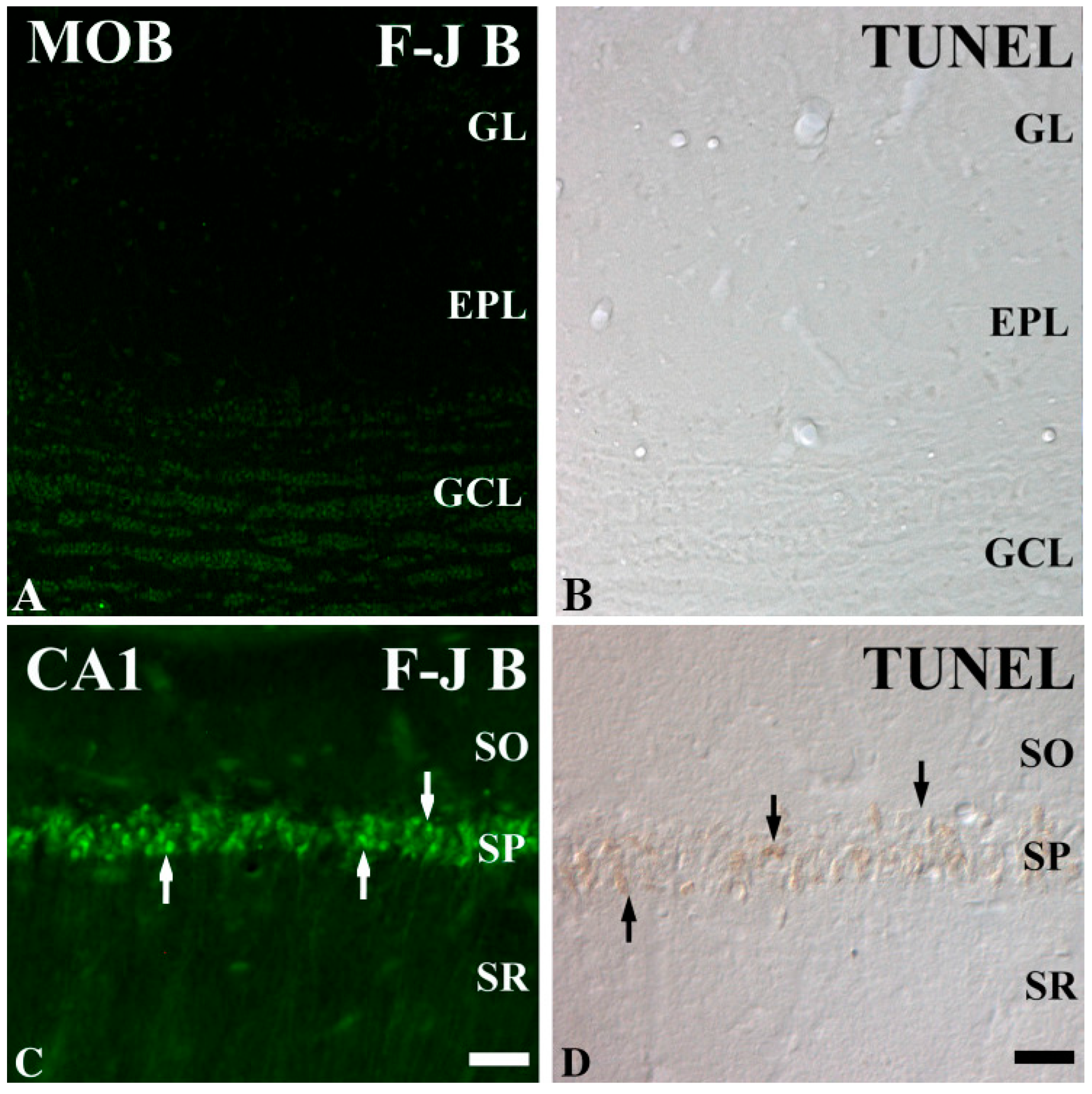
Figure 1. Fluoro Jade B (F-J B) histofluorescence and terminal deoxynucleotidyl transferase dUTP nick end labeling (TUNEL) staining in the main olfactory bulb (MOB; (A,B)) and hippocampal CA1 region (C,D) of the gerbil. F-J B and TUNEL-positive cells are rare in the MOB of the IR group, but many F-J B (white arrows) and TUNEL (black arrows)-positive cells are shown in the CA1 region after IR injury. EPL external plexiform layer; GCL granule cell layer; GL glomerular layer; SO stratum radiatum, SP stratum pyramidale; SR stratum radiatum. Scale Bars = 200 μm (A,B), 50 μm (C,D). This figure was published by Choi et al. (2010) [12].
Ijms 26 05079 g001
Figure 2. Schematic representation of the translaminar organization of the olfactory bulb (OB) and its position. The olfactory receptor (or sensory) neurons (ORN) in the olfactory epithelium (OE) project to the glomerular cell layer (GL), where they connect to periglomerular (PG), as well as mitral (MC) and middle tufted (mTC) cells, which connect to granule cells (GC). The OB output propagates to the olfactory cortex through the MC and mTC axons in the lateral olfactory tract (LOT). EPL, external plexiform layer; GCL, granule cell layer. This figure was published by Cavarretta et al. (2018) [16].
Ijms 26 05079 g002
Figure 3. Mechanisms of brain IR injury. This figure represents the cascade of pathological events occurring during IR injury. The interplay between excitotoxicity, oxidative stress, inflammation, and mitochondrial dysfunction contributes to neuronal death and brain damage. Excessive Ca2+ influx and ROS lead to mitochondrial dysfunction and the opening of the mitochondrial permeability transition pore (mPTP). Cytochrome C is released into the cytoplasm and activates caspases, which drive apoptosis. This figure was originally published by Zheng et al. (2023) [6].
Ijms 26 05079 g003
Figure 4. (A) Arterial supply of the OB and olfactory nerve. The OB is mainly supplied by the olfactory artery, a branch of the anterior cerebral artery. This figure was published by Hendrix et al. (2014). (B) Three-dimensional reconstruction of vessels and glomeruli. Vessel colors vary with depth, and glomeruli are indicated in light blue. This figure was published by Lecoq et al. (2009) [26].
Ijms 26 05079 g004
Table 1. Types of neurons vulnerable and resistant to IR injury.
Neuron TypeVulnerability to IR InjuryLocationPrimary FunctionReferences
CA1 pyramidal neuronsHighly vulnerableHippocampus (CA1 region)Long-term memory formation, synaptic plasticity[39]
Cerebellar Purkinje CellsHighly vulnerableCerebellumCoordination of movement, motor learning[40]
Neocortical Pyramidal NeuronsModerately VulnerableNeocortex (Layer 5)Motor control, cognition, sensory processing[41]
Dopaminergic NeuronsResistantSubstantia Nigra, Ventral Tegmental AreaMotor control, reward processing, mood regulation[42]
Spinal Motor NeuronsResistantSpinal CordMotor function, voluntary movement[40,43]
Striatal InterneuronsResistantStriatumRegulation of motor activity, reward and emotional processing[44]
Dentate granule cellsHighly resistantDentate gyrusLearning, memory, and spatial navigation[45]
Olfactory mitral cellsResistantOlfactory BulbOlfaction, processing odor signals[9]
Disclaimer/Publisher’s Note: The statements, opinions and data contained in all publications are solely those of the individual author(s) and contributor(s) and not of MDPI and/or the editor(s). MDPI and/or the editor(s) disclaim responsibility for any injury to people or property resulting from any ideas, methods, instructions or products referred to in the content.