
Recent Advances in Synergistic Effect of Nanoparticles and Its Biomedical Application


Abstract
1. Introduction
2. Properties of Metallic Nanoparticles
2.1. Comparison between Mono- and Bimetallic Nanoparticles
2.2. Structure and Characteristics of Bimetallic Nanoparticles
3. Nanoalloy Synergistic Applications
3.1. Gold Nanoalloys
3.2. Magnetic Nanoalloys
4. Mechanism of Nanoparticle–Cell Interaction
5. Green Synthesis Trends in Bimetallic Nanoparticles
6. Conclusions
Funding
Acknowledgments
Conflicts of Interest
References
- Amarnath, C.A.; Nanda, S.S.; Papaefthymiou, G.C.; Yi, D.K.; Paik, U. Nanohybridization of low-dimensional nanomaterials: Synthesis, classification, and application. Crit. Rev. Solid State Mater. Sci. 2013, 38, 1–56. [Google Scholar] [CrossRef]
- Xu, L.; Ma, W.; Wang, L.; Xu, C.; Kuang, H.; Kotov, N.A. Nanoparticle assemblies: Dimensional transformation of nanomaterials and scalability. Chem. Soc. Rev. 2013, 42, 3114–3126. [Google Scholar] [CrossRef] [PubMed]
- Dreaden, E.C.; Alkilany, A.M.; Huang, X.; Murphy, C.J.; El-Sayed, M.A. The golden age: Gold nanoparticles for biomedicine. Chem. Soc. Rev. 2012, 41, 2740–2779. [Google Scholar] [CrossRef] [PubMed]
- Joudeh, N.; Linke, D. Nanoparticle classification, physicochemical properties, characterization, and applications: A comprehensive review for biologists. J. Nanobiotechnol. 2022, 20, 262. [Google Scholar] [CrossRef]
- Lee, D.E.; Koo, H.; Sun, I.C.; Ryu, J.H.; Kim, K.; Kwon, I.C. Multifunctional nanoparticles for multimodal imaging and theragnosis. Chem. Soc. Rev. 2012, 41, 2656–2672. [Google Scholar] [CrossRef]
- Nanda, S.S.; Hembram, K.P.S.S.; Lee, J.K.; Kim, K.; Selvan, S.T.; Yi, D.K. Experimental and theoretical structural characterization of Cu–Au tripods for photothermal anticancer therapy. ACS Appl. Nano Mater. 2019, 2, 3735–3742. [Google Scholar] [CrossRef]
- Kundu, A.; Nandi, S.; Nandi, A.K. Nucleic acid based polymer and nanoparticle conjugates: Synthesis, properties and applications. Prog. Mater. Sci. 2017, 88, 136–185. [Google Scholar] [CrossRef]
- Thi, E.P.; Mire, C.E.; Lee, A.C.H.; Geisbert, J.B.; Zhou, J.Z.; Agans, K.N.; Snead, N.M.; Deer, D.J.; Barnard, T.R.; Fenton, K.A.; et al. Lipid nanoparticle siRNA treatment of Ebola-virus-Makona-infected nonhuman primates. Nature 2015, 521, 362. [Google Scholar] [CrossRef]
- Cutler, J.I.; Auyeung, E.; Mirkin, C.A. Spherical nucleic acids. J. Am. Chem. Soc. 2012, 134, 1376–1391. [Google Scholar] [CrossRef]
- Stengel, D.; Jörgensen, A.M.; Polidori, I.; Kapitza, P.; Ricci, F.; Bernkop-Schnürch, A. The power of sulfhydryl groups: Thiolated lipid-based nanoparticles enhance cellular uptake of nucleic acids. J. Colloid Interface Sci. 2024, 654, 1136–1145. [Google Scholar] [CrossRef]
- Haizan, I.; Park, D.H.; Choi, M.Y.; Lee, H.; Choi, J.H. Nanomaterials-Based Exosomes for the Diagnostics and Drug Deliveries of Central Nervous System Diseases. BioChip J. 2023, 17, 293–307. [Google Scholar] [CrossRef]
- Lauschke, V.M.; Zhou, Y.; Ingelman-Sundberg, M. Pharmacogenomics beyond single common genetic variants: The way forward. Annu. Rev. Pharmacol. Toxicol. 2023, 64, 33–51. [Google Scholar] [CrossRef] [PubMed]
- Mead, R.J.; Shan, N.; Reiser, H.J.; Marshall, F.; Shaw, P.J. Amyotrophic lateral sclerosis: A neurodegenerative disorder poised for successful therapeutic translation. Nat. Rev. Drug Discov. 2023, 22, 185–212. [Google Scholar] [CrossRef] [PubMed]
- Wang, T.; Yeom, K.S.; Nanda, S.S.; An, S.S.A.; Yi, D.K. Cancer cell growth in the near infrared region by using silica coated gold nanorods. Nano 2020, 15, 2050001. [Google Scholar] [CrossRef]
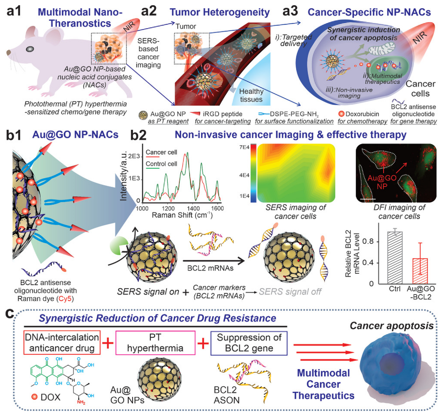
- Yang, L.; Kim, T.H.; Cho, H.Y.; Luo, J.; Lee, J.M.; Chueng, S.T.D.; Hou, Y.; Yin, P.T.T.; Han, J.; Kim, J.H.; et al. Hybrid graphene-gold nanoparticle-based nucleic acid conjugates for cancer-specific multimodal imaging and combined therapeutics. Adv. Funct. Mater. 2021, 31, 2006918. [Google Scholar] [CrossRef] [PubMed]
- Galan-Ladero, M.M.; Sarmento, M.; Marques, S. Social Marketing to achieve the Sustainable Development Goals (SDGs) in 2030 Agenda by the United Nations. Int. Rev. Public Nonprofit Mark. 2023, 20, 521–527. [Google Scholar] [CrossRef]
- Mangukiya, R.D.; Sklarew, D.M. Analyzing three pillars of sustainable development goals at sub-national scales within the USA. World Dev. Sustain. 2023, 2, 100058. [Google Scholar] [CrossRef]
- Walsh, P.P.; Murphy, E.; Horan, D. The role of science, technology and innovation in the UN 2030 agenda. Technol. Forecast. Soc. Change 2020, 154, 119957. [Google Scholar] [CrossRef]
- United Nations. The future is now—Science for achieving sustainable development. In Global Sustainable Development Report by the Independent Group of Scientists; United Nations: New York, NY, USA, 2019. [Google Scholar]
- Bairagi, S.; Kamali, M.R. Review on green biomass-synthesized metallic nanoparticles and composites and their photocatalytic water purification applications: Progress and perspectives. Chem. Eng. J. Adv. 2023, 14, 100460. [Google Scholar]
- Medina-Cruz, D.; Saleh, B.; Vernet-Crua, A.; Nieto-Argüello, A.; Lomelí-Marroquín, D.; Vélez-Escamilla, L.Y.; Cholula-Díaz, J.L.; García-Martín, J.M.; Webster, T. Bimetallic nanoparticles for biomedical applications: A review. In Racing for the Surface: Antimicrobial and Interface Tissue Engineering; Springer: Cham, Switzerland, 2020; pp. 397–434. [Google Scholar] [CrossRef]
- Blosi, M.; Ortelli, S.; Costa, A.L.; Dondi, M.; Lolli, A.; Andreoli, S.; Benito, P.; Albonetti, S. Bimetallic nanoparticles as efficient catalysts: Facile and green microwave synthesis. Materials 2016, 9, 550. [Google Scholar] [CrossRef]
- Ren, G.; Wan, K.; Kong, H.; Guo, L.; Wang, Y.; Liu, X.; Wei, G. Recent advance in biomass membranes: Fabrication, functional regulation, and antimicrobial applications. Carbohydr. Polym. 2023, 305, 120537. [Google Scholar] [CrossRef]
- Kumar, N.; Gusain, R.; Pandey, S.; Ray, S.S. Hydrogel Nanocomposite Adsorbents and Photocatalysts for Sustainable Water Purification. Adv. Mater. Interfaces 2023, 10, 2201375. [Google Scholar] [CrossRef]
- Trinh, B.M.; Chang, B.P.; Mekonnen, T.H. The barrier properties of sustainable multiphase and multicomponent packaging materials: A review. Prog. Mater. Sci. 2023, 133, 101071. [Google Scholar] [CrossRef]
- Larrañaga-Tapia, M.; Betancourt-Tovar, B.; Videa, M.; Antunes-Ricardo, M.; Cholula-Díaz, J.L. Green synthesis trends and potential applications of bimetallic nanoparticles towards the sustainable development goals 2030. Nanoscale Adv. 2024, 6, 51–71. [Google Scholar] [CrossRef] [PubMed]
- Shrivastava, P.; Jain, V.K.; Nagpal, S. Nanoparticle intervention for heavy metal detection: A review. Environ. Nanotechnol. Monit. Manag. 2022, 17, 100667. [Google Scholar] [CrossRef]
- Idris, D.S.; Roy, A. Synthesis of Bimetallic Nanoparticles and Applications—An Updated Review. Crystals 2023, 13, 637. [Google Scholar] [CrossRef]
- Nasrollahzadeh, M.; Sajjadi, M.; Iravani, S.; Varma, R.S. Trimetallic nanoparticles: Greener synthesis and their applications. Nanomaterials 2020, 10, 1784. [Google Scholar] [CrossRef] [PubMed]
- Vance, M.E.; Kuiken, T.; Vejerano, E.P.; McGinnis, S.P.; Hochella, M.F., Jr.; Rejeski, D.; Hull, M.S. Nanotechnology in the real world: Redeveloping the nanomaterial consumer products inventory. Beilstein J. Nanotechnol. 2015, 6, 1769–1780. [Google Scholar] [CrossRef] [PubMed]
- Cholula-Díaz, J.L.; Lomelí-Marroquín, D.; Pramanick, B.; Nieto-Argüello, A.; Cantú-Castillo, L.A.; Hwang, H. Synthesis of colloidal silver nanoparticle clusters and their application in ascorbic acid detection by SERS. Colloids Surf. B Biointerfaces 2018, 163, 329–335. [Google Scholar] [CrossRef] [PubMed]
- Geleta, G.S. A colorimetric aptasensor based on two dimensional (2D) nanomaterial and gold nanoparticles for detection of toxic heavy metal ions: A review. Food Chem. Adv. 2023, 2, 100184. [Google Scholar] [CrossRef]
- Soliman, M.K.; Salem, S.S.; Abu-Elghait, M.; Azab, M.S. Biosynthesis of silver and gold nanoparticles and their efficacy towards antibacterial, antibiofilm, cytotoxicity, and antioxidant activities. Appl. Biochem. Biotechnol. 2023, 195, 1158–1183. [Google Scholar] [CrossRef]
- Yang, W.; Wang, C.; Arrighi, V. One step preparation of copper–silver self-catalyzed hybrid conductive ink with reduced sintering temperature for flexible electronics. J. Mater. Sci. Mater. Electron. 2019, 30, 11607–11618. [Google Scholar] [CrossRef]
- Alahdal, F.A.; Qashqoosh, M.T.; Manea, Y.K.; Mohammed, R.K.; Naqvi, S. Green synthesis and characterization of copper nanoparticles using Phragmanthera austroarabica extract and their biological/environmental applications. Sustain. Mater. Technol. 2023, 35, e00540. [Google Scholar] [CrossRef]
- Gomez, F.J.; Chumanov, G.; Silva, M.F.; Garcia, C.D. CO2 reduction using paper-derived carbon electrodes modified with copper nanoparticles. RSC Adv. 2019, 9, 33657–33663. [Google Scholar] [CrossRef]
- Song, D.; Li, Y.; Lu, X.; Sun, M.; Liu, H.; Yu, G.; Gao, F. Palladium-copper nanowires-based biosensor for the ultrasensitive detection of organophosphate pesticides. Anal. Chim. Acta 2017, 982, 168–175. [Google Scholar] [CrossRef] [PubMed]
- Behera, A.; Mittu, B.; Padhi, S.; Patra, N.; Singh, J. Bimetallic nanoparticles: Green synthesis, applications, and future perspectives. In Multifunctional Hybrid Nanomaterials for Sustainable Agri-Food and Ecosystems; Elsevier: Amsterdam, The Netherlands, 2020; pp. 639–682. [Google Scholar]
- Anjo, L.; Khajehnezhad, A.; Sari, A.H.; Sebt, S.A.; Ismail, M.M. Coexistence of Plasmonic and Magnetic Properties in Bimetallic Fe/Ag Nanoparticles Synthesized by Pulsed Laser Ablation. Plasmonics 2022, 17, 941–948. [Google Scholar] [CrossRef]
- Malik, M.A.; Alshehri, A.A.; Patel, R. Facile one-pot green synthesis of Ag–Fe bimetallic nanoparticles and their catalytic capability for 4-nitrophenol reduction. J. Mater. Res. Technol. 2021, 12, 455–470. [Google Scholar] [CrossRef]
- Rajeev, R.; Datta, R.; Varghese, A.; Sudhakar, Y.N.; George, L. Recent advances in bimetallic based nanostructures: Synthesis and electrochemical sensing applications. Microchem. J. 2021, 163, 105910. [Google Scholar] [CrossRef]
- Wang, R.; Tang, T.; Lu, G.; Zheng, Z.; Huang, K.; Li, H.; Tao, X.; Yin, H.; Shi, Z.; Lin, Z.; et al. Mechanisms and pathways of debromination of polybrominated diphenyl ethers (PBDEs) in various nano-zerovalent iron-based bimetallic systems. Sci. Total Environ. 2019, 661, 18–26. [Google Scholar] [CrossRef] [PubMed]
- Ngamaroonchote, A.; Sanguansap, Y.; Wutikhun, T.; Karn-Orachai, K. Highly branched gold–copper nanostructures for non-enzymatic specific detection of glucose and hydrogen peroxide. Microchim. Acta 2020, 187, 559. [Google Scholar] [CrossRef] [PubMed]
- Srinoi, P.; Chen, Y.T.; Vittur, V.; Marquez, M.D.; Lee, T.R. Bimetallic nanoparticles: Enhanced magnetic and optical properties for emerging biological applications. Appl. Sci. 2018, 8, 1106. [Google Scholar] [CrossRef]
- Das, P.; Borthakur, P.; Boruah, P.K.; Das, M.R. Peroxidase mimic activity of Au-Ag/l-Cys-rGO nanozyme toward detection of Cr (VI) ion in water: Role of 3, 3′, 5, 5′-tetramethylbenzidine adsorption. J. Chem. Eng. Data 2019, 64, 4977–4990. [Google Scholar] [CrossRef]
- Jiang, L.Y.; Na, L. (Eds.) Membrane-Based Separations in Metallurgy: Principles and Applications; Elsevier: Amsterdam, The Netherlands, 2017. [Google Scholar]
- Sardar, R.; Funston, A.M.; Mulvaney, P.; Murray, R.W. Gold nanoparticles: Past, present, and future. Langmuir 2009, 25, 13840–13851. [Google Scholar] [CrossRef] [PubMed]
- Kaushal, S.; Nanda, S.S.; Samal, S.; Yi, D.K. Strategies for the development of metallic-nanoparticle-based label-free biosensors and their biomedical applications. ChemBioChem 2020, 21, 576–600. [Google Scholar] [CrossRef] [PubMed]
- Kaushal, S.; Nanda, S.S.; Yi, D.K.; Ju, H. Effects of aspect ratio heterogeneity of an assembly of gold nanorod on localized surface plasmon resonance. J. Phys. Chem. Lett. 2020, 11, 5972–5979. [Google Scholar] [CrossRef]
- Jain, P.K.; Lee, K.S.; El-Sayed, I.H.; El-Sayed, M.A. Calculated absorption and scattering properties of gold nanoparticles of different size, shape, and composition: Applications in biological imaging and biomedicine. J. Phys. Chem. B 2006, 110, 7238–7248. [Google Scholar] [CrossRef]
- Sperling, R.A.; Gil, P.R.; Zhang, F.; Zanella, M.; Parak, W.J. Biological applications of gold nanoparticles. Chem. Soc. Rev. 2008, 37, 1896–1908. [Google Scholar] [CrossRef]
- Feng, Q.L.; Wu, J.; Chen, G.Q.; Cui, F.Z.; Kim, T.N.; Kim, J.O. A mechanistic study of the antibacterial effect of silver ions on Escherichia coli and Staphylococcus aureus. J. Biomed. Mater. Res. 2000, 52, 662–668. [Google Scholar] [CrossRef]
- Liu, H.L.; Dai, S.A.; Fu, K.Y.; Hsu, S.H. Antibacterial properties of silver nanoparticles in three different sizes and their nanocomposites with a new waterborne polyurethane. Int. J. Nanomed. 2010, 5, 1017–1028. [Google Scholar] [CrossRef]
- Liu, J.; Hurt, R.H. Ion release kinetics and particle persistence in aqueous nano-silver colloids. Environ. Sci. Technol. 2010, 44, 2169–2175. [Google Scholar] [CrossRef]
- Chernousova, S.; Epple, M. Silver as antibacterial agent: Ion, nanoparticle, and metal. Angew. Chem. Int. Ed. 2013, 52, 1636–1653. [Google Scholar] [CrossRef]
- Mohamed, R.M.; Fawzy, E.M.; Shehab, R.A.; Abdel-Salam, M.O.; Salah El Din, R.A.; Abd El Fatah, H.M. Production, characterization, and cytotoxicity effects of silver nanoparticles from Brown alga (Cystoseira myrica). J. Nanotechnol. 2022, 2022, 6469090. [Google Scholar] [CrossRef]
- Karakaş, İ. Some biological potential of silver nanoparticles synthesized from Ocimum basilicum L. GSC Biol. Pharm. Sci. 2023, 22, 107–113. [Google Scholar] [CrossRef]
- Rajan, R.; Huo, P.; Chandran, K.; Dakshinamoorthi, B.M.; Yun, S.I.; Liu, B. A review on the toxicity of silver nanoparticles against different biosystems. Chemosphere 2022, 292, 133397. [Google Scholar] [CrossRef]
- Choudhary, A.; Singh, S.; Ravichandiran, V. Toxicity, preparation methods and applications of silver nanoparticles: An update. Toxicol. Mech. Methods 2022, 32, 650–661. [Google Scholar] [CrossRef]
- Taha, R.H. Green synthesis of silver and gold nanoparticles and their potential applications as therapeutics in cancer therapy; A review. Inorg. Chem. Commun. 2022, 143, 109610. [Google Scholar] [CrossRef]
- Milan, J.; Niemczyk, K.; Kus-Liśkiewicz, M. Treasure on the Earth—Gold nanoparticles and their biomedical applications. Materials 2022, 15, 3355. [Google Scholar] [CrossRef] [PubMed]
- Patil, T.; Gambhir, R.; Vibhute, A.; Tiwari, A.P. Gold nanoparticles: Synthesis methods, functionalization and biological applications. J. Clust. Sci. 2023, 34, 705–725. [Google Scholar] [CrossRef]
- Anik, M.I.; Mahmud, N.; Al Masud, A.; Hasan, M. Gold nanoparticles (GNPs) in biomedical and clinical applications: A review. Nano Sel. 2022, 3, 792–828. [Google Scholar] [CrossRef]
- Siddique, S.; Chow, J.C. Gold nanoparticles for drug delivery and cancer therapy. Appl. Sci. 2020, 10, 3824. [Google Scholar] [CrossRef]
- Nejati, K.; Dadashpour, M.; Gharibi, T.; Mellatyar, H.; Akbarzadeh, A. Biomedical applications of functionalized gold nanoparticles: A review. J. Clust. Sci. 2021, 33, 1–16. [Google Scholar] [CrossRef]
- Bansal, S.A.; Kumar, V.; Karimi, J.; Singh, A.P.; Kumar, S. Role of gold nanoparticles in advanced biomedical applications. Nanoscale Adv. 2020, 2, 3764–3787. [Google Scholar] [CrossRef]
- Bai, X.; Wang, Y.; Song, Z.; Feng, Y.; Chen, Y.; Zhang, D.; Feng, L. The basic properties of gold nanoparticles and their applications in tumor diagnosis and treatment. Int. J. Mol. Sci. 2020, 21, 2480. [Google Scholar] [CrossRef]
- Yafout, M.; Ousaid, A.; Khayati, Y.; El Otmani, I.S. Gold nanoparticles as a drug delivery system for standard chemotherapeutics: A new lead for targeted pharmacological cancer treatments. Sci. Afr. 2021, 11, e00685. [Google Scholar] [CrossRef]
- Yu, Y.; Yang, T.; Sun, T. New insights into the synthesis, toxicity and applications of gold nanoparticles in CT imaging and treatment of cancer. Nanomedicine 2020, 15, 1127–1145. [Google Scholar] [CrossRef]
- Tang, J.; Shi, H.; Ma, G.; Luo, L.; Tang, Z. Ultrasmall Au and Ag nanoclusters for biomedical applications: A review. Front. Bioeng. Biotechnol. 2020, 8, 1019. [Google Scholar] [CrossRef]
- Babawale, O.E.; Gundlach, L. Fabrication of Large-Area Fully Alloyed Ag/Au Nanoparticle Arrays. ACS Appl. Nano Mater. 2023, 6, 21866–21875. [Google Scholar] [CrossRef]
- Petkov, V.; Prasai, B.; Ren, Y.; Shan, S.; Luo, J.; Joseph, P.; Zhong, C.J. Solving the nanostructure problem: Exemplified on metallic alloy nanoparticles. Nanoscale 2014, 6, 10048–10061. [Google Scholar] [CrossRef]
- Chen, D.H.; Chen, C.J. Formation and characterization of Au–Ag bimetallic nanoparticles in water-in-oil microemulsions. J. Mater. Chem. 2002, 12, 1557–1562. [Google Scholar] [CrossRef]
- Devarajan, S.; Bera, P.; Sampath, S. Bimetallic nanoparticles: A single step synthesis, stabilization, and characterization of Au–Ag, Au–Pd, and Au–Pt in sol–gel derived silicates. J. Colloid Interface Sci. 2005, 290, 117–129. [Google Scholar] [CrossRef]
- Gonzalez, C.M.; Liu, Y.; Scaiano, J.C. Photochemical strategies for the facile synthesis of gold−silver alloy and core−shell bimetallic nanoparticles. J. Phys. Chem. C 2009, 113, 11861–11867. [Google Scholar] [CrossRef]
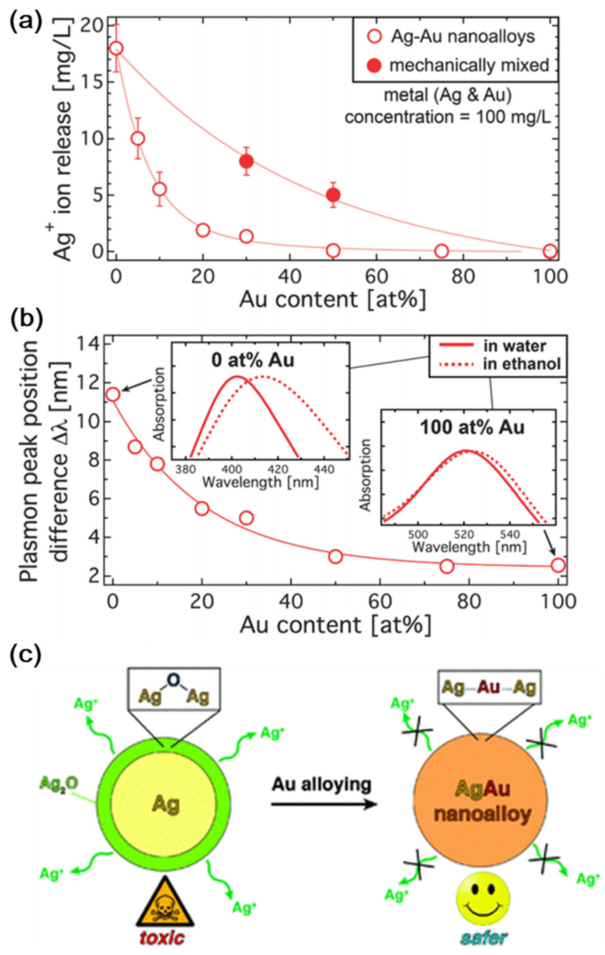
- Grade, S.; Eberhard, J.; Jakobi, J.; Winkel, A.; Stiesch, M.; Barcikowski, S. Alloying colloidal silver nanoparticles with gold disproportionally controls antibacterial and toxic effects. Gold Bull. 2014, 47, 83–93. [Google Scholar] [CrossRef]
- Neumeister, A.; Jakobi, J.; Rehbock, C.; Moysig, J.; Barcikowski, S. Monophasic ligand-free alloy nanoparticle synthesis determinants during pulsed laser ablation of bulk alloy and consolidated microparticles in water. Phys. Chem. Chem. Phys. 2014, 16, 23671–23678. [Google Scholar] [CrossRef]
- Taylor, U.; Tiedemann, D.; Rehbock, C.; Kues, W.A.; Barcikowski, S.; Rath, D. Influence of gold, silver and gold–silver alloy nanoparticles on germ cell function and embryo development. Beilstein J. Nanotechnol. 2015, 6, 651–664. [Google Scholar] [CrossRef]
- Wang, C.; Peng, S.; Chan, R.; Sun, S. Synthesis of AuAg Alloy Nanoparticles from Core/Shell-Structured Ag/Au. Small 2009, 5, 567–570. [Google Scholar] [CrossRef]
- Radziuk, D.V.; Zhang, W.; Shchukin, D.; Möhwald, H. Ultrasonic alloying of preformed gold and silver nanoparticles. Small 2010, 6, 545–553. [Google Scholar] [CrossRef]
- Sotiriou, G.A.; Etterlin, G.D.; Spyrogianni, A.; Krumeich, F.; Leroux, J.C.; Pratsinis, S.E. Plasmonic biocompatible silver–gold alloyed nanoparticles. Chem. Commun. 2014, 50, 13559–13562. [Google Scholar] [CrossRef]
- Anker, J.N.; Hall, W.P.; Lyandres, O.; Shah, N.C.; Zhao, J.; Van Duyne, R.P. Biosensing with plasmonic nanosensors. Nat. Mater. 2008, 7, 442–453. [Google Scholar] [CrossRef]
- Cortie, M.B.; McDonagh, A.M. Synthesis and optical properties of hybrid and alloy plasmonic nanoparticles. Chem. Rev. 2011, 111, 3713–3735. [Google Scholar] [CrossRef]
- Barnes, W.L.; Dereux, A.; Ebbesen, T.W. Surface plasmon subwavelength optics. Nature 2003, 424, 824–830. [Google Scholar] [CrossRef]
- Pryshchepa, O.; Pomastowski, P.; Buszewski, B. Silver nanoparticles: Synthesis, investigation techniques, and properties. Adv. Colloid Interface Sci. 2020, 284, 102246. [Google Scholar] [CrossRef]
- Li, Y.; Deng, Z. Ag ion soldering: An emerging tool for sub-nanomeric plasmon coupling and beyond. Acc. Chem. Res. 2019, 52, 3442–3454. [Google Scholar] [CrossRef]
- Lee, H.; Yoo, Y.; Kang, T.; In, J.; Seo, M.K.; Kim, B. Topotaxial Fabrication of Vertical AuxAg1−x Nanowire Arrays: Plasmon-Active in the Blue Region and Corrosion Resistant. Small 2012, 8, 1527–1533. [Google Scholar] [CrossRef]
- Hussain, S.M.; Hess, K.L.; Gearhart, J.M.; Geiss, K.T.; Schlager, J.J. In vitro toxicity of nanoparticles in BRL 3A rat liver cells. Toxicol. In Vitro 2005, 19, 975–983. [Google Scholar] [CrossRef]
- Sotiriou, G.A.; Meyer, A.; Knijnenburg, J.T.; Panke, S.; Pratsinis, S.E. Quantifying the origin of released Ag+ ions from nanosilver. Langmuir 2012, 28, 15929–15936. [Google Scholar] [CrossRef]
- Sotiriou, G.A.; Pratsinis, S.E. Engineering nanosilver as an antibacterial, biosensor and bioimaging material. Curr. Opin. Chem. Eng. 2011, 1, 3–10. [Google Scholar] [CrossRef]
- Zhao, S.; Yu, X.; Qian, Y.; Chen, W.; Shen, J. Multifunctional magnetic iron oxide nanoparticles: An advanced platform for cancer theranostics. Theranostics 2020, 10, 6278. [Google Scholar] [CrossRef]
- Khizar, S.; Ahmad, N.M.; Zine, N.; Jaffrezic-Renault, N.; Errachid-el-salhi, A.; Elaissari, A. Magnetic nanoparticles: From synthesis to theranostic applications. ACS Appl. Nano Mater. 2021, 4, 4284–4306. [Google Scholar] [CrossRef]
- Li, X.; Li, W.; Wang, M.; Liao, Z. Magnetic nanoparticles for cancer theranostics: Advances and prospects. J. Control. Release 2021, 335, 437–448. [Google Scholar] [CrossRef]
- Ferreira, M.; Sousa, J.; Pais, A.; Vitorino, C. The role of magnetic nanoparticles in cancer nanotheranostics. Materials 2020, 13, 266. [Google Scholar] [CrossRef]
- Baki, A.; Wiekhorst, F.; Bleul, R. Advances in magnetic nanoparticles engineering for biomedical applications—A Review. Bioengineering 2021, 8, 134. [Google Scholar] [CrossRef]
- Díez, A.G.; Rincón-Iglesias, M.; Lanceros-Méndez, S.; Reguera, J.; Lizundia, E. Multicomponent magnetic nanoparticle engineering: The role of structure-property relationship in advanced applications. Mater. Today Chem. 2022, 26, 101220. [Google Scholar] [CrossRef]
- Stueber, D.D.; Villanova, J.; Aponte, I.; Xiao, Z.; Colvin, V.L. Magnetic nanoparticles in biology and medicine: Past, present, and future trends. Pharmaceutics 2021, 13, 943. [Google Scholar] [CrossRef]
- Farinha, P.; Coelho, J.M.; Reis, C.P.; Gaspar, M.M. A comprehensive updated review on magnetic nanoparticles in diagnostics. Nanomaterials 2021, 11, 3432. [Google Scholar] [CrossRef]
- Brito, B.; Price, T.W.; Gallo, J.; Bañobre-López, M.; Stasiuk, G.J. Smart magnetic resonance imaging-based theranostics for cancer. Theranostics 2021, 11, 8706. [Google Scholar] [CrossRef]
- Wu, K.; Su, D.; Liu, J.; Saha, R.; Wang, J.P. Magnetic nanoparticles in nanomedicine: A review of recent advances. Nanotechnology 2019, 30, 502003. [Google Scholar] [CrossRef]
- Cheng, H.W.; Tsao, H.Y.; Chiang, C.S.; Chen, S.Y. Advances in magnetic nanoparticle-mediated cancer immune-theranostics. Adv. Healthc. Mater. 2021, 10, 2001451. [Google Scholar] [CrossRef]
- Wallyn, J.; Anton, N.; Akram, S.; Vandamme, T.F. Biomedical imaging: Principles, technologies, clinical aspects, contrast agents, limitations and future trends in nanomedicines. Pharm. Res. 2019, 36, 78. [Google Scholar] [CrossRef]
- Li, H.; Meade, T.J. Molecular magnetic resonance imaging with Gd (III)-based contrast agents: Challenges and key advances. J. Am. Chem. Soc. 2019, 141, 17025–17041. [Google Scholar] [CrossRef]
- Yan, G.P.; Robinson, L.; Hogg, P. Magnetic resonance imaging contrast agents: Overview and perspectives. Radiography 2007, 13, e5–e19. [Google Scholar] [CrossRef]
- Kim, J.; Piao, Y.; Hyeon, T. Multifunctional nanostructured materials for multimodal imaging, and simultaneous imaging and therapy. Chem. Soc. Rev. 2009, 38, 372–390. [Google Scholar] [CrossRef]
- Bulte, J.W.; Kraitchman, D.L. Iron oxide MR contrast agents for molecular and cellular imaging. NMR Biomed. 2004, 17, 484–499. [Google Scholar] [CrossRef]
- McNamara, K.; Tofail, S.A. Nanosystems: The use of nanoalloys, metallic, bimetallic, and magnetic nanoparticles in biomedical applications. Phys. Chem. Chem. Phys. 2015, 17, 27981–27995. [Google Scholar] [CrossRef]
- Yi, D.K.; Lee, S.S.; Papaefthymiou, G.C.; Ying, J.Y. Nanoparticle architectures templated by SiO2/Fe2O3 nanocomposites. Chem. Mater. 2006, 18, 614–619. [Google Scholar] [CrossRef]
- Yi, D.K.; Lee, S.S.; Ying, J.Y. Synthesis and applications of magnetic nanocomposite catalysts. Chem. Mater. 2006, 18, 2459–2461. [Google Scholar] [CrossRef]
- Yi, D.K.; Selvan, S.T.; Lee, S.S.; Papaefthymiou, G.C.; Kundaliya, D.; Ying, J.Y. Silica-coated nanocomposites of magnetic nanoparticles and quantum dots. J. Am. Chem. Soc. 2005, 127, 4990–4991. [Google Scholar] [CrossRef]
- Duong, H.K.; Abdibastami, A.; Gloag, L.; Barrera, L.; Gooding, J.J.; Tilley, R.D. A guide to the design of magnetic particle imaging tracers for biomedical applications. Nanoscale 2022, 14, 13890–13914. [Google Scholar] [CrossRef]
- Nowak-Jary, J.; Machnicka, B. Pharmacokinetics of magnetic iron oxide nanoparticles for medical applications. J. Nanobiotechnol. 2022, 20, 305. [Google Scholar] [CrossRef]
- Fernández-Bertólez, N.; Costa, C.; Brandão, F.; Teixeira, J.P.; Pásaro, E.; Valdiglesias, V.; Laffon, B. Toxicological aspects of iron oxide nanoparticles. In Nanotoxicology in Safety Assessment of Nanomaterials; Springer International Publishing: Cham, Switzerland, 2022; pp. 303–350. [Google Scholar]
- Ma, Z.; Mohapatra, J.; Wei, K.; Liu, J.P.; Sun, S. Magnetic nanoparticles: Synthesis, anisotropy, and applications. Chem. Rev. 2021, 123, 3904–3943. [Google Scholar] [CrossRef]
- Capelli, S.; Cattaneo, S.; Stucchi, M.; Villa, A.; Prati, L. Iron as modifier of Pd and Pt-based catalysts for sustainable and green processes. Inorganica Chim. Acta 2022, 535, 120856. [Google Scholar] [CrossRef]
- McNamara, K.; Tofail, S.A.; Thorat, N.D.; Bauer, J.; Mulvihill, J.J. Biomedical applications of nanoalloys. In Nanoalloys, 2nd ed.; Elsevier: Amsterdam, The Netherlands, 2020; pp. 381–432. [Google Scholar] [CrossRef]
- Coviello, V.; Forrer, D.; Amendola, V. Recent developments in plasmonic alloy nanoparticles: Synthesis, modelling, properties and applications. ChemPhysChem 2022, 23, e202200136. [Google Scholar] [CrossRef]
- Dik, G.; Ulu, A.; Ates, B. Medicinal and Biological Application of Magnetic Alloy Nanoparticles and Their Polymer Nanocomposites. In Handbook of Magnetic Hybrid Nanoalloys and their Nanocomposites; Springer International Publishing: Cham, Switzerland, 2022; pp. 1–27. [Google Scholar]
- Semaltianos, N.G.; Karczewski, G. Laser synthesis of magnetic nanoparticles in liquids and application in the fabrication of polymer–nanoparticle composites. ACS Appl. Nano Mater. 2021, 4, 6407–6440. [Google Scholar] [CrossRef]
- Thomas, S.; Nochehdehi, A.R. (Eds.) Handbook of Magnetic Hybrid Nanoalloys and Their Nanocomposites; Springer Nature: Berlin/Heidelberg, Germany, 2022. [Google Scholar]
- Simon, J.; Nampoori, V.P.N.; Kailasnath, M. Concentration dependent thermo-optical properties and nonlinear optical switching behavior of bimetallic Au-Ag nanoparticles synthesized by femtosecond laser ablation. Opt. Laser Technol. 2021, 140, 107022. [Google Scholar] [CrossRef]
- Aslan, N.; Koç, M.M. X-ray Computed Tomography and Magnetic Resonance Imaging Applications of Magnetic Nanoalloys and Nanocomposites. In Handbook of Magnetic Hybrid Nanoalloys and their Nanocomposites; Springer International Publishing: Cham, Switzerland, 2022; pp. 1–20. [Google Scholar]
- Gabbani, A.; Fantechi, E.; Albino, M.; Sangregorio, C.; Pineider, F. Intermetallic Au3LixM1−x (M = Fe, Ni or Co) nanoalloys: Effect of synthetic conditions on the composition and order-disorder transition. Inorganica Chim. Acta 2023, 554, 121545. [Google Scholar] [CrossRef]
- Abduljawad, M. Synthesis and Functionalization of Hybrid Magnetic Nanoparticle Composites for Energy Conversion, Light Harvesting and Optical and Biomedical Applications. Doctoral Dissertation, The University of Arizona, Tucson, AZ, USA, 2021. [Google Scholar]
- Basagni, A.; Torresan, V.; Marzola, P.; van Raap, M.B.F.; Nodari, L.; Amendola, V. Structural evolution under physical and chemical stimuli of metastable Au–Fe nanoalloys obtained by laser ablation in liquid. Faraday Discuss. 2023, 242, 286–300. [Google Scholar] [CrossRef]
- Lin, L.S.; Yang, X.; Zhou, Z.; Yang, Z.; Jacobson, O.; Liu, Y.; Yang, A.; Niu, G.; Song, J.; Yang, H.H.; et al. Cooperation of endogenous and exogenous reactive oxygen species induced by zinc peroxide nanoparticles to enhance oxidative stress-based cancer therapy. Theranostics 2019, 9, 7200. [Google Scholar] [CrossRef]
- Singh, S.; Seehra, M.S. Testing the validity of the core-shell-surface layer model on the size dependence of effective magnetic anisotropy in magnetic nanoparticles. Front. Mater. 2022, 9, 1050600. [Google Scholar] [CrossRef]
- Tran, H.B.; Matsushita, Y.I. Temperature and size dependence of energy barrier for magnetic flips in L10 FePt nanoparticles: A theoretical study. Scr. Mater. 2024, 242, 115947. [Google Scholar] [CrossRef]
- Zhang, Z.; He, P.; Ma, W.; Zuo, P.; Liu, X.; Zhuang, Q. Freely Tailorable Yolk-Shell Encapsulation: Versatile Applications in Ultralow-k Dielectric, Drug Delivery Systems, and Catalysts. Adv. Funct. Mater. 2023, 33, 2302212. [Google Scholar] [CrossRef]
- Liu, Y.; Wu, P.C.; Guo, S.; Chou, P.T.; Deng, C.; Chou, S.W.; Yuan, Z.; Liu, T.M. Low-toxicity FePt nanoparticles for the targeted and enhanced diagnosis of breast tumors using few centimeters deep whole-body photoacoustic imaging. Photoacoustics 2020, 19, 100179. [Google Scholar] [CrossRef]
- Zhang, C.; Xu, C.; Gao, X.; Yao, Q. Platinum-based drugs for cancer therapy and anti-tumor strategies. Theranostics 2022, 12, 2115. [Google Scholar] [CrossRef]
- Rehbock, C.; Barcikowski, S. Toxicity of Colloidal Alloy Nanoparticles. In Nanoalloys; Elsevier: Amsterdam, The Netherlands, 2020; pp. 433–449. [Google Scholar]
- Bayda, S.; Adeel, M.; Tuccinardi, T.; Cordani, M.; Rizzolio, F. The history of nanoscience and nanotechnology: From chemical–physical applications to nanomedicine. Molecules 2019, 25, 112. [Google Scholar] [CrossRef]
- Yamanaka, S. A fresh look at iPS cells. Cell 2009, 137, 13–17. [Google Scholar] [CrossRef]
- Malgieri, A.; Kantzari, E.; Patrizi, M.P.; Gambardella, S. Bone marrow and umbilical cord blood human mesenchymal stem cells: State of the art. Int. J. Clin. Exp. Med. 2010, 3, 248. [Google Scholar]
- Dawson, E.; Mapili, G.; Erickson, K.; Taqvi, S.; Roy, K. Biomaterials for stem cell differentiation. Adv. Drug Deliv. Rev. 2008, 60, 215–228. [Google Scholar] [CrossRef]
- Kerativitayanan, P.; Carrow, J.K.; Gaharwar, A.K. Nanomaterials for engineering stem cell responses. Adv. Healthc. Mater. 2015, 4, 1600–1627. [Google Scholar] [CrossRef]
- Cha, C.; Liechty, W.B.; Khademhosseini, A.; Peppas, N.A. Designing biomaterials to direct stem cell fate. ACS Nano 2012, 6, 9353–9358. [Google Scholar] [CrossRef]
- Wei, M.; Li, S.; Le, W. Nanomaterials modulate stem cell differentiation: Biological interaction and underlying mechanisms. J. Nanobiotechnol. 2017, 15, 75. [Google Scholar] [CrossRef]
- Sapsford, K.E.; Algar, W.R.; Berti, L.; Gemmill, K.B.; Casey, B.J.; Oh, E.; Stewart, M.H.; Medintz, I.L. Functionalizing nanoparticles with biological molecules: Developing chemistries that facilitate nanotechnology. Chem. Rev. 2013, 113, 1904–2074. [Google Scholar] [CrossRef]
- Smith, B.R.; Gambhir, S.S. Nanomaterials for in vivo imaging. Chem. Rev. 2017, 117, 901–986. [Google Scholar] [CrossRef]
- Zhou, X.; Yuan, L.; Wu, C.; Luo, G.; Deng, J.; Mao, Z. Recent review of the effect of nanomaterials on stem cells. RSC Adv. 2018, 8, 17656–17676. [Google Scholar] [CrossRef]
- Yi, D.K.; Nanda, S.S.; Kim, K.; Selvan, S.T. Recent progress in nanotechnology for stem cell differentiation, labeling, tracking and therapy. J. Mater. Chem. B 2017, 5, 9429–9451. [Google Scholar] [CrossRef] [PubMed]
- El-Sayed, M.A. Some interesting properties of metals confined in time and nanometer space of different shapes. Acc. Chem. Res. 2001, 34, 257–264. [Google Scholar] [CrossRef] [PubMed]
- Dos Santos Ramos, M.A.; Da Silva, P.B.; Spósito, L.; De Toledo, L.G.; Bonifacio, B.V.; Rodero, C.F.; Dos Santos, K.C.; Chorilli, M.; Bauab, T.M. Nanotechnology-based drug delivery systems for control of microbial biofilms: A review. Int. J. Nanomed. 2018, 13, 1179–1213. [Google Scholar] [CrossRef] [PubMed]
- Melo, M.A., Jr.; Santos, L.S.S.; Gonçalves, M.D.C.; Nogueira, A.F. Preparation of silver and gold nanoparticles: A simple method to introduce nanotechnology into teaching laboratories. Quím. Nova 2012, 35, 1872–1878. [Google Scholar]
- Iravani, S.; Thota, S.; Crans, D.C. Methods for Preparation of Metal Nanoparticles; Wiley: Weinheim, Germany, 2017; Volume 63, pp. 15–31. [Google Scholar]
- Thakkar, K.N.; Mhatre, S.S.; Parikh, R.Y. Biological synthesis of metallic nanoparticles. Nanomed. Nanotechnol. Biol. Med. 2010, 6, 257–262. [Google Scholar] [CrossRef] [PubMed]
- Kumar, M.; Varshney, L.; Francis, S. Radiolytic formation of Ag clusters in aqueous polyvinyl alcohol solution and hydrogel matrix. Radiat. Phys. Chem. 2005, 73, 21–27. [Google Scholar] [CrossRef]
- Constantin, C.; Neagu, M.; Ion, R.M.; Gherghiceanu, M.; Stavaru, C. Fullerene–porphyrin nanostructures in photodynamic therapy. Nanomedicine 2010, 5, 307–317. [Google Scholar] [CrossRef]
- Nanda, S.S.; Wang, T.; Yoon, H.Y.; An, S.S.A.; Hembram, K.P.S.S.; Kim, K.; Yi, D.K. Enhanced proliferation of rabbit chondrocytes by using a well circulated nanoshock system. Sci. Rep. 2021, 11, 19388. [Google Scholar] [CrossRef]
- Ferrari, M. Cancer nanotechnology: Opportunities and challenges. Nat. Rev. Cancer 2005, 5, 161–171. [Google Scholar] [CrossRef]
- Boulaiz, H.; Alvarez, P.J.; Ramirez, A.; Marchal, J.A.; Prados, J.; Rodríguez-Serrano, F.; Perán, M.; Melguizo, C.; Aranega, A. Nanomedicine: Application areas and development prospects. Int. J. Mol. Sci. 2011, 12, 3303–3321. [Google Scholar] [CrossRef]
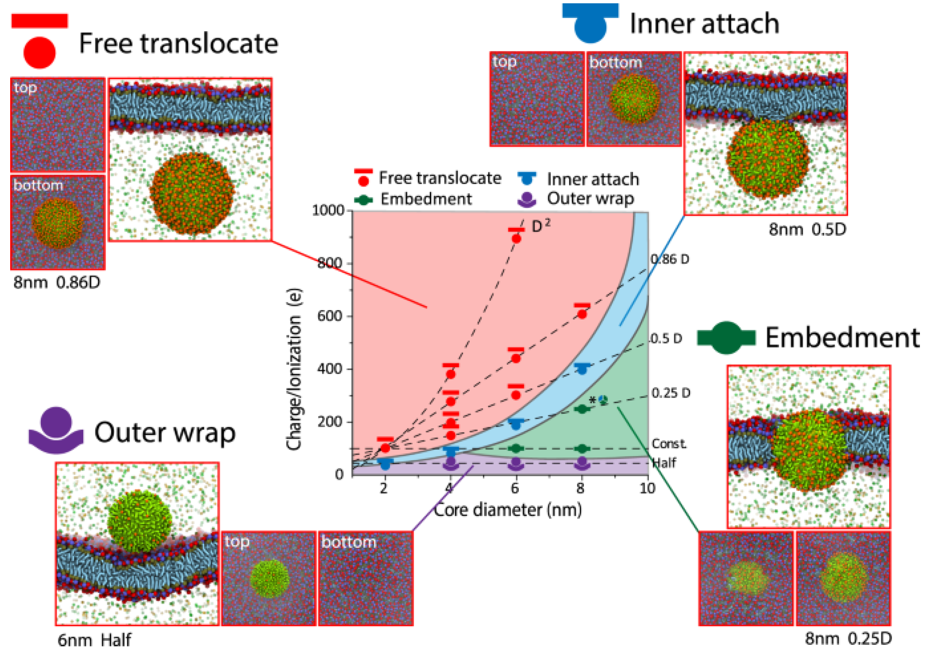
- Lin, J.; Miao, L.; Zhong, G.; Lin, C.H.; Dargazangy, R.; Alexander-Katz, A. Understanding the synergistic effect of physicochemical properties of nanoparticles and their cellular entry pathways. Commun. Biol. 2020, 3, 205. [Google Scholar] [CrossRef] [PubMed]
- Aithal, S.; Aithal, P.S. Green and eco-friendly Nanotechnology–concepts and industrial prospects. Int. J. Manag. Technol. Soc. Sci. 2021, 6, 1–31. [Google Scholar] [CrossRef]
- Duan, H.; Wang, D.; Li, Y. Green chemistry for nanoparticle synthesis. Chem. Soc. Rev. 2015, 44, 5778–5792. [Google Scholar] [CrossRef] [PubMed]
- Nasrollahzadeh, M.; Sajjadi, M.; Sajadi, S.M.; Issaabadi, Z. Green nanotechnology. In Interface Science and Technology; Elsevier: Amsterdam, The Netherlands, 2019; Volume 28, pp. 145–198. [Google Scholar]
- Puja, P.; Kumar, P. A perspective on biogenic synthesis of platinum nanoparticles and their biomedical applications. Spectrochim. Acta Part A Mol. Biomol. Spectrosc. 2019, 211, 94–99. [Google Scholar] [CrossRef] [PubMed]
- Zhao, X.; Zhou, L.; Riaz Rajoka, M.S.; Yan, L.; Jiang, C.; Shao, D.; Zhu, J.; Shi, J.; Huang, Q.; Yang, H.; et al. Fungal silver nanoparticles: Synthesis, application and challenges. Crit. Rev. Biotechnol. 2018, 38, 817–835. [Google Scholar] [CrossRef] [PubMed]
- Taherzadeh, M.J.; Fox, M.; Hjorth, H.; Edebo, L. Production of mycelium biomass and ethanol from paper pulp sulfite liquor by Rhizopus oryzae. Bioresour. Technol. 2003, 88, 167–177. [Google Scholar] [CrossRef]
- Nayak, S.S.; Wadhawa, G.C.; Pathade, K.B.; Shivankar, V.S.; Mirgane, N.A. Green synthesis of the plant assisted nanoparticles from Euphorbia neriifolia L. and its application in the degradation of dyes from industrial waste. Plant Sci. Today 2021, 8, 380–385. [Google Scholar] [CrossRef]
- Bairwa, P.; Devra, V. Experimental Investigation on Green Synthesis of Bimetallic Nanoparticles by Using Plant Extract: A Review. J. Nanoworld 2022, 8, 6–18. [Google Scholar]
- Selim, A.A.; Sakr, T.M.; Essa, B.M. Gold Nanoparticles: Synthesis, Functionalization and Biomedical Applications Especially in Cardiovascular Therapy. Pharm. Chem. J. 2023, 59, 29–39. [Google Scholar] [CrossRef]
- Ogidi, C.O.; Emmanuel, O.P.; Daramola, O.O.; Bamigboye, O.; Malomo, O. Synthesis of Silver Nanoparticles using Cellulose and Starch Extracted from Brewer Spent Grain: Assessment of their Antimicrobial and Preservatives Activities. Turk. J. Agric. Food Sci. Technol. 2023, 11, 227–238. [Google Scholar] [CrossRef]
Disclaimer/Publisher’s Note: The statements, opinions and data contained in all publications are solely those of the individual author(s) and contributor(s) and not of MDPI and/or the editor(s). MDPI and/or the editor(s) disclaim responsibility for any injury to people or property resulting from any ideas, methods, instructions or products referred to in the content. |
© 2024 by the authors. Licensee MDPI, Basel, Switzerland. This article is an open access article distributed under the terms and conditions of the Creative Commons Attribution (CC BY) license (https://creativecommons.org/licenses/by/4.0/).
Share and Cite
Nanda, S.S.; Yi, D.K. Recent Advances in Synergistic Effect of Nanoparticles and Its Biomedical Application. Int. J. Mol. Sci. 2024, 25, 3266. https://doi.org/10.3390/ijms25063266
Nanda SS, Yi DK. Recent Advances in Synergistic Effect of Nanoparticles and Its Biomedical Application. International Journal of Molecular Sciences. 2024; 25(6):3266. https://doi.org/10.3390/ijms25063266
Chicago/Turabian StyleNanda, Sitansu Sekhar, and Dong Kee Yi. 2024. "Recent Advances in Synergistic Effect of Nanoparticles and Its Biomedical Application" International Journal of Molecular Sciences 25, no. 6: 3266. https://doi.org/10.3390/ijms25063266
APA StyleNanda, S. S., & Yi, D. K. (2024). Recent Advances in Synergistic Effect of Nanoparticles and Its Biomedical Application. International Journal of Molecular Sciences, 25(6), 3266. https://doi.org/10.3390/ijms25063266