Review

HTRA2/OMI-Mediated Mitochondrial Quality Control Alters Macrophage Polarization Affecting Systemic Chronic Inflammation

Key Laboratory of Pathobiology, Department of Pathophysiology, Ministry of Education, College of Basical Medical Sciences, Jilin University, 126 Xinmin Street, Changchun 130012, China
*
Author to whom correspondence should be addressed.
Int. J. Mol. Sci. 2024, 25(3), 1577; https://doi.org/10.3390/ijms25031577
Submission received: 28 December 2023 / Revised: 22 January 2024 / Accepted: 24 January 2024 / Published: 27 January 2024

Abstract

:
Systemic chronic inflammation (SCI) due to intrinsic immune over-activation is an important factor in the development of many noninfectious chronic diseases, such as neurodegenerative diseases and diabetes mellitus. Among these immune responses, macrophages are extensively involved in the regulation of inflammatory responses by virtue of their polarization plasticity; thus, dysregulation of macrophage polarization direction is one of the potential causes of the generation and maintenance of SCI. High-temperature demand protein A2 (HtrA2/Omi) is an important regulator of mitochondrial quality control, not only participating in the degradation of mis-accumulated proteins in the mitochondrial unfolded protein response (UPRmt) to maintain normal mitochondrial function through its enzymatic activity, but also participating in the regulation of mitochondrial dynamics-related protein interactions to maintain mitochondrial morphology. Recent studies have also reported the involvement of HtrA2/Omi as a novel inflammatory mediator in the regulation of the inflammatory response. HtrA2/Omi regulates the inflammatory response in BMDM by controlling TRAF2 stabilization in a collagen-induced arthritis mouse model; the lack of HtrA2 ameliorates pro-inflammatory cytokine expression in macrophages. In this review, we summarize the mechanisms by which HtrA2/Omi proteins are involved in macrophage polarization remodeling by influencing macrophage energy metabolism reprogramming through the regulation of inflammatory signaling pathways and mitochondrial quality control, elucidating the roles played by HtrA2/Omi proteins in inflammatory responses. In conclusion, interfering with HtrA2/Omi may become an important entry point for regulating macrophage polarization, providing new research space for developing HtrA2/Omi-based therapies for SCI.

1. Introduction

SCI is the cause of noninfectious disease, a state of low-grade, persistent, noninfectious inflammation [1], involving autoimmune disease as well as neuroinflammation. In the absence of acute infectious injury or activation of pathogen-associated molecular patterns (PAMPs), SCI is usually triggered by damage-associated molecular patterns (DAMPs) [2,3,4]. Macrophages are important immune cells of the organism and are deeply involved in the regulatory process of SCI by virtue of their extensive plasticity, which is characterized by a complex balance between pro-inflammatory and anti-inflammatory responses [2]. Macrophages are classified into pro-inflammatory macrophages and anti-inflammatory macrophages, where pro-inflammatory macrophages are mainly involved in the maintenance of the pro-inflammatory state in the inflammatory response, while anti-inflammatory macrophages prevent excessive activation of the inflammatory response primarily through an anti-inflammatory effects [5]. In addition to their well-characterized gene and protein expression profiles, the metabolic processes of pro-inflammatory and anti-inflammatory macrophages are distinctly different. In the resting state, macrophages meet their energy requirements by oxidative phosphorylation (OXPHOS). After lipopolysaccharide stimulation, macrophages polarize toward pro-inflammatory macrophages, glucose uptake increases, and aerobic glycolysis becomes the major metabolic pathway [6]. In contrast to inflammatory macrophages that rely on glycolysis, IL-4 induces macrophage polarization into “anti-inflammatory macrophages with enhanced mitochondrial oxidative respiration”. Following IL-4 stimulation, anti-inflammatory macrophages are supplied with energy via fatty acid (FA) oxidation in addition to glucose consumption to support mitochondrial OXPHOS [7].
Mitochondria are key regulators of the cell fate during disease. UPRmt is a major pathway in cells that protects mitochondria and maintains homeostasis and normal function within the mitochondria. This pathway protects and repairs damaged mitochondria by transmitting signals of mitochondrial damage to the nucleus, which promotes the high expression of nuclear-encoded mitochondrial stress genes [8]. Mitochondrial fission and fusion ensure that damaged mitochondria are separated and removed in a timely manner and maintain the homeostasis of mitochondria and their contents [9]. Current studies have shown that mitochondria first initiate the fusion process to restore normal mitochondrial function; when mitochondrial fusion fails to maintain normal physiological function when the injury is further aggravated then mitochondrial fission is initiated to remove severely damaged mitochondria [10]. Studies have shown that many immune cells, including macrophages, alter their metabolism during inflammatory responses, and mitochondrial quality control (MQC) may be involved in these changes, leading to their close association with inflammatory diseases [11].
HtrA2/Omi is located in the mitochondrial membrane gap and was originally described as a serine protease that induces stress responses in mammalian cells [12]. Previous studies have mostly focused on the potential regulation of apoptosis by HtrA2/Omi proteins in neurodegenerative diseases [13], sarcopenia [14], and tumors [15]. With the progress of research, another role of HtrA2/Omi proteins in cells has been gradually discovered, as a key regulator of mitochondrial molecular quality control [16]. Studies have shown that HtrA2/Omi proteins can participate in the degradation of error-accumulated proteins in the UPRmt to maintain the normal mitochondrial function through the enzymatic activity they possess; they can also participate in mitochondrial kinetic-related protein interactions, maintenance of mitochondrial morphology, and many other intra-mitochondrial signaling cascades, suggesting that HtrA2/Omi proteins play a role in MQC. Rheumatoid arthritis (RA) is a systemic autoimmune disease involving SCI. In a collagen-induced arthritis mouse model, HtrA2/Omi modulated inflammatory responses in BMDM by enhancing the stability of TNF receptor-associated factor 2 [17]. In addition, inhibition of HtrA2 attenuated the lipopolysaccharide-induced inflammatory response and apoptosis in acute pneumonia in rats [18], suggesting that HtrA2/Omi changes act as inflammatory signaling factors involved in the SCI response process. Therefore, ameliorating the inflammatory environment by regulating the activation status of macrophages is an effective way to treat the disease.
In this review, we summarize the progress of studies by summarizing the role of HtrA2 in mitochondrial quality control and the imbalance of mitochondrial quality control to promote pro-inflammatory macrophages polarization. A possible mechanism by which HtrA2 deficiency promotes pro-inflammatory macrophages polarization is proposed, suggesting that targeting HtrA2 may be an important direction for the treatment of SCI.

2. Dysregulated Macrophage Polarization Promotes SCI Progression

The current study found that alterations in mitochondrial metabolism and physiology lead to macrophage activation into different states, such as alterations in oxidative metabolism, mtROS, mitochondrial ultrastructure, and membrane potentials [19]. With the release of ROS and damage-associated molecular patterns of mitochondrial origin, mitochondrial dysfunction initiates an inflammatory response by regulating the secretion of proinflammatory cytokines that interact with receptors, such as those involved in pathogen-related responses [20]. Thus, mitochondrial quality control plays an important role in the homeostasis of macrophage polarization and function by regulating mitochondrial morphostructure, function, lifespan, and bioenergetic properties. Macrophages act as key mediators of the inflammatory response and they are involved in the progression of several SCI diseases. Rheumatoid arthritis (RA) is a chronic disease that causes systemic inflammation, characterized by joint inflammation and damage; it is usually associated with an imbalance of pro-inflammatory and anti-inflammatory macrophages [21]. Elevated levels of anti-inflammatory macrophages have been associated with therapeutic response in RA and are considered an important therapeutic target for it [22]. Activation of inflammatory vesicles in microglia in the brain induces a shift in the metabolic pattern of microglia from oxidative phosphorylation to aerobic glycolysis and leads to polarization of microglia to a pro-inflammatory phenotype, which ultimately induces neuroinflammation and neurodegeneration and promotes the progression of Parkinson’s disease [23]. This suggests that abnormal macrophage polarization is an important factor influencing SCI. Coordination of the balance between pro-inflammatory and anti-inflammatory responses is essential for disease development, and dysregulation of macrophage polarization plays an important role in the development of excessive inflammation. Therefore, exploring the factors affecting macrophage polarization becomes a potential target for the treatment of inflammatory diseases.

3. HtrA2 Is a Novel Inflammatory Mediator and a Potential Target for Anti-Inflammatory Therapies

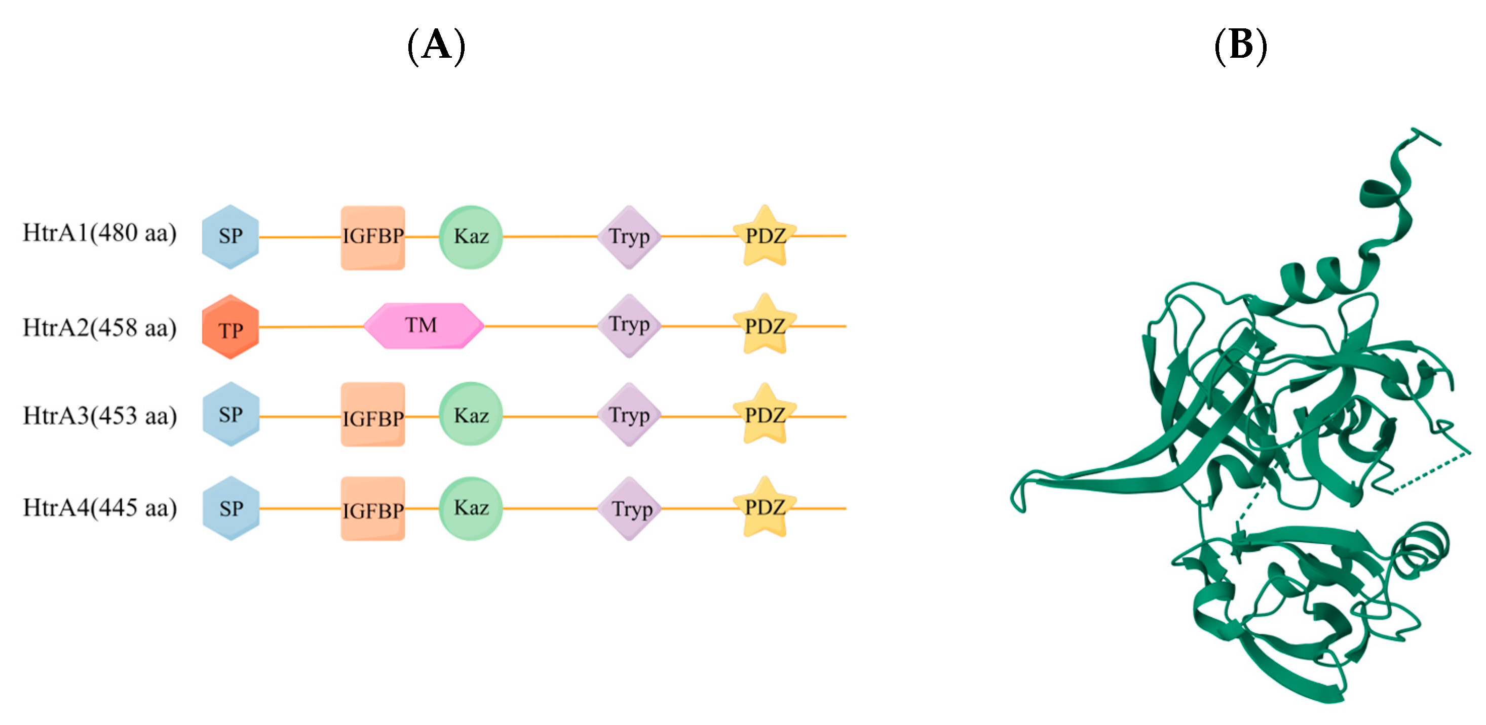
3.1. Structural Features of HtrA2

The highly conserved high temperature requirement A (HtrA) family of serine proteases has many different physiological functions and constitutes the core group of cellular proteases [24]. All four human HtrAs contain the characteristic motif of HtrA, a conserved protease domain and a C-terminal PDZ domain [25]. However, these enzymes show significant differences in their N-terminal regions, which may be necessary to accommodate their different functional properties. The families are naturally divided into two distinct groups based on their different N-terminal regions. The N-terminal regions of HtrA1, HtrA3, and HtrA4 contain a signal peptide (SP), an insulin growth factor binding protein domain (IGFBP), and a Kazal-type S protease inhibitor domain (Kaz) [25]. The N-terminal region of HtrA2 is completely different and includes a transient peptide (TP) and a transmembrane domain (TM) (Figure 1) [26].
Among the four identified human HtrAs, HtrA2 has been extensively studied for its enigmatic structural features and profound functional relevance. Studies have shown that HtrA2/Omi plays a role in the progression of neurodegenerative diseases, prostate cancer, and hepatocellular carcinoma [15,27]. Notably, HtrA2/Omi is involved in the development of several SCIs including rheumatoid arthritis (RA) and neuroinflammation. Jeong et al. showed that during endoplasmic reticulum stress-induced apoptosis, synoviocytes release HtrA2 into the synovial lumen of the RA, which is a key regulator of the production of inflammatory cytokines [28]. HtrA2 contributes to the pathogenesis of autoimmune arthritis through the differentiation of Th17 cells by STAT3 [29]. Motor neuron degeneration (mnd)2 mice lack HtrA2 activity due to a missense mutation Ser276Cys in the structural domain of the HtrA2 protease [30]. Lee et al. found that HtrA2mnd2 mice significantly increased pSTAT3 expression, and HtrA2 induced STAT3 degradation in vitro, demonstrating that HtrA2 improves RA by inhibiting STAT3 [31]. Hu et al. showed that increased transcription of pro-inflammatory genes, as well as activation of astrocytes and microglia, were observed in the brain of HtrA2mnd2 mice, suggesting that HtrA2/Omi is an intrinsic cytokine that inhibits neuroinflammation [32]. In addition, HtrA2/Omi expression correlates with the level of infiltration of multiple immune cell populations [33]. However, there is currently little understanding of the molecular mechanisms by which HtrA2/Omi controls inflammation and immune responses are unknown. Studies have shown that HtrA2/Omi plays a role in mitochondrial homeostasis [34]. Thus, the molecular mechanisms by which HtrA2/Omi controls inflammation and immune responses may be closely related to the role of HtrA2/Omi in mitochondrial homeostasis.

3.2. Functions of HtrA2 in MQC

Mitochondria are the primary site of cellular energy supply, where most cellular ATP is produced by oxidative phosphorylation in mitochondria, accounting for more than 80% of the energy required for life activities. Also, mitochondria are key regulators of cell fate during disease. They control cell survival by producing ATP that promotes cellular processes and, conversely, control cell death by inducing apoptosis through the release of pro-apoptotic factors (e.g., cytochrome C). Therefore, stringent quality control mechanisms must be in place to ensure a healthy mitochondrial network. Current studies have identified a central role for mitochondria in SCI aseptic inflammation [35]. MQC plays a key role in these processes. In the MQC system, there exists an important proteasome, HtrA2/Omi, located in the mitochondrial membrane interstitial space, originally described as a serine protease that induces a stress response in mammalian cells [12] and coordinates the MQC system by degrading its specific substrates thereby [36]. The current study shows that HtrA2/Omi promotes cell survival by maintaining mitochondrial homeostasis [30,37,38].

3.2.1. HtrA2 Dysregulation Triggers UPRmt

HtrA2 is a key stress-protective protease that cleaves misfolded proteins in an ATP-independent manner to protect cells from stress induced by toxic protein aggregates [39]. When proteins do not fold correctly, intermediate ‘misfolded’ forms called unfolded protein deposits (UPODs), stick together to form protein aggregates. There is growing evidence that HtrA2 exerts prominent neuroprotective effects by degrading toxic aggregates in mitochondria and that its aberrant function leads to mitochondrial dysregulation [40,41,42,43]. HtrA2/Omi is involved in the degradation of misfolded mitochondrial proteins, thus contributing to the maintenance of cellular homeostasis [36]. Felicity et al. showed that in neuronal cells HtrA2 deficiency has been shown to result in the accumulation of unfolded proteins in the mitochondria, a decrease in mitochondrial respiration, an increase in ROS production, and ultimately neuronal cell death [44]. Recent studies on isolated mitochondria from HtrA2/Omi knockout mice have shown increased accumulation of unfolded subunits of respiratory complexes I-IV and systemic respiratory chain dysfunction [42]. In addition, HtrA2/Omi is a protein quality control protease important for mitochondrial homeostasis and plays a major role in protein quality control and maintenance of cellular homeostasis. It has been shown that one of the main triggers of UPRmt is a disturbance of protein stabilization in the mitochondrial intermembrane space (IMS) [41]. Luena et al. showed that accumulation of proteins in the mitochondrial intermembrane space in the breast cancer cell line MCF-7 activates a unique UPRmt [45]. Under physiological conditions, proteins accumulated in the IMS are degraded by IMS proteases such as HtrA2, OMA1, and Yme1L [41,46,47]. The above suggests that HtrA2/Omi is a protease that is localized to the IMS in the physiological state and is involved in MQC to maintain mitochondrial protein homeostasis [45,48]. HtrA2 deficiency increases protein accumulation in the IMS to promote UPRmt formation.
UCF 101 is a HtrA2-specific inhibitor [49]. Klupsch et al. found that in MEF cells, UCF 101 activated CHOP transcription and expression of UPR response-associated protein [50]. It can lead to accumulation of unfolded proteins in mitochondria, oxidative stress, and defective mitochondrial respiration [51]. In addition, Meng et al. showed increased expression of the UPRmt-related proteins HSP10, HSP60, and LONP in HtrA2Hetero mice [52], suggesting that HtrA2/Omi deletion leads to MQC imbalance through an increase in UPRmt. UPRmt activation of PKR (double-stranded-RNA-activated protein kinase) is dependent on ClpP activity; Pkr−/− mice were unable to activate the UPRmt target gene CPN60 in intestinal epithelial cells after the administration of dextran sodium sulfate and were protected against DSS-induced colitis [53]. Induction of PKR links the mitochondrial unfolded protein response to the pathogenesis of intestinal inflammation. In addition, NF-κB is also upregulated when the UPR is activated [54]. Thus, the UPRmt may be involved in the balance between inflammatory and anti-inflammatory responses.

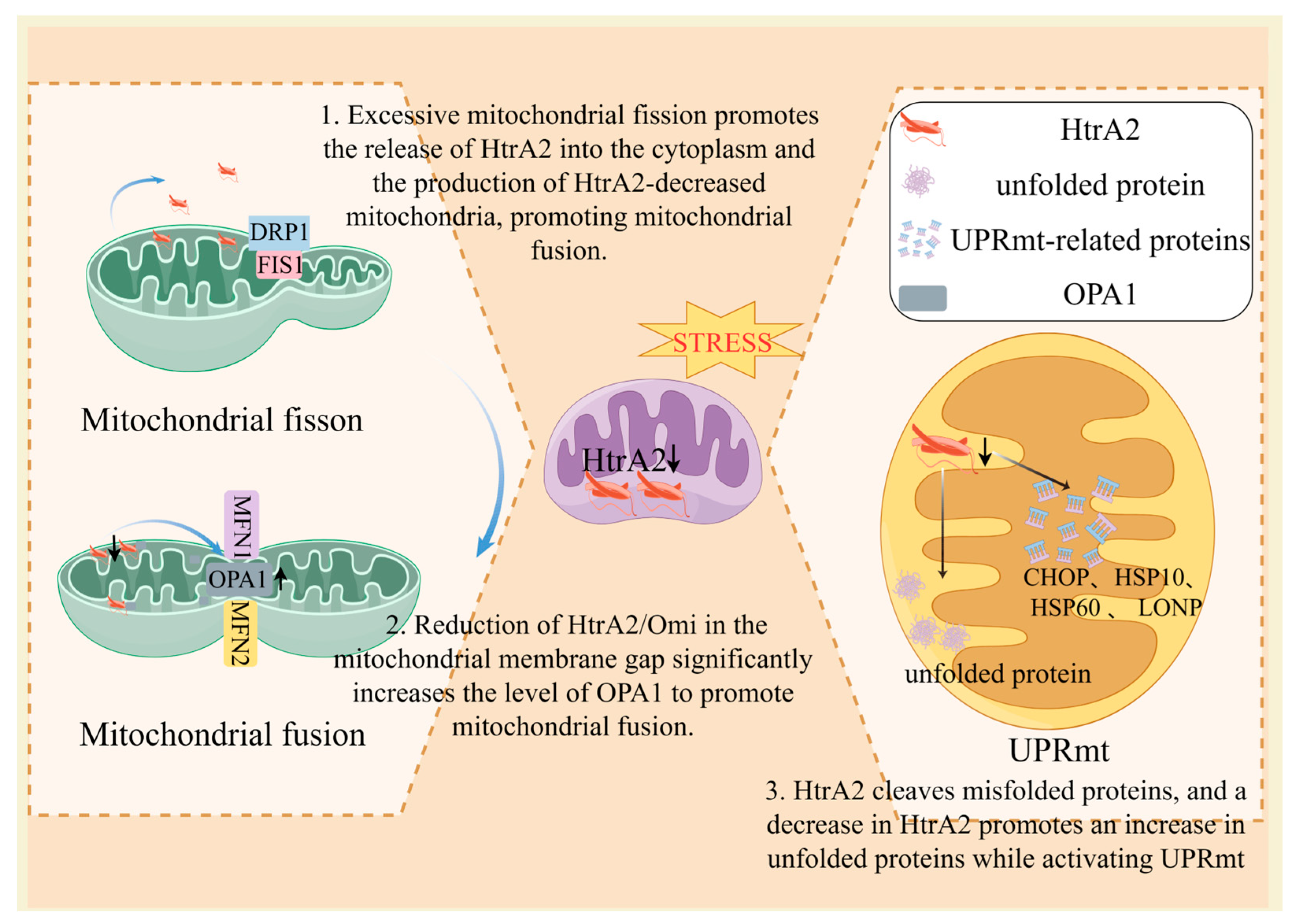
3.2.2. HtrA2 Dysregulation Induces an Imbalance in Mitochondrial Dynamics

Mitochondria can form complex networks within cells. Depending on the health of the mitochondrial network and the energy requirements of the cell, the size, morphology, and number of mitochondria are constantly changing [55]. The number and morphology of mitochondria are controlled by tightly regulated fission and fusion events [56]. Mitochondrial fusion maintains mitochondrial function and prevents the production of defective mtDNA [57]. Fusion is mediated by fusion proteins in the mitochondrial outer and inner membranes (OMM and IMM), termed mitofusin 1 (Mfn1), mitofusin 2 (Mfn2), and optic atrophy 1 (OPA1), respectively [58]. Mitochondrial fusion is a two-step process that involves OMM fusion mediated by Mfn1/2 and inner mitochondrial membrane (IMM) fusion mediated by OPA1. Mfn1 and Mfn2 share significant sequence and structural similarities, but they appear to have distinct cellular functions [59]. Mfn1 more efficiently mediates mitochondrial fusion, which may be due to having approximately eight times the GTPase activity [60]. OPA1 also relies on Mfn1, but not Mfn2, to promote fusion [58]. The fusion of two mitochondria produces hyperfused mitochondria, which can be stimulated by abnormal stimuli to produce more ATP. Mitochondrial fission is controlled by dynamin-related protein 1 (DRP1), which interacts with mitochondrial fission 1 (FIS1) after recruitment to the OMM to promote mitochondrial contraction and eventual fission [61,62].
Kieper et al. found that HtrA2/Omi interacts directly with OPA1 both functionally and physically, and that OPA1 levels were significantly increased in XMEF of HtrA2/Omi knockout mice. Transfection of HtrA2/Omi into HtrA2/Omi knockout cells restored increased OPA1 levels to those in wild-type (WT) mice [63]. These results demonstrated that HtrA2/Omi deletion is involved in regulating mitochondrial kinetic processes through the promotion of mitochondrial fusion, which affects disease progression. In addition, it has been shown that silencing of Yes-associated protein drives JNK phosphorylation, which induces Drp1 activation and translocation to the mitochondrial surface, thereby initiating mitochondrial fission. Excessive mitochondrial fission mediates the leakage of HtrA2/Omi from the mitochondria into the cytoplasm, demonstrating that excessive mitochondrial fission promotes HtrA2/Omi mitochondrial leakage into the cytoplasm [64] which in turn produces HtrA2/Omi-reduced mitochondria, further exacerbates mitochondrial fusion, and promotes MQC imbalance (Figure 2). The above suggests that HtrA2/Omi is associated with mitochondrial fission, but whether HtrA2/Omi deficiency regulates the mitochondrial fission response requires further verification.
The current study suggests that mitochondrial dynamics-related MQC is associated with the activation of the inflammatory response [65]. An imbalance between mitochondrial fusion and fission leads to mitochondrial dysfunction, which in turn results in mitochondrial fragmentation, diminished oxidative phosphorylation energy supply, reduction of mtDNA, and increased ROS production. This ROS burst represents a major pro-inflammatory factor by regulating NF-κB expression and activity [66]. Abnormal mitochondrial fusion and division activate different early inflammatory mediators such as tumor necrosis factor α, interleukins, and IFN-γ [67]. Mitochondrial dysfunction with upregulation of mitochondrial fission prevents the elimination of damaged mitochondria; the generation of ROS in excess of antioxidant activity may be an initiating factor for SCI [68]. With the release of ROS and mitochondria-derived DAMPs, mitochondrial dysfunction initiates an inflammatory response by promoting the release of proinflammatory cytokines that interact with DAMPs receptors [20].

4. HtrA2-Mediated Modulation of Mitochondrial Quality Control Signaling Is Involved in the Regulation of Macrophage Polarization Phenotype in SCI

HtrA2/Omi activity is critical for the inflammatory response. Inflammation and oxidative stress interact with each other, with inflammation increasing ROS production and ROS exacerbating inflammation [69]. HtrA2/Omi is normally present in the mitochondrial membrane interstitial space; however, under stressful conditions such as oxidative or heat stress, it disrupts the inner membrane and migrates into the mitochondrial matrix [70]. This results in a decrease in HtrA2/Omi in the membrane interstitial space. Under apoptotic signaling, HtrA2/Omi is released from the mitochondria into the cytoplasmic lysate and is involved in disease progression in neurodegeneration and rheumatoid arthritis [27]. In addition, in endoplasmic reticulum stress-induced RA-FLS, HtrA2 is released from mitochondria to the extracellular space [28]. It suggests that long-term exposure of macrophages to oxidative stress and heat stress within the SCI response releases HtrA2/Omi from the mitochondrial membrane interstitial space to the mitochondrial matrix, cytoplasm, and extracellularly, which ultimately leads to a decrease in mitochondrial membrane interstitial HtrA2/Omi.
Data obtained from the database BioGPS suggest that HtrA2 levels are high in immune cells, especially precursor cells of myeloid-derived immune cells. Mitochondrial membrane gap HtrA2/Omi deficiency reduces pro-inflammatory cytokine production in LPS or CpG-triggered BMDM, suggesting that HtrA2/Omi is an immune response regulator [17]. Current studies have found that alterations in mitochondrial metabolism and physiology lead to macrophage activation into different states, such as alterations in oxidative metabolism, mtROS, mitochondrial ultrastructure, and membrane potential [19]. HtrA2/Omi plays an important role in the homeostasis of macrophage polarization and function by modulating the morphostructure, function, lifespan, and bioenergetic properties of mitochondria.

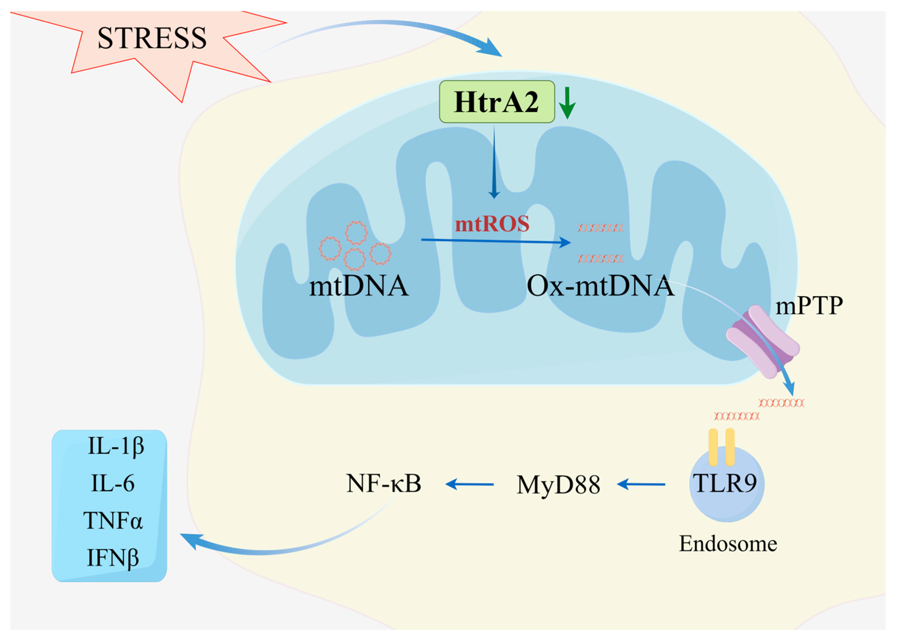
4.1. Aberrant HtrA2 Function Promotes ROS Generation Inducing Ox-mtDNA-Catalyzed Macrophage Immune Activation

Studies have shown that mitochondrial DNA (mtDNA) regulates energy production and cellular metabolism, and is a key signaling molecule that triggers the inflammatory response. mtDNA contains inflammatory unmethylated CpG motifs similar to those found in bacterial DNA, which makes it more of an “exogenous” than an “endogenous” DNA [71]. Without histone protection, mtDNA is more susceptible to mitochondrial reactive oxygen species (mtROS) attack [72], leading to oxidative damage to mtDNA. Min-Kyung et al. showed that HtrA2 regulates α-Synuclein-mediated mtROS production in microglial cell mitochondria. HtrA2 inactivation led to an increase in mtROS in microglia [73]. Restoration of HtrA2/Omi expression rescued CCl4-induced hepatic fibrosis in hepatocytes and reversed mitochondrial dysfunction (e.g., excessive ROS production) [51]. This suggests that HtrA2/Omi acts as an important proteasome in the MQC system to regulate mtROS production.
Increased mitochondrial ROS release inhibits macrophage anti-inflammatory function. ROS promote macrophages to undergo pro-inflammatory macrophages polarization. Mitochondria are the main source of ROS in macrophage polarization [74]. It has been shown that decreased mitochondrial ROS levels inhibit pro-inflammatory macrophages polarization and promote macrophage polarization to anti-inflammatory macrophages [75]. NADPH oxidase 4 (NOX4) plays an important role in the transport of electrons in the mitochondrial respiratory chain, which is one of the major sources of ROS. In a study of inflammatory bowel disease it was shown that NOX4 is able to promote polarization of intestinal macrophages in the pro-inflammatory macrophages direction through increased ROS production [76], and that elevated glucose uptake induces an overproduction of ROS, leading to macrophage activation in the pro-inflammatory direction in vitro [77,78]. Furthermore, systemic inflammation with predominant pro-inflammatory macrophages polarization was observed in Ndufs1 knockout (a key component of mitochondrial complex I) mice, which could be attenuated by the use of the mitochondria-targeted antioxidant Mito-TEMPO. This supports a critical role for mitochondrial ROS in macrophage polarization to pro-inflammatory macrophages [79]. Thus, ROS are critical for anti-inflammatory macrophages polarization, while mitochondria also play an important role in ROS. So, how does ROS regulate macrophage polarization?
Goo et al. showed that in MEF cells and mouse brain tissues, HtrA2 protein deletion led to mtDNA conformational changes by promoting mtROS production, targeting only HtrA2/Omi overexpression; mtDNA repair genes, including POLG2, Twinkle, and APTX1 were significantly up-regulated in HtrA2/Omi knockdown cells, demonstrating that HtrA2/Omi was able to restore mtDNA conformational stability in HtrA2/Omi knockdown MEF cells [34]. Hui-Gwan et al. showed that HTRA2 helps to maintain mitochondrial DNA conformation changes in MEF cells and HeLa cells, and HtrA2/Omi deficiency through ROS production and mutation leading to mtDNA damage [80]. We speculate that this mtDNA conformational change is associated with resistant oxidative-damaged mtDNA (Ox-mtDNA). It has been shown that HtrA2/Omi deficiency induces Ox-mtDNA production by promoting mtROS survival. Increased ROS cause an alteration in mitochondrial membrane potential, which subsequently induces the opening of the mitochondrial permeability transition pore (MPTP) [81], which releases Ox-mtDNA into the cytoplasm, where it is then recognized by the cytoplasmic receptor for DAMPs [82]. DAMPs activate pathways related to innate immunity through toll-like receptors (TLR) [83,84]. MtDNA contains an unmethylated CpG-DNA sequence; these sequences are TLR9 ligands that can mediate inflammation through the NF-κB pathway [85]. TLR9 is a classic TLR that recognizes CpG DNA and senses nucleic acids to initiate myeloid differentiation primary response protein 88 (MyD88) triggering a downstream signaling cascade that activates the nuclear transcription factor NF-κB [86], promotes the production of pro-inflammatory cytokines such as interleukin-1β (IL-1β) and interleukin-12 (IL-12), as well as interferon I (IFN), and promotes macrophage polarization toward pro-inflammatory macrophages (Figure 3) [87,88].

4.2. OXPHOS Complex Abnormalities Promote Pro-Inflammatory Macrophages Polarization

MQC has a profound effect on macrophage function. Mitochondria are the main site of cellular energy supply, in which most cellular ATP is produced by oxidative phosphorylation in the mitochondria, accounting for more than 80% of the energy required for life activities. In the resting state, macrophages meet their energy requirements through oxidative phosphorylation. After LPS stimulation, macrophages are polarized towards pro-inflammatory macrophages; the tricarboxylic acid cycle, fatty acid oxidation, and oxidative phosphorylation activity of the pro-inflammatory macrophages subpopulation is impaired and relies on increased levels of glycolysis (also known as the Warburg effect) and the pentose phosphate pathway to satisfy the cellular energy requirements [6]. Cytokines such as tumor necrosis factor α, IL-6, and IL-1β and chemicals such as ROS and NO are characteristic of pro-inflammatory macrophages [89].
One of the hallmark features of HtrA2 as a stress-protective protease is its ability to cleave aggregated proteins [42,90,91]. HtrA2/Omi deficiency promotes the generation of unfolded proteins, in particular components of respiratory complexes on mitochondrial membranes. The current study shows that HtrA2/Omi protease deficiency leads to oxidative phosphorylation and a reduction in the number of mitochondrial genes encoding ETC subunits [14]. Moisoi et al. showed that an increase in the unfolded subunits of the HtrA2/Omi KO brain mitochondrial complexes I-IV leads to systemic respiratory dysfunction [42]. This suggests that when the protease of HtrA2 activity is absent, the inability to remove unfolded respiratory complex proteins leads to mitochondrial complex abnormalities that inhibit OXPHOS levels. In addition, mitochondrial respiratory chain complexes I and II were significantly reduced in HtrA2mnd2 littermate mice, and mitochondrial synthesis of ATP energy was reduced, leading to increased ROS production, which further impaired the respiratory chain complexes and exacerbated the mitochondrial damage, resulting in a vicious cycle [92]. It was shown above that HtrA2/Omi deficiency leads to abnormal mitochondrial dynamics by promoting mitochondrial division. In the hippocampus of heart failure mice, mitochondrial fission gene DRP1 has been reported to be increased, and damaged mitochondria release excess ROS and have a reduced ability to produce ATP [93]. Furthermore, reduced mitochondrial mass and activity was found after overexpression of mitochondrial fission factor in the breast cancer cell line MCF7, and analysis of metabolic fluxes using Seahorse XFe96 revealed reduced levels of oxidative phosphorylation and glycolysis in this cell [94]. This suggests that increased mitochondrial fission due to HtrA2/Omi deficiency may impair mitochondrial energy metabolism and thus increase macrophage polarization towards pro-inflammatory macrophages.
In summary, HtrA2/Omi plays an important role in the regulation of macrophage activation by affecting MQC. MQC abnormalities affect the direction of macrophage polarization regulated by ROS generation on the one hand; on the other hand, they lead to abnormal energy metabolism driving the direction of pro-inflammatory macrophages polarization.

5. Conclusions

Systemic SCI is a low-grade, persistent inflammation that may cause tissue and organ damage through, for example, oxygen stress. The persistence of inflammatory factors (e.g., IL-1β, IL-6, and TNF-α) and damage to tissues is the underlying cause of SCI. MQC is an integrated network that monitors the quality of mitochondria and an endogenous cellular protective program that is essential for maintaining mitochondrial homeostasis and function. Many mitochondrial components and metabolites can function as DAMPs and promote inflammation upon release into the cytoplasm or extracellular environment. As mentioned previously, in SCI, oxidative stress and other induced release of mitochondrial membrane interstitial HtrA2/Omi into the mitochondrial matrix or cytoplasm leads to a reduction of HtrA2/Omi in the mitochondrial membrane interstitial space. One of the hallmark features of HtrA2 as a stress-protective protease is its ability to cleave aggregated proteins, which in turn promotes UPRmt disruption and a reduction in the OXPHOS complex, contributing to an increase in ROS; on the other hand, a decrease in HtrA2/Omi promotes an increase in OPA1, which in turn promotes mitochondrial fragmentation leading to mitochondrial kinetic derangement, contributing to increased ROS release. The increased ROS causes oxidative damage to mtDNA, and Ox-mtDNA escapes from the mitochondria to the cytoplasmic matrix or even extracellular space. Ox-mtDNA acts as DAMPs to initiate TLR9/MyD88/NF-κB, promoting pro-inflammatory macrophages polarization and releasing pro-inflammatory cytokines, which further amplifies the inflammatory response. Targeting HtrA2/Omi is therefore a new direction for the treatment of SCI.
The SCI response is associated with mitochondrial dysfunction as well as high levels of ROS production [95]. It has been shown that impaired mitochondrial quality control due to loss of function of the mitochondrial protease HtrA2 is associated with the accumulation of damaged mitochondria [96]. Neurodegenerative phenotypes in mice completely lacking HtrA2/Omi or expressing an enzymatically inactivated mnd2 mutant suggest that the proteolytic activity of HtrA2/Omi exerts a protective role in the mitochondria of neuronal cells [30,37]. Mitochondria are the primary site for the production of ROS or free radicals, which are necessary to fight infection. However excessive and uncontrolled ROS production can be detrimental to cells, leading to mitochondrial and tissue damage [95]. The results of the study showed that BMDMs with dysregulated mitochondrial quality control have high levels of ROS production, suggesting that high levels of oxidative stress mediated by dysregulated mitochondrial quality control may serve as an additional important pathway for the maintenance of SCI. In addition, the high levels of ROS produced could in turn act as new damage factors to make the already precarious mitochondrial quality control even more vulnerable to mitochondrial quality control dysregulation and oxidative damage promoting inflammatory progression. It has been shown that HtrA2, in addition to its classical role as a serine protease in mitochondria, can acquire new functions as a regulator of inflammation, demonstrating the functional diversity of mitochondrial proteins. Inhibitors of apoptosis (IAPs) proteins were initially characterized for their ability to directly bind and inhibit apoptotic caspases. Interestingly, the current study demonstrates the importance of IAPs in modulating inflammatory signaling and immune responses [97,98]. It was shown that inhibition of cIAPs modulated the activity of tumor-associated macrophages and reversed the status of anti-inflammatory macrophages to pro-inflammatory macrophages, with massive recruitment of neutrophils [99]. At the same time, levels of pro-inflammatory cytokines, including TNFα, IFNγ, and IL-12, were increased and promoted SCI progression. In addition, Kavanagh et al. suggested that cIAP-2 may be a therapeutic target to reduce the pro-inflammatory activation of microglial cells [100]. Many articles have shown that cytoplasmic HtrA2 can bind and antagonize IAPs [40,101]. Thus, HtrA2 may affect SCI by regulating proteins of the IAP family.
This review highlights the importance of HtrA2/Omi as an important regulator of mitochondrial quality control, in addition to its involvement in protein degradation by mis-accumulation through the UPRmt and in the regulation of mitochondrial dynamics. In addition, HtrA2/Omi is involved in the regulation of mitochondrial biogenesis and mitochondrial autophagy of HtrA2/Omi to renew the mitochondrial reservoir by interacting with the GSK3β/PGC-1α pathway. On the one hand, Xu et al. showed that Omi cleaves glycogen synthase kinase 3β (GSK3β), a kinase that promotes the degradation of PPARγ coactivator-1α (PGC-1α), and thus regulates PGC-1α, an important factor in mitochondrial biogenesis; moreover, in HTRA2mnd2 mice, the abundance of GSK3β was significantly increased, while the abundance of PGC-1α was significantly decreased [102]. On the other hand, Kang et al. showed that HTRA2mnd2 mice exhibited marked cardiac enlargement with left ventricular hypertrophy, which was associated with increased autophagosome activity in cardiac tissue. The increase in autophagosome activity may be due to an increase in mitochondrial autophagy, as their in vitro experiments showed that two mitochondrial proteins, PPID/peptidylprolyl isomerase D (cyclophilin D) and SOD2/MnSOD, were degraded at an increased rate in HTRA2-expressing MEFs in response to starvation compared to HTRA2-deficient mouse embryonic fibroblasts (MEFs) [103]. In addition, Cilenti et al. showed the accumulation of Mulan protein in various tissues of HTRA2mnd2 mice as well as in HTRA2-deficient MEFs, which resulted in a significant reduction of mitofusin 2 (Mfn2) protein and an increase in mitochondrial autophagy [104]. Furthermore, this work highlights the importance of HtrA2 as a potential therapeutic target for SCI, as it is a common trigger of the inflammatory process. HtrA2/Omi is a potential biomarker for SCI. However, its therapeutic utility is speculative and further studies are needed to fully grasp its role. The location of elevated HtrA2/Omi varies in different inflammatory diseases, with higher concentrations of HtrA2 in rheumatoid arthritis (RA) synovial fluid (SF) than in osteoarthritis (OA) SF, which correlates with the number of immune cells in RA SF, and with the finding that treatment of RA FLS with an endoplasmic reticulum stress inducer resulted in the release of HtrA2/Omi from the mitochondria into the extracellular space [28]. In addition, HtrA2/Omi was released from mitochondria to the cytoplasm in pneumonia [18]. Therefore, targeting drugs to the specific location of HtrA2/Omi expression in different inflammatory conditions is more therapeutically promising.

Author Contributions

Conceptualization, J.S., writing—original draft preparation, Q.L., visualization, X.Y., R.L. and Y.Y.; writing—review and editing, Y.Z., J.W. and J.F. All authors have read and agreed to the published version of the manuscript.

Funding

This research was funded by the Jilin Province Science and Technology Development Plan Project (20230505046ZP); the Jilin Province Health Science and Technology Ability Improvement Project (2023JC018, 2022JC045); the Norman Bethune Project Plan of Jilin University (2023B32); the National Natural Science Foundation of China (82303668).

Institutional Review Board Statement

Not applicable.

Informed Consent Statement

Not applicable.

Data Availability Statement

Not applicable.

Conflicts of Interest

The authors declare no conflicts of interest.

References

  1. Zhou, W.B.S.; Meng, J.; Zhang, J. Does Low Grade Systemic Inflammation Have a Role in Chronic Pain? Front. Mol. Neurosci. 2021, 14, 785214. [Google Scholar] [CrossRef]
  2. Franceschi, C.; Garagnani, P.; Vitale, G.; Capri, M.; Salvioli, S. Inflammaging and ‘Garb-Aging’. Trends Endocrinol. Metab. 2017, 28, 199–212. [Google Scholar] [CrossRef]
  3. Liston, A.; Masters, S.L. Homeostasis-altering molecular processes as mechanisms of inflammasome activation. Nat. Rev. Immunol. 2017, 17, 208–214. [Google Scholar] [CrossRef]
  4. Frank, D.; Vince, J.E. Pyroptosis versus necroptosis: Similarities, differences, and crosstalk. Cell Death Differ. 2019, 26, 99–114. [Google Scholar] [CrossRef]
  5. Alivernini, S.; MacDonald, L.; Elmesmari, A.; Finlay, S.; Tolusso, B.; Gigante, M.R.; Petricca, L.; Di Mario, C.; Bui, L.; Perniola, S.; et al. Distinct synovial tissue macrophage subsets regulate inflammation and remission in rheumatoid arthritis. Nat. Med. 2020, 26, 1295–1306. [Google Scholar] [CrossRef]
  6. Kelly, B.; O’Neill, L.A. Metabolic reprogramming in macrophages and dendritic cells in innate immunity. Cell Res. 2015, 25, 771–784. [Google Scholar] [CrossRef]
  7. Jha, A.K.; Huang, S.C.; Sergushichev, A.; Lampropoulou, V.; Ivanova, Y.; Loginicheva, E.; Chmielewski, K.; Stewart, K.M.; Ashall, J.; Everts, B.; et al. Network integration of parallel metabolic and transcriptional data reveals metabolic modules that regulate macrophage polarization. Immunity 2015, 42, 419–430. [Google Scholar] [CrossRef]
  8. Shao, L.W.; Peng, Q.; Dong, M.; Gao, K.; Li, Y.; Li, Y.; Li, C.Y.; Liu, Y. Histone deacetylase HDA-1 modulates mitochondrial stress response and longevity. Nat. Commun. 2020, 11, 4639. [Google Scholar] [CrossRef]
  9. Yao, B.F.; Luo, X.J.; Peng, J. A review for the correlation between optic atrophy 1-dependent mitochondrial fusion and cardiovascular disorders. Int. J. Biol. Macromol. 2023, 254 Pt 2, 127910. [Google Scholar] [CrossRef]
  10. Ng, M.Y.W.; Wai, T.; Simonsen, A. Quality control of the mitochondrion. Dev. Cell 2021, 56, 881–905. [Google Scholar] [CrossRef]
  11. Kawano, I.; Bazila, B.; Ježek, P.; Dlasková, A. Mitochondrial Dynamics and Cristae Shape Changes during Metabolic Reprogramming. Antioxid. Redox Signal. 2023, 39, 684–707. [Google Scholar] [CrossRef]
  12. Faccio, L.; Fusco, C.; Chen, A.; Martinotti, S.; Bonventre, J.V.; Zervos, A.S. Characterization of a novel human serine protease that has extensive homology to bacterial heat shock endoprotease HtrA and is regulated by kidney ischemia. J. Biol. Chem. 2000, 275, 2581–2588. [Google Scholar] [CrossRef]
  13. Goo, H.G.; Rhim, H.; Kang, S. Pathogenic Role of Serine Protease HtrA2/Omi in Neurodegenerative Diseases. Curr. Protein Pept. Sci. 2017, 18, 746–757. [Google Scholar] [CrossRef] [PubMed]
  14. Sun, H.; Shen, L.; Zhang, P.; Lin, F.; Ma, J.; Wu, Y.; Yu, H.; Sun, L. Inhibition of High-Temperature Requirement Protein A2 Protease Activity Represses Myogenic Differentiation via UPRmt. Int. J. Mol. Sci. 2022, 23, 11761. [Google Scholar] [CrossRef] [PubMed]
  15. Wang, H.; Jiang, F.; Hao, F.; Ju, R. The expression of HtrA2 and its diagnostic value in patients with hepatocellular carcinoma. Medicine 2018, 97, e0128. [Google Scholar] [CrossRef]
  16. Dagda, R.K.; Chu, C.T. Mitochondrial quality control: Insights on how Parkinson’s disease related genes PINK1, parkin, and Omi/HtrA2 interact to maintain mitochondrial homeostasis. J. Bioenerg. Biomembr. 2009, 41, 473–479. [Google Scholar] [CrossRef]
  17. Xu, Z.; Lin, J.; Xie, Y.; Tang, H.; Xie, J.; Zeng, R. HtrA2 is required for inflammatory responses in BMDMs via controlling TRAF2 stability in collagen-induced arthritis. Mol. Immunol. 2021, 129, 78–85. [Google Scholar] [CrossRef]
  18. Wang, X. Inhibition of HtrA2 alleviates inflammatory response and cell apoptosis in lipopolysaccharide-induced acute pneumonia in rats. Mol. Med. Rep. 2020, 22, 3127–3134. [Google Scholar] [CrossRef]
  19. Wang, Y.; Li, N.; Zhang, X.; Horng, T. Mitochondrial metabolism regulates macrophage biology. J. Biol. Chem. 2021, 297, 100904. [Google Scholar] [CrossRef]
  20. Chan, N.C.; Salazar, A.M.; Pham, A.H.; Sweredoski, M.J.; Kolawa, N.J.; Graham, R.L.; Hess, S.; Chan, D.C. Broad activation of the ubiquitin-proteasome system by Parkin is critical for mitophagy. Hum. Mol. Genet. 2011, 20, 1726–1737. [Google Scholar] [CrossRef] [PubMed]
  21. Zhang, B.; Lai, R.C.; Sim, W.K.; Lim, S.K. Therapeutic Efficacy of Mesenchymal Stem/Stromal Cell Small Extracellular Vesicles in Alleviating Arthritic Progression by Restoring Macrophage Balance. Biomolecules 2023, 13, 1501. [Google Scholar] [CrossRef]
  22. Tardito, S.; Martinelli, G.; Soldano, S.; Paolino, S.; Pacini, G.; Patane, M.; Alessandri, E.; Smith, V.; Cutolo, M. Macrophage M1/M2 polarization and rheumatoid arthritis: A systematic review. Autoimmun. Rev. 2019, 18, 102397. [Google Scholar] [CrossRef]
  23. Yu, H.; Chang, Q.; Sun, T.; He, X.; Wen, L.; An, J.; Feng, J.; Zhao, Y. Metabolic reprogramming and polarization of microglia in Parkinson’s disease: Role of inflammasome and iron. Ageing Res. Rev. 2023, 90, 102032. [Google Scholar] [CrossRef] [PubMed]
  24. Page, M.J.; Di Cera, E. Evolution of peptidase diversity. J. Biol. Chem. 2008, 283, 30010–30014. [Google Scholar] [CrossRef] [PubMed]
  25. Singh, H.; Nero, T.L.; Wang, Y.; Parker, M.W.; Nie, G. Activity-modulating monoclonal antibodies to the human serine protease HtrA3 provide novel insights into regulating HtrA proteolytic activities. PLoS ONE 2014, 9, e108235. [Google Scholar] [CrossRef] [PubMed]
  26. Nie, G.Y.; Hampton, A.; Li, Y.; Findlay, J.K.; Salamonsen, L.A. Identification and cloning of two isoforms of human high-temperature requirement factor A3 (HtrA3), characterization of its genomic structure and comparison of its tissue distribution with HtrA1 and HtrA2. Biochem. J. 2003, 371 Pt 1, 39–48. [Google Scholar] [CrossRef] [PubMed]
  27. Vande Walle, L.; Lamkanfi, M.; Vandenabeele, P. The mitochondrial serine protease HtrA2/Omi: An overview. Cell Death Differ. 2008, 15, 453–460. [Google Scholar] [CrossRef] [PubMed]
  28. Jeong, G.H.; Nam, M.K.; Hur, W.; Heo, S.; Lee, S.; Choi, E.; Park, J.H.; Park, Y.; Kim, W.U.; Rhim, H.; et al. Role of high-temperature requirement serine protease A 2 in rheumatoid inflammation. Arthritis Res. Ther. 2023, 25, 96. [Google Scholar] [CrossRef]
  29. Koch, A.E. Review: Angiogenesis: Implications for rheumatoid arthritis. Arthritis Rheum. 1998, 41, 951–962. [Google Scholar] [CrossRef] [PubMed]
  30. Jones, J.M.; Datta, P.; Srinivasula, S.M.; Ji, W.; Gupta, S.; Zhang, Z.; Davies, E.; Hajnóczky, G.; Saunders, T.L.; Van Keuren, M.L.; et al. Loss of Omi mitochondrial protease activity causes the neuromuscular disorder of mnd2 mutant mice. Nature 2003, 425, 721–727. [Google Scholar] [CrossRef]
  31. Lee, S.H.; Moon, Y.M.; Seo, H.B.; Kim, S.Y.; Kim, E.K.; Yi, J.; Nam, M.K.; Min, J.K.; Park, S.H.; Rhim, H.; et al. HtrA2 suppresses autoimmune arthritis and regulates activation of STAT3. Sci. Rep. 2016, 6, 39393. [Google Scholar] [CrossRef]
  32. Hu, Q.; Li, B.; Xu, R.; Chen, D.; Mu, C.; Fei, E.; Wang, G. The protease Omi cleaves the mitogen-activated protein kinase kinase MEK1 to inhibit microglial activation. Sci. Signal. 2012, 5, ra61. [Google Scholar] [CrossRef]
  33. Feng, L.; Li, Z.; Xiong, Y.; Yan, T.; Fu, C.; Zeng, Q.; Wang, H. HtrA2 Independently Predicts Poor Prognosis and Correlates with Immune Cell Infiltration in Hepatocellular Carcinoma. J. Oncol. 2023, 2023, 4067418. [Google Scholar] [CrossRef]
  34. Singh, S.; Datta, G.; Jain, S.; Thakur, V.; Arora, P.; Muneer, A.; Asad, M.; Ali, S.; Mohmmed, A. Dual role of an essential HtrA2/Omi protease in the human malaria parasite: Maintenance of mitochondrial homeostasis and induction of apoptosis-like cell death under cellular stress. PLoS Pathog. 2022, 18, e1010932. [Google Scholar] [CrossRef] [PubMed]
  35. López-Armada, M.J.; Riveiro-Naveira, R.R.; Vaamonde-García, C.; Valcárcel-Ares, M.N. Mitochondrial dysfunction and the inflammatory response. Mitochondrion 2013, 13, 106–118. [Google Scholar] [CrossRef] [PubMed]
  36. Chaganti, L.K.; Kuppili, R.R.; Bose, K. Intricate structural coordination and domain plasticity regulate activity of serine protease HtrA2. FASEB J. 2013, 27, 3054–3066. [Google Scholar] [CrossRef] [PubMed]
  37. Martins, L.M.; Morrison, A.; Klupsch, K.; Fedele, V.; Moisoi, N.; Teismann, P.; Abuin, A.; Grau, E.; Geppert, M.; Livi, G.P.; et al. Neuroprotective role of the Reaper-related serine protease HtrA2/Omi revealed by targeted deletion in mice. Mol. Cell. Biol. 2004, 24, 9848–9862. [Google Scholar] [CrossRef] [PubMed]
  38. Krick, S.; Shi, S.; Ju, W.; Faul, C.; Tsai, S.Y.; Mundel, P.; Böttinger, E.P. Mpv17l protects against mitochondrial oxidative stress and apoptosis by activation of Omi/HtrA2 protease. Proc. Natl. Acad. Sci. USA 2008, 105, 14106–14111. [Google Scholar] [CrossRef] [PubMed]
  39. Toyama, Y.; Harkness, R.W.; Kay, L.E. Structural basis of protein substrate processing by human mitochondrial high-temperature requirement A2 protease. Proc. Natl. Acad. Sci. USA 2022, 119, e2203172119. [Google Scholar] [CrossRef] [PubMed]
  40. Suzuki, Y.; Imai, Y.; Nakayama, H.; Takahashi, K.; Takio, K.; Takahashi, R. A serine protease, HtrA2, is released from the mitochondria and interacts with XIAP, inducing cell death. Mol. Cell 2001, 8, 613–621. [Google Scholar] [CrossRef] [PubMed]
  41. Radke, S.; Chander, H.; Schäfer, P.; Meiss, G.; Krüger, R.; Schulz, J.B.; Germain, D. Mitochondrial protein quality control by the proteasome involves ubiquitination and the protease Omi. J. Biol. Chem. 2008, 283, 12681–12685. [Google Scholar] [CrossRef] [PubMed]
  42. Moisoi, N.; Klupsch, K.; Fedele, V.; East, P.; Sharma, S.; Renton, A.; Plun-Favreau, H.; Edwards, R.E.; Teismann, P.; Esposti, M.D.; et al. Mitochondrial dysfunction triggered by loss of HtrA2 results in the activation of a brain-specific transcriptional stress response. Cell Death Differ. 2009, 16, 449–464. [Google Scholar] [CrossRef] [PubMed]
  43. Desideri, E.; Martins, L.M. Mitochondrial Stress Signalling: HTRA2 and Parkinson’s Disease. Int. J. Cell Biol. 2012, 2012, 607929. [Google Scholar] [CrossRef] [PubMed]
  44. Johnson, F.; Kaplitt, M.G. Novel mitochondrial substrates of omi indicate a new regulatory role in neurodegenerative disorders. PLoS ONE 2009, 4, e7100. [Google Scholar] [CrossRef] [PubMed]
  45. Papa, L.; Germain, D. Estrogen receptor mediates a distinct mitochondrial unfolded protein response. J. Cell Sci. 2011, 124 Pt 9, 1396–1402. [Google Scholar] [CrossRef] [PubMed]
  46. Rainbolt, T.K.; Saunders, J.M.; Wiseman, R.L. YME1L degradation reduces mitochondrial proteolytic capacity during oxidative stress. EMBO Rep. 2015, 16, 97–106. [Google Scholar] [CrossRef] [PubMed]
  47. Head, B.; Griparic, L.; Amiri, M.; Gandre-Babbe, S.; van der Bliek, A.M. Inducible proteolytic inactivation of OPA1 mediated by the OMA1 protease in mammalian cells. J. Cell Biol. 2009, 187, 959–966. [Google Scholar] [CrossRef]
  48. Skorko-Glonek, J.; Zurawa-Janicka, D.; Koper, T.; Jarzab, M.; Figaj, D.; Glaza, P.; Lipinska, B. HtrA protease family as therapeutic targets. Curr. Pharm. Des. 2013, 19, 977–1009. [Google Scholar] [CrossRef]
  49. Zhang, C.; He, A.; Liu, S.; He, Q.; Luo, Y.; He, Z.; Chen, Y.; Tao, A.; Yan, J. Inhibition of HtrA2 alleviated dextran sulfate sodium (DSS)-induced colitis by preventing necroptosis of intestinal epithelial cells. Cell Death Dis. 2019, 10, 344. [Google Scholar] [CrossRef]
  50. Klupsch, K.; Downward, J. The protease inhibitor Ucf-101 induces cellular responses independently of its known target, HtrA2/Omi. Cell Death Differ. 2006, 13, 2157–2159. [Google Scholar] [CrossRef]
  51. Hur, W.; Kang, B.Y.; Kim, S.M.; Lee, G.W.; Kim, J.H.; Nam, M.K.; Rhim, H.; Yoon, S.K. Serine Protease HtrA2/Omi Deficiency Impairs Mitochondrial Homeostasis and Promotes Hepatic Fibrogenesis via Activation of Hepatic Stellate Cells. Cells 2019, 8, 1119. [Google Scholar] [CrossRef] [PubMed]
  52. Meng, H.; Sun, L.K.; Su, J.; Yan, W.Y.; Jin, Y.; Luo, X.; Jiang, X.R.; Wang, H.L. Serine protease HtrA2/Omi regulates adaptive mitochondrial reprogramming in the brain cortex after ischemia/reperfusion injury via UCP2-SIRT3-PGC1 axis. Hum. Cell 2022, 35, 63–82. [Google Scholar] [CrossRef] [PubMed]
  53. Rath, E.; Berger, E.; Messlik, A.; Nunes, T.; Liu, B.; Kim, S.C.; Hoogenraad, N.; Sans, M.; Sartor, R.B.; Haller, D. Induction of dsRNA-activated protein kinase links mitochondrial unfolded protein response to the pathogenesis of intestinal inflammation. Gut 2012, 61, 1269–1278. [Google Scholar] [CrossRef] [PubMed]
  54. Begum, M.E.; Sen, D. DOR agonist (SNC-80) exhibits anti-parkinsonian effect via downregulating UPR/oxidative stress signals and inflammatory response in vivo. Neurosci. Lett. 2018, 678, 29–36. [Google Scholar] [CrossRef] [PubMed]
  55. Mishra, P.; Chan, D.C. Metabolic regulation of mitochondrial dynamics. J. Cell Biol. 2016, 212, 379–387. [Google Scholar] [CrossRef]
  56. Hoppins, S.; Lackner, L.; Nunnari, J. The machines that divide and fuse mitochondria. Annu. Rev. Biochem. 2007, 76, 751–780. [Google Scholar] [CrossRef]
  57. Nunnari, J.; Suomalainen, A. Mitochondria: In sickness and in health. Cell 2012, 148, 1145–1159. [Google Scholar] [CrossRef]
  58. Cipolat, S.; de Brito, O.M.; Dal Zilio, B.; Scorrano, L. OPA1 requires mitofusin 1 to promote mitochondrial fusion. Proc. Natl. Acad. Sci. USA 2004, 101, 15927–15932. [Google Scholar] [CrossRef]
  59. Schrepfer, E.; Scorrano, L. Mitofusins, from Mitochondria to Metabolism. Mol. Cell 2016, 61, 683–694. [Google Scholar] [CrossRef] [PubMed]
  60. Ishihara, N.; Eura, Y.; Mihara, K. Mitofusin 1 and 2 play distinct roles in mitochondrial fusion reactions via GTPase activity. J. Cell Sci. 2004, 117 Pt 26, 6535–6546. [Google Scholar] [CrossRef] [PubMed]
  61. Mishra, P.; Chan, D.C. Mitochondrial dynamics and inheritance during cell division, development and disease. Nat. Rev. Mol. Cell Biol. 2014, 15, 634–646. [Google Scholar] [CrossRef] [PubMed]
  62. Zhan, M.; Brooks, C.; Liu, F.; Sun, L.; Dong, Z. Mitochondrial dynamics: Regulatory mechanisms and emerging role in renal pathophysiology. Kidney Int. 2013, 83, 568–581. [Google Scholar] [CrossRef] [PubMed]
  63. Kieper, N.; Holmström, K.M.; Ciceri, D.; Fiesel, F.C.; Wolburg, H.; Ziviani, E.; Whitworth, A.J.; Martins, L.M.; Kahle, P.J.; Krüger, R. Modulation of mitochondrial function and morphology by interaction of Omi/HtrA2 with the mitochondrial fusion factor OPA1. Exp. Cell Res. 2010, 316, 1213–1224. [Google Scholar] [CrossRef] [PubMed]
  64. Li, H.; He, F.; Zhao, X.; Zhang, Y.; Chu, X.; Hua, C.; Qu, Y.; Duan, Y.; Ming, L. YAP Inhibits the Apoptosis and Migration of Human Rectal Cancer Cells via Suppression of JNK-Drp1-Mitochondrial Fission-HtrA2/Omi Pathways. Cell. Physiol. Biochem. 2017, 44, 2073–2089. [Google Scholar] [CrossRef] [PubMed]
  65. Moehlman, A.T.; Youle, R.J. Mitochondrial Quality Control and Restraining Innate Immunity. Annu. Rev. Cell Dev. Biol. 2020, 36, 265–289. [Google Scholar] [CrossRef] [PubMed]
  66. Schreck, R.; Rieber, P.; Baeuerle, P.A. Reactive oxygen intermediates as apparently widely used messengers in the activation of the NF-kappa B transcription factor and HIV-1. EMBO J. 1991, 10, 2247–2258. [Google Scholar] [CrossRef] [PubMed]
  67. Picca, A.; Lezza, A.M.S.; Leeuwenburgh, C.; Pesce, V.; Calvani, R.; Landi, F.; Bernabei, R.; Marzetti, E. Fueling Inflamm-Aging through Mitochondrial Dysfunction: Mechanisms and Molecular Targets. Int. J. Mol. Sci. 2017, 18, 933. [Google Scholar] [CrossRef]
  68. Hernández-Aguilera, A.; Rull, A.; Rodríguez-Gallego, E.; Riera-Borrull, M.; Luciano-Mateo, F.; Camps, J.; Menéndez, J.A.; Joven, J. Mitochondrial dysfunction: A basic mechanism in inflammation-related non-communicable diseases and therapeutic opportunities. Mediat. Inflamm. 2013, 2013, 135698. [Google Scholar] [CrossRef]
  69. Mittal, M.; Siddiqui, M.R.; Tran, K.; Reddy, S.P.; Malik, A.B. Reactive oxygen species in inflammation and tissue injury. Antioxid. Redox Signal. 2014, 20, 1126–1167. [Google Scholar] [CrossRef]
  70. Martins, L.M.; Iaccarino, I.; Tenev, T.; Gschmeissner, S.; Totty, N.F.; Lemoine, N.R.; Savopoulos, J.; Gray, C.W.; Creasy, C.L.; Dingwall, C.; et al. The serine protease Omi/HtrA2 regulates apoptosis by binding XIAP through a reaper-like motif. J. Biol. Chem. 2002, 277, 439–444. [Google Scholar] [CrossRef]
  71. West, A.P.; Shadel, G.S. Mitochondrial DNA in innate immune responses and inflammatory pathology. Nat. Rev. Immunol. 2017, 17, 363–375. [Google Scholar] [CrossRef]
  72. Collins, L.V.; Hajizadeh, S.; Holme, E.; Jonsson, I.M.; Tarkowski, A. Endogenously oxidized mitochondrial DNA induces in vivo and in vitro inflammatory responses. J. Leukoc. Biol. 2004, 75, 995–1000. [Google Scholar] [CrossRef] [PubMed]
  73. Nam, M.K.; Seong, Y.; Jeong, G.H.; Yoo, S.A.; Rhim, H. HtrA2 regulates α-Synuclein-mediated mitochondrial reactive oxygen species production in the mitochondria of microglia. Biochem. Biophys. Res. Commun. 2023, 638, 84–93. [Google Scholar] [CrossRef]
  74. Van den Bossche, J.; Baardman, J.; Otto, N.A.; van der Velden, S.; Neele, A.E.; van den Berg, S.M.; Luque-Martin, R.; Chen, H.J.; Boshuizen, M.C.; Ahmed, M.; et al. Mitochondrial Dysfunction Prevents Repolarization of Inflammatory Macrophages. Cell Rep. 2016, 17, 684–696. [Google Scholar] [CrossRef] [PubMed]
  75. Padgett, L.E.; Burg, A.R.; Lei, W.; Tse, H.M. Loss of NADPH oxidase-derived superoxide skews macrophage phenotypes to delay type 1 diabetes. Diabetes 2015, 64, 937–946. [Google Scholar] [CrossRef]
  76. Han, C.; Sheng, Y.; Wang, J.; Zhou, X.; Li, W.; Zhang, C.; Guo, L.; Yang, Y. NOX4 promotes mucosal barrier injury in inflammatory bowel disease by mediating macrophages M1 polarization through ROS. Int. Immunopharmacol. 2022, 104, 108361. [Google Scholar] [CrossRef] [PubMed]
  77. Torres-Castro, I.; Arroyo-Camarena, Ú.D.; Martínez-Reyes, C.P.; Gómez-Arauz, A.Y.; Dueñas-Andrade, Y.; Hernández-Ruiz, J.; Béjar, Y.L.; Zaga-Clavellina, V.; Morales-Montor, J.; Terrazas, L.I.; et al. Human monocytes and macrophages undergo M1-type inflammatory polarization in response to high levels of glucose. Immunol. Lett. 2016, 176, 81–89. [Google Scholar] [CrossRef] [PubMed]
  78. Kumar, P.; Swain, M.M.; Pal, A. Hyperglycemia-induced inflammation caused down-regulation of 8-oxoG-DNA glycosylase levels in murine macrophages is mediated by oxidative-nitrosative stress-dependent pathways. Int. J. Biochem. Cell Biol. 2016, 73, 82–98. [Google Scholar] [CrossRef]
  79. Jin, Z.; Wei, W.; Yang, M.; Du, Y.; Wan, Y. Mitochondrial complex I activity suppresses inflammation and enhances bone resorption by shifting macrophage-osteoclast polarization. Cell Metab. 2014, 20, 483–498. [Google Scholar] [CrossRef]
  80. Goo, H.G.; Jung, M.K.; Han, S.S.; Rhim, H.; Kang, S. HtrA2/Omi deficiency causes damage and mutation of mitochondrial DNA. Biochim. Biophys. Acta BBA—Mol. Cell Res. 2013, 1833, 1866–1875. [Google Scholar] [CrossRef]
  81. Xiaopeng, C.; Jin, T. Perfluorooctane sulfonate (PFOS) causes aging damage in the liver through the mt-DNA-mediated NLRP3 signaling pathway. Ecotoxicol. Environ. Saf. 2023, 262, 115121. [Google Scholar] [CrossRef]
  82. Marchi, S.; Guilbaud, E.; Tait, S.W.G.; Yamazaki, T.; Galluzzi, L. Mitochondrial control of inflammation. Nat. Rev. Immunol. 2023, 23, 159–173. [Google Scholar] [CrossRef]
  83. Laplante, M.; Sabatini, D.M. mTOR signaling in growth control and disease. Cell 2012, 149, 274–293. [Google Scholar] [CrossRef]
  84. Zhang, Q.; Raoof, M.; Chen, Y.; Sumi, Y.; Sursal, T.; Junger, W.; Brohi, K.; Itagaki, K.; Hauser, C.J. Circulating mitochondrial DAMPs cause inflammatory responses to injury. Nature 2010, 464, 104–107. [Google Scholar] [CrossRef]
  85. Lin, M.M.; Liu, N.; Qin, Z.H.; Wang, Y. Mitochondrial-derived damage-associated molecular patterns amplify neuroinflammation in neurodegenerative diseases. Acta Pharmacol. Sin. 2022, 43, 2439–2447. [Google Scholar] [CrossRef]
  86. Huang, H.; Sun, Z.; Xu, J.; Wang, L.; Zhao, J.; Li, J.; Zhang, S.; Yuan, F.; Liu, M.; Fang, Z. Yang-Xin-Shu-Mai granule alleviates atherosclerosis by regulating macrophage polarization via the TLR9/MyD88/NF-κB signaling pathway. J. Ethnopharmacol. 2024, 318, 116868. [Google Scholar] [CrossRef]
  87. Wang, P.; Zhang, S.; Liu, W.; Lv, X.; Wang, B.; Hu, B.; Shao, Z. Bardoxolone methyl breaks the vicious cycle between M1 macrophages and senescent nucleus pulposus cells through the Nrf2/STING/NF-κB pathway. Int. Immunopharmacol. 2023, 127, 111262. [Google Scholar] [CrossRef]
  88. Zhang, Y.; Xu, Q.; Wang, Y.; Zhang, C.; Xu, S.; Luo, M.; Yang, S. Caragana sinica (Buc’hoz) Rehd. (jin ji er) polysaccharide regulates the immune function and intestinal microbiota of cyclophosphamide (CTX) induced immunosuppressed mice. J. Ethnopharmacol. 2024, 322, 117551. [Google Scholar] [CrossRef] [PubMed]
  89. Orecchioni, M.; Ghosheh, Y.; Pramod, A.B.; Ley, K. Macrophage Polarization: Different Gene Signatures in M1(LPS+) vs. Classically and M2(LPS−) vs. Alternatively Activated Macrophages. Front. Immunol. 2019, 10, 1084. [Google Scholar] [CrossRef] [PubMed]
  90. Kooistra, J.; Milojevic, J.; Melacini, G.; Ortega, J. A new function of human HtrA2 as an amyloid-beta oligomerization inhibitor. J. Alzheimer. Dis. 2009, 17, 281–294. [Google Scholar] [CrossRef] [PubMed]
  91. Chung, H.J.; Jamal, M.; Hong, S.T. The function of bacterial HtrA is evolutionally conserved in mammalian HtrA2/Omi. Sci. Rep. 2020, 10, 5284. [Google Scholar] [CrossRef] [PubMed]
  92. Jiang, X. The role of HtrA2/Omi in regulating mitochondrial unfolded proteinresponse and its role in cerebral ischemia reperfusion injury. Master’s Thesis, Jilin University, Changchun, China, 2017. [Google Scholar]
  93. Wu, Y.; Zhou, K.; Liu, B.; Xu, J.; Lei, L.; Hu, J.; Cheng, X.; Zhong, F.; Wang, S. Glial Activation, Mitochondrial Imbalance, and Akt/mTOR Signaling May Be Potential Mechanisms of Cognitive Impairment in Heart Failure Mice. Neurotox. Res. 2023, 41, 589–603. [Google Scholar] [CrossRef] [PubMed]
  94. Sánchez-Alvarez, R.; De Francesco, E.M.; Fiorillo, M.; Sotgia, F.; Lisanti, M.P. Mitochondrial Fission Factor (MFF) Inhibits Mitochondrial Metabolism and Reduces Breast Cancer Stem Cell (CSC) Activity. Front. Oncol. 2020, 10, 1776. [Google Scholar] [CrossRef] [PubMed]
  95. Andrieux, P.; Chevillard, C.; Cunha-Neto, E.; Nunes, J.P.S. Mitochondria as a Cellular Hub in Infection and Inflammation. Int. J. Mol. Sci. 2021, 22, 11338. [Google Scholar] [CrossRef] [PubMed]
  96. Temelie, M.; Talpur, R.; Dominguez-Prieto, M.; Dantas Silva, A.; Cenusa, C.; Craciun, L.; Savu, D.I.; Moisoi, N. Impaired Integrated Stress Response and Mitochondrial Integrity Modulate Genotoxic Stress Impact and Lower the Threshold for Immune Signalling. Int. J. Mol. Sci. 2023, 24, 5891. [Google Scholar] [CrossRef]
  97. Sharma, S.; Kaufmann, T.; Biswas, S. Impact of inhibitor of apoptosis proteins on immune modulation and inflammation. Immunol. Cell Biol. 2017, 95, 236–243. [Google Scholar] [CrossRef]
  98. Nadella, V.; Mohanty, A.; Sharma, L.; Yellaboina, S.; Mollenkopf, H.J.; Mazumdar, V.B.; Palaparthi, R.; Mylavarapu, M.B.; Maurya, R.; Kurukuti, S.; et al. Inhibitors of Apoptosis Protein Antagonists (Smac Mimetic Compounds) Control Polarization of Macrophages during Microbial Challenge and Sterile Inflammatory Responses. Front. Immunol. 2017, 8, 1792. [Google Scholar] [CrossRef]
  99. Lecis, D.; De Cesare, M.; Perego, P.; Conti, A.; Corna, E.; Drago, C.; Seneci, P.; Walczak, H.; Colombo, M.P.; Delia, D.; et al. Smac mimetics induce inflammation and necrotic tumour cell death by modulating macrophage activity. Cell Death Dis. 2013, 4, e920. [Google Scholar] [CrossRef]
  100. Kavanagh, E.; Rodhe, J.; Burguillos, M.A.; Venero, J.L.; Joseph, B. Regulation of caspase-3 processing by cIAP2 controls the switch between pro-inflammatory activation and cell death in microglia. Cell Death Dis. 2014, 5, e1565. [Google Scholar] [CrossRef]
  101. Yang, Q.H.; Church-Hajduk, R.; Ren, J.; Newton, M.L.; Du, C. Omi/HtrA2 catalytic cleavage of inhibitor of apoptosis (IAP) irreversibly inactivates IAPs and facilitates caspase activity in apoptosis. Genes Dev. 2003, 17, 1487–1496. [Google Scholar] [CrossRef]
  102. Xu, R.; Hu, Q.; Ma, Q.; Liu, C.; Wang, G. The protease Omi regulates mitochondrial biogenesis through the GSK3β/PGC-1α pathway. Cell Death Dis. 2014, 5, e1373. [Google Scholar] [CrossRef] [PubMed]
  103. Kang, S.; Fernandes-Alnemri, T.; Alnemri, E.S. A novel role for the mitochondrial HTRA2/OMI protease in aging. Autophagy 2013, 9, 420–421. [Google Scholar] [CrossRef] [PubMed]
  104. Cilenti, L.; Ambivero, C.T.; Ward, N.; Alnemri, E.S.; Germain, D.; Zervos, A.S. Inactivation of Omi/HtrA2 protease leads to the deregulation of mitochondrial Mulan E3 ubiquitin ligase and increased mitophagy. Biochim Biophys Acta BBA—Mol. Cell Res. 2014, 1843, 1295–1307. [Google Scholar] [CrossRef] [PubMed]
Figure 1. Schematic representation of the protein structural domain organization of the four human HtrAs. (A). Comparison of the structural domain organization of the human HtrA family, including HtrA1, HtrA2, HtrA3, and HtrA4. (B). HtrA2 crystal structure (PDB ID: 1LCY). By Figdraw (https://www.figdraw.com, accessed on 1 December 2023).
Ijms 25 01577 g001
Figure 2. Functions of HtrA2 in MQC. Macrophages exposed to SCI for a long period of time release HtrA2/Omi from the mitochondrial membrane gap into the mitochondrial matrix, cytoplasm, and extracellularly under stress conditions such as oxidative stress and heat stress, which ultimately leads to a decrease in mitochondrial membrane gap HtrA2/Omi. HtrA2/Omi interacts directly with OPA1 functionally and physically, and HtrA2/Omi decreases OPA1 levels significantly and inhibits mitochondrial fusion. Thus, a decrease in HtrA2/Omi in the mitochondrial membrane gap would allow an increase in OPA1. Excessive mitochondrial division promotes the release of HtrA2 into the cytoplasm and the production of HtrA2-decreased mitochondria, promoting mitochondrial fusion. Furthermore, HtrA2 cleaves misfolded proteins, and a decrease in HtrA2 promotes an increase in unfolded proteins while activating UPRmt. By Figdraw (https://www.figdraw.com, accessed on 1 December 2023).
Ijms 25 01577 g002
Figure 3. HtrA2/Omi deficiency is involved in pro-inflammatory macrophages formation by promoting Ox-mtDNA. Macrophages are chronically exposed to SCI and under stress conditions such as oxidative stress, heat stress, etc., mitochondrial membrane gap HtrA2/Omi is reduced. HtrA2/Omi deletion promotes an increase in the level of ROS leads to oxidative damage to mitochondrial mtDNA as well as induces the mitochondrial permeability transition pore MPTP to open, releasing Ox-mtDNA into the cytoplasm, which is then recognized by TLR9 (a receptor for DAMPs on macrophage endosomes), which activates innate immunity-related pathways to promote pro-inflammatory macrophages polarization and release inflammatory factors such as IL-1β, IL-6, TNFα, and IFNβ, which further exacerbate SCI progression. By Figdraw (https://www.figdraw.com, accessed on 1 December 2023.).
Ijms 25 01577 g003
Disclaimer/Publisher’s Note: The statements, opinions and data contained in all publications are solely those of the individual author(s) and contributor(s) and not of MDPI and/or the editor(s). MDPI and/or the editor(s) disclaim responsibility for any injury to people or property resulting from any ideas, methods, instructions or products referred to in the content.

