Article

Organelle Genomes of Epipogium roseum Provide Insight into the Evolution of Mycoheterotrophic Orchids

Key Laboratory of National Forestry and Grassland Administration for Orchid Conservation and Utilization at College of Landscape Architecture and Art, Fujian Agriculture and Forestry University, Fuzhou 350002, China
*
Authors to whom correspondence should be addressed.
Int. J. Mol. Sci. 2024, 25(3), 1578; https://doi.org/10.3390/ijms25031578
Submission received: 22 December 2023 / Revised: 23 January 2024 / Accepted: 25 January 2024 / Published: 27 January 2024
(This article belongs to the Special Issue Molecular Research on Orchid Plants)

Abstract

:
Epipogium roseum, commonly known as one of the ghost orchids due to its rarity and almost transparent color, is a non-photosynthetic and fully mycoheterotrophic plant. Given its special nutritional strategies and evolutionary significance, the mitogenome was first characterized, and three plastomes sampled from Asia were assembled. The plastomes were found to be the smallest among Orchidaceae, with lengths ranging from 18,339 to 19,047 bp, and exhibited high sequence variety. For the mitogenome, a total of 414,552 bp in length, comprising 26 circular chromosomes, were identified. A total of 54 genes, including 38 protein-coding genes, 13 tRNA genes, and 3 rRNA genes, were annotated. Multiple repeat sequences spanning a length of 203,423 bp (45.47%) were discovered. Intriguingly, six plastid regions via intracellular gene transfer and four plastid regions via horizontal gene transfer to the mitogenome were observed. The phylogenomics, incorporating 90 plastomes and 56 mitogenomes, consistently revealed the sister relationship of Epipogium and Gastrodia, with a bootstrap percentage of 100%. These findings shed light on the organelle evolution of Orchidaceae and non-photosynthetic plants.

1. Introduction

Mycoheterotrophic plants have long fascinated botanists and mycologists due to their unique ability to obtain carbohydrates from fungi rather than through photosynthesis [1,2]. In the Orchidaceae family, the seeds are characterized by their small, dust-like size and lack of endosperm. Consequently, mycoheterotrophy is a common phenomenon during the germination stage of their life cycle [3], and most species continue to derive carbohydrates from fungi even after they have developed the capacity for photosynthesis [4,5,6]. However, some orchids, such as Epipogium, Gastrodia, and Rhizanthella, fully rely on fungal nutrients throughout their entire life cycle, making them invaluable resources for studying the occurrence and evolution of mycoheterotrophy.
Plastids and mitochondria play crucial roles in plants. The plastid genome (plastome) generally exhibits a stable genome structure and size, making it more amenable to sequencing and assembly [7]. In contrast, the mitochondrial genome (mitogenome) exhibits significant variation in size and structure among species and even populations [8,9]. The availability of substantial evolutionary information in organelle genomes has contributed to their widespread use in phylogeny and evolutionary biology studies. To date, there have been 11,634 complete plastomes and 593 mitogenome deposited in the NCBI Organelle Genome Resources database [10]. Orchidaceae is one of the largest families of angiosperms [11]. More than 900 plastomes of Orchidaceae have been reported, while only six mitogenomes have been published, including four autotrophic species (Apostasia shenzhenica, Cymbidium lancifolium, C. macrorhizon, and Paphiopedilum micranthum) and two fully mycoheterotrophic species (Gastrodia elata and G. pubilabiata) [12,13,14,15,16].
Intercellular gene transfers (IGTs) occurring frequently among the nucleus, plastids, and mitochondria enhance genetic material movement within organisms [17]. This process enables the nucleus to exert control over the organelles through encoding organelle-specific proteins and tRNA genes. Horizontal gene transfers (HGTs) have played a significant role in the evolution of eukaryotic genomes. The number of well-supported cases of HGTs with functional implications is rapidly expanding, highlighting their importance in niche adaptation and their varying impact across different lineages [18]. Plant mitogenomes exhibit diverse structures characterized by multiple rearrangements. In addition, IGTs and HGTs have been reported in plant mitogenomes, further complicating the prediction of their characteristics [19,20,21].
The fully mycoheterotrophic orchid Epipogium roseum (D.Don) Lindl., commonly known as one of the ghost orchids due to its rarity, almost transparent color, and underground life history except during flowering, is the focus of this study. E. roseum primarily grows on densely shaded forest floors and exhibits an obligate self-pollination system [22]. It is widely distributed in tropical and subtropical regions of Asia, Australia, and Africa. Previous research has reported and discussed five E. roseum plastomes from Cameroon, Vanuatu, and Vietnam [23]. The results revealed several unique characteristics of E. roseum, including possessing the smallest plastome (ca. 19 kb) within the Orchidaceae, as well as exhibiting intraspecific plastome polymorphism, multiple rearrangements, highly biased nucleotide composition, and an unprecedentedly high substitution rate. The morphology, reproductive patterns, and plastome evolution of E. roseum have undergone investigation. However, the plastome from Asia and the mitogenome remain unexplored and unreported to date.
In this study, three plastomes from Asia and the first complete mitogenome of E. roseum were sequenced, assembled, and annotated, aiming to address several key questions: (1) How do plastomes vary across different regions? (2) What are the characteristics of the mitogenome? (3) Are there gene transfer events existing between organelle genomes?

2. Results

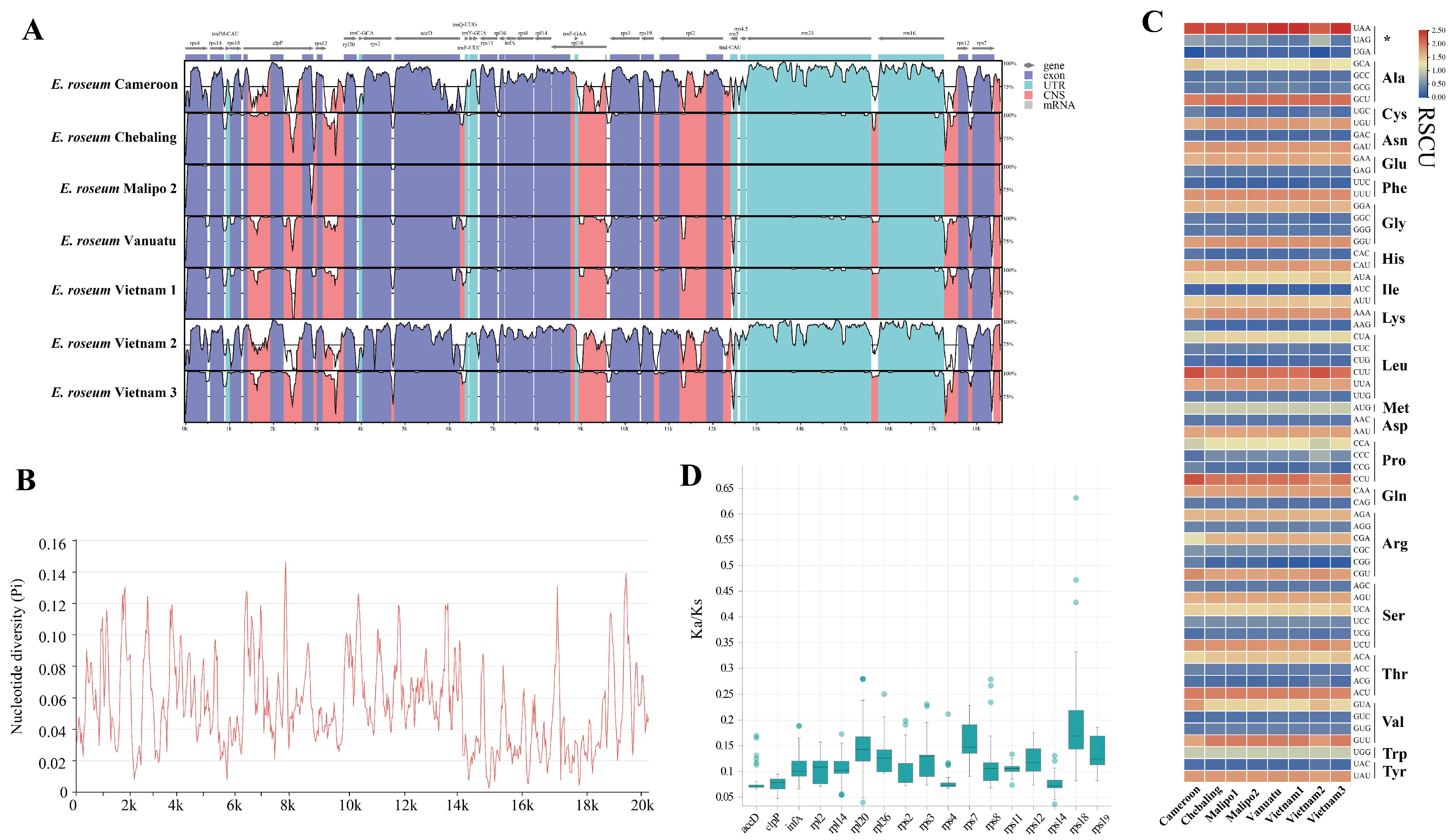
2.1. Characterization and Comparative Analysis of Plastomes

In this study, three complete plastomes from Asia were newly assembled and annotated, as presented in Supplementary Table S1. These plastomes exhibited extreme reduction, being the smallest known among orchids, with lengths ranging from 18,339 to 19,047 bp and low GC contents (30.03–30.96%). Each plastome comprised two single-copy (SC) regions and two considerably contracted inverted repeat (IR) regions (198–292 bp). The length of the SC1 regions ranged from 8326 bp to 8911 bp, with a GC content of 23.14% to 24.49%. The SC2 regions spanned from 9427 bp to 9618 bp, with a GC content of 35.93% to 36.84% (Supplementary Figure S1, Supplementary Table S6). A total of 29 common genes, including 18 protein-coding genes (PCGs), 7 tRNA genes, and 4 rRNA genes, were annotated in the plastomes (Supplementary Figure S1, Supplementary Table S1).
To explore the divergence of complete sequences among E. roseum plastomes with increased sampling, comparative analyses were conducted. Visual alignment results (Figure 1A) and nucleotide diversity (Pi) calculations (Figure 1B) illustrated high intraspecific plastome polymorphism among these sequences. Due to their distinctive features, the plastome of sample “Malipo 1” was selected as the reference sequence. The sequence of sample Malipo 2 exhibited the highest similarity, and Cameroon and Vietnam 2 displayed the highest polymorphism. E. roseum plastomes showed high Pi values ranging from 0.003 to 0.146 (Figure 1B, Supplementary Table S2). Although the plastomes shared common PCGs, the codons of these genes and the RSCU varied (Figure 1C, Supplementary Table S3). Furthermore, non-synonymous substitutions (Ka) and synonymous substitutions (Ks) were calculated for the 17 PCGs in E. roseum plastomes, along with five other orchids. The Ka/Ks ratios for all PCGs were found to be much lower than 1, indicating the stability of protein function for these genes during evolution (Figure 1D, Supplementary Table S4).

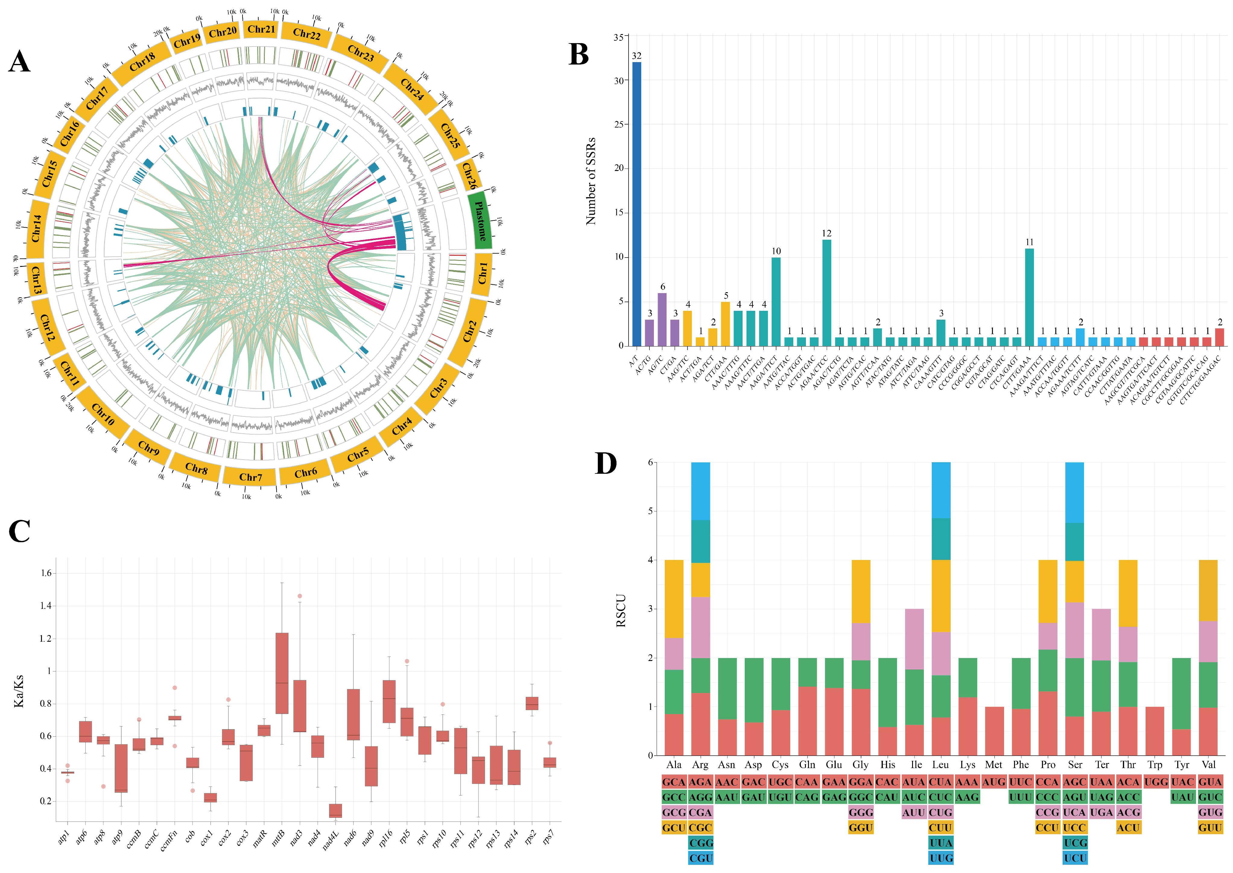
2.2. Comprehensive Analysis of Mitogenome Characteristics

The mitogenome of E. roseum was assembled into 26 circular chromosomes, with lengths ranging from 9865 to 23,000 bp, resulting in a total length of 414,552 bp (Table 1, Supplementary Figure S2). The average GC content of the E. roseum mitogenome was 45.2%, with individual chromosomes ranging from 41.6% to 48.0% in GC content. It contained a total of 54 genes, including 38 protein-coding genes (PCGs), 13 tRNA genes, and 3 rRNA genes (rrn5, rrn18, and rrn26). The PCGs comprised five ATP synthase genes (atp1, atp4, atp6, atp8, and atp9), nine NADH dehydrogenase genes (nad1, nad2, nad3, nad4, nad4L, nad5, nad6, nad7, and nad9), four cytochrome c biogenesis genes (ccmB, ccmC, and ccmFN), three cytochrome c oxidase genes (cox1, cox2, and cox3), one Maturases gene (matR), one membrane transport protein gene (mttB), and one ubichinol cytochrome c reductase gene (cob). Six PCGs, namely nad1, nad2, nad5, nad7, ccmFc, and rpl2, were either incomplete or pseudogenized. Among the genes, nad2 and rps10 contained one intron each, cox2 contained two introns, and nad4 contained three introns. Additionally, the tRNA genes trnE-TTC and trnM-CAT were present in two copies.
A total of 139 simple sequence repeats (SSRs) comprising 1- to 6-nucleotide motifs were identified in the mitogenome of E. roseum (Figure 2A,B; Supplementary Table S5). Among these SSRs, tetranucleotide repeats were the most abundant, accounting for 47.48% of the total, and pentanucleotide and hexanucleotide repeats had nine occurrences each. Mononucleotide repeats were exclusively composed of A/T SSRs, and dinucleotide and tetranucleotide repeats were observed 12 times each. The most frequently observed repeat motif was the A/T mononucleotide, occurring 32 times (Figure 2B, Supplementary Table S5). Furthermore, the E. roseum mitogenome contained 47 tandem repeats, all with a 100% match. These repeats ranged in length from 25 to 150 bp (Figure 2A, Supplementary Table S6). Additionally, 997 dispersed repeats were identified in the E. roseum mitogenome, consisting of 511 forward repeats and 486 palindromic repeats (Figure 2A, Supplementary Table S7).
To explore the selection pressure on the mitochondrial PCGs, the non-synonymous substitution rate (Ka) and synonymous substitution rate (Ks) with five other orchids—Apostasia shenzhenica, Cymbidium lancifolium, Dendrobium amplum, Gastrodia pubilabiata, and Paphiopedilum micranthum—were separately calculated (Figure 2C, Supplementary Table S9). Overall, the Ka/Ks ratios among these orchids were generally similar, with the majority of ratios being less than 1. However, we observed Ka/Ks ratios greater than 1 for the nad6 gene with A. shenzhenica, the rpl16 gene with D. amplum, the nad3 gene with G. pubilabiata, and the mttB and rpl5 genes with P. micranthum (Figure 2C, Supplementary Table S8).
A codon preference analysis was performed on 30 PCGs and 6828 codons in the E. roseum mitogenome (Figure 2D, Supplementary Table S9). Among these codons, stop codons were observed 40 times. The most frequently occurring amino acid was leucine (Leu), appearing 696 times, while cysteine (Cys) was the least frequent, occurring only 90 times. Analysis of the relative synonymous codon usage (RSCU) revealed that GCU and CUU had the highest codon usage bias (CUB), with values of 1.589 and 1.470, respectively. On the other hand, UAC and CCG had the lowest CUB, with values of 0.538 and 0.545, respectively. Out of the codons analyzed, 28 exhibited RSCU values greater than 1, while 34 had values below 1. The RSCU values for the codons AUG (encoding methionine, Met) and UGG (encoding threonine, Thr) were both determined to be 1.

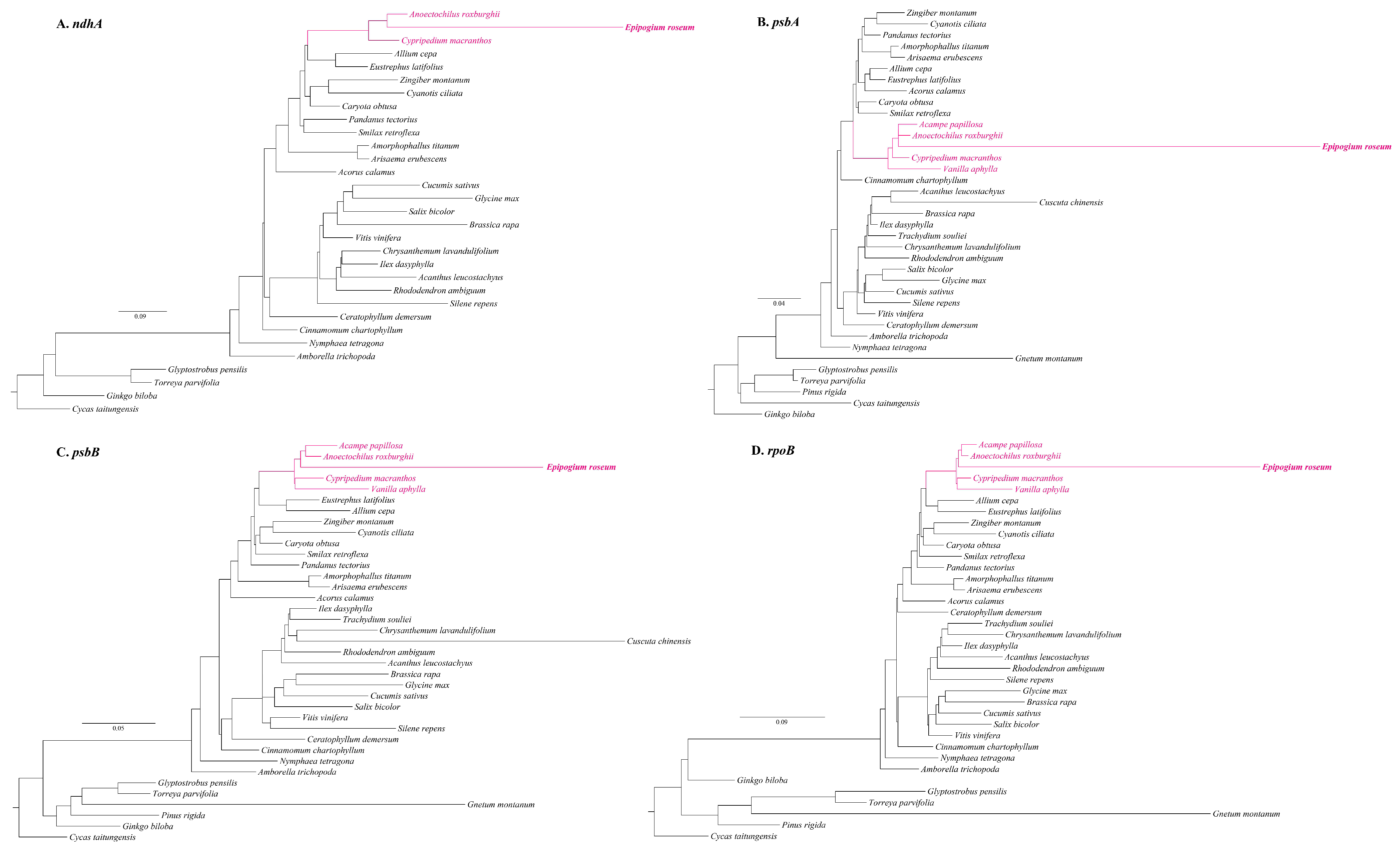
2.3. Migration of Plastid Regions in Mitogenome

To investigate intracellular gene transfers (IGTs), a homologous fragment analysis was performed between E. roseum mitogenome and two plastomes that had sequence differences (sample Malipo 1 and Vietnam 2). The analysis revealed the presence of homologous regions, with sample Malipo 1 having 4549 bp and Vietnam 2 having 6513 bp of homologous regions (Supplementary Table S10). Within these homologous regions, certain plastid genes were identified, including accD, clpP, rpl2, rps2, rrn16, and rrn23 genes. However, it was observed that the majority of these regions were rRNA regions, while the coding gene regions were too short for further alignment or comparison.
A database of 5366 published plastomes of vascular plants was established to search for additional plastid regions in the E. roseum mitogenome. To focus on coding gene regions, the plastome database was curated by removing intergenic spacer regions and rRNA and tRNA genes. The analysis revealed that the E. roseum mitogenome had 37,008 blast hits against the plastome database (Supplementary Table S11). Based on the continuity of blast hits and the length of the plastid regions in the mitogenome, four distinct blocks, namely nadA-like, psbA-like, psbB-like, and rpoB-like, were identified as representatives for further alignment and phylogenetic analysis (Figure 3). Alignments and phylogenetic analyses were performed on these four blocks, comparing them with related genes in 36 other plant plastomes to determine their phylogenetic positions. The results demonstrated that all four plastome-related sequences nested within the Orchidaceae family and plastome-related regions displayed long branches in the phylogenetic topology (Figure 3).

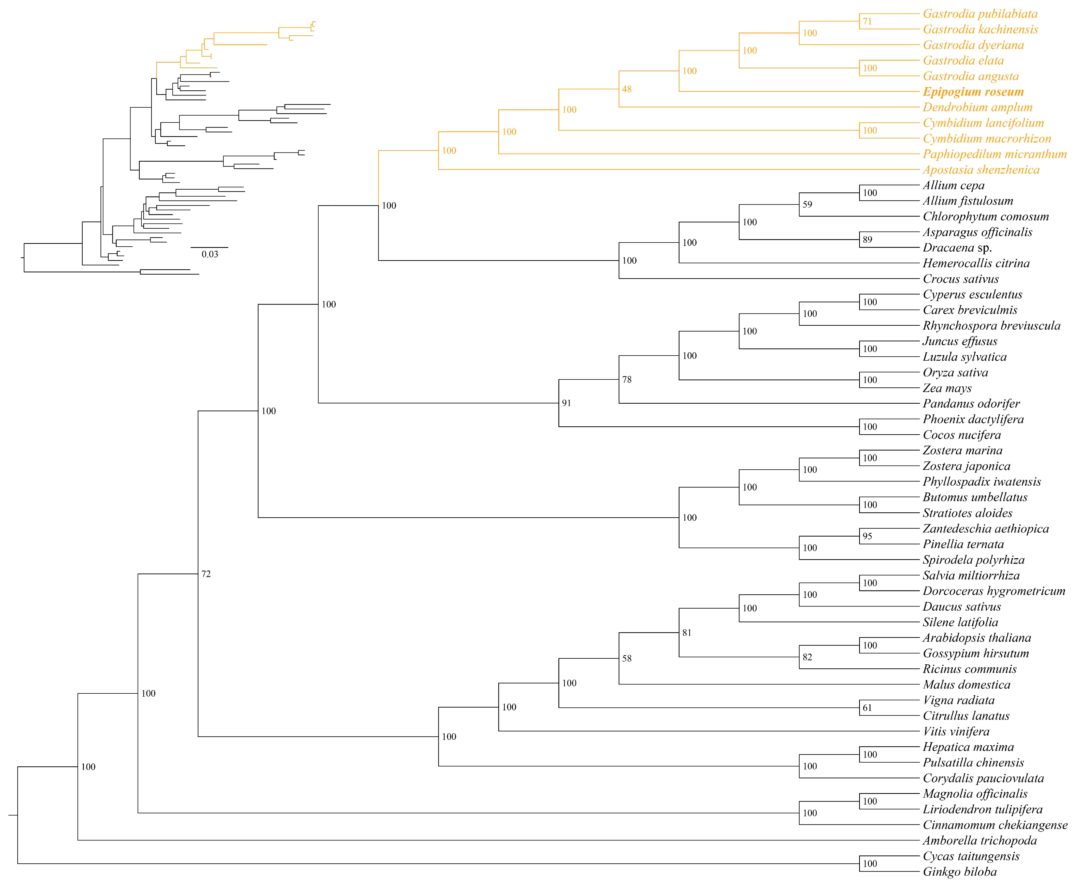
2.4. Phylogenetic Position of E. roseum

The phylogenetic analysis was conducted using 14 PCGs from the plastomes of 88 Orchidaceae species along with two outgroup species (Curculigo orchioides and Asparagus cochinchinensis) (Figure 4, Supplementary Table S12). The alignment matrix comprised 10,919 bp, with 5643 variable sites and 4236 parsimony informative sites. The resulting phylogenetic tree displayed several long branches, including Epipogium, Gastrodia, Rhizanthella gardneri, Lecanorchis japonica, and Vanilla planifolia. These long branches primarily consisted of mycoheterotrophic orchids, except for V. planifolia, which may be closely related to L. japonica. The phylogenetic analysis confirmed the monophyly of the genus Epipogium, consisting of two monophyletic groups: E. aphyllum and E. roseum. Furthermore, the analysis revealed the relationships among the eight samples of E. roseum as follows: (Cameroon, Vietnam 2), (Vietnam 3, (Vietnam 1, Vanuatu)), (Chebaling, (Malipo 1, Malipo 2)). Based on branch lengths, E. roseum can be divided into two clades: one clade comprising Cameroon and Vietnam 2, and the other clade comprising the other samples.
In the mitochondrial phylogenetic analysis, 30 conserved mitochondrial protein-coding genes (PCGs) were analyzed from a total of 56 vascular plants (Figure 5, Supplementary Table S12). The mitogenomes of Ginkgo biloba and Cycas revoluta were used as outgroups for comparison. The alignment matrix used for analysis comprised a total of 50,220 bp, with 19,775 variable sites and 11,404 parsimony informative sites. The phylogenetic tree showed that E. roseum was closely related to Gastrodia with robust support (Figure 5). Additionally, the mitogenomes of Orchidaceae family shared one clade and comprised a monophyletic group. The relationships among the mitogenomes of Orchidaceae are as follows: (Apostasia shenzhenica, (Paphiopedilum micranthum, (Cymbidum, (Dendrobium amplum, (Epipogium roseum, Gastrodia))))). This analysis provides valuable insights into the evolutionary relationships and genetic connections among Orchidaceae species and other vascular plants based on their mitogenomes.

3. Discussion

The newly assembled plastomes of E. roseum exhibited structural characteristics, sequence length, gene content, and GC content that fell within the range reported for previous plastomes [23]. The comparative genomic results revealed unusual structure, genome reduction, and high intraspecific polymorphism in E. roseum plastomes. Most plastomes within the Orchidaceae family displayed the typical quadripartite structure, consisting of a large single-copy (LSC) region, a small single-copy (SSC) region, and two inverted repeat (IR) regions [24,25]. In contrast, the plastomes of E. roseum had two small IR regions, ranging from 198 to 292 bp, and two single-copy (SC) regions. Within the smaller SC region, genes such as accD and clpP shared similarities with the LSC regions of other Orchidaceae plastomes; genes within the larger SC region, particularly the four rRNA genes, exhibited similarity to the IR regions of other Orchidaceae plastomes. Previous studies reported nucleotide diversity values (Pi) of 0.14 for five Epidendrum plastomes and 0.129 for seven Trichoglottis plastomes [26,27]. In the case of E. roseum, the high intraspecific plastome polymorphism is manifested by Pi values reaching up to 0.146. Additionally, the relative synonymous codon usage (RSCU) values were found to vary among different populations.
In this study, the complete mitogenome of E. roseum was reported for the first time. It was found to have 26 circular chromosomes with a total length of 414,552 bp. Previous studies have explored the complex structures and varying genome sizes of plant mitogenomes [9,15,28]. Many orchid mitogenomes published before were multichromosomal, including species like Cymbidium lancifolium, Cymbidium macrorhizon, Gastrodia elata, Gastrodia pubilabiata, and Paphiopedilum micranthum, with total lengths ranging from 447,368 bp (P. micranthum) to 1,340,105 bp (G. elata). Hence, the multichromosomal structure observed in E. roseum seems reasonable. A total of 54 genes, including 38 protein-coding genes (PCGs), 13 tRNA genes, and 3 rRNA genes, were successfully annotated. However, some PCGs were incomplete or showed pseudogenization. Some genes contained multiple introns that spanned several chromosomes, such as nad1, nad2, nad5, and nad7, which posed challenges for perfect assembly and annotation. The ccmFc gene exhibited significant sequence similarity with other orchids, but the CDS could not be successfully annotated due to unexpected stop codons.
Sequences with repetitive elements have been shown to facilitate various rearrangements in plant mitogenomes [29]. Repeat-mediated recombination has been observed in plant mitogenomes, including those of P. micranthum, Ipomoea batatas, and Gelsemium elegans [15,19,29]. In this study, we identified tandem repeats, dispersed repeats, and SSRs within E. roseum mitogenome, with a total length of 203,423 bp (45.47%). However, the potential for recombination and the hypothesis of a master circle were not further explored. To gain insights into the evolution of PCGs in E. roseum organelle genomes, Ka/Ks analysis was performed. Despite the loss of multiple genes in the plastomes, the lower Ka/Ks ratios indicated the stability of the remaining PCGs. The Ka/Ks ratios observed in both plastid and mitochondrial PCGs indicated a consistent pattern of negative selection and their evolutionary significance.
The presence of plastome-derived sequences in plant mitogenomes can be attributed to IGTs and horizontal gene transfers (HGTs). For instance, in Paphiopedilum micranthum (Orchidaceae), 59% of the plastome-origin mitochondrial sequences were identified [15]. In Vitis vinifera (Vitaceae), it was 8.8% [30], and in Actinidia (Actinidiaceae), it ranged from 1.16% to 4.05% [31]. Photosynthetic plants often exhibit plastome-origin mitochondrial sequences through IGTs occurring between plastomes and mitogenomes themselves [15,20,32]. Notably, the identification of HGTs appears to be more significant in non-photosynthetic plants, leading to further research on foreign plastid sequences in plant mitogenomes [16,33,34]. To investigate the IGTs in the E. roseum mitogenome, two representative samples were utilized, considering the sequence variations in plastomes. The analysis resulted in the identification of six plastid regions with lengths of 4549 and 6513 bp. However, the coding gene regions were too short for meaningful alignment or comparison. Consequently, an exploration of homologous regions among a database of plant plastomes and mitogenomes was carried out. Through alignment and phylogenetic analysis, four plastid regions transferred via HGTs were confirmed.
The phylogenetic relationships of Epipogium have been a subject of intense debate in orchid systematics [35]. Different studies using various genetic markers have proposed conflicting placements for Epipogium. For instance, an analysis based on nuclear ITS suggested a close relationship between Epipogium and Nervilieae [36], while another study indicated it to be sister to Triphoreae. On the other hand, a phylogenetic analysis using mitochondrial nad1 sequences strongly supported E. aphyllum as the sister species to Nervilia shirensis [35]. In a larger-scale study involving 1450 low-copy nuclear genes from 610 orchid species, the phylogeny indicated Epipogium to be sister to Nervilia with moderate support [37]. Consequently, these studies led to the placement of Epipogium within the tribe Nervilieae and the subtribe Epipogiinae. In this particular study, phylogenetic trees using 14 PCGs from 90 plastomes and 30 PCGs from 56 mitogenomes were reconstructed. The results supported Epipogium being sister to Gastrodia with 100% bootstrap percentages for both plastid and mitochondrial datasets.
This study reports three E. roseum plastomes and conducts a comparative analysis to reveal their sequence variety. The mitogenome was sequenced and annotated, followed by a comprehensive analysis of genome characteristics. IGTs and HGTs were identified by sequence similarity and phylogeny. Morever, system evolution analysis was conducted to comprehend the phylogenetics of it. This research provides a theoretical foundation for exploring the organelle genomes of mycoheterotrophic orchids.

4. Materials and Methods

4.1. Sample Sampling, DNA Extraction and Sequencing, Genome Assembly and Annotation

Plant samples were collected from Guangdong Chebaling National Nature Reserve (Shixing County, China) and Malipo County (China) (Supplementary Table S1) and stored at the herbarium of the College of Forestry, Fujian Agriculture and Forestry University (FJFC). Genomic DNA was extracted from fresh tubers, stems, and flowers using a modified CTAB protocol [38]. Short reads were sequenced using an Illumina HiSeq 4000 platform (San Diego, CA, USA). To construct genomic libraries (SMRTbell libraries) for PacBio long-read sequencing, high-molecular-weight genomic DNA was sheared into fragments of approximately 20 kb. Then, large-fragment genomic DNA was concentrated with AMPure PacBio beads and used for SMRTbell preparation according to the manufacturer’s specifications (Pacific Biosciences, Menlo Park, CA, USA). The libraries were constructed and sequenced using the PacBio Seque II sequencing platform (Pacific Biosciences, Menlo Park, CA, USA).
Plastome assembly and annotation were conducted following the methodologies described in our previous study [39]. The short reads were de novo assembled into the mitogenome using GetOrganelle v1.7.6.1 [40]. Upon assembly, the original contigs exhibited a pattern of ‘fireworks’, with a length of 437,794 bp (Supplementary Figure S3). The visualization of this assembly was conducted using Bandage v0.8.1 [41]. Subsequently, the long reads were filtered utilizing BLASR v5.1 [42], using the original assemblies as a reference. The filtered long reads were then assembled using CANU v1.8 [43]. The resulting contigs obtained from CANU assembly were considered as the reference, and the assembly process was iterated until stable contigs were obtained. Finally, the circular nature of the contigs was manually determined through overlap analysis.
For gene annotation, the coding and rRNA genes were annotated in Geneious Prime (Biomatters, Inc., Auckland, New Zealand), employing published orchid mitogenomes as references. These annotations were then manually refined. Furthermore, tRNA genes were annotated using tRNAscan-SE v2.0 [44]. To visualize the genome maps, Organellar Genome DRAW [45] was employed.

4.2. Genome Characters and Comparative Genomics

To analyze the diversity of E. roseum plastome sequences, the online program mVISTA was employed, specifically utilizing the Shuffle-LAGAN alignment program [46]. The protein-coding genes (PCGs) were extracted using PhyloSuite v1.2.2 [47] and aligned using MAFFT 7 [48]. The nucleotide variability (Pi) for the eight plastomes was calculated using DnaSP 6 [49], with a window length of 100 bp and a step size of 25 bp. The relative synonymous codon usage (RSCU) was analyzed using DAMBE 7 [50]. Furthermore, the Ka/Ks ratios were calculated using KaKs_Calculator2 [51].
To identify tandem repeats, the Tandem Repeats Finder v4.09 program [52] was employed with default parameters. The REPuter [53] was used with default parameters to detect four types of long repeats, namely forward (F), palindrome (P), reverse (R), and complement (C) repeats. SSRs were identified using the Perl script MISA [54], with the minimum thresholds for mono-, di-, tri-, tetra-, penta-, and hexa-motif microsatellites set at 10, 5, 4, 3, 3, and 3 nucleotide repeats, respectively. ChiPlot Online Tools was used to visualize the obtained results [55].

4.3. Gene Transfer and Phylogenetic Analysis

The plastomes and mitogenomes used in this study were obtained from the NCBI Organelle Genome Resources database, and a summary of the sources can be found in Supplementary Table S11. To analyze sequence similarity between the plastomes and mitogenome and detect transferred DNA fragments, BLASTN [56] was utilized with an e-value cut-off of 1 × 10⁻⁵. For visualization purposes, the Circos module implemented in TBtools [57] was employed to generate a circle diagram.
A total of 90 plastomes (two outgroup species: Curculigo orchioides and Asparagus cochinchinensis) and 56 mitogenomes (two outgroup species: Ginkgo biloba and Cycas revoluta) were selected to conduct phylogenetic analysis. Supplementary Table S12 provides details of the taxa. To elucidate the phylogenetic analysis, maximum likelihood (ML) analysis was conducted using the RAxML-HPC2 on XSEDE 8.2.10 tool available on the CIPRES Science Gateway web server. The GTRCAT model was specified for all datasets, and 1000 repeated self-expanding analyses were performed [58].

5. Conclusions

In this study, we present the first complete mitogenome of the fully mycoheterotrophic orchid Epipogium roseum. Additionally, three plastomes sampled from China were assembled and analyzed. Our findings shed light on the intraspecific plastome polymorphism, general characteristics of the mitogenome, and the phylogenetic position of E. roseum. The mitogenome of E. roseum exhibited multiple sequence repeats, comprising a total length of 203,423 bp (45.47%). Intracellular gene transfers (IGTs) and horizontal gene transfer (HGT) events were identified within organelle genomes. The phylogenomics supported the robust sister relationship between Epipogium and Gastrodia. Overall, this study adds to the understanding of organelle genomes in non-photosynthetic orchids and fully mycoheterotrophic plants.

Supplementary Materials

The following supporting information can be downloaded at: https://www.mdpi.com/article/10.3390/ijms25031578/s1.

Author Contributions

Conceptualization, M.-H.L. and Z.-J.L.; methodology, Z.Z.; software, Z.Z. and Y.L.; validation, Z.Z., M.-H.L. and J.-W.Z.; formal analysis, Z.Z.; investigation, Z.Z. and Y.L.; resources, J.-W.Z.; data curation, Z.Z. and Y.L.; writing—original draft preparation, Z.Z.; writing—review and editing, M.-H.L. and Z.-J.L.; visualization, Z.Z.; supervision, M.-H.L. and Z.-J.L.; project administration, M.-H.L. and Z.-J.L.; funding acquisition, M.-H.L. and Z.-J.L. All authors have read and agreed to the published version of the manuscript.

Funding

This research was funded by the National Key Research and Development Program of China (no. 2023YFD1000500) and the Forestry Peak Discipline Construction Project of Fujian Agriculture and Forestry University (no. 72202200205).

Institutional Review Board Statement

Not applicable.

Informed Consent Statement

Not applicable.

Data Availability Statement

The newly obtained complete mitogenome sequences that support the findings of the study have been deposited in the NCBI with accession numbers as follows: OR871626–OR871651; the accession numbers of plastome sequences were: OR871652–OR871654.

Acknowledgments

The authors thank the Editor and the anonymous reviewers for their insightful comments and suggestions on the manuscript.

Conflicts of Interest

The authors declare no conflicts of interest.

References

  1. Leake, J.R. The biology of myco-heterotrophic (‘saprophytic’) plants. New Phytol. 1994, 127, 171–216. [Google Scholar] [CrossRef]
  2. Bidartondo, M.I. The evolutionary ecology of myco-heterotrophy. New Phytol. 2005, 167, 335–352. [Google Scholar] [CrossRef]
  3. Li, M.-H.; Liu, K.-W.; Li, Z.; Lu, H.-C.; Ye, Q.-L.; Zhang, D.; Wang, J.-Y.; Li, Y.-F.; Zhong, Z.-M.; Liu, X.; et al. Genomes of leafy and leafless Platanthera orchids illuminate the evolution of mycoheterotrophy. Nat. Plants 2022, 8, 373–388. [Google Scholar] [CrossRef]
  4. Suetsugu, K.; Matsubayashi, J. Evidence for mycorrhizal cheating in Apostasia nipponica, an early-diverging member of the Orchidaceae. New Phytol. 2021, 229, 2302–2310. [Google Scholar] [CrossRef]
  5. Motomura, H.; Marc-André, S.; Martos, F.; Kagawa, A. Mycoheterotrophy evolved from mixotrophic ancestors: Evidence in Cymbidium (orchidaceae). Ann. Bot. 2010, 106, 573. [Google Scholar] [CrossRef]
  6. Yagame, T.; Orihara, T.; Selosse, M.A.; Yamato, M.; Iwase, K. Mixotrophy of Platanthera minor, an orchid associated with ectomycorrhiza-forming Ceratobasidiaceae fungi. New Phytol. 2012, 193, 178–187. [Google Scholar] [CrossRef]
  7. Wang, J.; Kan, S.; Liao, X.; Zhou, J.; Tembrock, L.R.; Daniell, H.; Jin, S.; Wu, Z. Plant organellar genomes: Much done, much more to do. Trends Plant Sci. 2024. [Google Scholar] [CrossRef]
  8. Wu, Z.; Sloan, D.B. Recombination and intraspecific polymorphism for the presence and absence of entire chromosomes in mitochondrial genomes. Heredity 2019, 122, 647–659. [Google Scholar] [CrossRef]
  9. Lin, Y.; Li, P.; Zhang, Y.; Akhter, D.; Pan, R.; Fu, Z.; Huang, M.; Li, X.; Feng, Y. Unprecedented organelle genomic variations in morning glories reveal independent evolutionary scenarios of parasitic plants and the diversification of plant mitochondrial complexes. BMC Biol. 2022, 20, 49. [Google Scholar] [CrossRef]
  10. Organelle Genome Resources Database. Available online: https://www.ncbi.nlm.nih.gov/genome/organelle/ (accessed on 22 November 2023).
  11. Chase, M.W.; Cameron, K.M.; Freudenstein, J.V.; Pridgeon, A.M.; Salazar, G.; Van den Berg, C.; Schuiteman, A. An updated classification of Orchidaceae. Bot. J. Linn. Soc. 2015, 177, 151–174. [Google Scholar] [CrossRef]
  12. Ke, S.J.; Liu, D.K.; Tu, X.D.; He, X.; Zhang, M.M.; Zhu, M.J.; Zhang, D.Y.; Zhang, C.L.; Lan, S.R.; Liu, Z.J. Apostasia Mitochondrial Genome Analysis and Monocot Mitochondria Phylogenomics. Int. J. Mol. Sci. 2023, 24, 7837. [Google Scholar] [CrossRef]
  13. Li, X.; Zhe, M.; Huang, Y.; Fan, W.; Yang, J.; Zhu, A. The Evolution of Mitochondrial Genomes between Two Cymbidium Sister Species: Dozens of Circular Chromosomes and the Maintenance and Deterioration of Genome Synteny. Genes 2023, 14, 864. [Google Scholar] [CrossRef]
  14. Yuan, Y.; Jin, X.; Liu, J.; Zhao, X.; Zhou, J.; Wang, X.; Wang, D.; Lai, C.; Xu, W.; Huang, J.; et al. The Gastrodia elata genome provides insights into plant adaptation to heterotrophy. Nat. Commun 2018, 9, 1615. [Google Scholar] [CrossRef]
  15. Yang, J.X.; Dierckxsens, N.; Bai, M.Z.; Guo, Y.Y. Multichromosomal mitochondrial genome of Paphiopedilum micranthum: Compact and fragmented genome, and rampant intracellular gene transfer. Int. J. Mol. Sci. 2023, 24, 3976. [Google Scholar] [CrossRef]
  16. Kim, Y.K.; Jo, S.; Cheon, S.H.; Hong, J.R.; Kim, K.J. Ancient Horizontal Gene Transfers from Plastome to Mitogenome of a Nonphotosynthetic Orchid, Gastrodia pubilabiata (Epidendroideae, Orchidaceae). Int. J. Mol. Sci. 2023, 24, 11448. [Google Scholar] [CrossRef]
  17. Woodson, J.D.; Chory, J. Coordination of gene expression between organellar and nuclear genomes. Nat. Rev. Genet. 2008, 9, 383–395. [Google Scholar] [CrossRef]
  18. Keeling, P.J.; Palmer, J.D. Horizontal gene transfer in eukaryotic evolution. Nat. Rev. Genet. 2008, 9, 605–618. [Google Scholar] [CrossRef]
  19. You, C.; Cui, T.; Zhang, C.; Zang, S.; Su, Y.; Que, Y. Assembly of the Complete Mitochondrial Genome of Gelsemium elegans Revealed the Existence of Homologous Conformations Generated by a Repeat Mediated Recombination. Int. J. Mol. Sci. 2022, 24, 527. [Google Scholar] [CrossRef]
  20. Park, S.; Grewe, F.; Zhu, A.; Ruhlman, T.A.; Sabir, J.; Mower, J.P.; Jansen, R.K. Dynamic evolution of Geranium mitochondrial genomes through multiple horizontal and intracellular gene transfers. New Phytol. 2015, 208, 570–583. [Google Scholar] [CrossRef]
  21. Zhou, S.; Zhi, X.; Yu, R.; Liu, Y.; Zhou, R. Factors contributing to mitogenome size variation and a recurrent intracellular DNA transfer in Melastoma. BMC Genom. 2023, 24, 370. [Google Scholar] [CrossRef]
  22. Zhou, X.; Lin, H.; Fan, X.L.; Gao, J.Y. Autonomous self-pollination and insect visitation in a saprophytic orchid, Epipogium roseum (D. Don) Lindl. Aust. J. Bot. 2012, 60, 154–159. [Google Scholar] [CrossRef]
  23. Schelkunov, M.I.; Shtratnikova, V.Y.; Nuraliev, M.S.; Selosse, M.A.; Penin, A.A.; Logacheva, M.D. Exploring the limits for reduction of plastid genomes: A case study of the mycoheterotrophic orchids Epipogium aphyllum and Epipogium roseum. Genome Biol. Evol. 2015, 7, 1179–1191. [Google Scholar] [CrossRef]
  24. Kim, Y.K.; Jo, S.; Cheon, S.H.; Joo, M.J.; Hong, J.R.; Kwak, M.; Kim, K.J. Plastome evolution and phylogeny of Orchidaceae, with 24 new sequences. Front. Plant Sci. 2020, 11, 22. [Google Scholar] [CrossRef]
  25. Li, L.; Wu, Q.; Fang, L.; Wu, K.; Li, M.; Zeng, S. Comparative chloroplast genomics and phylogenetic analysis of Thuniopsis and closely related genera within Coelogyninae (Orchidaceae). Front. Genet. 2022, 13, 850201. [Google Scholar] [CrossRef]
  26. Zhao, Z.; Zeng, M.Y.; Wu, Y.W.; Li, J.W.; Zhou, Z.; Liu, Z.J.; Li, M.H. Characterization and Comparative Analysis of the Complete Plastomes of Five Epidendrum (Epidendreae, Orchidaceae) Species. Int. J. Mol. Sci. 2023, 24, 14437. [Google Scholar] [CrossRef]
  27. Zhou, C.Y.; Zeng, M.Y.; Gao, X.; Zhao, Z.; Li, R.; Wu, Y.; Liu, Z.J.; Zhang, D.; Li, M.H. Characteristics and Comparative Analysis of Seven Complete Plastomes of Trichoglottis s.l. (Aeridinae, Orchidaceae). Int. J. Mol. Sci. 2023, 24, 14544. [Google Scholar] [CrossRef]
  28. Wu, Z.Q.; Liao, X.Z.; Zhang, X.N.; Tembrock, L.R.; Broz, A. Genomic architectural variation of plant mitochondria—A review of multichromosomal structuring. J. Syst. Evol. 2022, 60, 160–168. [Google Scholar] [CrossRef]
  29. Yang, Z.; Ni, Y.; Lin, Z.; Yang, L.; Chen, G.; Nijiati, N.; Hu, Y.; Chen, X. De novo assembly of the complete mitochondrial genome of sweet potato (Ipomoea batatas [L.] Lam) revealed the existence of homologous conformations generated by the repeat-mediated recombination. BMC Plant Biol. 2022, 22, 285. [Google Scholar] [CrossRef]
  30. Goremykin, V.V.; Salamini, F.; Velasco, R.; Viola, R. Mitochondrial DNA of Vitis vinifera and the issue of rampant horizontal gene transfer. Mol. Biol. Evol. 2009, 26, 99–110. [Google Scholar] [CrossRef]
  31. Wang, S.; Li, D.; Yao, X.; Song, Q.; Wang, Z.; Zhang, Q.; Zhong, C.; Liu, Y.; Huang, H. Evolution and diversification of kiwifruit mitogenomes through extensive whole-genome rearrangement and mosaic loss of intergenic sequences in a highly variable region. Genome Biol. Evol. 2019, 11, 1192–1206. [Google Scholar] [CrossRef]
  32. Niu, Y.; Gao, C.; Liu, J. Complete mitochondrial genomes of three Mangifera species, their genomic structure and gene transfer from chloroplast genomes. BMC Genom. 2022, 23, 147. [Google Scholar] [CrossRef]
  33. Rice, D.W.; Alverson, A.J.; Richardson, A.O.; Young, G.J.; Sanchez-Puerta, M.V.; Munzinger, J.; Barry, K.; Boore, J.L.; Zhang, Y.; DePamphilis, C.W.; et al. Horizontal transfer of entire genomes via mitochondrial fusion in the angiosperm Amborella. Science 2013, 342, 1468–1473. [Google Scholar] [CrossRef]
  34. Gandini, C.L.; Sanchez-Puerta, M.V. Foreign plastid sequences in plant mitochondria are frequently acquired via mitochondrion-to-mitochondrion horizontal transfer. Sci. Rep. 2017, 7, 43402. [Google Scholar] [CrossRef]
  35. Li, M.H.; Zhang, G.Q.; Lan, S.R.; Liu, Z.J.; China Phylogeny Consortium. A molecular phylogeny of Chinese orchids. J. Syst. Evol. 2016, 54, 349–362. [Google Scholar] [CrossRef]
  36. Molvray, M.; Kores, P.J.; Chase, M.W. Polyphyly of mycoheterotrophic orchids and functional influences on floral and molecular characteristics. In Monocots: Systematics and Evolution; Wilson, K.L., Mossison, D.A., Eds.; CSIRO: Melbourne, Australia, 2000; pp. 441–448. [Google Scholar]
  37. Zhang, G.; Hu, Y.; Huang, M.Z.; Huang, W.C.; Liu, D.K.; Zhang, D.; Hu, H.; Downing, J.L.; Liu, Z.J.; Ma, H. Comprehensive phylogenetic analyses of Orchidaceae using nuclear genes and evolutionary insights into epiphytism. J. Integr. Plant. Biol. 2023, 65, 1204–1225. [Google Scholar] [CrossRef]
  38. Arseneau, J.R.; Steeves, R.; Laflamme, M. Modified low-salt CTAB extraction of high-quality DNA from contaminant-rich tissues. Mol. Ecol. Resour. 2017, 17, 686–693. [Google Scholar] [CrossRef]
  39. Liu, D.K.; Tu, X.D.; Zhao, Z.; Zeng, M.Y.; Zhang, S.; Ma, L.; Zhang, G.Q.; Wang, M.M.; Liu, Z.J.; Lan, S.R.; et al. Plastid phylogenomic data yield new and robust insights into the phylogeny of CleisostomaGastrochilus clades (Orchidaceae, Aeridinae). Mol. Phyl. Evol. 2020, 145, 106729. [Google Scholar] [CrossRef]
  40. Jin, J.J.; Yu, W.B.; Yang, J.B.; Song, Y.; De Pamphilis, C.W.; Yi, T.S.; Li, D.Z. GetOrganelle: A fast and versatile toolkit for accurate de novo assembly of organelle genomes. Genome Biol. 2020, 21, 241. [Google Scholar] [CrossRef] [PubMed]
  41. Wick, R.R.; Schultz, M.B.; Zobel, J.; Holt, K.E. Bandage: Interactive visualization of de novo genome assemblies. Bioinformatics 2015, 31, 3350–3352. [Google Scholar] [CrossRef] [PubMed]
  42. Chaisson, M.J.; Tesler, G. Mapping single molecule sequencing reads using basic local alignment with successive refinement (BLASR): Application and theory. BMC Bioinform. 2012, 13, 238. [Google Scholar] [CrossRef] [PubMed]
  43. Koren, S.; Walenz, B.P.; Berlin, K.; Miller, J.R.; Bergman, N.H.; Phillippy, A.M. Canu: Scalable and accurate long-read assembly via adaptive k-mer weighting and repeat separation. Genome Res. 2017, 27, 722–736. [Google Scholar] [CrossRef]
  44. Chan, P.P.; Lin, B.Y.; Mak, A.J.; Lowe, T.M. tRNAscan-SE 2.0: Improved detection and functional classification of transfer RNA genes. Nucleic Acids Res. 2021, 49, 9077–9096. [Google Scholar] [CrossRef] [PubMed]
  45. Greiner, S.; Lehwark, P.; Bock, R. Organellar Genome DRAW (OGDRAW) version 1.3. 1: Expanded toolkit for the graphical visualization of organellar genomes. Nucleic Acids Res. 2019, 47, W59–W64. [Google Scholar] [CrossRef]
  46. Brudno, M.; Malde, S.; Poliakov, A.; Do, C.B.; Couronne, O.; Dubchak, I.; Baĵoglou, S. Glocal alignment: Finding rearrangements during alignment. Bioinformatics 2003, 19, i54–i62. [Google Scholar] [CrossRef] [PubMed]
  47. Zhang, D.; Gao, F.; Jakovlić, I.; Zou, H.; Zhang, J.; Li, W.X.; Wang, G.T. PhyloSuite: An integrated and scalable desktop platform for streamlined molecular sequence data management and evolutionary phylogenetics studies. Mol. Ecol. Resour. 2020, 20, 348–355. [Google Scholar] [CrossRef] [PubMed]
  48. Katoh, K.; Standley, D.M. MAFFT multiple sequence alignment software version 7: Improvements in performance and usability. Mol. Biol. Evol. 2013, 30, 772–780. [Google Scholar] [CrossRef]
  49. Rozas, J.; Ferrer-Mata, A.; Sánchez-DelBarrio, J.C.; Guirao-Rico, S.; Librado, P.; Ramos-Onsins, S.E.; Sánchez-Gracia, A. DnaSP 6: DNA sequence polymorphism analysis of large data sets. Mol. Biol. Evol. 2017, 34, 3299–3302. [Google Scholar] [CrossRef]
  50. Xia, X. DAMBE7: New and improved tools for data analysis in molecular biology and evolution. Mol. Biol. Evol. 2018, 35, 1550–1552. [Google Scholar] [CrossRef]
  51. Wang, D.; Zhang, Y.; Zhang, Z.; Zhu, J.; Yu, J. KaKs_Calculator 2.0: A toolkit incorporating gamma-series methods and sliding window strategies. Genom. Proteom. Bioinform. 2010, 8, 77–80. [Google Scholar] [CrossRef]
  52. Gao, H.; Kong, J. Distribution characteristics and biological function of tandem repeat sequences in the genomes of different organisms. Zool. Res. 2005, 26, 555–564. [Google Scholar]
  53. Kurĵ, S.; Choudhuri, J.V.; Ohlebusch, E.; Schleiermacher, C.; Stoye, J.; Giegerich, R. REPuter: The manifold applications of repeat analysis on a genomic scale. Nucleic Acids Res. 2001, 29, 4633–4642. [Google Scholar]
  54. Beier, S.; Thiel, T.; Münch, T.; Scholz, U.; Mascher, M. MISA-web: A web server for microsatellite prediction. Bioinformatics 2017, 33, 2583–2585. [Google Scholar] [CrossRef]
  55. ChiPlot. Available online: https://www.chiplot.online/ (accessed on 20 November 2023).
  56. Chen, Y.; Ye, W.; Zhang, Y.; Xu, Y. High speed BLASTN: An accelerated Mega BLAST search tool. Nucleic Acids Res. 2015, 43, 7762–7768. [Google Scholar] [CrossRef]
  57. Chen, C.; Chen, H.; Zhang, Y.; Thomas, H.R.; Frank, M.H.; He, Y.; Xia, R. TBtools: An integrative toolkit developed for interactive analyses of big biological data. Mol. Plant 2020, 13, 1194–1202. [Google Scholar] [CrossRef]
  58. Stamatakis, A.; Hoover, P.; Rougemont, J. A rapid bootstrap algorithm for the RAxML web servers. Syst. Biol. 2008, 57, 758–771. [Google Scholar] [CrossRef]
Figure 1. Comparative analyses of eight E. roseum plastomes. (A) Sequence alignment of the plastomes. Thick, gray arrows above the alignment indicate the orientation and position of each gene; the Y-axis represents the identity percentage, ranging from 50 to 100%. (B) The nucleotide diversity of plastomes using a sliding window test. The window size was set to 100 bp, and the sliding window size was 25 bp. X-axis, position of the midpoint of a window; Y-axis, Pi values of each window. (C) The RSCU values of 18 concatenated protein-coding genes for the plastomes. * indicates stop codon. The red values mean higher RSCU, and the blue values mean lower RSCU. (D) The boxplots of Ka/Ks values among 17 PCGs in plastomes with five orchids. The “X” axis shows the names of protein-coding genes, and the “Y” axis shows the Ka/Ks values.
Ijms 25 01578 g001
Figure 2. Comparative analysis of E. roseum mitogenome. (A) Features of mitogenome tabulated in a Circos plot. The circles from outside to inside represent lengths of mitochondrial contigs and plastome; positions of SSRs (colored red) and tandem repeats (colored green); GC ratios; gene density; homologous fragments among plastome and mitogenome (colored red); forward repeats (colored blue); and palindromic repeats (colored yellow). (B) SSR distribution of mitogenome. (C) Ka/Ks ratios of mitogenome PCGs. (D) RSCU of mitogenome PCGs.
Ijms 25 01578 g002
Figure 3. A maximum likelihood (ML) phylogenetic tree of four regions found to be plastid-origin coding regions in the mitogenome. The numbers near the nodes are bootstrap percentages. The position of Orchidaceae based on mitogenome is highlighted in red, and position of E. roseum is bold.
Ijms 25 01578 g003
Figure 4. A maximum likelihood (ML) phylogenetic tree of 90 plastomes based on 14 PCGs. The numbers near the nodes are bootstrap percentages; upper left corner shows the phylogenetic topology structure. The position of E. roseum is highlighted in green and bold.
Ijms 25 01578 g004
Figure 5. A maximum likelihood (ML) phylogenetic tree of 56 mitogenomes based on 38 PCGs. The numbers near the nodes are bootstrap percentages; upper left corner shows the phylogenetic topology structure. The position of Orchidaceae based on mitogenome is highlighted in yellow, and position of E. roseum is bold.
Ijms 25 01578 g005
Table 1. Feathers of E. roseum mitogenome. * Genes with one intron. ** Genes with two introns. *** Genes with three introns. ψ Genes were incomplete or showed pseudogenization.
ChromosomeLength (bp)GC Content (%)GenesPacBio Reads Coverage
Chr 114,12742.5rps722.27
Chr 223,00045.8ψ atp4, atp8, nad4L22.24
Chr 317,94044.3nad2 *15.70
Chr 414,74644.8psbE, trnQ-TTC10.66
Chr 517,46446.2atp1, ccmFn, ψ nad510.49
Chr 616,75644.7atp917.81
Chr 717,44144.8rps220.79
Chr 816,69644.7atp6, cox2 **, ψ nad3, rps11, trnM-CAT16.41
Chr 915,23145.4cob, ψ nad1, rpl5, rps14, trnC-GCA12.31
Chr 1020,23446.6nad4 ***20.48
Chr 1112,24145.2ψ nad510.43
Chr 1218,96445.6nad3, rps12, trnE-TTC, trnH-GTG19.73
Chr 1310,97644.3trnD-GTC, trn-GTT14.64
Chr 1419,26446.4atp9, rps1, trnY-GTA22.61
Chr 1515,51045.8matR12.59
Chr 1611,63348.0ψ nad7, trnE-TTC6.56
Chr 1715,70346.0ψ rpl2, rpl16, rps1912.40
Chr 1820,49345.7mttB, rps1330.44
Chr 19997344.7ccmB6.38
Chr 2011,81645.4ψ nad55.82
Chr 2111,46041.6nad9, trnF-GAA, trnW-CCA15.88
Chr 2216,38844.8cox1, trnL-CAA15.47
Chr 2319,05345.0ccmC, nad6, rps10 *21.72
Chr 2420,24545.3cox321.19
Chr 2517,33345.1rrn5, rrn18, rrn26, trnM-CAT32.24
Chr 26986545.4ψ ccmFc6.56
Disclaimer/Publisher’s Note: The statements, opinions and data contained in all publications are solely those of the individual author(s) and contributor(s) and not of MDPI and/or the editor(s). MDPI and/or the editor(s) disclaim responsibility for any injury to people or property resulting from any ideas, methods, instructions or products referred to in the content.

