Article

Triterpenoid Saponins and Flavonoid Glycosides from the Flower of Camellia flavida and Their Cytotoxic and α-Glycosidase Inhibitory Activities

Medical School of Guangxi University, Nanning 530004, China
*
Authors to whom correspondence should be addressed.
These authors contributed equally to this work.
Int. J. Mol. Sci. 2024, 25(20), 10977; https://doi.org/10.3390/ijms252010977
Submission received: 9 September 2024 / Revised: 8 October 2024 / Accepted: 9 October 2024 / Published: 12 October 2024

Abstract

Camellia flavida var. flavida, commonly known as “Jinhua Tea”, has its flowers and leaves traditionally utilized as tea and functional food sources. However, there is limited knowledge about its bioactive components and their biological activities. This study isolated ten previously unidentified glycoside compounds from the flowers of Camellia flavida, including three oleanane-type triterpenoid saponins (compounds 13) and seven flavonoid glycosides (compounds 410), collectively named flavidosides A–J. This study assessed the cytotoxicity of these compounds against a panel of human cancer cell lines and their α-glucosidase inhibitory activities. Notably, flavidoside C showed significant cytotoxicity against BEL-7402 and MCF-7 cell lines, with IC50 values of 4.94 ± 0.41 and 1.65 ± 0.39 μM, respectively. Flavidoside H exhibited potent α-glucosidase inhibitory activity, with an IC50 value of 1.17 ± 0.30 mM. These findings underscore the potential of Camellia flavida in the development of functional foods.

1. Introduction

Yellow camellia, belonging to the Theaceae family and referred to as “Jinhua Tea” in China, was designated as a new food source by the State Food and Drug Administration of China in 2010 and was approved as a food ingredient in 2023. Numerous studies have demonstrated its pharmacological benefits, including antibacterial activity [1], potential hepatitis treatment [2], blood sugar reduction [3], antioxidant effects [4], and cancer prevention capabilities [5]. Camellia flavida var. flavida, a distinguished species of yellow camellia, was identified in Chongzuo, Guangxi, in 1982. This species is characterized by its year-round blooming and substantially higher flower yield compared to other species. Due to its high commercial value and potential as a tea and functional food source, its cultivation has expanded significantly in recent years.
While recent studies by our group have focused on the chemical and biological properties of C. flavida leaves [6,7], research on its flowers has not yet been conducted. Consequently, this study investigates the bioactive constituents and biological activities of C. flavida flowers. Preliminary extraction studies revealed that C. flavida exhibited cytotoxic effects against tumor cells. A substantial body of evidence demonstrates that extracts of C. flavida exert inhibitory actions on non-small cell lung cancer [8], hepatocellular carcinoma [9], and colon cancer [10]. However, these studies predominantly employ crude extracts of C. flavida, whereas many modern chemotherapeutic agents are derived from single, purified natural compounds, such as paclitaxel, vincristine, and onychomycotoxin analogs [11]. Consequently, there is a compelling need to isolate and evaluate individual compounds from C. flavida for their antitumor potential; we assessed the cytotoxic activities of individual compounds against cancer cells lines using the MTT assay.
In traditional Chinese medicine, yellow camellia has long been consumed as a health beverage believed to lower blood sugar levels. Notably, the flavonoids of Camellia nitidissima flower extracts have demonstrated in vitro α-glucosidase inhibitory activity with an IC50 value of 45 μg/mL [12]. The activity of α-glucosidase plays a pivotal role in modulating carbohydrate utilization and absorption in the small intestine. The inhibition of α-glucosidase can effectively diminish carbohydrate digestion and absorption, leading to a notable reduction in postprandial blood glucose levels. This, in turn, aids in the management of metabolic disorders, including diabetes and obesity [13]. Although clinical medications, such as acarbose and voglibose, are available for diabetic patients, they are often associated with and accompanied by undesirable side effects, encompassing flatulence, diarrhea, and abdominal discomfort. As a result, numerous reports have underscored the efficacy of safe, natural phytochemical enzyme inhibitors derived from sources like mulberry leaves, guava fruits, and oil tea seeds in mitigating postprandial hyperglycemia. These findings indicate the potential of C. flavida as a functional food component to control blood glucose and alleviate symptoms of diabetes mellitus.
In this research, ten new compounds (Figure 1), including three triterpenoid saponins (13) and seven flavonoid glycosides (410), along with five previously identified flavonoids (1115), were isolated from C. flavida. The cytotoxic activities of compounds 110 were assessed against six cancer cell lines using the MTT assay, and their α-glucosidase inhibitory activities were also evaluated.

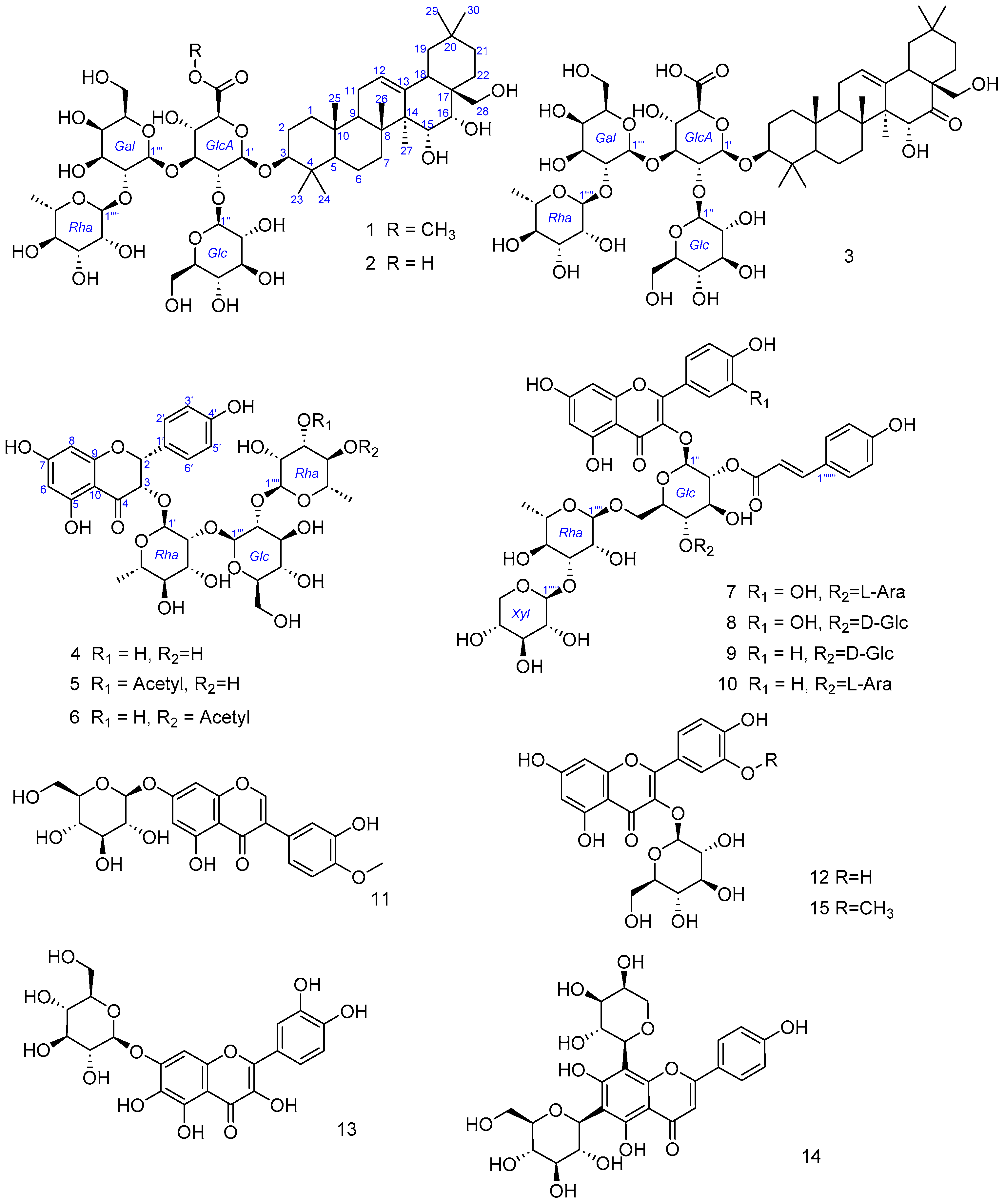
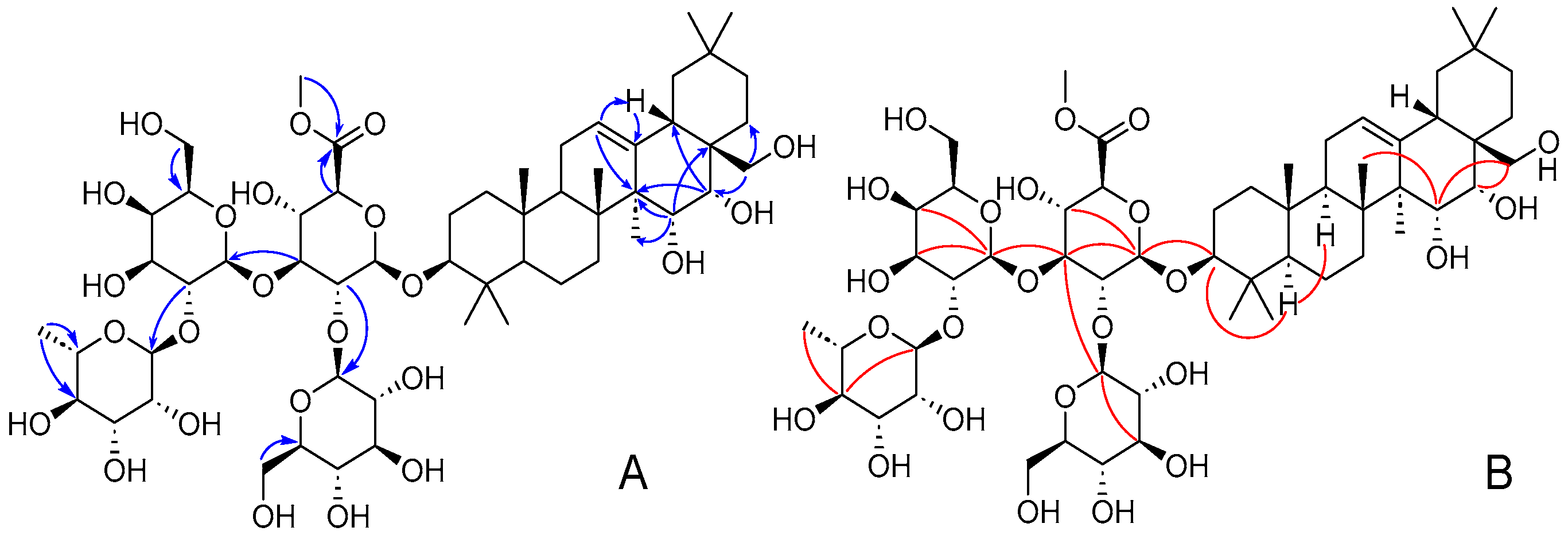
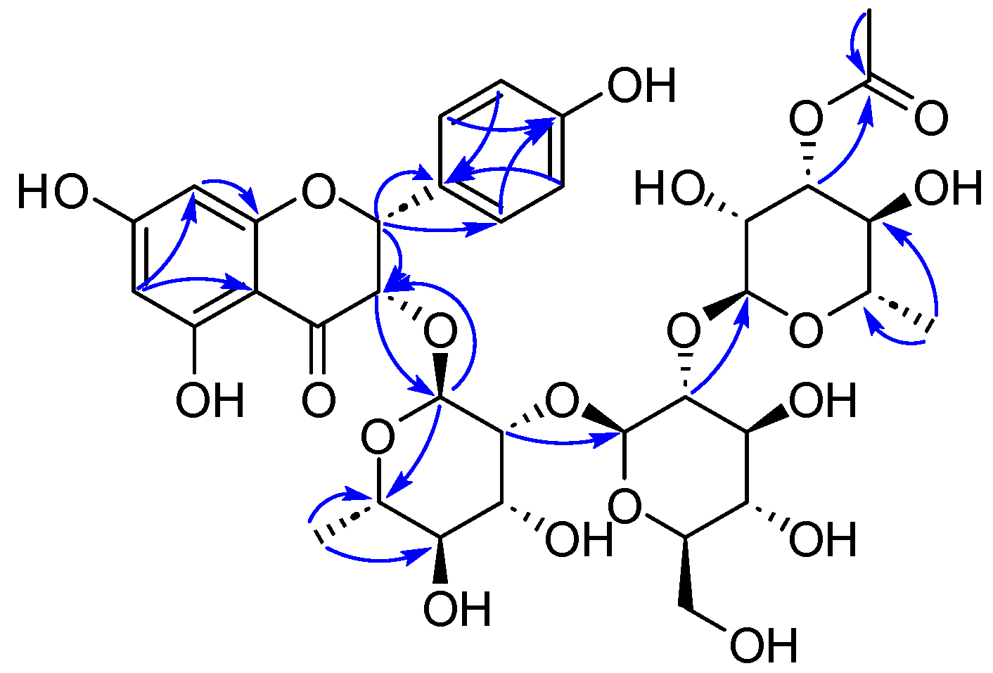
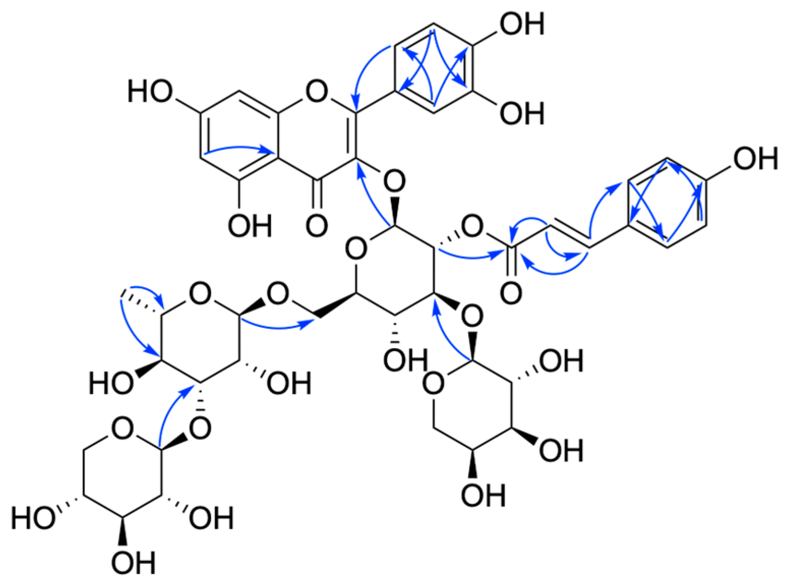
2. Results and Discussion

Compound 1 was isolated as a white powder, and its molecular formula was determined as C55H90O24 based on HR-ESI-MS analysis (Figure S1, m/z 1157.5726, [M + Na]+, calcd 1157.5720). The Liebermann–Burchard reaction for compound 1 was positive, indicating its identity as a potential triterpenoid. NMR analysis (Table 1 and Table 2, and Figures S2–S10) revealed seven singlet methyl groups at chemical shifts δH 0.82, 1.02, 1.05 × 2, 1.10, 1.12, and 1.81 in the up-field region, and δH 2.45 (dd, J = 13.5, 3.8 Hz, H-18), a notable proton signal with two characteristic unsaturated carbons at δC 146.0 (C-13) and δC 123.9 (C-12), indicating an olean-12-ene pentacyclic triterpenoid skeleton. Additionally, three oxymethine protons were observed at δH 3.17, 4.48, and 4.55, corresponding to carbons C-3 (δC 89.7), C-16 (δC 78.7), and C-15 (δC 67.4), respectively. Two methylene protons attached to C-28 (δC 69.6) appeared as a doublet at δH 3.60 and 3.79 (both d, J = 10.5 Hz). Moreover, HMBC correlations (Figure 2A) indicated three hydroxy groups linked to C-15, C-16, and C-28, demonstrated by HMBC cross-peaks from δH 4.48 (H-15) to δC 48.0 (C-14), 41.1 (C-17), and 21.0 (C-27), from δH 4.55 (H-16) to δC 48.0 (C-14) and 41.1 (C-17), and from δH 3.60/3.79 (H2-28) to δC 41.1 (C-17) and 31.0 (C-22). The α-orientation of H-3 was confirmed by the trans-diaxial coupling constant of a large 3J value (10.2 Hz) and NOESY correlations (Figure 2B) of δH 3.17 (H-3)/0.77 (H-5). The cis-orientations of H-15/H-16 were suggested by a small 3J value of H-16 (3.8 Hz), and NOESY correlations of δH 4.48 (H-15)/1.05 (H3-26)/3.60 (H2-28), δH 4.55 (H-16)/3.60 (H2-28), and δH 3.60 (H2-28)/2.45 (H-18)/1.05 (H3-29) established that H-15 and H-16 were β-oriented. Thus, the aglycone part of compound 1 was established as a 3,15,16,28-tetrahydroxyolean-12-ene framework. Moreover, the NMR characteristics were found to be very similar to those of camellianol F [14].
Anomeric proton signals at δH 4.86, 5.87, 6.10, and 6.23 were observed and displayed HSQC correlations with the anomeric carbon signals at δC 105.2, 102.9, 101.6, and 102.3, respectively, characteristic of the four monosaccharide units. The 13C-NMR signals of the four sugars were determined from the HMBC and TOCSY spectra. Specifically, δC 105.2, 79.5, 82.8, 70.9, 76.6, and 170.3 were assigned to glucopyranuronic acid. δC 102.9, 76.4, 78.5, 72.6, 78.2, and 63.5 were associated with glucopyranose. δC 101.6, 76.2, 76.1, 71.1, 77.0, and 62.0 were identified as galactopyranose, and δC 102.3, 72.6, 72.6, 73.9, 69.8, and 18.3 were attributed to rhamnopyranose. Additionally, acid hydrolysis of 1 yielded D-glucuronic acid, L-rhamnose, D-glucose, and D-galactopy, analyzed by HPLC.
Further analysis of the HMBC correlations of δH 3.69 (GlcA-6-OCH3) and 4.45 (GlcA-H-5) to δC 170.3 (GlcA-C-6) indicated a methyl ester group was present at GlcA-C-5. The β-configuration of glucopyranosyl and galactopyranosyl units was confirmed by the relatively large coupling constants (J = 7.4 and 7.7 Hz) of their anomeric protons. The broad singlet of its anomeric proton confirmed the α-configuration of the rhamnopyranosyl unit. The β-configuration of the 6-methyl glucopyranuronoyl unit was indicated by the NOESY correlations of δH 4.86 (GlcA-H-1)/4.67 (GlcA-H-3)/4.45 (GlcA-H-5).
The HMBC correlations from Gal-H-1 (δH 6.10) to GlcA-C-3 (δC 70.9) and from Glc-H-1 (δH 5.87) to GlcA-C-2 (δC 79.5) suggested that the GlcA was linked to Gal-C-1 and Glc-C-1 via GlcA-C-2 and GlcA-C-3, respectively. The rhamnose was connected to Gal-C-2, as evidenced by the HMBC correlation from Rha-H-1 (δH 6.23) to Gal-C-2 (δC 76.2). The HMBC correlation from GlcA-H-1 (δH 4.86) to C-3 (δC 89.7) of the aglycone unit confirmed that the tetrasaccharide moiety was located at C-3 of the triterpene skeleton. Therefore, the structure of 1 was established as 3β,15α,16α,28-tetrahydroxyolean-12-en-3-O-β-D-glucopyranosyl-(1→2)-[α-L-rhamnopyranosyl-(1→2)-β-D-galactopyranosyl-(1→3)]-β-D-glucuronopyranoside methyl ester, assigned the trivial name flavidoside A.
Compound 2 was obtained as a white powder and displayed a molecular formula of C54H88O24 by HR-ESI-MS (Figure S11, m/z 1143.5554, [M + Na]+, calcd 1143.5564). The NMR data (Table 1 and Table 2, and Figures S12–S19) for 2 were very similar to 1. Compounds 1 and 2 shared the same glycoside moiety, specifically camellianol F. However, they differ in the type of sugar moiety that was attached to this common glycoside. The HSQC correlations from δH 4.86 to δC 105.4, from δH 5.90 to δC 102.7, from δH 6.18 to δC 101.4, and from δH 6.23 to δC 102.4 suggested the presence of four sugar units. According to other NMR spectra and acid hydrolysis followed by HPLC analysis, compound 2 comprised β-D-glucopyranuronic acid (GlcA), β-D-glucopyranosyl (Glc), α-L-rhamnopyranosyl (Rha), and β-D-galactopyranosyl (Gal) sugar units. GlcA was linked to Gal-C-1 and Glc-C-1 via GlcA-C-2 and C-3, respectively, and rhamnose was connected to Gal-C-2, as evidenced by the HMBC correlations (Figure S20) from δH 6.18 (Gal-H-1) to δC 82.8 (GlcA-C-3), from δH 5.90 (Glc-H-1) to δC 79.5 (GlcA-C-2), and from δH 6.23 (Rha-H-1) to δC 76.4 (Gal-C-2). Thus, the structure of compound 2 was established as 3β,15α,16α,28-tetrahydroxyolean-12-en-3-O-β-D-glucopyranosyl-(1→2)-[α-L-rhamnopyranosyl-(1→2)-β-D-galactopyranosyl-(1→3)]-β-D-glucuronopyranoside (flavidoside B).
Compound 3 was isolated as a white powder. The molecular formula was deduced as C54H86O24 by HR-ESI-MS (Figure S21, m/z 1141.5389 [M + Na]+, calcd 1141.5407). The 1H and 13C NMR data were very similar to those of compound 2, except for the absence of the hydroxy group at C-16, which was replaced by a carbonyl group in the triterpene skeleton. The sugar chain was identical in compounds 3 and 2, as confirmed by the NMR data (Table 1 and Table 2, and Figures S22–S31). As a result, compound 3 was elucidated as 3β,15α,28-trihydroxyolean-12-en-16-on-3-O-β-D-glucopyranosyl-(1→2)-[α-L-rhamnopyranosyl-(1→2)-β-D-galactopyranosyl-(1→3)]-β-D-glucuronopyranoside (flavidoside C).
Compound 4 was isolated as a yellow amorphous powder. The molecular formula was determined to be C33H42O19 from its positive HR-ESI-MS (Figure S32, m/z 765.2207 [M + Na]+, calcd 765.2218). δH 5.92 (H-6) and 5.90 (H-8) in the 1H-NMR spectrum (Table 3 and Figures S33–S40) displayed a pair of doublets (J = 1.9 Hz) representing aromatic protons. The aromatic proton signals at δH 6.85 (2H, d, J = 8.4 Hz) and 7.36 (2H, d, J = 8.4 Hz) of the A2B2 type suggested a 1,4-disubstituted B-ring. Two oxygenated methines were observed at δH 4.55 and 5.48 (both d, J = 10.4 Hz). These findings indicated that compound 4 was a 3,5,7,4′-tetrahydroxyflavanone (dihydrokaempferol) [15].
Compound 4 contained three sugar moieties, deduced from the anomeric signals of δH/C 4.71/102.1, 4.02/105.3, and 5.21/101.8. The 13C-NMR, HMBC, and TOCSY spectra displayed the signal attribution of three sugars: δC 102.1, 82.4, 71.6, 73.9, and 70.9 were assigned to one rhamnopyranose; δC 101.8, 71.9, 71.8, 74.2, 70.3, and 17.9 were associated with another rhamnopyranose; δC 105.3, 78.9, 78.3, 70.9, 77.2, and 62.1 were identified as the glucopyranose. Moreover, the configurations of L-rhamnose and D-glucose were determined by acid hydrolysis and HPLC identification. The anomeric proton at δH 4.02 (d, J = 7.6 Hz) suggested the β-configuration of glucopyranosyl. The two α-configuration units of rhamnopyranosyl were determined by the broad singlet of their anomeric protons. Thus, compound 4 was identified as a 3,5,7,4′-tetrahydroxyflavanone substituted with two α-L-rhamnopyranosyl moieties and a β-D-glucopyranosyl moiety.
The linkage of the rhamnopyranosyl unit at C-2 of the glucopyranosyl unit and the glucopyranosyl moiety was attached to C-2 of another rhamnopyranosyl moiety, confirmed by the HMBC correlations (Figure 3) from δH 5.21 (Rha2-H-1) to δC 78.9 (Glc-C-2) and from δH 4.02 (Glc-H-1) to δC 82.4 (Rha1-C-2). Moreover, the oligosaccharide chain was linked at C-3 of the aglycone, also confirmed by the HMBC correlation from δH 4.71 (Rha1-H-1) to δC 81.2 (C-3) and the reverse correlation from δH 4.55 (H-3) to Rha1-C-1 (δC 102.1).
The large 3JH-2/H-3 value (10.4 Hz) hinted at the trans-form orientation of H-2 and H-3. The ECD spectrum of 4 (Figure 4) showed a positive Cotton effect at 330 nm (Δε +2.19) and a strong negative Cotton effect at 292 nm (Δε −9.50), establishing that the aglycone of 4 was 2R and 3R [16]; this was confirmed by the calculated ECD of 4 as well. Based on the above analysis, compound 4 could be assigned as (2R,3R)-3,5,7,4′-tetrahydroxyflavanone-3-O-α-L-rhamnopyranosyl-(1→2)-β-D-glucopyranosyl-(1→2)-α-L-rhamnoside (flavidoside D).
Compound 5 was isolated as a yellow powder with the molecular formula determined to be C35H44O20 using HR-ESI-MS data (Figure S41, m/z 807.2327, [M + Na]+, calcd 807.2324). The 1H and 13C NMR data (Table 4, Figures S42–S49) for 5 were similar to 4, with an added acetyl moiety at position Rha2-C-3, suggested by the HMBC correlations (Figure 5) from δH 5.03 (Rha2-H-3) and 2.08 (acetyl-H-2) to δC 172.8 (acetyl-C-1). The small 3JH-2/H-3 value (2.3 Hz) hinted at the cis-form orientation of H-2/H-3. The absolute configuration of 2R, 3S was confirmed by comparing the experimental ECD and calculated ECD data of 5 (Figure 6). Thus, compound 5 was assigned as (2R,3S)-3,5,7,4′-tetrahydroxyflavanone-3-O-[3-acetyl-O-α-L-rhamnopyranosyl-(1→2)-β-D-glucopyranosyl-(1→2)]-α-L-rhamnoside (flavidoside E).
Compound 6 was obtained as a yellow solid, with the same molecular formula as 5 (C35H44O20) deduced by HR-ESI-MS (Figure S50, m/z 807.2311 [M + Na]+, calcd 807.2324). The 1H and 13C NMR data (Table 4, Figures S51–S57) were very similar to compound 5 except for the position of the acetyl unit. The acetyl unit linked to C-4 of the rhamnopyranosyl moiety was confirmed by the HMBC correlation (Figure S58) from δH 4.72 (Rha2-H-4) to δC 170.2 (acetyl-C-1). The large 3JH-2/H-3 value (8.4 Hz) revealed the trans-form orientation of H-2/H-3 and the absolute configuration of 2R,3R, which was displayed by the ECD spectra (Figure S59). Thus, compound 6 was identified as (2R,3R)-3,5,7,4′-tetrahydroxyflavanone-3-O-[4-acetyl-O-α-L-rhamnopyranosyl-(1→2)-β-D-glucopyranosyl-(1→2)]-α-L-rhamnoside (flavidoside F).
Compound 7 was a yellow powder and displayed the molecular formula of C46H52O26 (Figure S60, m/z 1021.2837, [M + H]+, calcd 1021.2825) based on the HR-ESI-MS data. The presence of 3,5,7,3′,4′-pentahydroxyflavone aglycone (quercetin) [17] was confirmed by a pair of aromatic proton signals at δH 6.13 and 6.32 (both br s) along with 6.88 (d, J = 7.8 Hz), 7.55 (br d, J = 7.8 Hz), and 7.60 (1H, br s), as well as the HMBC correlations (Figure 7) from δH 7.60 (H-2′) and 7.55 (H-6′) to δC 158.9 (C-2) in the 1H-NMR spectrum (Table 5).
The 1D and 2D NMR spectra (Figures S61–S67) of 7 suggested that it contains four sugar units: glucopyranose (δC 100.7, 74.5, 84.5, 70.3, 76.8, 68.4), arabinopyranose (δC 105.2, 72.2, 73.9, 69.5, 67.2), rhamnopyranose (δC 102.2, 71.6, 82.4, 72.7, 69.5, 17.9), and xylopyranose (δC 106.2, 75.3, 77.5, 71.1, 66.8). Acid hydrolysis and HPLC confirmed the configuration of these sugars.
HMBC correlations were observed from δH 3.88 (Glc-H-3) to δC 105.2 (Ara-C-1) and from δH 3.51 (Glc-H-6) to δC 102.2 (Rha-C-1), establishing that Glc is linked to Ara-C-1 and Rha-C-1 through Glc-C-2 and Glc-C-6, respectively. The xylopyranoyl unit was linked to Rha-C-3, as shown by the HMBC correlation from δH 4.39 (Xyl-H-1) to δC 82.4 (Rha-C-3). The sugar chain was positioned at C-3 of the flavone, confirmed by the HMBC correlation from δH 5.58 (Glc-H-1) to δC 134.8 (C-3). Signals for a 1,4-disubstituted benzene ring proton at δH 6.80 (2H, Coumaroyl-H-2 and 6) and 7.45 (2H, Coumaroyl-H-3 and 5), and for trans-oriented vinylic protons at δH 6.42 (Coumaroyl-H-8) and 7.68 (Coumaroyl-H-7), were noted. Moreover, HMBC correlations from δH 6.42 (Coumaroyl-H-8) to δC 127.3 (Coumaroyl-C-1), 168.8 (Coumaroyl-C-9), from δH 7.68 (Coumaroyl-H-7) to δC 131.3 (Coumaroyl-C-2 and Coumaroyl-C-6), and 115.1 (Coumaroyl-C-8) suggested a p-E-coumaroyl unit. The HMBC correlation from δH 5.24 (Glc-H-2) to δC 168.8 (Coumaroyl-C-9) indicated that coumaroyl was attached to Glc-C-2. Therefore, compound 7 was identified as 3,5,7,3′,4′-pentahydroxyflavonol-3-O-α-L-arabinopyranosyl-(1→3)-[β-D-xylopyranosyl-(1→3)-α-L-rhamnopyranosyl-(1→6)]-2-O-p-E-coumaryl-β-D-glucopyranoside, named flavidoside G.
Compound 8, isolated as a yellow solid, had its molecular formula determined as C47H54O27 from the ion peak at m/z 1073.2747 [M + Na]+ (calcd 1073.2750, Figure S68) in the HR-ESI-MS. The 1H- NMR spectrum (Table 5) showed that 8 contained the same flavone aglucone structure of quercetin as 7. The difference between 8 and 7 was in the sugar units. The sugar chain in 8 included two β-D-glucopyranosyl units, a α-L-rhamnopyranosyl unit, and a β-D-xylopyranoyl unit, as suggested by the 1D and 2D NMR data (Figures S69–S75) and acid hydrolysis. HMBC correlations (Figure S76) from δH 4.24 (Xyl-H-1) to δC 81.1 (Rha-C-3), from δH 4.38 (Rha-H-1) to δC 67.9 (Glc1-C-6), from δH 4.31 (Glc2-H-1) to δC 83.1 (Glc1-C-3), from δH 5.09 (Glc1-H-2) to δC 165.7 (Coumaroyl-C-9), and from δH 5.57 (Glc1-H-1) to δC 132.8 (C-3) revealed the positions of the sugars and confirmed that the sugar chain was located at C-3 of the aglucone. Based on this evidence, the structure of 8 was identified as 3,5,7,3′,4′-pentahydroxyflavonol-3-O-β-D-glucopyranosyl-(1→3)-[β-D-xylopyranosyl-(1→3)-α-L-rhamnopyranosyl-(1→6)]-2-p-E-coumaryl-β-D-glucopyranoside (flavidoside H).
Compound 9, obtained as a yellow powder, had its molecular formula identified as C47H54O26 by HR-ESI-MS (Figure S77, m/z 1035.3014, [M + H]+, calcd 1035.2981). The 1H and 13C NMR data (Table 5 and Table 6, and Figures S78–S86) were similar to those of compound 8. The distinction of 9 was its 1,4-disubstituted B-ring, evidenced by its A2B2-type aromatic proton signals at δH 6.89 and 7.98 (both d, J = 8.3 Hz). The flavone aglucone structure of 9 was kaempferol [18]. Thus, compound 9 was designated as 3,5,7,4′-tetrahydroxyflavonol-3-O-β-D-glucopyranosyl-(1→3)-[β-D-xylopyranosyl-(1→3)-α-L-rhamnopyranosyl-(1→6)]-2-p-E-coumaryl-β-D-glucopyranoside (flavidoside I).
Compound 10 was obtained as a yellow powder. Its molecular formula was determined to be C46H52O25 (Figure S87, m/z 1005.2885, [M + H]+, calcd 1005.2876) based on HR-ESI-MS. Its 1H and 13C NMR data (Table 5 and Table 6, and Figures S88–S96) were very similar to those of compound 7. The difference in 10 was the presence of para substitution on the B-ring. Thus, compound 10 was designated as 3,5,7,4′-tetrahydroxyflavonol-3-O-α-L-arabinopyranosyl-(1→3)-[β-D-xylopyranosyl-(1→3)-α-L-rhamnopyranosyl-(1→6)]-2-p-E-coumaryl-β-D-glucopyranoside (flavidoside J).
The known compounds 11 to 15 were identified as pratensein-7-O-β-D-glucoside (11) [19], quercetagetin-7-O-β-D-glucoside (12) [20], quercetin-3-O-β-D-glucoside (13) [21] apigenin-8-C-α-L-arabinosyl-6-C-β-D-glucoside (14) [22], and 3,4′-,5,7-tetrahydroxy-3′-methoxyflavone-3-O-β-D-glucopyranoside (15) [23], respectively (Figures S97–S106).
The cytotoxicity of compounds 110 was evaluated against six human cancer cell lines and two normal human cell lines using the MTT method. Compounds 13, representing newly identified triterpenoid saponins, exhibited inhibitory effects against Hela, MCF-7, BEL-7402, Hep G2, and MDA-MB-231 cell lines. Notably, compound 3 demonstrated potent cytotoxic effects against the MCF-7 and BEL-7402 cell lines, surpassing the positive control with IC50 values of 1.65 ± 0.39 and 4.94 ± 0.41 μM, respectively. Moreover, its inhibitory effect on MDA-MB-231 cells was comparable to that of cisplatin. As a novel oleanane-type saponin analog, compound 3 fell within a class of triterpenoid saponins renowned for their potent antitumor properties, positioning it as a promising lead candidate for antitumor drug development [24]. Notably, the IC50 value of compound 3 against MCF-7 cells (1.65 ± 0.39 µM) was significantly lower than the reported range (6.1~16.0 µM) for oleanane-type saponins in the study by Huyen, L.T. et al. [25], highlighting its enhanced potency. A thorough literature review on the cytotoxicity of C. flavida revealed a lack of prior studies investigating the effects of its active components on BEL-7402 cells. Our findings contribute to bridging this knowledge gap and underscore the potential of compound 3 in the treatment of breast and liver cancer.
Compound 2 demonstrated significantly greater cytostatic effects on Hela cells compared to the positive control, with an IC50 value of 4.17 ± 0.85 μM. In contrast, compound 3, along with compound 1, exhibited less pronounced inhibitory effects. A review of the literature suggested that the n-butanol and water-soluble fractions of the ethanolic extract from C. flavida were more potent in inhibiting Hela cell proliferation. Quantitative analysis identifies saponins as the primary constituents in these fractions [26]. Consequently, compound 2 from C. flavida emerged as a promising lead candidate for cervical cancer treatment.
In contrast, all flavonoid saponins (4 to 10) exhibited only weak cytotoxicity against the six cancer cell lines. The triterpenoid saponins exhibited significantly stronger cytotoxicity than the flavonoid glycosides across all six cancer cells. Moreover, none of the compounds displayed cytotoxicity towards normal cells (Table 7).
Despite the cytotoxicity results of compounds 410 being unsatisfactory, the flavonoid glycosides exhibited strong α-glucosidase inhibitory activity. In contrast, none of the triterpenoid saponins exhibited any inhibitory effect on α-glucosidase. The inhibitory activity of compounds 7 and 10 towards α-glucosidase was equivalent to that of acarbose, exhibiting IC50 values of 2.38 ± 0.22 mM and 2.26 ± 0.48 mM, respectively. Compound 9, on the other hand, demonstrated a moderate level of inhibitory activity against α-glucosidase, with an IC50 value of 3.33 ± 0.48 mM. Compound 8 demonstrated strong inhibitory activity against α-glucosidase, with an IC50 value of 1.17 ± 0.30 mM, which was superior to that of acarbose (2.04 ± 0.27 mM). The initial analysis of SAR (Structure-Activity Relationship) revealed that the presence of a double bond at positions C-2 and C-3 in the parent nucleus diminished the inhibitory effect on α-glycosidase. Additionally, the number of hydroxyl groups on the skeleton and the type of monosaccharide in the sugar chain had minimal impact on this inhibitory effect. Notably, the introduction of the p-E-coumaroyl group significantly enhanced the inhibitory effect of the compound against α-glycosidase. Accordingly, we could hypothesize that flavonoid glycosides 410 derived from the water fraction of C. flavida constitute a significant basis for its hypoglycemic activity (Table 8).
To explore the reasons behind the inhibitory effects of compounds 1 to 10 on α-glucosidase, molecular analysis docking was conducted to uncover the potential mechanism (Figure 8 and Figures S107–S115). Among these, compound 8 exhibited a binding affinity of −5.48 kcal/mol and effectively occupied the active site pockets, indicating multiple binding interactions. A hydrogen bond was formed between the C-8-OH group and residue A307 at a distance of 2.9 Å. Additionally, two hydrogen bonds were identified between the carbonyl group at C-4 and the C-5-OH group, both with residue A307 at distances of 3.5 Å and 3.1 Å, respectively. The Glc-3-OH group simultaneously formed two hydrogen bonds with residues T310 and A307 at distances of 2.9 Å and 3.2 Å, respectively (Table S1).

3. Experimental Procedures

3.1. General Experimental Procedures

UV spectra were obtained using a UNICO UV-2802 Spectrometer (Shanghai UNICO Instruments Co., Ltd., Shanghai, China). ECD spectra were measured at room temperature on a Jasco J-815 CD spectrometer (JASCO Corporation, Tokyo, Japan). IR spectra were collected using a Shimadzu IRAffinity-1S FTIR spectrometer (Shimadzu Corporation, Tokyo, Japan). NMR spectra were acquired on Bruker AVANCE III HD 600 MHz spectrometers (Bruker Corporation, Billerica, MA, USA) with Pyr-d5 (δH 7.20, 7.57, 8.72 and δC 123.44, 135.43, 149.84), CD3OD (δH 3.31 and δC 49.00), and DMSO-d6 (δH 2.50 and δC 39.52) as solvents. HR-ESI-MS data were collected on a Waters G2-XS Q-TOF mass spectrometer (Waters Corporation, Milford, MA, USA) equipped with a BEH-C18 column. Column chromatography was performed using Toyopearl Butyl 650C (Nippon Chemi-Con Corporation, Tokyo, Japan), MCI GEL (CHP20, Mitsubishi Chemical Corporation, Tokyo, Japan), Toyopearl HW-40F (Nippon Chemi-Con Corporation, Tokyo, Japan), macroporous resin of D101-type (Tianjin Berens Biotechnology Co., Ltd., Tianjin, China), and C18 reverse-phase silica gel (SMB 100-20/45, Fuji Silysia Chemical Ltd., Tokyo, Japan). Semipreparative HPLC was conducted with a Welch XB-C18 column (5 μm, 10 × 250 mm, 4 mL/min, Shanghai Welch Technology Co., Ltd., Shanghai, China) on a Laballiance HPLC system (LabAlliance, State College, PA, USA) with a Model 500 UV detector. Analytical HPLC was performed using an SSI HPLC system (Scientific Systems, Inc., State College, PA, USA) with a Welch XB-C18 column (5 μm, 4.6 × 250 mm, 1.0 mL/min) and a Model 201 UV detector. Medium-pressure liquid chromatography was conducted on a Büchi B-688 chromatography pump (BÜCHI Labortechnik Aktiengesellschaft, Flawil, Switzerland) with a C-635 UV photometer.

3.2. Plant Material

The flowers of C. flavida were authenticated and provided by Guangxi Fangcheng Golden Camellias National Nature Reserve Management Centre in January 2022. A voucher specimen (No. 20220201-8126) of the C. flavida plant was deposited in the Medical School of Guangxi University.

3.3. Extraction and Isolation

Air-dried flowers of C. flavida (2.5 kg) were extracted with 70% EtOH (9 L) for 7 days, yielding a concentrated extract (1 kg). This extract was diluted with water and extracted sequentially with petroleum ether and ethyl acetate. The aqueous layer was filtered and subjected to chromatography on D101 macroporous resin, eluted with an EtOH/H2O gradient (0:1, 1:9, 3:7, 5:5, 7:3, and 9:1), resulting in 5 fractions (Fr.1–Fr.5). Fraction Fr.3 (130 g, 50% EtOH) was further separated on MCI resin with a MeOH/H2O gradient (0:100 to 95:5), yielding 12 sub-fractions (Fr.3.1–Fr.3.12). Sub-fraction Fr.3.8, eluted with 80% MeOH, was subjected to medium-pressure liquid chromatography on an ODS column, eluted with a MeOH/H2O gradient (30% to 100% MeOH), and produced 9 sub-fractions (Fr.3.8.1–Fr.3.8.9). Fr.3.8.9 was further chromatographed on a Butyl 650C column, eluted with a MeOH/H2O gradient (10% to 100% MeOH), isolating compound 5 (15.1 mg) and 6 (19.0 mg). Fr.3.8.7 was purified using semi-preparative with CH3CN/0.1% TFA-H2O (35:65, v/v) under isocratic conditions, yielding compounds 1 (tR = 20.57 min, 60.3 mg), 2 (tR = 12.53 min, 11.4 mg), and 3 (tR = 18.45 min, 201.4 mg). The 90% EtOH-eluted fraction (Fr.10) was separated using medium-pressure liquid chromatography on a C18 column, eluted with a MeOH/H2O gradient (50% to 100% MeOH), producing 7 sub-fractions (Fr.3.10.1–Fr.3.10.7). Fr.3.10.1 was further chromatographed on HW-40F resin with an EtOH/H2O gradient (0:1, 1:9, 3:7, 5:5, 7:3, 9:1), yielding compounds 7 (47.2 mg) and 10 (9.4 mg). Fr.3.10.2 was purified using a semi-preparative Welch XB-C18 column with CH3CN/0.1% TFA-H2O (18:82, v/v), yielding compounds 8 (tR = 22.02 min, 161.7 mg), 9 (tR = 41.95 min, 22.8 mg), and 15 (tR = 95.82 min, 30.2 mg). Finally, Fr.3.12 (20 g) was separated on MCI resin with a MeOH/H2O gradient (from 0:100 to 95:5), affording 7 fractions (Fr.3.12.1–Fr.3.12.7). Fr.3.12.7 (4155 mg) was chromatographed on the Butyl 650C, eluted with a MeOH/H2O gradient (10% to 100% MeOH), isolating compounds 11 (427.3 mg) and 14 (52.7 mg). Meanwhile, Fr.3.12.6 (6074 mg) was purified using a semi-preparative Welch XB-C18 column with CH3CN/0.1% TFA-H2O (56:44, v/v), yielding compounds 4 (tR = 45.37 min, 12.8 mg), 12 (tR = 69.08 min, 55.4 mg), and 13 (tR = 95.44 min, 333.1 mg).

3.3.1. Compound 1

Flavidoside A (1): Appears as a white amorphous powder; [α]25D +25.9 (c 0.1, CH3OH); UV (CH3OH) λmax (log ε) 204 (1.06); IR (ATR) vmax 3402, 2945, 1750, 1304, 1080 cm−1; NMR spectra recorded in Pyr-d5, refer to Table 1 and Table 2; HR-ESI-MS m/z 1157.5726 [M + Na]+ (calcd for C55H90O24Na+, 1157.5720).

3.3.2. Compound 2

Flavidoside B (2): Appears as a white amorphous powder; [α]25D +40.1 (c 0.1, CH3OH); UV (CH3OH) λmax (log ε) 211 (1.35); IR (ATR) vmax 3390, 2933, 1742, 1295, 1081 cm−1; NMR spectra recorded in Pyr-d5, refer to Table 1 and Table 2; HR-ESI-MS m/z 1143.5554 [M + Na]+ (calcd for C54H88O24Na+, 1143.5564).

3.3.3. Compound 3

Flavidoside C (3): Appears as a white powder; [α]25D +84.6 (c 0.1, CH3OH); UV (CH3OH) λmax (log ε) 200 (0.92); IR (ATR) vmax 3382, 2921, 1751, 1294, 1083 cm−1; NMR spectra recorded in Pyr-d5, refer to Table 1 and Table 2; HR-ESI-MS m/z 1141.5389 [M + Na]+ (calcd for C54H86O24Na+, 1141.5407).

3.3.4. Compound 4

Flavidoside D (4): Appears as a yellow amorphous powder; [α]25D −19.5 (c 0.1, CH3OH); UV (CH3OH) λmax (log ε) 196 (2.03), 236 (0.96), 290 (0.76); IR (ATR) vmax 3375, 1716, 1625 cm−1; ECD (c 1.0 mg/mL, CH3OH) λmaxε) 254 (0.81), 292 (−10.50), 330 (2.09) nm; 1H-NMR and 13C-NMR in CD3OD, refer to Table 3; HR-ESI-MS m/z 765.2207 [M + Na]+ (calcd for C33H42O19Na+, 765.2218).

3.3.5. Compound 5

Flavidoside E (5): Appears as a yellow amorphous powder; [α]25D −28.6 (c 0.1, CH3OH); UV (CH3OH) λmax (log ε) 190 (1.82), 223 (1.07), 294 (0.66); IR (ATR) vmax 3380, 1721, 1633 cm−1; ECD (c 1.0 mg/mL, CH3OH) λmaxε) 260 (1.07), 300 (−8.71), 350 (2.44) nm; 1H-NMR and 13C-NMR in CD3OD, see Table 4; HR-ESI-MS m/z 807.2327 [M + Na]+ (calcd for C35H44O20Na+, 807.2324).

3.3.6. Compound 6

Flavidoside F (6): Appears as a yellow solid; [α]25D −16.3 (c 0.1, CH3OH); UV (CH3OH) λmax (log ε) 191 (1.09), 220 (061), 294 (0.28); ECD (c 1.0 mg/mL, CH3OH) λmax (log ε) 252 (0.94), 292 (-5.84), 334 (1.96); IR (ATR) vmax 3385, 1708, 1637 cm−1; 1H-NMR and 13C-NMR in DMSO-d6, see Table 4; HR-ESI-MS m/z 807.2311 [M + Na]+ (calcd for C35H44O20Na+, 807.2324).

3.3.7. Compound 7

Flavidoside G (7): Appears as a yellow powder; [α]25D −85.9 (c 0.1, CH3OH); UV (CH3OH) λmax (log ε) 209 (2.06), 251 (1.62), 270 (1.44), 310 (1.73), 358 (1.70); IR (ATR) vmax 3380, 1702, 1640, 1442, 1165 cm−1; 1H-NMR and 13C-NMR in CD3OD, see Table 5 and Table 6; HR-ESI-MS m/z 1021.2837 [M + H]+ (calcd for C46H53O26+, 1021.2825).

3.3.8. Compound 8

Flavidoside H (8): Appears as a yellow solid; [α]25D −111.2 (c 0.1, CH3OH); UV (CH3OH) λmax (log ε) 204 (1.85), 255 (150), 281 (1.29), 310 (1.42), 350 (1.51); IR (ATR) vmax 3395, 1711, 1651, 1444, 1160 cm−1; 1H-NMR and 13C-NMR in DMSO-d6, see Table 5 and Table 6; HR-ESI-MS m/z 1073.2747 [M + Na]+ (calcd for C47H54O27Na+, 1073.2750).

3.3.9. Compound 9

Flavidoside I (9): Appears as a yellow powder; [α]25D −71.4 (c 0.1, CH3OH); UV (CH3OH) λmax (log ε) 200 (2.52), 244 (2.01), 277 (1.85), 304 (2.09), 366 (1.97); IR (ATR) vmax 3383, 1708, 1643, 1450, 1162 cm−1; 1H-NMR and 13C-NMR in CD3OD, see Table 5 and Table 6; HR-ESI-MS m/z 1035.3014 [M + H]+ (calcd for C47H55O26+, 1035.2981).

3.3.10. Compound 10

Flavidoside J (10): Appears as a yellow powder; [α]25D −42.6 (c 0.1, CH3OH); UV (CH3OH) λmax (log ε) 201 (1.47), 262 (1.08), 282 (0.96), 305 (0.82), 364 (0.94); IR (ATR) vmax 3395, 1713, 1640, 1436, 1159 cm−1; 1H-NMR and 13C-NMR in CD3OD, see Table 5 and Table 6; HR-ESI-MS m/z 1005.2885 [M + H]+ (calcd for C46H53O25+, 1005.2876).

3.3.11. Purity Statement

The purity of all compounds was verified to be ≥95% by HPLC analysis.

3.4. Acid Hydrolysis

Acid hydrolysis and the identification of monosaccharides followed the method described in the previous literature [27].

3.5. ECD Calculation

ECD calculations were performed using the method described in a previous paper from our laboratory [7].

3.6. Cytotoxicity Assays

3.6.1. Cell Culture and Treatment

The human cervical cancer cell line Hela, the human lung adenocarcinoma cell line A549, the human breast cancer cell lines MCF-7 and MDA-MB-231, the human hepatoma cell lines BEL-7402 and Hep G2, the human embryonic lung fibroblast cell line MRC-5, and the human nasal epithelial cell line HNEpC were all sourced from the China Center for Type Culture Collection (CCTCC, Wuhan University, Hubei, China). Hela, A549, MCF-7, MDA-MB-231, BEL-7402, MRC-5, and Hep G2 cells were maintained in DMEM media, while HNEpC cells were cultured in RPMI 1640. All media were supplemented with 10% FBS (v/v) and 1% penicillin-streptomycin, and cells were grown in a humidified atmosphere containing 5% CO2 at 37 °C.

3.6.2. Cell Viability Assay

The MTT assay was conducted to assess cell viability, as previously described [28]. Cells were seeded onto 96-well plates at a density of 1 × 10⁴ cells per well, with each experiment replicated eight times to ensure reproducibility. Following incubation at 37 °C for 24 h, cells were exposed to various concentrations (0.25, 0.5, 1.0, 2.0, 4.0, 8.0, 16.0, 32.0 µM) of compounds 110 for 24 h. Subsequently, 10 μL MTT reagent was added to each well, and the plates were incubated for a further 4 h at 37 °C, adhering strictly to the manufacturer’s instructions. The optical density (OD) values were then measured at 570 nm using a microplate reader. The cell survival rate for each compound was calculated employing the following formula:
O D   v a l u e   o f   t h e   e x p e r i m e n t a l   g r o u p O D   v a l u e   o f   t h e   b l a n k   g r o u p O D   v a l u e   o f   t h e   c o n t r o l   g r o u p O D   v a l u e   o f   B l a n k   g r o u p × 100 %

3.7. α-Glycosidase Inhibitory Assays

The anti-α-glycosidase effects of compound 110 were screened using a previously reported method. Briefly, 40 µL of each target compound 110 at various concentrations and 20 µL of α-glycosidase solution (0.25 U/mL) were added to individual wells of a 96-well plate, constituting the experimental group (EG). These were incubated for 10 min at 37 °C. For the sample background control group (SBCG), 40 μL of solutions containing compounds 110 and 20 µL of phosphate buffer solution (50 mM) were added. In the blank group (BG), 20 µL of α-glucosidase solution (0.25 U/mL) and 40 µL of 50 mmol/L phosphate buffer solution (PBS) were combined. Additionally, 60 µL of PBS (50 mM) were added to wells designated as the blank control group (BCG). Subsequently, 50 µL of p-nitrophenyl-α-D-glucopyranoside solution (1 mM), serving as the substrate, were added to each well of the EG and incubated for 30 min. After incubation, the reaction was terminated by adding 50 µL of sodium carbonate solution (0.2 mM). The absorbance of each well was measured at 405 nm and the values were recorded as A1 for EG, A2 for SBCG, B1 for BG, and B2 for BCG.
The inhibition rate for each compound was calculated using the following formula:
( 1 A 1 A 2 B 1 B 2 ) × 100 %

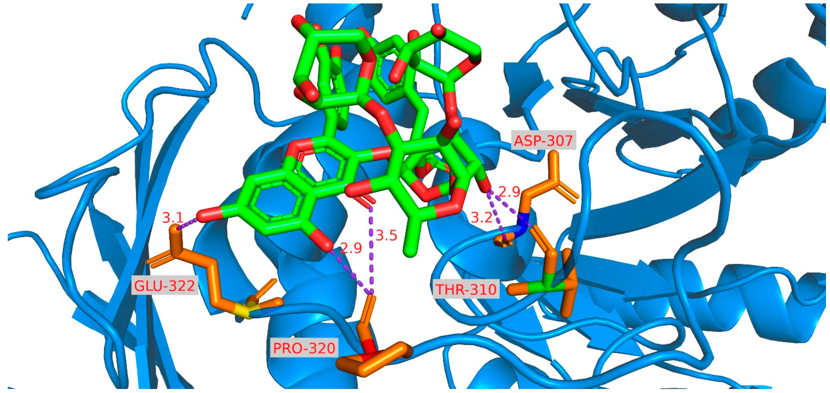
3.8. Molecular Docking Investigation

Autodock Vina (Version 1.1.2), a protein-ligand docking software, was used to evaluate the binding affinity and interaction modes of compounds 110 with their target. The 3D coordinates of α-glucosidase (PDB ID: 3A4A, resolution 1.6 Å) were obtained from the Protein Data Bank (RCSB PDB. Available online: https://www.rcsb.org, Accessed on 8 September 2024). All water molecules were removed, and polar hydrogen atoms were added. Grid boxes were designed to encompass the structural domains of each protein, permitting the ligands to move freely. The docking pocket was defined as a cubic region measuring 126 Å × 126 Å × 126 Å, with grid spacing set at 0.375 Å. Optimal molecular interactions were determined by the binding characteristics of α-glucosidase residues and their corresponding binding affinity scores.

3.9. Statistical Analysis

Statistical analysis data from the cytotoxicity and α-glycosidase inhibitory assays were analyzed using the mean and standard deviation. The 50% cytotoxic and inhibitory concentrations were calculated and compared to the control via non-linear regression. These analyses were performed using SPSS® Statistics (Version 18.0, IBM Software, Armonk, NY, USA).

4. Conclusions

This study examined the bioactive constituents and activities of Camellia flavida as sources for tea and functional foods. Ten new glycoside compounds (flavidosides A–J), comprising three oleanane-type triterpenoid saponins and seven flavonoid glycosides, were isolated from the flowers of C. flavida. Tests were conducted on the cytotoxic effects of the compounds against cancer cells and their α-glucosidase inhibitory activity. The IC50 values recorded for flavidoside C against MCF-7 and BEL-7402 cells were 1.65 ± 0.39 and 4.94 ± 0.41 μM, respectively. The semi-inhibitory concentration of flavidoside H on α-glucosidase was 1.17 ± 0.30 mM. These findings suggest that the bioactive constituents in C. flavida could potentially slow tumor growth and inhibit α-glucosidase activity, highlighting their promise for further research in anti-tumor treatments and metabolic regulation. We are now planning to undertake more comprehensive research to gain a deeper understanding of the underlying mechanisms, with the goal of identifying superior lead compounds.

Supplementary Materials

The following supporting information can be downloaded at https://www.mdpi.com/article/10.3390/ijms252010977/s1.

Author Contributions

Writing—original draft preparation, S.M.; methodology, Y.W.; data curation, H.M.; investigation, H.M. and L.G.; project administration, K.Y. All authors have read and agreed to the published version of the manuscript.

Funding

This research was funded by the Basic Ability Improvement Project for Young Teachers in Guangxi Colleges and Universities (2024KY1614) and the Guangxi Vocational College of Safety Engineering (GAZY2023KYA001).

Institutional Review Board Statement

Not applicable.

Informed Consent Statement

Not applicable.

Data Availability Statement

The data presented in this study are available in the Supplementary Materials.

Conflicts of Interest

The authors declare no conflicts of interest.

References

  1. Yang, R.; Guan, Y.; Zhou, J.; Sun, B.; Wang, Z.; Chen, H.; He, Z.; Jia, A. Phytochemicals from Camellia nitidissima Chi flowers reduce the pyocyanin production and motility of Pseudomonas aeruginosa PAO1. Front. Microbiol. 2018, 8, 2640. [Google Scholar] [CrossRef] [PubMed]
  2. Zhang, X.; Feng, J.; Su, S.; Huang, L. Hepatoprotective effects of Camellia nitidissima aqueous ethanol extract against CCl4-induced acute liver injury in SD rats related to Nrf2 and NF-κB signalling. Pharm. Biol. 2020, 58, 239–246. [Google Scholar] [CrossRef] [PubMed]
  3. Wang, W.; Liu, H.; Wang, Z.; Qi, J.; Yuan, S.; Zhang, W.; Chen, H.; Finley, J.W.; Gu, L.; Jia, A. Phytochemicals from Camellia nitidissima Chi inhibited the formation of advanced glycation end-products by scavenging methylglyoxal. Food Chem. 2016, 205, 204–211. [Google Scholar] [CrossRef] [PubMed]
  4. Yang, R.; Guan, Y.; Wang, W.; Chen, H.; He, Z.; Jia, A. Antioxidant capacity of phenolics in Camellia nitidissima Chi flowers and their identification by HPLC Triple TOF MS/MS. PLoS ONE 2018, 13, e0195508. [Google Scholar] [CrossRef]
  5. Hou, X.; Du, H.; Yang, R.; Qi, J.; Huang, Y.; Feng, S.; Wu, Y.; Lin, S.; Liu, Z.; Jia, A.; et al. The antitumor activity screening of chemical constituents from Camellia nitidissima Chi. Int. J. Mol. Med. 2018, 41, 2793–2801. [Google Scholar] [CrossRef]
  6. Ma, S.; Weng, M.; Yang, T.; Ge, L.; Yang, K. Triterpenes and pheophorbides from Camellia ptilosperma and their cytotoxicity, photocytotoxicity, and photodynamic antibacterial activity. Molecules 2023, 28, 7058. [Google Scholar] [CrossRef]
  7. Ma, S.; Ge, L.; Li, Y.; Liao, N.; Xie, J.; Yang, K. Ursane-type triterpenes with a phenylpropanoid unit from Camellia ptilosperma and evaluation of their cytotoxic activities. J. Nat. Prod. 2023, 86, 1793–1800. [Google Scholar] [CrossRef]
  8. Wang, Z.; Hou, X.; Li, M.; Ji, R.; Li, Z.; Wang, Y.; Guo, Y.; Liu, D.; Huang, B.; Du, H. Active fractions of golden-flowered tea (Camellia nitidissima Chi) inhibit epidermal growth factor receptor mutated non-small cell lung cancer via multiple pathways and targets in vitro and in vivo. Front. Nutr. 2022, 9, 1014414. [Google Scholar] [CrossRef]
  9. Zhou, A.; Zhou, C.; Liu, M.; He, J.; Huang, L. Anticancer effects of Camellia nitidissima Chi polysaccharide on hepa-tocellular carcinoma cells via apoptosis. J. Herb. Med. 2023, 42, 100800. [Google Scholar] [CrossRef]
  10. Chen, Y.; Zhang, F.; Du, Z.; Xie, J.; Xia, L.; Hou, X.; Hao, E.; Deng, J. Proteome analysis of Camellia nitidissima Chi revealed its role in colon cancer through the apoptosis and ferroptosis pathway. Front. Oncol. 2021, 11, 727130. [Google Scholar] [CrossRef]
  11. Naeem, A.; Hu, P.; Yang, M.; Zhang, J.; Liu, Y.; Zhu, W.; Zheng, Q. Natural products as anticancer agents: Current status and future perspectives. Molecules 2022, 27, 8367. [Google Scholar] [CrossRef] [PubMed]
  12. Zhang, H.; Wu, Q.; Qin, X. Camellia nitidissima Chi flower extracts inhibit α-amylase and α-glucosidase: In vitro by analysis of optimization of addition methods, inhibitory kinetics and mechanisms. Process Biochem. 2019, 86, 177–185. [Google Scholar] [CrossRef]
  13. Song, X.; Cao, P.; Bai, X.; Zhao, Y.; Zhang, Y.; Kong, H.; Zhao, Y.; Qu, H. The effects of carbon dots from hordei fructus Germinatus Carbonisatus on Glycometabolism and α-glycosidase activity. J. Biomed. Nanotechnol. 2022, 18, 2750–2758. [Google Scholar] [CrossRef]
  14. Xiong, J.; Wan, J.; Ding, J.; Wang, P.; Ma, G.; Li, J.; Hu, J. Camellianols A–G, barrigenol-like triterpenoids with PTP1B inhibitory effects from the endangered ornamental plant Camellia crapnelliana. J. Nat. Prod. 2017, 80, 2874–2882. [Google Scholar] [CrossRef]
  15. Zhang, H.; Li, X.; Wu, K.; Wang, M.; Liu, P.; Wang, X.; Deng, R. Antioxidant activities and chemical constituents of flavonoids from the flower of Paeonia ostii. Molecules 2016, 22, 5. [Google Scholar] [CrossRef]
  16. William, G.; Anthony, C.; Waiss, J. Structural relationships and interconversions of isomeric astilbins. J. Org. Chem. 1975, 40, 1057–1061. [Google Scholar] [CrossRef]
  17. Linh, N.T.T.; Thuy, T.T.; Tam, N.T.; Cham, B.T.; Tam, K.T.; Sa, N.H.; Thao, D.T.; Chinh, V.T.; Anh, N.T.H. Chemical constituents of Impatiens chapaensis Tard. and their α-glucosidase inhibition activities. Nat. Prod. Res. 2021, 36, 3229–3233. [Google Scholar] [CrossRef]
  18. Kim, S.; Li, Y.; Lin, L.; Sayasith, P.R.; Tarr, A.T.; Wright, E.B.; Yasmin, S.; Lannigan, D.A.; O’Doherty, G.A. Synthesis and biological evaluation of 4′-substituted kaempfer-3-ols. J. Org. Chem. 2020, 85, 4279–4288. [Google Scholar] [CrossRef]
  19. Noccioli, C.; Luciardi, L.; Barsellini, S. Flavonoids from two Italian Genista species: Genista cilentina and Genista sulcitana. Chem. Nat. Compd. 2012, 48, 672–673. [Google Scholar] [CrossRef]
  20. de Beer, D.; Joubert, E.; Malherbe, C.J.; Jacobus Brand, D. Use of countercurrent chromatography during isolation of 6-hydroxyluteolin-7-O-β-glucoside, a major antioxidant of Athrixia phylicoides. J. Chromatogr. A 2011, 1218, 6179–6186. [Google Scholar] [CrossRef]
  21. Jin, H.; Chen, G.; Li, X.; Shen, Y.; Yan, S.; Zhang, L.; Yang, M.; Zhang, W. Flavonoids from Rhododendron decorum. Chem. Nat. Compd. 2009, 45, 85–86. [Google Scholar] [CrossRef]
  22. de Oliveira, A.P.; Coppede, J.S.; Bertoni, B.W.; Crotti, A.E.M.; França, S.C.; Pereira, A.M.S.; Taleb-Contini, S.H. Costus spiralis (Jacq.) roscoe: A novel source of flavones with α-glycosidase inhibitory activity. Chem. Biodivers. 2017, 15, el700421. [Google Scholar] [CrossRef] [PubMed]
  23. Benahmed, M.; Akkal, S.; Elomri, A.; Laouer, H.; Vérité, P.; Seguin, E. Constituents from Bupleurum montanum (Coss. & Dur.) (Apiaceae). Arab. J. Chem. 2014, 7, 1065–1069. [Google Scholar] [CrossRef]
  24. Sporn, M.B.; Liby, K.T.; Yore, M.M.; Fu, L.; Lopchuk, J.M.; Gribble, G.W. New Synthetic Triterpenoids: Potent Agents for Prevention and Treatment of Tissue Injury Caused by Inflammatory and Oxidative Stress. J. Nat. Prod. 2011, 74, 537–545. [Google Scholar] [CrossRef]
  25. Huyen, L.T.; Bao, D.G.; Son, N.T.; Anh, N.T.V.; Huu Tai, B.H.; Huong, P.T.T.; Kiem, P.V.; Thuy, N.T.K.; Hanh, N.T.; Park, S.; et al. Cytotoxic Effects of Oleanane-type Saponins from Lysimachia laxa. Chem. Biodivers. 2024, e202401024. [Google Scholar] [CrossRef]
  26. Han, L.; Shi, L.; Yu, D.; Tang, Q.; Tang, L.; Feng, B.; Wang, Y. Inhibitive Effect of Seeds of Camellia chrysantha (Hu) Tuyama on Gonadal Hormones Dependent Tumour in vitro. Lishizhen Med. Mater. Medica Res. 2009, 20, 3146–3148. [Google Scholar]
  27. Luo, Z.; Zeng, J.; Yu, H.; Huang, H.; Bao, X.; Qin, S.; Chen, G.; Zhou, Z.; Zhi, H.; Yao, X. Astramalabaricosides A–T, highly oxygenated malabaricane triterpenoids with migratory inhibitory activity from Astragalus membranaceus var. mongholicus. J. Nat. Prod. 2022, 85, 2312–2331. [Google Scholar] [CrossRef]
  28. Zhang, Y.; Lv, P.; Ma, J.; Chen, N.; Guo, H.; Chen, Y.; Gan, X.; Wang, R.; Liu, X.; Fan, S.; et al. Antrodia cinnamomea exerts an anti-hepatoma effect by targeting PI3K/AKT-mediated cell cycle progression in vitro and in vivo. Acta Pharm. Sin. B 2022, 12, 890–906. [Google Scholar] [CrossRef]
Figure 1. Structure of compounds 115 from the flower of C. flavida.
Ijms 25 10977 g001
Figure 2. Key HMBC (A) and NOESY (B) correlations of compound 1.
Ijms 25 10977 g002
Figure 3. Key HMBC correlations of compound 4.
Ijms 25 10977 g003
Figure 4. The experimental ECD of 4 and calculated ECD of (2R, 3R)-4.
Ijms 25 10977 g004
Figure 5. Key HMBC correlations of compound 5.
Ijms 25 10977 g005
Figure 6. The experimental ECD of 5 and calculated ECD of (2R, 3S)-5.
Ijms 25 10977 g006
Figure 7. Key HMBC correlations of compound 7.
Ijms 25 10977 g007
Figure 8. Molecular docking simulations obtained at the lowest energy conformation, highlighting potential hydrogen contacts of 8.
Ijms 25 10977 g008
Table 1. 1H-NMR (600 MHz) data of compounds 13 in Pyr-d5 (δH, mult; J in Hz).
No.123
10.83 m
1.42 m
0.81 m
1.37 m
0.76 m
1.35 m
21.68 m
1.97 m
1.71 m
2.06 m
1.72 m
2.05 m
33.17 dd (11.7, 4.1)3.20 dd (11.3, 3.9)3.17 dd (11.5, 3.8)
50.77 br d (12.3)0.77 br d (11.9)0.72 br d (11.6)
61.33 m
1.53 m
1.34 m
1.54 m
1.31 m
1.50 m
72.03 m
2.15 m
2.03 m
2.15 m
1.85–1.97 m
91.68 m1.68 m1.49, m
111.82, m1.84, m1.85, m
125.45 t (3.4)5.44 t (3.4)5.46 t (3.4)
154.48 overlapped4.48 overlapped5.09 s
164.55 d (3.8)4.54 d (3.7)
182.45 dd (13.5, 3.8)2.44 dd (13.5, 3.8)2.71 dd, 13.9, 3.4)
191.30 overlapped
2.70 dd (13.5, 12.9)
1.29 overlapped
2.70 dd (13.5, 12.9)
1.21 overlapped
1.56 t-like (13.9)
211.40 m
2.39 m
1.39 m
2.39 m
1.29 m
1.76 m
222.17 m
2.27 m
2.16 m
2.27 m
1.53 overlapped
2.88 br d (13.7)
231.12 s1.13, s1.10, s
241.02 s1.04, s1.04, s
250.82 s0.81, s0.79, s
261.05 s1.04, s1.13, s
271.81 s1.82, s1.34, s
283.60 d (10.5)
3.79 d (10.5)
3.60, d (10.5)
3.79, d (10.5)
3.81, d (10.8)
4.42, overlapped
291.05 s1.04, s0.85, s
301.10 s1.10, s0.89, s
GlcA-14.86 overlapped4.86, overlapped4.82, d (7.1)
24.68 m4.73, m4.71, m
34.67 m4.74, m4.74, m
44.52 m4.64, m4.63, dd (9.5, 9.0)
54.45 overlapped4.57, m4.57, d (9.5)
6-OCH33.69 s
Glc-15.87 d (7.4)5.90 d (7.4)5.90 d (7.4)
24.10 dd (9.0, 7.4)4.12 dd (9.3, 7.4)4.10 dd (9.0, 7.4)
34.40 m4.41 m4.39 m
44.07 t-like (9.1)4.07 t-like (9.0)4.04 t-like (9.1)
54.40 m4.41 m4.39 m
64.31 m
4.66 m
4.31 m
4.66 m
4.28 m
4.66 m
Gal-16.10 d (7.7)6.18 d (7.6)6.21 overlapped
24.69 m4.71 m4.71 m
34.45 m4.48 m4.49 br d (9.2)
44.45 m4.46 m4.44 br s
54.23 m4.26 m4.27 m
64.31 m4.32 m4.33 m
Rha-16.23 br s6.23 br s6.21 br s
24.77 br s4.78 br s4.76 br s
34.71 m4.72 m4.69 m
44.20 t-like (9.2)4.21 t-like (9.1)4.20 t-like (9.1)
54.84 m4.88 m4.89 m
61.41 d (6.1)1.42 d (5.8)1.40 d (5.8)
Table 2. 13C-NMR (151 MHz) data of compounds 13 in Pyr-d5 (δC).
No.123
139.039.039.0
226.426.526.6
389.789.889.8
439.639.739.7
555.655.655.6
618.818.918.7
736.836.836.1
841.441.541.8
947.447.347.1
1037.037.137.0
1124.024.024.1
12123.9123.9126.0
13146.0146.0142.0
1448.048.054.3
1567.467.473.8
1678.778.7216.1
1741.141.153.2
1843.343.348.1
1948.048.048.0
2031.331.331.0
2137.137.135.8
2231.031.027.1
2328.128.028.0
2416.816.816.9
2515.915.915.8
2617.617.617.7
2721.021.021.7
2869.669.670.9
2933.533.533.1
3024.624.623.6
GlcA-1105.2105.4105.4
279.579.579.6
382.882.882.7
470.971.371.3
576.677.477.4
6170.3172.9172.2
6-OCH352.1
Glc-1102.9102.7102.8
276.476.476.5
378.578.578.5
472.672.672.7
578.278.178.2
663.563.663.7
Gal-1101.6101.4101.4
276.276.476.5
376.176.076.1
471.171.271.3
577.077.077.1
662.062.062.1
Rha-1102.3102.4102.4
272.672.672.7
372.672.672.7
473.974.074.0
569.869.869.9
618.318.318.3
Table 3. 1H (600 MHz) and 13C (151 MHz) NMR data of compound 4 in CD3OD.
No.δH (J in Hz)δCNo.δH (J in Hz)δC
25.19 d (10.4)83.543.29 overlapped73.9
34.55 d (10.4)81.254.18 m70.9
4 195.761.20 d (6.2)17.9
5 165.5Glc-14.02 d (7.6)105.3
65.92 d (1.9)97.423.30 overlapped78.9
7 168.633.41 t-like (9.1)78.3
85.90 d (1.9)96.343.33 overlapped70.9
9 164.053.10 dt (9.5, 3.1)77.2
10 102.563.68 d (3.1)62.1
1′ 128.7Rha2-15.21 d (1.5)101.8
2′, 6′7.36 d (8.4)130.123.88 dd (3.3, 1.5)71.9
3′, 5′6.85 d (8.4)116.533.79 dd (9.5, 3.3)71.8
4′ 159.243.33 overlapped74.2
Rha1-14.71 d (1.4)102.154.02 m70.3
23.46 dd (3.2, 1.4)82.461.26 d (6.2)17.9
33.72 dd (9.8, 3.2)71.6
Table 4. 1H (600 MHz) and 13C (151 MHz) NMR data of compounds 5 and 6.
No.5 16 2
δH (J in Hz)δCδH (J in Hz)δC
25.48 d (2.3)82.15.42 d (8.4)80.9
34.17 d (2.3)75.84.71 d (8.4)76.9
4 194.4 193.8
5 166.1 163.4
65.93 d (1.7)97.55.90 s96.0
7 168.9 167.1
85.97 d (1.7)96.35.90 s95.1
9 164.6 161.9
10 101.9 101.0
1′ 128.0 126.2
2′, 6′7.31 d (8.5)129.07.25 d (8.4)128.8
3′, 5′6.82 d (8.5)116.36.78 d (8.4)115.3
4′ 158.7 157.7
5-OH 11.71 br s
7-OH 10.96 br s
4′-OH 9.62 br s
Rha1-15.36 d (1.2)99.64.77 br s99.7
23.73 br s81.83.49 br s77.9
33.53 dd (9.7 3.4)71.63.09 m70.1
43.18 t-like (9.7)73.43.15 m72.1
52.38 dq (9.7 6.1)70.33.60 m69.0
60.88 d (6.1)17.91.07 d (6.0)17.6
Glc-14.47 d (7.7)105.54.31 d (7.8)102.5
23.41 overlapped78.43.17 m75.6
33.46 t-like (9.0)78.73.28 m77.5
43.39 overlapped71.13.46 m70.0
53.20 m77.43.07 m76.6
63.74 m
3.85 m
62.43.48 m
3.64 m
60.8
Rha2-15.27 d (1.8)101.35.18 br s99.5
24.00 dd (3.2 1.8)69.83.70 br s70.3
35.03 dd (9.8 3.2)75.53.75 m67.7
43.50 t-like (9.8)71.64.72 t-like (9.8)74.1
54.19 m70.14.07 m65.7
61.24 d (6.2)17.90.94 d (6.1)17.4
Acetyl-1 172.8 170.2
22.08 s21.12.00 s21.0
1 Measured in CD3OD. 2 Measured in DMSO-d6.
Table 5. 1H-NMR (600 MHz) data (J in Hz) of compounds 710.
No.7 18 29 110 1
66.13 br s6.15 br s6.17 br s6.15 br s
86.32 br s6.33 br s6.35 br s6.35 br s
2′7.60 br s7.51 overlapped7.98 d (8.3)7.98 d (8.6)
3′ 6.89 d (8.3)6.90 d (8.6)
5′6.88 d (7.8)6.84 d (8.8)6.89 d (8.3)6.90 d (8.6)
6′7.55 br d (7.8)7.51 overlapped7.98 d (8.3)7.98 d (8.6)
5-OH 12.58 br s
Glc1-15.58 d (7.8)5.57 d (7.9)5.53 d (8.0)5.57 d (8.0)
25.24 dd (9.5, 7.8)5.09 dd (9.5, 7.9)5.20 dd (9.5, 8.0)5.19 dd (9.5, 8.0)
33.88 m3.82 t-like (9.0)3.90 t-like (9.2)3.87 t-like (9.0)
43.48 m3.29 m3.43 m3.42 t-like (9.2)
53.58 m3.52 m3.52 m3.54 m
63.51 m
3.88 m
3.41 overlapped
3.73 br d (11.3)
3.47 m
3.86 m
3.47 m
3.87 m
Ara-14.34 d (6.8) 4.34 d (7.0)
23.54 m 3.53 m
33.49 m 3.47 m
43.77 br s 3.77 br s
53.56 m
3.85 m
3.56 m
3.87 m
Glc2-1 4.31 d (7.9)4.42 d (7.7)
2 2.95 m3.18 dd (9.0, 7.7)
3 3.20 m3.28 m
4 3.26 m3.27 m
5 3.10 m3.32 m
6 3.41 overlapped
3.68 m
3.62 dd (11.6, 6.3)
3.86 m
Rha-14.58 br s4.38 br s4.54 br s4.54 d (1.5)
23.85 br s3.57 br s3.80 br s3.81 dd (3.1, 1.5)
33.60 dd (9.2, 3.0)3.32 m3.55 dd (9.3, 3.1)3.56 m
43.44 t-like (9.4)3.26 m3.42 t-like (9.4)3.43 t-like (9.4)
53.53 m3.32 m3.49 m3.51 m
61.13 d (6.0)0.96 d (6.0)1.11 d (6.1)1.12 d (6.1)
Xyl-14.39 d (7.4)4.24 d (7.5)4.35 d (7.5)4.36 d (7.5)
23.26 dd (9.1, 7.4)3.02 m3.26 dd (9.0, 7.5)3.25 dd (9.1, 7.5)
33.37 t-like, (9.0)3.12 m3.35 t-like (9.0)3.35 t-like (9.0)
43.49 m3.03 m3.49 m3.49 m
53.17 t-like (10.5)
3.80 m
3.00 m
3.60 dd (11.1, 5.0)
3.15 t-like (10.7)
3.79 m
3.15 t-like (10.8)
3.79 m
Coumaroyl-2, 67.45 d (8.0)7.51 d (8.4)7.47 d (8.2)7.46 d (8.2)
3, 56.80 d (8.0)6.79 d (8.4)6.81 d (8.2)6.80 d (8.2)
77.68 d (15.9)7.54 d (15.9)7.70 d (15.9)7.69 d (15.9)
86.42 d (15.9)6.36 d (15.9)6.39 d (15.9)6.39 d (15.9)
1 Measured in CD3OD. 2 Measured in DMSO-d6.
Table 6. 13C-NMR (151 MHz) data of compounds 710 (δC).
No.7 18 29 110 1
2158.9156.8159.1159.1
3134.8132.8134.6134.7
4178.9177.0179.0179.0
5162.9161.2163.0163.0
699.998.699.999.9
7165.5164.1165.6165.6
894.893.694.994.9
9158.3156.4158.4158.5
10105.9104.0105.8105.9
1′123.3120.9122.8122.9
2′117.5116.3132.3132.3
3′145.8144.8116.2116.2
4′149.6148.6161.3161.3
5′116.2115.2116.2116.2
6′123.5121.7132.3132.3
Glc1-1100.798.8100.7100.7
274.572.274.474.5
384.583.184.684.3
470.369.270.470.4
576.875.376.976.9
668.467.968.668.5
Ara-1105.2 105.3
272.2 72.2
373.9 73.9
469.5 69.5
567.2 67.2
Glc2-1 103.3104.8
2 73.274.7
3 76.877.6
4 69.571.3
5 76.478.0
6 61.062.5
Rha-1102.2101.3102.2102.2
271.669.871.771.7
382.481.182.482.4
472.770.772.672.7
569.568.069.569.5
617.917.617.917.9
Xyl-1106.2105.4106.3106.3
275.373.975.275.3
377.576.077.577.5
471.169.971.071.1
566.865.566.866.8
Coumaroyl-1127.3125.3127.3127.4
2, 6131.3130.2131.3131.3
3, 5116.8115.8116.8116.8
4161.1159.7161.2161.2
7147.3144.6147.2147.3
8115.1114.4115.2115.2
9168.8165.7168.6168.6
1 Measured in CD3OD. 2 Measured in DMSO-d6.
Table 7. Cytotoxicity (IC50, μM ± SD, n = 8) of compounds 110 against cell lines.
HelaMCF-7BEL-7402A549Hep G2MB-231MRC-5HNEpC
115.07 ± 1.069.06 ± 0.457.62 ± 1.06>3025.60 ± 3.46>30>30>30
24.17 ± 0.8519.24 ± 3.71>30>3014.04 ± 3.2926.53 ± 3.07>30>30
3>301.65 ± 0.394.94 ± 0.4123.42 ± 7.268.77 ± 1.5610.08 ± 2.28>30>30
4>30>30>30>30>30>30>30>30
5>30>30>30>3020.76 ± 3.79>30>30>30
6>30>3011.40 ± 3.11>30>30>30>30>30
7>30>30>30>30>30>30>30>30
8>30>30>30>30>30>30>30>30
924.72 ± 4.39>30>30>3028.80 ± 5.55>30>30>30
10>30>30>3017.38 ± 2.81>30>30>30>30
PC11.06 ± 0.72 13.79 ± 0.26 29.94 ± 0.80 14.20 ± 0.37 16.31 ± 0.18 19.06 ± 1.01 1NDND
Positive control (PC): 1 cisplatin and 2 doxorubicin.
Table 8. α-Glucosidase inhibition activity (IC50, mM ± SD, n = 3) of compounds 110.
IC50 IC50
1>2072.38 ± 0.22
2>2081.17 ± 0.30
3>2093.33 ± 0.48
46.15 ± 0.43102.26 ± 0.48
512.73 ± 1.08acarbose2.04 ± 0.27
68.09 ± 2.61
Disclaimer/Publisher’s Note: The statements, opinions and data contained in all publications are solely those of the individual author(s) and contributor(s) and not of MDPI and/or the editor(s). MDPI and/or the editor(s) disclaim responsibility for any injury to people or property resulting from any ideas, methods, instructions or products referred to in the content.

