Correction: Scimeca et al. Microcalcifications Drive Breast Cancer Occurrence and Development by Macrophage-Mediated Epithelial to Mesenchymal Transition. Int. J. Mol. Sci. 2019, 20, 5633

 ,
,  ,
,  , ,
, , 
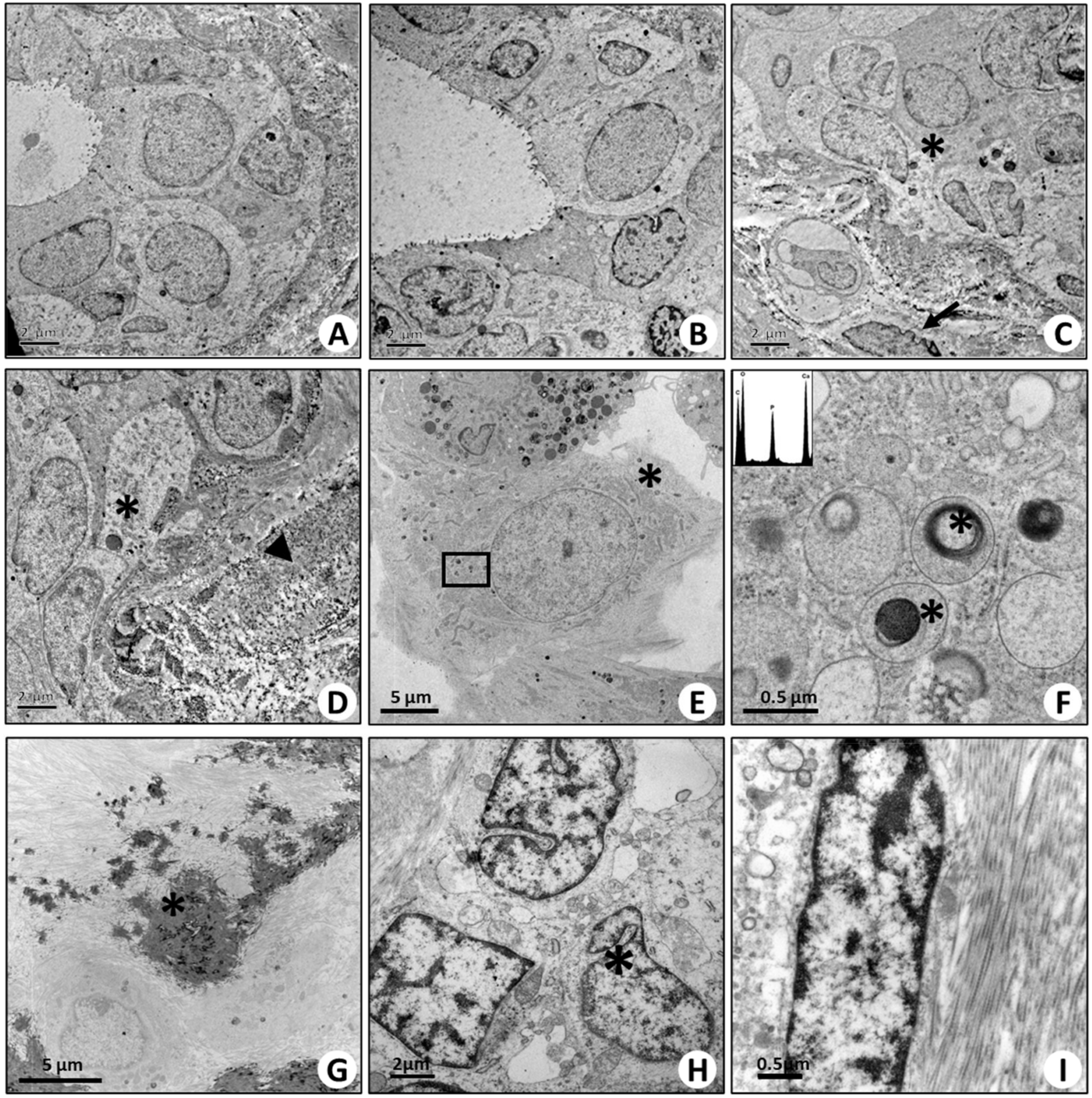
Error in Figure

Reference
- Scimeca, M.; Bonfiglio, R.; Menichini, E.; Albonici, L.; Urbano, N.; De Caro, M.T.; Mauriello, A.; Schillaci, O.; Gambacurta, A.; Bonanno, E. Microcalcifications Drive Breast Cancer Occurrence and Development by Macrophage-Mediated Epithelial to Mesenchymal Transition. Int. J. Mol. Sci. 2019, 20, 5633. [Google Scholar] [CrossRef] [PubMed]
Disclaimer/Publisher’s Note: The statements, opinions and data contained in all publications are solely those of the individual author(s) and contributor(s) and not of MDPI and/or the editor(s). MDPI and/or the editor(s) disclaim responsibility for any injury to people or property resulting from any ideas, methods, instructions or products referred to in the content. |
© 2024 by the authors. Licensee MDPI, Basel, Switzerland. This article is an open access article distributed under the terms and conditions of the Creative Commons Attribution (CC BY) license (https://creativecommons.org/licenses/by/4.0/).
Share and Cite
Scimeca, M.; Bonfiglio, R.; Menichini, E.; Albonici, L.; Urbano, N.; De Caro, M.T.; Mauriello, A.; Schillaci, O.; Gambacurta, A.; Bonanno, E. Correction: Scimeca et al. Microcalcifications Drive Breast Cancer Occurrence and Development by Macrophage-Mediated Epithelial to Mesenchymal Transition. Int. J. Mol. Sci. 2019, 20, 5633. Int. J. Mol. Sci. 2024, 25, 8016. https://doi.org/10.3390/ijms25158016
Scimeca M, Bonfiglio R, Menichini E, Albonici L, Urbano N, De Caro MT, Mauriello A, Schillaci O, Gambacurta A, Bonanno E. Correction: Scimeca et al. Microcalcifications Drive Breast Cancer Occurrence and Development by Macrophage-Mediated Epithelial to Mesenchymal Transition. Int. J. Mol. Sci. 2019, 20, 5633. International Journal of Molecular Sciences. 2024; 25(15):8016. https://doi.org/10.3390/ijms25158016
Chicago/Turabian StyleScimeca, Manuel, Rita Bonfiglio, Erika Menichini, Loredana Albonici, Nicoletta Urbano, Maria Teresa De Caro, Alessandro Mauriello, Orazio Schillaci, Alessandra Gambacurta, and Elena Bonanno. 2024. "Correction: Scimeca et al. Microcalcifications Drive Breast Cancer Occurrence and Development by Macrophage-Mediated Epithelial to Mesenchymal Transition. Int. J. Mol. Sci. 2019, 20, 5633" International Journal of Molecular Sciences 25, no. 15: 8016. https://doi.org/10.3390/ijms25158016
APA StyleScimeca, M., Bonfiglio, R., Menichini, E., Albonici, L., Urbano, N., De Caro, M. T., Mauriello, A., Schillaci, O., Gambacurta, A., & Bonanno, E. (2024). Correction: Scimeca et al. Microcalcifications Drive Breast Cancer Occurrence and Development by Macrophage-Mediated Epithelial to Mesenchymal Transition. Int. J. Mol. Sci. 2019, 20, 5633. International Journal of Molecular Sciences, 25(15), 8016. https://doi.org/10.3390/ijms25158016