Constitutive Neurogenesis and Neuronal Plasticity in the Adult Cerebellum and Brainstem of Rainbow Trout, Oncorhynchus mykiss


Abstract
1. Introduction
2. Results
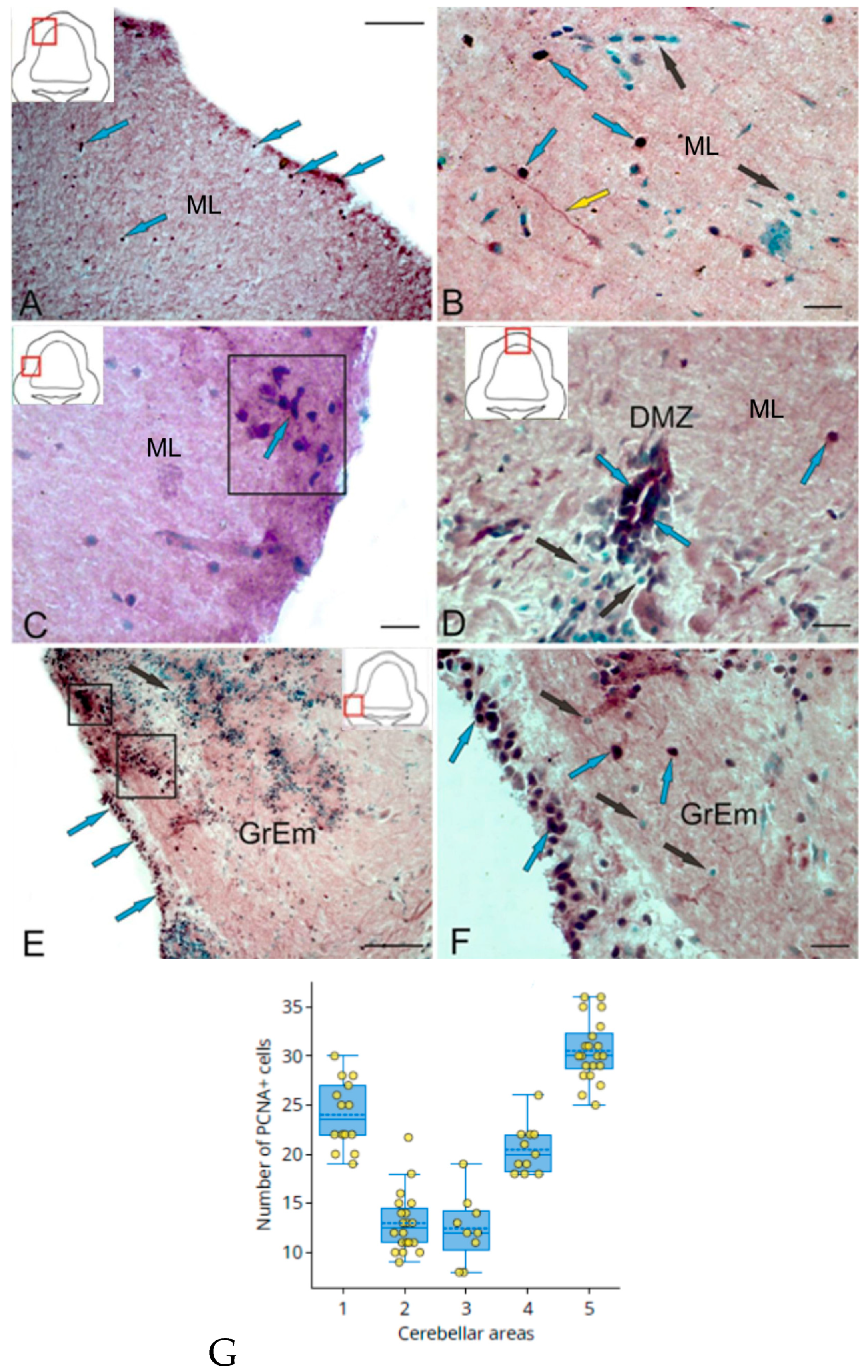
2.1. Proliferating Cell Nuclear Antigen (PCNA) in Trout Cerebellum
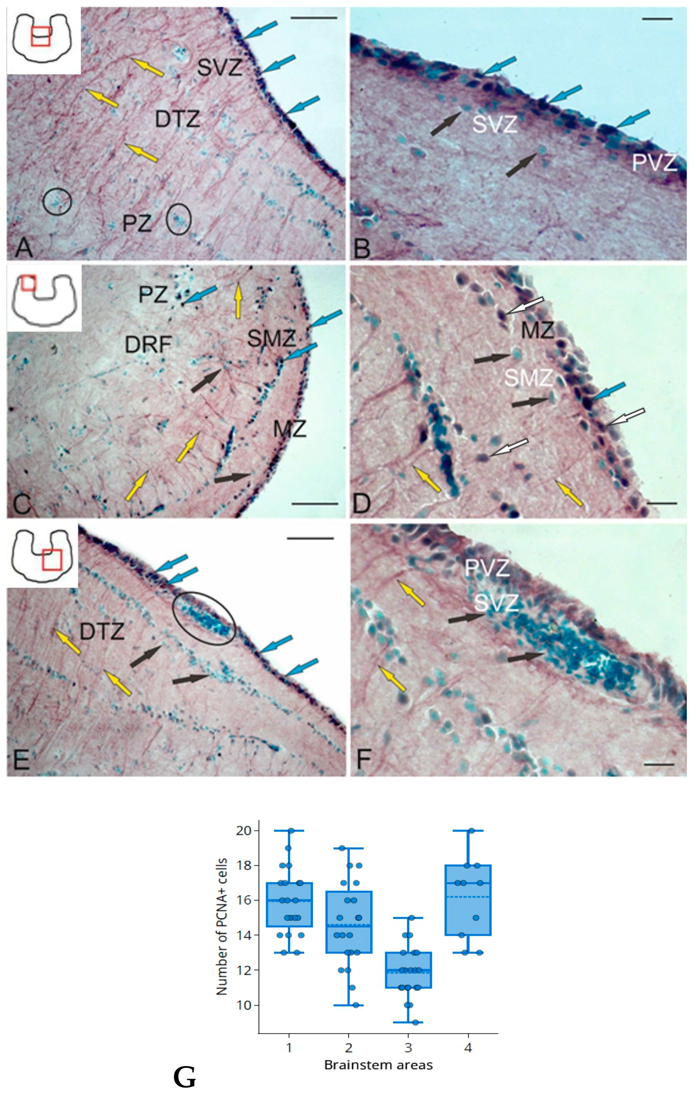
2.2. PCNA in the Trout Brainstem
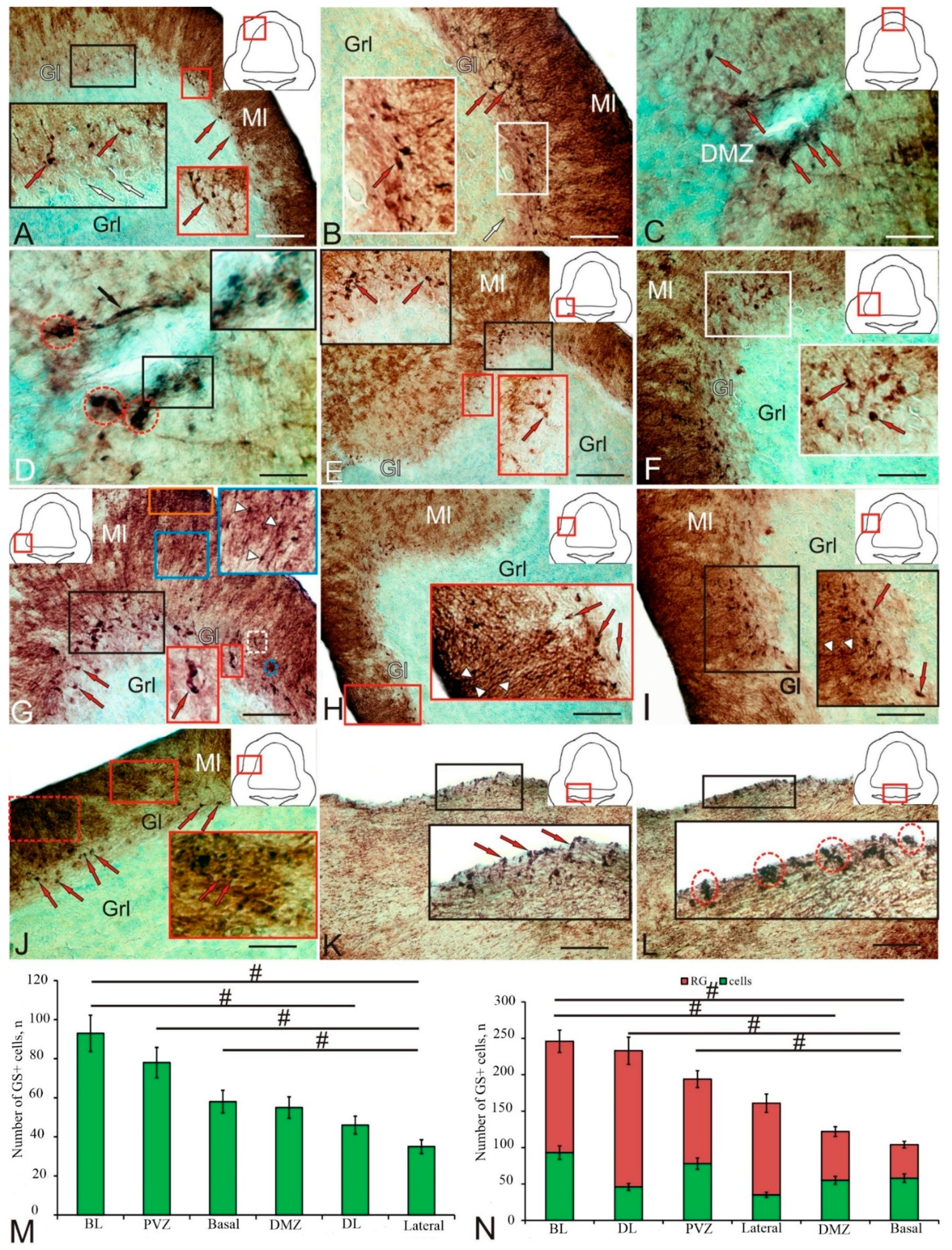
2.3. Glutamine Synthetase in the Trout Cerebellum
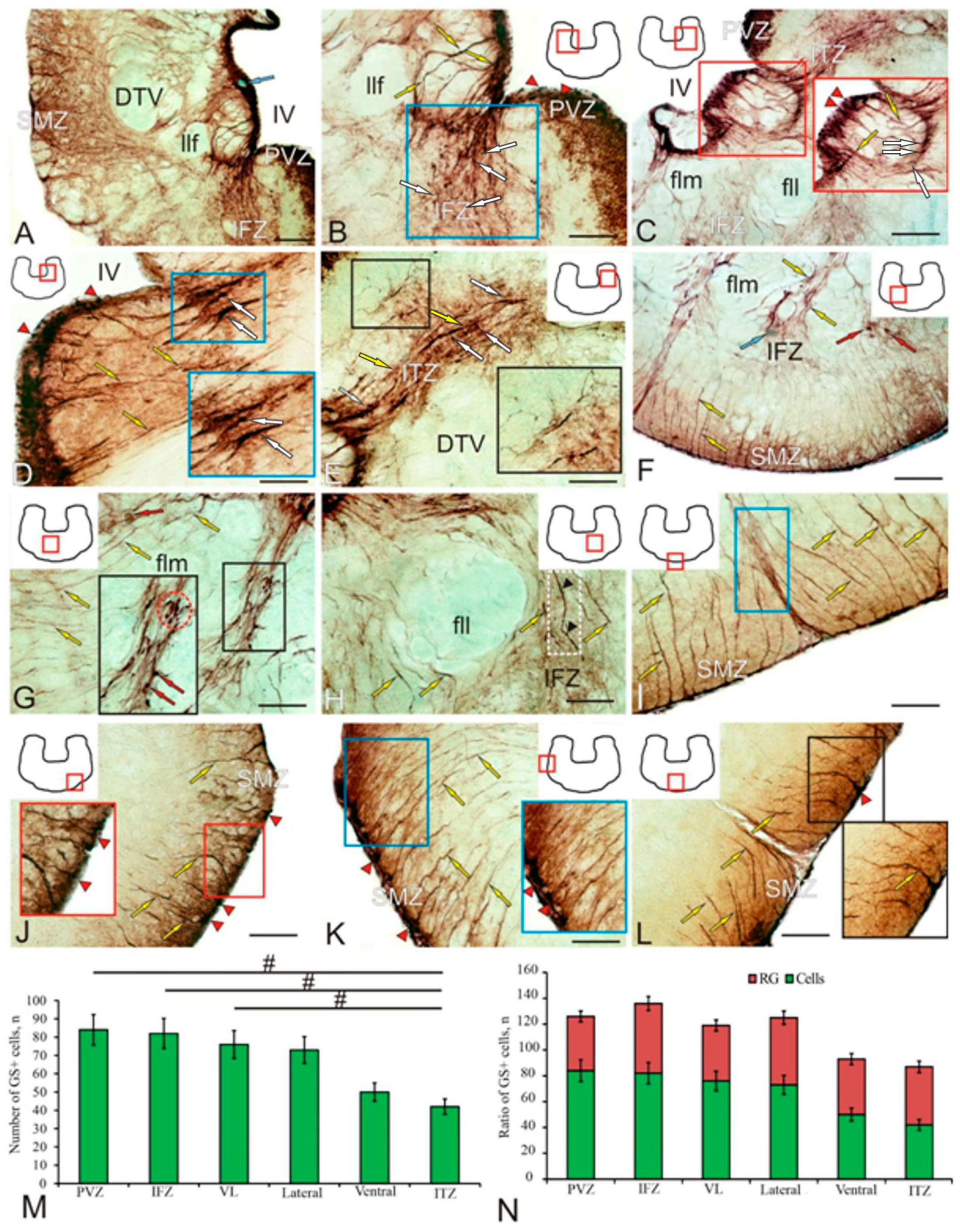
2.4. Glutamine Synthetase in the Trout Brainstem
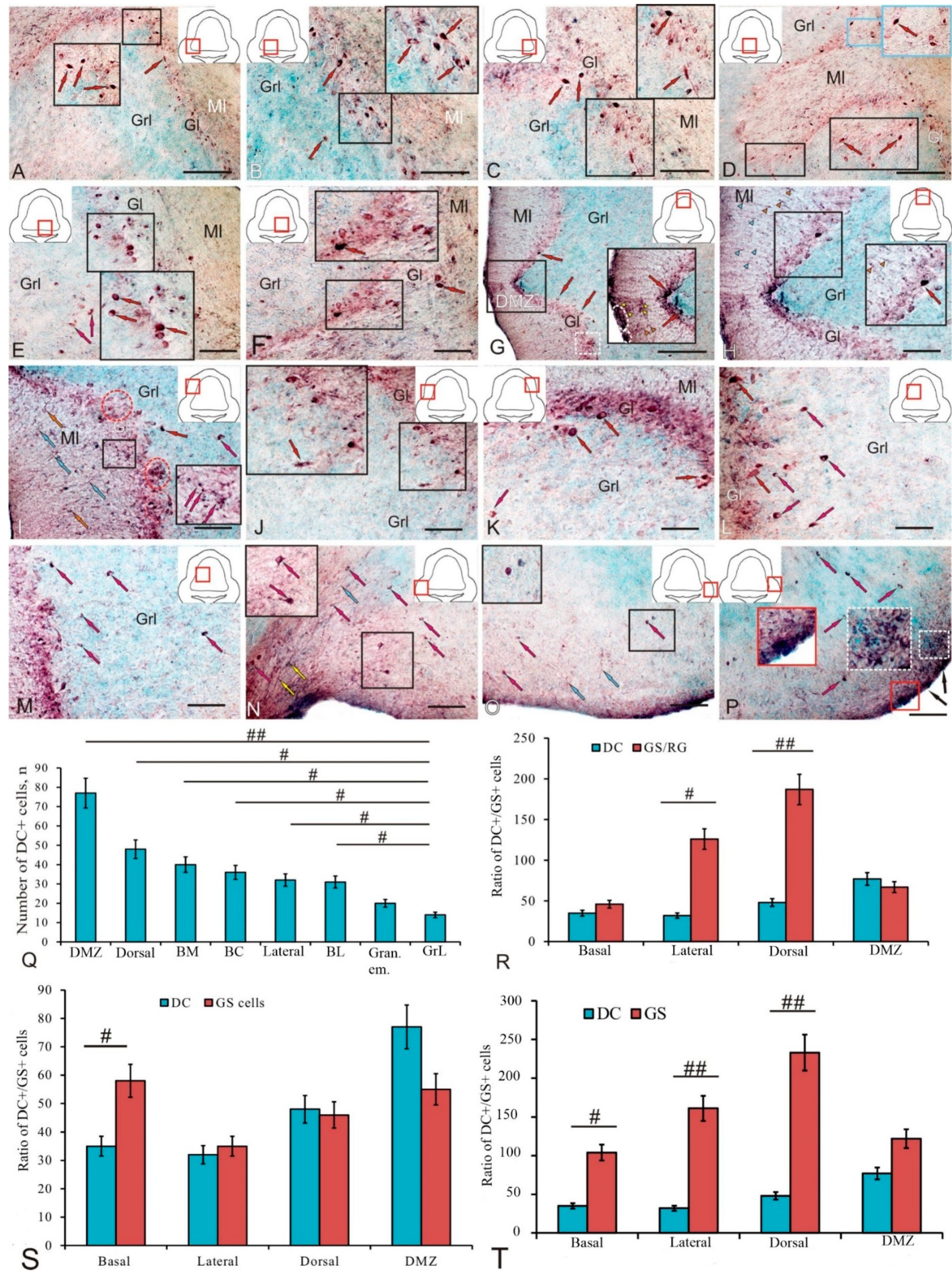
2.5. Doublecortin in the Trout Cerebellum
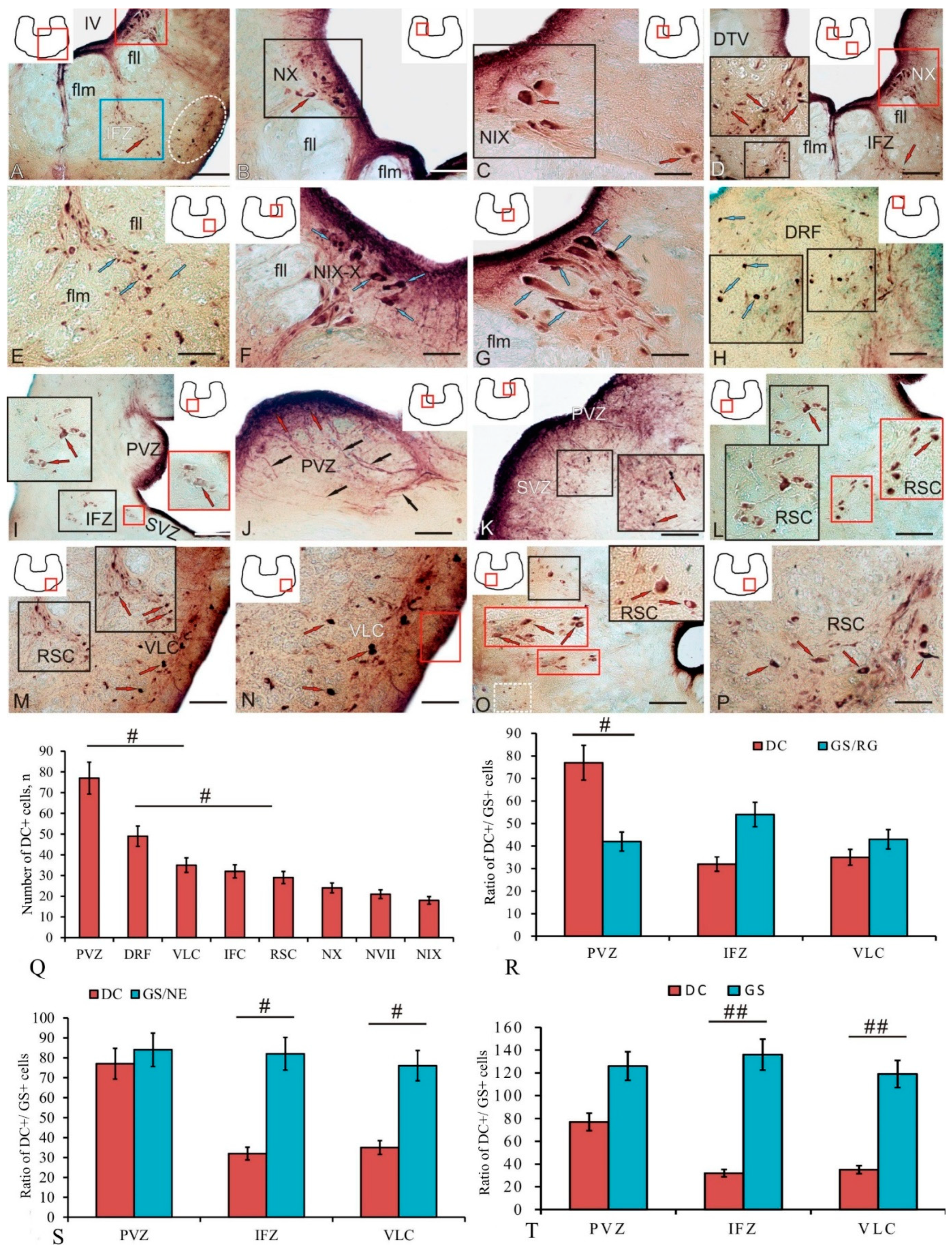
2.6. Doublecortin in the Trout Brainstem
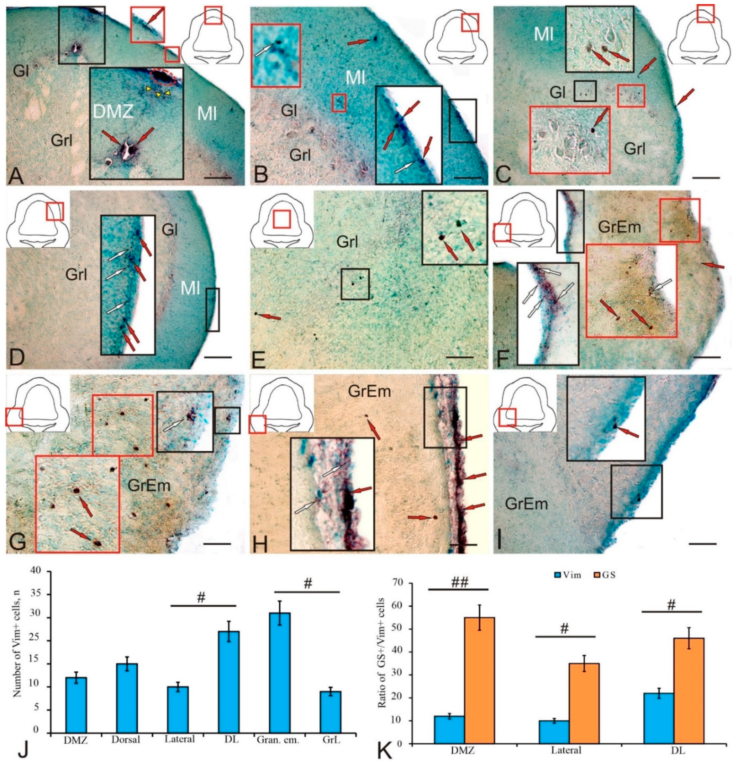
2.7. Vimentin in the Trout Cerebellum
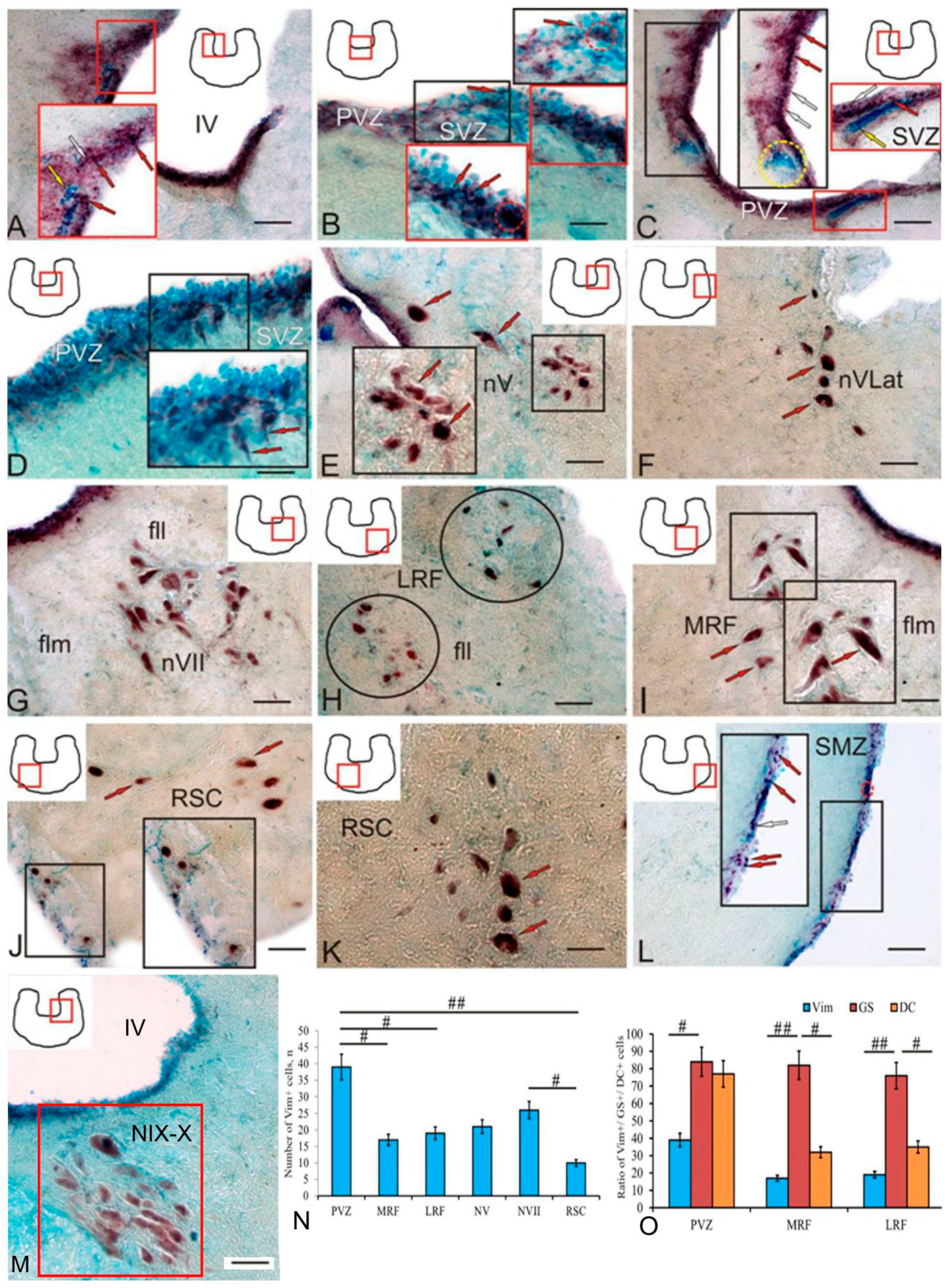
2.8. Vimentin in the Trout Brainstem
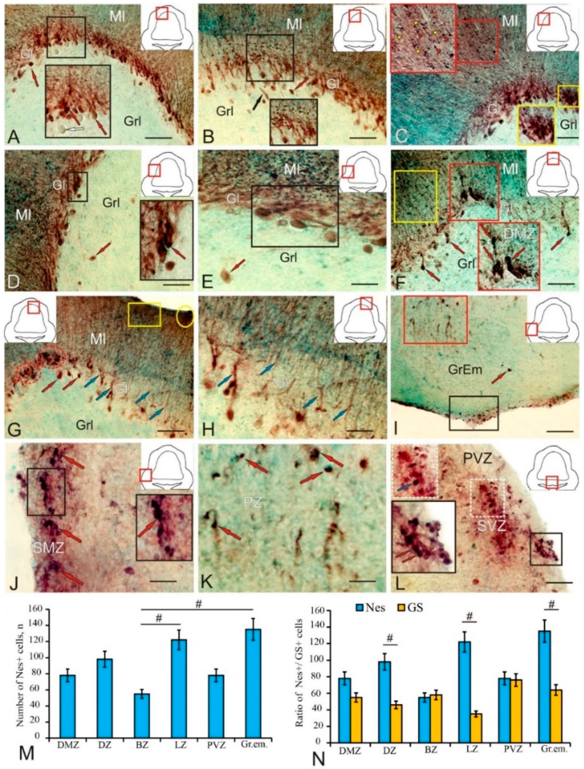
2.9. Nestin in the Trout Cerebellum
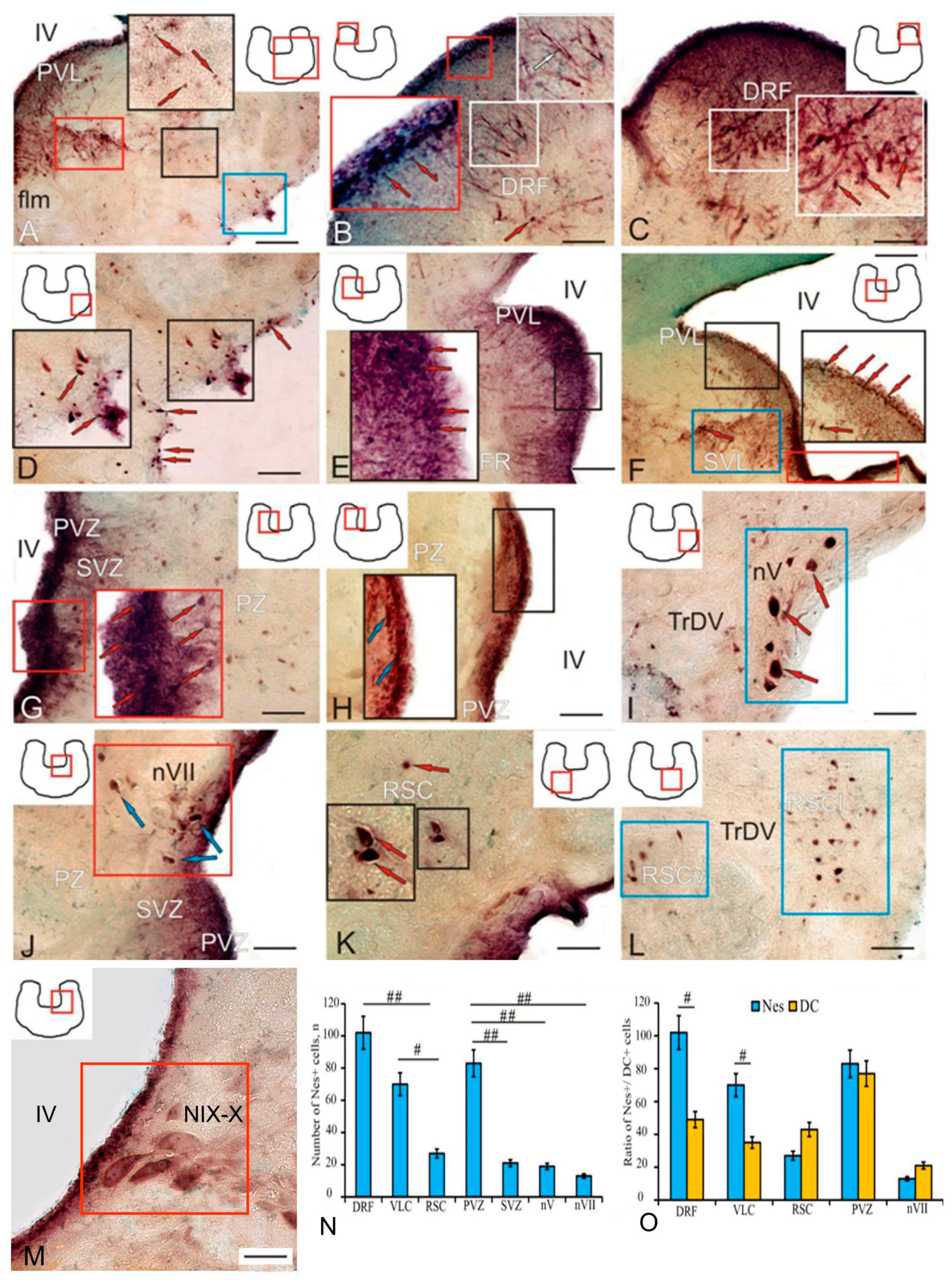
2.10. Nestin in the Trout Brainstem
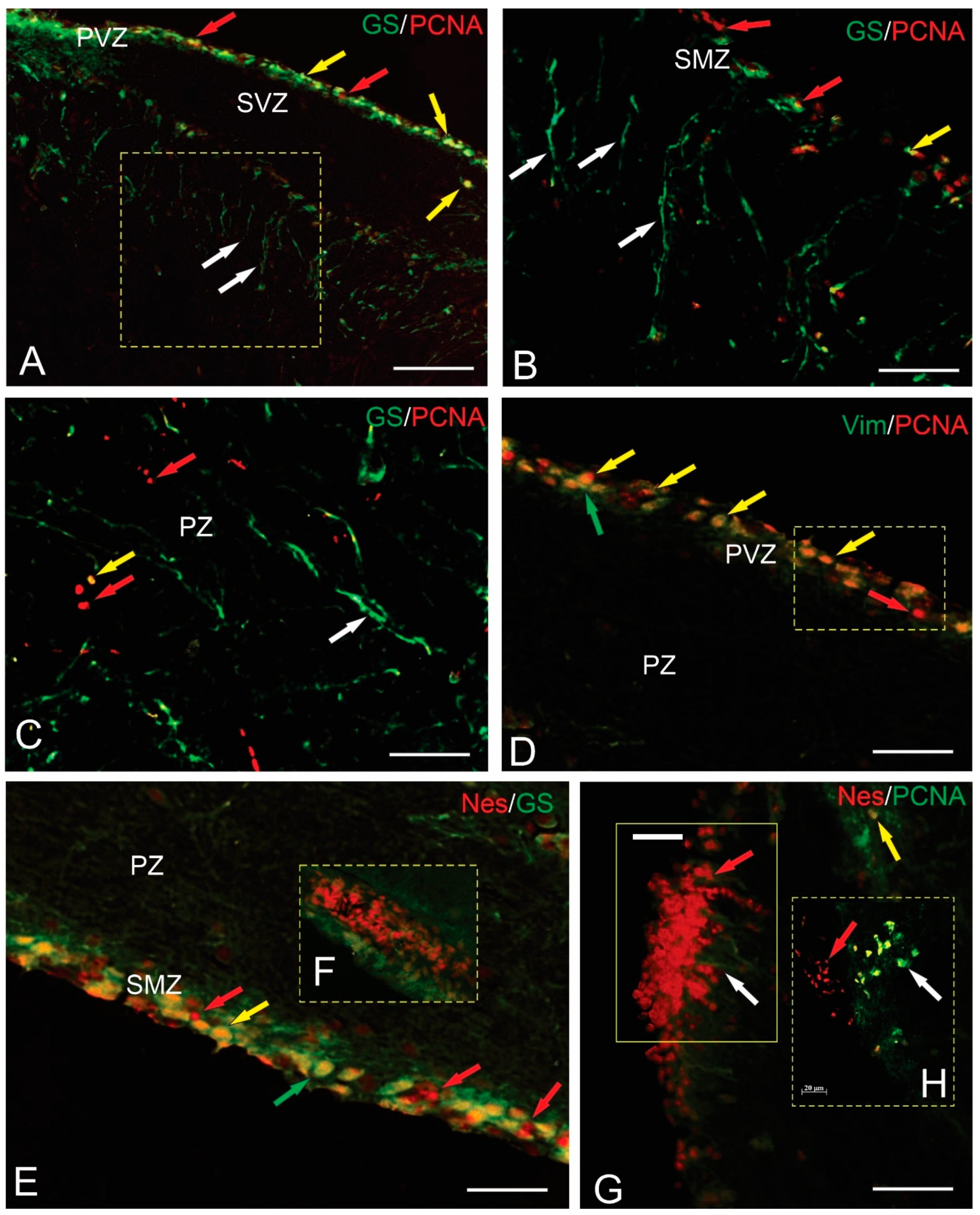
2.11. Double Immunolabeling in the Brainstem and Cerebellum of Trout
3. Discussion
3.1. Adult Cerebellar Neurogenesis and Expression of Molecular Markers by Various Types of aNSPCs
3.2. Adult Neurogenesis in the Brainstem
4. Material and Methods
4.1. Experimental Animals
4.2. Preparation of Material for Immunohistochemical Studies
4.2.1. Anesthesia and Prefixation
4.2.2. Control and Specificity of Antibodies
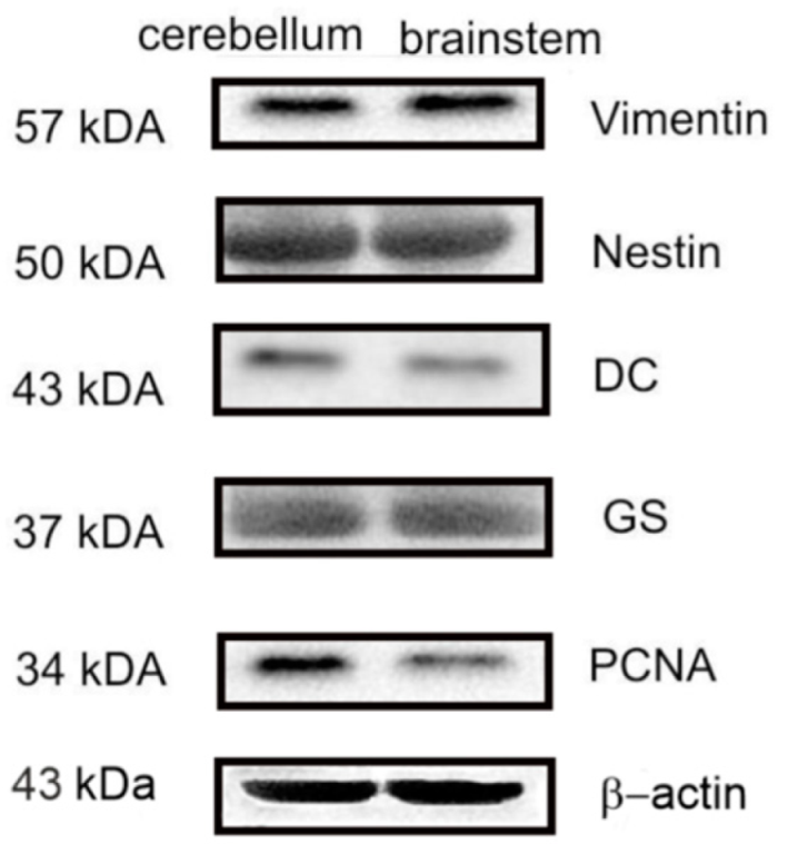
4.2.3. Western Immunoblotting
4.2.4. Immunohistochemistry
4.2.5. Double Immunofluorescent Labeling
4.2.6. Microscopy
4.2.7. Densitometry
4.2.8. Statistical Analysis
5. Conclusions
Supplementary Materials
Author Contributions
Funding
Institutional Review Board Statement
Informed Consent Statement
Data Availability Statement
Acknowledgments
Conflicts of Interest
Abbreviations
References
- Kriegstein, A.; Alvarez-Buylla, A. The glial nature of embryonic and adult neural stem cells. Annu. Rev. Neurosci. 2009, 32, 149–184. [Google Scholar] [CrossRef] [Scilit] [PubMed]
- Alunni, A.; Bally-Cui, L. A comparative view of regenerative neurogenesis in vertebrates. Development 2016, 143, 741–753. [Google Scholar] [CrossRef] [Scilit] [PubMed]
- Pushchina, E.V.; Varaksin, A.A.; Obukhov, D.K. Molecular markers of adult neurogenesis in the telencephalon and tectum of rainbow trout, Oncorhynchus mykiss. Int. J. Mol. Sci. 2022, 23, 1188. [Google Scholar] [CrossRef] [Scilit] [PubMed]
- Brandstätter, R.; Kotrschal, K. Brain growth patterns in four European cyprinid fish species (Cyprinidae, Teleostei): Roach (Rutilus rutilus), bream (Abramis brama), common carp (Cyprinus carpio) and sabre carp (Pelecus cultratus). Brain Behav. Evol. 1990, 35, 195–211. [Google Scholar] [CrossRef] [Scilit] [PubMed]
- Candal, E.; Anadón, R.; Bourrat, F.; Rodríguez-Moldes, I. Cell proliferation in the developing and adult hindbrain and midbrain of trout and medaka (teleosts): A segmental approach. Brain Res. Dev. Brain Res. 2005, 160, 157–175. [Google Scholar] [CrossRef] [Scilit] [PubMed]
- Sîrbulescu, R.F.; Ilieş, I.; Vitalo, A.G.; Trull, K.; Zhu, J.; Traniello, I.M.; Zupanc, G.K. Adult stem cells in the knifefish cerebellum. Dev. Neurobiol. 2015, 75, 39–65. [Google Scholar] [CrossRef] [Scilit] [PubMed]
- Ekström, P.; Johnsson, C.M.; Ohlin, L.M. Ventricular proliferation zones in the brain of an adult teleost fish and their relation to neuromeres and migration (secondary matrix) zones. J. Comp. Neurol. 2001, 436, 92–110. [Google Scholar] [CrossRef] [PubMed]
- Pushchina, E.V.; Kapustyanov, I.A.; Varaksin, A.A. Neural stem cells/neuronal precursor cells and postmitotic neuroblasts in constitutive neurogenesis and after, traumatic injury to the mesencephalic tegmentum of juvenile chum salmon, Oncorhynchus keta. Brain Sci. 2020, 10, 65. [Google Scholar] [CrossRef] [Scilit]
- Puschina, E.V.; Varaksin, A.A.; Obukhov, D.K. Reparative Neurogenesis in the Brain and Changes in the Optic Nerve of Adult Trout Oncorhynchus mykiss after Mechanical Damage of the Eye. Ontogenez 2016, 47, 15–39. (In Russian) [Google Scholar] [PubMed]
- Dray, N.; Bedu, S.; Vuillemin, N.; Alunni, A.; Coolen, M.; Krecsmarik, M. Large-scale live imaging of adult neural stem cells in their endogenous niche. Development 2015, 142, 3592–3600. [Google Scholar]
- Ganz, J.; Kroehne, V.; Freudenreich, D.; Machate, A.; Geffarth, M.; Braasch, I. Subdivisions of the adult zebrafish pallium based on molecular marker analysis. F1000Research 2015, 520, 633–655. [Google Scholar]
- Caron, A.; Trzuskot, L.; Lindsey, B.W. Uncovering the spectrum of adult zebrafish neural stem cell cycle regulators. Front. Cell Dev. Biol. 2022, 10, 941893. [Google Scholar] [CrossRef] [Scilit] [PubMed]
- Pushchina, E.V.; Stukaneva, M.E.; Varaksin, A.A. Hydrogen Sulfide Modulates Adult and Reparative Neurogenesis in the Cerebellum of Juvenile Masu Salmon, Oncorhynchus masou. Int. J. Mol. Sci. 2020, 21, 9638. [Google Scholar] [CrossRef] [Scilit] [PubMed]
- Pushchina, E.V.; Bykova, M.E.; Varaksin, A.A. Posttraumatic expression of Aromatase B, Glutamine Synthetase, and Cysta-thionine-β-Synthase in the Cerebellum of Juvenile Chum Salmon, Oncorhynchus keta. Int. J. Mol. Sci. 2024, 25, 3299. [Google Scholar] [CrossRef] [Scilit] [PubMed]
- Wan, J.; Zhao, X.-F.; Vojtek, A.; Goldman, D. Retinal injury, growth factors, and cytokines converge on β-catenin and pStat3 signaling to stimulate retina regeneration. Cell Rep. 2014, 9, 285–297. [Google Scholar] [CrossRef] [Scilit] [PubMed]
- Pereida-Jaramillo, E.; Gómez-González, G.B.; Espino-Saldaña, A.E.; Martínez-Torres, A. Calcium Signaling in the Cerebellar Radial Glia and Its Association with Morphological Changes during Zebrafish Development. Int. J. Mol. Sci. 2021, 22, 13509. [Google Scholar] [CrossRef] [Scilit] [PubMed] [PubMed Central]
- Mu, Y.; Bennett, D.V.; Rubinov, M.; Narayan, S.; Yang, C.T.; Tanimoto, M.; Mensh, B.D.; Looger, L.L.; Ahrens, M.B. Glia Accumulate Evidence that Actions Are Futile and Suppress Unsuccessful Behavior. Cell 2019, 178, 27–43.e19. [Google Scholar] [CrossRef] [Scilit] [PubMed]
- Labusch, M.; Mancini, L.; Morizet, D.; Bally-Cuif, L. Conserved and divergent features of adult neurogenesis in zebrafish. Front. Cell Dev. Biol. 2020, 8, 525. [Google Scholar] [CrossRef] [Scilit]
- Mazzitelli-Fuentes, L.S.; Román, F.R.; Castillo Elías, J.R.; Deleglise, E.B.; Mongiat, L.A. Spatial learning promotes adult neurogenesis in specific regions of the zebrafish pallium. Front. Cell Dev. Biol. 2022, 10, 840964. [Google Scholar] [CrossRef] [Scilit]
- Stukaneva, M.E.; Pushchina, E.V.; Varaksin, A.A. GFAP and PCNA marking in the cerebellum of masu salmon’s (Oncorhynchus masou) juvenile after mechanical injury. Rus. J. Devel. Biol. 2017, 48, 321–329. [Google Scholar] [CrossRef] [Scilit]
- Coolen, M.; Labusch, M.; Mannioui, A.; Bally-Cuif, L. Mosaic heterochrony in neural progenitors sustains accelerated brain growth and neurogenesis in the juvenile killifish N. furzeri. Curr. Biol. 2020, 30, 736–745. [Google Scholar] [CrossRef] [Scilit] [PubMed]
- Pushchina, E.V.; Zharikova, E.I.; Varaksin, A.A. Mechanical brain injury increases cells’ production of cystathionine β-synthase and glutamine synthetase, but reduces Pax2 expression in the telencephalon of juvenile chum salmon, Oncorhynchus keta. Int. J. Mol. Sci. 2021, 22, 1279. [Google Scholar] [CrossRef] [Scilit] [PubMed]
- Beamish, R. The need to see a bigger picture to understand the ups and downs of pacific salmon abundances. ICES J. Mar. Sci. 2022, 79, 1005–1014. [Google Scholar] [CrossRef] [Scilit]
- Lindsey, B.W.; Hall, Z.J.; Heuzé, A.; Joly, J.S.; Tropepe, V.; Kaslin, J. The role of neuro-epithelial-like and radial-glial stem and progenitor cells in development, plasticity, and repair. Prog. Neurobiol. 2018, 170, 99–114. [Google Scholar] [CrossRef] [Scilit]
- Alexandrou, M.A.; Swartz, B.A.; Matzke, N.J.; Oakley, T.H. Genome duplication and multiple evolutionary origins of complex migratory behavior in Salmonidae. Mol. Phylogenetics Evol. 2013, 69, 514–523. [Google Scholar] [CrossRef] [Scilit] [PubMed]
- Berthelot, C.; Brunet, F.; Chalopin, D.; Juanchich, A.; Bernard, M.; Noël, B.; Bento, P.; Da Silva, C.; Labadie, K.; Alberti, A.; et al. The rainbow trout genome provides novel insights into evolution after whole-genome duplication in vertebrates. Nat. Commun. 2014, 5, 3657. [Google Scholar] [CrossRef] [Scilit] [PubMed]
- Sémon, M.; Wolfe, K.H. Consequences of genome duplication. Curr. Opin. Genet. Dev. 2007, 17, 505–512. [Google Scholar] [CrossRef] [Scilit] [PubMed]
- Ramachandran, R.; Zhao, X.-F.; Goldman, D. Asc1a/Dkk/β-catenin signaling pathway is necessary and glycogen synthase kinase-3B inhibition is sufficient for zebrafish retina regeneration. Proc. Natl. Acad. Sci. USA 2011, 108, 15858–15863. [Google Scholar] [CrossRef] [Scilit] [PubMed]
- Wu, C.-C.; Tsai, T.-H.; Chang, C.; Lee, T.-T.; Lin, C.; Cheng, I.H.-J.; Sun, M.-C.; Chuang, Y.-J.; Chen, B.-S. On the crucial cerebellar wound healing-related pathways and their cross-talks after traumatic brain injury in Danio rerio. PLoS ONE 2014, 9, e97902. [Google Scholar] [CrossRef] [Scilit]
- Kaslin, J.; Kroehne, V.; Ganz, J.; Hans, S.; Bran, M. Distinct roles of neuroepithelial-like and radial glial-like progenitor cells in cerebellar regeneration. Development 2017, 144, 1462–1471. [Google Scholar] [CrossRef] [Scilit]
- Brulet, R.; Matsuda, T.; Zhang, L.; Miranda, C.; Giacca, M.; Kaspar, B.K.; Nakashima, K.; Hsieh, J. NEUROD1 instructs neuronal conversion in non-reactive astrocytes. Stem Cell Rep. 2017, 8, 1506–1515. [Google Scholar] [CrossRef] [Scilit] [PubMed]
- Gascón, S.; Masserdotti, G.; Russo, G.L.; Götz, M. Direct neuronal reprogramming: Achievements, hurdles, and new roads to success. Cell Stem Cell 2017, 21, 18–34. [Google Scholar] [CrossRef] [Scilit] [PubMed]
- Carlén, M.; Meletis, K.; Göritz, C.; Darsalia, V.; Evergren, E.; Tanigaki, K.; Amandola, M.; Barnabé-Heider, F.; Yeung, M.S.; Naldini, L.; et al. Forebrain ependymal cells are Notch-dependent and generate neuroblasts and astrocytes after stroke. Nat. Neurosci. 2009, 12, 259–267. [Google Scholar] [CrossRef] [Scilit] [PubMed]
- Li, H.; Chen, G. In vivo reprogramming for CNS repair: Regenerating neurons from endogenous glial cells. Neuron 2009, 91, 728–738. [Google Scholar] [CrossRef] [Scilit]
- Zupanc, G.K. Adult neurogenesis and neuronal regeneration in the brain of teleost fish. J. Physiol.-Paris 2008, 102, 357–373. [Google Scholar] [CrossRef] [Scilit] [PubMed]
- Pushchina, E.V.; Shukla, S.; Varaksin, A.A.; Obukhov, D.K. Cell proliferation and apoptosis in optic nerve and brain integration centers of adult trout Oncorhynchus mykiss after optic nerve injury. Neural Regen. Res. 2016, 11, 578–590. [Google Scholar] [CrossRef] [Scilit] [PubMed]
- Grosche, J.; Härtig, W.; Reichenbach, A. Expression of glial fibrillary acidic protein (GFAP), glutamine synthetase (GS), and Bcl-2 protooncogene protein by Müller (glial)cells in retinal light damage of rats. Neurosci. Lett. 1995, 185, 119–122. [Google Scholar] [CrossRef] [Scilit]
- Zupanc, G.K.; Sîrbulescu, R.F. Teleost fish as a model system to study successful regeneration of the central nervous system. Curr. Top. Microbiol. 2013, 367, 193–223. [Google Scholar]
- Sánchez-Farías, N.; Candal, E. Identification of radial glia progenitors in the developing and adult retina of sharks. Front. Neuroanat. 2016, 10, 65. [Google Scholar] [CrossRef] [Scilit]
- Schaar, B.T.; Kinoshita, K.; Mcconnell, S.K. Doublecortin microtubule affinity is regulated by a balance of kinase and phosphatase activity at the leading edge of migrating neurons. Neuron 2004, 41, 203–213. [Google Scholar] [CrossRef] [Scilit]
- Ambrogini, P.; Lattanzi, D.; Ciuffoli, S.; Agostini, D.; Bertini, L.; Stocchi, V.; Santi, S.; Cuppini, R. Morphofunctional characterization of neuronal cells at different stages of maturation in granule cell layer of adult rat dentate gyrus. Brain Res. 2004, 1017, 21–31. [Google Scholar] [CrossRef] [Scilit] [PubMed]
- Brandt, M.D.; Jessberger, S.; Steiner, B.; Kronenberg, G.; Reuter, K.; Bick-Sander, A.; Von deer Behrens, W.; Kempermann, G. Transient calretinin-expression defines early postmitotic step of neuronal differentiation in adult hippocampus neurogenesis of mice. Mol. Cell. Neurosci. 2003, 24, 603–613. [Google Scholar] [CrossRef] [Scilit] [PubMed]
- Dahl, D.; Rueger, D.C.; Bignami, A.; Weber, K.; Osbom, M. Vimentin, the 57000 molecular weight protein of fibroblast filaments, is the major cytoskeletal component in immature glia. Eur. J. Cell Biol. 1981, 24, 191–196. [Google Scholar] [PubMed]
- Arochena, M.; Anadon, R.; Diaz Regueira, S.M. Development of vimentin and glial fibrillary acidic protein immunoreactivities in the brain of gray mullet (Chelon labrosus), an advanced teleost. J. Comp. Neurol. 2004, 46, 413–436. [Google Scholar] [CrossRef] [Scilit] [PubMed]
- Kalman, M.; Ari, C. Distribution of GFAP immunoreactive structures in the rhombencephalon of the sterlet (Acipenser ruthenus) and its evolutionary implication. J. Exp. Zool. 2002, 293, 395–406. [Google Scholar] [CrossRef] [Scilit] [PubMed]
- Pecchi, E.; Dallaporta, M.; Charrier, C.; Pio, J.; Jean, A.; Moyse, E.; Troadec, J.D. Glial fibrillary acidic protein (GFAP)-positive radial-like cells are present in the vicinity of proliferative progenitors in the nucleus tractus solitarius of adult rat. J. Comp. Neurol. 2007, 501, 353–368. [Google Scholar] [CrossRef] [Scilit] [PubMed]
- Chen, H.L.; Yuh, C.H.; Wu, K.K. Nestin is essential for zebrafish brain and eye development through control of progenitor cell apoptosis. PLoS ONE 2010, 5, e9318. [Google Scholar] [CrossRef] [Scilit] [PubMed]
- Mahler, J.; Driever, W. Expression of the zebrafish intermediate neurofilament nestin in the developing nervous system and in neural proliferation zones at postembryonic stages. BMC Dev. Biol. 2007, 7, 89. [Google Scholar] [CrossRef] [Scilit] [PubMed]
- Carmona, I.C.; Luesma Bartolomé, M.J.; Lavoie-Gagnon, C.; Junquera, C. Distribution of nestin protein: Immunohistochemical study in enteric plexus of rat duodenum. Microsc. Res. Tech. 2011, 74, 148–152. [Google Scholar] [CrossRef] [Scilit] [PubMed]
- Kaslin, J.; Ganz, J.; Geffarth, M.; Grandel, H.; Hans, S.; Brand, M. Stem cells in the adult zebrafish cerebellum: Initiation and maintenance of a novel stem cell niche. J. Neurosci. 2009, 29, 6142–6153. [Google Scholar] [CrossRef] [Scilit]
- Grandel, H.; Brand, M. Comparative aspects of adult neural stem cell activity in vertebrates. Dev. Genes Evol. 2013, 22, 131–147. [Google Scholar] [CrossRef] [Scilit] [PubMed]
- Tozzini, E.T.; Baumgart, M.; Battistoni, G.; Cellerino, A. Adult neurogenesis in the short-lived teleost Nothobranchius furzeri: Localization of neurogenic niches, molecular characterization and effects of aging. Aging Cell 2012, 11, 241–251. [Google Scholar] [CrossRef] [Scilit] [PubMed]
- Ben Abdallah, N.M.; Slomianka, B.L.; Vyssotski, A.L.; Lipp, H.P. Early age-related changes in adult hippocampal neurogenesis in C57 mice. Neurobiol. Aging 2010, 31, 151–161. [Google Scholar] [CrossRef] [Scilit] [PubMed]
- Ceriani, R.; Whitlock, K. Gonadotropin releasing hormone (Gnrh) triggers neurogenesis in the hypothalamus of adult zebrafish. Int. J. Mol. Sci. 2021, 22, 5926. [Google Scholar] [CrossRef] [Scilit] [PubMed]
- Lindsey, B.W.; Tropepe, V. Changes in the social environment induce neurogenic plasticity predominantly in niches structures of the zebrafish brain independently residing in sensory of cortisol levels: Social context regulates adult neurogenesis. Dev. Neurobiol. 2014, 74, 1053–1077. [Google Scholar] [CrossRef] [Scilit] [PubMed]
- Dunlap, K.D.; Teles, M.C.; Oliveira, R.F. Social stimuli increase activity of adult-born cells in the telencephalon of zebrafish (Danio rerio). J. Exp. Biol. 2021, 224, jeb242253. [Google Scholar] [CrossRef] [Scilit] [PubMed]
- Benítez-Santana, T.; Simion, M.; Corraze, G.; Médale, F.; Joly, J.S. Effect of nutrient availability on progenitor cells in zebrafish (Danio rerio). Dev. Neurobiol. 2016, 77, 26–38. [Google Scholar] [CrossRef] [Scilit] [PubMed]
- Pushchina, E.V.; Zharikova, E.I.; Varaksin, A.A. Expression of Doublecortin, Glial Fibrillar Acidic Protein, and Vimentin in the Intact Subpallium and after Traumatic Injury to the Pallium in Juvenile Salmon, Oncorhynchus masou. Int. J. Mol. Sci. 2022, 23, 1334. [Google Scholar] [CrossRef] [Scilit]
- Grandel, H.; Kaslin, J.; Ganz, J.; Wenzel, I.; Brand, M. Neural stem cells and neurogenesis in the adult zebrafish brain: Origin, proliferation dynamics, migration and cell fate. Dev. Biol. 2006, 295, 263–277. [Google Scholar] [CrossRef] [Scilit]
- Mack, A.F.; DeOliveira-Mello, L.; Mattheus, U.; Neckel, P.H. Organization of radial glia reveals growth pattern in the telencephalon of a percomorph fish Astatotilapia burtoni. J. Comp. Neurol. 2021, 529, 2813–2823. [Google Scholar] [CrossRef] [Scilit]
- Baumgart, E.V.; Barbosa, J.S.; Bally-Cuif, L.; Götz, M.; Ninkovic, J. Stab wound injury of the zebrafish telencephalon: A model for comparative analysis of reactive gliosis. Glia 2012, 60, 343–357. [Google Scholar] [CrossRef] [Scilit] [PubMed]
- Kaslin, J.; Kroehne, V.; Benato, F.; Argenton, F.; Brand, M. Development and specification of cerebellar stem and progenitor cells in zebrafish: From embryo to adult. Neural. Dev. 2013, 8, 9. [Google Scholar] [CrossRef] [Scilit]
- Costagli, A.; Kapsimali, M.; Wilson, S.W.; Mione, M. Conserved and divergent patterns of Reelin expression in the zebrafish central nervous system. J. Comp. Neurol. 2002, 450, 73–93. [Google Scholar] [CrossRef] [Scilit]
- Ganz, J.; Brand, M. Adult neurogenesis in fish. Cold Spring Harb. Perspect. Biol. 2016, 8, a019018. [Google Scholar] [CrossRef] [Scilit]
- Louvi, A.; Alexandr, P.; Meti, C.; Wurst, W.; Wassef, M. The isthmic neuroepithelium is essential for cerebellar midline fusion. Development 2003, 130, 5319–5330. [Google Scholar] [CrossRef] [Scilit] [PubMed]
- Sgaier, S.K.; Millet, S.; Villanueva, M.P.; Berenshteyn, F.; Song, C.; Joyner, A.L. Morphogenetic and cellular movements that shape the mouse cerebellum; insights from genetic fate mapping. Neuron 2005, 45, 27–40. [Google Scholar]
- Chaplin, N.; Tendeng, C.; Wingate, R.J. Absence of an external germinal layer in zebrafish and shark reveals a distinct, anamniote ground plan of cerebellum development. J. Neurosci. 2010, 30, 3048–3057. [Google Scholar] [CrossRef] [Scilit]
- Reimer, M.M.; Sorensen, I.; Kuscha, V.; Frank, R.E.; Liu, C.; Becker, C.G.; Becker, T. Motor neuron regeneration in adult zebrafish. J. Neurosci. 2008, 28, 8510–8516. [Google Scholar] [CrossRef] [Scilit]
- Blažek, R.; Polacik, M.; Reichard, M. Rapid growth, early maturation and short generation time in African annual fishes. EvoDevo 2013, 4, 24. [Google Scholar] [CrossRef] [Scilit]
- Zupanc, G.K.; Hinsch, K.; Gage, F.H. Proliferation, migration, neuronal differentiation, and long-term survival of new cells in the adult zebrafish brain. J. Comp. Neurol. 2005, 488, 290–319. [Google Scholar] [CrossRef] [Scilit]
- von Krogh, K.; Sørensen, C.; Nilsson, G.E.; Øverli, Ø. Forebrain cell proliferation, behavior, and physiology of zebrafish, Danio rerio, kept in enriched or barren environments. Physiol. Behav. 2010, 101, 32–39. [Google Scholar] [CrossRef] [Scilit] [PubMed]
- Venables, M.J.; Navarro-Martína, L.; Basak, A.; Bauma, B.R.; Zhangc, D.; Trudeau, V.L. Characterization of multiple nestin isoforms in the goldfish brain. Comp. Biochem. Physiol. Part D 2016, 19, 8–17. [Google Scholar] [CrossRef] [Scilit] [PubMed]
- Pushchina, E.V.; Varaksin, A.A.; Obukhov, D.K.; Prudnikov, I.M. GFAP expression in the optic nerve and increased H2S generation in the integration centers of the rainbow trout (Oncorhynchus mykiss) brain after unilateral eye injury. Neural Regen. Res. 2020, 15, 1867–1886. [Google Scholar] [CrossRef] [Scilit] [PubMed]





| Brain Area | Type of Cell, Brain Localization | Cell Size | Intensity of Immunolabeling |
|---|---|---|---|
| Cerebellum | |||
| Dorso-lateral part rostral area | Undifferentiated (PVZ) Elongated (PZ, ML) | 3.9 ± 0.4/2.8 ± 0.6 5.7 ± 0.6/4.3 ± 0.7 | +++ +++ |
| Dorsal matrix zone (DMZ) | Undifferentiated (PZ, ML) Elongated (PZ, ML) | 4.3 ± 0.4/3.0 ± 0.8 5.4 ± 0.3/3.2 ± 0.6 | +++/++ +++ |
| Lateral part | Undifferentiated (PVZ) Elongated (PZ, ML) | 4.0 ± 0.4/2.9 ± 0.5 5.6 ± 0.5/4.2 ± 0.5 | +++ +++ |
| Basal part | Undifferentiated (PZ, ML) Elongated (PZ, ML) | 4.1 ± 0.2/2.7 ± 0.4 5.6 ± 0.5/4.2 ± 0.5 | +++ +++ |
| Periventricular zone (PVZ) | Undifferentiated (PVZ) Elongated (ML, GL) Oval (GrL) | 4.4 ± 0.5/3.7 ± 0.3 5.8 ± 0.4/3.8 ± 0.8 7.7 ± 1.8/4.2 ± 0.5 | +++ +++/++ +++/++ |
| Granular eminences (GrEms) | Undifferentiated (SMZ) Elongated (GrEm) Oval (GrEm) | 5.2 ± 0.6/3.6 ± 0.8 6.7 ± 0.4/4.8 ± 0.7 10.1 ± 0.7/6.6 ± 0.5 | +++ +++/++ +++/++ |
| Brainstem | |||
| Periventricular zone (PVZ) | Undifferentiated Elongated 1 Radial glia 1 Radial glia 2 | 5.8 ± 0.6/5.4 ± 0.7 7.6 ± 0.6/5.2 ± 0.7 9.4 ± 0.7/5.4 ± 0.9 11.6 ± 0.9/6.6 ± 0.9 | +++ +++/++ +++/++ +++/++ |
| Interfascicular zone (IFZ) | Undifferentiated Elongated 1 | 6.1 ± 0.8/3.5 ± 0.7 7.7 ± 0.8/3.9 ± 1.0 | +++/++ +++/++ |
| Submarginal zone (SMZ) | Undifferentiated Elongated 1 (PZ) Radial glia 1 | 5.3 ± 0.7/3.3 ± 0.8 6.8 ± 0.3/4.1 ± 0.7 8.8 ± 0.2/5.2 ± 0.9 | +++ +++ +++/++ |
| Brain Area | Type of Cell | Cell Size, µm | Intensity of Labeling |
|---|---|---|---|
| Cerebellum | |||
| Dorso-lateral part | Undifferentiated Elongated 1 Elongated 2 Elongated 3 | 5.9 ± 0.6/3.7 ± 0.6 7.9 ± 0.3/5.3 ± 0.5 9.7 ± 0.7/5.4 ± 1.2 13.2 ± 1.3/7.6 ± 0.3 | +++ +++ +++ +++/++ |
| Dorsal matrix zone (DMZ) | Undifferentiated Elongated | 4.5 ± 0.5/3.2 ± 0.6 5.5 ± 0.4/3.2 ± 0.5 | +++ +++ |
| Basal part | Undifferentiated Elongated 1 Elongated 2 Elongated 3 | 6.5 ± 0.2/5.2 ± 0.4 8.4 ± 0.3/6.2 ± 0.6 9.6 ± 0.4/6.9 ± 1.4 12.6 ± 0.5/7.4 ± 2.4 | +++ +++ +++/++ +++/++ |
| Baso-lateral part | Undifferentiated Elongated 1 Elongated 2 Elongated 3 | 6.2 ± 0.8/5.7 ± 1.0 7.6 ± 0.1/4.9 ± 0.3 9.7 ± 0.2/6.3 ± 1.5 11.8 ± 0.6/6.9 ± 1.9 | +++ +++ +++/++ +++/++ |
| Periventricular zone (PVZ) | Undifferentiated Elongated 1 Elongated 2 Elongated 3 | 4.5 ± 0.5/3.7 ± 0.4 7.0 ± 0.5/4.4 ± 0.5 8.4 ± 0.5/4.6 ± 0.6 11.8 ± 0.6/6.4 ± 1.0 | +++ +++ +++/++ +++/++ |
| Brainstem | |||
| Periventricular zone (PVZ) | Undifferentiated Elongated 1 Radial glia 1 Radial glia 2 | 6.2 ± 0.4/5.2 ± 1.1 7.9 ± 0.5/5.3 ± 0.6 9.6 ± 0.6/5.2 ± 1.3 13.7 ± 0.7/6.3 ± 1.1 | +++/++ +++ +++/++ +++/++ |
| Interfascicular zone (IFZ) | Undifferentiated Elongated 1 | 6.1 ± 0.8/3.5 ± 0.7 7.7 ± 0.8/3.9 ± 1.0 | +++/++ +++/++ |
| Submarginal zone (SMZ) | Undifferentiated Elongated 1 Radial glia 1 | 5.3 ± 0.7/3.3 ± 0.8 6.8 ± 0.3/4.1 ± 0.7 8.8 ± 0.2/5.2 ± 0.9 | +++ +++ +++/++ |
| Brain Area | Type of Cell | Cell Size, µm | Intensity of Labeling |
|---|---|---|---|
| Cerebellum | |||
| Basal zone (BZ) | Elongated (ML, GL) Oval (GL) Eurydendroid (GL) Pear-shaped (GL) | 11.3 ± 1.6/8.7 ± 1.9 16.2 ± 1.0/10.8 ± 1.7 20.1 ± 1.2/12.4 ± 2.3 24.6 ± 1.6/13.4 ± 1.7 | +++ +++ +++/++ +++/++ |
| Dorsal zone (DZ) | Undifferentiated (ML, GL) Elongated 1 (ML, GL) Elongated 2 (ML) Oval (ML) Pear-shaped (GL) | 4.9 ± 0.6/4.0 ± 0.4 7.1 ± 0.7/5.2 ± 0.5 8.9 ± 0.9/6.3 ± 1.3 13.9 ± 0.9/8.3 ± 1.9 22.2 ± 3.8/11.8 ± 5.7 | +++ +++ +++/++ +++/++ +++/++ |
| Dorsal matrix zone (DMZ) | Undifferentiated (ML, GL) Elongated (ML, GL) | 4.2 ± 0.4/3.1 ± 0.55 8 ± 0.8/3.8 ± 0.8 | +++ +++ |
| Lateral zone (LZ) | Undifferentiated (ML) Elongated 1 (ML) Oval (ML, GL) Pear-shaped 1 (GL) Pear-shaped 2 (GL) | 4.2 ± 0.5/3.0 ± 0.8 8.9 ± 1.2/6.5 ± 2.0 15.4 ± 2.2/11.7 ± 1.2 23.6 ± 1.4/18.7 ± 3.4 26.9 ± 1.2/15.5 ± 2.4 | +++ +++/++ +++/++ +++/++ +++/++ |
| Granular layer (Grl) | Oval (GrL) Polygonal 1 (GrL) Polygonal 2 (GrL) | 13.2 ± 0.8/8.8 ± 0.8 17.8 ± 2.5/11.0 ± 2.6 23.9 ± 2.3/12.7 ± 2.6 | ++ +++/++ +++/++ |
| Granular eminence (GrEm) | Elongated (GrEm) Oval 1 (GrEm) Oval 2 (GrEm) Polygonal 1 (GrEm) Polygonal 2 (GrEm) | 5.5 ± 0.7/4.2 ± 0.5 10.3 ± 0.2/6.3 ± 0.3 13.3 ± 0.7/8.6 ± 1.7 15.0 ± 0.4/9.0 ± 1.0 18.7 ± 1.0/11.7 ± 1.4 | +++/++ ++ +++/++ +++/++ +++/++ |
| Brainstem | |||
| Nucleus nervus vagus (NX) | Oval (SVZ) Polygonal 1 (SVZ) Polygonal 2 (SVZ) Polygonal 3 (PZ) | 10.7 ± 0.4/8.9 ± 0.5 16.0 ± 0.7/9.2 ± 0.8 22.4 ± 2.7/9.6 ± 1.8 28.9 ± 3.4/11.9 ± 2.7 | +++/++ +++/++ +++/++ ++ |
| Nucleus nervus glossopharyngeus (NIX) | Polygonal 1 (SVZ) Polygonal 2 (SVZ) Polygonal 3 (PZ) | 19.7 ± 0.9/11.8 ± 0.7 28.5 ± 1.7/16.6 ± 1.4 31.0 ± 2.6/11.6 ± 1.8 | ++ ++ ++ |
| Interfascicular zone (IFZ) | Oval (IFZ) Polygonal 1 (IFZ) Polygonal 2 (IFZ) Polygonal 3 (IFZ) Polygonal 4 (IFZ) | 8.4 ± 0.4/7.2 ± 0.6 14.9 ± 1.8/7.4 ± 3.3 19.5 ± 0.6/12.7 ± 0.4 25.7 ± 1.3/12.8 ± 1.8 31.5 ± 0.7/18.6 ± 4.5 | ++ ++ +++/++ +++/++ +++/++ |
| Nucleus nervus facialis (NVII) | Polygonal 1 (PZ) Polygonal 2 (PZ) Polygonal 3 (SVZ, PZ) Polygonal 4 (SVZ) Polygonal 5 (PZ) Polygonal 6 (PZ) Polygonal 7 (PZ) | 19.7 ± 0.8/11.8 ± 0.7 29.4 ± 1.8/13.4 ± 2.2 40.7 ± 2.3/17.2 ± 3.6 48.2 ± 2.3/27.8 ± 8.0 69.5 ± 4.6/23.7 ± 7.3 80.7 ± 3.7/23.6 ± 2.6 95.4 ± 1.2/20.4 ± 4.5 | +++/++ +++/++ +++ ++/+ ++/+ +++/++ ++ |
| Dorsal reticular formation (DRF) | Undifferentiated (PZ) Elongated 1 (PZ) Elongated 2 (PZ) Oval 1 (PZ) Oval 2 (PZ) Polygonal (PZ) | 4.2 ± 0.3/3.2 ± 1.4 6.9 ± 0.6/4.5 ± 0.7 8.6 ± 0.5/5.3 ± 0.3 11.0 ± 0.9/8.0 ± 1.0 13.7 ± 0.4/10.5 ± 1.4 21.4 ± 1.2/11.3 ± 2.3 | +++ +++ ++/+ ++ +++/++ +++/++ |
| Periventricular layer (PVl) reticulospinal cells (RSCs) | Undifferentiated (PVZ) Elongated (PVZ) Elongated (PVZ, SVZ) Elongated 2 (PZ) Oval (PZ) Polygonal 1 (SVZ, PZ) Polygonal 2 (SVZ) Polygonal 3 (PZ) Polygonal 4 (PZ) Polygonal 5 (PZ) Polygonal 6 (PZ) Polygonal 7 (PZ) | 4.4 ± 0.7/3.2 ± 0.5 6.8 ± 0.6/4.5 ± 0.8 8.9 ± 0.6/5.2 ± 0.4 8.4 ± 0.4/7.3 ± 0.6 12.0 ± 1.7/7.5 ± 2.3 15.0 ± 0.3/11.2 ± 0.6 18.3 ± 1.3/11.5 ± 1.9 23.0 ± 1.4/12.2 ± 1.7 27.4 ± 1.3/13.4 ± 4.9 31.2 ± 0.8/17.4 ± 3.6 35.0 ± 1.7/17.4 ± 8.7 41.3 ± 2.5/16.3 ± 2.3 | +++ +++ ++/+ +++ +++ +++/++ +++/++ +++/++ ++/+ +++/++ +++/++ +++/++ |
| Ventro-lateral cells (VLCs) | Undifferentiated (PVZ) Elongated 1 (PVZ) Elongated 2 (PVZ, SVZ) Oval (PZ) Polygonal 1 (SVZ, PZ) Polygonal 2 (SVZ) | 4.3 ± 0.5/2.7 ± 0.3 7.8 ± 0.9/5.1 ± 0.9 9.9 ± 0.5/6.5 ± 1.5 12.6 ± 0.4/7.5 ± 2.3 15.3 ± 1.0/7.2 ± 1.3 18.9 ± 1.8/11.5 ± 1.6 | +++ +++ ++ +++ ++/+ +++/++ |
| Brain Area | Type of Cell | Cell Size, µm | Intensity of Labeling |
|---|---|---|---|
| Cerebellum | |||
| Dorso-lateral part of rostral area | Undifferentiated (PVZ) Elongated (PZ, ML) Granules (PZ, ML) | 3.9 ± 0.4/2.8 ± 0.6 10.9 ± 0.4/7.5 ± 0.4 2.4 ± 0.4/2.4 ± 0.3 | +++ +++ +++ |
| Dorsal part of caudal area | Elongated 1 (ML) Elongated 2 (ML) Pear-shaped 1 (GL) Pear-shaped 2 (GL) | 7.4 ± 0.2/4.9 ± 0.4 9.5 ± 0.3/6.5 ± 0.6 11.6 ± 0.4/9.3 ± 1.4 13.8 ± 0.5/11.1 ± 2.4 | +++ +++ +++/++ +++/++ |
| Dorsal matrix zone DMZ | Undifferentiated (PZ, ML) Elongated (PZ, ML) | 4.3 ± 0.4/3.0 ± 0.8 5.4 ± 0.3/3.2 ± 0.6 | +++ +++ |
| Lateral part of CC | Undifferentiated (PZ, ML) Granules (PZ, ML) | 4.1 ± 0.2/2.7 ± 0.4 2.3 ± 0.4/1.8 ± 0.1 | +++ +++ |
| Granular layer (Grl) | Elongated 1 (GrL) Elongated 2 (GrL) Oval (GrL) | 6.2 ± 0.8/5.2 ± 1.3 8.0 ± 0.7/6.0 ± 0.5 9.6 ± 0.5/6.9 ± 0.6 | +++ +++ +++ |
| Periventricular zone (PVZ) | Granules (PVZ) | 2.4 ± 0.3/2.2 ± 0.3 | +++ |
| Granular eminence (GrEm) | Undifferentiated (SMZ) Elongated (GrEm) Oval (GrEm) Polygonal 1 (GrEm) Polygonal 2 (GrEm) | 5.2 ± 0.6/3.6 ± 0.8 6.7 ± 0.4/4.8 ± 0.7 10.3 ± 0.8/6.8 ± 0.6 12.2 ± 1.1/8.4 ± 3.5 16.1 ± 0.4/11.4 ± 1.7 | +++ +++/++ +++/++ +++/++ +++/++ |
| Brainstem | |||
| Periventricular zone (PVZ) | Undifferentiated (PVZ) Elongated 1 (PVZ) Elongated 2 (PVZ) Radial glia 1 (PVZ) Radial glia 2 (PVZ) | 5.2 ± 0.4/3.3 ± 0.5 6.5 ± 0.3/3.7 ± 0.7 7.5 ± 0.3/4.0 ± 0.6 8.7 ± 0.5/5.9 ± 1.2 10.6 ± 0.7/4.5 ± 0.3 | +++ +++ +++/++ +++/++ +++/++ |
| Principal and lateral nucleus of nervi trigeminalis (NV) | Polygonal 1 (PZ) Polygonal 2 (PZ) Polygonal 3 (SVZ) Polygonal 4 (PZ) Polygonal 5 (PZ) | 19.3 ± 1.4/18.9 ± 1.6 27.0 ± 2.0/16.7 ± 4.6 40.6 ± 2.0/24.4 ± 7.5 46.3 ± 0.9/24.2 ± 10.0 54.3 ± 1.5/30.4 ± 4.0 | +++/++ +++/++ +++/++ +++/++ +++/++ |
| Nucleus nervus facialis (NVII) | Polygonal 1 (PZ) Polygonal 2 (PZ) Polygonal 3 (SVZ) Polygonal 4 (PZ) Polygonal 5 (PZ) Polygonal 4 (PZ) Polygonal 5 (PZ) | 24.4 ± 2.1/14.8 ± 3.3 28.7 ± 1.5/19.2 ± 4.4 35.3 ± 2.8/20.9 ± 6.7 42.2 ± 0.7/15.6 ± 3.0 47.8 ± 1.5/23.4 ± 1.8 52.9 ± 1.9/22.8 ± 5.8 74.5 ± 3.5/19.5 ± 4.0 | +++/++ +++/++ +++/++ +++/++ +++/++ +++/++ +++/++ |
| Nucleus nervus vagus (NX) | Oval (SVZ) Polygonal 1 (SVZ) Polygonal 2 (SVZ) Polygonal 3 (PZ) Polygonal 4 (PZ) | 11.4 ± 0.3/7.8 ± 0.5 16.0 ± 0.9/9.5 ± 0.7 21.5 ± 3.4/10.2 ± 2.5 30.2 ± 4.2/12.7 ± 2.7 41.9 ± 5.2/16.5 ± 3.2 | ++ ++ +++/++ +++ +++ |
| Lateral reticular formation (LRF) | Polygonal 1 (PZ) Polygonal 2 (PZ) Polygonal 3 (PZ) Polygonal 4 (PZ) Polygonal 5 (PZ) | 14.7 ± 0.4/7.7 ± 0.6 20.5 ± 0.5/14.3 ± 3.4 27.4 ± 1.1/17.0 ± 3.1 31.1 ± 2.9/16.8 ± 2.1 56.1 ± 6.6/32.0 ± 8.1 | +++/++ +++/++ +++/++ +++/++ +++/++ |
| Medial reticular formation (MRF) | Polygonal 3 (SVZ) Polygonal 4 (PZ) Bipolar (PZ) | 28.3 ± 2.1/14.6 ± 2.4 35.3 ± 3.1/17.4 ± 2.8 46.4 ± 5.2/19.3 ± 7.2 | +++/++ +++/++ +++/++ |
| Reticulospinal cells (RSCs) | Polygonal 2 (IFZ) Polygonal 3 (IFZ) Polygonal 4 (IFZ) Polygonal 5 (IFZ) | 31.7 ± 3.9/24.0 ± 1.5 38.6 ± 1.3/23.3 ± 8.6 45.6 ± 2.2/30.8 ± 0.9 56.2 ± 3.6/27.6 ± 5.1 | +++/++ +++/++ +++/++ +++/++ |
| Submarginal zone (SMZ) | Undifferentiated (PVZ) Undifferentiated (PVZ) Elongated (PVZ) | 3.4 ± 0.3/2.3 ± 0.4 5.2 ± 0.6/3.4 ± 0.7 6.8 ± 0.6/3.6 ± 0.4 | +++ +++ +++/++ |
| Brain Area | Type of Cell, Brain Localization | Cell Size, µm | Intensity of Labeling |
|---|---|---|---|
| Cerebellum | |||
| Dorsal zone of molecular layer (DZ) | Undifferentiated (ML, GL) Elongated 1 (ML, GL) Elongated 2 (ML) Oval (ML) | 4.2 ± 0.4/3.1 ± 0.5 5.7 ± 0.6/4.3 ± 0.7 8.0 ± 0.5/5.2 ± 1.6 11.4 ± 0.8/6.4 ± 1.5 | +++ +++ +++/++ +++/++ |
| Dorsal zone of Purkinje cells (PCs) | Pear-shaped (GL) Pear-shaped (GL) Pear-shaped (GL) Pear-shaped (GL) Pear-shaped (GL) | 18.2 ± 1.2/11.6 ± 1.6 23.0 ± 1.8/14.6 ± 1.4 27.6 ± 1.9/16.4 ± 3.4 23.6 ± 1.4/18.7 ± 3.4 31.5 ± 1.8/11.6 ± 2.4 | +++/++ +++/++ +++/++ +++/++ +++/++ |
| Dorsal matrix zone (DMZ) | Undifferentiated (ML, GL) Elongated (ML, GL) | 4.1 ± 0.6/2.5 ± 0.5 5.7 ± 0.5/3.2 ± 0.4 | +++ +++ |
| Lateral zone (LZ) of Purkinje cells (PCs) | Pear-shaped (GL) Pear-shaped (GL) Pear-shaped (GL) Pear-shaped (GL) | 16.6 ± 2.2/13.1 ± 1.2 20.0 ± 1.5/15.7 ± 3.2 24.1 ± 1.2/12.8 ± 2.3 28.9 ± 0.8/13.3 ± 1.7 | +++/++ +++/++ +++/++ +++/++ |
| Basal zone (BZ) of Purkinje cells (PCs) | Pear-shaped (GL) Pear-shaped (GL) Pear-shaped (GL) Pear-shaped (GL) Pear-shaped (GL) | 14.3 ± 1.3/11.5 ± 2.7 17.9 ± 0.4/14.3 ± 2.5 18.9 ± 0.9/12.9 ± 1.3 22.2 ± 0.7/10.6 ± 2.5 26.0 ± 0.9/11.7 ± 1.0 | +++/++ +++/++ +++/++ +++/++ +++/++ |
| Periventricular zone (PVZ) | Undifferentiated (PVZ) Elongated (ML, GL) Oval (GrL) | 4.4 ± 0.5/3.7 ± 0.3 5.8 ± 0.4/3.8 ± 0.8 7.7 ± 1.8/4.2 ± 0.5 | ++ +++/++ +++/++ |
| Granular eminence (GrEm) | Undifferentiated (PVZ) Elongated (GrEm) Oval (GrEm) | 4.7 ± 0.8/3.3 ± 0.5 6.8 ± 0.6/4.6 ± 1.0 12.3 ± 0.7/7.5 ± 1.7 | +++/++ ++ +++/++ |
| Brainstem | |||
| Dorsal reticular formation (DRF) | Undifferentiated (PZ) Elongated 1 (PZ) Elongated 2 (PZ) Oval 1 (PZ) Oval 2 (PZ) Polygonal (PZ) | 4.5 ± 0.4/3.2 ± 0.4 6.7 ± 0.4/4.3 ± 0.7 8.4 ± 0.5/5.3 ± 0.3 11.2 ± 0.7/7.7 ± 1.2 14.0 ± 0.6/11.3 ± 1.6 20.2 ± 1.6/11.4 ± 2.3 | +++ +++ +++/++ +++/++ +++/++ +++/++ |
| Nucleus nervus trigeminalis (NV) | Polygonal 1 (PZ) Polygonal 2 (PZ) Polygonal 3 (SVZ, PZ) Polygonal 4 (SVZ) Polygonal 5 (PZ) | 31.3 ± 1.8/21.9 ± 2.7 44.9 ± 2.6/32.0 ± 3.1 57.2 ± 2.8/32.4 ± 6.6 60.9 ± 5.6/28.2 ± 5.6 74.8 ± 5.8/42.4 ± 4.6 | +++/++ +++/++ +++ +++/++ +++/++ |
| Nucleus nervus facialis (NVII) | Polygonal 1 (PZ) Polygonal 2 (PZ) Polygonal 3 (SVZ, PZ) | 29.4 ± 2.6/19.6 ± 3.4 41.5 ± 2.8/29.7 ± 4.7 52.7 ± 3.4/35.1 ± 5.8 | +++/++ +++/++ +++ |
| Nucleus nervus vagus (NX) | Oval (SVZ) Polygonal 1 (SVZ) Polygonal 2 (SVZ) Polygonal 3 (PZ) Polygonal 4 (PZ) | 11.6 ± 0.7/8.3 ± 0.6 15.2 ± 1.8/9.8 ± 0.8 24.3 ± 4.6/12.3 ± 3.2 36.3 ± 5.1/13.4 ± 3.8 42.7 ± 6.3/17.2 ± 4.7 | ++ ++ +++/++ +++/++ +++/++ |
| Periventricular layer (PVL) | Undifferentiated (PVZ) Elongated 1 (PVZ) Elongated 2 (PVZ) | 4.9 ± 0.5/3.3 ± 0.5 6.7 ± 0.5/4.3 ± 0.4 8.6 ± 0.5/5.0 ± 0.7 | +++ +++ +++/++ |
| Reticulospinal cells (RSCs) | Elongated (PVZ, SVZ) Elongated 2 (PZ) Oval (PZ) Polygonal 1 (SVZ, PZ) Polygonal 2 (SVZ) Polygonal 3 (PZ) Polygonal 4 (PZ) Polygonal 5 (PZ) Polygonal 6 (PZ) Polygonal 7 (PZ) | 8.9 ± 0.6/5.2 ± 0.4 8.4 ± 0.4/7.3 ± 0.6 12.0 ± 1.7/7.5 ± 2.3 15.0 ± 0.3/11.2 ± 0.6 18.3 ± 1.3/11.5 ± 1.9 23.0 ± 1.4/12.2 ± 1.7 27.4 ± 1.3/13.4 ± 4.9 31.2 ± 0.8/17.4 ± 3.6 35.0 ± 1.7/17.4 ± 8.7 41.3 ± 2.5/16.3 ± 2.3 | +++/++ +++ +++ +++/++ +++/++ +++/++ +++/++ +++/++ +++/++ +++/++ |
| Ventro-lateral external cells (VLCs) | Undifferentiated (PVZ) Elongated (PVZ) Elongated (PVZ, SVZ) Oval (PZ) Polygonal 1 (SVZ, PZ) Polygonal 2 (SVZ) | 4.3 ± 0.5/2.7 ± 0.3 7.8 ± 0.9/5.1 ± 0.9 9.9 ± 0.5/6.5 ± 1.5 12.6 ± 0.4/7.5 ± 2.3 15.3 ± 1.0/7.2 ± 1.3 18.9 ± 1.8/11.5 ± 1.6 | +++ +++ +++/++ +++ +++/++ +++/++ |
Disclaimer/Publisher’s Note: The statements, opinions and data contained in all publications are solely those of the individual author(s) and contributor(s) and not of MDPI and/or the editor(s). MDPI and/or the editor(s) disclaim responsibility for any injury to people or property resulting from any ideas, methods, instructions or products referred to in the content. |
© 2024 by the authors. Licensee MDPI, Basel, Switzerland. This article is an open access article distributed under the terms and conditions of the Creative Commons Attribution (CC BY) license (https://creativecommons.org/licenses/by/4.0/).
Share and Cite
Pushchina, E.V.; Varaksin, A.A. Constitutive Neurogenesis and Neuronal Plasticity in the Adult Cerebellum and Brainstem of Rainbow Trout, Oncorhynchus mykiss. Int. J. Mol. Sci. 2024, 25, 5595. https://doi.org/10.3390/ijms25115595
Pushchina EV, Varaksin AA. Constitutive Neurogenesis and Neuronal Plasticity in the Adult Cerebellum and Brainstem of Rainbow Trout, Oncorhynchus mykiss. International Journal of Molecular Sciences. 2024; 25(11):5595. https://doi.org/10.3390/ijms25115595
Chicago/Turabian StylePushchina, Evgeniya Vladislavovna, and Anatoly Alekseevich Varaksin. 2024. "Constitutive Neurogenesis and Neuronal Plasticity in the Adult Cerebellum and Brainstem of Rainbow Trout, Oncorhynchus mykiss" International Journal of Molecular Sciences 25, no. 11: 5595. https://doi.org/10.3390/ijms25115595
APA StylePushchina, E. V., & Varaksin, A. A. (2024). Constitutive Neurogenesis and Neuronal Plasticity in the Adult Cerebellum and Brainstem of Rainbow Trout, Oncorhynchus mykiss. International Journal of Molecular Sciences, 25(11), 5595. https://doi.org/10.3390/ijms25115595