Review

Polymorphisms in Lymphotoxin-Alpha as the “Missing Link” in Prognosticating Favourable Response to Omega-3 Supplementation for Dry Eye Disease: A Narrative Review

1
Lee Kong Chian School of Medicine, Nanyang Technological University, Singapore 308232, Singapore
2
Department of Cornea and External Eye Disease, Singapore National Eye Center, Singapore 168751, Singapore
3
Ocular Surface Research Group, Singapore Eye Research Institute, Singapore 169856, Singapore
4
Ophthalmology and Visual Sciences Academic Clinical Program, Duke-NUS Medical School, Singapore 169857, Singapore
*
Author to whom correspondence should be addressed.
Int. J. Mol. Sci. 2023, 24(4), 4236; https://doi.org/10.3390/ijms24044236
Submission received: 17 November 2022 / Revised: 8 February 2023 / Accepted: 16 February 2023 / Published: 20 February 2023
(This article belongs to the Special Issue Metabolism and the Biological Functions of Oxylipins)

Abstract

:
Elements of inflammation are found in almost all chronic ocular surface disease, such as dry eye disease. The chronicity of such inflammatory disease speaks to the dysregulation of innate and adaptive immunity. There has been a rising interest in omega-3 fatty acids to attenuate inflammation. While many cell-based (in vitro) studies verify the anti-inflammatory effects of omega-3, different human trials report discordant outcomes after supplementation. This may be due to underlying inter-individual differences in inflammatory cytokine metabolism (such as tumor necrosis factor alpha (TNF-α)), in which genetic differences might play a role, such as polymorphisms in the lymphotoxin alpha (LT-α) gene. Inherent TNF-α production affects omega-3 response and is also associated with LT-α genotype. Therefore, LT-α genotype might predict omega-3 response. Using the NIH dbSNP, we analyzed the relative frequency of LT-α polymorphisms among various ethnicities, each weighted by the genotype’s probability of positive response. While the probability of response for unknown LT-α genotypes are 50%, there is greater distinction in response rates between various genotypes. Hence, there is value in genetic testing to prognosticate an individual’s response to omega-3.

Graphical Abstract

1. Introduction

1.1. Pathogenesis of DED Is Inflammatory

Dry eye disease (DED), characterized by tear film dysfunction, is a disease of the ocular surface characterized by a loss of homeostasis of the tear film [1]. DED is multifactorial, with varying components of aqueous deficiency (due to lacrimal gland dysfunction), mucin deficiency (due to goblet cell loss), and lipid deficiency (due to meibomian gland disease) [2,3,4].
Regardless of the initial precipitating factor, the “final common pathway” in the pathogenesis of DED is localized stress on the corneal surface epithelium (due to localized defects in tear film exposing the underlying cornea), which can lead to the release of inflammatory mediators, progressing to globalized inflammation along the ocular surface, making for an “inflammatory soup” of pro-inflammatory cytokines, lipid mediators, and an influx of proinflammatory cells, which in turn cause further ocular surface damage [2,3].
Such processes potentiate chronic inflammation and are driven by the immune response, which involve both the ocular mucosa, and in many cases, a systemic immune response. These processes are modelled as the “immune response arc” [5,6].

1.2. Normal Immune Response Arc: Innate and Adaptive Immunity

In DED, antigens at a tissue site are phagocytosed by antigen-presenting cells (APCs) such as macrophages and dendritic cells, in which the antigen is cleaved and combined with a unique major histocompatibility complex (MHC) class II peptide (HLA-DR, HLA-DP, HLA-DQ) to form a peptide-MHC complex on their cell surface membrane. These APCs then migrate to lymph nodes and use their peptide-MHC complex to bind to specific receptors on naïve B and T cells, activating them to undergo clonal expansion and differentiation to form effector T cells (CD4+ and CD8+ T cells) and effector B cells (antibody-secreting plasma cells).
CD4+ T helper (Th) cells play important roles in chronic inflammation, with each subset secreting a unique repertoire of cytokines which regulate the immune response and may act synergistically or antagonistically to each other, of which Th1 cells participate in cell-mediated and delayed hypersensitivity reactions via secretion of cytokines such as tumor necrosis factor-alpha (TNF-α) & interferon-gamma (IFN-y) [7]. Th2 cells also potentiate allergic diseases via secreting interleukins (IL), such as IL-4, IL-5, and IL-10 [8]. A new subset of CD4+ T cells, the Th17 cells, secrete inflammatory cytokines IL-17, IL-21, IL-22, and IL-24 and have been implicated in autoimmune diseases like rheumatoid arthritis [9,10].

1.3. Inflammation in DED Is Due to Dysregulation of Innate and Adaptive Immunity

In health, any inflammation is usually acute, self-limiting, and self-resolving. However, failure of resolution of existing acute inflammation can cause continuous chronic inflammation [5]. Elements of inflammation are found in many chronic ocular surface disease, such as DED, blepharitis, meibomian gland disease, allergic conjunctivitis, and rosacea, to varying extents [11,12].

1.3.1. Innate Immunity

Desiccating stress induces secretion of IL-1, TNF-α, and IL-6 by ocular surface tissues, which promote migration of ocular resident APCs to the regional draining lymph nodes (LNs) [13]. Tear film hyperosmolarity induces hyperosmolar stress on ocular surface epithelial cells, stimulating production of a mitogen-activated protein kinase (MAPK), nuclear factor-kappa B (NF-KB), IL-1a, TNF-α, and matrix metalloproteinase-9 (MMP-9) [14]. Natural Killer (NK) cells also secrete IFN-y, TNF-α, IL-1, and IL-6, which induce MMP production from corneal epithelial cells to cause corneal epithelial barrier dysfunction [15]. They also promote APC maturation through upregulating MHC-II [15]. Under appropriate stimuli, conjunctival epithelial cells and macrophages secrete many cytokines, such as IL-1B, which in turn stimulates the production of TNF-α and other MMPs [15].

1.3.2. Adaptive Immunity

Th1 cells secrete IFN-y, which upregulates the production of IL-9, CXCL10, and CAMs, facilitating recruitment of more inflammatory cells to the ocular surface as well as TNF-α to supplement those from innate immune cells [15,16]. Th17 cells secrete IL-17, which promotes secretion of a variety of vascular endothelial growth factors (VEGF-A, VEGF-C, and VEGFR-2) to cause corneal lymphangiogenesis, which in turn facilitates an ingress of immune cells to the ocular surface and antagonizes Treg activity, hence permitting Th17 and Th1 cells to expand further, migrate to the ocular surface, and cause more epithelial damage [15,17,18,19,20].

1.4. Inflammation Is Regulated by Genetics and Environment

These aforementioned inflammatory processes are regulated by both genetic and environmental factors and, hence, must be considered as potential therapeutic targets in the treatment strategy for DED. In particular, genetic factors may affect immune response and, hence, autoimmunity, manifesting in the form of autoimmune disease. However, even in the absence of such autoimmune disease, genetic factors play a role in the immune response in dry eye disease. For instance, polymorphisms in IL-10 (anti-inflammatory) lower its mRNA expression and, hence, the risk of chronic gastritis in Asians [21]. Furthermore, polymorphisms in receptors downstream of cytokine signaling pathways are also associated with dysregulated inflammation, such as in the association of JAK2 polymorphisms with Behcet’s disease, TKY2 polymorphisms with Crohn’s, systemic lupus erythematosus (SLE), STAT3 with psoriasis, STAT4 with rheumatoid arthritis (RA), and STAT6 with atopy and asthma [22,23,24,25,26,27].

2. Role of Omega-3 in Dampening Inflammation

2.1. Omega-3 Fatty Acids as a Treatment for DED

Omega-3 fatty acids have been advocated as a treatment for ocular surface inflammatory disease such as DED. Their use as anti-inflammatory compounds dates back to the 18th century when cod liver oil was used in the Manchester Infirmary to treat rheumatism and other chronic inflammatory conditions like tuberculosis [28]. Studies since then have identified the key compounds in cod liver oil responsible for the anti-inflammatory properties, which are also abundant in seafood and some nuts. These compounds include alpha-linoleic acid (ALA), docosahexaenoic acid (DHA), and eicosapentaenoic acid (EPA), together referred to as the omega-3 fatty acids, which are obtained from the diet alongside omega-6 fatty acids (arachidonic acid) [29,30]. Since these cannot be synthesized from other precursors, they are also referred to as essential fatty acids [31].
After absorption in the gastrointestinal tract, these fatty acids compete for metabolism by the same intracellular enzymes (cyclooxygenase (COX) and lipoxygenase (LOX)) to form a variety of lipid mediators of inflammation (eicosanoids), which have various effects on modulating innate and adaptive immune cells [32,33,34]. Supplementation with omega-3 saturates COX and LOX enzymes at the expense of omega-6 metabolism, such that omega-3 metabolites (5-series leukotrienes and 3-series prostaglandins), which are less potent inflammatory mediators than omega-6 metabolites (4-series leukotrienes and 2-series prostaglandins), are preferentially produced, hence shifting the eicosanoid profile from that of a pro-inflammatory milieu to a pro-resolving and anti-inflammatory one [5,32,35]. To this end, studies point towards an optimal omega-3-omega-6 ratio of <4:1 to reduce inflammation [36,37]. Recent evidence has elucidated other metabolites exclusively derived from omega-3, such as resolvins, protectins, and maresins, which promote resolution of inflammation through a variety of mechanisms [38,39].
Over the years, omega-3 in DED has been the subject of numerous studies and meta-analyses [1,36,40,41,42]. The pooled effect sizes vary widely, prohibiting the establishment of definitive conclusions. This is because not all studies report positive results, including the recent multicentre randomized trial, the Dry Eye Assessment and Management (DREAM) study, which found no added benefit of omega-3 over placebo, and results from the DREAM extension study showed that patients who stopped omega-3 supplementation did not do worse off than their counterparts who continued on omega-3 [43,44].

2.2. Effect of Omega-3 Fatty Acids on Innate and Adaptive Immunity

Not only do omega-3 fatty acids lead to a shift in eicosanoid profile, but they also modulate innate—and to a certain extent—adaptive immunity, and these have been reviewed separately [45]. For example, DHA and EPA can blunt macrophage to M1 phenotype (pro-inflammatory) and instead promote polarization [46] to M2 (involved in promoting tissue repair) [47]. They also have effects on reducing neutrophil migration and reduced antigen presentation by APCs. Regarding adaptive immunity, omega-3 fatty acids can also act on T cell lineages to abrogate Th17 differentiation and promote Treg proliferation.
Furthermore, omega-3 PUFAs may downregulate inflammatory gene expression. For example, cells pre-treated with DHA or EPA can downregulate TNF-α, IL-1B, and IL-6 gene expression [48,49,50]. In particular, omega-3 decreases TNF-α expression in monocytes and macrophages, which are the predominant synthesizers of TNF-α [48,51]. A few possible mechanisms include the following: omega-3-mediated decrease in thromboxane A2 production (which itself facilitates cytokine production), or omega-3 fatty acids directly binding to peroxisome proliferator-activated receptors (PPARs), hence inhibiting nuclear factor kappa B (otherwise a stimulant of TNF-α synthesis) [52,53]. Synthesis of omega-3 metabolites like protectin D1 have also been shown to inhibit TNF-α production [54].
As shown above, the bioactive metabolites of omega-3 fatty acids play crucial roles in modulating innate and adaptive immunity. As shown, the downstream metabolites of omega-3s are dominated by anti-inflammatory oxylipins, which dampen the production of cytokines by the aforementioned immune cells, of which cytokines from the tumor necrosis factor (TNF) superfamily are extensively implicated in both innate and adaptive immunity.

3. Roles of Inflammatory Cytokines TNF-α and LT-α in DED Inflammation

3.1. Tumor Necrosis Factor Alpha (TNF-α)

TNF-α is produced in all nucleated cells, including the epithelial cells of the cornea and conjunctiva [55], as well as in macrophages and monocytes. It is elevated in the tears of DED patients [56,57]. TNF-α performs a vast variety of roles in inflammation via the innate immune response through acting on TNF receptors (TNFR) and TNFR1 and TNFR2, which are found on endothelial cells and leukocytes. TNF-α signaling at these receptors leads to macrophage activation, proliferation, and differentiation [58,59,60]. TNF-α-TNFR1 signaling also upregulates intracellular adhesion molecule-1 (ICAM-1) expression on the vascular (lymph and blood) endothelium on the ocular surface in DED patients, and is, therefore, key in DED pathogenesis [56,61]. TNF-α-TNFR2 signaling stimulates expression of E- and P-selectins which adhere neutrophils, monocytes, and memory T cells to the vascular endothelium, facilitating the ingress of leukocytes to the site of inflammation. TNF-α also induces nitric oxide expression in macrophages, causing local vasodilatation and hyperemia [62,63], and mediates ocular surface epithelial dysfunction (apoptosis, increased cell turnover, squamous metaplasia, staining, etc.) [64]. These processes are summarized in Figure 1. Given TNF-α’s many roles in inflammation, it is no surprise that the TNF-α blockade has been effective in treating ocular inflammatory disorders in both mouse models [65,66] and humans [67,68].
Apart from bolstering innate immunity, there is emerging evidence that chronic TNF-α stimulation may potentiate adaptive immune responses by promoting the survival of effector T cells at sites of inflammation, as well as suppressing cell lineages which inhibit cytokine production, such as Treg cells [69,70].

3.2. Lymphotoxin Alpha (LT-α)

Lymphotoxin-alpha (LT-α) is another cytokine of the TNF superfamily. LT-α is structurally similar to TNF-α and also binds to the TNF1 and TNF2 receptors [71]. It is produced by CD4+, Th17, CD8+, NK, and B cells [72]. It is thought to be mainly pro-inflammatory and exerts its effect through two signaling pathways: Firstly, it binds as a LT-α trimer to TNF1 and TNF2 receptors, through which it causes lymphoangiogenesis, upregulates the secretion of chemokines like Chemokine Ligand 5 (CCL5) and monocyte-chemoattractant-protein-1 (mcp-1), and increases the expression of ICAM in endothelial cells [73,74,75]. Secondly, it can compete with LT-β to form a trimeric ligand which binds to the LT-β receptor, activating pathways like NF-KB [76,77].

4. Role of Genetics in Prognosticating Anti-Inflammatory Effects of Omega-3

4.1. Genetic Basis for Variation in Omega-3 Response

Much of the differences in response to omega-3 supplementation between individuals might be due to differences in baseline levels of these omega-3 fatty acids. Wilder’s law of initial value states that the level of response must take into account the initial levels of that substance [78]. Levels of these fatty acids in the eye are not routinely measured, if at all, in the ocular surface where the inflammatory soup of DED resides. This is important, because not all patients with DED will have deficiency of omega-3 in the eye. If the baseline levels of omega-3, omega-6, and their downstream eicosanoid metabolites in the eye are unknown and variable, the outcomes of omega-3 supplementation will likewise be unpredictable [79,80]. In the same vein, baseline pro-inflammatory cytokine levels may affect the level of response sufficient to have a significant effect on inflammation [81,82].
Genetic factors may also account for the discrepancies in the anti-inflammatory response to omega-3. This is because single nucleotide polymorphisms (SNPs) in gene coding for inflammatory cytokines can influence the severity and extent of inflammation, of which polymorphisms in TNF-α and LT-α have been the most extensively studied due to their involvement in both innate and adaptive immunity. For instance, polymorphisms in TNF-α show an association with inflammatory connective tissue disease like SLE, RA, and ankylosing spondylitis (AS), and other inflammatory disease like Celiac disease [83,84]. Furthermore, polymorphisms in receptors downstream of TNF-α signaling are also associated with dysregulated inflammation, such as RA, Crohn’s disease, AS, and graft-versus-host disease, which alter binding kinetics between TNFR2 and TNF-α and, hence, the frequency of TNF signal transduction [85].

4.2. Genetic Basis for Variation in Production of TNF-α

The regulation of TNF-α production is through many epigenetic mechanisms, none of which has a monopoly on TNF-α production. Germane to the discussion is an understanding of the location of the TNF-α gene. TNF-α is part of an MHC gene cluster located on chromosome 6, adjacent to LT-α and LT-β; furthermore, TNF-α and LT-α share the same transcriptional orientation [86]. At the transcriptional level of regulation, polymorphisms in the TNF-α promoter region may affect the affinity of RNA polymerase for the promoter and, hence, the frequency of transcription initiation [87,88]. However, evidence supporting the effect of TNF-α promoter SNPs on TNF-α production is not robust. For example, a meta-analysis on a variety of patients (healthy, septic, and autoimmune disease) across a variety of TNF-α promoter SNPs (−238G/A, −308G/A, and −857C/T) points to the absence of any association between TNF-α promoter SNPs and TNF-α production [89].
Intriguingly, polymorphisms in other genes nearby (LT-α) can also affect TNF-α production [71]. This might be due to a linkage disequilibrium between LT-α alleles, TNF-α alleles, and even other alleles part of the same gene cluster, such as HLA-DR and HLA-DQ, to form extended haplotypes [89]. In such situations, polymorphisms of LT-α are potentially inherited together as part of an extended haplotype with other genetic polymorphisms, which may affect TNF-α secretion. For example, certain HLA-DR subtypes have been correlated with an increased cytokine secretion capacity from monocytes [90]. All these can affect TNF-α production.
At the post-translational level of regulation, soluble TNF receptors may sequester TNF and, hence, attenuate its activity [91]. Additionally, the density of membrane-bound TNF receptors will affect the magnitude of TNF signaling [92]. Additionally, the expression of TNF-binding receptors (TNFR1 and TNFR2) can be regulated. Genetic differences may affect transcription rates or post-translational stability or trafficking of the receptor. TNFR1 promoter polymorphisms (e.g., TNFR1-609G/T) may also affect the binding of transcription factors like interferon consensus sequence-binding protein/interferon regulatory factor 8 (ICSBP/IRF-8) to the promoter, altering TNFR1 expression [93]. Similarly, differences in variable tandem repeat sequences (VNTRs) within the TNFR2 gene promoter correlate with increased promoter activity and, hence, TNFR2 expression [94]. At the genomic level, recent studies have shown that chromatin remodeling and higher-order intra-chromosomal interactions can bring TNF promoter into close proximity with distal enhancers, and histone acetylation can loosen DNA packing around histones and increase promoter accessibility for transcription factor binding [86].

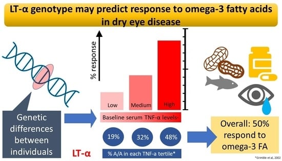
4.3. Polymorphisms to TNF-α Related Genes Might Predict Omega-3 Response

Part of the anti-inflammatory effect from omega-3 comes from suppression of TNF-α production. However, if inherent TNF-α production is already low to begin with, any attempts to suppress TNF-α might not further benefit. Conversely, patients with inherently high TNF-α production will be more sensitive to the TNF-α lowering effects of omega-3.
This hypothesis was evaluated for the first time by Grimble et al. in a study of 111 healthy subjects [95]. To do so, they stratified all subjects by their levels of inherent TNF-α production and pooled all data according to tertiles. In line with their hypothesis, when all subjects were supplemented with omega-3, they found that the highest tertile exhibited a fall in TNF-α. The middle tertile did not respond, and the bottom tertile actually suffered an increase in TNF-α. Lending further support to their hypothesis was the finding that the proportion of subjects with TNF-α suppression was the largest in the highest tertile (86%), followed by the middle tertile (43%) and the smallest (22%) in the lowest tertile. This is shown in Table 1. These differences in baseline TNF-α production and, hence, their responsiveness to omega-3 supplementation might reflect intrinsic differences in the regulation of immunity at baseline.
Next, Grimble et al. sought to investigate if inherent TNF-α production could be associated with TNF-α or LT-α genotypes. These genotypes arise due to polymorphisms in the TNF-α (TNF-α-308G and TNF-α-308A, also known as TNF*1 and TNF*2), as well as Lymphotoxin A (LT-α+252G and LT-α+252A a.k.a. TNFB*1 and TNFB*2) genes, giving rise to three alleles at each loci [96]. In their analysis, Grimble et al. evaluated the relative proportion of each genotype in each of the three tertiles of inherent TNF-α production. Three important findings were made. Firstly, TNF-α genotype was unrelated to TNF-α production, as the relative proportions of each genotype were consistent across all tertiles. Secondly, homozygosity for the LT-α+252A allele was positively correlated with inherent TNF-α production (41% homozygosity in the highest tertile and 16% homozygosity in the lowest tertile). Thirdly, heterozygosity at the LT-α locus was inversely correlated with inherent TNF-α production. Therefore, the LT-α genotype (but not the TNF-α genotype) can affect the inherent TNF-α. These findings regarding LT-α are shown in Table 2.
Therefore, since the LT-α genotype is associated with baseline TNF-α, and that the baseline TNF-α tertile predicts direction of omega-3 response, we can deduce that the LT-α genotype can potentially predict omega-3 response. Based on the above information, the calculated probability of a particular genotype responding favorably to omega-3 supplementation is the frequency of each tertile in that genotype (Table 2) multiplied by their respective proportion of individuals of each tertile responding favorably to omega-3 supplementation (Table 1). For example, the probability of the B1/B1 genotype response is 0.238 × 0.86 + 0.381 × 0.43 + 0.381 × 0.22 = 0.452. These findings are given in Table 3.
Since the genetic data reported by Grimble is based on a purely Caucasian population. In order to extrapolate these findings to other ethnicities, we have to assume that the frequency of each TNF-α tertile in a particular genotype, as well as the proportion of individuals responding favorably in each tertile, is similar to that of other ethnicities.
Therefore, if the patient’s genotype is known, the probability of response is taken to be the same amongst all ethnicities. However, if the patient’s genotype is unknown, we can calculate the probability of favorable response in a given ethnic group by considering the relative frequencies of each genotype (B1/B1, B1/B2, and B2/B2) in a given ethnic population. To do so, we obtained the allelic frequencies for each polymorphism from the refSNP database for each ethnic population (Caucasian, African, Asian, etc.). We then used the Hardy–Weidenburg Equilibrium to estimate the proportion of each genotype (B1/B1, B1/B2, and B2/B2) in each ethnic population, as described below [97]:
Heterozygosity = 2pq
Homozygous B1/B1 = p2
Homozygous B2/B2 = q2
where frequency of allele B1 = p; frequency of allele B2 = q.
These probabilities, referred to as P(unknown), are shown in Table 4 for the major ethnic populations.
As shown, P(unknown) does not vary much between ethnicities and hovers around 50%, making the probability of favorable response to omega-3 about 50%. However, response rates differ greatly between the various genotypes B1/B1, B1/B2 and B2/B2, as shown in Table 3. Therefore, genotyping will provide a more definitive answer as to whether or not a patient will respond. The aforementioned workflow is summarized in Figure 2.
While genetic testing may be costly, the cost of omega-3 taken as a supplement over a lifetime justifies its cost, since it allows each patient to make an informed decision on the efficacy of omega-3 supplementation. Furthermore, since this is testing for a single SNP, the cost is not expected to be high.

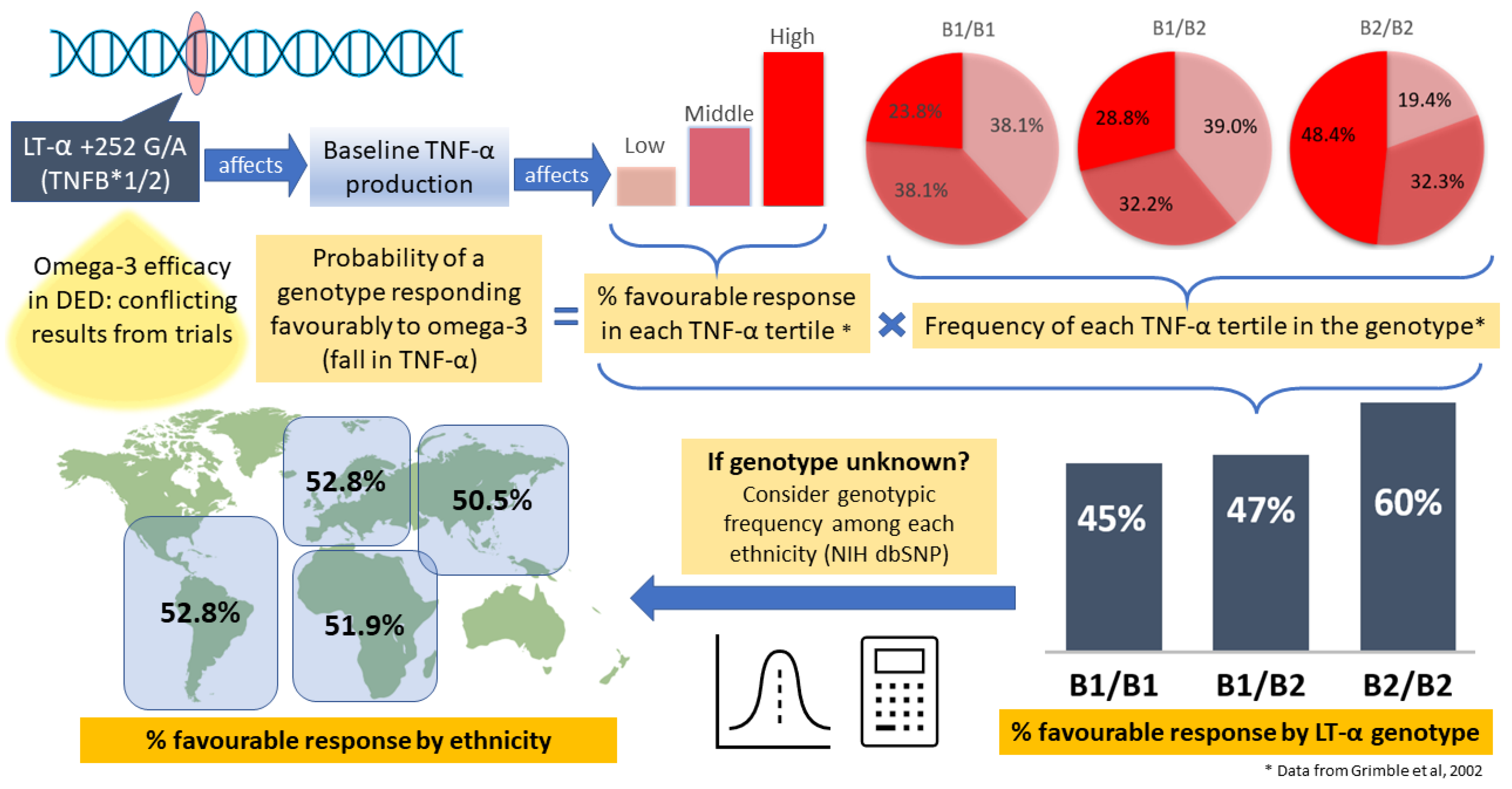
4.4. Accounting for How Polymorphisms in LT-α Alter the Expression and Activity of Cytokines

While the effect (while discordant) of TNF-α promoter polymorphisms on TNF-α production is clear, the mechanisms behind LT-α polymorphisms influencing TNF-α production is less well elucidated. One possible mechanism is that due to the extremely close proximity of the LT-α and TNF-α gene loci, these LT-α polymorphisms may be in linkage disequilibrium with other unidentified susceptibility polymorphisms responsible for the differences in baseline TNF-α [98].
Alternatively, polymorphism in LT-α might affect the formation of the transcription initiation complex at the TNF-α promoter, again, due to their proximity. The TNF-α promoter is just around 2 kilobases downstream of the LT-α polymorphism in question [99,100]. Therefore, alterations to the stability of the transcription initiation complex will affect the frequency of the transcription initiation of TNF-α and, hence, its levels. However, these need to be verified with future rigorous transcriptomics and metabolomics experiments.
The polymorphisms in LT-α may also affect differences in the expression of tear cytokines which drive DED pathogenesis, although the underlying mechanisms are highly complex. On the one hand, LT-α polymorphisms correlate with inherent TNF-α levels, which itself increases pro-inflammatory cytokines IL-6, IFN-y, and IL-1b and suppresses immunoregulatory TGF-β and IL-10 on the ocular surface [65,66]. On the other hand, LT-α polymorphisms correlate with plasma LT-α levels, which vary widely among DED patients, and, after a certain LT-α threshold (>700 pg/mL), LT-α positively correlates with increases in both pro-inflammatory (TNF-α, IL-12/23) and anti-inflammatory (IL-10, IL-1b, and IL-1Ra) cytokines, as revealed in a study by Chen et al. in 2020 [101]. Evidently, when DED patients are classified based on LT-α levels, the pathomechanisms are different. Further studies are required to correlate the inherent levels of these other cytokines (individually and in combinations) with omega-3 response.
Intriguingly, the same study by Chen et al. [101] found no significant differences in tear protein markers between the low LT-α DED (<700 pg/mL) and a healthy control group. The controls could have a mix of both high and low inherent levels of LT-α, leading to no difference when compared to the low LT-α or high LT-α groups. It may be arbitrary to divide LT-α into two categories when it is probably a spectrum. It is unlikely that the entire cytokine profile is dependent on one polymorphism; multiple genetic factors are likely at play. The study also did not report the clinical severity of DED in each of the three groups. Further studies are required to justify the threshold of 700 pg/mL to segregate “high” and “low” LT-α and to perform comparisons which are matched for DED severities. This will elucidate if LT-α is an independent predictor of the tear cytokine profile.

4.5. Implications for Testing and Other Potential Testing Targets

The aforementioned results also emphasize the need for LT-α genotyping prior to starting omega-3 in clinical trials. This is so that patients with similar LT-α genotype can be analyzed together so that effects of any variables being tested (e.g., concentration of omega-3, type of omega-3, combinations of omega-3 fatty acids given, etc.) are not confounded by genetic factors which also affect the individual’s response to omega-3. Otherwise, the results might be heterogenous with no clear direction of effect, which is the case with the DREAM study and the DREAM extension study.
The levels of lipid mediators of inflammation, such as prostaglandin E2 (PGE2), may also be measured in the tears of DED patients. This is because high levels of PGE2 suggest an underlying imbalance of pro-inflammatory lipids, which may be counterbalanced by omega-3 supplementation. This can be achieved using mass spectrometry as described previously [102].
The polymorphisms in LT-α may also affect the results of certain diagnostic tests for DED. For instance, although Schirmer’s I test has been used to confirm severe aqueous-deficient DED, its variability and invasiveness precludes its utility as a routine diagnostic test of tear volume, in particular for evaporative DED where subtle reduction in basal tear volume will be masked by reflex tearing. It also has poor sensitivity in identifying groups of patients with DED [103,104]. This is because LT-α polymorphisms (TNFB*2 allele) not only increase inherent TNF-α levels [103] but also increase inherent LT-α levels [105,106], both of which are strongly correlated with increased IL-17A, which in turn correlates with reduced tear production in the Schirmer’s I test [101]. This shows that LT-α polymorphisms may affect Schirmer’s test results. That being said, to the extent that symptoms and signs of DED reflect the underlying ocular surface inflammation (e.g., mediated by TNF-α), the conclusion that certain LT-α polymorphisms favor omega-3 supplementation should still be valid.
Current meta-analyses are plagued by data heterogeneity due to discordant results from different trials, such that there is no clear direction of effect. For instance, the latest meta-analysis in 2022 of eight studies found no improvement to corneal staining, tear breakup time, or Schirmer’s test due to substantial statistical heterogeneity throughout all outcome measures [42], leading to poor quality evidence and perpetually inconclusive results. Judging from clear effect directions in individual patients and some individual studies, it may simply be due to differences in underlying pathophysiology and cytokine profiles among DED patients [4], whether they involve or are independent of TNF-α. When results are adjusted for and stratified according to genotype, then clearer directions of effect may emerge. As elucidated in our review, LT-α and TNF-α are good places to start. In an era of increasing screen use when remote work arrangements become more popular, DED incidence is likely to rise and so will the number of patients that can benefit from omega-3 supplementation, hence making pre-treatment genotyping more important than ever.

4.6. Implications for Specific Patient Populations

Cytokines in patients with systemic diseases with DED manifestation may also influence the response to omega-3, although no studies have been in these specific patient populations. For example, Sjogren’s DED patients have higher inherent TNF-α levels than their non-Sjogren’s DED counterparts [107], which may predict more favorable omega-3 response. However, since the levels of TNF-α production are also affected by cytokines, such as IL-10, IL-4, TGF-β, IL-1, IL-2, and IFN-y [108,109,110], diseases with elevations to these cytokines can affect inherent TNF-α production and, hence, omega-3 response. For example, schizophrenia patients have elevated IL-1 relative to healthy controls [111]. However, while these remain speculative, they suggest targets for future research.

5. Conclusions

In conclusion, the polymorphisms in LT-α can prognosticate the direction of response to omega-3 supplementation as proxied by changes in TNF-α production. Greater inherent levels of TNF-α production is predictive for favorable response to omega-3 supplementation. While the calculated probability of favorable and therapeutic response to omega-3 ranges around 50% for all ethnicities, there is greater distinction in response rates between various genotypes. This emphasizes the need for LT-α genotyping before starting omega-3 in both long-term treatment and in clinical trials. There may be other cytokines which act on or independently of TNF-α to affect the response to omega-3 supplementation and merit further investigation.

Author Contributions

Conceptualization, B.P. and L.T.; methodology, B.P. and L.T.; validation, B.P. and L.T.; formal analysis, B.P. and L.T.; investigation, B.P. and L.T.; resources, B.P. and L.T.; data curation, B.P. and L.T.; writing—original draft preparation, B.P. and L.T.; writing—review and editing, B.P. and L.T.; visualization, B.P. and L.T.; supervision, L.T.; project administration, B.P. and L.T. All authors have read and agreed to the published version of the manuscript.

Funding

This research was funded by National Medical Research Council Singapore, grant number NMRC/CSA/017/2017.

Data Availability Statement

The data presented in this study may be available on request from the correspondence author.

Conflicts of Interest

The authors declare no conflict of interest.

References

  1. Willcox, M.D.; Argüeso, P.; Georgiev, G.A.; Holopainen, J.M.; Laurie, G.W.; Millar, T.J.; Papas, E.B.; Rolland, J.P.; Schmidt, T.A.; Stahl, U.; et al. TFOS DEWS II Tear Film Report. Ocul. Surf. 2017, 15, 366–403. [Google Scholar] [CrossRef] [PubMed] [Green Version]
  2. Baudouin, C.; Messmer, E.M.; Aragona, P.; Geerling, G.; Akova, Y.; Benítez-Del-Castillo, J.; Boboridis, K.; Merayo-Lloves, J.; Rolando, M.; Labetoulle, M. Revisiting the vicious circle of dry eye disease: A focus on the pathophysiology of meibomian gland dysfunction. Br. J. Ophthalmol. 2016, 100, 300–306. [Google Scholar] [CrossRef] [PubMed]
  3. Messmer, E.M. The pathophysiology, diagnosis, and treatment of dry eye disease. Dtsch. Arztebl. Int. 2015, 112, 71–81. [Google Scholar] [CrossRef] [PubMed] [Green Version]
  4. Bron, A.J.; de Paiva, C.S.; Chauhan, S.K.; Bonini, S.; Gabison, E.E.; Jain, S.; Knop, E.; Markoulli, M.; Ogawa, Y.; Perez, V.; et al. TFOS DEWS II pathophysiology report. Ocul. Surf. 2017, 15, 438–510. [Google Scholar] [CrossRef] [PubMed]
  5. Wang, H.; Daggy, B.P. The Role of Fish Oil in Inflammatory Eye Diseases. Biomed. Hub 2017, 2, 455818. [Google Scholar] [CrossRef] [PubMed] [Green Version]
  6. 2020–2021 BCSC Basic and Clinical Science CourseTM. Available online: https://www.aao.org/bcscsnippetdetail.aspx?id=e15abb31-d82f-4c41-abe1-8a4d9aece885 (accessed on 16 November 2022).
  7. Annunziato, F.; Cosmi, L.; Liotta, F.; Maggi, E.; Romagnani, S. Human T helper type 1 dichotomy: Origin, phenotype and biological activities. Immunology 2014, 144, 343–351. [Google Scholar] [CrossRef] [PubMed]
  8. Walker, J.A.; McKenzie, A.N.J. TH2 cell development and function. Nat. Rev. Immunol. 2018, 18, 121–133. [Google Scholar] [CrossRef]
  9. Tesmer, L.A.; Lundy, S.; Sarkar, S.; Fox, D.A. Th17 cells in human disease. Immunol. Rev. 2008, 223, 87–113. [Google Scholar] [CrossRef]
  10. Wu, X.; Tian, J.; Wang, S. Insight into Non-Pathogenic Th17 Cells in Autoimmune Diseases. Front. Immunol. 2018, 9, 1112. [Google Scholar] [CrossRef] [Green Version]
  11. Soifer, M.; Azar, N.S.; Mousa, H.M.; Perez, V.L. Ocular Surface Inflammatory Disorders (OSID): A Collective of Systemic Etiologies Which Cause or Amplify Dry Eye Syndrome. Front. Med. 2022, 9, 1112. [Google Scholar] [CrossRef]
  12. Ong, H.S.; Dart, J.K. Managing ocular surface disease: A common-sense approach. Community Eye Health 2016, 29, 44–46. [Google Scholar] [PubMed]
  13. Guzmán, M.; Keitelman, I.; Sabbione, F.; Trevani, A.S.; Giordano, M.N.; Galletti, J.G. Desiccating stress-induced disruption of ocular surface immune tolerance drives dry eye disease. Clin. Exp. Immunol. 2016, 184, 248–256. [Google Scholar] [CrossRef] [PubMed] [Green Version]
  14. Luo, L.; Li, D.-Q.; Corrales, R.M.; Pflugfelder, S.C. Hyperosmolar Saline Is a Proinflammatory Stress on the Mouse Ocular Surface. Eye Contact Lens Sci. Clin. Pract. 2005, 31, 186–193. [Google Scholar] [CrossRef] [PubMed]
  15. Tsubota, K.; Pflugfelder, S.; Liu, Z.; Baudouin, C.; Kim, H.; Messmer, E.; Kruse, F.; Liang, L.; Liang, L.; Galeano, J.; et al. Defining Dry Eye from a Clinical Perspective. Int. J. Mol. Sci. 2020, 21, 9271. [Google Scholar] [CrossRef]
  16. Jang, D.-I.; Lee, A.-H.; Shin, H.-Y.; Song, H.-R.; Park, J.-H.; Kang, T.-B.; Lee, S.-R.; Yang, S.-H. The Role of Tumor Necrosis Factor Alpha (TNF-α) in Autoimmune Disease and Current TNF-α Inhibitors in Therapeutics. Int. J. Mol. Sci. 2021, 22, 2719. [Google Scholar] [CrossRef]
  17. Enríquez-De-Salamanca, A.; Castellanos, E.; Stern, M.E.; Fernández, I.; Carreño, E.; García-Vázquez, C.; Herreras, J.M.; Calonge, M. Tear cytokine and chemokine analysis and clinical correlations in evaporative-type dry eye disease. Mol. Vis. 2010, 16, 862–873. [Google Scholar]
  18. Chauhan, S.K.; Annan, J.; Ecoiffier, T.; Goyal, S.; Zhang, Q.; Saban, D.R.; Dana, R. Autoimmunity in Dry Eye Is Due to Resistance of Th17 to Treg Suppression. J. Immunol. 2009, 182, 1247–1252. [Google Scholar] [CrossRef] [Green Version]
  19. De Paiva, C.S.; Chotikavanich, S.; Pangelinan, S.B.; Pitcher, J.D.; Fang, B.; Zheng, X.; Ma, P.; Farley, W.J.; Siemasko, K.F.; Niederkorn, J.Y.; et al. IL-17 disrupts corneal barrier following desiccating stress. Mucosal Immunol. 2009, 2, 243–253. [Google Scholar] [CrossRef] [Green Version]
  20. Chennakesavalu, M.; Somala, S.R.R.; Dommaraju, S.R.; Peesapati, M.P.; Guo, K.; Rosenblatt, M.I.; Chang, J.-H.; Azar, D.T. Corneal lymphangiogenesis as a potential target in dry eye disease—A systematic review. Surv. Ophthalmol. 2021, 66, 960–976. [Google Scholar] [CrossRef]
  21. Qi, M.; Liu, D.; Pan, L.; Lin, Y. Interleukin-10 gene -592C > A polymorphism and susceptibility to gastric cancer. Genet. Mol. Res. 2014, 13, 8954–8961. [Google Scholar] [CrossRef]
  22. Schwartz, D.M.; Bonelli, M.; Gadina, M.; O’Shea, J.J. Type I/II cytokines, JAKs, and new strategies for treating autoimmune diseases. Nat. Rev. Rheumatol. 2016, 12, 25–36. [Google Scholar] [CrossRef] [Green Version]
  23. Franke, A.; McGovern, D.P.; Barrett, J.C.; Wang, K.; Radford-Smith, G.L.; Ahmad, T.; Lees, C.W.; Balschun, T.; Lee, J.; Roberts, R.; et al. Genome-wide meta-analysis increases to 71 the number of confirmed Crohn’s disease susceptibility loci. Nat. Genet. 2010, 42, 1118–1125. [Google Scholar] [CrossRef] [Green Version]
  24. Hu, K.; Hou, S.; Jiang, Z.; Kijlstra, A.; Yang, P. JAK2 and STAT3 polymorphisms in a Han Chinese population with Behçet’s disease. Investig. Ophthalmol. Vis. Sci. 2012, 53, 538–541. [Google Scholar] [CrossRef] [Green Version]
  25. Ellinghaus, D.; Ellinghaus, E.; Nair, R.P.; Stuart, P.E.; Esko, T.; Metspalu, A.; Debrus, S.; Raelson, J.V.; Tejasvi, T.; Belouchi, M.; et al. Combined analysis of genome-wide association studies for Crohn disease and psoriasis identifies seven shared susceptibility loci. Am. J. Hum. Genet. 2012, 90, 636–647. [Google Scholar] [CrossRef] [Green Version]
  26. Remmers, E.F.; Plenge, R.M.; Lee, A.T.; Graham, R.R.; Hom, G.; Behrens, T.W.; de Bakker, P.I.; Le, J.M.; Lee, H.S.; Batliwalla, F.; et al. STAT4 and the risk of rheumatoid arthritis and systemic lupus erythematosus. N. Engl. J. Med. 2007, 357, 977–986. [Google Scholar] [CrossRef] [Green Version]
  27. Duetsch, G.; Illig, T.; Loesgen, S.; Rohde, K.; Klopp, N.; Herbon, N.; Gohlke, H.; Altmueller, J.; Wjst, M. STAT6 as an asthma candidate gene: Polymorphism-screening, association and haplotype analysis in a Caucasian sib-pair study. Hum Mol Genet. 2002, 11, 613–621. [Google Scholar] [CrossRef] [Green Version]
  28. Grad, R. Cod and the consumptive: A brief history of cod-liver oil in the treatment of pulmonary tuberculosis. Pharm. Hist. 2004, 46, 106–120. [Google Scholar] [PubMed]
  29. Gammone, M.A.; Riccioni, G.; Parrinello, G.; D’Orazio, N. Omega-3 Polyunsaturated Fatty Acids: Benefits and Endpoints in Sport. Nutrients 2018, 11, 46. [Google Scholar] [CrossRef] [PubMed] [Green Version]
  30. Yashodhara, B.M.; Umakanth, S.; Pappachan, J.M.; Bhat, S.K.; Kamath, R.; Choo, B.H. Omega-3 fatty acids: A comprehensive review of their role in health and disease. Postgrad. Med. J. 2009, 85, 84–90. [Google Scholar] [CrossRef] [PubMed] [Green Version]
  31. Griffing, G.T. Mother was right about cod liver oil. Am. J. Med. 2008, 10, 8. [Google Scholar]
  32. Ishihara, T.; Yoshida, M.; Arita, M. Omega-3 fatty acid-derived mediators that control inflammation and tissue homeostasis. Int. Immunol. 2019, 31, 559–567. [Google Scholar] [CrossRef] [PubMed] [Green Version]
  33. Sheppe, A.E.F.; Edelmann, M.J. Roles of Eicosanoids in Regulating Inflammation and Neutrophil Migration as an Innate Host Response to Bacterial Infections. Infect. Immun. 2021, 89, e00095-21. [Google Scholar] [CrossRef] [PubMed]
  34. Alvarez, Y.; Valera, I.; Municio, C.; Hugo, E.; Padrón, F.; Blanco, L.; Rodríguez, M.; Fernández, N.; Crespo, M.S. Eicosanoids in the Innate Immune Response: TLR and Non-TLR Routes. Mediat. Inflamm. 2010, 2010, 201929. [Google Scholar] [CrossRef] [PubMed] [Green Version]
  35. Bagga, D.; Wang, L.; Farias-Eisner, R.; Glaspy, J.A.; Reddy, S.T. Differential effects of prostaglandin derived from ω-6 and omega-3 polyunsaturated fatty acids on COX-2 expression and IL-6 secretion. Proc. Natl. Acad. Sci. USA 2003, 100, 1751–1756. [Google Scholar] [CrossRef] [PubMed] [Green Version]
  36. Downie, L.; Ng, S.M.; Lindsley, K.B.; Akpek, E.K. Omega-3 and omega-6 polyunsaturated fatty acids for dry eye disease. Cochrane Database Syst. Rev. 2019. [Google Scholar] [CrossRef] [PubMed]
  37. Barabino, S.; Horwath-Winter, J.; Messmer, E.M.; Rolando, M.; Aragona, P.; Kinoshita, S. The role of systemic and topical fatty acids for dry eye treatment. Prog. Retin. Eye Res. 2017, 61, 23–34. [Google Scholar] [CrossRef]
  38. Duvall, M.G.; Levy, B.D. DHA- and EPA-derived resolvins, protectins, and maresins in airway inflammation. Eur. J. Pharmacol. 2016, 785, 144–155. [Google Scholar] [CrossRef] [Green Version]
  39. Hong, S.; Lu, Y. Omega-3 fatty acid-derived resolvins and protectins in inflammation resolution and leukocyte functions: Targeting novel lipid mediator pathways in mitigation of acute kidney injury. Front. Immunol. 2013, 4, 13. [Google Scholar] [CrossRef] [Green Version]
  40. Giannaccare, G.; Pellegrini, M.; Sebastiani, S.; Bernabei, F.; Roda, M.; Taroni, L.; Versura, P.; Campos, E.C. Efficacy of Omega-3 Fatty Acid Supplementation for Treatment of Dry Eye Disease: A Meta-Analysis of Randomized Clinical Trials. Cornea 2019, 38, 565–573. [Google Scholar] [CrossRef]
  41. Liu, A.; Ji, J. Omega-3 essential fatty acids therapy for dry eye syndrome: A meta-analysis of randomized controlled studies. Med. Sci. Monit. 2014, 20, 1583–1589. [Google Scholar]
  42. O’Byrne, C.; O’Keeffe, M. Omega-3 fatty acids in the management of dry eye disease—An updated systematic review and meta-analysis. Acta Ophthalmol. 2022, 101, e118–e134. [Google Scholar] [CrossRef]
  43. Dry Eye Assessment and Management Study Research Group. n-3 Fatty Acid Supplementation for the Treatment of Dry Eye Disease. N. Engl. J. Med. 2018, 378, 1681–1690.
  44. Hussain, M.; Shtein, R.M.; Pistilli, M.; Maguire, M.G.; Oydanich, M.; Asbell, P.A. The Dry Eye Assessment and Management (DREAM) extension study—A randomized clinical trial of withdrawal of supplementation with omega-3 fatty acid in patients with dry eye disease. Ocul. Surf. 2020, 18, 47–55. [Google Scholar] [CrossRef] [PubMed]
  45. Gutiérrez, S.; Svahn, S.L.; Johansson, M.E. Effects of Omega-3 Fatty Acids on Immune Cells. Int. J. Mol. Sci. 2019, 20, 5028. [Google Scholar] [CrossRef] [PubMed] [Green Version]
  46. Titos, E.; Rius, B.; González-Périz, A.; López-Vicario, C.; Morán-Salvador, E.; Martínez-Clemente, M.; Arroyo, V.; Clària, J. Resolvin D1 and Its Precursor Docosahexaenoic Acid Promote Resolution of Adipose Tissue Inflammation by Eliciting Macrophage Polarization toward an M2-Like Phenotype. J. Immunol. 2011, 187, 5408–5418. [Google Scholar] [CrossRef] [PubMed] [Green Version]
  47. Orecchioni, M.; Ghosheh, Y.; Pramod, A.B.; Ley, K. Macrophage Polarization: Different Gene Signatures in M1(LPS+) vs. Classically and M2(LPS–) vs. Alternatively Activated Macrophages. Front. Immunol. 2019, 10, 1084. [Google Scholar] [CrossRef] [PubMed]
  48. Ramirez-Ramirez, V.; Macias-Islas, M.A.; Ortiz, G.G.; Pacheco-Moises, F.; Torres-Sanchez, E.D.; Sorto-Gomez, T.E.; Cruz-Ramos, J.A.; Orozco-Aviña, G.; de la Rosa, A.J.C. Efficacy of Fish Oil on Serum of TNFα, IL-1β, and IL-6 Oxidative Stress Markers in Multiple Sclerosis Treated with Interferon Beta-1b. Oxidative Med. Cell. Longev. 2013, 2013, 709493. [Google Scholar] [CrossRef] [PubMed] [Green Version]
  49. Moghadam, A.M.; Saedisomeolia, A.; Djalali, M.; Djazayery, A.; Pooya, S.; Sojoudi, F. Efficacy of omega-3 fatty acid supplementation on serum levels of tumour necrosis factor-alpha, C-reactive protein and interleukin-2 in type 2 diabetes mellitus patients. Singap. Med. J. 2012, 53, 615–619. [Google Scholar]
  50. Natto, Z.S.; Yaghmoor, W.; Alshaeri, H.K.; Van Dyke, T.E. Omega-3 Fatty Acids Effects on Inflammatory Biomarkers and Lipid Profiles among Diabetic and Cardiovascular Disease Patients: A Systematic Review and Meta-Analysis. Sci. Rep. 2019, 9, 18867. [Google Scholar] [CrossRef] [Green Version]
  51. Allam-Ndoul, B.; Guénard, F.; Barbier, O.; Vohl, M.-C. Effect of n-3 fatty acids on the expression of inflammatory genes in THP-1 macrophages. Lipids Health Dis. 2016, 15, 69. [Google Scholar] [CrossRef] [Green Version]
  52. Li, K.; Huang, T.; Zheng, J.; Wu, K.; Li, D. Effect of Marine-Derived n-3 Polyunsaturated Fatty Acids on C-Reactive Protein, Interleukin 6 and Tumor Necrosis Factor α: A Meta-Analysis. PLoS ONE 2014, 9, e88103. [Google Scholar] [CrossRef] [PubMed] [Green Version]
  53. Caughey, G.; Mantzioris, E.; Gibson, R.; Cleland, L.G.; James, M.J. The effect on human tumor necrosis factor alpha and interleukin 1 beta production of diets enriched in n-3 fatty acids from vegetable oil or fish oil. Am. J. Clin. Nutr. 1996, 63, 116–122. [Google Scholar] [CrossRef] [PubMed] [Green Version]
  54. Calder, P.C. Omega-3 fatty acids and inflammatory processes: From molecules to man. Biochem. Soc. Trans. 2017, 45, 1105–1115. [Google Scholar] [CrossRef] [PubMed] [Green Version]
  55. Cannon, J.G. Inflammatory Cytokines in Nonpathological States. Physiology 2000, 15, 298–303. [Google Scholar] [CrossRef]
  56. Massingale, M.L.; Li, X.; Vallabhajosyula, M.; Chen, D.; Wei, Y.; Asbell, P. Analysis of Inflammatory Cytokines in the Tears of Dry Eye Patients. Cornea 2009, 28, 1023–1027. [Google Scholar] [CrossRef]
  57. Pflugfelder, S.C.; Jones, D.; Ji, Z.; Afonso, A.; Monroy, D. Altered cytokine balance in the tear fluid and conjunctiva of patients with Sjögren’s syndrome keratoconjunctivitis sicca. Curr. Eye Res. 1999, 19, 201–211. [Google Scholar] [CrossRef]
  58. Wesemann, D.R.; Benveniste, E.N. STAT-1 alpha and IFN-gamma as modulators of TNF-αlpha signaling in macrophages: Regulation and functional implications of the TNF receptor 1:STAT-1 alpha complex. J. Immunol. 2003, 171, 5313–5319. [Google Scholar] [CrossRef] [Green Version]
  59. Yarilina, A.; Park-Min, K.-H.; Antoniv, T.T.; Hu, X.; Ivashkiv, L.B. TNF activates an IRF1-dependent autocrine loop leading to sustained expression of chemokines and STAT1-dependent type I interferon–response genes. Nat. Immunol. 2008, 9, 378–387. [Google Scholar] [CrossRef]
  60. Takada, Y.; Aggarwal, B.B. Evidence that genetic deletion of the TNF receptor p60 or p80 in macrophages modulates RANKL-induced signaling. Blood 2004, 104, 4113–4121. [Google Scholar] [CrossRef]
  61. Yoon, K.-C.; Jeong, I.-Y.; Park, Y.-G.; Yang, S.-Y. Interleukin-6 and Tumor Necrosis Factor-α Levels in Tears of Patients with Dry Eye Syndrome. Cornea 2007, 26, 431–437. [Google Scholar] [CrossRef]
  62. Vallance, P.; Chan, N. Endothelial function and nitric oxide: Clinical relevance. Heart 2001, 85, 342–350. [Google Scholar] [CrossRef] [PubMed] [Green Version]
  63. Sanders, D.B.; Larson, D.F.; Hunter, K.; Gorman, M.; Yang, B. Comparison of tumor necrosis factor-α effect on the expression of iNOS in macrophage and cardiac myocytes. Perfusion 2001, 16, 67–74. [Google Scholar] [CrossRef]
  64. Luo, L.; Li, D.-Q.; Pflugfelder, S.C. Hyperosmolarity-Induced Apoptosis in Human Corneal Epithelial Cells Is Mediated by Cytochrome c and MAPK Pathways. Cornea 2007, 26, 452–460. [Google Scholar] [CrossRef] [PubMed]
  65. Ji, Y.W.; Byun, Y.J.; Choi, W.; Jeong, E.; Kim, J.S.; Noh, H.; Kim, E.S.; Song, Y.J.; Park, S.K.; Lee, H.K. Neutralization of Ocular Surface TNF-α Reduces Ocular Surface and Lacrimal Gland Inflammation Induced by In Vivo Dry Eye. Investig. Opthalmol. Vis. Sci. 2013, 54, 7557–7566. [Google Scholar] [CrossRef] [PubMed] [Green Version]
  66. Choi, W.; Noh, H.; Yeo, A.; Jang, H.; Ahn, H.K.; Song, Y.J.; Lee, H.K. The Effect of TNF-α Blocker HL036337 and Its Best Concentration to Inhibit Dry Eye Inflammation. Korean J. Ophthalmol. 2016, 30, 302–308. [Google Scholar] [CrossRef] [PubMed] [Green Version]
  67. Shettle, L.; McLaurin, E.; Martel, J.; Seaman, J.W.; Weissgerber, G. Topical Anti-TNFα Agent Licaminlimab (OCS-02) Relieves Persistent Ocular Discomfort in Severe Dry Eye Disease: A Randomized Phase II Study. Clin. Ophthalmol. 2022, 16, 2167–2177. [Google Scholar] [CrossRef]
  68. Dong, Y.; Wang, S.; Cong, L.; Zhang, T.; Cheng, J.; Yang, N.; Qu, X.; Li, D.; Zhou, X.; Wang, H.; et al. TNF-α inhibitor tanfanercept (HBM9036) improves signs and symptoms of dry eye in a phase 2 trial in the controlled adverse environment in China. Int. Ophthalmol. 2022, 42, 2459–2472. [Google Scholar] [CrossRef]
  69. Rogler, G.; Herfarth, H.; Hibi, T.; Nielsen, O.H. Anti-Tumor Necrosis Factor Therapy in Inflammatory Bowel Disease; Karger Medical and Scientific Publishers: Basel, Switzerland, 2015. [Google Scholar]
  70. Clark, J.; Vagenas, P.; Panesar, M.; Cope, A.P. What does tumour necrosis factor excess do to the immune system long term? Ann. Rheum. Dis. 2005, 64, iv70–iv76. [Google Scholar] [CrossRef] [Green Version]
  71. Calmon-Hamaty, F.; Combe, B.; Hahne, M.; Morel, J. Lymphotoxin α revisited: General features and implications in rheumatoid arthritis. Arthritis Res. Ther. 2011, 13, 232–235. [Google Scholar] [CrossRef] [Green Version]
  72. Chiang, E.Y.; Kolumam, G.; Yu, X.; Francesco, M.; Ivelja, S.; Peng, I.; Gribling, P.; Shu, J.; Lee, W.P.; Refino, C.J.; et al. Targeted depletion of lymphotoxin-α–expressing TH1 and TH17 cells inhibits autoimmune disease. Nat. Med. 2009, 15, 766–773. [Google Scholar] [CrossRef]
  73. Etemadi, N.; Holien, J.K.; Chau, D.; Dewson, G.; Murphy, J.M.; Alexander, W.S.; Parker, M.W.; Silke, J.; Nachbur, U. Lymphotoxin α induces apoptosis, necroptosis and inflammatory signals with the same potency as tumour necrosis factor. FEBS J. 2013, 280, 5283–5297. [Google Scholar] [CrossRef] [PubMed]
  74. Cuff, C.; Schwartz, J.; Bergman, C.M.; Russell, K.S.; Bender, J.R.; Ruddle, N.H. Lymphotoxin alpha3 induces chemokines and adhesion molecules: Insight into the role of LT alpha in inflammation and lymphoid organ development. J. Immunol. 1998, 161, 6853–6860. [Google Scholar] [CrossRef] [PubMed]
  75. Kratz, A.; Campos-Neto, A.; Hanson, M.S.; Ruddle, N.H. Chronic inflammation caused by lymphotoxin is lymphoid neogenesis. J. Exp. Med. 1996, 183, 1461–1472. [Google Scholar] [CrossRef] [PubMed] [Green Version]
  76. Buhrmann, C.; Shayan, P.; Aggarwal, B.B.; Shakibaei, M. Evidence that TNF-β (lymphotoxin α) can activate the inflammatory environment in human chondrocytes. Arthritis Res. Ther. 2013, 15, R202. [Google Scholar] [CrossRef] [PubMed] [Green Version]
  77. Jiang, X.; Takahashi, N.; Matsui, N.; Tetsuka, T.; Okamoto, T. The NF-κB Activation in Lymphotoxin β Receptor Signaling Depends on the Phosphorylation of p65 at Serine 536 *. J. Biol. Chem. 2003, 278, 919–926. [Google Scholar] [CrossRef] [PubMed] [Green Version]
  78. Messerli, F.H.; Bangalore, S.; Schmieder, R.E. Wilder’s principle: Pre-treatment value determines post-treatment response. Eur. Heart J. 2014, 36, 576–579. [Google Scholar] [CrossRef] [Green Version]
  79. Hom, M.M.; Asbell, P.; Barry, B. Omegas and Dry Eye: More Knowledge, More Questions. Optom. Vis. Sci. 2015, 92, 948–956. [Google Scholar] [CrossRef]
  80. Wierzejska, R.E. Dietary Supplements—For Whom? The Current State of Knowledge about the Health Effects of Selected Supplement Use. Int. J. Environ. Res. Public Health 2021, 18, 8897. [Google Scholar] [CrossRef]
  81. Takeuchi, T.; Miyasaka, N.; Tatsuki, Y.; Yano, T.; Yoshinari, T.; Abe, T.; Koike, T. Baseline tumour necrosis factor alpha levels predict the necessity for dose escalation of infliximab therapy in patients with rheumatoid arthritis. Ann. Rheum. Dis. 2011, 70, 1208–1215. [Google Scholar] [CrossRef]
  82. Zhang, R.-F.; Liu, S.; Wang, Y.-W.; Li, J. Potential molecular biomarkers used to predict the response to biological therapies in ulcerative colitis. Chin. Med. J. 2021, 134, 1058–1060. [Google Scholar] [CrossRef]
  83. Khan, S.; Mandal, R.K.; Jawed, A.; Dar, S.A.; Wahid, M.; Panda, A.K.; Areeshi, M.Y.; Khan, E.A.; Haque, S. TNF-α -308 G > A (rs1800629) Polymorphism is Associated with Celiac Disease: A Meta-analysis of 11 Case-Control Studies. Sci. Rep. 2016, 6, 32677. [Google Scholar] [CrossRef] [PubMed]
  84. El-Tahan, R.R.; Ghoneim, A.M.; El-Mashad, N. TNF-α gene polymorphisms and expression. Springerplus 2016, 5, 1508. [Google Scholar] [CrossRef] [PubMed] [Green Version]
  85. Faustman, D.L.; Davis, M. TNF Receptor 2 and Disease: Autoimmunity and Regenerative Medicine. Front. Immunol. 2013, 4, 478. [Google Scholar] [CrossRef] [PubMed] [Green Version]
  86. Falvo, J.V.; Tsytsykova, A.V.; Goldfeld, A.E. Transcriptional Control of the TNF Gene. Curr. Dir. Autoimmun. 2010, 11, 27–60. [Google Scholar] [CrossRef] [PubMed] [Green Version]
  87. Bayley, J.-P.; Ottenhoff, T.H.M.; Verweij, C.L. Is there a future for TNF promoter polymorphisms? Genes Immun. 2004, 5, 315–329. [Google Scholar] [CrossRef] [PubMed] [Green Version]
  88. Elahi, M.M.; Asotra, K.; Matata, B.M.; Mastana, S.S. Tumor necrosis factor alpha-308 gene locus promoter polymorphism: An analysis of association with health and disease. Biochim. Biophys. Acta (BBA)—Mol. Basis Dis. 2009, 1792, 163–172. [Google Scholar] [CrossRef] [Green Version]
  89. Mekinian, A.; Tamouza, R.; Pavy, S.; Gestermann, N.; Ittah, M.; Mariette, X.; Miceli-Richard, C. Functional study of TNF-α promoter polymorphisms: Literature review and meta-analysis. Eur. Cytokine Netw. 2011, 22, 88–102. [Google Scholar] [CrossRef] [Green Version]
  90. Pociot, F.; Briant, L.; Jongeneel, C.V.; Mölvig, J.; Worsaae, H.; Abbal, M.; Thomsen, M.; Nerup, J.; Cambon-Thomsen, A. Association of tumor necrosis factor (TNF) and class II major histocompatibility complex alleles with the secretion of TNF-αlpha and TNF-beta by human mononuclear cells: A possible link to insulin-dependent diabetes mellitus. Eur. J. Immunol. 1993, 23, 224–231. [Google Scholar] [CrossRef]
  91. Aderka, D. The potential biological and clinical significance of the soluble tumor necrosis factor receptors. Cytokine Growth Factor Rev. 1996, 7, 231–240. [Google Scholar] [CrossRef]
  92. Hehlgans, T.; Pfeffer, K. The intriguing biology of the tumour necrosis factor/tumour necrosis factor receptor superfamily: Players, rules and the games. Immunology 2005, 115, 1–20. [Google Scholar] [CrossRef]
  93. Sainz, J.; Salas-Alvarado, I.; López-Fernández, E.; Olmedo, C.; Comino, A.; García, F.; Blanco, A.; Gómez-Lopera, S.; Oyonarte, S.; Bueno, P.; et al. TNFR1 mRNA Expression Level and TNFR1 Gene Polymorphisms are Predictive Markers for Susceptibility to Develop Invasive Pulmonary Aspergillosis. Int. J. Immunopathol. Pharmacol. 2010, 23, 423–436. [Google Scholar] [CrossRef] [Green Version]
  94. Li, H.; Anderson, S.K. Association of TNFRSF1B Promoter Polymorphisms with Human Disease: Further Studies Examining T-Regulatory Cells Are Required. Front. Immunol. 2018, 9, 443. [Google Scholar] [CrossRef] [PubMed] [Green Version]
  95. Grimble, R.F.; Howell, W.M.; O’Reilly, G.; Turner, S.J.; Markovic, O.; Hirrell, S.; East, J.M.; Calder, P.C. The ability of fish oil to suppress tumor necrosis factor α production by peripheral blood mononuclear cells in healthy men is associated with polymorphisms in genes that influence tumor necrosis factor α production. Am. J. Clin. Nutr. 2002, 76, 454–459. [Google Scholar] [CrossRef] [Green Version]
  96. Bilolikar, H.; Nam, A.R.; Rosenthal, M.; Davies, J.; Henderson, D.C.; Balfour-Lynn, I.M. Tumour necrosis factor gene polymorphisms and childhood wheezing. Eur. Respir. J. 2005, 26, 637–646. [Google Scholar] [CrossRef] [PubMed] [Green Version]
  97. Abramovs, N.; Brass, A.; Tassabehji, M. Hardy-Weinberg Equilibrium in the Large Scale Genomic Sequencing Era. Front. Genet. 2020, 11, 210. [Google Scholar] [CrossRef]
  98. Vejbaesya, S.; Luangtrakool, P.; Luangtrakool, K.; Kalayanarooj, S.; Vaughn, D.W.; Endy, T.P.; Mammen, M.P.; Green, S.; Libraty, D.H.; Ennis, F.A.; et al. TNF and LTA Gene, Allele, and Extended HLA Haplotype Associations with Severe Dengue Virus Infection in Ethnic Thais. J. Infect. Dis. 2009, 199, 1442–1448. [Google Scholar] [CrossRef] [PubMed] [Green Version]
  99. Clark, T.G.; Diakite, M.; Auburn, S.; Campino, S.; Fry, A.E.; Green, A.; Richardson, A.; Small, K.; Teo, Y.Y.; Wilson, J.; et al. Tumor Necrosis Factor and Lymphotoxin-α Polymorphisms and Severe Malaria in African Populations. J. Infect. Dis. 2009, 199, 569–575. [Google Scholar] [CrossRef] [Green Version]
  100. Tsytsykova, A.V.; Rajsbaum, R.; Falvo, J.V.; Ligeiro, F.; Neely, S.R.; Goldfeld, A.E. Activation-dependent intrachromosomal interactions formed by the TNF gene promoter and two distal enhancers. Proc. Natl. Acad. Sci. USA 2007, 104, 16850–16855. [Google Scholar] [CrossRef] [PubMed] [Green Version]
  101. Chen, H.; Chen, H.; Liang, L.; Zhong, Y.; Liang, Y.; Yu, Y.; Huang, S.; Lu, X. Evaluation of Tear Protein Markers in Dry Eye Disease with Different Lymphotoxin-Alpha Expression Levels. Am. J. Ophthalmol. 2020, 217, 198–211. [Google Scholar] [CrossRef]
  102. Ambaw, Y.A.; Chao, C.; Ji, S.; Raida, M.; Torta, F.; Wenk, M.R.; Tong, L. Tear eicosanoids in healthy people and ocular surface disease. Sci. Rep. 2018, 8, 11296. [Google Scholar] [CrossRef] [PubMed] [Green Version]
  103. Savini, G.; Prabhawasat, P.; Kojima, T.; Grueterich, M.; Espana, E.; Goto, E. The challenge of dry eye diagnosis. Clin. Ophthalmol. 2008, 2, 31–55. [Google Scholar] [CrossRef] [PubMed] [Green Version]
  104. Wolffsohn, J.S.; Arita, R.; Chalmers, R.; Djalilian, A.; Dogru, M.; Dumbleton, K.; Gupta, P.K.; Karpecki, P.; Lazreg, S.; Pult, H.; et al. TFOS DEWS II Diagnostic Methodology report. Ocul. Surf. 2017, 15, 539–574. [Google Scholar] [CrossRef] [PubMed]
  105. Messer, G.; Spengler, U.; Jung, M.C.; Honold, G.; Blömer, K.; Pape, G.R.; Riethmüller, G.; Weiss, E.H. Polymorphic structure of the tumor necrosis factor (TNF) locus: An NcoI polymorphism in the first intron of the human TNF-beta gene correlates with a variant amino acid in position 26 and a reduced level of TNF-beta production. J. Exp. Med. 1991, 173, 209–219. [Google Scholar] [CrossRef] [PubMed] [Green Version]
  106. Whichelow, C.E.; Hitman, G.A.; Raafat, I.; Bottazzo, G.F.; Sachs, J.A. The effect of tnf*b gene polymorphism on tnf-? and -? secretion levels in patients with insulin-dependent diabetes mellitus and healthy controls. Eur. J. Immunogenet. 1996, 23, 425–435. [Google Scholar] [CrossRef]
  107. Lee, S.Y.; Han, S.J.; Nam, S.M.; Yoon, S.; Ahn, J.M.; Kim, T.-I.; Kim, E.K.; Seo, K.Y. Analysis of Tear Cytokines and Clinical Correlations in Sjögren Syndrome Dry Eye Patients and Non–Sjögren Syndrome Dry Eye Patients. Am. J. Ophthalmol. 2013, 156, 247–253.e1. [Google Scholar] [CrossRef]
  108. Hart, P.H.; Vitti, G.F.; Burgess, D.R.; Whitty, G.; Piccoli, D.S.; Hamilton, J. Potential antiinflammatory effects of interleukin 4: Suppression of human monocyte tumor necrosis factor alpha, interleukin 1, and prostaglandin E2. Proc. Natl. Acad. Sci. USA 1989, 86, 3803–3807. [Google Scholar] [CrossRef] [Green Version]
  109. Bogdan, C.; Paik, J.; Vodovotz, Y.; Nathan, C. Contrasting mechanisms for suppression of macrophage cytokine release by transforming growth factor-beta and interleukin-10. J. Biol. Chem. 1992, 267, 23301–23308. [Google Scholar] [CrossRef]
  110. Renz, H.; Gong, J.H.; Schmidt, A.; Nain, M.; Gemsa, D. Release of tumor necrosis factor-alpha from macrophages. Enhancement and suppression are dose-dependently regulated by prostaglandin E2 and cyclic nucleotides. J. Immunol. 1988, 141, 2388–2393. [Google Scholar] [CrossRef]
  111. Chen, Q.; Wei, Z.; Wang, L.; Xu, X.; Wei, Z.; Zheng, P.; Cao, K.; Zhang, Z.; Chen, K.; Liang, Q. Dry Eye Disease in Patients with Schizophrenia: A Case-Control Study. Front. Med. 2022, 9, 23. [Google Scholar] [CrossRef]
Figure 1. Many roles of TNF-α in potentiating DED inflammation (ICAM = intracellular adhesion molecule).
Ijms 24 04236 g001
Figure 2. Polymorphisms in lymphotoxin-alpha may prognosticate response to omega-3 supplementation. Data marked with an * is reproduced from Grimble et al., 2002 [95].
Ijms 24 04236 g002
Table 1. Response of TNF-α production levels by various tertiles of inherent baseline TNF-α production.
TNF-α Production
Before Supplementation (ng/L)After
Supplementation (ng/L)
All Subjects (n = 111)4821 ± 41774643 ± 3388Subjects with Reduction of TNF-α
Tertile of inherent (baseline) TNF-α productionLowest1458 ± 6003809 ± 257122%
Middle3728 ± 9364796 ± 327043%
Highest9277 ± 43385323 ± 394186%
Table 2. Proportion of genotype in each tertile of inherent (baseline) TNF-α production.
LT-α Genotype
B1/B1B1/B2B2/B2
All Subjects (n)215931
Tertile of inherent (baseline) TNF-α productionLowest8
0.381
23
0.390
6
0.194
Middle8
0.381
19
0.322
10
0.323
Highest5
0.238
17
0.288
15
0.484
Note: bolded figures represent frequency of each tertile in the genotype.
Table 3. Probability of response to omega-3 supplementation in each genotype.
CaucasianProb in Tertile × Chance of RespondingProbability of Response to Omega-3
B1/B10.238 × 0.86 + 0.381 × 0.43 + 0.381 × 0.22=0.452
B1/B20.288 × 0.86 + 0.322 × 0.43 + 0.39 × 0.22=0.472
B2/B20.484 × 0.86 + 0.323 × 0.43 + 0.194 × 0.22=0.598
Table 4. Response to omega-3 supplementation in each ethnicity.
Proportion B1/B1P(B1/B1)Proportion B1/B2P(B1/B2)Proportion B2/B2P(B2/B2)P(unknown)
European0.1020.4520.4350.4720.4630.5980.528
African0.1420.4520.4700.4720.3890.5980.519
Asian0.2040.4520.4950.4720.3000.5980.505
Latin America0.1020.4520.4340.4720.4640.5980.528
Disclaimer/Publisher’s Note: The statements, opinions and data contained in all publications are solely those of the individual author(s) and contributor(s) and not of MDPI and/or the editor(s). MDPI and/or the editor(s) disclaim responsibility for any injury to people or property resulting from any ideas, methods, instructions or products referred to in the content.

