Article

Inositol in Disease and Development: Roles of Catabolism via myo-Inositol Oxygenase in Drosophila melanogaster

by
Altagracia Contreras
1,2,
Melissa K. Jones
1,3,
Elizabeth D. Eldon
1 and
Lisa S. Klig
1,*
1
Department of Biological Sciences, California State University Long Beach, Long Beach, CA 90840, USA
2
Department of Biology, Johns Hopkins University, Baltimore, MD 21218, USA
3
Genentech, South San Francisco, CA 94080, USA
*
Author to whom correspondence should be addressed.
Int. J. Mol. Sci. 2023, 24(4), 4185; https://doi.org/10.3390/ijms24044185
Submission received: 1 September 2022 / Revised: 14 February 2023 / Accepted: 15 February 2023 / Published: 20 February 2023
(This article belongs to the Special Issue Role of Drosophila in Human Disease Research 3.0)

Abstract

:
Inositol depletion has been associated with diabetes and related complications. Increased inositol catabolism, via myo-inositol oxygenase (MIOX), has been implicated in decreased renal function. This study demonstrates that the fruit fly Drosophila melanogaster catabolizes myo-inositol via MIOX. The levels of mRNA encoding MIOX and MIOX specific activity are increased when fruit flies are grown on a diet with inositol as the sole sugar. Inositol as the sole dietary sugar can support D. melanogaster survival, indicating that there is sufficient catabolism for basic energy requirements, allowing for adaptation to various environments. The elimination of MIOX activity, via a piggyBac WH-element inserted into the MIOX gene, results in developmental defects including pupal lethality and pharate flies without proboscises. In contrast, RNAi strains with reduced levels of mRNA encoding MIOX and reduced MIOX specific activity develop to become phenotypically wild-type-appearing adult flies. myo-Inositol levels in larval tissues are highest in the strain with this most extreme loss of myo-inositol catabolism. Larval tissues from the RNAi strains have inositol levels higher than wild-type larval tissues but lower levels than the piggyBac WH-element insertion strain. myo-Inositol supplementation of the diet further increases the myo-inositol levels in the larval tissues of all the strains, without any noticeable effects on development. Obesity and blood (hemolymph) glucose, two hallmarks of diabetes, were reduced in the RNAi strains and further reduced in the piggyBac WH-element insertion strain. Collectively, these data suggest that moderately increased myo-inositol levels do not cause developmental defects and directly correspond to reduced larval obesity and blood (hemolymph) glucose.

1. Introduction

Alterations in myo-inositol metabolism are often associated with human diseases including diabetes, cancer, reproductive defects, and neurological disorders [1,2,3,4,5,6,7,8]. The depletion of myo-inositol has been associated with diabetic complications such as nephropathies, cataracts, retinopathies, and neuropathies [9,10,11,12]. Inositol depletion could arise from increased myo-inositol catabolism. Rats with upregulated myo-inositol catabolism have increased blood glucose and related pathobiological stress [13,14,15]. In the fruit fly, Drosophila melanogaster, reduced myo-inositol synthesis was shown to cause defective spermatogenesis [16], and increased myo-inositol synthesis caused severe developmental defects [17]. This led to the current study investigating the role of myo-inositol catabolism in fruit fly development and metabolism.
Inositol is a six-carbon sugar alcohol found in all eukaryotes and some prokaryotes. It can serve as a precursor of the membrane lipid phosphatidylinositol, act as a second messenger in signal transduction pathways, aid in osmoregulation, mediate endoplasmic reticulum stress (unfolded protein response), affect nucleic acid synthesis, or function as a carbon and/or energy source [18,19,20,21,22,23]. myo-Inositol oxygenase (MIOX) catalyzes the first step of myo-inositol catabolism and is essential in the regulation of myo-inositol levels in vivo [24,25,26,27]. MIOX was first reported in rat kidney extracts, and later in oat seedlings, hog and human kidney, many plants, and yeast [13,25,28,29,30,31,32,33,34,35]. In most organisms, MIOX is an approximately 33 kDa monomeric single-domain protein [36,37]. This highly conserved enzyme catalyzes the ring cleavage of myo-inositol with the incorporation of oxygen, converting myo-inositol into D-glucuronic acid. D-glucuronic acid can enter in multiple metabolic pathways, including the step-wise conversion into D-xylulose-5-phosphate and then the pentose phosphate cycle, eventually producing nucleic acids and providing energy [25,26,28,29]. The glucuronate–xylulose pathway has been documented as the only myo-inositol catabolic pathway in eukaryotes [25].
In recent years, D. melanogaster has emerged as an ideal model organism for studying metabolic diseases including diabetes [38,39,40,41,42,43,44,45,46]. It has also been shown, via established assays, to display a wide array of diabetic-like traits similar to humans such as increased circulating glucose, insulin resistance, excess lipid storage, and decreased longevity [38,39,42,45,47,48,49,50,51,52,53,54,55,56]. The development of the fruit fly consists of gametic, embryonic, larval, pupal, and adult stages. During embryogenesis, the imaginal disc primordia are established, and head involution occurs. Head involution includes the rearrangement of lobes that form larval head structures, concurrent with the relocation of the imaginal disc primordia that later contribute to adult head structures, including the proboscis [57,58]. Rivera et al. [17] demonstrated that dietary myo-inositol and/or increasing myo-inositol synthesis via genetic manipulation alleviated obesity and high-hemolymph glucose; however, extremely high levels of constitutive myo-inositol synthesis resulted in pupal lethality and developmental defects (lacking proboscises and with structural alterations of the legs and wings).
In the current study, myo-inositol catabolism and its role in growth, development, and adaptation to varied environments were explored in the model organism D. melanogaster. D. melanogaster were shown to survive with myo-inositol as the sole dietary sugar. Moreover, this study seems to be the first demonstration in animals that MIOX mRNA levels and MIOX specific activity levels are regulated in response to dietary myo-inositol. A piggyBac WH-element insertion strain, with MIOX specific activity eliminated, displayed high levels of pupal lethality and pharate adult developmental defects (no proboscises). Third-instar larvae of three independent D. melanogaster strains with reduced MIOX mRNA levels and reduced MIOX specific activity levels were shown to have a dramatic reduction in obesity and high-hemolymph glucose. Lastly, genetic modifications cause high levels of myo-inositol mitigate diabetic traits but display developmental defects, while dietary myo-inositol supplementation mitigates diabetic traits without inducing developmental defects. These studies contribute to the understanding of the role of myo-inositol in metabolism and development.

2. Results

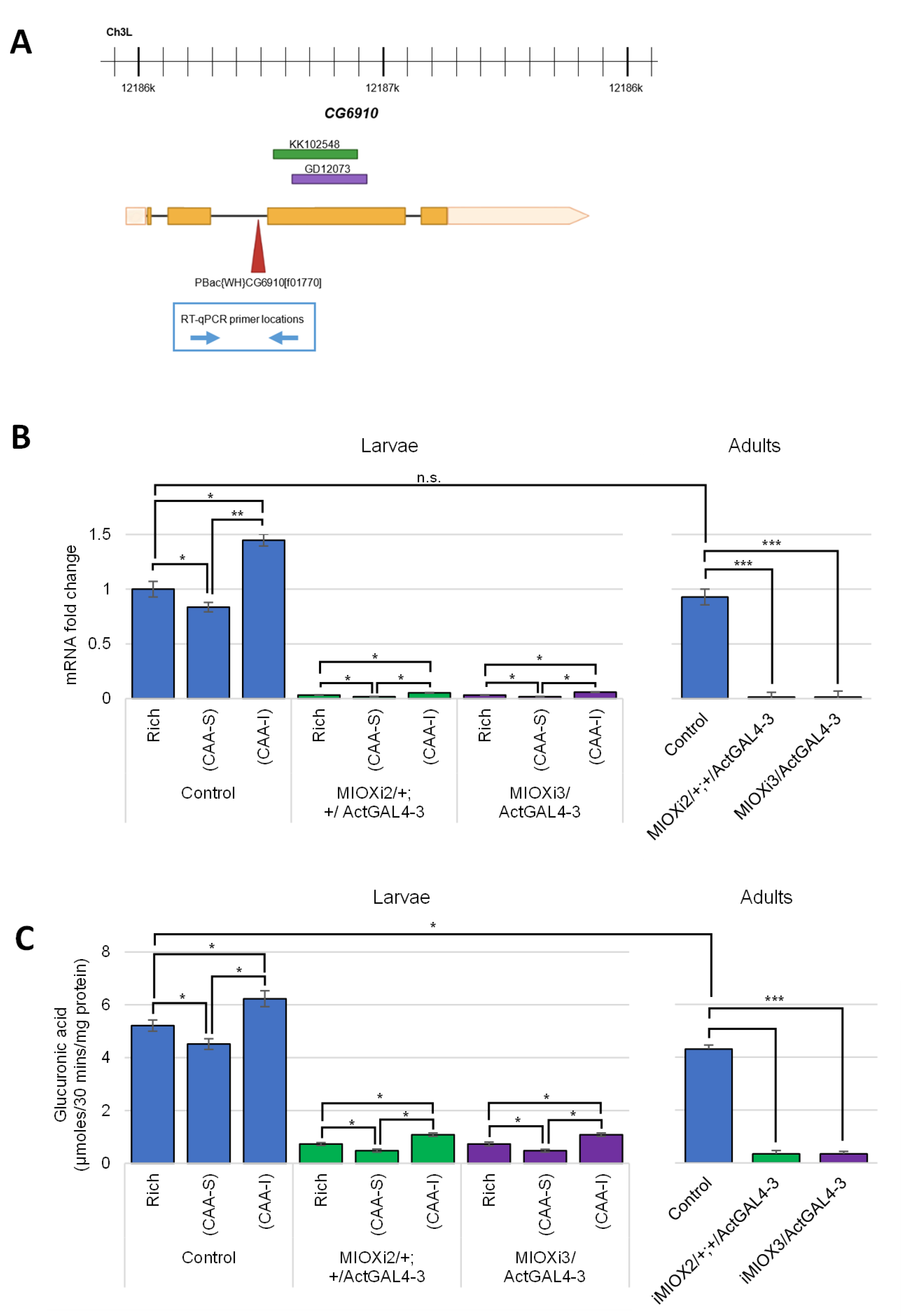
2.1. MIOX Homolog Identified in D. melanogaster (CG6910) Is Regulated in Response to Dietary myo-Inositol

To identify myo-inositol oxygenase (MIOX) in Drosophila melanogaster, BLASTP was performed using the 285 amino acid human MIOX protein sequence as a query. This revealed one candidate (CG6910) with ~55% identity spanning the entire protein. Although there are two splice variants of CG6910, B and C, listed in Flybase [59], 5′RACE experiments previously performed in this lab did not detect the C variant in larvae or adults. Moreover, high-throughput expression data (RNA -Seq Signal by Region) and the G-browse visual display reveal that RNA transcripts unique to the C variant region are rare or undetectable in all stages of development and in all tissues examined [59]. To experimentally verify that CG6910 encoded the MIOX protein, three strains were obtained; two RNAi strains to reduce CG6910 transcript levels and one strain with a piggyBac WH-element inserted into CG6910 (Figure 1A).
To determine if the levels of CG6910 (MIOX) mRNA are regulated in response to myo-inositol, qRT-PCR experiments were performed. RNA was extracted from third-instar larvae and adults of five D. melanogaster strains. Two of the strains have different MIOX (CG6910) RNAi constructs with UASGAL4 sequences. Both are controlled by GAL4 driven by the Actin 5C (Act5C) promoter (MIOXi2/+; +/Act5CGal4-3 and MIOXi3/Act5CGal4-3). Three control strains were also used (CS, ActGal4-3/Tb, and CyOGFP/+; ActGal4-3/Tb). The adults had slightly lower MIOX mRNA levels than the larvae. MIOX (CG6910) mRNA levels in MIOXi2/+; +/Act5Cgal4-3 and MIOXi3/Act5CGal4-3 were significantly lower than in the control strains. When larvae were grown on semi-defined food with myo-inositol as the sole sugar (CAA-I), the level of MIOX mRNA was significantly higher than when grown on semi-defined food with sucrose as the sole sugar (CAA-S) (Figure 1B).
To examine if dietary myo-inositol affected MIOX specific activity, the conversion of myo-inositol to glucuronic acid by crude lysates of third-instar larvae and adults was monitored in the strains described above. There was slightly less activity detectable in the adults than the larvae. MIOX specific activity in larvae was significantly higher for all the strains when grown on CAA-I. Moreover, the specific activity of MIOX in crude lysates of MIOXi2/+, +/Act5CGal4-3, and MIOXi3/Act5CGal4-3 larvae was significantly lower than that of the control larvae (Figure 1C). Even more striking is that there was no detectable MIOX activity in crude lysates of the homozygous piggyBac WH-element insertion strain (P-mioxf01770/P-mioxf01770).

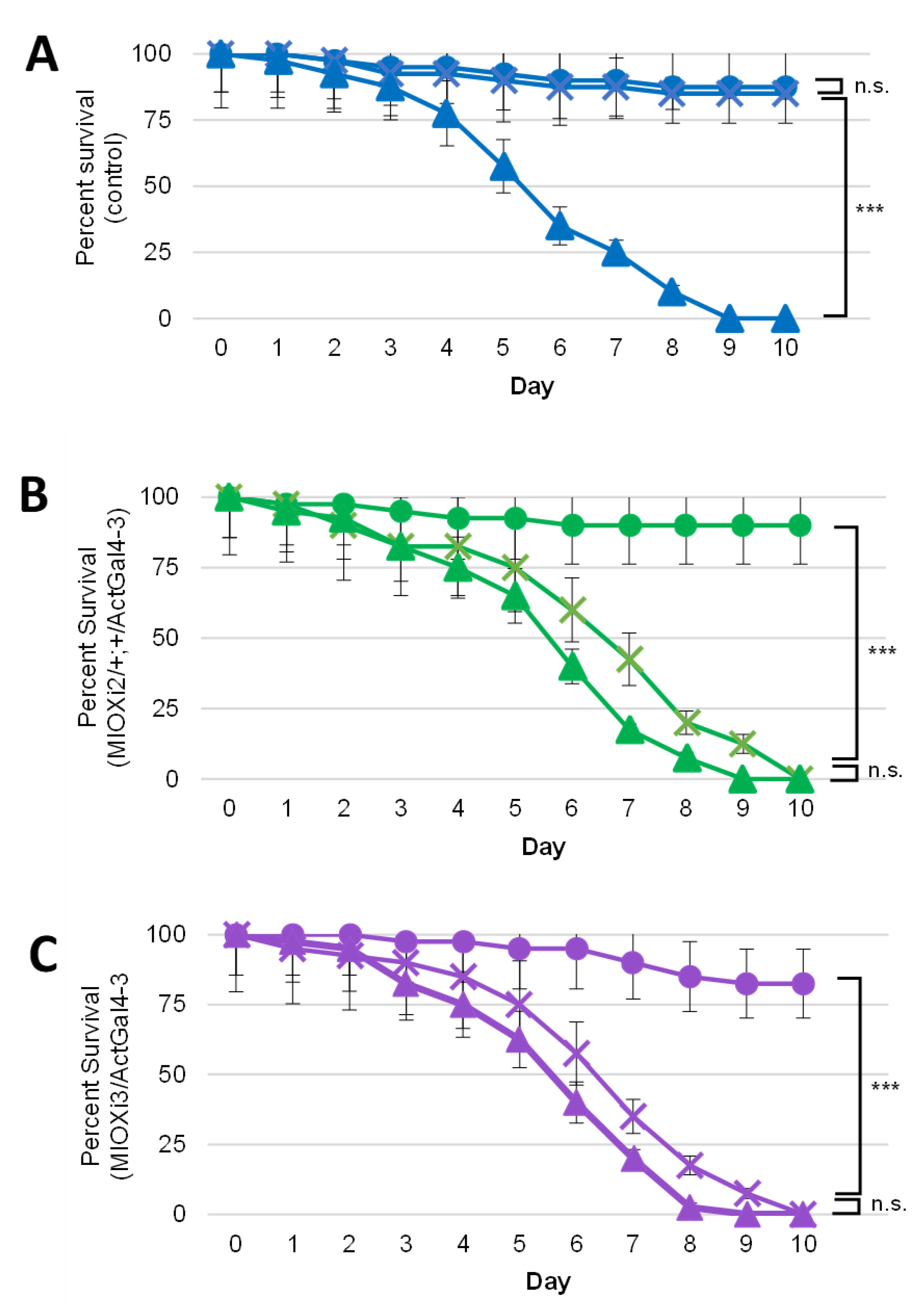
2.2. Dietary myo-Inositol Supports Survival of Wild-Type (CS) D. melanogaster Adults but Not of MIOX Knockdown Strains

To determine if MIOX plays a role in fruit fly survival, pupae of MIOXi2/+; +/Act5CGal4-3, MIOXi3/Act5CGal4-3, and the wild-type control strain (CS) were transferred to tubes with semi-defined food containing sucrose (CAA-S) or myo-inositol as the sole sugar (CAA-I), or no sugar (CAA-0). It was exciting to note that wild-type (CS) adult flies survived equally well on semi-defined food with either myo-inositol or sucrose as the sole sugar, demonstrating that there is sufficient myo-inositol catabolism to support survival of D. melanogaster (Figure 2A).
Moreover, control, MIOXi2/+; +/Act5CGal4-3, and MIOXi3/Act5CGal4-3 adults survived comparably well on semi-defined food with sucrose (CAA-S). On semi-defined no-sugar food (CAA-0), all three strains died within ten days (Figure 2A–C). In contrast to wild-type flies, MIOXi2/+; +/Act5CGal4-3 and MIOXi3/Act5CGal4-3 adults died by day 10 on semi-defined food with myo-inositol as the sole sugar (CAA-I). The survival of these two strains on CAA-I was similar to their survival on food with no-sugar (CAA-0) (Figure 2B,C).

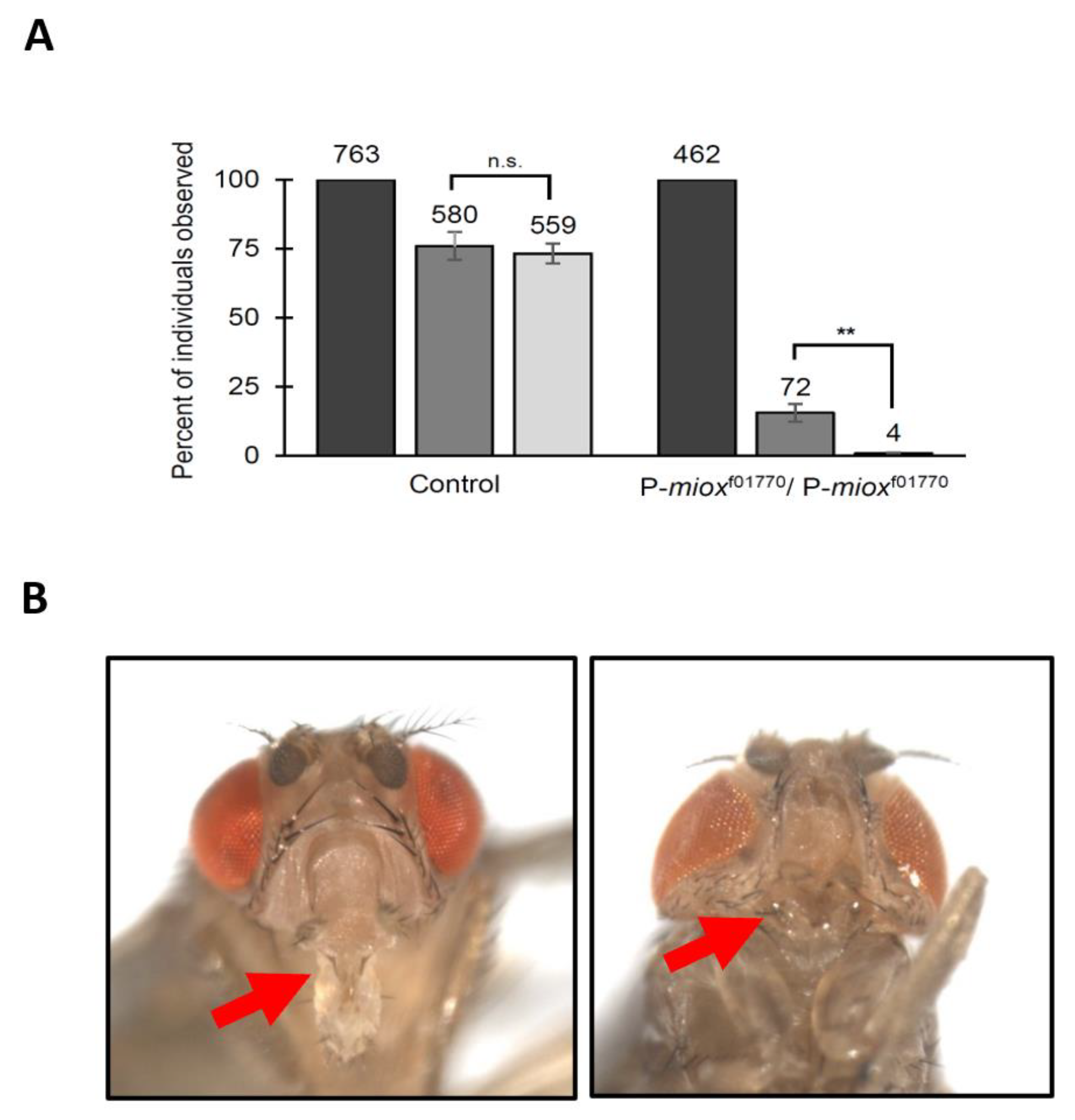
2.3. Disruption of myo-Inositol Catabolism via Piggybac WH-Element Insertion in MIOX Results in Developmental Defects

The survival experiments described above did not include adult P-mioxf01770/P-mioxf01770 flies, because these homozygotes are not viable as adults. As displayed in Figure 3A, ~16% of the homozygous P-mioxf01770/P-mioxf01770 and ~76% of the control embryos developed to the pupal stage. Even more striking is that only ~6% of the homozygous P-mioxf01770/P-mioxf01770 pupae eclosed as adults, with most dying as pharate adults (not eclosing from the pupal case), in contrast to ~96% of the control strain eclosing as adults. The few P-mioxf01770/P-mioxf01770 adults that eclosed from the pupal case died within two days and exhibited severe head morphological defects, most noticeably the lack of a proboscis (Figure 3B). To confirm that the piggyBac WH-element in P-mioxf01770 caused the pupal lethality and morphological defect, heterozygous strains with the element excised were generated. All three independently obtained excision lines (eleven stocks), with TM6, Tb balancer chromosomes, produced viable homozygous progeny in the expected ratio. Of the 998 pupae examined (350 homozygotes; 648 heterozygotes (Tb)), the same percentage of homozygotes and heterozygotes eclosed. Neither pupal lethality nor morphological defects were observed in the homozygous excision progeny. The excision of the element reverted the phenotype.

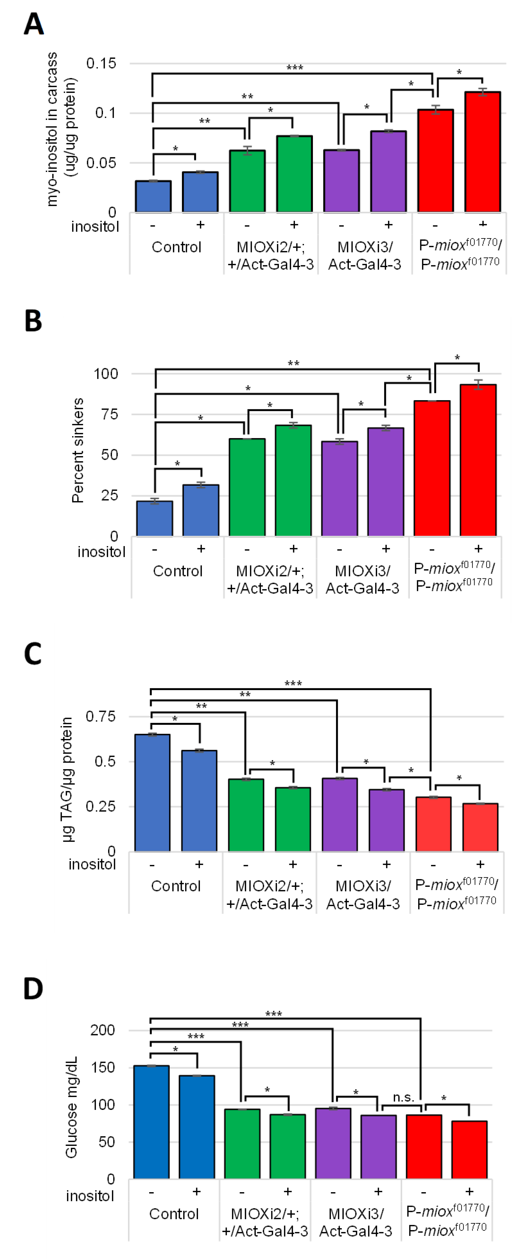
2.4. Reduced myo-Inositol Catabolism Increases myo-Inositol Levels in Larvae

To assess if the developmental defect observed in the P-mioxf01770/P-mioxf01770 strain was due to reduced catabolism yielding increased myo-inositol levels, assays were performed using carcasses of third-instar larvae grown on rich food with 0 or 50 µM myo-inositol supplementation. Higher myo-inositol levels were observed in the tissues of larvae with decreased myo-inositol catabolism (Figure 4A). The highest level of myo-inositol is apparent in the P-mioxf01770/P-mioxf01770 larval tissues which had no detectable myo-inositol catabolic activity via MIOX. In all the strains, myo-inositol levels increased when the standard rich food was supplemented with 50 µM of myo-inositol (Figure 4A).

2.5. Increased myo-Inositol Decreases Larval Obesity and Hemolymph Glucose

To examine the relationship between MIOX and the diabetic hallmarks, obesity and high hemolymph glucose, third-instar larvae grown on standard rich food with 0 or 50 µM myo-inositol supplementation were assayed. Buoyancy, TAG, and glucose assays revealed that P-mioxf01770/P-mioxf01770 larvae were the least obese with the lowest levels of TAG and hemolymph glucose. In these assays, the two RNAi knockdown strains, MIOXi2/+; +/Act5CGal4-3 and MIOXi3/Act5CGal4-3, had intermediate levels relative to the control and the P-mioxf01770/P-mioxf01770 strains (Figure 4B–D). Dietary myo-inositol supplementation (50 µM) further reduced the proportion of obese larvae, TAG, and hemolymph glucose in all strains (Figure 4B–D).

3. Discussion

This study examines the roles of myo-inositol catabolism using the model organism D. melanogaster. CG6910 was identified as the myo-inositol catabolic gene encoding myo-inositol oxygenase (MIOX), which is more than 55% identical (>70% similar) to human MIOX. The high level of identity among these two evolutionarily distant organisms demonstrates the conservation of MIOX structures in eukaryotes. There are two splice variants, B and C, listed in Flybase [59]; however, multiple experiments suggest that RNA transcripts unique to the C variant region are rare or undetectable in all stages of development and in all tissues examined [59]. If the C variant exists in larvae and adults, it comprises a small proportion of the MIOX transcripts and protein. Developmental proteome experiments reveal high levels of MIOX expression in late third-instar larvae (wandering and prepupae) and adults [60]. Temporal microarray and RNAseq data have shown the peak expression of MIOX (CG6910) mRNA in late third-instar larvae and adults [61]; therefore, an emphasis has been placed on examining third-instar larvae and adults.
This appears to be the first report to examine MIOX (CG6910) mRNA levels in animals, D. melanogaster larvae and adults, in response to dietary myo-inositol (Figure 1B). A significantly higher level of MIOX mRNA is apparent in qRT-PCR experiments examining larvae fed myo-inositol as the sole sugar (CAA-I) compared to larvae fed sucrose as the sole sugar (CAA-S). MIOX mRNA levels were reduced in larvae and adults via two different RNAi constructs (MIOXi2/+; +/Act5CGal4-3 and MIOXi3/Act5CGal4-3) (Figure 1B). Similar to the wild-type control, these strains have higher levels of MIOX mRNA when grown on CAA-I than when grown on CAA-S (Figure 1B). Increased myo-inositol catabolism, via MIOX, has been implicated in decreased renal function [62]. Decreased MIOX mRNA levels via siRNA in transgenic mice expressing high levels of MIOX mRNA has been shown to reduce renal damage and associated endoplasmic reticulum stressors [63]. Yet, in this study using D. melanogaster, neither the RNAi strain nor the wild-type controls exhibited any gross morphological abnormalities, even with increased levels of MIOX mRNA when fed CAA-I.
An assay to measure MIOX-specific activity in D. melanogaster was established based on previously existing protocols for rat kidneys, hog kidneys, and fungi [28,31,32] (Figure 1C). Similar to the mRNA levels, MIOX-specific activity levels in MIOXi2/+; +/Act5CGal4-3 and MIOXi3/Act5CGal4-3 larvae and adults were lower than the control (CS) strain. Moreover, all the strains had increased levels of MIOX activity when fed CAA-I. Homozygous CG6910-MIOX (P-mioxf01770/P-mioxf01770) larvae with a 7.2 kb piggyBac WH-element insertion disrupting the MIOX gene had no detectable MIOX activity. The MIOX-specific activity in crude lysates of control D. melanogaster larvae is 5.2 µmol/30 mins/mg, slightly more than that observed in adults, much more than that in rat kidney [29], and similar to that in the fungus Cryptococcus neoformans [32].
Survival experiments revealed that adult D. melanogaster have sufficient myo-inositol catabolism and transport to remain viable on semi-defined food with myo-inositol as the sole sugar/energy source (CAA-I) (Figure 2). The statistically significant results of six independent trials also showed that a reduction in MIOX expression via RNAi diminishes viability on CAA-I, mimicking survival on food with no sugar. The identical loss of the viability of both fly strains, MIOXi2/+; +/Act5CGal4-3 and MIOXi3/Act5CGal4-3, is particularly compelling, because the RNAi constructs are located on separate chromosomes and the strains were generated from two separate Vienna Drosophila Resource Center (VDRC) libraries (KK (phiC31) and GD (P-element), respectively). These libraries used different vectors and cloning methods and have been shown to have different off-target effects [64,65,66,67]. Since the two strains appeared phenotypically identical, the observed phenotypes in this study should be due to the decreased MIOX mRNA levels. Moreover, these results indicate that MIOX is a component of the primary myo-inositol catabolic pathway in D. melanogaster.
Adult P-mioxf01770/P-mioxf01770 were not included in experiments because they are rarely viable, with only ~6% of the pupae eclosing and the flies that eclose dying within two days (Figure 3A). These results are similar to previously published findings that highly upregulated myo-inositol synthesis reduces eclosion to ~9% [17]. As myo-inositol is a precursor of the phosphatidylinositol phosphates (PIPs), it is interesting to note that inositol phosphate kinase 2 (Ipk2) deletions and dysregulation of the expression of the phosphatidylinositol synthase gene (Pis) also cause pupal lethality [68,69]. Among the few P-mioxf01770/P-mioxf01770 adults, the most jarring morphological defect is the lack of proboscises (Figure 3B). This phenotype has been previously described when myo-inositol synthesis was highly upregulated [17]. The MIOX RNAi knockdown strains with intermediate levels of of myo-inositol in larval tissues did not display morphological abnormalities, paralleling previously published findings that lower but still elevated levels of myo-inositol synthesis did not produce the developmental defect [17]. Together, these data suggest that increased myo-inositol, not the process of synthesis or catabolism, contributes to or causes developmental defects. Deformities of fruit fly head structures have been observed with mutations disrupting the head involution defective (hid) [70,71] or the decapentaplegic (dpp) genes [72,73,74]. The morphological abnormalities observed in this study, however, seem to be unique to flies with elevated myo-inositol levels.
As could be predicted, less myo-inositol catabolism results in more myo-inositol in larval tissues, with the highest myo-inositol level observed in the strain with the lowest level of catabolism (P-mioxf01770/P-mioxf01770). Not surprisingly, the intermediate levels of myo-inositol catabolism in the two RNAi knockdown strains MIOXi2/+; +/Act5CGal4-3 and MIOXi3/Act5CGal4-3 showed intermediate levels of myo-inositol in larval tissues (Figure 4A). When rich food was supplemented with 50 µM of myo-inositol, the myo-inositol levels in all the strains increased (Figure 4A). Interestingly, whole MIOXi2/+; +/Act5CGal4-3 and MIOXi3/Act5CGal4-3 larvae fed rich food with 50 µM myo-inositol supplementation had more total myo-inositol than P-mioxf01770/P-mioxf01770 larvae fed rich food without myo-inositol supplementation, yet only P-mioxf01770/P-mioxf01770 displayed morphological defects (Figure 3B). Larvae with hemolymph removed (carcasses) of MIOXi2/+; +/Act5CGal4-3 and MIOXi3/Act5CGal4-3 fed rich food with 50 µM myo-inositol supplementation had lower total myo-inositol than carcasses of P-mioxf01770/P-mioxf01770 larvae fed rich food without myo-inositol supplementation (Figure 4A). Apparently, adding dietary myo-inositol, and by doing so increasing the circulating myo-inositol levels, does not affect development but does further reduce obesity (buoyancy and TAG) and hemolymph glucose levels in all the strains (Figure 4B,C). It is tantalizing to speculate that at least some of the developmental defects observed in P-mioxf01770/P-mioxf01770 stem from abnormally high myo-inositol levels during embryogenesis prior to the organism feeding. Notably, abnormally high myo-inositol levels contribute to the pathology of some human disorders of neurological development and dysfunction [6,7,75,76,77].
Alterations of myo-inositol metabolism been implicated in many human diseases and disorders including diabetes, obesity, and hyperglycemia. Low MIOX expression/activity, which should elevate myo-inositol levels, rescued mice and rats from renal injury and oxidative stress [27,62]. In this study, low MIOX levels were shown to reduce obesity and hyperglycemia in populations of D. melanogaster larvae. In addition, the supplementation of the rich food with 50 µM of myo-inositol further reduced obesity and hyperglycemia. In humans, dietary myo-inositol augmentation may mitigate obesity and hyperglycemia (high blood glucose) [4,7,78]. These results complement studies which established that reduction in the inositol 1,4,5-trisphosphate receptor (InsP3R), either by knockdown or mutation, resulted in obese adult fruit flies [79]. Moreover, these results are in agreement with studies demonstrating that an increase in myo-inositol, via the overexpression of the myo-inositol synthetic gene or the addition of dietary myo-inositol, decreased obesity and hyperglycemia in D. melanogaster [17]. In summary, increased myo-inositol, regardless of its source, can mitigate diabetes-associated obesity and hyperglycemia. This study, at the junction of metabolism and development, furthers the understanding of the importance of myo-inositol catabolism and the regulation of intracellular myo-inositol levels and may have implications for the treatment of diabetes and developmental disorders.

4. Materials and Methods

4.1. Fly Stocks and Maintenance

Stocks obtained from the Bloomington Drosophila Stock Center (Bloomington, IN, USA) include the Canton-S (#1, CS) strain, w [1118]; PBac{w[+mC] = WH}CG6910[f01770]/ TM6B, Tb[1] (#18471, hereafter identified as P-mioxf01770/Tb), y[1] w[*]; +;P(w[+mC] = Act5C-GAL4)17bFO1/TM6B, Tb[1] (#3954, hereafter identified as Act-Gal4-3/Tb), w[1118]; CyO, P{w[+mC] = FRT(w[+])Tub-PBac\T}2/wg[Sp-1] (#8283), w[*]; TM3, Sb [1] Ser [1]/TM6B, Tb[1] (#2537, hereafter identified as w[*]; Tb/Sb), and w[1118]; Df(3L) Ly, sens(Ly-1)/TM6B, P{w[+mW.hs]=Ubi-GFP.S65T}PAD2, Tb[1] (#4887) and w[1]; sna[Sco]/CyO, P{w[+mC] = GAL4-Hsp70.PB}TR1, P{w[+mC] = UAS-GFP.Y}TR1 (#5702) used to introduce GFP-marked chromosomes 3 and 2, respectively). The two RNAi strains P{KK102548}VIE-260B (#103766, hereafter identified as MIOXi2/MIOXi2) and w[1118]; P{GD12073} v22464/TM3, Tb (#22464, hereafter identified as MIOXi3/Tb) were obtained from the Vienna Drosophila Research Center (Vienna, Austria). The RNAi strain MIOXi2/MIOXi2 is homozygous for an RNAi construct complementary to the third exon of CG6910 that was inserted via P-element to chromosome 2. The other RNAi strain, MIOXi3/Tb, also containing sequences complementary to the third exon of CG6910, is heterozygous for a different RNAi construct that was inserted via a phiC31 sequence to chromosome 3. Both RNAi constructs contain UASGAL4 sequences controlled by GAL4 expression. MIOXi2/+; +/ActGal4-3 and MIOXi3/ActGal4-3 were generated by mating strains marked with GFP on corresponding balancer chromosomes. The third strain was generated by introducing a GFP-marked balancer third chromosome (TM6) into the P-mioxf01770/Tb, an existing strain with an approximately 7.2kb piggyBac WH-element inserted into the second intron of CG6910, and non-GFP non-tubby homozygotes (P-mioxf01770/ P-mioxf01770) were then readily identified. The piggyBac-element insertion in CG6910 was remapped and its location confirmed using flanking sequence data [80]. To create a double-marked (Tb/Sb) transposase strain, the strain harboring the transposase (#8283) was crossed to a third chromosome double balancer strain (#2537) introducing the Tb marked TM6B chromosome. These Tb marked flies were again crossed to the third chromosome double balancer strain (#2537) to introduce the Sb marked TM3 chromosome creating the double marked transposase strain w [1118]; CyO, P{w[+mC] = FRT(w[+])Tub-PBac\T}2; TM3, Sb [1] Ser [1]/TM6B, Tb [1] (hereafter identified as w[+]; piggyBac transposase; Tb/Sb). To excise the piggyBac WH-element inserted in the MIOX gene (CG6910), the w[+]; piggyBac transposase; Tb/Sb strain was crossed to P-mioxf01770/Tb (#18471). According to Thibault et al. 2004 [81], excisions of this piggyBac WH-element are precise. Seventy-two single Tubby (Tb) progeny with dark red eyes (double w[+mC]) with curly wings (CyO) harboring both the transposase and the piggyBac WH-element were individually mated with w[*]; Tb/Sb. Eight of the seventy-two crosses produced white-eyed, straight-winged progeny, carrying neither the piggyBac WH-element (chromosome 3, CG6910) nor the transposase (chromosome 2, CyO, P{w[+mC] = FRT(w[+])Tub-PBac\T}2). Of these eight, three produced multiple white-eyed, straight-winged progeny which were used to establish stocks. Three or four individuals from each of the three independent lines (eleven flies) with putative piggyBac WH excisions (from the MIOX gene (CG6910)) were individually crossed to w[*]; Tb/Sb, and Tb progeny were selected.
Flies were maintained in standard laboratory conditions at 25 °C and 70–80% humidity on a 12 h:12 h light–dark cycle. All fly stocks were grown on either rich food (BDSC cornmeal food, https://bdsc.indiana.edu/information/recipes/bloomfood.html (accessed on 29 April 2011)) or modified food (per liter, 10 g of agar (Fisher Scientific, Waltham, MA, USA), 80 g of brewer’s yeast (Genesee), 20 g of yeast extract (Fisher Scientific), 20 g of peptone (Fisher Scientific), and sucrose (Fisher Scientific) as indicated, [49]) with or without 50 µM of myo-inositol (Sigma, St. Louis, MO, USA) as indicated, which is sufficient to support growth of a homozygous Inos deletion mutant (inosΔDF/inosΔDF) [16]. Semi-defined food (casamino acids sucrose (CAA-S) and casamino acids myo-inositol (CAA-I)) was prepared essentially as described [82] with modifications [83]. Briefly, defined food contained 0.4 g of lecithin, 0.613 g of vitamin mix, 5.5 g of casamino acids, 3.15 g of agar, and 7.5 g of sugar (sucrose or myo-inositol) or no sugar per 100 mL. The vitamin mix was composed of 3 g of cholesterol, 0.02 g of thiamine, 0.01 g of riboflavin, 0.12 g of nicotinic acid, 0.16 g of calcium pantothenate, 0.25 g of pyridoxine, 0.016 g of biotin, 0.03 g of folic acid, 14 g of NaHCO3, 18.3 g of KH2PO4, 18.9 g of K2HPO4, and 6.2 g of MgSO4. Then, 350 μL of 30% Tegosept was added to the 100 mL of food.

4.2. RNA Extraction and qRT-PCR

Total RNA was extracted from 10–20 third-instar larvae or adult flies grown on the food indicated using TrizolTM (Life Technologies, Carlsbad, CA, USA) [84]. Total RNA (1 µg) was DNase-treated using the DNA-free Kit (Ambion, Foster City, CA, USA) with inactivation buffer (DNA-free DNA Removal Kit, Invitrogen, Carlsbad, CA, USA). cDNAs were generated using oligo (dT) 18 primers (Eurofins, Luxembourg), dNTPs (ThermoFisher, Waltham, MA, USA), and Moloney Murine Leukemia Virus Reverse Transcriptase (M-MLV RT) (Fisher, Waltham, MA, USA). After amplification, the samples were treated with RNAse H (New England BioLabs, Ipswich, MA, USA). The cDNA was diluted in RNase/DNase-free water (ThermoFisher) (1:16), and qRT-PCR experiments were performed using ThermoScientific Absolute qPCR Mix, SYBR Green, ROX (Fisher) in an Applied Biosystems StepOnePlus System. Triplicate samples were used in all the experiments including linearizations and melt curves. All the experiments were performed at least three independent times (separate biological samples), as indicated in the figure legends. The results were normalized to the transcript levels of Drosophila melanogaster ribosomal protein L32 (RpL32). The following primers were used: MIOX exon 2-3 forward GACACCACCGATCCTCTAAAGG and reverse GGAAGGCGTGGATGATGT, RpL32 forward CCAGCATACAGGCCCAAGAT and reverse GCACTCTGTTGTCGATACCC.

4.3. Protein Extraction and the MIOX Activity Assay

The MIOX activity assays were established for D. melanogaster based on protocols for hog kidneys and fungi [31,32]. For each sample, ten flies or ten larvae were homogenized in 300 μL of 20 mM sodium acetate (pH 6.0), 2 mM L-cysteine (pH 4.5), 1 mM glutathione (pH 3.5), 1mM ferrous ammonium sulfate (pH 4.5), and 10 μL of protease inhibitor (Halt™ Protease Inhibitor Cocktail (100X), Thermo Scientific). Protein concentrations of the crude lysate cleared supernatants were determined using the Bradford Assay [85] with bovine serum albumin (Pierce™ Bovine Serum Albumin Standard, Thermo Scientific) and using a dye concentrate (5000006, Bio-Rad, Hercules, CA, USA) for the colorimetric analysis. myo-Inositol catabolism was assayed in 450 μL of 50 mM sodium acetate (pH 6.0), 2 mM L-cysteine (pH 3.5), 1 mM ferrous ammonium sulfate (pH 4.5), and 60 mM myo-inositol (or water) with 15 μg of crude lysate protein. The reactions were incubated at 30 °C. Then, 200 μL aliquots were removed at 0 and 30 min, immediately added to 55 μL of 30% trichloroacetic acid (TCA), and incubated at 100 °C for two minutes. The glucuronic acid concentrations of the cleared supernatants were determined by adding 500 μL of orcinol reagent (0.08 g of orcinol, 0.018 g of FeCl3 and 20 mL of concentrated HCl) to 250 μL of the sample and were measured at 660 nm [28].

4.4. Survival Studies

Twenty CS or Act-Gal4-3/TbGFP virgin females and ten CS or MIOXi2/MIOXi2 or MIOXi3/TbGFP males were mated on rich food. For each of the six independent trials, twenty pupae from each cross were transferred onto rich food with 0 or 50 µM of myo-inositol added, CAA-S, CAA-I, or CAA-0 (no sugar added). Survival was monitored daily.

4.5. Pupariation and Eclosion

Female and male adults (2:1) were placed in vials of standard rich food in a 25 °C incubator at 70–80% humidity on a 12 h:12 h light–dark cycle. The progeny (embryos) were sorted using the GFP marker and reconfirmed as larvae. To allow sufficient time for genotypes with developmental delays to eclose, the vials were checked daily for up to 28 days. The number of pupae were recorded, as was the number of adults that eclosed.

4.6. Light Microscopy

Fifteen control CS and sixteen independent experimental P-mioxf01770/P-mioxf01770 specimens were viewed on a Nikon SMZ1500 microscope, and images were captured using a Micropublisher 6 color CCD camera system (Teledyne Q imaging, Surrey, BC, Canada).

4.7. myo-Inositol Assay

Five third-instar larval carcasses per sample were collected by puncturing five larvae and draining the hemolymph via centrifugation. The samples were homogenized in dH2O, and the Megazyme myo- inositol assay kit (K-INOSL) was used as per the manufacturer’s instructions. Three independent trials were performed.

4.8. Buoyancy Assay

Experiments were conducted essentially as described by Reis [48,54] using 20–30 3rd-instar larvae per sample, with initial results confirmed by the method of Hazegh and Reis [54]. A relationship between the percentage of larvae floating in a buoyancy assay and the percent body fat of the larvae has been established [48,86]. In this study, the relative obesity of a population of larvae is defined as the proportion of the larvae floating in the buoyancy assay.

4.9. Triacylglyceride (TAG) Assay

Experiments were performed essentially as previously described [39] and normalized to total protein (using the Bradford Assay described above). Six third-instar larvae per sample were homogenized in 1xPBS with 9.1% Tween, and the Serum Triglyceride Determination Kit (TR0100, Sigma, St. Louis, MO, USA) and Triglyceride Reagent (T2449, Sigma) were used as per the manufacturer’s instructions for three independent trials.

4.10. Hemolymph Glucose Assay

Experiments were performed essentially as described by Tennessen et al. [39]. Hemolymph was collected by puncturing five third-instar larvae per replicate, and the Sigma Glucose (GO) Assay Kit GAGO-20 was used. Three independent trials were performed.

4.11. Computational Analyses

The National Center for Biotechnology Information’s (NCBI) Basic Local Alignment Search Tool (BLASTP) with default settings (BLOSSUM 62) was used to identify the MIOX homolog in D. melanogaster.

4.12. Statistical Analyses

Standard error was calculated for all experiments. Mantel–Cox (log rank) tests were performed to calculate significance in survival studies. The p-values of pairwise comparisons were determined using Student’s two-tailed t-test.

5. Conclusions

In the model organism Drosophila melanogaster, the elimination of myo-inositol catabolism and the associated high levels of myo-inositol cause severe developmental defects. A reduction in myo-inositol catabolism or dietary myo-inositol supplementation yields the beneficial effects of higher myo-inositol levels (reduced obesity and hemolymph (blood) glucose in Drosophila melanogaster third-instar larvae) without causing developmental defects. This suggests that dietary inositol supplementation may serve as a therapeutic agent.

Author Contributions

Conceptualization, L.S.K.; validation, A.C., M.K.J., E.D.E. and L.S.K.; formal analysis, A.C. and M.K.J.; investigation, A.C., M.K.J., E.D.E. and L.S.K.; resources, E.D.E. and L.S.K.; writing—original draft preparation, A.C., M.K.J., E.D.E. and L.S.K.; writing—review and editing, M.K.J., E.D.E. and L.S.K.; visualization, A.C., M.K.J. and E.D.E.; supervision, L.S.K.; project administration, L.S.K.; funding acquisition, L.S.K. All authors have read and agreed to the published version of the manuscript.

Funding

This work was supported by funds for A.C. from RISE National Institutes of Health R25GM071638 and for L.S.K. from Research, Scholarly, and Creative Activity (RSCA) CSULB Division of Academic Affairs.

Data Availability Statement

Strains available upon request. All the relevant data are within the paper.

Acknowledgments

We thank Sandy Rodriguez and Chantana Bun for their assistance with the MIOX assay, Matt Smith for the RACE data, and Keona Wang for her help with the hemolymph glucose assays. Stocks obtained from the Bloomington Drosophila Stock Center (National Institutes of Health P40OD018537) and the Vienna Drosophila Research Center (VDRC, www.vdrc.at (accessed on 27 September 2010)) were used in this study.

Conflicts of Interest

The authors declare no conflict of interest.

References

  1. Bevilacqua, A.; Bizzarri, M. Inositols in insulin signaling and glucose metabolism. Int. J. Endocrinol. 2018, 2018, 1968450. [Google Scholar] [CrossRef] [Green Version]
  2. Chhetri, D.R. Myo-inositol and its derivatives: Their emerging role in the treatment of human diseases. Front. Pharmacol. 2019, 10, 1172. [Google Scholar] [CrossRef] [Green Version]
  3. Guo, T.; Nan, Z.; Miao, C.; Jin, X.; Yang, W.; Wang, Z.; Tu, Y.; Bao, H.; Lyu, J.; Zheng, H.; et al. The autophagy-related gene Atg101 in Drosophila regulates both neuron and midgut homeostasis. J. Biol. Chem. 2019, 294, 5666–5676. [Google Scholar] [CrossRef] [PubMed]
  4. D’Anna, R.; Corrado, F.; Loddo, S.; Gullo, G.; Giunta, L.; Di Benedetto, A. Myoinositol plus α-lactalbumin supplementation, insulin resistance and birth outcomes in women with gestational diabetes mellitus: A randomized, controlled study. Sci. Rep. 2021, 11, 8866. [Google Scholar] [CrossRef]
  5. Kiani, A.K.; Paolacci, S.; Calogero, A.E.; Cannarella, R.; Di Renzo, G.C.; Gerli, S.; Della Morte, C.; Busetto, G.M.; De Berardinis, E.; Del Giudice, F.; et al. From Myo-inositol to D-chiro-inositol molecular pathways. Eur. Rev. Med. Pharmacol. Sci. 2021, 25, 2390–2402. [Google Scholar] [CrossRef]
  6. Patkee, P.A.; Baburamani, A.A.; Long, K.R.; Dimitrova, R.; Ciarrusta, J.; Allsop, J.; Hughes, E.; Kangas, J.; McAlonan, G.M.; Rutherford, M.A.; et al. Neurometabolite mapping highlights elevated myo-inositol profiles within the developing brain in down syndrome. Neurobiol. Dis. 2021, 153, 105316–105325. [Google Scholar] [CrossRef]
  7. Dinicola, S.; Unfer, V.; Facchinetti, F.; Soulage, C.O.; Greene, N.D.; Bizzarri, M.; Laganà, A.S.; Chan, S.-Y.; Bevilacqua, A.; Pkhaladze, L.; et al. Inositols: From established knowledge to novel approaches. Int. J. Mol. Sci. 2021, 22, 10575. [Google Scholar] [CrossRef]
  8. Weinberg, S.E.; Sun, L.Y.; Yang, A.L.; Liao, J.; Yang, G.Y. Overview of inositol and inositol phosphates on chemoprevention of colitis-induced carcinogenesis. Molecules 2020, 26, 31. [Google Scholar] [CrossRef] [PubMed]
  9. Dyck, P.J.; Zimmerman, B.R.; Vilen, T.H.; Minnerath, S.R.; Karnes, J.L.; Yao, J.K.; Poduslo, J.F. Nerve Glucose, Fructose, Sorbitol, Myoinositol and Fiber Degeneration and Regeneration in Diabetic Neuropathy. New Engl. J. Med. 1988, 319, 542–548. [Google Scholar] [CrossRef] [PubMed]
  10. Lin, L.-R.; Reddy, V.; Giblin, F.; Kador, P.; Kinoshita, J. Polyol accumulation in cultured human lens epithelial cells. Exp. Eye Res. 1991, 52, 93–100. [Google Scholar] [CrossRef]
  11. Henry, D.N.; Del Monte, M.; A Greene, D.; Killen, P.D. Altered aldose reductase gene regulation in cultured human retinal pigment epithelial cells. J. Clin. Investig. 1993, 92, 617–623. [Google Scholar] [CrossRef]
  12. Cohen, A.M.; Wald, H.; Popovtzer, M.; Rosenmann, E. Effect of myo-inositol supplementation on the development of renal pathological changes in the Cohen diabetic (type 2) rat. Diabetologia 1995, 38, 899–905. [Google Scholar] [CrossRef]
  13. Arner, R.J.; Prabhu, K.S.; Krishnan, V.; Johnson, M.C.; Reddy, C.C. Expression of myo-inositol oxygenase in tissues susceptible to diabetic complications. Biochem. Biophys. Res. Commun. 2006, 339, 816–820. [Google Scholar] [CrossRef]
  14. Lu, Y.; Liu, C.; Miao, X.; Xu, K.; Wu, X.; Liu, C. Increased expression of myo-inositol oxygenase is involved in the tubulointerstitial injury of diabetic nephropathy. Exp. Clin. Endocrinol. Diabetes 2009, 117, 257–265. [Google Scholar] [CrossRef] [PubMed]
  15. Nayak, B.; Kondeti, V.K.; Xie, P.; Lin, S.; Viswakarma, N.; Raparia, K.; Kanwar, Y.S. Transcriptional and post-translational modulation of myo-inositol oxygenase by high glucose and related pathobiological stresses. J. Biol. Chem. 2011, 286, 27594–27611. [Google Scholar] [CrossRef] [PubMed] [Green Version]
  16. Jackson, N.A.; Flores, A.M.; Eldon, E.D.; Klig, L.S. Disruption of INOS, a gene encoding myo-inositol phosphate synthase, causes male sterility in Drosophila melanogaster. G3 Genes Genomes Genet. 2018, 8, 2913–2922. [Google Scholar] [CrossRef] [Green Version]
  17. Rivera, M.J.; Contreras, A.; Nguyen, L.T.; Eldon, E.D.; Klig, L.S. Regulated inositol synthesis is critical for balanced metabolism and development in Drosophila melanogaster. Biol. Open 2021, 10, bio058833. [Google Scholar] [CrossRef] [PubMed]
  18. Loewus, F.A.; Loewus, M.W. Myo-Inositol: Its biosynthesis and metabolism. Annu. Rev. Plant Physiol. 1983, 34, 137–161. [Google Scholar] [CrossRef]
  19. Holub, B.J. Metabolism and function of myo-inositol and inositol phospholipids. Annu. Rev. Nutr. 1986, 6, 563–597. [Google Scholar] [CrossRef]
  20. Holub, B.J. The nutritional importance of inositol and the phosphoinositides. New Engl. J. Med. 1992, 326, 1285–1287. [Google Scholar] [CrossRef]
  21. Henry, S.A.; Gaspar, M.L.; Jesch, S.A. The response to inositol: Regulation of glycerolipid metabolism and stress response signaling in yeast. Chem. Phys. Lipids 2014, 180, 23–43. [Google Scholar] [CrossRef] [Green Version]
  22. Basak, P.; Sangma, S.; Mukherjee, A.; Agarwal, T.; Sengupta, S.; Ray, S.; Majumder, A.L. Functional characterization of two myo-inositol-1-phosphate synthase (MIPS) gene promoters from the halophytic wild rice (Porteresia coarctata). Planta 2018, 248, 1121–1141. [Google Scholar] [CrossRef] [PubMed]
  23. Cui, W.; Ma, A.; Wang, X.; Huang, Z. Myo-inositol enhances the low-salinity tolerance of turbot (Scophathalmus maximus) by modulating cortisol synthesis. Biochem. Biophys. Res. Commun. 2020, 526, 913–919. [Google Scholar] [CrossRef] [PubMed]
  24. Howard, C.F.; Anderson, L. Metabolism of myo-inositol in animals. II. Complete catabolism of myo-inositol-14C by rat kidney slices. Arch. Biochem. Biophys. 1967, 118, 332–339. [Google Scholar] [CrossRef] [PubMed]
  25. Hankes, L.V.; Politzer, W.M.; Touster, O.; Anderson, L. Myo-inositol catabolism in human pentosurics: The predominant role of the glucuronate-xylulose-pentose phosphate pathway. Ann. N. Y. Acad. Sci. 1969, 165, 564–576. [Google Scholar] [CrossRef] [PubMed]
  26. Arner, R.J.; Prabhu, K.S.; Thompson, J.T.; Hildenbrandt, G.R.; Liken, A.D.; Reddy, C.C. Myo-Inositol oxygenase: Molecular cloning and expression of a unique enzyme that oxidizes myo-inositol and d-chiro-inositol. Biochem. J. 2001, 360, 313–320. [Google Scholar] [CrossRef]
  27. Chang, H.-H.; Chao, H.-N.; Walker, C.S.; Choong, S.-Y.; Phillips, A.; Loomes, K.M. Renal depletion of myo-inositol is associated with its increased degradation in animal models of metabolic disease. Am. J. Physiol. Physiol. 2015, 309, F755–F763. [Google Scholar] [CrossRef] [Green Version]
  28. Charalampous, F.C.; Lyras, C. Biochemical studies on inositol. IV. Conversion of inositol to glucuronic acid by rat kidney extracts. J. Biol. Chem. 1957, 228, 1–13. [Google Scholar] [CrossRef]
  29. Charalampous, F.C. Biochemical studies on inositol. V. Purification and properties of the enzyme that cleaves inositol to D-glucuronic acid. J. Biol. Chem. 1959, 234, 220–227. [Google Scholar] [CrossRef] [PubMed]
  30. Koller, E.; Koller, F.; Hoffmann-Ostenhof, O. Myo-inositol oxygenase from oat seedlings. Mol. Cell. Biochem. 1976, 10, 33–39. [Google Scholar] [CrossRef]
  31. Reddy, C.C.; Swan, J.S.; Hamilton, G.A. Myo-inositol oxygenase from hog kidney. I. Purification and characterization of the oxygenase and of an enzyme complex containing the oxygenase and D-glucuronate reductase. J. Biol. Chem. 1981, 256, 8510–8518. [Google Scholar] [CrossRef] [PubMed]
  32. Molina, Y.; Ramos, S.E.; Douglass, T.; Klig, L.S. Inositol synthesis and catabolism in Cryptococcus neoformans. Yeast 1999, 15, 1657–1667. [Google Scholar] [CrossRef]
  33. Mackenzie, E.A.; Klig, L.S. Computational modeling and in silico analysis of differential regulation of myo-inositol catabolic enzymes in Cryptococcus neoformans. BMC Mol. Biol. 2008, 9, 88. [Google Scholar] [CrossRef] [Green Version]
  34. Endres, S.; Tenhaken, R. Down-regulation of the myo-inositol oxygenase gene family has no effect on cell wall composition in Arabidopsis. Planta 2011, 234, 157–169. [Google Scholar] [CrossRef] [Green Version]
  35. Li, Z.; Liu, Z.; Wei, Y.; Liu, Y.; Xing, L.; Liu, M.; Li, P.; Lu, Q.; Peng, R. Genome-wide identification of the MIOX gene family and their expression profile in cotton development and response to abiotic stress. PLoS ONE 2021, 16, e0254111. [Google Scholar] [CrossRef]
  36. Brown, P.M.; Caradoc-Davies, T.T.; Dickson JM, J.; Cooper GJ, S.; Loomes, K.M.; Baker, E.N. Crystal structure of a substrate complex of myo-inositol oxygenase, A di-iron oxygenase with a key role in inositol metabolism. Proc. Natl. Acad. Sci. USA 2006, 103, 15032–15037. [Google Scholar] [CrossRef] [Green Version]
  37. Thorsell, A.-G.; Persson, C.; Voevodskaya, N.; Busam, R.D.; Hammarström, M.; Graslund, S.; Gräslund, A.; Hallberg, B.M. Structural and biophysical characterization of human myo-inositol oxygenase. J. Biol. Chem. 2008, 283, 15209–15216. [Google Scholar] [CrossRef] [PubMed] [Green Version]
  38. Musselman, L.P.; Kühnlein, R.P. Drosophilaas a model to study obesity and metabolic disease. J. Exp. Biol. 2018, 221, jeb163881. [Google Scholar] [CrossRef] [PubMed] [Green Version]
  39. Tennessen, J.M.; Barry, W.E.; Cox, J.; Thummel, C.S. Methods for studying metabolism in Drosophila. Methods 2014, 68, 105–115. [Google Scholar] [CrossRef] [Green Version]
  40. Ugrankar, R.; Berglund, E.; Akdemir, F.; Tran, C.; Kim, M.S.; Noh, J.; Schneider, R.; Ebert, B.; Graff, J.M. Drosophila glucome screening identifies Ck1alpha as a regulator of mammalian glucose metabolism. Nat. Commun. 2015, 6, 7102. [Google Scholar] [CrossRef] [Green Version]
  41. Williams, M.J.; Klockars, A.; Eriksson, A.; Voisin, S.; Dnyansagar, R.; Wiemerslage, L.; Kasagiannis, A.; Akram, M.; Kheder, S.; Ambrosi, V.; et al. The drosophila ETV5 homologue Ets96B: Molecular link between obesity and bipolar disorder. PLoS Genet. 2016, 12, e1006104. [Google Scholar] [CrossRef] [PubMed] [Green Version]
  42. Graham, P.; Pick, L. Drosophila as a model for diabetes and diseases of insulin resistance. Curr. Top. Dev. Biol. 2017, 121, 397–419. [Google Scholar] [CrossRef] [PubMed] [Green Version]
  43. Mattila, J.; Hietakangas, V. Regulation of carbohydrate energy metabolism in Drosophila melanogaster. Genetics 2017, 207, 1231–1253. [Google Scholar] [CrossRef] [PubMed]
  44. Huang, Y.; Wan, Z.; Wang, Z.; Zhou, B. Insulin signaling in Drosophila melanogaster mediates Aβ toxicity. Commun. Biol. 2019, 2, 13. [Google Scholar] [CrossRef] [Green Version]
  45. Gillette, C.M.; Tennessen, J.M.; Reis, T. Balancing energy expenditure and storage with growth and biosynthesis during Drosophila development. Dev. Biol. 2021, 475, 234–244. [Google Scholar] [CrossRef] [PubMed]
  46. Chatterjee, N.; Perrimon, N. What fuels the fly: Energy metabolism in Drosophila and its application to the study of obesity and diabetes. Sci. Adv. 2021, 7, eabg4336. [Google Scholar] [CrossRef] [PubMed]
  47. Baker, K.D.; Thummel, C.S. Diabetic larvae and obese flies—Emerging studies of metabolism in Drosophila. Cell Metab. 2007, 6, 257–266. [Google Scholar] [CrossRef] [Green Version]
  48. Reis, T.; Van Gilst, M.R.; Hariharan, I.K. A buoyancy-based screen of Drosophila larvae for fat-storage mutants reveals a role for Sir2 in coupling fat storage to nutrient availability. PLoS Genet. 2010, 6, e1001206. [Google Scholar] [CrossRef] [Green Version]
  49. Musselman, L.P.; Fink, J.L.; Narzinski, K.; Ramachandran, P.V.; Sukumar Hathiramani, S.; Cagan, R.L.; Baranski, T.J. A high-sugar diet produces obesity and insulin resistance in wild-type Drosophila. Dis. Model. Mech. 2011, 4, 842–849. [Google Scholar] [CrossRef] [PubMed] [Green Version]
  50. Pandey, U.B.; Nichols, C.D. Human disease models in Drosophila melanogaster and the role of the fly in therapeutic drug discovery. Pharmacol. Rev. 2011, 63, 411–436. [Google Scholar] [CrossRef] [PubMed] [Green Version]
  51. Grewal, S. Controlling animal growth and body size–Does fruit fly physiology point the way? F1000 Biol. Rep. 2012, 4, 12. [Google Scholar] [CrossRef] [Green Version]
  52. Rovenko, B.M.; Kubrak, O.I.; Gospodaryov, D.V.; Perkhulyn, N.V.; Yurkevych, I.S.; Sanz, A.; Lushchak, O.V.; Lushchak, V.I. High sucrose consumption promotes obesity whereas its low consumption induces oxidative stress in Drosoph melanogaster. J. Insect Physiol. 2015, 79, 42–54. [Google Scholar] [CrossRef] [PubMed]
  53. Hazegh, K.E.; Reis, T. A Buoyancy-based method of determining fat levels in Drosophila. J. Vis. Exp. 2016, 117, 54744. [Google Scholar] [CrossRef]
  54. Reis, T. Effects of synthetic diets enriched in specific nutrients on Drosophila development, body fat, and lifespan. PLoS ONE 2016, 11, e0146758. [Google Scholar] [CrossRef] [Green Version]
  55. Alfa, R.W.; Kim, S.K. Using Drosophila to discover mechanisms underlying type 2 diabetes. Dis. Model. Mech. 2016, 9, 365–376. [Google Scholar] [CrossRef] [PubMed] [Green Version]
  56. Pereira, M.T.; Brock, K.; Musselman, L.P. Meep, a novel regulator of insulin signaling, supports development and insulin sensitivity via maintenance of protein homeostasis in Drosophila melanogaster. G3 Genes Genomes Genet. 2020, 10, 4399–4410. [Google Scholar] [CrossRef]
  57. Campos-Ortega, J.A.; Hartenstein, V. The Embryonic Development of Drosophila melanogaster; Springer: Berlin/Heidelberg, Germany, 1985. [Google Scholar]
  58. Younossi-Hartenstein, A.; Tepass, U.; Hartenstein, V. Embryonic origin of the imaginal discs of the head of Drosophila melanogaster. Roux’s Arch. Dev. Biol. 1993, 203, 60–73. [Google Scholar] [CrossRef] [PubMed]
  59. Gramates, L.S.; Agapite, J.; Attrill, H.; Calvi, B.R.; Crosby, M.A.; Dos Santos, G.; Goodman, J.L.; Goutte-Gattat, D.; Jenkins, V.K.; Kaufman, T.; et al. FlyBase: A guided tour of highlighted features. Genetics 2022, 220, iyac035. [Google Scholar] [CrossRef] [PubMed]
  60. Casas-Vila, N.; Bluhm, A.; Sayols, S.; Dinges, N.; Dejung, M.; Altenhein, T.; Kappei, D.; Altenhein, B.; Roignant, J.Y.; Butter, F. The developmental proteome of Drosophila melanogaster. Genome Res. 2017, 27, 1273–1285. [Google Scholar] [CrossRef] [Green Version]
  61. Larkin, A.; Marygold, S.J.; Antonazzo, G.; Attrill, H.; Dos Santos, G.; Garapati, P.V.; Goodman, J.L.; Gramates, L.S.; Millburn, G.; Strelets, V.B.; et al. FlyBase: Updates to the Drosophila melanogaster knowledge base. Nucleic Acids Res. 2021, 49, D899–D907. [Google Scholar] [CrossRef]
  62. Sharma, I.; Deng, F.; Liao, Y.; Kanwar, Y.S. Myo-inositol Oxygenase (MIOX) Overexpression Drives the Progression of Renal Tubulointerstitial Injury in Diabetes. Diabetes 2020, 69, 1248–1263. [Google Scholar] [CrossRef] [PubMed]
  63. Tominaga, T.; Sharma, I.; Fujita, Y.; Doi, T.; Wallner, A.K.; Kanwar, Y.S. Myo-inositol oxygenase accentuates renal tubular injury initiated by endoplasmic reticulum stress. Am. J. Physiology. Ren. Physiol. 2019, 316, F301–F315. [Google Scholar] [CrossRef]
  64. Dietzl, G.; Chen, D.; Schnorrer, F.; Su, K.C.; Barinova, Y.; Fellner, M.; Gasser, B.; Kinsey, K.; Oppel, S.; Scheiblauer, S.; et al. A genome-wide transgenic RNAi library for conditional gene inactivation in Drosophila. Nature 2007, 448, 151–156. [Google Scholar] [CrossRef] [PubMed]
  65. Green, E.W.; Fedele, G.; Giorgini, F.; Kyriacou, C.P. A Drosophila RNAi collection is subject to dominant phenotypic effects. Nature Methods 2014, 11, 222–223. [Google Scholar] [CrossRef] [PubMed]
  66. Vissers, J.H.; Manning, S.A.; Kulkarni, A.; Harvey, K.F. A Drosophila RNAi library modulates Hippo pathway-dependent tissue growth. Nat. Commun. 2016, 7, 10368. [Google Scholar] [CrossRef] [Green Version]
  67. Evangelou, A.; Ignatiou, A.; Antoniou, C.; Kalanidou, S.; Chatzimatthaiou, S.; Shianiou, G.; Ellina, S.; Athanasiou, R.; Panagi, M.; Apidianakis, Y.; et al. Unpredictable effects of the genetic background of transgenic lines in physiological quantitative traits. G3 2019, 9, 3877–3890. [Google Scholar] [CrossRef] [Green Version]
  68. Seeds, A.M.; Tsui, M.M.; Sunu, C.; Spana, E.P.; York, J.D. Inositol phosphate kinase 2 is required for imaginal disc development in Drosophila. Proc. Natl. Acad. Sci. USA 2015, 112, 15660–15665. [Google Scholar] [CrossRef] [Green Version]
  69. Janardan, V.; Sharma, S.; Basu, U.; Raghu, P. A genetic screen in Drosophila to identify novel regulation of cell growth by phosphoinositide signaling. G3 2020, 10, 57–67. [Google Scholar] [CrossRef] [Green Version]
  70. Abbott, M.K.; Lengyel, J.A. Embryonic head involution and rotation of male terminalia require the Drosophila locus head involution defective. Genetics 1991, 129, 783–789. [Google Scholar] [CrossRef] [PubMed]
  71. Bergmann, A.; Agapite, J.; McCall, K.; Steller, H. The Drosophila gene hid is a direct molecular target of Ras-dependent survival signaling. Cell 1998, 95, 331–341. [Google Scholar] [CrossRef] [Green Version]
  72. Hursh, D.A.; Stultz, B.G. Odd-Paired: The Drosophila Zic Gene. Adv. Exp. Med. Biol. 2018, 1046, 41–58. [Google Scholar] [CrossRef] [PubMed]
  73. Setiawan, L.; Pan, X.; Woods, A.L.; O’Connor, M.B.; Hariharan, I.K. The BMP2/4 ortholog DPP can function as an inter-organ signal that regulates developmental timing. Life Sci. Alliance 2018, 1, e201800216. [Google Scholar] [CrossRef] [PubMed] [Green Version]
  74. Simon, E.; de la Puebla, S.F.; Guerrero, I. Drosophila Zic family member odd-paired is needed for adult post-ecdysis maturation. Open Biol. 2019, 9, 190245. [Google Scholar] [CrossRef] [Green Version]
  75. Lepore, E.; Lauretta, R.; Bianchini, M.; Mormando, M.; Di Lorenzo, C.; Unfer, V. Inositols depletion and resistance: Principal mechanisms and therapeutic strategies. Int. J. Mol. Sci. 2021, 22, 6796. [Google Scholar] [CrossRef] [PubMed]
  76. Hagen-Lillevik, S.; Johnson, J.; Siddiqi, A.; Persinger, J.; Hale, G.; Lai, K. Harnessing the power of purple sweet potato color and myo-inositol to treat classic galactosemia. Int. J. Mol. Sci. 2022, 23, 8654. [Google Scholar] [CrossRef]
  77. Lazcano, P.; Schmidtke, M.W.; Onu, C.; Greenberg, M.L. Phosphatidic acid inhibits inositol synthesis by inducing nuclear translocation of kinase IP6K1 and repression of myo-inositol-3-P synthase. J. Biol. Chem. Adv. Online Publ. 2022, 298, 102363. [Google Scholar] [CrossRef]
  78. Croze, M.L.; Soulage, C.O. Potential role and therapeutic interests of myo-inositol in metabolic diseases. Biochimie 2013, 95, 1811–1827. [Google Scholar] [CrossRef]
  79. Subramanian, M.; Metya, S.K.; Sadaf, S.; Kumar, S.; Schwudke, D.; Hasan, G. Altered lipid homeostasis in Drosophila InsP3 receptor mutants leads to obesity and hyperphagia. Dis. Model. Mech. 2013, 6, 734–744. [Google Scholar] [CrossRef] [Green Version]
  80. Bellen, H.J.; Levis, R.W.; He, Y.; Carlson, J.W.; Evans-Holm, M.; Bae, E.; Kim, J.; Metaxakis, A.; Savakis, C.; Schulze, K.L.; et al. The Drosophila gene disruption project: Progress using transposons with distinctive site specificities. Genetics 2011, 188, 731–743. [Google Scholar] [CrossRef] [Green Version]
  81. Thibault, S.T.; Singer, M.A.; Miyazaki, W.Y.; Milash, B.; Dompe, N.A.; Singh, C.M.; Buchholz, R.; Demsky, M.; Fawcett, R.; Francis-Lang, H.L.; et al. A complementary transposon tool kit for Drosophila melanogaster using P and piggyBac. Nat. Genet. 2004, 36, 283–287. [Google Scholar] [CrossRef]
  82. Sang, J.H. Quantitative nutritional requirements of Drosophila melanogaster. J. Exp. Biology 1955, 33, 45–72. [Google Scholar] [CrossRef]
  83. Falk, D.R.; Nash, D. Sex-linked auxotrophic and putative auxotrophic mutants of Drosophila melanogaster. Genetics 1974, 76, 755–766. [Google Scholar] [CrossRef] [PubMed]
  84. Green, M.R. Total RNA Extraction from Drosophila melanogaster. In Molecular Cloning; Cold Spring Harbor Laboratory Press: Woodbury, NY, USA, 2012; p. 2028. [Google Scholar]
  85. Bradford, M.M. A rapid and sensitive method for the quantitation of microgram quantities of protein utilizing the principle of protein-dye binding. Anal. Biochem. 1976, 72, 248–254. [Google Scholar] [CrossRef] [PubMed]
  86. Tsuda-Sakurai, K.; Seong, K.H.; Horiuchi, J.; Aigaki, T.; Tsuda, M. Identification of a novel role for Drosophila MESR4 in lipid metabolism. Genes Cells Devoted Mol. Cell. Mech. 2015, 20, 358–365. [Google Scholar] [CrossRef] [PubMed]
Figure 1. D.melanogaster MIOX (CG6910) is regulated in response to dietary myo-inositol. (A) The D. melanogaster myo-inositol catabolic gene (myo-inositol oxygenase) CG6910, the surrounding genomic region of chromosome 3, and the transcript (isoform B) is displayed. The 5′ and 3′ UTRs are peach, and the exons are orange. The locations of the MIOXi2 (KK102548, green) and MIOXi3 (GD12073, purple) sequences and the ~7.2 kb piggyBac WH-element insertion (red) are also displayed. The primer locations for the qRT-PCR experiments are blue arrows. (B) qRT-PCR experiments examining MIOX mRNA levels of larvae (left) and adults (right) grown on rich or semi-defined sucrose (CAA-S) or semi-defined myo-inositol (CAA-I) food. Normalized to RpL32, mean ± SE of three independent trials are represented. Control strains ActGal4-3/TbGFP and CyOGFP/+; ActGal4-3/+ were indistinguishable from the wild-type control Canton-S results. n.s. = not significant, * p < 0.05, ** p< 0.005, *** p < 0.0001 as indicated, determined by two-tailed t-test. (C) MIOX enzyme assays to determine myo-inositol oxygenase specific activity in crude lysates of larvae and adults (as indicated) grown on rich, CAA-S, or CAA-I food. Mean ± SE of three independent trials are represented. There was no detectable MIOX activity in crude lysates of the homozygous piggyBac WH-element insertion strain (P-mioxf01770/P-mioxf01770). Control strains ActGal4-3/TbGFP and CyOGFP/+: ActGal4-3/+ were indistinguishable from the wild-type control Canton-S results shown. * p < 0.05, *** p < 0.0001 as indicated, determined by two-tailed t-test.
Ijms 24 04185 g001
Figure 2. Wild-type (CS) D. melanogaster adults, but not MIOXi2/+; +/Act5CGal4-3 and MIOXi3/Act5CGal4-3 adults, are viable on food with myo-inositol as the sole sugar (CAA-I). On CAA-S, all the strains survive, and without sugar (CAA-0), all the strains die. Survivals on three foods are displayed: CAA-S (●), CAA-I (x), and CAA-0 (▲). (A) Wild-type control strain Canton S. (B) MIOXi2/+; +/Act5CGal4-3. (C) MIOXi3/Act5CGal4-3. Mean ± SE of six independent trials with twenty flies (half male/half female) per trial. n.s. = not significant, *** p < 10−6 as indicated determined by Mantel–Cox (log rank) test.
Ijms 24 04185 g002
Figure 3. Disruption of myo-inositol catabolism via piggyBac WH-element insertion in MIOX results in developmental defects. (A) The percent of adults (light grey bars) eclosing from pupae (medium grey bars), normalized to the number of embryos (dark grey bars) on standard rich food. Strains as indicated. Wild-type control (CS) results shown. N = total number of individuals examined. Mean ± SE of three independent trials are represented. n.s. = not significant, ** p < 0.005, as determined by two-tailed t-test. (B) Brightfield microscope images of adult flies after eclosion. On the left is the control heterozygous P-mioxf01770/TbGFP (N = 15) (indistinguishable from the wild-type control Canton-S), and on the right is P-mioxf01770/P-mioxf01770 (N = 16). The arrows indicate the proboscis in the wild-type or the region lacking the proboscis in the piggyBac WH-element insertion strain.
Ijms 24 04185 g003
Figure 4. Reduced myo-inositol catabolism increases myo-inositol levels but decreases larval obesity and hemolymph glucose. Larvae grown on standard rich food with myo-inositol supplementation as indicated. (A) Larval carcasses assayed for myo-inositol; values indicated are normalized to total protein. N = 5 per condition per trial. (B) Buoyancy assay; the percentage of larvae that sink are displayed. N = 20 per condition per trial. (C) TAG assay; values indicated are normalized to total protein. N = 6 per condition per trial. (D) Glucose (mg/dL) assay of hemolymph. N = 5 per condition per trial. Mean ± SE of three independent trials of each experiment are represented. * p < 0.05; ** p < 0.005; *** p < 0.0001 as indicated, determined by two-tailed t-test. Control strains ActGal4-3/TbGFP and CyOGFP/+: ActGal4-3/+ were indistinguishable from the wild-type control Canton-S results shown for all four experiments.
Ijms 24 04185 g004
Disclaimer/Publisher’s Note: The statements, opinions and data contained in all publications are solely those of the individual author(s) and contributor(s) and not of MDPI and/or the editor(s). MDPI and/or the editor(s) disclaim responsibility for any injury to people or property resulting from any ideas, methods, instructions or products referred to in the content.

