Article

Integrative Roles of Dopamine Pathway and Calcium Channels Reveal a Link between Schizophrenia and Opioid Use Disorder

1
Department of Mechatronics and Biomedical Engineering, Universiti Tunku Abdul Rahman, Kajang 43000, Malaysia
2
Jeffrey Cheah School of Medicine and Health Sciences, Monash University Malaysia, Subang Jaya 47500, Malaysia
*
Author to whom correspondence should be addressed.
Int. J. Mol. Sci. 2023, 24(4), 4088; https://doi.org/10.3390/ijms24044088
Submission received: 30 December 2022 / Revised: 5 February 2023 / Accepted: 6 February 2023 / Published: 17 February 2023
(This article belongs to the Special Issue Zebrafish Models to Study New Pathways in Biology)

Abstract

:
Several theories have been proposed to explain the mechanisms of substance use in schizophrenia. Brain neurons pose a potential to provide novel insights into the association between opioid addiction, withdrawal, and schizophrenia. Thus, we exposed zebrafish larvae at 2 days post-fertilization (dpf) to domperidone (DPM) and morphine, followed by morphine withdrawal. Drug-induced locomotion and social preference were assessed, while the level of dopamine and the number of dopaminergic neurons were quantified. In the brain tissue, the expression levels of genes associated with schizophrenia were measured. The effects of DMP and morphine were compared to vehicle control and MK-801, a positive control to mimic schizophrenia. Gene expression analysis revealed that α1C, α1Sa, α1Aa, drd2a, and th1 were up-regulated after 10 days of exposure to DMP and morphine, while th2 was down-regulated. These two drugs also increased the number of positive dopaminergic neurons and the total dopamine level but reduced the locomotion and social preference. The termination of morphine exposure led to the up-regulation of th2, drd2a, and c-fos during the withdrawal phase. Our integrated data implicate that the dopamine system plays a key role in the deficits in social behavior and locomotion that are common in the schizophrenia-like symptoms and opioid dependence.

1. Introduction

Substance use disorders and schizophrenia often co-occur. According to epidemiological studies, patients with schizophrenia have serious problems with alcohol dependence, smoking, and illicit drug dependence [1,2,3]. Addiction to several drug classes, including cannabis, methamphetamine, and cocaine, occurs commonly in patients with schizophrenia [2]. The prevalence of these substance use disorders is significantly elevated among schizophrenics compared to individuals with other psychiatric diagnoses or the general population [4,5,6]. Patients with first episode psychosis were more likely to be cannabis users [7], and therefore adolescent cannabis might increase the risk of schizophrenia [8,9,10]. Moreover, illicit drugs worsen schizophrenia symptoms, limit treatment compliance, increase psychotic relapse and hospitalization, and increase the risks of readmission and suicide [11,12,13,14,15,16,17]. There are some explanations for the link between schizophrenia and addiction, and one of them suggests that these two disorders are influenced by a common biological trait.
Dopaminergic dysfunction is thought to play a role in the biological hypotheses of both schizophrenia and addiction. It was suggested that dopaminergic hyperactivity could be responsible for the positive symptoms of schizophrenia [18]. Patients with schizophrenia show increases in subcortical synaptic dopamine content [19,20] and increased basal dopamine synthesis capacity [21,22,23]. On the contrary, the negative symptoms are associated with dopaminergic deficiency in the prefrontal cortex [18,24]. Due to the heterogeneity of negative symptoms, certain symptoms such as motivation impairment, underlying avolition, asociality, and anhedonia can derive from specific dopamine (DA) pathophysiological mechanisms [25]. Dopaminergic innervations from the ventral tegmental area to the basolateral amygdala are involved in emotional processing and associative learning of rewarding and aversive stimuli. An anatomical difference in these projections between males and females indicates sex differences in motivational and emotional behaviors and related psychiatric dysfunctions [26]. Alterations of the DA-dependent response of the ventral striatum to reward anticipation could be the cause of these motivational deficits, which are associated with a reduced sensitivity to reinforcers and pleasure [25,27]. Elevated striatal DA levels, which may be important in schizophrenia, are attributable to deficits in glutamate synapses and synaptic plasticity [28].
Addiction is a dysregulation by a pathological imbalance of the brain’s reward systems. Most of the drugs of abuse induce the increase of extracellular DA concentration in the limbic regions, including the nucleus accumbens (NAc) [29,30], via interaction of different DA receptors [31]. These psychoactive drugs include stimulants [32,33], nicotine [34], alcohol [35], and marijuana [36]. Increases in DA levels are crucial in coding, predicting, and motivating the acquisition of reward [37]. Through conditioning of motivation of reward procurement, a neutral stimulus which is linked with the reinforcer, activates rapid DA release in the striatum, encoding reward-directed behaviors [38].
Various clusters of dopaminergic neurons with different anatomical positions and functions are localized in the diencephalon, mesencephalon, and the olfactory bulb [39]. DA is synthesized in the neurite of dopaminergic neurons, regulated by a set of enzymes including tyrosine hydroxylase (TH) and dopa decarboxylase (DDC). The amino acid tyrosine is converted into 3,4-dihydroxyphenylalanine (DOPA), the precursor of DA by TH, a rate-limiting enzyme in DA biosynthesis. Following that, DDC catalyzes the irreversible decarboxylation of DOPA, followed by the production of DA [40,41]. Finally, DA is released at the nerve terminals to activate post-synaptic DA (D1- or D2-type) receptors, and presynaptic D2-type autoreceptors. D1- and D2-type receptors are linked to stimulation and inhibition of adenylate cyclase respectively, where the latter is involved in the regulation of DA synthesis, metabolism, and release [42,43].
The firing of an action potential in response to depolarization of neurons relies on a number of different types of voltage-gated ion channels that are permeable to sodium, potassium, chloride, and calcium. The former three ions support a predominantly electrogenic role, while calcium ions alter membrane potentials and serve as important signaling entities [44]. Voltage-gated calcium channels (CaVs) are categorized into two major groups. High voltage-activated (HVA) channels are activated by large membrane depolarizations, while low voltage-activated (LVA) channels are triggered by smaller voltage changes [45,46]. L-type calcium channels (LTCCs or Cav1), members of the high-voltage activated family, are widely expressed in the central nervous system [47]. LTCCs have been suggested to mediate DA D2 receptors (D2R) responses [48]. Recently, genes encoding CaVs have been implicated in psychiatric disorders [49,50,51]. Notably, cacna1 gene encoding CaV subunit alpha 1, the pore-forming α1 subunit, has been associated with schizophrenia, as elucidated by previous genetic and biological studies [52,53,54,55,56]. However, the role of these CaV-encoding genes in the pathophysiology of schizophrenia remains unexplored.
Zebrafish (Danio rerio) are fast-emerging as a powerful model to study psychiatric diseases due to its numerous advantages [57]. Zebrafish possess about 70% gene homology to humans [58] and share similar brain architecture with humans [59]. Furthermore, neurotransmitter systems are conserved between zebrafish and mammals, allowing for the translation of neurotransmission changes and associated developmental and disease pathways [60]. The dopaminergic system in zebrafish, which is fully formed by 96 h post-fertilization (hpf), has been widely studied [61]. Another important advantage is their high fecundity, which makes them easy for high-throughput drug screening [57]. Lastly, their transparency at early stages of development allows for non-invasive in vivo imaging using transgenic reporter lines.
Considering drug abuse and negative symptoms in schizophrenia are largely related to the DA activity in the ventral striatum [30,62,63,64,65], it is suggested that there is a possible link between addiction and schizophrenia. Based on the considerable comorbidity between schizophrenia and drug abuse, we were interested in using a zebrafish model to elucidate the differential effects of morphine and domperidone (DMP), a D2R antagonist on dopaminergic neurons leading to alterations in gene expression, dopamine synthesis, locomotor, and social behaviors.

2. Results

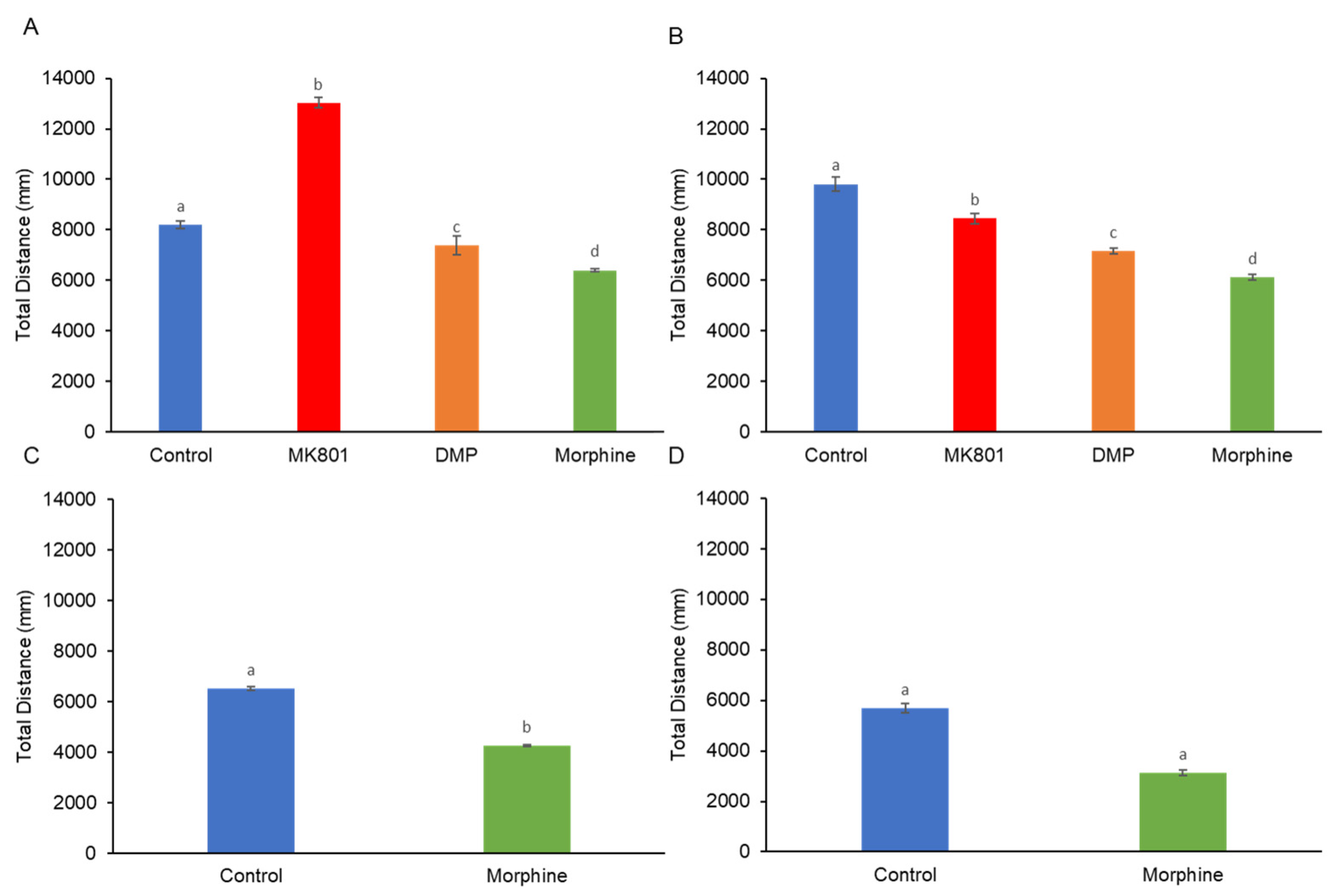
The short-term exposure to MK-801 significantly enhanced their locomotion activity. In contrast to MK-801, DMP and morphine reduced swimming activity (Figure 1A). When the larvae were exposed to these three drugs for 10 days, we observed a depression of locomotor activity (Figure 1B). Increased locomotor activity in zebrafish larvae in response to morphine withdrawal was observed after the termination of short- and long-term morphine exposure (Figure 1C,D). Apart from the difference in locomotor activity, all treated groups demonstrated a different travelling distance from the control group. When comparing with locomotor activity (Figure 1A,B), the short- and long-term treated larvae showed a similar trend in travelling distance (Figure 2A,B). However, the morphine withdrawal groups displayed a different swimming pattern. Although they crossed the central line with higher frequency (Figure 1C,D), they merely swam close to the central line, resulting in low travelling distance (Figure 2C,D).
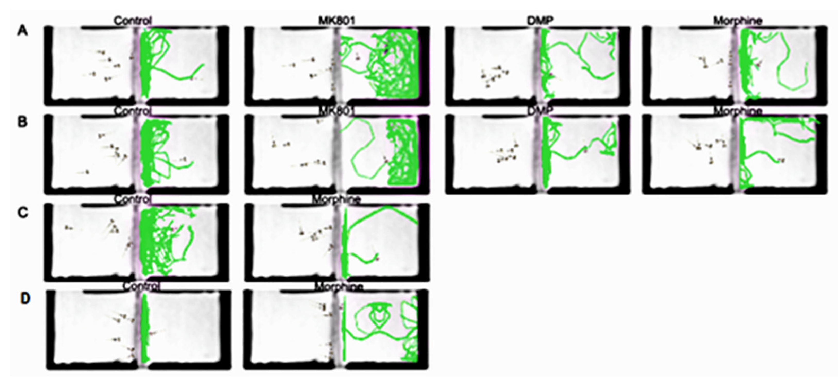
While the controls exhibited a strong, positive, assortative preference toward conspecifics, the other three treated groups, by contrast, showed significantly decreased sociability (Figure 3A,B). During the morphine withdrawal periods, the larvae still did not preferentially associate with conspecifics (Figure 3C,D). The larvae from all the treated groups displayed different degrees of avoidance to conspecifics (Figure 4).
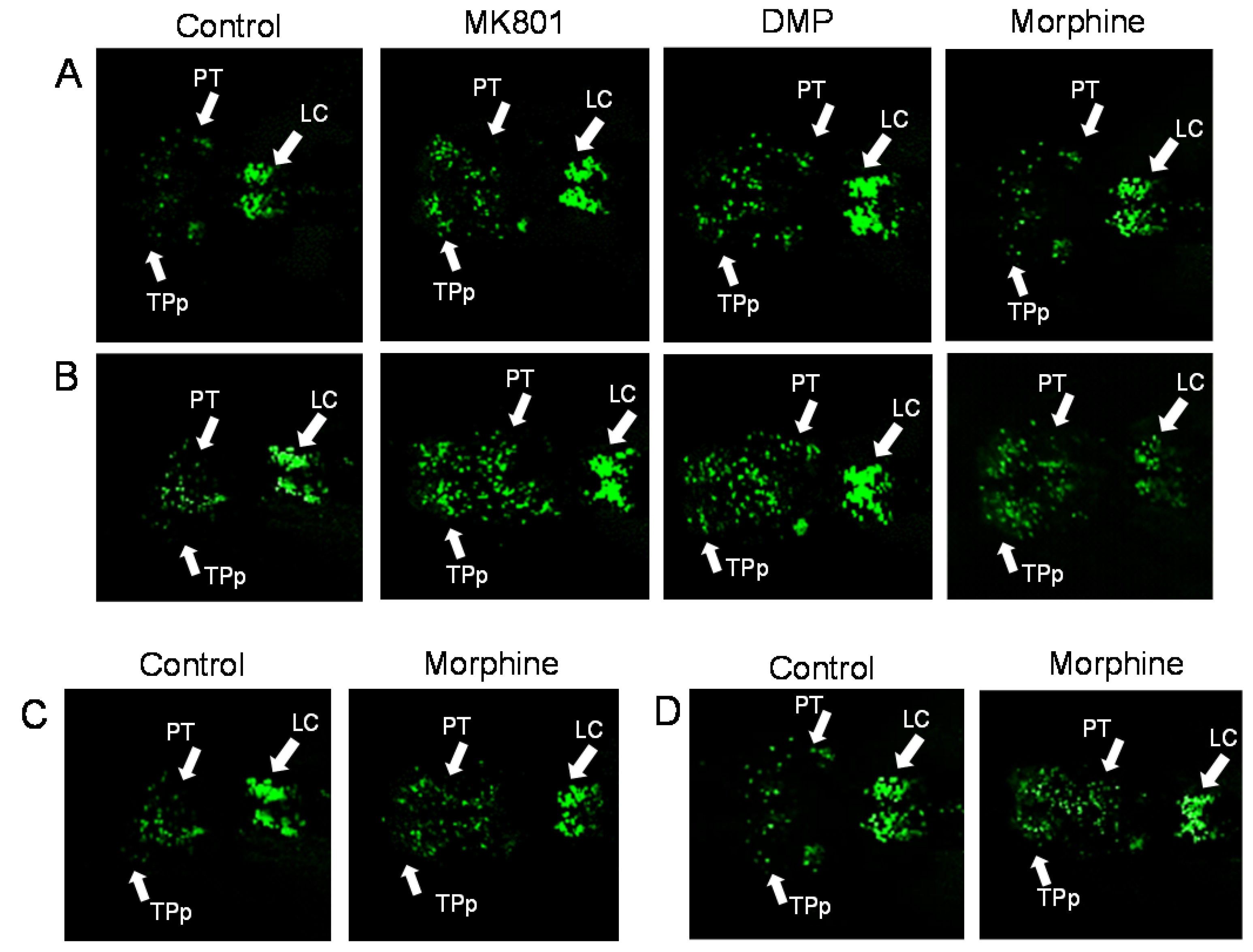
Dopaminergic neurons were seen distributed in brain regions mainly in the periventricular nucleus of posterior tubercule (TPp), posterior tuberculum (PT), caudal hypothalamus (Hc), and locus coeruleus (LC). A noticeable increase was seen in the distribution of dopaminergic neurons, especially in the PT region, between the control and the drug-treated larvae (Figure 5A,B). Exposure to drugs, particularly MK801, was associated with a significant increment of dopaminergic cell number (Figure 6A,B). Nonetheless, during the morphine withdrawal, there was no significant difference in the number of dopaminergic neurons between the control and the morphine treated group (Figure 5C,D and Figure 6C,D). Histograms (Figure 7A,B) show that all treatments increased concentration of DA, reflecting increment of dopaminergic cell number. However, DA levels remained high during morphine withdrawal (Figure 7C,D).
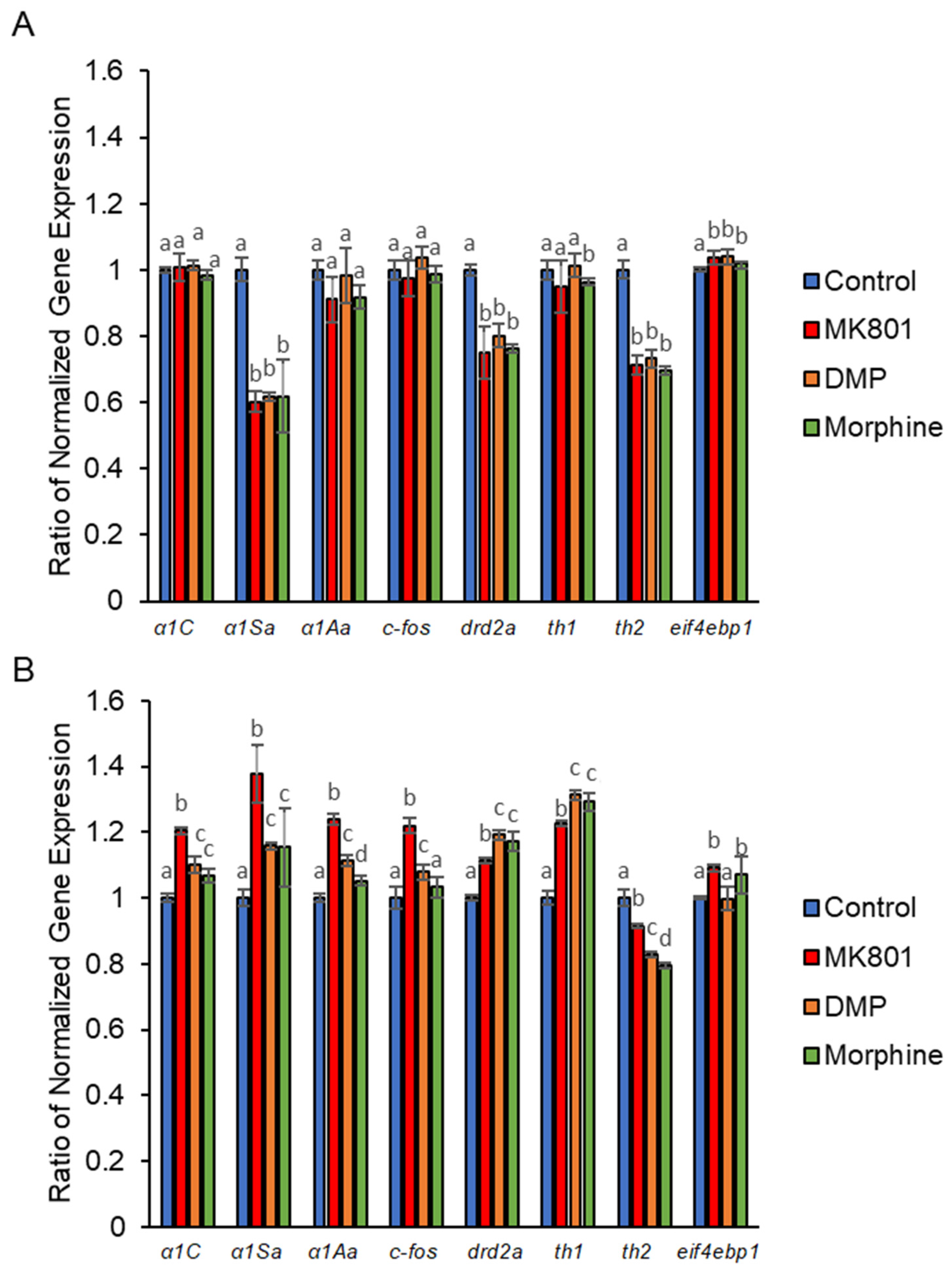
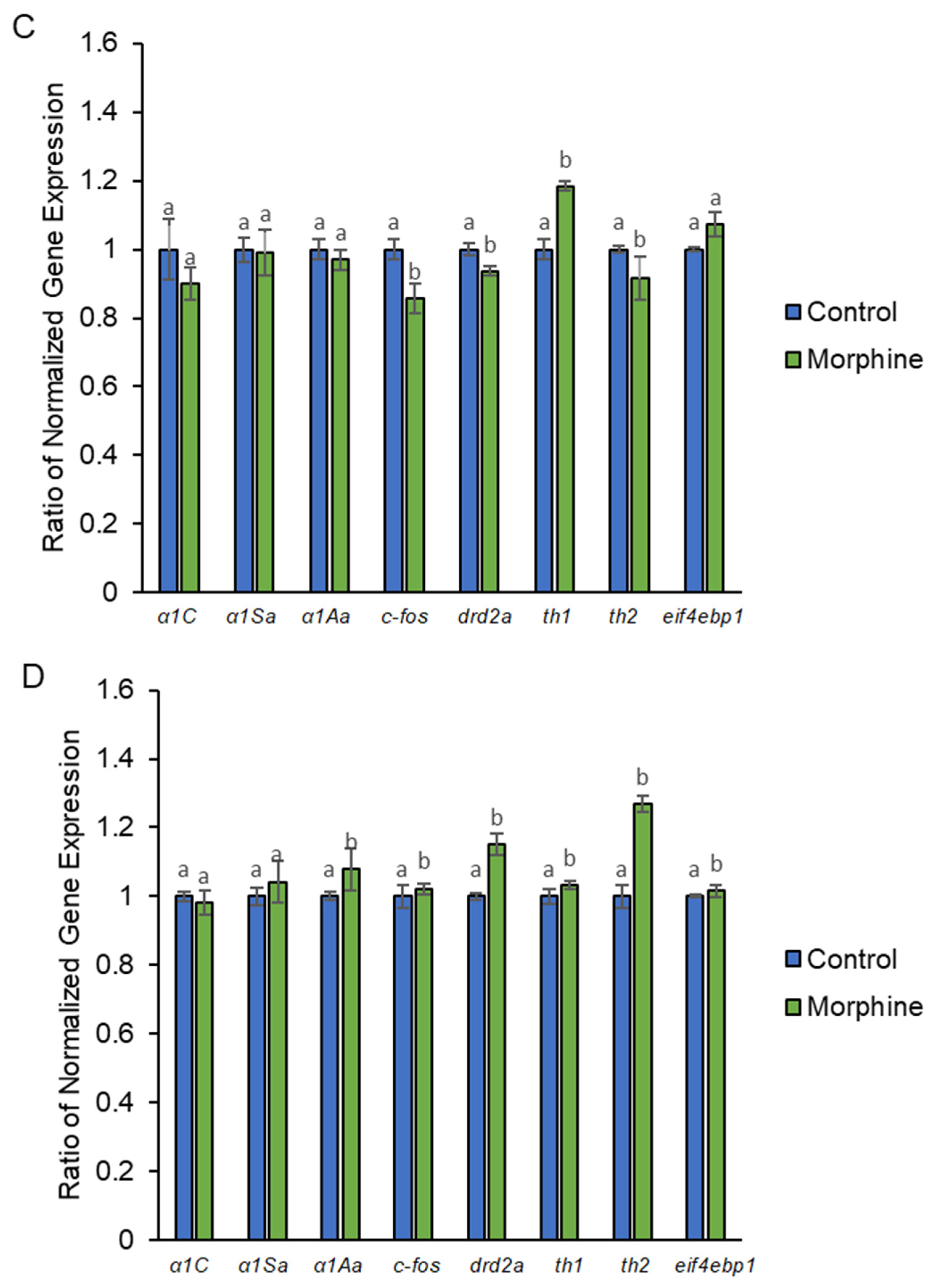
We found that eif4bp1 was up-regulated by all short-term drug treatments. In contrast, α1Sa, drd2a, and th2 were down-regulated. Of the three drug treatments, only morphine down-regulated th1 (Figure 8A). After long-term drug treatments, the gene expression changed more dramatically as compared to short-term treatments. α1C, α1Sa, α1Aa, drd2a, and th1 were up-regulated by all three drugs. Among these genes, MK-801 gave the most prominent effect to α1C, α1Sa, and α1Aa, while DMP and morphine extensively up-regulated drd2a and th1. C-fos was up-regulated by MK-801 and DMP, whereas eif4bp1 was up-regulated by both MK-801 and morphine. Th2 was the only gene down-regulated after long-term drug treatments (Figure 8B). When the larvae underwent morphine withdrawal after short-term morphine exposure, c-fos, th2, and drd2a were down-regulated while th1 and eifbbp1 were up-regulated (Figure 8C). After long-term exposure to morphine, α1Aa, c-fos, drd2a, th1, and eif4bp1 remained up-regulated during withdrawal. Surprisingly, the expression of th2 was highly up-regulated (Figure 8D).

3. Discussion

DA neurons are characterized by the expression of D2R [66]. Findings suggest elevated density of D2R in the brains of schizophrenia patients [19,67]. The encoding gene, drd2 expression, was found to be related with negative symptoms and positively correlated with the deficit syndrome severity [68]. There are two drd2 orthologs in zebrafish, drd2a and drd2b, due to the whole genome duplication in teleosts [69], which show a similarity of 27% and 66%, respectively, with human drd2 [70]. In the current study, all short-term drug exposures led to suppression of drd2a. However, the long-term exposure to these drugs induced upregulation of drd2a. NMDA antagonists such as MK-801 and 3-(2-carboxypiperazin-4-yl)-propyl-1-phosphonic acid, a competitive NMDA receptor antagonist, have been reported to upregulate postsynaptic drd2 [71,72,73,74]. These antagonists block the presynaptic NMDA receptors on the dopaminergic projections [75,76] and inhibit the release of DA, which results in an upregulation of postsynaptic drd2. Our observations regarding drd2a elevation in long-term morphine treated and morphine withdrawn larvae further support the previous finding regarding the association of increased drd2 to morphine dependence and withdrawal [77]. In the present study, confocal microscopy analysis revealed that the elevation of drd2a could be explained by the increased number of DA neurons.
TH is a rate-limiting enzyme which catalyzes the transformation of L-tyrosine to the dopamine precursor (L-DOPA). This enzyme has been widely used as a molecular marker of dopaminergic neurons. In zebrafish, TH is coded by th1 and th2 [78,79]. We found that the expression of th1 after long-term drug treatments was comparable with the increase of dopaminergic neurons. The increase of th1 was reflected by the number of DA neurons, and this was further verified by the raise of DA level. In contrast, the expression of th2 was significantly reduced in this study. Unlike th1, which is widely expressed in various brain regions [79], the expression of th2 is restricted to the preoptic nucleus and the cerebrospinal fluid-contacting neurons in the PT, and the intermediate and caudal hypothalamus, where there is little co-expression of th1 [79,80]. These th2-expressing neurons are involved in the maintenance of normal levels of spontaneous activity and production of swimming behaviors [81,82]. Our data support the reduction of th2 expression could be associated with the low number of movements after short- and long-term exposure to MK-801, DMP and morphine. The highly expressed th2 in the larvae during morphine withdrawal after long-term morphine exposure might be reflected by their hyperactive swimming behavior.
As an immediate early gene, c-fos serves as a marker of neuronal activation [83]. It has been reported that MK-801 increased c-fos expression in the schizophrenia rat model [84]. The MK-801-induced c-fos expression in the long-term treatment group is in line with previous findings. Surprisingly, morphine failed to induce c-fos expression after long-term exposure. An increase of c-fos expression was observed only after termination of long-term morphine treatment. Previous studies reported that morphine does not induce FOS [85,86]. However, opiate withdrawal increases the expression of the c-fos in different brain regions, such as the NAc, amygdala, and GABAergic tail of the ventral tegmental area [87,88,89,90]. Although c-fos mRNA in zebrafish has been evaluated following various experimental manipulations [91], the effect of D2R antagonist on c-fos expression has not been investigated in the larval brain. The blockade of D2R in the prefrontal cortex was reported to induce c-FOS expression in the dorsomedial striatum, dorsolateral striatum, NAc shell, lateral septal nucleus ventral part, and bed nucleus of the stria terminalis in mice [92]. This could explain the significant upregulation of c-fos after long-term DMP treatment.
Voltage-gated calcium channels (Cavs) are transmembrane proteins activated by depolarization of membrane potential. Classically, dysfunction of Cavs has been linked to neurological disorders including Parkinson’s disease, ataxia, migraine, and neuropathic pain [93]. Cavs are being considered as molecular targets to treat several neurological conditions including psychiatric disorders [50]. Cav1 channel subfamily comprises Cav1.1. Cav1.2, Cav1.2, and Cav1.4 channels, while the Cav2 channel subfamily comprises Cav2.1, Cav2.2, and Cav2.3. These channels are involved in the release of neurotransmitters. Calcium ion influx via Cav1 postsynaptic channels activates phosphorylation of cyclic-AMP response element binding protein (CREB) [94,95,96,97] and NFATc4 neuronal nuclear transcription factors [98]. Cav2.1 and Cav2.2 channels play a vital role in the release of fast transmitters such as GABA, acetylcholine, and glutamate [99,100]. In the current study, genes encoding Cav1.1, Cav1.2, and Cav2.1 were up-regulated by long-term drug treatments. The up-regulation of these Cavs will lead to a sustained activation of CREB in the NAc, which leads to anhedonia-like and pro-depression-like symptoms [101,102]. This could be further supported by our previous study, where creb1 was up-regulated after treatment with DMP and morphine [103]. The upregulation of these Cavs will also allow excessive influx of calcium ions inside the cells. The overloaded calcium will reduce the amount of ATP production while increasing the accumulation of reactive oxygen species and release of cytochrome C that induces apoptosis of neuronal cells [104]. Excessive accumulation of calcium ions in the mitochondria of the neurons can also lead to excessive neuronal firing and eventually cause neuronal death. The dysfunction of mitochondria can lead to several diseases, including intellectual disability [105]. Moreover, a gain-of-function mutation in Cav1.2 causes Timothy syndrome [106], which is associated with neurological developmental defects, including manifestation of neuropsychiatric phenotypes [106,107,108]. This can be reflected by the lower social preference in all the MK-801, DMP, and morphine-treated larvae. Noteworthily, for the morphine withdrawn larvae, low social preference was still shown, and this could be due to the high expression of α1Aa which encodes Cav2.1.
The eukaryotic translation initiation factor 4E binding protein 1 (EIF4EBP1), encoded by eif4ebp1, is one member of a family of small proteins that act as repressors of mRNA translation initiation by binding the mRNA cap-binding protein eIF4E [109]. The activity of phosphatidylinositol 3-kinase (PI3K)/AKT pathway negatively regulates the transcription of EIF4EBP1 via mechanistic target of rapamycin (mTOR) [110]. The significance of mTOR in neuron development and its regulation of translational control and protein synthesis implicated that mTOR is involved in the pathology of schizophrenia [111,112,113,114]. Although the role of mTOR was not tested in the current study, upregulation of eif4ebp1 after drug exposure and morphine withdrawal could be explained by the downregulation of pi3k and akt1 in our previous study [103].

4. Materials and Methods

4.1. Zebrafish Strains and Housing Conditions

Wild-type zebrafish embryos at 0 hpf were collected from Danio Assay Laboratories Pvt. Ltd., University Putra Malaysia, Serdang, Selangor, Malaysia. Transgenic Tg (dat:EGFP) zebrafish embryos were obtained from Jeffrey Cheah School of Medicine and Health Sciences, Monash University Malaysia, Subang Jaya, Selangor, Malaysia, after 2 hpf. This strain was developed by the Center for Advanced Research in Environmental Genomics, Department of Biology, University of Ottawa. The embryos were maintained at 27 °C in embryo medium (5 mM sodium chloride (NaCl), 0.17 mM potassium chloride (KCl), 0.33 mM calcium chloride (CaCl2), and 0.33 mM magnesium sulfate (MgSO4), pH 7.4). Unfertilized, unhealthy, and dead embryos were discarded. The hatched larvae were fed at 7 days post-fertilization (dpf) with live paramecium. All experiments were done with the approval of animal ethics by Universiti Tunku Abdul Rahman Research Ethics and Code of Conduct (U/SERC/18/2020). Visual screening was performed within the time frame of experimental procedure to monitor the health of the zebrafish larvae by removing the larvae with abnormal mortality, heartbeat, and swimming activity.

4.2. Drug Optimization and Treatments

The optimization of maximum-tolerated concentration (MTC) of each drug and identification of its optimal condition in inducing schizophrenia were based on our previous study [103]. At 2 dpf, both strains of the zebrafish were dosed at MTC according to the short- (3 days) and long-term (10 days) treatment protocols. DMP and morphine were added to the embryo medium at a final concentration of 3.13 μM and 0.80 μM, respectively. Additionally, 5.0 μM of dizocilpine (MK-801) (Sigma-Aldrich, Saint Louis, Missouri, USA), a schizophrenia-mimicking compound, was used as a positive control, while embryo medium served as vehicle control. Three groups (n = 50 /group) of embryos were examined for the effect of short- and long-term treatments. Another two groups of embryos were assessed for withdrawal syndrome five days after the termination of short- and long-term morphine treatments.

4.3. Behavioral Analyses

Calculating the total number of crossings is a simple and effective method to determine the locomotor activity of zebrafish [115,116]. Our assay was modified from the method of Boehmler et al. (2007) [115]. A single vertical line was drawn on the bottom of a clear petri dish (60 mm × 15 mm) which divided the dish into 2 equal halves. Three larvae were placed in the petri dish filled with embryo medium at 27 °C. Locomotor activity was measured for 30 s by counting the number of times each larva crossed the line. Total distance traveled was also used as a measure of locomotor activity. A recording camera at 30 frames per second was positioned above the arena. Videos were captured for 5 min and processed using ToxTrac [117]. Each treatment was subjected to 12 independent trials with 3 independent experiments.

4.4. Social Preference

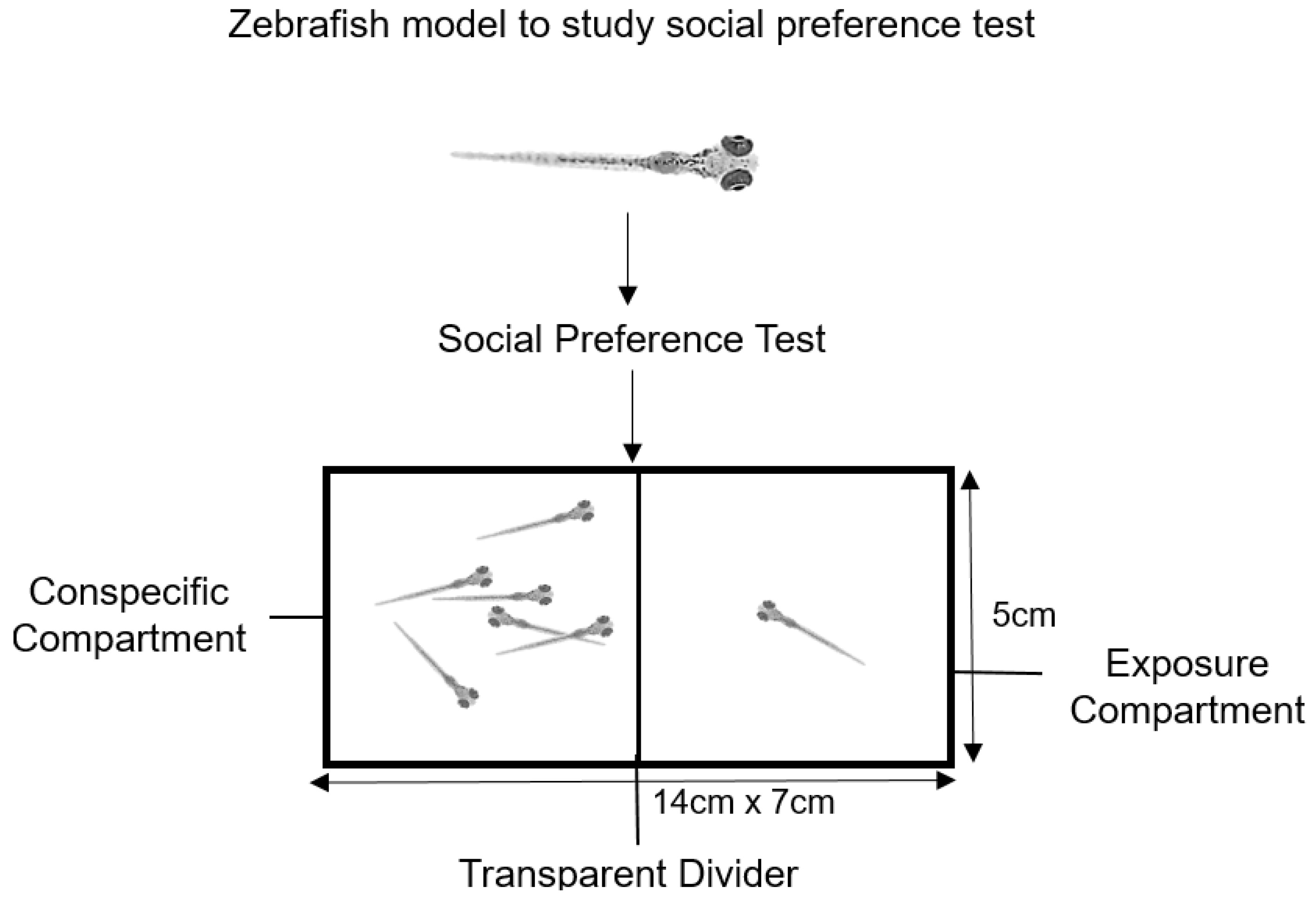
The experimental tank (14 cm length, 7 cm width, 5 cm height) was separated by a transparent divider into exposure compartment and conspecific compartment (Figure A1). To avoid lateral bias in zebrafish cohorts, the left/right location of target/conspecific larvae were alternated between the trials. The target larvae were pre-exposed to a drug or drug-free water (control) for 20 min. Control or drug-exposed zebrafish (n = 12 in each group) were introduced individually to the central zone of the apparatus, temporarily separated (by transparent sliding dividing doors) from the two arms of the corridor.
The larva was subjected to an initial 30-s acclimation interval in order to reduce transfer/handling stress. The larva was allowed to explore the apparatus for 5 min. Videos were captured to record the time of each larva spent near the conspecific compartment.

4.5. Gene Expression Analysis by Real-Time PCR

The larvae were euthanized by rapid cooling in ice water (0–4 °C) for at least 20 min [118]. Total RNA from the dissected brains of zebrafish larvae pool (n = 50) was extracted using TRIzol reagent (Thermo Fisher Scientific, Waltham, Massachusetts, USA), with three independent pools used for each treatment [119]. The quality of RNA was assessed by gel electrophoresis and the total RNA concentration was determined using spectrophotometer. The double-stranded cDNA was synthesized using a Tetro cDNA Synthesis Kit (Bioline, London, UK). Eight genes related to the dopaminergic circuitry were selected. The primers used to amplify these genes are presented in Table 1. Quantitative real-time PCR was performed using the SensiFAST™ SYBR® No-ROX kit (Bioline, London, UK) in a qTOWER3 G real-time thermal cycler (Analytik Jena, Jena, Germany) with beta actin (β-actin) as the housekeeping gene. cDNA was diluted in series ranging from 100 ng/μL to 0.01 ng/μL to generate a standard curve. All reactions were performed in triplicate. The relative expression of each gene was calculated from the average CT (cycle threshold) readings at each point of the standard curve using the formula E = 10 (1/slope). Expression of genes of interest was then normalized to the housekeeping gene.

4.6. Total Protein Purification

Zebrafish larvae were washed with cold phosphate buffered saline (PBS) and stored overnight at −20 °C. Cell lysis was conducted by two freeze–thaw cycles. The pooled larval brain tissue was homogenized in PBS containing 1% Triton-X and sonicated for 10 min. The homogenates were centrifuged at 5000× g for 5 min. The supernatant was collected, and subsequently the protein concentration was determined using Bradford assay. The purified protein was subjected to biochemical assays immediately.

4.7. Biochemical Assay

The concentration of DA was determined using the commercial enzyme-linked immunosorbent assay (ELISA) kits (Cusabio Biotech®, Houston, Texas, USA) according to the manufacturer’s instructions. The optical density (OD value) of each well was determined at 450 nm for the assay.

4.8. Confocal Microscopy

Tg (dat:EGFP) larvae were treated with 1-phenyl-2-thiourea (0.003% phenylthiourea, Sigma-Aldrich, Saint Louis, Missouri, USA) to prevent pigment formation. Preceding the treatments, the larvae were fixed by 4% paraformaldehyde (PFA) overnight at 4 °C and stored in PBS until analysis. Prior to imaging, each larva was embedded in 2% low melting point agarose gel (Sigma, Darmstadt, Germany). A small incision was made to allow the dorsal of the larvae to face down in order to image the brain region. The brain image of the larva was then captured by the laser scanning confocal microscope (Nikon C1si, Nikon Inc., Tokyo, Japan) and the NIS Elements AR software version 4.1 (Nikon Inc., Tokyo, Japan). The dopaminergic neurons in the PT were determined based on the z-stacks of confocal images using the ImageJ (Bethesda, Maryland, USA) 3D object counter plugin described by Tay et al. [127].

4.9. Statistical Analysis

All results were expressed as mean ± SEM. The differences between samples were analyzed using the one-way analysis of variance (ANOVA) followed by post-hoc Tukey’s test. Means comparisons between the control and withdrawal groups were performed using Student’s t-test. All statistical tests were performed using Statistical Program for Social Sciences Statistical Program for Social Sciences (SPSS version 22). A p-value of less than 0.05 (p < 0.05) was considered statistically significant.

5. Conclusions

In summary, our study, which integrated behavioral, biochemical, gene expression, and confocal microscopy imaging data in drug-treated zebrafish larvae, revealed that there is an association between opioid addiction and schizophrenia. The alterations in dopaminergic neurons, calcium channels, and gene expression induced by DMP and morphine could contribute to the abnormal locomotor and social behaviors. Future work is required to elucidate the specificity of these changes in the context of these two disorders.

Author Contributions

Conceptualization, P.Y.T., S.Y.M., S.O. and I.S.P.; methodology, S.K.T.; software, S.K.T.; validation, P.Y.T. and S.Y.M.; formal analysis, S.K.T.; investigation, S.K.T.; data curation, S.K.T. and P.Y.T.; writing—original draft preparation, S.K.T.; writing—review and editing, P.Y.T., S.Y.M. and S.O.; visualization, S.K.T.; supervision, P.Y.T. and S.Y.M.; project administration, P.Y.T. All authors have read and agreed to the published version of the manuscript.

Funding

This research was funded by the Ministry of Higher Education (MOHE) through Fundamental Research Grant Scheme project FRGS/1/2018/STG03/UTAR/02/1 and Malaysia Toray Science Foundation 4417/M01.

Institutional Review Board Statement

The animal study protocol was approved by the Scientific and Ethical Review Committee of UNIVERSITI TUNKU ABDUL RAHMAN (U/SERC/18/2020, 4 February 2020).

Informed Consent Statement

Not applicable.

Data Availability Statement

Not applicable.

Conflicts of Interest

The authors declare no conflict of interest.

Appendix A

Figure A1. Social preference in zebrafish larvae.
Ijms 24 04088 g0a1

References

  1. Karam, E.G.; Yabroudi, P.F.; Melhem, N.M. Comorbidity of substance abuse and other psychiatric disorders in acute general psychiatric admissions: A study from Lebanon. Compr. Psychiatry 2002, 43, 463–468. [Google Scholar] [CrossRef] [PubMed] [Green Version]
  2. Volkow, N.D. Substance Use Disorders in Schizophrenia—Clinical Implications of Comorbidity. Schizophr. Bull. 2009, 35, 469–472. [Google Scholar] [CrossRef] [Green Version]
  3. Hartz, S.M.; Pato, C.N.; Medeiros, H.; Cavazos-Rehg, P.; Sobell, J.L.; Knowles, J.A.; Bierut, L.J.; Pato, M.T.; Genomic Psychiatry Cohort Consortium. Comorbidity of severe psychotic disorders with measures of substance use. JAMA Psychiatry 2014, 71, 248–254. [Google Scholar] [CrossRef] [PubMed] [Green Version]
  4. Helzer, J.E.; Pryzbeck, T.R. The co-occurrence of alcoholism with other psychiatric disorders in the general population and its impact on treatment. J. Stud. Alcohol 1988, 49, 219–224. [Google Scholar] [CrossRef] [PubMed]
  5. LeDuc, P.A.; Mittleman, G. Schizophrenia and psychostimulant abuse: A review and re-analysis of clinical evidence. Psychopharmacology 1995, 121, 407–427. [Google Scholar] [CrossRef]
  6. Diwan, A.; Castine, M.; Pomerleau, C.S.; Meador-Woodruff, J.H.; Dalack, G.W. Differential prevalence of cigarette smoking in patients with schizophrenic vs mood disorders. Schizophr. Res. 1998, 33, 113–118. [Google Scholar] [CrossRef]
  7. Di Forti, M.; Morgan, C.; Dazzan, P.; Pariante, C.M.; Mondelli, V.; Marques, T.R.; Handley, R.; Luzi, S.; Russo, M.; Paparelli, A.; et al. High-potency cannabis and the risk of psychosis. Br. J. Psychiatry 2009, 195, 488–491. [Google Scholar] [CrossRef] [Green Version]
  8. Moore, T.H.; Zammit, S.; Lingford-Hughes, A.; Barnes, T.R.; Jones, P.B.; Burke, M.; Lewis, G. Cannabis use and risk of psychotic or affective mental health outcomes: A systematic review. Lancet 2007, 370, 319–328. [Google Scholar] [CrossRef] [Green Version]
  9. Gage, S.H.; Hickman, M.; Zammit, S. Association between cannabis and psychosis: Epidemiologic evidence. Biol. Psychiatry 2016, 79, 549–556. [Google Scholar] [CrossRef] [Green Version]
  10. Gage, S.H.; Jones, H.J.; Burgess, S.; Bowden, J.; Smith, G.D.; Zammit, S.; Munafo, M.R. Assessing causality in associations between cannabis use and schizophrenia risk: A two-sample Mendelian randomization study. Psychol. Med. 2017, 47, 971–980. [Google Scholar] [CrossRef] [Green Version]
  11. Kovasznay, B.; Fleischer, J.; Tanenberg-Karant, M.; Jandorf, L.; Miller, A.D.; Bromet, E. Substance use disorder and the early course of illness in schizophrenia and affective psychosis. Schizophr. Bull. 1997, 23, 195–201. [Google Scholar] [CrossRef] [PubMed] [Green Version]
  12. Addington, J.; Addington, D. Substance abuse and cognitive functioning in schizophrenia. J. Psychiatry Neurosci. 1997, 22, 99–104. [Google Scholar] [PubMed]
  13. Hunt, G.E.; Bergen, J.; Bashir, M. Medication compliance and comorbid substance abuse in schizophrenia: Impact on community survival 4 years after a relapse. Schizophr. Res. 2002, 54, 253–264. [Google Scholar] [CrossRef] [PubMed]
  14. Swartz, M.S.; Wagner, H.R.; Swanson, J.W.; Stroup, T.S.; McEvoy, J.P.; Canive, J.M.; Miller, D.D.; Reimherr, F.; McGee, M.; Khan, A.; et al. Substance use in persons with schizophrenia: Baseline prevalence and correlates from the NIMH CATIE study. J. Nerv. Ment. Dis. 2006, 194, 164–172. [Google Scholar] [CrossRef]
  15. Schmidt, L.M.; Hesse, M.; Lykke, J. The impact of substance use disorders on the course of schizophrenia—A 15-year follow-up study: Dual diagnosis over 15 years. Schizophr. Res. 2011, 130, 228–233. [Google Scholar] [CrossRef]
  16. Janssen, B.; Gaebel, W.; Haerter, M.; Komaharadi, F.; Lindel, B.; Weinmann, S. Evaluation of factors influencing medication compliance in inpatient treatment of psychotic disorders. Psychopharmacology 2006, 187, 229–236. [Google Scholar] [CrossRef]
  17. Strømme, M.F.; Mellesdal, L.S.; Bartz-Johannesen, C.A.; Kroken, R.A.; Krogenes, M.L.; Mehlum, L.; Johnsen, E. Use of benzodiazepines and antipsychotic drugs are inversely associated with acute readmission risk in schizophrenia. J. Clin. Psychopharmacol. 2022, 42, 37–42. [Google Scholar] [CrossRef]
  18. Goldstein, M.; Deutch, A.Y. Dopaminergic mechanisms in the pathogenesis of schizophrenia. FASEB J. 1992, 6, 2413–2421. [Google Scholar] [CrossRef]
  19. Abi-Dargham, A.; Rodenhiser, J.; Printz, D.; Zea-Ponce, Y.; Gil, R.; Kegeles, L.S.; Weiss, R.; Cooper, T.B.; Mann, J.J.; Van Heertum, R.L.; et al. Increased baseline occupancy of D2 receptors by dopamine in schizophrenia. Proc. Natl. Acad. Sci. USA 2000, 97, 8104–8109. [Google Scholar] [CrossRef] [Green Version]
  20. Abi-Dargham, A.; van de Giessen, E.; Slifstein, M.; Kegeles, L.S.; Laruelle, M. Baseline and amphetamine-stimulated dopamine activity are related in drug-naive schizophrenic subjects. Biol. Psychiatry 2009, 65, 1091–1093. [Google Scholar] [CrossRef]
  21. Howes, O.O.; Fusar-Poli, P.; Bloomfield, M.; Selvaraj, S.; McGuire, P. From the prodrome to chronic schizophrenia: The neurobiology underlying psychotic symptoms and cognitive impairments. Curr. Pharm. Des. 2012, 18, 459–465. [Google Scholar] [CrossRef] [PubMed] [Green Version]
  22. Howes, O.D.; Kambeitz, J.; Kim, E.; Stahl, D.; Slifstein, M.; Abi-Dargham, A.; Kapur, S. The nature of dopamine dysfunction in schizophrenia and what this means for treatment: Meta-analysis of imaging studies. Arch. Gen. Psychiatry 2012, 69, 776–786. [Google Scholar] [CrossRef] [PubMed] [Green Version]
  23. Howes, O.D.; Williams, M.; Ibrahim, K.; Leung, G.; Egerton, A.; McGuire, P.K.; Turkheimer, F. Midbrain dopamine function in schizophrenia and depression: A post-mortem and positron emission tomographic imaging study. Brain 2013, 136, 3242–3251. [Google Scholar] [CrossRef] [PubMed] [Green Version]
  24. Davis, K.L.; Kahn, R.S.; Ko, G.; Davidson, M. Dopamine in schizophrenia: A review and reconceptualization. Am. J. Psychiatry 1991, 148, 1474–1486. [Google Scholar] [CrossRef] [PubMed]
  25. Galderisi, S.; Mucci, A.; Buchanan, R.W.; Arango, C. Negative symptoms of schizophrenia: New developments and unanswered research questions. Lancet Psychiatry 2018, 5, 664–677. [Google Scholar] [CrossRef]
  26. Manion, M.T.C.; Glasper, E.R.; Wang, K.H. A sex difference in mouse dopaminergic projections from the midbrain to basolateral amygdala. Biol. Sex Differ. 2022, 13, 75. [Google Scholar] [CrossRef]
  27. Radua, J.; Schmidt, A.; Borgwardt, S.; Heinz, A.; Schlagenhauf, F.; McGuire, P.; Fusar-Poli, P. Ventral striatal activation during reward processing in psychosis: A neurofunctional meta-analysis. JAMA Psychiatry 2015, 72, 1243–1251. [Google Scholar] [CrossRef] [Green Version]
  28. Panayi, M.C.; Boerner, T.; Jahans-Price, T.; Huber, A.; Sprengel, R.; Gilmour, G.; Sanderson, D.J.; Harrison, P.J.; Walton, M.E.; Bannerman, D.M. Glutamatergic dysfunction leads to a hyper-dopaminergic phenotype through deficits in short-term habituation: A mechanism for aberrant salience. Mol. Psychiatry 2022, 28, 579–587. [Google Scholar] [CrossRef]
  29. Di Chiara, G.; Imperato, A. Drugs abused by humans preferentially increase synaptic dopamine concentrations in the mesolimbic system of freely moving rats. Proc. Natl. Acad. Sci. USA 1988, 85, 5274–5278. [Google Scholar] [CrossRef] [Green Version]
  30. Koob, G.F.; Bloom, F.E. Cellular and molecular mechanisms of drug dependence. Science 1988, 242, 715–723. [Google Scholar] [CrossRef]
  31. Koob, G.F. Neural mechanisms of drug reinforcement. Ann. N. Y. Acad. Sci. 1992, 654, 171–191. [Google Scholar] [CrossRef] [PubMed]
  32. Volkow, N.D.; Wang, G.J.; Fowler, J.S.; Logan, J.; Gatley, S.J.; Wong, C.; Hitzemann, R.; Pappas, N.R. Reinforcing effects of psychostimulants in humans are associated with increases in brain dopamine and occupancy of D2receptors. J. Pharmacol. Exp. Ther. 1999, 291, 409–415. [Google Scholar] [PubMed]
  33. Drevets, W.C.; Gautier, C.; Price, J.C.; Kupfer, D.J.; Kinahan, P.E.; Grace, A.A.; Price, J.L.; Mathis, C.A. Amphetamine-induced dopamine release in human ventral striatum correlates with euphoria. Biol. Psychiatry 2001, 49, 81–96. [Google Scholar] [CrossRef] [PubMed]
  34. Brody, A.L.; Mandelkern, M.A.; Olmstead, R.E.; Allen-Martinez, Z.; Scheibal, D.; Abrams, A.L.; Costello, M.R.; Farahi, J.; Saxena, S.; Monterosso, J.; et al. Ventral striatal dopamine release in response to smoking a regular vs a denicotinized cigarette. Neuropsychopharmacology 2009, 34, 282–289. [Google Scholar] [CrossRef] [PubMed] [Green Version]
  35. Boileau, I.; Assaad, J.M.; Pihl, R.O.; Benkelfat, C.; Leyton, M.; Diksic, M.; Tremblay, R.E.; Dagher, A. Alcohol promotes dopamine release in the human nucleus accumbens. Synapse 2003, 49, 226–231. [Google Scholar] [CrossRef]
  36. Bossong, M.G.; Van Berckel, B.N.; Boellaard, R.; Zuurman, L.; Schuit, R.C.; Windhorst, A.D.; van Gerven, J.; Ramsey, N.F.; Lammertsma, A.A.; Kahn, R.S. Δ9-tetrahydrocannabinol induces dopamine release in the human striatum. Neuropsychopharmacology 2009, 34, 759–766. [Google Scholar] [CrossRef]
  37. Wise, R.A. Brain reward circuitry: Insights from unsensed incentives. Neuron 2002, 36, 229–240. [Google Scholar] [CrossRef] [Green Version]
  38. Owesson-White, C.A.; Ariansen, J.; Stuber, G.D.; Cleaveland, N.A.; Cheer, J.F.; Mark Wightman, R.; Carelli, R.M. Neural encoding of cocaine-seeking behavior is coincident with phasic dopamine release in the accumbens core and shell. Eur. J. Neurosci. 2009, 30, 1117–1127. [Google Scholar] [CrossRef] [Green Version]
  39. Björklund, A.; Lindvall, O. Dopamine-containing systems in the CNS. In Handbook of Chemical Neuroanatomy Vol. 2: Classical Transmitters in the CNS; Björklund, A., Hokfelt, T., Eds.; Elsevier: Toronto, ON, Canada, 1984; pp. 55–122. [Google Scholar]
  40. Blenau, W.; Baumann, A. Molecular and pharmacological properties of insect biogenic amine receptors: Lessons from Drosophila melanogaster and Apis mellifera. Arch. Insect Biochem. Physiol. 2001, 48, 13–38. [Google Scholar] [CrossRef]
  41. Roeder, T. Biochemistry and molecular biology of receptors for biogenic amines in locusts. Microsc. Res. Tech. 2002, 56, 237–247. [Google Scholar] [CrossRef]
  42. Perrone-Capano, C.; Tino, A.; di Porzio, U. Target cells modulate dopamine transporter gene expression during brain development. Neuroreport 1994, 5, 1145–1148. [Google Scholar] [CrossRef] [PubMed]
  43. Zheng, X.; Hasegawa, H. Pharmacological intervention of brain neurotransmission affects exercise capacity. In Physical Activity and the Aging Brain; Watson, R.R., Ed.; Academic Press: Cambridge, MA, USA, 2017; pp. 53–64. [Google Scholar] [CrossRef]
  44. Clapham, D.E. Calcium signaling. Cell 2007, 131, 1047–1058. [Google Scholar] [CrossRef] [Green Version]
  45. Armstrong, C.M.; Matteson, D.R. Two distinct populations of calcium channels in a clonal line of pituitary cells. Science 1985, 227, 65–67. [Google Scholar] [CrossRef] [PubMed]
  46. Bean, B.P. Two kinds of calcium channels in canine atrial cells. Differences in kinetics, selectivity, and pharmacology. J. Gen. Physiol. 1985, 86, 1–30. [Google Scholar] [CrossRef] [PubMed]
  47. Striessnig, J.; Pinggera, A.; Kaur, G.; Bock, G.; Tuluc, P. L-type Ca2+ channels in heart and brain. Wiley Interdiscip. Rev. Membr. Transp. Signal. 2014, 3, 15–38. [Google Scholar] [CrossRef] [PubMed]
  48. Borgkvist, A.; Mosharov, E.V.; Sulzer, D. Calcium currents regulate dopamine autoreceptors. Brain 2014, 137, 2113–2115. [Google Scholar] [CrossRef] [Green Version]
  49. Heyes, S.; Pratt, W.S.; Rees, E.; Dahimene, S.; Ferron, L.; Owen, M.J.; Dolphin, A.C. Genetic disruption of voltage-gated calcium channels in psychiatric and neurological disorders. Prog. Neurobiol. 2015, 134, 36–54. [Google Scholar] [CrossRef] [Green Version]
  50. Zamponi, G.W. Targeting voltage-gated calcium channels in neurological and psychiatric diseases. Nat. Rev. Drug Discov. 2016, 15, 19–34. [Google Scholar] [CrossRef]
  51. Nanou, E.; Catterall, W.A. Calcium channels, synaptic plasticity, and neuropsychiatric disease. Neuron 2018, 98, 466–481. [Google Scholar] [CrossRef] [Green Version]
  52. Schizophrenia Working Group of the Psychiatric Genomics Consortium. Biological insights from 108 schizophrenia-associated genetic loci. Nature 2014, 511, 421–427. [Google Scholar] [CrossRef] [Green Version]
  53. Simms, B.A.; Zamponi, G.W. Neuronal voltage-gated calcium channels: Structure, function, and dysfunction. Neuron 2014, 82, 24–45. [Google Scholar] [CrossRef] [Green Version]
  54. Hall, J.; Trent, S.; Thomas, K.L.; O’Donovan, M.C.; Owen, M.J. Genetic risk for schizophrenia: Convergence on synaptic pathways involved in plasticity. Biol. Psychiatry 2015, 77, 52–58. [Google Scholar] [CrossRef]
  55. Moon, A.L.; Haan, N.; Wilkinson, L.S.; Thomas, K.L.; Hall, J. CACNA1C: Association with psychiatric disorders, behavior, and neurogenesis. Schizophr. Bull. 2018, 44, 958–965. [Google Scholar] [CrossRef] [PubMed]
  56. Bhat, S.; Dao, D.T.; Terrillion, C.E.; Arad, M.; Smith, R.J.; Soldatov, N.M.; Gould, T.D. CACNA1C (Cav1. 2) in the pathophysiology of psychiatric disease. Prog. Neurobiol. 2012, 99, 1–14. [Google Scholar] [CrossRef] [PubMed] [Green Version]
  57. Kalueff, A.V.; Stewart, A.M.; Gerlai, R. Zebrafish as an emerging model for studying complex brain disorders. Trends Pharmacol. Sci. 2014, 35, 63–75. [Google Scholar] [CrossRef] [Green Version]
  58. Howe, K.; Clark, M.D.; Torroja, C.F.; Torrance, J.; Berthelot, C.; Muffato, M.; Collins, J.E.; Humphray, S.; McLaren, K.; Matthews, L.; et al. The zebrafish reference genome sequence and its relationship to the human genome. Nature 2013, 496, 498–503. [Google Scholar] [CrossRef] [PubMed] [Green Version]
  59. Guo, S. Using zebrafish to assess the impact of drugs on neural development and function. Expert Opin. Drug Discov. 2009, 4, 715–726. [Google Scholar] [CrossRef] [Green Version]
  60. Horzmann, K.A.; Freeman, J.L. Zebrafish get connected: Investigating neurotransmission targets and alterations in chemical toxicity. Toxics 2016, 4, 19. [Google Scholar] [CrossRef] [Green Version]
  61. Rink, E.; Wullimann, M.F. Development of the catecholaminergic system in the early zebrafish brain: An immunohistochemical study. Dev. Brain Res. 2002, 137, 89–100. [Google Scholar] [CrossRef]
  62. Wise, R.A. Addictive drugs and brain stimulation reward. Annu. Rev. Neurosci. 1996, 19, 319–340. [Google Scholar] [CrossRef]
  63. Berridge, K.C. From prediction error to incentive salience: Mesolimbic computation of reward motivation. Eur. J. Neurosci. 2012, 35, 1124–1143. [Google Scholar] [CrossRef] [PubMed]
  64. Juckel, G.; Schlagenhauf, F.; Koslowski, M.; Filonov, D.; Wüstenberg, T.; Villringer, A.; Knutson, B.; Kienast, T.; Gallinat, J.; Wrase, J.; et al. Dysfunction of ventral striatal reward prediction in schizophrenic patients treated with typical, not atypical, neuroleptics. Psychopharmacology 2006, 187, 222–228. [Google Scholar] [CrossRef]
  65. Wolf, D.H.; Satterthwaite, T.D.; Kantrowitz, J.J.; Katchmar, N.; Vandekar, L.; Elliott, M.A.; Ruparel, K. Amotivation in schizophrenia: Integrated assessment with behavioral, clinical, and imaging measures. Schizophr. Bull. 2014, 40, 1328–1337. [Google Scholar] [CrossRef] [PubMed]
  66. Anzalone, A.; Lizardi-Ortiz, J.E.; Ramos, M.; De Mei, C.; Hopf, F.W.; Iaccarino, C.; Halbout, B.; Jacobsen, J.; Kinoshita, C.; Welter, M.; et al. Dual control of dopamine synthesis and release by presynaptic and postsynaptic dopamine D2 receptors. J. Neurosci. 2012, 32, 9023–9034. [Google Scholar] [CrossRef] [Green Version]
  67. Stelzel, C.; Basten, U.; Montag, C.; Reuter, M.; Fiebach, C.J. Frontostriatal involvement in task switching depends on genetic differences in d2 receptor density. J. Neurosci. 2010, 30, 14205–14212. [Google Scholar] [CrossRef] [PubMed] [Green Version]
  68. Liu, L.; Luo, Y.; Zhang, G.; Jin, C.; Zhou, Z.; Cheng, Z.; Yuan, G. Correlation of DRD2 mRNA expression levels with deficit syndrome severity in chronic schizophrenia patients receiving clozapine treatment. Oncotarget 2017, 8, 86515–86526. [Google Scholar] [CrossRef]
  69. Glasauer, S.M.K.; Neuhauss, S.C.F. Whole-genome duplication in teleost fishes and its evolutionary consequences. Mol. Genet. Genom. 2014, 289, 1045–1060. [Google Scholar] [CrossRef] [PubMed] [Green Version]
  70. Boehmler, W.; Obrecht-Pflumio, S.; Canfield, V.; Thisse, C.; Thisse, B.; Levenson, R. Evolution and expression of D2 and D3 dopamine receptor genes in zebrafish. Dev. Dyn. 2004, 230, 481–493. [Google Scholar] [CrossRef]
  71. Micheletti, G.; Lannes, B.; Haby, C.; Borrelli, E.; Kempf, E.; Warter, J.M.; Zwiller, J. Chronic administration of NMDA antagonists induces D2 receptor synthesis in rat striatum. Mol. Brain Res. 1992, 14, 363–368. [Google Scholar] [CrossRef]
  72. Lannes, B.; Bernard, V.; Bloch, B.; Micheletti, G. Chronic treatment with dizocilpine maleate increases the number of striatal neurons expressing the D2 receptor gene. Neuroscience 1995, 65, 431–438. [Google Scholar] [CrossRef]
  73. Healy, J.D.; Meador-Woodruff, H.J. Differential regulation, by MK801, of dopamine receptor gene expression in rat nigrostriatal and mesocorticolimbic systems. Brain Res. 1996, 708, 38–44. [Google Scholar] [CrossRef]
  74. Nair, V.D.; Savelli, J.E.; Mishra, R.K. Modulation of dopamine D2 receptor expression by an NMDA receptor antagonist in rat brain. J. Mol. Neurosci. 1998, 11, 121–126. [Google Scholar] [CrossRef] [PubMed]
  75. Carlsson, M.; Carlsson, A. Interactions between glutamatergic and monoaminergic system within the basal ganglia-implications for schizophrenia and Parkinson’s disease. Trends Neurosci. 1990, 13, 272–276. [Google Scholar] [CrossRef] [PubMed]
  76. Grace, A.A. The depolarization block hypothesis of neuroleptic action: Implications for the etiology and treatment of schizophrenia. J. Neural Transm. Suppl. 1992, 36, 91–131. [Google Scholar] [CrossRef] [PubMed]
  77. García-Pérez, D.; Núñez, C.; Laorden, M.L.; Milanés, M.V. Regulation of dopaminergic markers expression in response to acute and chronic morphine and to morphine withdrawal. Addict. Biol. 2016, 21, 374–386. [Google Scholar] [CrossRef] [PubMed]
  78. Kastenhuber, E.; Kratochwil, C.F.; Ryu, S.; Schweitzer, J.; Driever, W. Genetic dissection of dopaminergic and noradrenergic contributions to catecholaminergic tracts in early larval zebrafish. J. Comp. Neurol. 2010, 518, 439–458. [Google Scholar] [CrossRef] [PubMed] [Green Version]
  79. Filippi, A.; Mahler, J.; Schweitzer, J.; Driever, W. Expression of the paralogous tyrosine hydroxylase encoding genes th1 and th2 reveals the full complement of dopaminergic and noradrenergic neurons in zebrafish larval and juvenile brain. J. Comp. Neurol. 2010, 518, 423–438. [Google Scholar] [CrossRef] [Green Version]
  80. Semenova, S.A.; Chen, Y.C.; Zhao, X.; Rauvala, H.; Panula, P. The tyrosine hydroxylase 2 (TH2) system in zebrafish brain and stress activation of hypothalamic cells. Histochem. Cell Biol. 2014, 142, 619–633. [Google Scholar] [CrossRef]
  81. McPherson, A.D.; Barrios, J.P.; Luks-Morgan, S.J.; Manfredi, J.P.; Bonkowsky, J.L.; Douglass, A.D.; Dorsky, R.I. Motor behavior mediated by continuously generated dopaminergic neurons in the zebrafish hypothalamus recovers after cell ablation. Curr. Biol. 2016, 26, 263–269. [Google Scholar] [CrossRef] [Green Version]
  82. Barrios, J.P.; Wang, W.C.; England, R.; Reifenberg, E.; Douglass, A.D. Hypothalamic dopamine neurons control sensorimotor behavior by modulating brainstem premotor nuclei in zebrafish. Curr. Biol. 2020, 30, 4606–4618. [Google Scholar] [CrossRef]
  83. Gao, Y.J.; Ji, R.R. c-Fos and pERK, which is a better marker for neuronal activation and central sensitization after noxious stimulation and tissue injury? Open Pain J. 2009, 2, 11–17. [Google Scholar] [CrossRef]
  84. Vishnoi, S.; Raisuddin, S.; Parvez, S. Modulatory effects of an NMDAR partial agonist in MK-801-induced memory impairment. Neuroscience 2015, 311, 22–33. [Google Scholar] [CrossRef] [PubMed]
  85. Perrotti, L.I.; Bolaños, C.A.; Choi, K.H.; Russo, S.J.; Edwards, S.; Ulery, P.G.; Wallace, D.L.; Self, D.W.; Nestler, E.J.; Barrot, M. DeltaFosB accumulates in a GABAergic cell population in the posterior tail of the ventral tegmental area after psychostimulant treatment. Eur. J. Neurosci. 2005, 21, 2817–2824. [Google Scholar] [CrossRef] [PubMed]
  86. Kaufling, J.; Waltisperger, E.; Bourdy, R.; Valera, A.; Veinante, P.; Freund-Mercier, M.J.; Barrot, M. Pharmacological recruitment of the GABAergic tail of the ventral tegmental area by acute drug exposure. Br. J. Pharmacol. 2010, 161, 1677–1691. [Google Scholar] [CrossRef] [PubMed] [Green Version]
  87. Hayward, M.D.; Duman, R.S.; Nestler, E.J. Induction of the c-fos proto-oncogene during opiate withdrawal in the locus coeruleus and other regions of the rat brain. Brain Res. 1990, 525, 256–266. [Google Scholar] [CrossRef]
  88. Stornetta, R.L.; Norton, F.E.; Guyenet, P.G. Autonomic areas of rat brain exhibit increased Fos-like immunoreactivity during opiate withdrawal in rats. Brain Res. 1993, 624, 19–28. [Google Scholar] [CrossRef]
  89. Rasmussen, K.; Brodsky, M.; Inturrisi, C.E. NMDA antagonists and clonidine block c-Fos expression during morphine withdrawal. Synapse 1995, 20, 68–74. [Google Scholar] [CrossRef]
  90. Sánchez-Catalán, M.J.; Faivre, F.; Yalcin, I.; Muller, M.A.; Massotte, D.; Majchrzak, M.; Barrot, M. Response of the tail of the ventral tegmental area to aversive stimuli. Neuropsychopharmacology 2017, 42, 638–648. [Google Scholar] [CrossRef] [Green Version]
  91. Chatterjee, D.; Tran, S.; Shams, S.; Gerlai, R.A. Simple method for immunohistochemical staining of zebrafish brain sections for c-fos protein expression. Zebrafish 2015, 12, 414–420. [Google Scholar] [CrossRef]
  92. Wulaer, B.; Kunisawa, K.; Tanabe, M.; Yanagawa, A.; Saito, K.; Mouri, A.; Nabeshima, T. Pharmacological blockade of dopamine D1- or D2-receptor in the prefrontal cortex induces attentional impairment in the object-based attention test through different neuronal circuits in mice. Mol. Brain 2021, 14, 43. [Google Scholar] [CrossRef]
  93. Surmeier, D.J. Calcium, ageing, and neuronal vulnerability in Parkinson’s disease. Lancet Neurol. 2007, 6, 933–938. [Google Scholar] [CrossRef] [PubMed]
  94. Bito, H.; Deisseroth, K.; Tsien, R.W. CREB phosphorylation and dephosphorylation: A Ca2+-and stimulus duration–dependent switch for hippocampal gene expression. Cell 1996, 87, 1203–1214. [Google Scholar] [CrossRef] [PubMed] [Green Version]
  95. Dolmetsch, R.E.; Pajvani, U.; Fife, K.; Spotts, J.M.; Greenberg, M.E. Signaling to the nucleus by an L-type calcium channel-calmodulin complex through the MAP kinase pathway. Science 2001, 294, 333–339. [Google Scholar] [CrossRef] [Green Version]
  96. Weick, J.P.; Groth, R.D.; Isaksen, A.L.; Mermelstein, P.G. Interactions with PDZ proteins are required for L-type calcium channels to activate cAMP response element-binding protein-dependent gene expression. J. Neurosci. 2003, 23, 3446–3456. [Google Scholar] [CrossRef] [PubMed] [Green Version]
  97. Zhang, H.; Maximov, A.; Fu, Y.; Xu, F.; Tang, T.S.; Tkatch, T.; Surmeier, D.J.; Bezprozvanny, I. Association of CaV1. 3 L-type calcium channels with Shank. J. Neurosci. 2005, 25, 1037–1049. [Google Scholar] [CrossRef] [PubMed] [Green Version]
  98. Graef, I.A.; Mermelstein, P.G.; Stankunas, K.; Neilson, J.R.; Deisseroth, K.; Tsien, R.W.; Crabtree, G.R. L-type calcium channels and GSK-3 regulate the activity of NF-ATc4 in hippocampal neurons. Nature 1999, 401, 703–708. [Google Scholar] [CrossRef]
  99. Stanley, E.F. PresyNaptic calcium channels: Why is P selected before N? Biophys. J. 2015, 108, 451–452. [Google Scholar] [CrossRef] [Green Version]
  100. Stanley, E.F. The nanophysiology of fast transmitter release. Trends Neurosci. 2016, 39, 183–197. [Google Scholar] [CrossRef] [Green Version]
  101. Pliakas, A.M.; Carlson, R.R.; Neve, R.L.; Konradi, C.; Nestler, E.J.; Carlezon, W.A. Altered responsiveness to cocaine and increased immobility in the forced swim test associated with elevated cAMP response element-binding protein expression in nucleus accumbens. J. Neurosci. 2001, 21, 7397–7403. [Google Scholar] [CrossRef]
  102. Newton, S.S.; Thome, J.; Wallace, T.L.; Shirayama, Y.; Schlesinger, L.; Sakai, N.; Chen, J.; Neve, R.; Nestler, E.J.; Duman, R.S. Inhibition of cAMP response element-binding protein or dynorphin in the nucleus accumbens produces an antidepressant-like effect. J. Neurosci. 2002, 22, 10883–10890. [Google Scholar] [CrossRef] [Green Version]
  103. Thiagarajan, S.K.; Mok, S.Y.; Ogawa, S.; Parhar, I.S.; Tang, P.Y. Receptor-mediated AKT/PI3K signalling and behavioural alterations in zebrafish larvae reveal association between schizophrenia and opioid use disorder. Int. J. Mol. Sci. 2022, 23, 4715. [Google Scholar] [CrossRef]
  104. Kessi, M.; Chen, B.; Peng, J.; Yan, F.; Yang, L.; Yin, F. Calcium channelopathies and intellectual disability: A systematic review. Orphanet J. Rare Dis. 2021, 16, 219. [Google Scholar] [CrossRef]
  105. Kramer, P.; Bressan, P. Our (mother’s) mitochondria and our mind. Perspect. Psychol. Sci. 2018, 13, 88–100. [Google Scholar] [CrossRef] [Green Version]
  106. Splawski, I.; Timothy, K.W.; Sharpe, L.M.; Decher, N.; Kumar, P.; Bloise, R.; Napolitano, C.; Schwartz, P.J.; Joseph, R.M.; Condouris, K.; et al. CaV1. 2 calcium channel dysfunction causes a multisystem disorder including arrhythmia and autism. Cell 2004, 119, 19–31. [Google Scholar] [CrossRef] [PubMed] [Green Version]
  107. Splawski, I.; Timothy, K.W.; Decher, N.; Kumar, P.; Sachse, F.B.; Beggs, A.H.; Sanguinetti, M.C.; Keating, M.T. Severe arrhythmia disorder caused by cardiac L-type calcium channel mutations. Proc. Natl. Acad. Sci. USA 2005, 102, 8089–8096. [Google Scholar] [CrossRef] [PubMed] [Green Version]
  108. Gillis, J.; Burashnikov, E.; Antzelevitch, C.; Blaser, S.; Gross, G.; Turner, L.; Babul-Hirji, R.; Chitayat, D. Long QT, syndactyly, joint contractures, stroke and novel CACNA1C mutation: Expanding the spectrum of Timothy syndrome. Am. J. Med. Genet. A 2012, 158, 182–187. [Google Scholar] [CrossRef] [PubMed] [Green Version]
  109. Haghighat, A.; Mader, S.; Pause, A.; Sonenberg, N. Repression of cap-dependent translation by 4E-binding protein 1: Competition with p220 for binding to eukaryotic initiation factor-4E. EMBO J. 1995, 14, 5701–5709. [Google Scholar] [CrossRef] [PubMed]
  110. Mostaid, M.S.; Lee, T.T.; Chana, G.; Sundram, S.; Shannon Weickert, C.; Pantelis, C.; Everall, I.; Bousman, C. Peripheral transcription of NRG-ErbB pathway genes are upregulated in treatment-resistant schizophrenia. Front. Psychiatry 2017, 8, 225. [Google Scholar] [CrossRef] [PubMed] [Green Version]
  111. Kim, J.Y.; Duan, X.; Liu, C.Y.; Jang, M.H.; Guo, J.U.; Pow-anpongkul, N.; Kang, E.; Song, H.; Ming, G.L. DISC1 regulates new neuron development in the adult brain via modulation of AKT-mTOR signaling through KIAA1212. Neuron 2009, 63, 761–773. [Google Scholar] [CrossRef] [PubMed] [Green Version]
  112. Zhou, M.; Li, W.; Huang, S.; Song, J.; Kim, J.Y.; Tian, X.; Kang, E.; Sano, Y.; Liu, C.; Balaji, J.; et al. mTOR Inhibition ameliorates cognitive and affective deficits caused by Disc1 knockdown in adult-born dentate granule neurons. Neuron 2013, 77, 647–654. [Google Scholar] [CrossRef] [Green Version]
  113. Gururajan, A.; van den Buuse, M. Is the mTOR-signalling cascade disrupted in Schizophrenia? J. Neurochem. 2014, 129, 377–387. [Google Scholar] [CrossRef] [PubMed]
  114. English, J.A.; Fan, Y.; Föcking, M.; Lopez, L.M.; Hryniewiecka, M.; Wynne, K.; Dicker, P.; Matigian, N.; Cagney, G.; Mackay-Sim, A.; et al. Reduced protein synthesis in schizophrenia patient-derived olfactory cells. Transl. Psychiatry 2015, 5, e663. [Google Scholar] [CrossRef] [PubMed] [Green Version]
  115. Boehmler, W.; Carr, T.; Thisse, C.; Thisse, B.; Canfield, V.A.; Levenson, R. D4 Dopamine receptor genes of zebrafish and effects of the antipsychotic clozapine on larval swimming behaviour. Genes Brain Behav. 2007, 6, 155–166. [Google Scholar] [CrossRef]
  116. Sterling, M.E.; Chang, G.Q.; Karatayev, O.; Chang, S.Y.; Leibowitz, S.F. Effects of embryonic ethanol exposure at low doses on neuronal development, voluntary ethanol consumption and related behaviors in larval and adult zebrafish: Role of hypothalamic orexigenic peptides. Behav. Brain Res. 2016, 304, 125–138. [Google Scholar] [CrossRef] [PubMed] [Green Version]
  117. Rodriguez, A.; Zhang, H.; Klaminder, J.; Brodin, T.; Andersson, P.L.; Andersson, M. ToxTrac: A fast and robust software for tracking organisms. Methods Ecol. Evol. 2018, 9, 460–464. [Google Scholar] [CrossRef] [Green Version]
  118. Becker, A.; Peters, B.; Schroeder, H.; Mann, T.; Huether, G.; Grecksch, G. Ketamine-induced changes in rat behaviour: A possible animal model of schizophrenia. Prog. Neuro Psychopharmacol. Biol. Psychiatry 2003, 27, 687–700. [Google Scholar] [CrossRef]
  119. Chomczynski, P.; Mackey, K. Short technical reports. Modification of the TRI reagent procedure for isolation of RNA from polysaccharide-and proteoglycan-rich sources. Biotechniques 1995, 19, 942–945. [Google Scholar]
  120. Haverinen, J.; Hassinen, M.; Dash, S.N.; Vornanen, M. Expression of calcium channel transcripts in the zebrafish heart: Dominance of T-type channels. J. Exp. Biol. 2018, 221, jeb179226. [Google Scholar] [CrossRef] [Green Version]
  121. Keow, J. Physiological and Behavioral Changes in a Rotenone Model of Dopamine Neurotoxicity and Neurodegeneration in Zebrafish. Doctoral Dissertation, University of Ottawa, Ottawa, ON, Canada, September 2016. [Google Scholar] [CrossRef]
  122. Barcellos, H.H.; Koakoski, G.; Chaulet, F.; Kirsten, K.S.; Kreutz, L.C.; Kalueff, A.V.; Barcellos, L.J. The effects of auditory enrichment on zebrafish behavior and physiology. PeerJ 2018, 6, e5162. [Google Scholar] [CrossRef]
  123. Chen, Y.C.; Sundvik, M.; Rozov, S.; Priyadarshini, M.; Panula, P. MANF regulates dopaminergic neuron development in larval zebrafish. Dev. Biol. 2012, 370, 237–249. [Google Scholar] [CrossRef] [Green Version]
  124. Sundvik, M.; Puttonen, H.; Semenova, S.; Panula, P. The bullies are the leaders of the next generation: Inherited aminergic neurotransmitter system changes in socially dominant zebrafish, Danio rerio. Behav. Brain Res. 2021, 409, 113309. [Google Scholar] [CrossRef] [PubMed]
  125. Gu, Y.; Gao, L.; Han, Q.; Li, A.; Yu, H.; Liu, D.; Pang, Q. GSK-3β at the crossroads in regulating protein synthesis and lipid deposition in zebrafish. Cells 2019, 8, 205. [Google Scholar] [CrossRef] [PubMed] [Green Version]
  126. Senay, E.C.; Adams, E.H.; Geller, A.; Inciardi, J.A.; Munoz, A.; Schnoll, S.H.; Woody, G.E.; Cicero, T.J. Physical dependence on Ultram®(tramadol hydrochloride): Both opioid-like and atypical withdrawal symptoms occur. Drug Alcohol Depend. 2003, 69, 233–241. [Google Scholar] [CrossRef] [PubMed]
  127. Tay, T.L.; Ronneberger, O.; Ryu, S.; Nitschke, R.; Driever, W. Comprehensive catecholaminergic projectome analysis reveals single-neuron integration of zebrafish ascending and descending dopaminergic systems. Nat. Commun. 2011, 2, 171. [Google Scholar] [CrossRef] [Green Version]
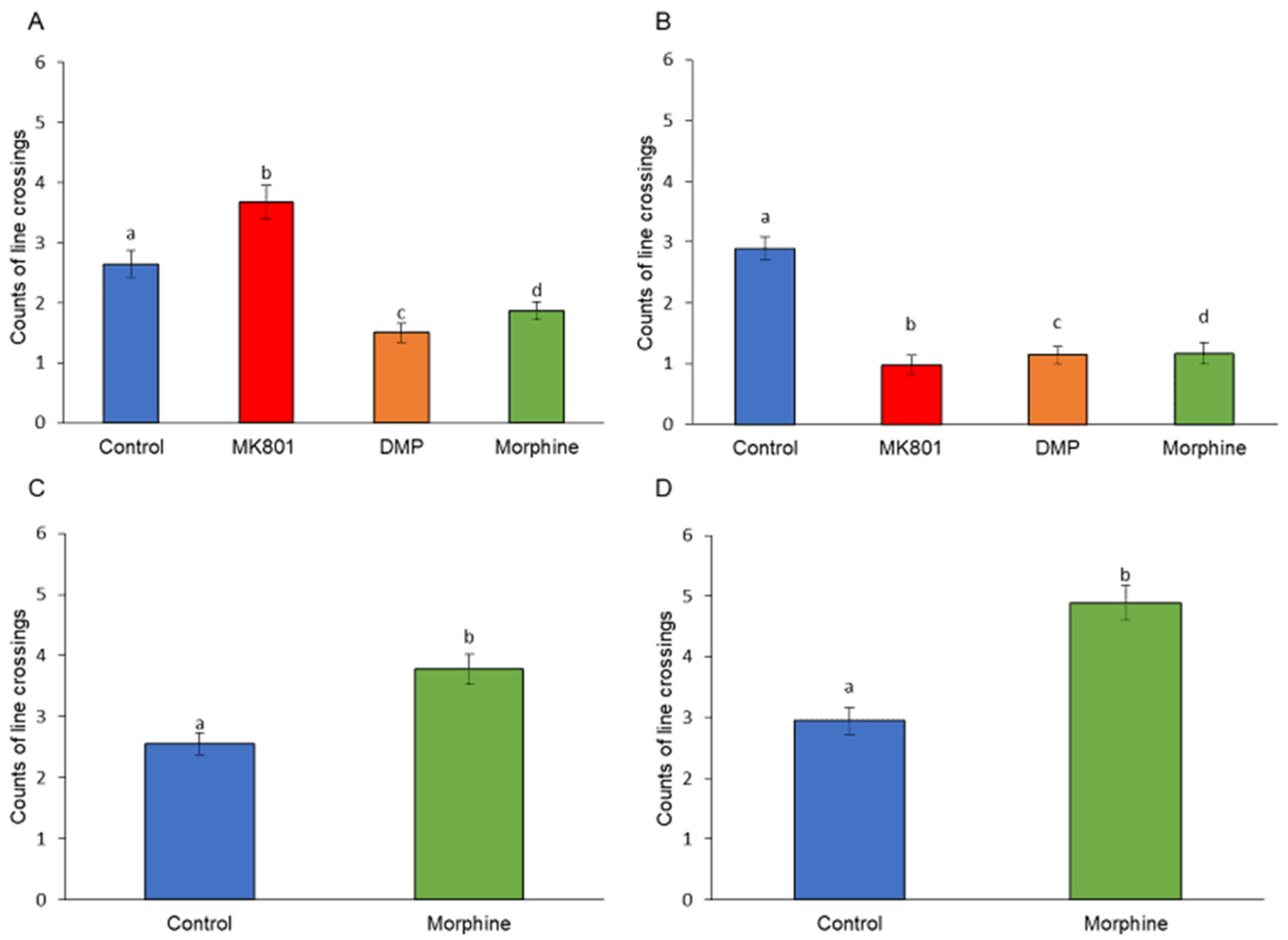
Figure 1. Effects of (A) short-term (F (3, 140) = 42.92, p = 0.01) and (B) long-term (F (3, 140) = 114.29, p = 0.00) exposure to MK801, DMP, and morphine, and after withdrawal of (C) short-term (t (70) = 35.30, p = 0.00) and (D) long-term (t (70) = 177.64, p = 0.00) morphine treatment on the locomotor activity. Data expressed as mean ± SEM, n = 12 of 3 independent experiments. Different alphabets indicate statistically significant values, p < 0.05. Drug abbreviation: DMP, domperidone.
Ijms 24 04088 g001
Figure 2. Effects of (A) short-term (F (3, 140) =46.84, p = 0.01) and (B) long-term (F (3, 140) = 124.51, p = 0.01) exposure to MK801, DMP, and morphine, and after withdrawal of (C) short-term (t (70) = 32.81, p = 0.04) and (D) long-term (t (70) = 184.32, p = 0.01) morphine treatment on the total distance travelled of zebrafish larvae (mm/5 min). Data expressed as mean ± SEM, n = 12 of 3 independent experiments. Different alphabets indicate statistically significant values, p < 0.05. Drug abbreviation: DMP, domperidone.
Ijms 24 04088 g002
Figure 3. Effects of (A) short-term (F (3, 140) =42.92, p = 0.00) and (B) long-term (F (3, 140) = 114.29, p = 0.00) exposure to MK801, DMP, and morphine, and after withdrawal of (C) short-term (t (70) = 35.31, p = 0.03) and (D) long-term (t (70) = 177.64, p = 0.00) morphine treatment on the social preferences of zebrafish larvae (sec/5 min). Data expressed as mean ± SEM, n = 12 of 3 independent experiments. Different alphabets indicate statistically significant values, p < 0.05. Drug abbreviation: DMP, domperidone.
Ijms 24 04088 g003
Figure 4. Tracked trajectories for control, MK801, DMP and morphine treated larvae after being subjected to conspecific larvae. Effects of (A) short-term and (B) long-term exposure to MK801, DMP and morphine, and after withdrawal of (C) short-term and (D) long-term morphine treatment on the social preference. Drug abbreviation: DMP, domperidone.
Ijms 24 04088 g004
Figure 5. Distribution of dopaminergic neurons in Tg (dat: EGFP) zebrafish. Representative confocal images showing the location of dopaminergic nuclei in the brains of the larval zebrafish following (A) short-term and (B) long-term exposure to MK801, DMP, and morphine, and after withdrawal of (C) short-term and (D) long-term treatment of morphine. Drug abbreviation: DMP, domperidone. Other abbreviations: LC, locus coeruleus; PT, posterior tuberculum; TPp, posterior tubercule.
Ijms 24 04088 g005
Figure 6. Effects of (A) short-term (F (3, 28) = 19.32, p = 0.00), (B) long-term (F (3, 28) = 10.74, p = 0.00) exposure to MK801, DMP, and morphine, and after withdrawal of (C) short-term (t (14) = 18.78, p = 0.00) and (D) long-term (t (14) = 6.33, p = 0.02) morphine treatment on cell counts in the PT region of the zebrafish larvae brain. Data expressed as mean ± SEM, n = 8. Different alphabets indicate statistically significant values, p < 0.05. Drug abbreviation: DMP, domperidone. Other abbreviation: PT, posterior tuberculum.
Ijms 24 04088 g006
Figure 7. Effects of (A) short-term (F (3, 32) = 20.09, p = 0.00) and (B) long-term (F (3, 32) = 22.17, p = 0.00) exposure to MK801, DMP, and morphine, and after withdrawal of (C) short-term (t (16) = 206.7, p = 0.00) and (D) long-term (t (16) = 56.83, p = 0.00) morphine treatment on the DA concentration (pg/mL) of the zebrafish larvae brain. Data expressed as mean ± SEM, n = 50 of 3 independent experiments. Different alphabets indicate statistically significant values, p < 0.05. Drug abbreviation: DMP, domperidone. Other abbreviation: DA, dopamine.
Ijms 24 04088 g007
Figure 8. Altered gene expression in zebrafish larvae following exposure to MK801, DMP, and morphine. (A) Short-term effect on expression of α1C (F (3, 32) = 1.62, p = 0.91), α1Sa (F (3, 32) = 1.57, p = 0.22), α1Aa (F (3,32) = 1.99, p = 0.13), c-fos (F (3, 32) = 1.50, p = 0.67), drd2a (F (3, 32) = 53.34, p = 0.00), th1 (F (3, 32) = 6.18, p = 0.02), th2 (F (3, 32) = 283.76, p = 0.00), and eif4ebp1 (F (3, 32) = 25.18, p = 0.00). (B) Long-term effect on expression of α1C (F (3,32) = 7.06, p = 0.00), α1Sa (F (3, 32) = 15.60, p = 0.00), α1Aa (F (3, 32) = 25.45, p = 0.00), c-fos (F (3,32) = 10.22, p = 0.01), drd2a (F (3, 32) = 264.97, p = 0.00), th1 (F (3, 32) = 292.13, p = 0.00), th2 (F (3, 32) = 617.59, p = 0.00), and eif4ebp1 (F (3,32) = 20.76, p = 0.00). (C) Effect of morphine withdrawal of short-term morphine treatment on α1C (t (16) = 0.19, p = 0.66), α1Sa (t (16) = 1.13, p = 0.06), α1Aa (t (16) = 3.84, p = 0.10), c-fos (t (16) = 6.54, p = 0.02), drd2a (t (16) = 42.67, p = 0.00), th1 (t (16) = 54.81, p = 0.00), th2 (t (16) = 0.81, p = 0.38), and eif4ebp1 (t (16) = 11.33, p = 0.00). (D) Effect of morphine withdrawal after long-term morphine treatment on α1C (t (16) = 0.69, p = 0.42), α1Sa (t (16) = 2.97, p = 0.10), α1Aa (t (16) = 24.37, p = 0.00), c-fos (t (16) = 7.49, p = 0.01), drd2a (t (16) = 18.02, p = 0.00), th1 (t (16) = 18.11, p = 0.01), th2 (t (16) = 28.78, p = 0.00), and eif4ebp1 (t (16) = 8.10, p = 0.01). Data expressed as mean ± SEM, n = 50 of 3 independent experiments. Different alphabets indicate statistically significant values, p < 0.05. Drug abbreviation: DMP, domperidone.
Ijms 24 04088 g008aIjms 24 04088 g008b
Table 1. Primer sequences of candidate genes.
ProteinGenePrimer Sequences (5′ → 3′)GenBank Accession NoReferences
Cav1.2α1CF: ACGGAGTCACTCTCCGACACXM_009300335[120]
R: AGAGAGGGCACAGGCTGATA
Cav1.1aα1SaF: TCTATAGGCGTGCTGGAGGTNM_001146150[120]
R: GCTATCTGCGAGCTGTAGGG
Cav2.1aα1AaF: TGCTACCCAGCCACATGATAENSDARG00000037905[120]
R: TGGTAGAGAGTGAGGGTAAA
Dopamine receptor D2adrd2aF: TGGTACTCCGGAAAAGACGNM_183068[121]
R: ACTCGGGATGGGTGCATTTC
c-FOSc-fosF:GCAGAGCATTGGCAGGAGDQ003339[122]
R: CCCTTCGGATTCTCCTTTTCT
Tyrosine hydroxylase 1th1F: GACGGAAGATGATCGGAGACAXM_682702.1[123]
R: CCGCCATGTTCCGATTTCT
Tyrosine hydroxylase 2th2F: CTCCAGAAGAGAATGCCACATGNM_001001829.1[124]
R: ACGTTCACTCTCCAGCTGAGTG
Eukaryotic translation initiation factor 4E-binding proteineif4ebp1F: AACGACAAGGTGCAAAGACNM_199645[125]
R: GTGGTTGGAATTGCCTGACT
Beta actin (β-actin)actb1F: AAGCTGTGACCCACCTCACGAF057040[126]
R:GGCTTTGCACATACCGGAGC
Disclaimer/Publisher’s Note: The statements, opinions and data contained in all publications are solely those of the individual author(s) and contributor(s) and not of MDPI and/or the editor(s). MDPI and/or the editor(s) disclaim responsibility for any injury to people or property resulting from any ideas, methods, instructions or products referred to in the content.