Article

A Receptor Tyrosine Kinase Inhibitor Sensitivity Prediction Model Identifies AXL Dependency in Leukemia

1
Division of Translational Cancer Research, Department of Laboratory Medicine, Lund University, 22381 Lund, Sweden
2
Lund Stem Cell Center, Department of Laboratory Medicine, Lund University, 22184 Lund, Sweden
3
Department of Hematology, Oncology and Radiation Physics, Skåne University Hospital, 22185 Lund, Sweden
*
Author to whom correspondence should be addressed.
Int. J. Mol. Sci. 2023, 24(4), 3830; https://doi.org/10.3390/ijms24043830
Submission received: 12 January 2023 / Revised: 5 February 2023 / Accepted: 11 February 2023 / Published: 14 February 2023
(This article belongs to the Special Issue Artificial Intelligence in Biomarker Discovery)

Abstract

:
Despite incredible progress in cancer treatment, therapy resistance remains the leading limiting factor for long-term survival. During drug treatment, several genes are transcriptionally upregulated to mediate drug tolerance. Using highly variable genes and pharmacogenomic data for acute myeloid leukemia (AML), we developed a drug sensitivity prediction model for the receptor tyrosine kinase inhibitor sorafenib and achieved more than 80% prediction accuracy. Furthermore, by using Shapley additive explanations for determining leading features, we identified AXL as an important feature for drug resistance. Drug-resistant patient samples displayed enrichment of protein kinase C (PKC) signaling, which was also identified in sorafenib-treated FLT3-ITD-dependent AML cell lines by a peptide-based kinase profiling assay. Finally, we show that pharmacological inhibition of tyrosine kinase activity enhances AXL expression, phosphorylation of the PKC-substrate cyclic AMP response element binding (CREB) protein, and displays synergy with AXL and PKC inhibitors. Collectively, our data suggest an involvement of AXL in tyrosine kinase inhibitor resistance and link PKC activation as a possible signaling mediator.

1. Introduction

The role of receptor tyrosine kinases (RTKs) in oncogenic transformation is well-documented [1]. Several RTKs are mutated or overexpressed in human cancers and are likely to be involved in constitutive activation of survival and proliferative signaling, and thereby, disease progression [2]. Therefore, inhibition of RTKs has become an attractive approach for the treatment of several cancers [3,4]. However, the development of primary and secondary resistance to RTK-targeted therapies limits its clinical application [5,6].
Several mechanisms explaining the development of therapy resistance have been documented during the past decades [7,8,9]. The most studied mechanisms include the selection of clones during drug treatment due to the pre-existing genetic alterations in a rare population or the acquisition of de novo mutations in cancer cells. During the drug treatment, epigenetic modifications can occur, which in turn contribute to alterations in gene expression of tumor suppressors and oncogenes, leading to the activation of cell survival pathways. Furthermore, dynamic fluctuation of gene expression during drug treatment can help cells escape therapy-induced death resulting in the accumulation of drug-tolerant cells [10,11].
Oncogenic mutations in the type III receptor tyrosine kinase FLT3 are the most commonly identified genetic alterations in acute myeloid leukemia (AML) [12]. Those type of mutations have also been reported in other hematological malignancies including acute lymphoblastic leukemia and myeloproliferative disorders [13,14,15]. Oncogenic mutations in FLT3 lead to constitutive activation of several pro-survival pathways including PI3K/AKT, MAPK pathways, and, with some exceptions, the STAT5 pathway [12]. Besides the identification of FLT3 mutations and their importance in pro-survival signaling, FLT3 inhibitors if used as monotherapy have not been as successful as expected. The first-generation FLT3 inhibitors, such as midostaurin, sorafenib, and sunitinib, were primarily developed to target other kinases and were later found to be useful in inhibiting FLT3 [16]. Such inhibitors display a low degree of specificity and higher toxicity profile by inhibiting a wide range of kinases [17]. The next-generation FLT3 inhibitors, e.g., crenolanib, AC220 (quizartinib), and gilteritinib, are more selective for FLT3 but still inhibit several other RTKs [18]. Several of those inhibitors underwent clinal trials and recently midostaurin and gilteritinib were approved by the FDA, and AC220 was approved for use for AML in Japan (a summary can be found in reference [18]).
The mechanisms by which AML patients develop resistance to the targeted therapies have been explored from different points of view. Patients carrying FLT3-ITD mutations develop resistance to FLT3-targeted therapies predominantly due to secondary mutations in the tyrosine kinase domain and also likely due to the activation of parallel survival signaling pathways during drug treatment [12,18,19,20].
In this study, we developed a machine learning model for drug sensitivity prediction and identified AXL as an important feature. We further show that AXL expression is upregulated during drug treatment and identify a possible link between protein kinase C activity and the regulation of AXL expression.

2. Results

2.1. Development of a TabNet Model to Predict Sorafenib Sensitivity in Leukemia

Several clinical trials have been initiated with the first-generation FLT3 inhibitor sorafenib for maintenance after stem cell transplantation [21], in combination with standard chemotherapy [22], and in combination with other drugs [23]. However, similar to other tyrosine kinase inhibitors, patients in many cases display sorafenib resistance due to the activation of parallel signaling pathways [20,24,25]. The elegant BeatAML studies used sorafenib to treat ex vivo 385 FLT3-ITD negative and 118 FLT3-ITD positive AML samples [26,27]. As shown in Supplementary Figure S1, we observed mixed responses to both FLT3-ITD positive and negative samples from BeatAML studies to sorafenib, although as expected, FLT3-ITD positive samples showed a significantly higher response. Our results suggest that the presence of FLT3-ITD does not exclusively predict sorafenib response. Therefore, we first aimed to develop a tool to predict sorafenib response in AML. We used pharmacogenomic and transcriptomics data from BeatAML, TCGA, and cell line studies. To identify genes that are consistently deregulated in AML we used highly variable genes (using ScanPy [28]) selected from BeatAML and TCGA studies. We then excluded genes that are absent in cell line data (Figure 1A). We identified a list of 732 genes that were then used to develop a binary classification model using a recently developed deep learning algorithm for tabular data (TabNet [29]). We used 1066 samples to train the TabNet model (831 samples for training and 235 samples for validation), and 234 samples for testing the model (Figure 1B). The model predicted 91 samples as sorafenib-resistant (out of 113 samples, 80.5% sensitivity) and 104 samples as sorafenib-sensitive (out of 121 samples, 86.0% specificity) (Figure 1C). Overall, the model achieved 83.3% accuracy and 84.3% precision. We have also tested the model by randomly sampling test samples in 500 different ways and achieved an average area under the curve (AUC) of 0.89 from the receiver operating characteristic (ROC) curve and 89.05% average accuracy. Furthermore, we attempted to reduce the number of features using the “Select From Model” function from the Sklearn feature selection [30], which allowed us to reduce the number of features to 36 (Figure 1D). The model developed using 36 features showed a slight improvement in terms of accuracy (87.6%), precision (93.3), and specificity (93.9) scores. However, when tested with random sampling, performance scores were similar to that of 732 features (Figure 1E).

2.2. Shapley Additive Explanations (SHAP) Identify AXL and HTRA4 as Important Features

Next, we applied the XGBoost classifier to check the predictive performance of the two sets of features described above. XGBoost is one of the most popular tree-based ensemble algorithms that has been widely used for classifications [31] and can be also used to identify important features when combined with Shapley additive explanations (SHAP) [32]. We used a random sampling method to sample test data (20%) from 1300 labeled samples. XGBoost models were built using 80% data and then tested on the corresponding 20% of the data. Models built using both 732 (Figure 2A) and 36 (Figure 2B) features were equally good as shown in average AUC scores in ROC curves. We then applied SHAP to identify leading predictors. We used both 732 and 36 features and observed that AXL and HTRA4 scored highest in both models (Figure 2C,D). The HTRA4 gene encodes a human protease that belong to the mammalian high temperature requirement A (HtrA) family. HtrA family proteins are involved in several cellular processes, including apoptosis and unfolded protein stress response [33]. A recent study suggested that the expression of HTRA4 enhances the effect of chemotherapy in breast and lung cancer cell lines [34]. AXL, a member of the TAM receptor tyrosine kinase family (TYRO3, AXL, and MER), is well known for its roles in cell proliferation, migration, invasion, and survival. The expression of AXL can also contribute to therapy resistance in many cancer types [35]. While HTRA4 expression may be important for sorafenib sensitivity prediction, AXL is well known for its role in therapy resistance [36].

2.3. Sorafenib Activates Protein Kinase C Signaling

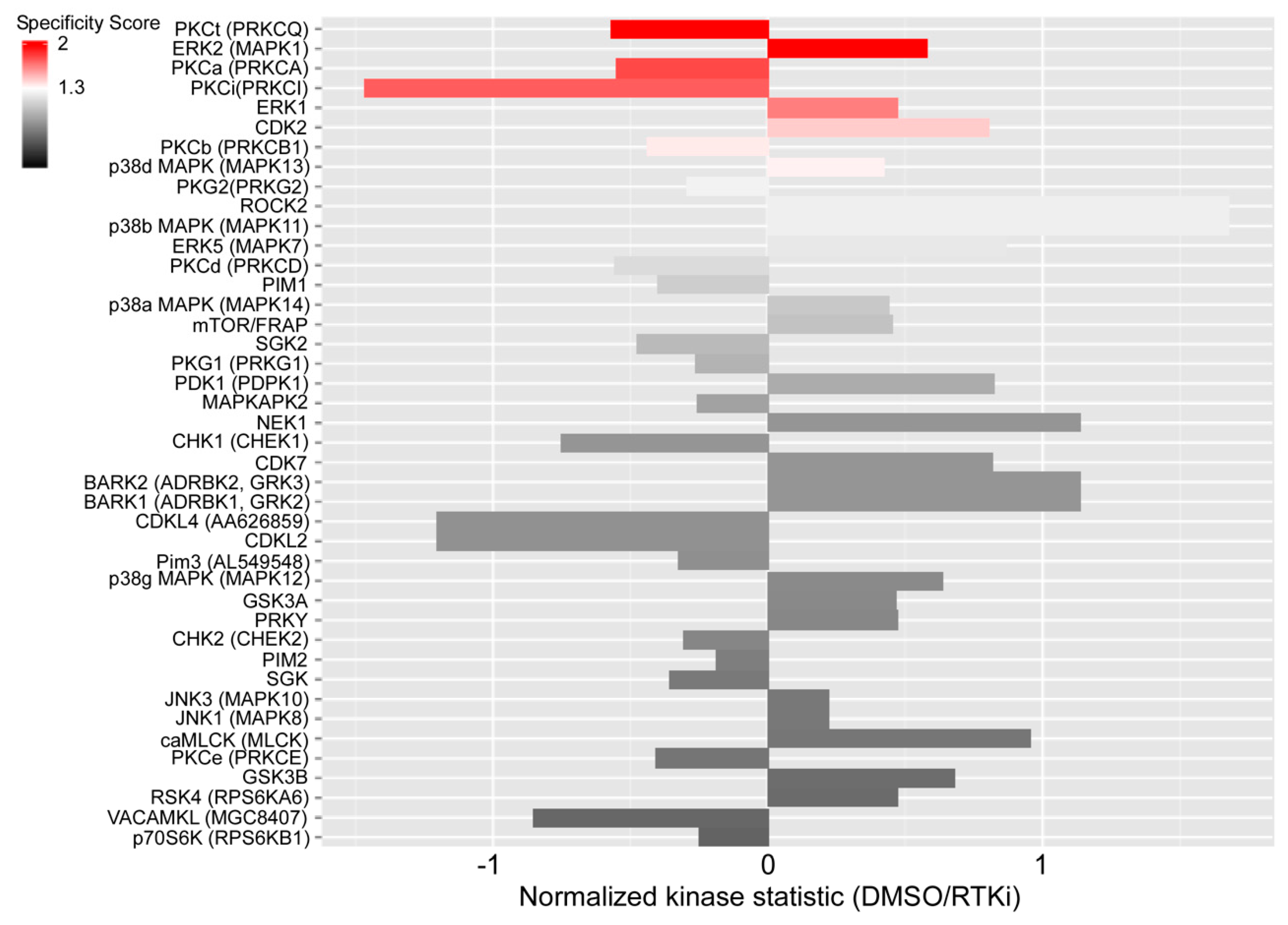
Next, we analyzed the enrichment of genes in sorafenib-resistant patients to map them on pathways in cancer using the BeatAML dataset. A sample was categorized as resistant or sensitive if the sample was both experimentally and predicted (by both TabNet models) in the same category. Using such criteria, 140 samples were labeled as sensitive and 85 samples were labeled as resistant. We mapped deregulated genes on KEGG [37] “Pathways in cancer” using iDEP [38] that uses Pathview [39] for mapping. Supplementary Figure S2 shows enriched pathways in resistant samples. We used a two-fold cut-off (scale shows log2 values) and observed enrichment of several serine/threonine kinase pathways. In a recent study, we have shown that pathways that are involved in therapy resistance were deregulated in the initial drug exposure [11]. Therefore, we treated an AML cell line (MOLM-13) with sorafenib for four hours and measured the activation of serine/threonine kinases using a peptide-substrate-based kinase profiling assay. MOLM-13 cells express a copy of FLT3-WT and a copy of FLT3-ITD, and therefore display constitutive activation of FLT3 downstream signaling pathways. We observed that while sorafenib suppressed ERK1/2 activation, which is one of the downstream effectors of FLT3 signaling, the activity of several protein kinase C (PKC) isoforms, including the classical isoform PKCα, the novel isoform PKCθ and the atypical isoform PKCι, were enhanced (Figure 3). PKC family members are involved in various cellular events including cell proliferation, survival, differentiation, apoptosis, and therapy resistance [40,41,42,43]. We observed the enrichment of at least two signaling pathways that activate PKC isoforms in pathway enrichment data (Supplementary Figure S2). Taken together, our data indicate a role of PKC isoforms in sorafenib resistance.

2.4. Inhibition of the Receptor Tyrosine Kinase FLT3 Results in Transcriptional Upregulation of AXL

Previously, we have demonstrated that dexamethasone-resistant B-cell acute lymphoblastic leukemia cells that express the oncogenic mutant of FLT3 display upregulation of AXL expression upon inhibition of FLT3 [44]. A cross-talk between FLT3-ITD and AXL has also been reported where AXL was identified as a positive regulator of FLT3-ITD [45]. Furthermore, a recent study suggested that AXL expression is upregulated in AC220-resistant cells and cells display sensitivity to AXL-targeted therapy [46]. These studies demonstrate a link between FLT3-ITD activity and AXL expression. To understand how targeted inhibition of FLT3 regulates global transcriptomics in AML cells, we analyzed global gene expression changes using microarray. MOLM-13 and MV4-11 cell lines were treated with AC220 for six hours to capture the early regulations in transcription. The total mRNA from inhibitor-treated cells was analyzed by microarray. As expected, AXL expression was upregulated in inhibitor-treated cells (Supplementary Figure S3A). Besides AXL, 225 genes were upregulated in MV4-11 and 145 genes in MOLM-13 (Supplementary Figure S3B), of which 86 genes were upregulated in both cell lines (Supplementary Figure S3C). Among those 86 genes, several of them are regulators of gene transcriptions (such as ARID5B, BTG2, CREBRF, HBP1, PRDM1, CALCOCO1, CARF, CIITA, HDAC5, MAP2K6, STAT2, TXNIP, TP53INP1, ZNF836, and ZNF846) (Supplementary Figure S3D). As we identified AXL as an important factor contributing to sorafenib response prediction and AXL expression was upregulated during AC220 treatment, we aimed to identify kinases that are involved in the regulation of AXL expression. We used MV4-11 and MOLM-13 cells that have constitutive activation of several pro-survival pathways due to the expression of FLT3-ITD [12,47,48,49,50]. These cells are highly responsive to FLT3-targeting drugs, such as AC220 and crenolanib, but also display sensitivity to a different extent to several pathway-specific inhibitors that target cell cycle regulatory kinases, mitogen-activated protein kinases (MAPKs), the PI3K/mTOR pathway, protein kinase C (PKC), and Janus tyrosine kinase (JAK) (Figure 4A and Supplementary Figure S4A,B). Several of these kinases, such as SYK, PKC, PI3K/mTOR, MAPK, and cell cycle kinases, are direct or indirect effectors of FLT3 signaling [12,50,51,52,53,54,55]. Therefore, inhibition of FLT3 downstream effectors may cooperate in growth inhibition by abrogating FLT3-ITD-mediated survival signaling. However, besides the strong growth inhibition, kinase inhibitors targeting FLT3 downstream effectors were poor in activating increased AXL transcription in both cell lines, as compared to FLT3-targeted inhibition (Figure 4B). The kinase inhibitors that displayed a considerable effect on AXL expression, e.g., hesperadin, may also target FLT3, since several other aurora kinase inhibitors are potential inhibitors of FLT3 [56]. Therefore, it is likely that transcriptional induction of AXL is caused by the direct inhibition of FLT3 and not by its downstream signaling components. However, we cannot exclude the possibility of the involvement of other tyrosine kinases, as inhibitors targeting FLT3 may also inhibit other tyrosine kinases due to similarity in the kinase domain. Furthermore, we observed that AXL expression was dynamically regulated upon FLT3 inhibition (Supplementary Figure S4C). The expression of AXL enhanced over time and reached its highest level after 16 h before declining. Upregulation was also observed at the protein level (Supplementary Figure S4D).

2.5. An Inhibitor Targeting FLT3 Displays Synergy with AXL and PKC Inhibitors

As we observed sorafenib-induced PKC activity in MOLM-13 cells, we checked whether the regulation of PKC activity is a result of the inhibition of FLT3-ITD. We measured the phosphorylation of cyclic AMP response element binding (CREB) protein and observed that CREB phosphorylation was enhanced during the AC220 treatment (Figure 5A). Furthermore, MV4-11 and MOLM13 cells displayed downregulation of AKT, ERK1/2, and GSK3β (Ser 9) phosphorylation in AC220-treated cells (Figure 5A). CREB is a substrate of several serine/threonine kinases including PKC [57,58]. Since we observed that several CREB-activating kinases, such as AKT [59], ERK [60], and GSK3β [61], were inhibited by AC220, it is likely that enhanced CREB phosphorylation was mediated by PKC. A recent report also suggested that AXL expression was regulated by serine/threonine protein kinase PKCα in triple-negative breast cancer [62]. Furthermore, like the AXL inhibitor bemcentinib (Figure 5B), pan-PKC inhibitor sotrastaurin, and LXS196 (inhibits PKCα, PKCθ, and GSK3β) displayed considerable synergy with AC220 (Figure 5C,D), suggesting possible involvement of PKC isoforms in AXL expression.

3. Discussion

In this study, we identified two gene signatures that can be used to develop machine-learning models for the prediction of tyrosine kinases inhibitor sorafenib sensitivity. The first gene signature (consisting of 732 genes) was identified using ScanPy’s “highly variable gene” function and the second gene signature (36 genes) was short-listed from the first gene signature by applying the Sklearn feature selection method. We developed a deep tabular data learning model using TabNet and gradient-boosted tree-based models using XGBoost. Both models displayed a similar performance. Furthermore, using Shapley additive explanations we identified AXL as a leading feature.
The TAM family receptor tyrosine kinases are well-studied for their role in leukemia [63]. More specifically, AXL has been shown to be involved in cell proliferation, survival, tumor metastasis, stem cell phenotype, and more importantly, therapy resistance [64]. Virtually all types of cancer treatment, such as chemotherapy, radiotherapy, and targeted therapy, can induce AXL expression, resulting in resistance to treatment. For example, chemotherapy-induced AXL expression was reported in AML [65], lung cancer [66], prostate cancer [67], ovarian cancer [68], and endometrial cancer [69] as conferring therapy resistance. Recently, a series of studies linked AXL to several receptor tyrosine kinase inhibitors including EGFR inhibitors in lung cancer [70], head and neck cancer [71] and colorectal cancer [72], HER2 inhibitors in breast cancer [73], and FLT3 inhibitors in AML [74]. Identification of AXL as a leading feature in the sorafenib sensitivity prediction model further emphasizes its importance in resistance to receptor tyrosine kinase inhibitors.
Several mechanisms by which AXL contributes to the receptor tyrosine kinase inhibitor resistance have been proposed. In AML, cytokine-induced activation of STAT5 enhances AXL expression, leading to AC220 resistance [74]. A natural product, apigenin, reduced AXL expression in ovarian cancer cell lines without affecting IL6 production and STAT3 phosphorylation [75], suggesting the selectivity of the STAT proteins in the regulation of AXL expression. The observation that AML cell lines treated with sorafenib or AC220 enhance PKC activity and AXL expression provides a possible additional mechanism for the regulation of AXL expression. In head and neck and esophageal squamous cell carcinomas, AXL dimerizes with EGFR and activates PKC through PLCγ [76], and in breast cancer pharmacological inhibition, PKC resulted in decreased AXL expression [62], suggesting a feedback loop between AXL expression and PKC activation. We observed that pharmacological inhibition of FLT3 in combination with AXL and PKC displays synergy, further suggesting that PKC may be involved in previously reported crosstalk between FLT3 and AXL [45].
In conclusion, we provide a framework of drug sensitivity prediction using a selected gene signature and provide evidence that AXL is involved in resistance to tyrosine kinase inhibitors, linking it to PKC activation as a possible signaling mediator.

4. Materials and Methods

Cell cultures: MOLM-13 and MV4-11were obtained from Deutsche Sammlung von Mikroorganismen und Zellkulturen (DSMZ, Braunschweig, Germany). The cell lines were cultured in RMPI-1640. All media were supplemented with 10% heat-inactivated fetal bovine serum (FBS), 100 units/mL penicillin, and 100 µg/mL streptomycin. Cells were maintained at 37 °C with 5% CO2 and regularly tested for mycoplasma contamination. All inhibitors were provided by Selleckchem and were dissolved in DMSO according to the manufacturer’s instructions. The final concentration of DMSO in cell cultures never exceeded 0.1%.
Cell viability assay: Cell viability assays were performed using PrestoBlue (Thermo Fisher Scientific, Waltham, MA, USA). Cells (1.8 × 10⁴) were plated into 96-well microplates in 100 μL cell culture medium with increasing doses of inhibitors for 48 h. For analysis, 10 μL PrestoBlue was added to each well, and plates were incubated for 3 h before reading in a microplate reader (excitation 530 nm, emission 590 nm; BioTek Instruments Inc., Winooski, VT, USA). Non-linear regression analysis was used to determine IC50.
Real-time quantitative PCR (RT-qPCR): Total RNA was isolated using the RNeasy Mini Kit following the manufacturer’s instructions (Qiagen, Venlo, Netherlands). Total RNA was measured using Nanodrop 2000 Spectrophotometer (Thermo Fisher Scientific). RNA was reverse-transcribed to cDNA using a High-Capacity cDNA Reverse Transcription Kit (Thermo Fisher Scientific) according to the manufacturer’s instructions. Gene expression was assessed using the TaqMan Gene Expression Master Mix and gene-specific probes (Thermo Fisher Scientific). AXL gene expression levels were normalized against the endogenous housekeeping genes GAPDH, β-actin, and α-tubulin. Normalization of gene expression was carried out by ΔΔCt analysis.
Western blot analysis: Cells were lysed in 1% Triton X-100 lysis buffer supplemented with phosphatase/protease inhibitors (Trasylol, PMSF, and Na3VO4). Protein concentration was determined using bicinchoninic acid (BCA) protein assay (Thermo Fisher Scientific). Lysates were mixed with SDS loading buffer and heated to 96 °C for 5 min. Equal amounts of protein (20 µg) were subjected to SDS-PAGE. Gels were transferred to polyvinylidene difluoride (PVDF) membranes, blocked with 5% non-fat dry milk, and immunoblotted with primary antibodies. The anti-pERK1/2 (1:200), pCREB1 (1:1000), and anti-β-actin (1:5000) antibodies were obtained from Santa Cruz Biotechnology (Dallas, TX, USA). Anti-pAKT Ser473 (1:5000) was obtained from Cell Signaling Technology (Danvers, MA, USA), Anti-pGSK3β (Ser9; 1:1000) was obtained from Thermo Fisher Scientific. Blots were next incubated with a secondary antibody conjugated with horseradish peroxidase (HRP) and developed using a Luminata Forte Western HRP substrate (Millipore, Burlington, MA, USA) and Amersham Imager 600 (GE Healthcare, Chicago, IL, USA).
Machine learning models: A brief description of machine learning models was described previously [77,78]. Pharmacogenomic data were collected from BeatAML, TCGA, and scDEAL studies. To develop binary classification models, Jupyter Notebook was used. The TabNet model was developed on CUDA and all other models were developed on the CPU.

Supplementary Materials

The following supporting information can be downloaded at: https://www.mdpi.com/article/10.3390/ijms24043830/s1.

Author Contributions

A.N., L.A.A., M.A., H.D., X.Z., L.R. and J.U.K. designed and performed the experiments, collected and analyzed the data, and wrote the manuscript. All authors have read and agreed to the published version of the manuscript.

Funding

This research was supported by the Kungliga Fysiografiska Sällskapet i Lund (A.N. and M.A.), the Crafoord Foundation (J.U.K), the Swedish Cancer Society (J.U.K. and L.R.), the Swedish Research Council (L.R.), the Swedish Childhood Cancer Foundation (J.U.K.), and SUS Stiftelser och Donationer (L.R.), Governmental Funding of Clinical Research within the National Health Service (ALF) (L.R.).

Institutional Review Board Statement

Not applicable.

Informed Consent Statement

Not applicable.

Data Availability Statement

All raw data are available upon request.

Acknowledgments

Open access funding was partially provided by Lund University.

Conflicts of Interest

The authors declare no conflict of interest.

References

  1. Esteban-Villarrubia, J.; Soto-Castillo, J.J.; Pozas, J.; San Roman-Gil, M.; Orejana-Martin, I.; Torres-Jimenez, J.; Carrato, A.; NAlonso-Gordoa, T.; Molina-Cerrillo, J. Tyrosine Kinase Receptors in Oncology. Int. J. Mol. Sci. 2020, 21, 8529. [Google Scholar] [CrossRef] [PubMed]
  2. Yamaoka, T.; Kusumoto, S.; Ando, K.; Ohba, M.; Ohmori, T. Receptor Tyrosine Kinase-Targeted Cancer Therapy. Int. J. Mol. Sci. 2018, 19, 3491. [Google Scholar] [CrossRef] [PubMed]
  3. Labbozzetta, M.; Barreca, M.; Spano, V.; Raimondi, M.V.; Poma, P.; Notarbartolo, M.; Barraja, P.; Montalbano, A. Novel insights on [1,2]oxazolo[5,4-e]isoindoles on multidrug resistant acute myeloid leukemia cell line. Drug Dev. Res. 2022, 83, 1331–1341. [Google Scholar] [CrossRef] [PubMed]
  4. Zhong, Y.; Qiu, R.Z.; Sun, S.L.; Zhao, C.; Fan, T.Y.; Chen, M.; Li, N.G.; Shi, Z.H. Small-Molecule Fms-like Tyrosine Kinase 3 Inhibitors: An Attractive and Efficient Method for the Treatment of Acute Myeloid Leukemia. J. Med. Chem. 2020, 63, 12403–12428. [Google Scholar] [CrossRef]
  5. Wang, Z.; Cai, J.; Cheng, J.; Yang, W.; Zhu, Y.; Li, H.; Lu, T.; Chen, Y.; Lu, S. FLT3 Inhibitors in Acute Myeloid Leukemia: Challenges and Recent Developments in Overcoming Resistance. J. Med. Chem. 2021, 64, 2878–2900. [Google Scholar] [CrossRef]
  6. Juchum, M.; Gunther, M.; Laufer, S.A. Fighting cancer drug resistance: Opportunities and challenges for mutation-specific EGFR inhibitors. Drug Resist. Updates 2015, 20, 12–28. [Google Scholar] [CrossRef]
  7. Wang, X.; Zhang, H.; Chen, X. Drug resistance and combating drug resistance in cancer. Cancer Drug Resist. 2019, 2, 141–160. [Google Scholar] [CrossRef]
  8. Boumahdi, S.; de Sauvage, F.J. The great escape: Tumour cell plasticity in resistance to targeted therapy. Nat. Rev. Drug Discov. 2020, 19, 39–56. [Google Scholar] [CrossRef]
  9. Kazi, J.U. Mechanisms of Anticancer Therapy Resistance: The Role of Cancer Stem Cells. Int. J. Mol. Sci. 2020, 21, 9006. [Google Scholar] [CrossRef]
  10. Shaffer, S.M.; Dunagin, M.C.; Torborg, S.R.; Torre, E.A.; Emert, B.; Krepler, C.; Beqiri, M.; Sproesser, K.; Brafford, P.A.; Xiao, M.; et al. Rare cell variability and drug-induced reprogramming as a mode of cancer drug resistance. Nature 2017, 546, 431–435. [Google Scholar] [CrossRef]
  11. Shah, K.; Ahmed, M.; Kazi, J.U. The Aurora kinase/beta-catenin axis contributes to dexamethasone resistance in leukemia. NPJ Precis. Oncol. 2021, 5, 13. [Google Scholar] [CrossRef] [PubMed]
  12. Kazi, J.U.; Rönnstrand, L. FMS-like Tyrosine Kinase 3/FLT3: From Basic Science to Clinical Implications. Physiol. Rev. 2019, 99, 1433–1466. [Google Scholar] [CrossRef] [PubMed]
  13. Lin, P.; Jones, D.; Medeiros, L.J.; Chen, W.; Vega-Vazquez, F.; Luthra, R. Activating FLT3 mutations are detectable in chronic and blast phase of chronic myeloproliferative disorders other than chronic myeloid leukemia. Am. J. Clin. Pathol. 2006, 126, 530–533. [Google Scholar] [CrossRef] [PubMed]
  14. Kabir, N.N.; Rönnstrand, L.; Kazi, J.U. FLT3 mutations in patients with childhood acute lymphoblastic leukemia (ALL). Med. Oncol. 2013, 30, 462. [Google Scholar] [CrossRef]
  15. Bains, A.; Luthra, R.; Medeiros, L.J.; Zuo, Z. FLT3 and NPM1 mutations in myelodysplastic syndromes: Frequency and potential value for predicting progression to acute myeloid leukemia. Am. J. Clin. Pathol. 2011, 135, 62–69. [Google Scholar] [CrossRef]
  16. Wander, S.A.; Levis, M.J.; Fathi, A.T. The evolving role of FLT3 inhibitors in acute myeloid leukemia: Quizartinib and beyond. Ther. Adv. Hematol. 2014, 5, 65–77. [Google Scholar] [CrossRef] [PubMed]
  17. Weisberg, E.; Roesel, J.; Furet, P.; Bold, G.; Imbach, P.; Florsheimer, A.; Caravatti, G.; Jiang, J.; Manley, P.; Ray, A.; et al. Antileukemic Effects of Novel First- and Second-Generation FLT3 Inhibitors: Structure-Affinity Comparison. Genes Cancer 2010, 1, 1021–1032. [Google Scholar] [CrossRef]
  18. Antar, A.I.; Otrock, Z.K.; Jabbour, E.; Mohty, M.; Bazarbachi, A. FLT3 inhibitors in acute myeloid leukemia: Ten frequently asked questions. Leukemia 2020, 34, 682–696. [Google Scholar] [CrossRef]
  19. Staudt, D.; Murray, H.C.; McLachlan, T.; Alvaro, F.; Enjeti, A.K.; Verrills, N.M.; Dun, M.D. Targeting Oncogenic Signaling in Mutant FLT3 Acute Myeloid Leukemia: The Path to Least Resistance. Int. J. Mol. Sci. 2018, 19, 3198. [Google Scholar] [CrossRef]
  20. Lindblad, O.; Cordero, E.; Puissant, A.; Macaulay, L.; Ramos, A.; Kabir, N.N.; Sun, J.; Vallon-Christersson, J.; Haraldsson, K.; Hemann, M.T.; et al. Aberrant activation of the PI3K/mTOR pathway promotes resistance to sorafenib in AML. Oncogene 2016, 35, 5119–5131. [Google Scholar] [CrossRef] [PubMed]
  21. Burchert, A.; Bug, G.; Fritz, L.V.; Finke, J.; Stelljes, M.; Röllig, C.; Wollmer, E.; Wäsch, R.; Bornhäuser, M.; Berg, T.; et al. Sorafenib Maintenance After Allogeneic Hematopoietic Stem Cell Transplantation for Acute Myeloid Leukemia With FLT3-Internal Tandem Duplication Mutation (SORMAIN). J. Clin. Oncol. 2020, 38, 2993–3002. [Google Scholar] [CrossRef]
  22. Pollard, J.A.; Alonzo, T.A.; Gerbing, R.; Brown, P.; Fox, E.; Choi, J.; Fisher, B.; Hirsch, B.; Kahwash, S.; Getz, K.; et al. Sorafenib in Combination With Standard Chemotherapy for Children With High Allelic Ratio FLT3/ITD+ Acute Myeloid Leukemia: A Report From the Children’s Oncology Group Protocol AAML1031. J. Clin. Oncol. 2022, 40, 2023–2035. [Google Scholar] [CrossRef] [PubMed]
  23. Borthakur, G.; Zeng, Z.; Cortes, J.E.; Chen, H.C.; Huang, X.; Konopleva, M.; Ravandi, F.; Kadia, T.; Patel, K.P.; Daver, N.; et al. Phase 1 study of combinatorial sorafenib, G-CSF, and plerixafor treatment in relapsed/refractory, FLT3-ITD-mutated acute myelogenous leukemia patients. Am. J. Hematol. 2020, 95, 1296–1303. [Google Scholar] [CrossRef]
  24. Scholl, S.; Spies-Weisshart, B.; Klink, A.; Muegge, L.O.; Fricke, H.J.; Hochhaus, A. Secondary resistance to sorafenib in two patients with acute myeloid leukemia (AML) harboring FLT3-ITD mutations. Ann. Hematol. 2011, 90, 473–475. [Google Scholar] [CrossRef] [PubMed]
  25. Damnernsawad, A.; Bottomly, D.; Kurtz, S.E.; Eide, C.A.; McWeeney, S.K.; Tyner, J.W.; Nechiporuk, T. Genome-wide CRISPR screen identifies regulators of MAPK and MTOR pathways mediating sorafenib resistance in acute myeloid leukemia. Haematologica 2022, 107, 77–85. [Google Scholar] [CrossRef] [PubMed]
  26. Bottomly, D.; Long, N.; Schultz, A.R.; Kurtz, S.E.; Tognon, C.E.; Johnson, K.; Abel, M.; Agarwal, A.; Avaylon, S.; Benton, E.; et al. Integrative analysis of drug response and clinical outcome in acute myeloid leukemia. Cancer Cell 2022, 40, 850–864. [Google Scholar] [CrossRef]
  27. Tyner, J.W.; Tognon, C.E.; Bottomly, D.; Wilmot, B.; Kurtz, S.E.; Savage, S.L.; Long, N.; Schultz, A.R.; Traer, E.; Abel, M.; et al. Functional genomic landscape of acute myeloid leukaemia. Nature 2018, 562, 526–531. [Google Scholar] [CrossRef]
  28. Wolf, F.A.; Angerer, P.; Theis, F.J. SCANPY: Large-scale single-cell gene expression data analysis. Genome Biol. 2018, 19, 15. [Google Scholar] [CrossRef]
  29. Arik, S.O.; Pfister, T. TabNet: Attentive Interpretable Tabular Learning. arXiv 2019, arXiv:1908.07442. [Google Scholar] [CrossRef]
  30. Pedregosa, F.; Varoquaux, G.; Gramfort, A.; Michel, V.; Thirion, B.; Grisel, O.; Blondel, M.; Prettenhofer, P.; Weiss, R.; Dubourg, V.; et al. Scikit-learn: Machine Learning in Python. J. Mach. Learn. Res. 2011, 12, 2825–2830. [Google Scholar]
  31. Chen, T.; Guestrin, C. XGBoost: A Scalable Tree Boosting System. arXiv 2016, arXiv:1603.02754. [Google Scholar]
  32. Lundberg, S.M.; Erion, G.; Chen, H.; DeGrave, A.; Prutkin, J.M.; Nair, B.; Katz, R.; Himmelfarb, J.; Bansal, N.; Lee, S.I. From Local Explanations to Global Understanding with Explainable AI for Trees. Nat. Mach. Intell. 2020, 2, 56–67. [Google Scholar] [CrossRef] [PubMed]
  33. Wang, Y.; Nie, G. Overview of Human HtrA Family Proteases and Their Distinctive Physiological Roles and Unique Involvement in Diseases, Especially Cancer and Pregnancy Complications. Int. J. Mol. Sci. 2021, 22, 10756. [Google Scholar] [CrossRef] [PubMed]
  34. Wenta, T.; Rychlowski, M.; Jarzab, M.; Lipinskka, B. HtrA4 Protease Promotes Chemotherapeutic-Dependent Cancer Cell Death. Cells 2019, 8, 1112. [Google Scholar] [CrossRef]
  35. Wium, M.; Ajayi-Smith, A.F.; Paccez, J.D.; Zerbini, L.F. The Role of the Receptor Tyrosine Kinase Axl in Carcinogenesis and Development of Therapeutic Resistance: An Overview of Molecular Mechanisms and Future Applications. Cancers 2021, 13, 1521. [Google Scholar] [CrossRef]
  36. Goyette, M.A.; Cote, J.F. AXL Receptor Tyrosine Kinase as a Promising Therapeutic Target Directing Multiple Aspects of Cancer Progression and Metastasis. Cancers 2022, 14, 466. [Google Scholar] [CrossRef]
  37. Kanehisa, M.; Furumichi, M.; Sato, Y.; Ishiguro-Watanabe, M.; Tanabe, M. KEGG: I ntegrating viruses and cellular organisms. Nucleic Acids Res. 2021, 49, D545–D551. [Google Scholar] [CrossRef]
  38. Ge, S.X.; Son, E.W.; Yao, R. iDEP: An integrated web application for differential expression and pathway analysis of RNA-Seq data. BMC Bioinform. 2018, 19, 534. [Google Scholar] [CrossRef]
  39. Luo, W.; Pant, G.; Bhavnasi, Y.K.; Blanchard, S.G., Jr.; Brouwer, C. Pathview Web: User friendly pathway visualization and data integration. Nucleic Acids Res. 2017, 45, W501–W508. [Google Scholar] [CrossRef]
  40. Kazi, J.U.; Kabir, N.N.; Rönnstrand, L. Protein kinase C (PKC) as a drug target in chronic lymphocytic leukemia. Med. Oncol. 2013, 30, 757. [Google Scholar] [CrossRef]
  41. Kabir, N.N.; Kazi, J.U. Selective mutation in ATP-binding site reduces affinity of drug to the kinase: A possible mechanism of chemo-resistance. Med. Oncol. 2013, 30, 448. [Google Scholar] [CrossRef] [PubMed]
  42. Nicholson, R.; Menezes, A.C.; Azevedo, A.; Leckenby, A.; Davies, S.; Seedhouse, C.; Gilkes, A.; Knapper, S.; Tonks, A.; Darley, R.L. Protein Kinase C Epsilon Overexpression Is Associated With Poor Patient Outcomes in AML and Promotes Daunorubicin Resistance Through p-Glycoprotein-Mediated Drug Efflux. Front. Oncol. 2022, 12, 840046. [Google Scholar] [CrossRef] [PubMed]
  43. Kazi, J.U.; Soh, J.W. Induction of the nuclear proto-oncogene c-fos by the phorbol ester TPA and v-H-Ras. Mol. Cells 2008, 26, 462–467. [Google Scholar] [PubMed]
  44. Chougule, R.A.; Shah, K.; Moharram, S.A.; Vallon-Christersson, J.; Kazi, J.U. Glucocorticoid-resistant B cell acute lymphoblastic leukemia displays receptor tyrosine kinase activation. NPJ Genom. Med. 2019, 4, 7. [Google Scholar] [CrossRef]
  45. Park, I.K.; Mishra, A.; Chandler, J.; Whitman, S.P.; Marcucci, G.; Caligiuri, M.A. Inhibition of the receptor tyrosine kinase Axl impedes activation of the FLT3 internal tandem duplication in human acute myeloid leukemia: Implications for Axl as a potential therapeutic target. Blood 2013, 121, 2064–2073. [Google Scholar] [CrossRef]
  46. Liu, Y.; Wei, J.; Liu, J.; Ma, W.; Duan, Y.; Liu, D. Novel AXL-targeted agents overcome FLT3 inhibitor resistance in FLT3-ITD(+) acute myeloid leukemia cells. Oncol. Lett. 2021, 21, 397. [Google Scholar] [CrossRef]
  47. Lin, D.C.; Yin, T.; Koren-Michowitz, M.; Ding, L.W.; Gueller, S.; Gery, S.; Tabayashi, T.; Bergholz, U.; Kazi, J.U.; Rönnstrand, L.; et al. Adaptor protein Lnk binds to and inhibits normal and leukemic FLT3. Blood 2012, 120, 3310–3317. [Google Scholar] [CrossRef]
  48. Kazi, J.U.; Sun, J.; Phung, B.; Zadjali, F.; Flores-Morales, A.; Rönnstrand, L. Suppressor of cytokine signaling 6 (SOCS6) negatively regulates Flt3 signal transduction through direct binding to phosphorylated tyrosines 591 and 919 of Flt3. J. Biol. Chem. 2012, 287, 36509–36517. [Google Scholar] [CrossRef]
  49. Chougule, R.A.; Kazi, J.U.; Rönnstrand, L. FYN expression potentiates FLT3-ITD induced STAT5 signaling in acute myeloid leukemia. Oncotarget 2016, 7, 9964–9974. [Google Scholar] [CrossRef]
  50. Kazi, J.U.; Rönnstrand, L. The role of SRC family kinases in FLT3 signaling. Int. J. Biochem. Cell Biol. 2019, 107, 32–37. [Google Scholar] [CrossRef]
  51. Puissant, A.; Fenouille, N.; Alexe, G.; Pikman, Y.; Bassil, C.F.; Mehta, S.; Du, J.; Kazi, J.U.; Luciano, F.; Rönnstrand, L.; et al. SYK is a critical regulator of FLT3 in acute myeloid leukemia. Cancer Cell 2014, 25, 226–242. [Google Scholar] [CrossRef] [PubMed]
  52. Dosil, M.; Wang, S.; Lemischka, I.R. Mitogenic signalling and substrate specificity of the Flk2/Flt3 receptor tyrosine kinase in fibroblasts and interleukin 3-dependent hematopoietic cells. Mol. Cell Biol. 1993, 13, 6572–6585. [Google Scholar] [PubMed]
  53. Arora, D.; Stopp, S.; Böhmer, S.A.; Schons, J.; Godfrey, R.; Masson, K.; Razumovskaya, E.; Rönnstrand, L.; Tänzer, S.; Bauer, R.; et al. Protein-tyrosine phosphatase DEP-1 controls receptor tyrosine kinase FLT3 signaling. J. Biol. Chem. 2011, 286, 10918–10929. [Google Scholar] [CrossRef]
  54. Takami, M.; Katayama, K.; Noguchi, K.; Sugimoto, Y. Protein kinase C alpha-mediated phosphorylation of PIM-1L promotes the survival and proliferation of acute myeloid leukemia cells. Biochem. Biophys. Res. Commun. 2018, 503, 1364–1371. [Google Scholar] [CrossRef] [PubMed]
  55. Moore, A.S.; Faisal, A.; Mak, G.W.Y.; Miraki-Moud, F.; Bavetsias, V.; Valenti, M.; Box, G.; Hallsworth, A.; de Haven Brandon, A.; Xavier, C.P.R.; et al. Quizartinib-resistant FLT3-ITD acute myeloid leukemia cells are sensitive to the FLT3-Aurora kinase inhibitor CCT241736. Blood Adv. 2020, 4, 1478–1491. [Google Scholar] [CrossRef] [PubMed]
  56. Moore, A.S.; Blagg, J.; Linardopoulos, S.; Pearson, A.D. Aurora kinase inhibitors: Novel small molecules with promising activity in acute myeloid and Philadelphia-positive leukemias. Leukemia 2010, 24, 671–678. [Google Scholar] [CrossRef]
  57. Xie, H.; Rothstein, T.L. Protein kinase C mediates activation of nuclear cAMP response element-binding protein (CREB) in B lymphocytes stimulated through surface Ig. J. Immunol. 1995, 154, 1717–1723. [Google Scholar] [CrossRef]
  58. Steven, A.; Seliger, B. Control of CREB expression in tumors: From molecular mechanisms and signal transduction pathways to therapeutic target. Oncotarget 2016, 7, 35454–35465. [Google Scholar] [CrossRef]
  59. Du, K.; Montminy, M. CREB is a regulatory target for the protein kinase Akt/PKB. J. Biol. Chem. 1998, 273, 32377–32379. [Google Scholar] [CrossRef]
  60. Koga, Y.; Tsurumaki, H.; Aoki-Saito, H.; Sato, M.; Yatomi, M.; Takehara, K.; Hisada, T. Roles of Cyclic AMP Response Element Binding Activation in the ERK1/2 and p38 MAPK Signalling Pathway in Central Nervous System, Cardiovascular System, Osteoclast Differentiation and Mucin and Cytokine Production. Int. J. Mol. Sci. 2019, 20, 1346. [Google Scholar] [CrossRef]
  61. Wang, H.; Xu, J.; Lazarovici, P.; Quirion, R.; Zheng, W. cAMP Response Element-Binding Protein (CREB): A Possible Signaling Molecule Link in the Pathophysiology of Schizophrenia. Front. Mol. Neurosci. 2018, 11, 255. [Google Scholar] [CrossRef] [PubMed]
  62. Yue, C.H.; Liu, L.C.; Kao, E.S.; Lin, H.; Hsu, L.S.; Hsu, C.W.; Lin, Y.Y.; Lin, Y.S.; Liu, J.Y.; Lee, C.J. Protein kinase C alpha is involved in the regulation of AXL receptor tyrosine kinase expression in triple-negative breast cancer cells. Mol. Med. Rep. 2016, 14, 1636–1642. [Google Scholar] [CrossRef]
  63. Huey, M.G.; Minson, K.A.; Earp, H.S.; DeRyckere, D.; Graham, D.K. Targeting the TAM Receptors in Leukemia. Cancers 2016, 8, 101. [Google Scholar] [CrossRef] [PubMed]
  64. Rankin, E.B.; Giaccia, A.J. The Receptor Tyrosine Kinase AXL in Cancer Progression. Cancers 2016, 8, 103. [Google Scholar] [CrossRef]
  65. Hong, C.C.; Lay, J.D.; Huang, J.S.; Cheng, A.L.; Tang, J.L.; Lin, M.T.; Lai, G.M.; Chuang, S.E. Receptor tyrosine kinase AXL is induced by chemotherapy drugs and overexpression of AXL confers drug resistance in acute myeloid leukemia. Cancer Lett. 2008, 268, 314–324. [Google Scholar] [CrossRef]
  66. Kim, K.C.; Lee, C. Reversal of Cisplatin resistance by epigallocatechin gallate is mediated by downregulation of axl and tyro 3 expression in human lung cancer cells. Korean J. Physiol. Pharmacol. 2014, 18, 61–66. [Google Scholar] [CrossRef]
  67. Lin, J.Z.; Wang, Z.J.; De, W.; Zheng, M.; Xu, W.Z.; Wu, H.F.; Armstrong, A.; Zhu, J.G. Targeting AXL overcomes resistance to docetaxel therapy in advanced prostate cancer. Oncotarget 2017, 8, 41064–41077. [Google Scholar] [CrossRef]
  68. Fang, S.; Luo, Y.; Zhang, Y.; Wang, H.; Liu, Q.; Li, X.; Yu, T. NTNG1 Modulates Cisplatin Resistance in Epithelial Ovarian Cancer Cells via the GAS6/AXL/Akt Pathway. Front. Cell Dev. Biol. 2021, 9, 652325. [Google Scholar] [CrossRef]
  69. Bruce, S.F.; Cho, K.; Noia, H.; Lomonosova, E.; Stock, E.C.; Oplt, A.; Blachut, B.; Mullen, M.M.; Kuroki, L.M.; Hagemann, A.R.; et al. GAS6-AXL Inhibition by AVB-500 Overcomes Resistance to Paclitaxel in Endometrial Cancer by Decreasing Tumor Cell Glycolysis. Mol. Cancer Ther. 2022, 21, 1348–1359. [Google Scholar] [CrossRef]
  70. Murakami, Y.; Kusakabe, D.; Watari, K.; Kawahara, A.; Azuma, K.; Akiba, J.; Taniguchi, M.; Kuwano, M.; Ono, M. AXL/CDCP1/SRC axis confers acquired resistance to osimertinib in lung cancer. Sci. Rep. 2022, 12, 8983. [Google Scholar] [CrossRef]
  71. Iida, M.; McDaniel, N.K.; Kostecki, K.L.; Welke, N.B.; Kranjac, C.A.; Liu, P.; Longhurst, C.; Bruce, J.Y.; Hong, S.; Salgia, R.; et al. AXL regulates neuregulin1 expression leading to cetuximab resistance in head and neck cancer. BMC Cancer 2022, 22, 447. [Google Scholar] [CrossRef]
  72. Cardone, C.; Blauensteiner, B.; Moreno-Viedma, V.; Martini, G.; Simeon, V.; Vitiello, P.P.; Ciardiello, D.; Belli, V.; Matrone, N.; Troiani, T.; et al. AXL is a predictor of poor survival and of resistance to anti-EGFR therapy in RAS wild-type metastatic colorectal cancer. Eur. J. Cancer 2020, 138, 1–10. [Google Scholar] [CrossRef]
  73. Adam-Artigues, A.; Arenas, E.J.; Martinez-Sabadell, A.; Braso-Maristany, F.; Cervera, R.; Tormo, E.; Hernando, C.; Martinez, M.T.; Carbonell-Asins, J.; Simon, S.; et al. Targeting HER2-AXL heterodimerization to overcome resistance to HER2 blockade in breast cancer. Sci. Adv. 2022, 8, eabk2746. [Google Scholar] [CrossRef]
  74. Dumas, P.Y.; Naudin, C.; Martin-Lannerée, S.; Izac, B.; Casetti, L.; Mansier, O.; Rousseau, B.; Artus, A.; Dufossée, M.; Giese, A.; et al. Hematopoietic niche drives FLT3-ITD acute myeloid leukemia resistance to quizartinib via STAT5-and hypoxia-dependent upregulation of AXL. Haematologica 2019, 104, 2017–2027. [Google Scholar] [CrossRef]
  75. Suh, Y.A.; Jo, S.Y.; Lee, H.Y.; Lee, C. Inhibition of IL-6/STAT3 axis and targeting Axl and Tyro3 receptor tyrosine kinases by apigenin circumvent taxol resistance in ovarian cancer cells. Int. J. Oncol. 2015, 46, 1405–1411. [Google Scholar] [CrossRef]
  76. Elkabets, M.; Pazarentzos, E.; Juric, D.; Sheng, Q.; Pelossof, R.A.; Brook, S.; Benzaken, A.O.; Rodon, J.; Morse, N.; Yan, J.J.; et al. AXL mediates resistance to PI3Kalpha inhibition by activating the EGFR/PKC/mTOR axis in head and neck and esophageal squamous cell carcinomas. Cancer Cell 2015, 27, 533–546. [Google Scholar] [CrossRef]
  77. Rafique, R.; Islam, S.M.R.; Kazi, J.U. Machine learning in the prediction of cancer therapy. Comput. Struct. Biotechnol. J. 2021, 19, 4003–4017. [Google Scholar] [CrossRef]
  78. Nasimian, A.; Ahmed, M.; Hedenfalk, I.; Kazi, J.U. A deep tabular data learning model predicting cisplatin sensitivity identifies BCL2L1 dependency in cancer. Comput. Struct. Biotechnol. J. 2023, 21, 956–964. [Google Scholar] [CrossRef]
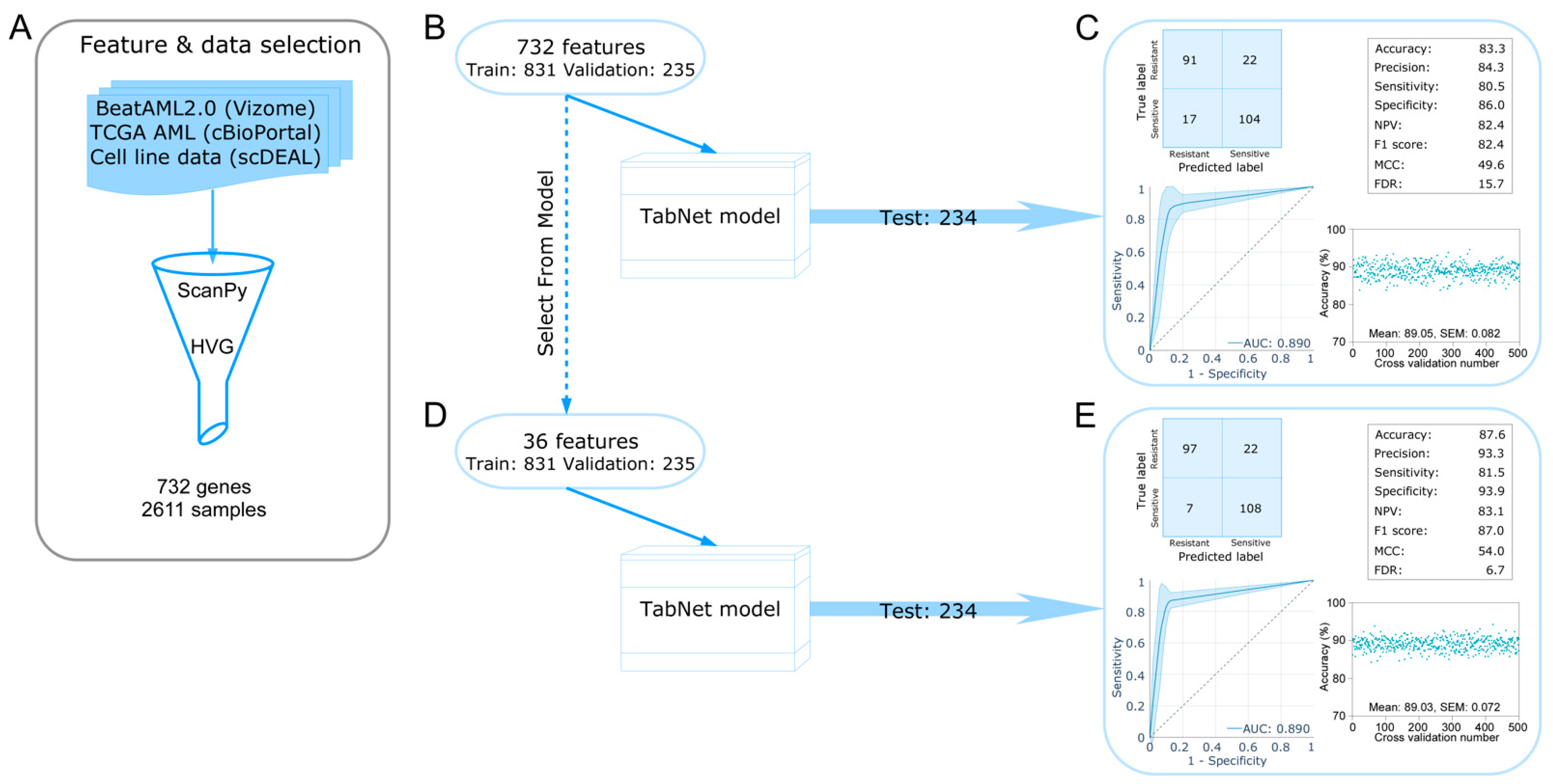
Figure 1. Development of deep learning models for tabular data. (A) Features were selected using the “highly variable genes” function from ScanPy. (B) Selected features (732) were used to develop a TabNet model. The model was built using 831 training samples and 235 validation samples and then was tested using 234 test samples. (C) The models’ performance was checked using a confusion matrix and several quality scores. Furthermore, from all samples (1300), we randomly sampled 20% of test samples 500 different times, and the rest of the 80% samples were used to build TabNet models. The ROC curve and accuracy scores represent results from all 500 tests. (D,E) A similar test was performed as described in (B) and (C) using 36 features selected using the “Select From Model” function from Sklearn feature selection.
Ijms 24 03830 g001
Figure 2. Identification of leading predictors using XGBoost and SHAP. (A,B) Performance of XGBoost models was checked by random sampling 20% of test samples 500 different times, and the rest of the 80% samples were used to build the models. The ROC curve represents the results from all 500 tests. (C,D) The SHAP explainer was used to determine SHAP values from test samples. XGBoost was used as the model.
Ijms 24 03830 g002
Figure 3. Regulation of serine/threonine signaling during tyrosine kinase inhibition. MOLM-13 cells were treated with DMSO or sorafenib for four hours before lysis. Lysates were applied to the Pamgene serine/threonine kinase profiling assays.
Ijms 24 03830 g003
Figure 4. Drug-induced AXL expression in AML cell lines. (A) MV4-11 and MOLM-13 cells were treated with different concentrations of kinase inhibitors for 48 h. Cell viability was measured using PrestoBlue. (B) MV4-11 and MOLM-13 cells were treated with different kinase inhibitors for 24 h with respective EC50 values and AXL expression was measured using RT-qPCR.
Ijms 24 03830 g004
Figure 5. FLT3 inhibition enhances CREB phosphorylation and displays synergy with AXL and PKC inhibitors. (A) MV4-11 and MOLM-13 cells were treated with AC220 for different time points before lysis. Lysates were used to separate by SDS-PAGE following western blotting using different antibodies. (BD) MV4-11 cells were treated with a single drug or a 1:1 drug combination for 48 h. Relative inhibition was used to calculate the synergy as described in material methods. A positive BLISS score represents the synergy (single drug response curves are presented in the Supplementary Figure S5).
Ijms 24 03830 g005
Disclaimer/Publisher’s Note: The statements, opinions and data contained in all publications are solely those of the individual author(s) and contributor(s) and not of MDPI and/or the editor(s). MDPI and/or the editor(s) disclaim responsibility for any injury to people or property resulting from any ideas, methods, instructions or products referred to in the content.