
Discovery of New Heterocyclic/Benzofuran Hybrids as Potential Anti-Inflammatory Agents: Design, Synthesis, and Evaluation of the Inhibitory Activity of Their Related Inflammatory Factors Based on NF-κB and MAPK Signaling Pathways

Abstract
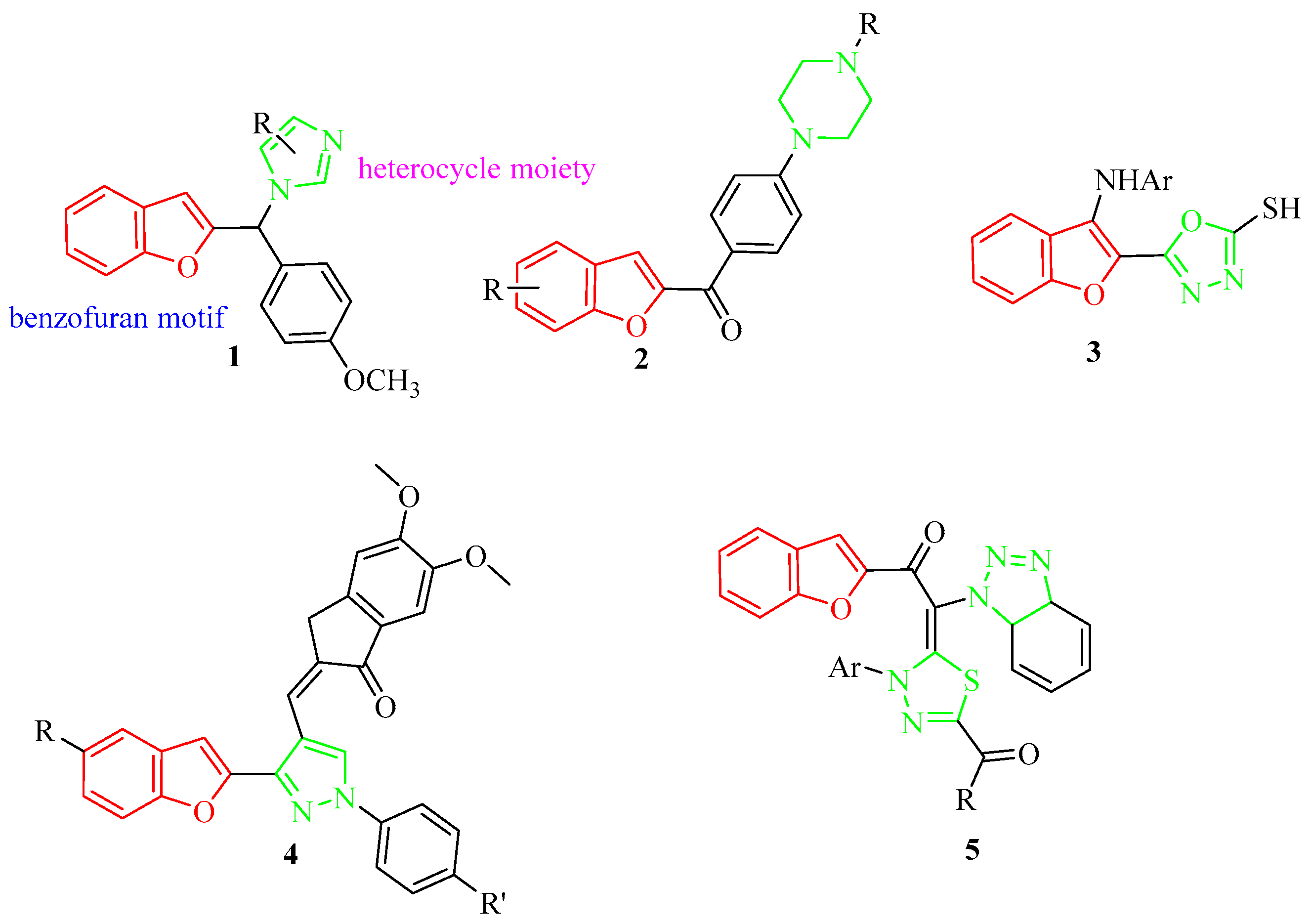
:1. Introduction
2. Results and Discussion
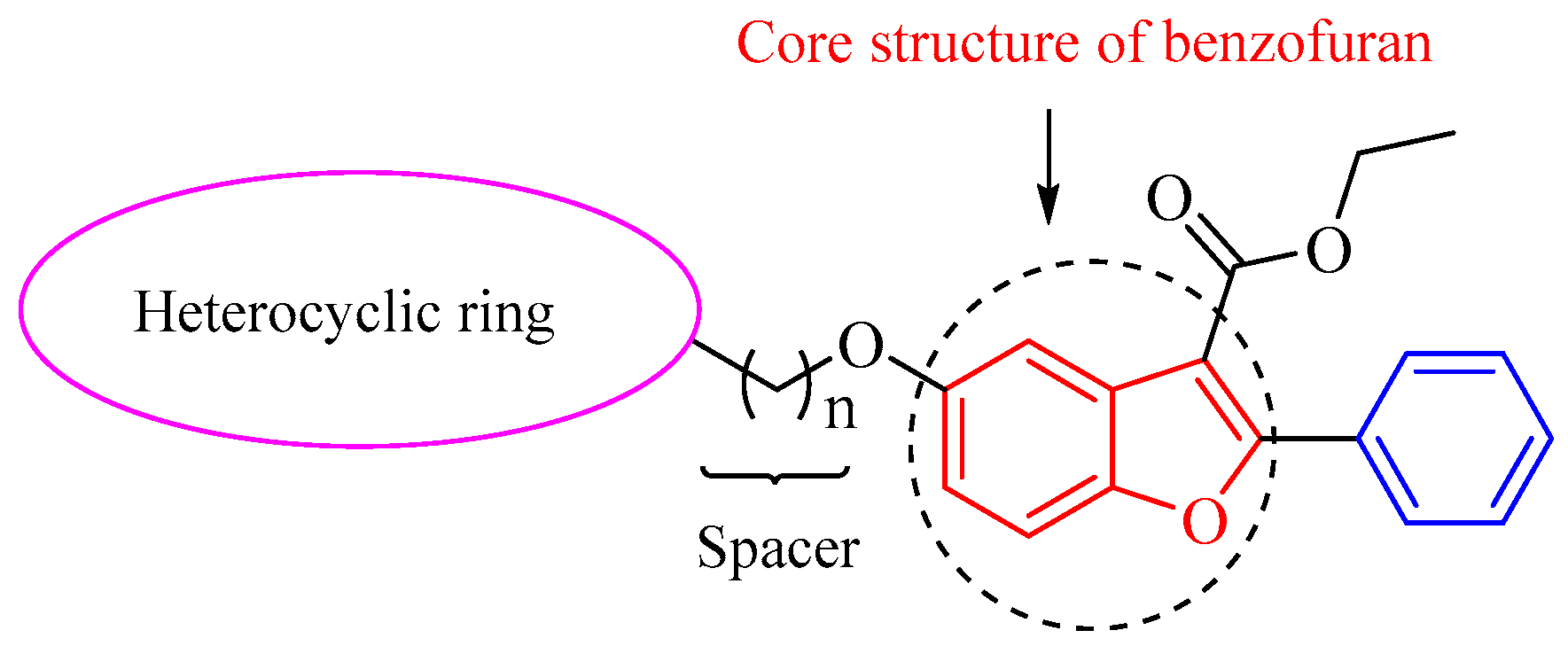
2.1. Chemistry
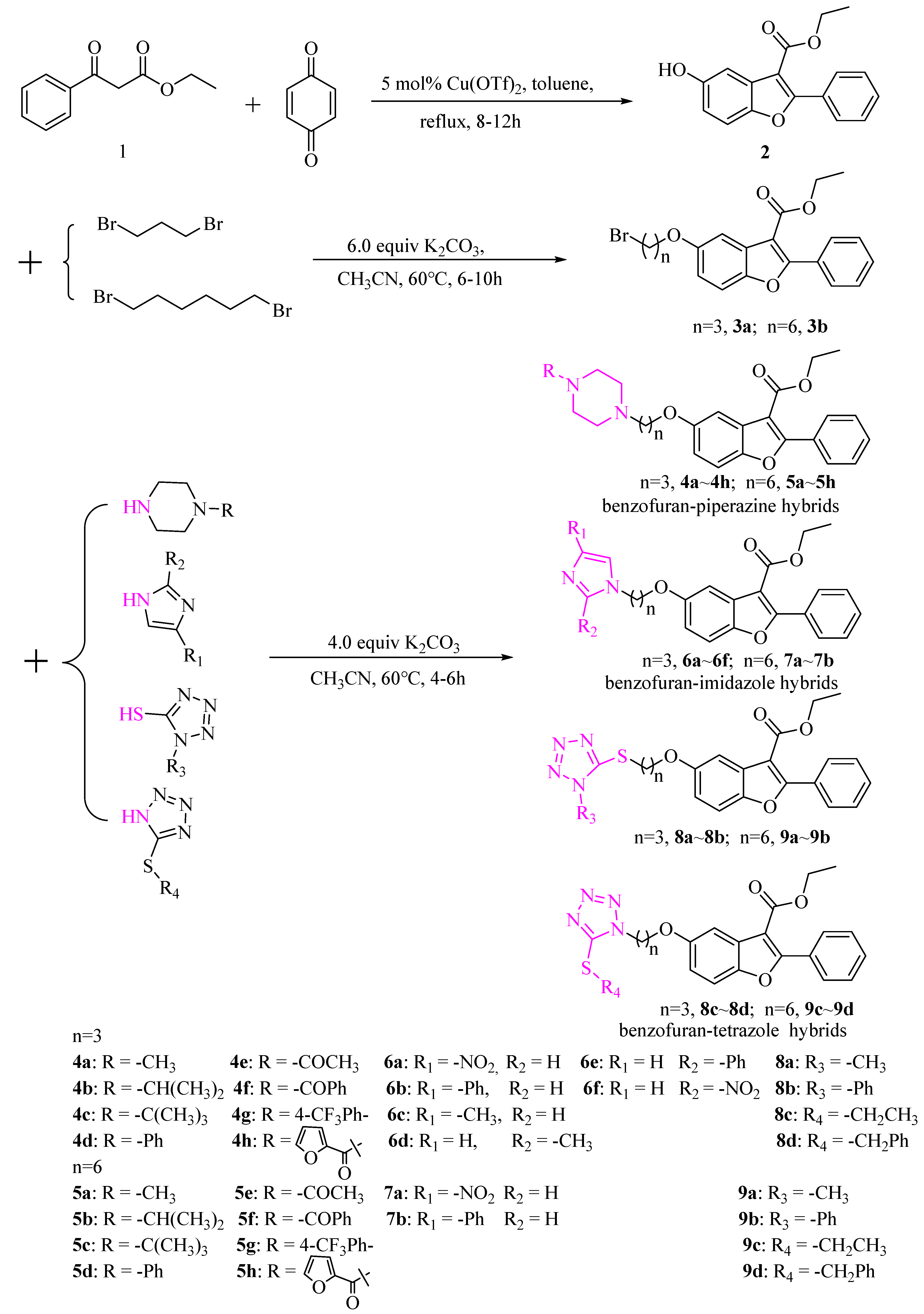
Synthetic Route of Benzofuran-Heterocycle Hybrids
2.2. In Vitro Studies
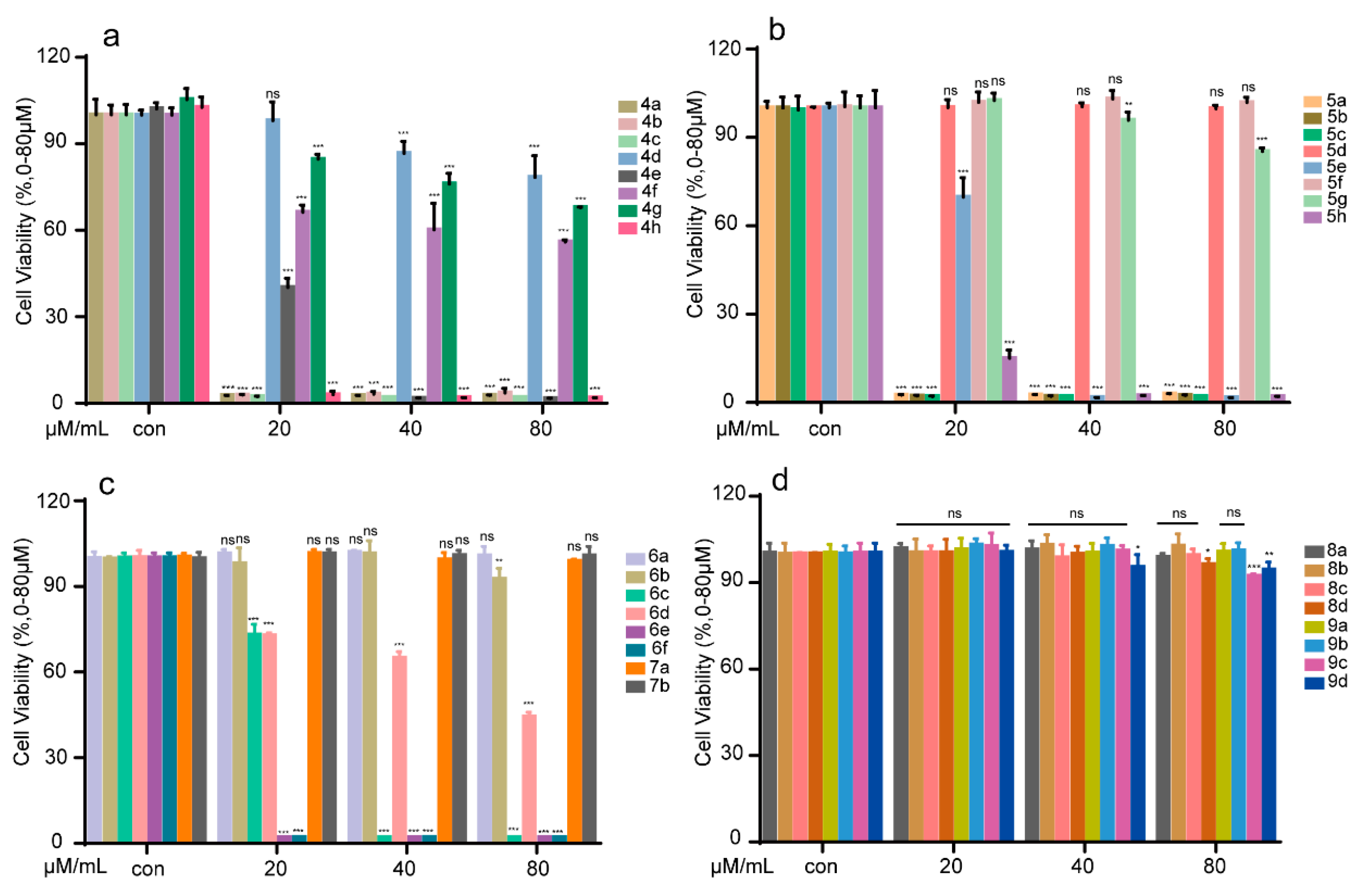
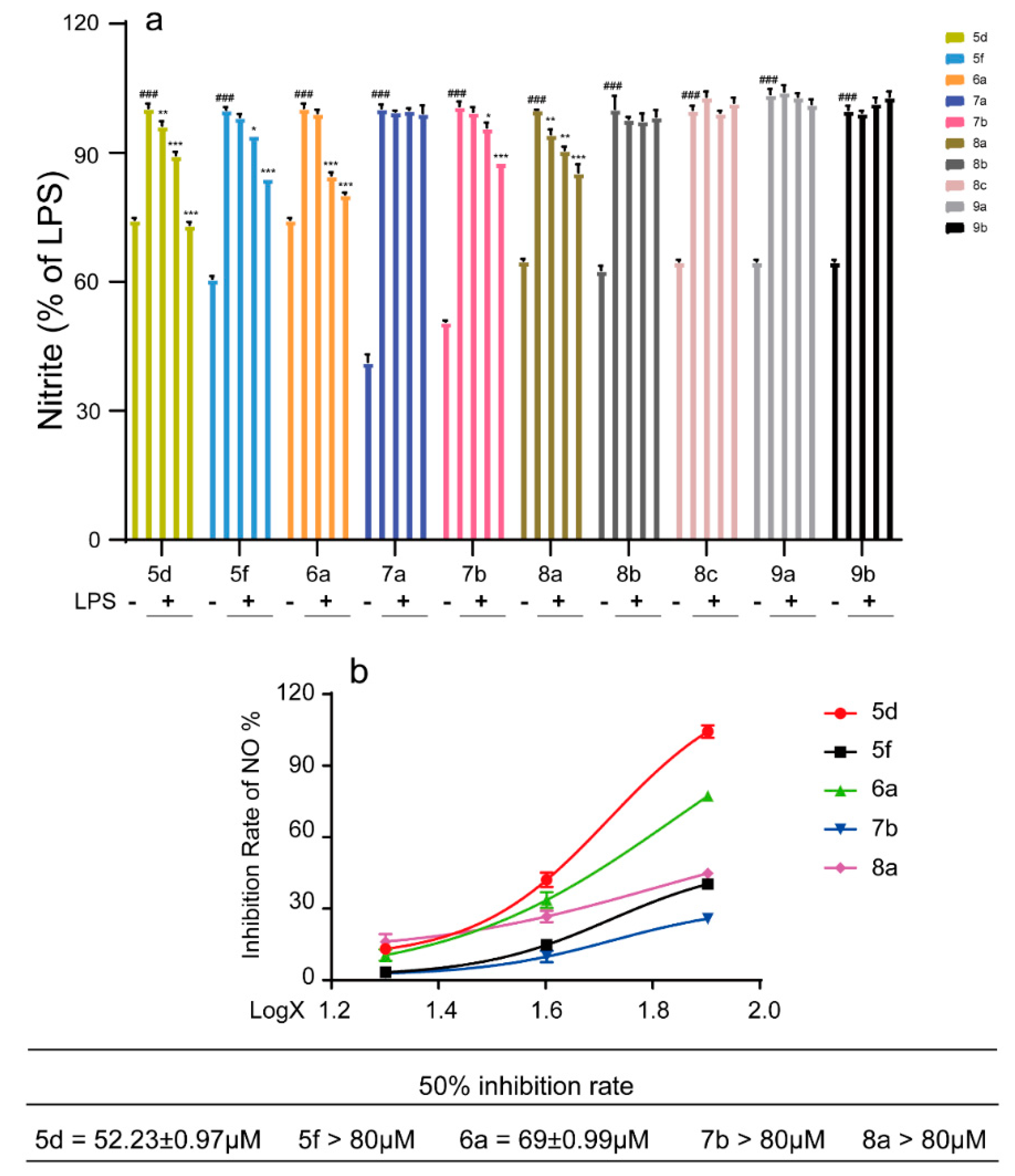
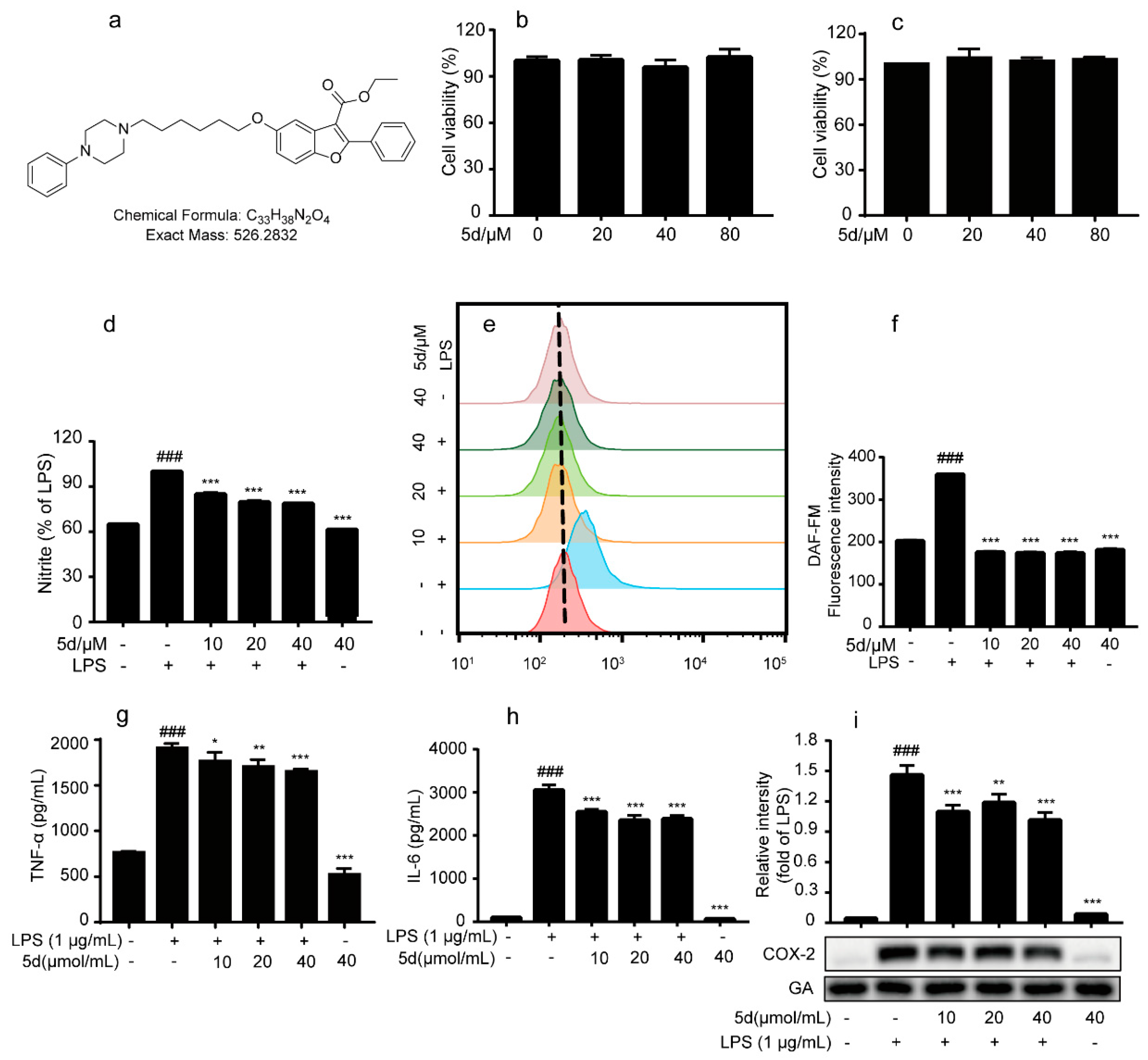
2.2.1. Determination of Cytotoxicity and Activity In Vitro
2.2.2. Effects of Compound 5d on Pro-Inflammatory Factors in LPS-Induced RAW264.7 Cells
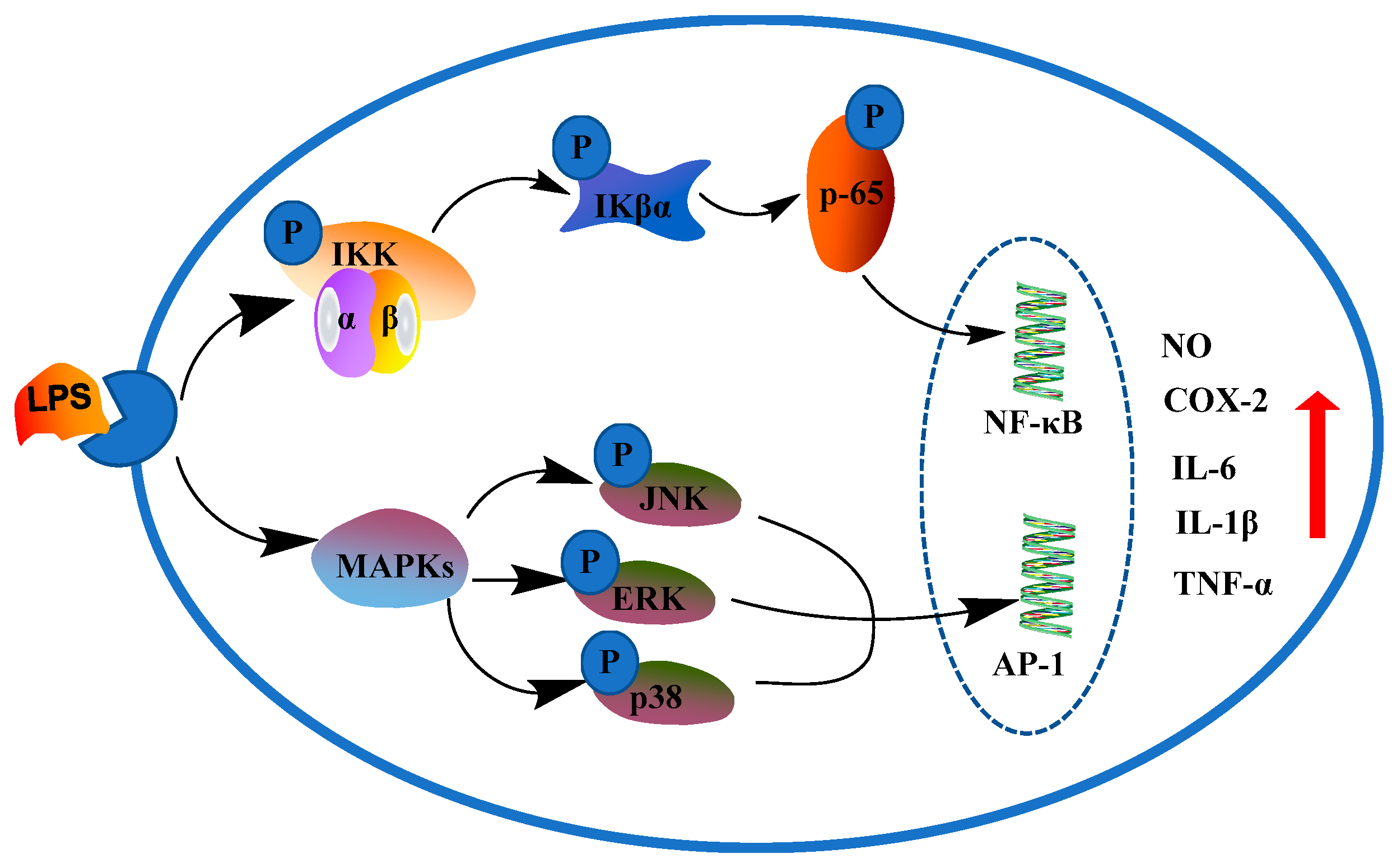
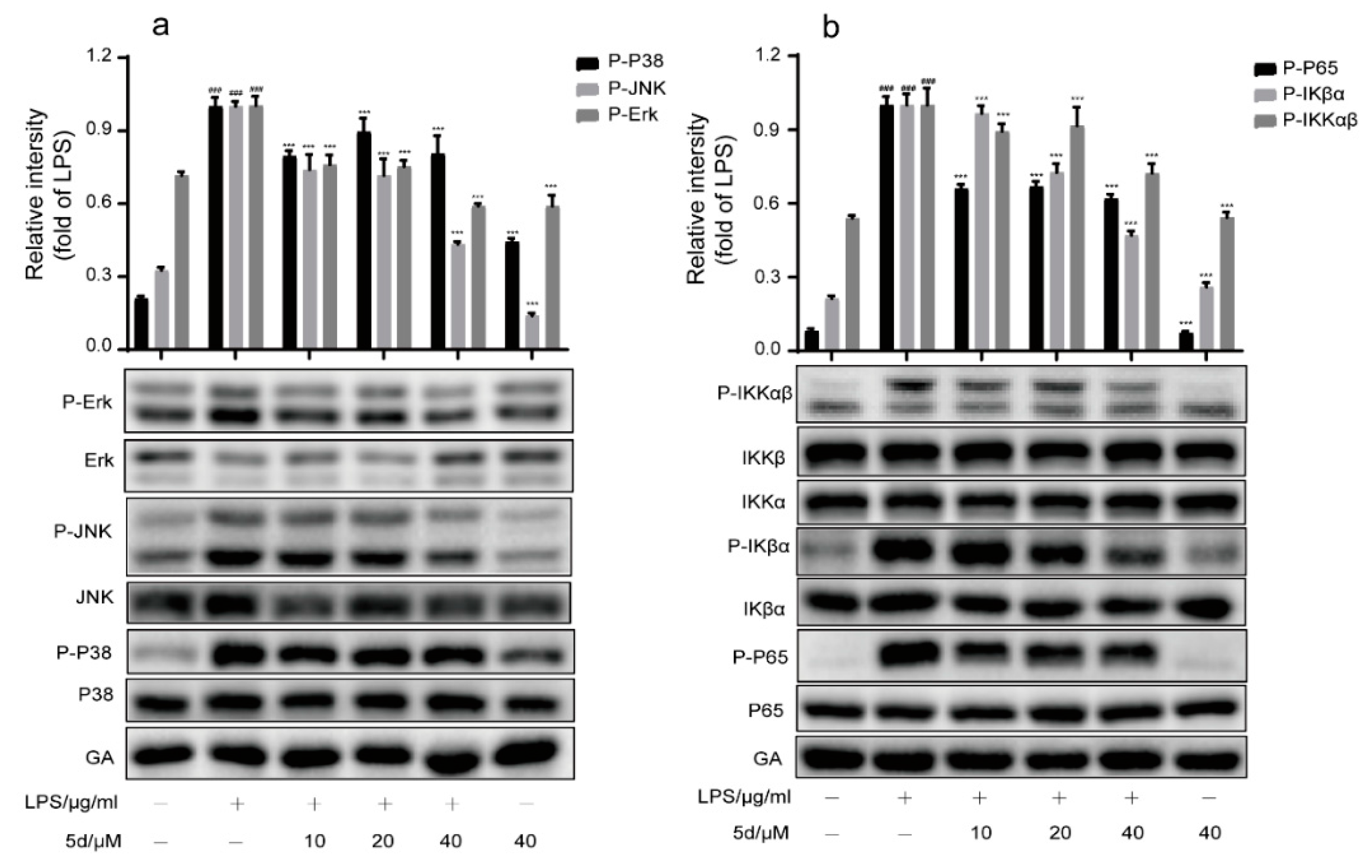
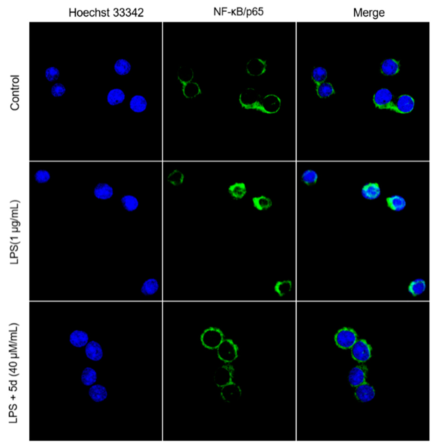
2.2.3. The Effect of 5d on NF-κB and MAPKs Pathways in RAW264.7 Cells
2.3. In Vivo Studies
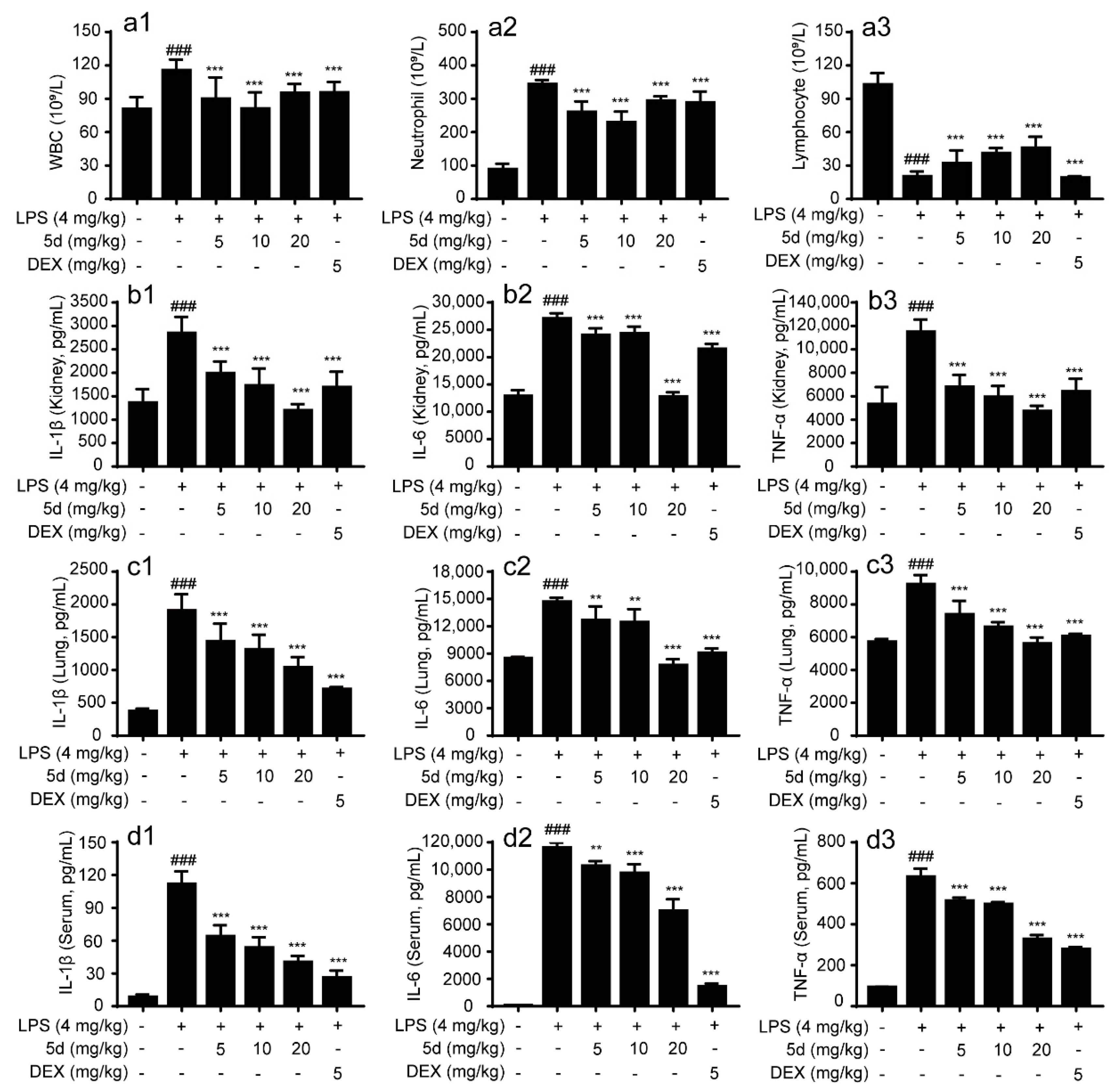
2.3.1. Protective Effect of the Compound 5d on Endotoxemic Mice
2.3.2. Effects of 5d on the Blood Routine and Anti-Inflammatory Cytokines in Serum and Tissue of Endotoxemic Mice
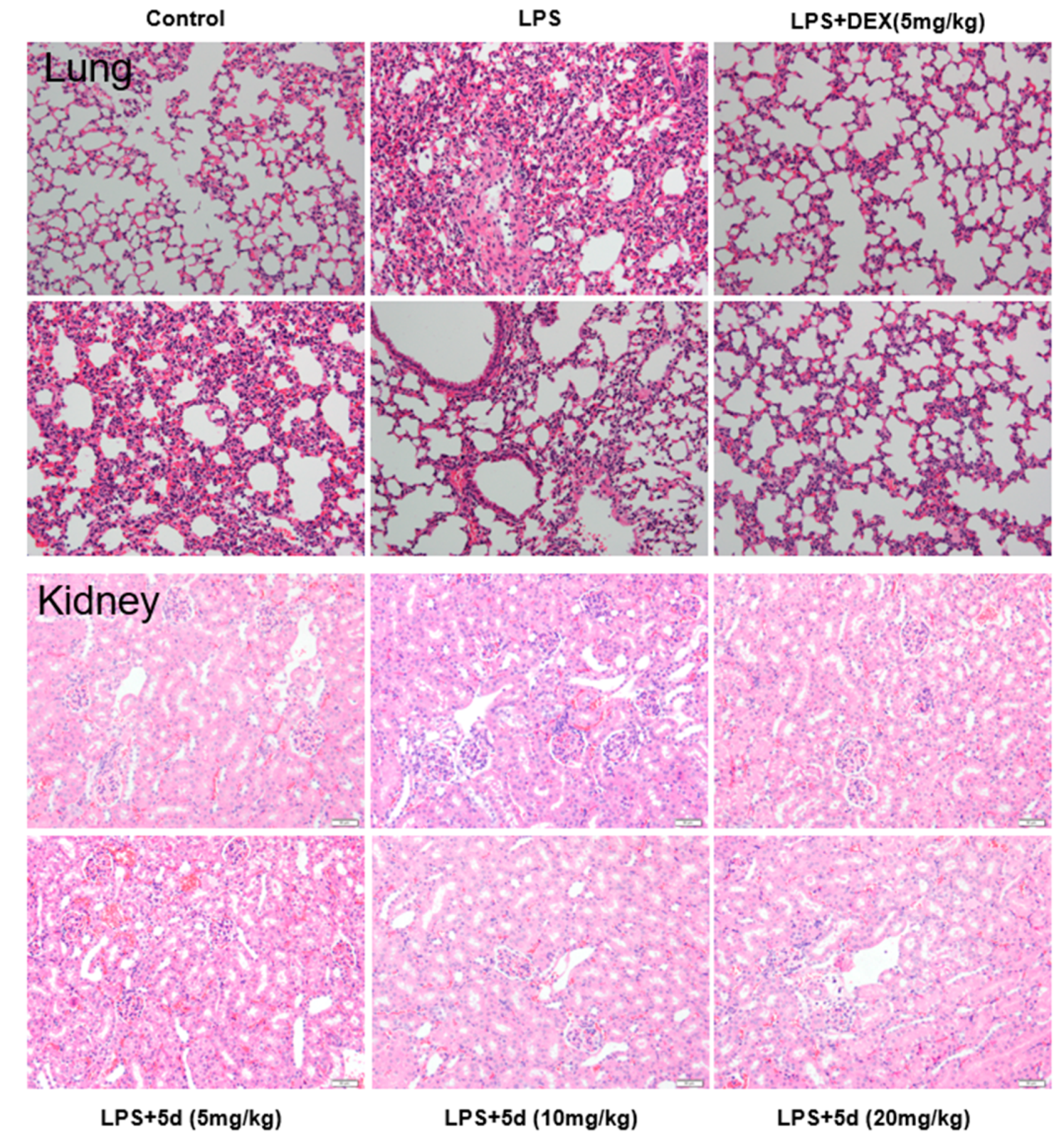
2.3.3. Effects of Compound 5d on Histopathological Changes of Lung and Kidney Tissues in the Endotoxemia Mice Model
3. Materials and Methods
3.1. Chemistry
3.1.1. Synthesis of Benzofuran-Heterocycle Hybrids
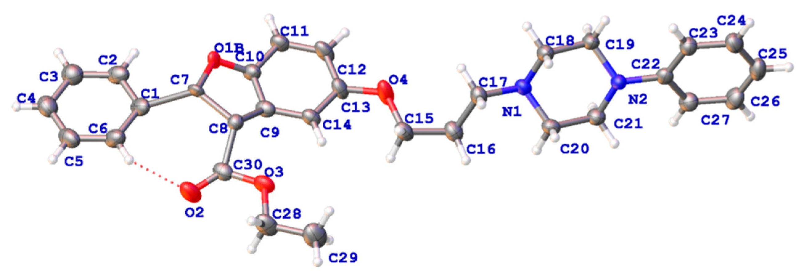
3.1.2. Preparation of Single Crystal Compound 4d and Its X-ray Single Crystal
3.2. Biological Evaluation
3.2.1. Cell Culture
3.2.2. MTT Assay
3.2.3. Griess Assay
3.2.4. Flow Cytometry Analysis
3.2.5. Fluorescence Imaging in RAW264.7 Cells by Confocal Microscopy
3.2.6. ELISA Assay
3.2.7. Western Blotting Analysis
3.2.8. Animal Experiments
3.2.9. Data Analysis
4. Conclusions
Author Contributions
Funding
Institutional Review Board Statement
Informed Consent Statement
Data Availability Statement
Acknowledgments
Conflicts of Interest
References
- Germolec, D.R.; Frawley, R.P.; Evans, E. Markers of inflammation. Methods Mol. Biol. 2010, 598, 53–73. [Google Scholar]
- Varela, M.L.; Mogildea, M.; Moreno, I.; Lopes, A. Acute Inflammation and Metabolism. Inflammation 2018, 41, 1115–1127. [Google Scholar]
- Baeuerle, P.A.; Henkel, T. Function and Activation of NF-kappaB in the Immune System. Annu. Rev. Immunol. 1994, 12, 141–179. [Google Scholar] [CrossRef]
- Mitchell, J.P.; Carmody, R.J. NF-κB and the Transcriptional Control of Inflammation. Int. Rev. Cell. Mol. Biol. 2018, 335, 41–84. [Google Scholar]
- Barnabei, L.; Laplantine, E.; Mbongo, W.; Rieux-Laucat, F.; Weil, R. NF-κB: At the Borders of Autoimmunity and Inflammation. Front. Immunol. 2021, 12, 3169. [Google Scholar] [CrossRef]
- Zhan, W.; Liu, H.T. MAPK signal pathways in the regulation of cell proliferation in mammalian cells. Cell Res. 2002, 12, 9–18. [Google Scholar] [CrossRef]
- Coulthard, L.R.; White, D.E.; Jones, D.L.; McDermott, M.F.; Burchill, S.A. p38(MAPK): Stress responses from molecular mechanisms to therapeutics. Trends Mol. Med. 2009, 15, 369–379. [Google Scholar] [CrossRef]
- Dong, Q.; Jie, Y.; Ma, J.; Li, C.; Xin, T.; Yang, D. Renal tubular cell death and inflammation response are regulated by the MAPK-ERK-CREB signaling pathway under hypoxia-reoxygenation injury. J. Recept. Signal. Transduct. Res. 2019, 39, 383–391. [Google Scholar] [CrossRef]
- Ko, J.H.; Yoon, S.O.; Lee, H.J.; Oh, J.Y. Rapamycin regulates macrophage activation by inhibiting NLRP3 inflammasome-p38 MAPK-NFκB pathways in autophagy- and p62-dependent manners. Oncotarget 2017, 8, 40817–40831. [Google Scholar] [CrossRef]
- Wang, X.; Yang, L.; Yang, L.; Xing, F.; Yang, H.; Qin, L.; Lan, Y.; Wu, H.; Zhang, B.; Shi, H.; et al. Gypenoside IX Suppresses p38 MAPK/Akt/NFκB Signaling Pathway Activation and Inflammatory Responses in Astrocytes Stimulated by Proinflammatory Mediators. Inflammation 2017, 40, 2137–2150. [Google Scholar] [CrossRef]
- Sun, X.G. Clinical application of non-steroidal anti-inflammatory drugs in ophthalmology. Ophthalmol. China 2013, 22, 147–148. [Google Scholar]
- Boynton, C.S.; Dick, C.F.; Mayor, G.H. NSAIDs: An overview. J. Clin. Pharmacol. 2013, 28, 512–517. [Google Scholar] [CrossRef]
- Stephen, K. Critical appraisal of ophthalmic ketorolac in treatment of pain and inflammation following cataract surgery. J. Clin. Pharmacol. 2011, 5, 751–758. [Google Scholar]
- Bindu, S.; Mazumder, S.; Bandyopadhyay, U. Non-steroidal anti-inflammatory drugs (NSAIDs) and organ damage: A current perspective. Biochem. Pharmacol. 2020, 180, 114147. [Google Scholar] [CrossRef]
- Khalil, H.S.; Sedky, N.K.; Amin, K.M.; Elhafez OM, A.; Arafa, R.K. Visnagin and benzofuran scaffold-based molecules as selective cyclooxygenase-2 inhibitors with anti-inflammatory and analgesic properties: Design, synthesis and molecular docking. Future Med. Chem. 2019, 11, 659–676. [Google Scholar] [CrossRef]
- Hayakawa, I.; Shioya, R.; Agatsuma, T.; Furukawa, H.; Sugano, Y. Thienopyridine and benzofuran derivatives as potent anti-tumor agents possessing different structure activity relationships. Bioorg. Med. Chem. Lett. 2004, 14, 3411–3414. [Google Scholar] [CrossRef]
- Xu, Z.; Xu, D.; Zhou, W.; Zhang, X. Therapeutic Potential of Naturally Occurring Benzofuran Derivatives and Hybrids of Benzofurans with other Pharmacophores as Antibacterial Agents. Curr. Top. Med. Chem. 2022, 22, 64–82. [Google Scholar] [CrossRef]
- Wang, W.J.; Wang, L.; Liu, Z.; Jiang, R.W.; Liu, Z.W.; Li, M.M.; Zhang, Q.W.; Dai, Y.; Li, Y.L.; Zhang, X.Q.; et al. Antiviral benzofurans from Eupatorium chinense. Phytochemistry 2016, 122, 238–245. [Google Scholar] [CrossRef]
- Gao, F.; Yang, H.; Lu, T.; Chen, Z.; Ma, L.; Xu, Z.; Schaffer, P.; Lu, G. Design, synthesis and anti-mycobacterial activity evaluation of benzofuran-isatin hybrids. Eur. J. Med. Chem. 2018, 59, 277–281. [Google Scholar] [CrossRef]
- Reddy, K.A.; Lohray, B.B.; Bhushan, V.; Bajji, A.C.; Reddy, K.V.; Reddy, P.R.; Krishna, T.H.; Rao, I.N.; Jajoo, H.K.; Rao, N.V.; et al. Novel antidiabetic and hypolipidemic agents. 3. Benzofuran-containing thiazolidinediones. J. Med. Chem. 1999, 42, 1927–1940. [Google Scholar] [CrossRef]
- Rech, T.d.S.T.; Alves, A.G.; Strelow, D.N.; Krüger, L.D.; Júnior, L.R.C.; Neto, J.S.D.S.; Braga, A.L.; Brüning, C.A.; Bortolatto, C.F. 2-Phenyl-3-(phenylselanyl) benzofuran elicits acute antidepressant-like action in male Swiss mice mediated by modulation of the dopaminergic system and reveals therapeutic efficacy in both sexes. Psychopharmacology 2021, 238, 3013–3024. [Google Scholar] [CrossRef]
- Hiremathad, A.; Chand, K.; Keri, R.S. Development of coumarin–benzofuran hybrids as versatile multitargeted compounds for the treatment of Alzheimer′s Disease. Chem. Biol. Drug. Des. 2018, 92, 1497–1503. [Google Scholar] [CrossRef]
- Nevagi, R.J.; Dighe, S.N.; Dighe, S.N. Biological and medicinal significance of benzofuran. Eur. J. Med. Chem. 2015, 97, 561–581. [Google Scholar] [CrossRef]
- Labib, M.B.; Fayez, A.M.; El-Nahass, E.S.; Awadallah, M.; Halim, P.A. Novel tetrazole-based selective COX-2 inhibitors: Design, synthesis, anti-inflammatory activity, evaluation of PGE2, TNF-α, IL-6 and histopathological study. Bioorg. Chem. 2020, 104, 104308. [Google Scholar] [CrossRef]
- Nascimento, M.V.P.; Munhoz, A.; Theindl, L.C.; Mohr, E.T.B.; Saleh, N.; Parisotto, E.B.; Rossa, T.A.; Zamoner, A.; Creczynski-Pasa, T.B.; Filippin-Monteiro, F.B.; et al. A Novel Tetrasubstituted Imidazole as a Prototype for the Development of Anti-inflammatory Drugs. Inflammation 2018, 41, 1334–1348. [Google Scholar] [CrossRef]
- Sharma, S.; Kumar, D.; Singh, G.; Monga, V.; Kumar, B. Recent advancements in the development of heterocyclic anti-inflammatory agents. Eur. J. Med. Chem. 2020, 200, 112438. [Google Scholar] [CrossRef]
- Chen, W.; Yang, X.D.; Li, Y.; Yang, L.J.; Wang, X.Q.; Zhang, G.L.; Zhang, H.B. Design, synthesis and cytotoxic activities of novel hybrid compounds between dihydrobenzofuran and imidazole. Org. Biomol. Chem. 2012, 9, 4250–4255. [Google Scholar] [CrossRef]
- Mao, Z.W.; Xi, Z.; Lin, Y.P.; Hu, C.Y.; Wang, X.L.; Wan, C.P.; Rao, G.X. Design, synthesis and anticancer activity of novel hybrid compounds between benzofuran and N-aryl piperazine. Bioorg. Med. Chem. Lett. 2016, 26, 3421–3424. [Google Scholar] [CrossRef]
- Mane, B.Y.; Vidyadhara, S. Microwave-assisted synthesis of carboxanilides as non-steroidal antiinflammatory agents. Res. J. Pharm. Biol. Chem. Sci. 2011, 2, 798–804. [Google Scholar]
- Kenchappa, R.; Bodke, Y.D. Synthesis, Analgesic and Anti-inflammatory activity of Benzofuran pyrazole Heterocycles. Chem. Data Collect. 2020, 28, 100453. [Google Scholar]
- Dawood, K.M.; Abdel-Gawad, H.; Rageb, E.A.; Ellithey, M.; Mohamed, H.A. Synthesis, anticonvulsant, and anti-inflammatory evaluation of some new benzotriazole and benzofuran-based heterocycles. Bioorg. Med. Chem. 2006, 14, 3672–3680. [Google Scholar] [CrossRef]
- Yadav, P.; Singh, P.; Tewari, A.K. Design, synthesis, docking and anti-inflammatory evaluation of novel series of benzofuran based prodrugs. Bioorg. Med. Chem. Lett. 2014, 24, 2251–2255. [Google Scholar] [CrossRef]
- Mothe, S.R.; Susanti, D.; Chan, P. Efficient synthesis of 3-acyl- 5-hydroxybenzofurans via copper(II) triflate-catalyzed cycloaddition of unactivated 1,4-benzoquinones with 1,3-dicarbonyl compounds. Tetra. Lett. 2010, 51, 2136–2140. [Google Scholar] [CrossRef]
- Cagiola, M.; Giulio, S.; Miriam, M.; Katia, F.; Paola, P.; Macrì, A.; Pasquali, P. In vitro down regulation of proinflammatory cytokines induced by LPS tolerance in pig CD14+ cells. Vet. Immunol. Immunopathol. 2006, 112, 316–320. [Google Scholar] [CrossRef]
- Ronchetti, D.; Impagnatiello, F.; Guzzetta, M.; Gasparini, L.; Borgatti, M.; Gambari, R.; Ongini, E. Modulation of iNOS expression by a nitric oxide-releasing derivative of the natural antioxidant ferulic acid in activated RAW 264.7 macrophages. Eur. J. Pharmacol. 2006, 532, 162–169. [Google Scholar] [CrossRef]


Disclaimer/Publisher’s Note: The statements, opinions and data contained in all publications are solely those of the individual author(s) and contributor(s) and not of MDPI and/or the editor(s). MDPI and/or the editor(s) disclaim responsibility for any injury to people or property resulting from any ideas, methods, instructions or products referred to in the content. |
© 2023 by the authors. Licensee MDPI, Basel, Switzerland. This article is an open access article distributed under the terms and conditions of the Creative Commons Attribution (CC BY) license (https://creativecommons.org/licenses/by/4.0/).
Share and Cite
Chen, Y.; Chen, R.; Yuan, R.; Huo, L.; Gao, H.; Zhuo, Y.; Chen, X.; Zhang, C.; Yang, S. Discovery of New Heterocyclic/Benzofuran Hybrids as Potential Anti-Inflammatory Agents: Design, Synthesis, and Evaluation of the Inhibitory Activity of Their Related Inflammatory Factors Based on NF-κB and MAPK Signaling Pathways. Int. J. Mol. Sci. 2023, 24, 3575. https://doi.org/10.3390/ijms24043575
Chen Y, Chen R, Yuan R, Huo L, Gao H, Zhuo Y, Chen X, Zhang C, Yang S. Discovery of New Heterocyclic/Benzofuran Hybrids as Potential Anti-Inflammatory Agents: Design, Synthesis, and Evaluation of the Inhibitory Activity of Their Related Inflammatory Factors Based on NF-κB and MAPK Signaling Pathways. International Journal of Molecular Sciences. 2023; 24(4):3575. https://doi.org/10.3390/ijms24043575
Chicago/Turabian StyleChen, Yangling, Rui Chen, Renyikun Yuan, Lini Huo, Hongwei Gao, Youqiong Zhuo, Xinxin Chen, Chenwei Zhang, and Shilin Yang. 2023. "Discovery of New Heterocyclic/Benzofuran Hybrids as Potential Anti-Inflammatory Agents: Design, Synthesis, and Evaluation of the Inhibitory Activity of Their Related Inflammatory Factors Based on NF-κB and MAPK Signaling Pathways" International Journal of Molecular Sciences 24, no. 4: 3575. https://doi.org/10.3390/ijms24043575
APA StyleChen, Y., Chen, R., Yuan, R., Huo, L., Gao, H., Zhuo, Y., Chen, X., Zhang, C., & Yang, S. (2023). Discovery of New Heterocyclic/Benzofuran Hybrids as Potential Anti-Inflammatory Agents: Design, Synthesis, and Evaluation of the Inhibitory Activity of Their Related Inflammatory Factors Based on NF-κB and MAPK Signaling Pathways. International Journal of Molecular Sciences, 24(4), 3575. https://doi.org/10.3390/ijms24043575