
Advancing Stroke Research on Cerebral Thrombi with Omic Technologies


Abstract
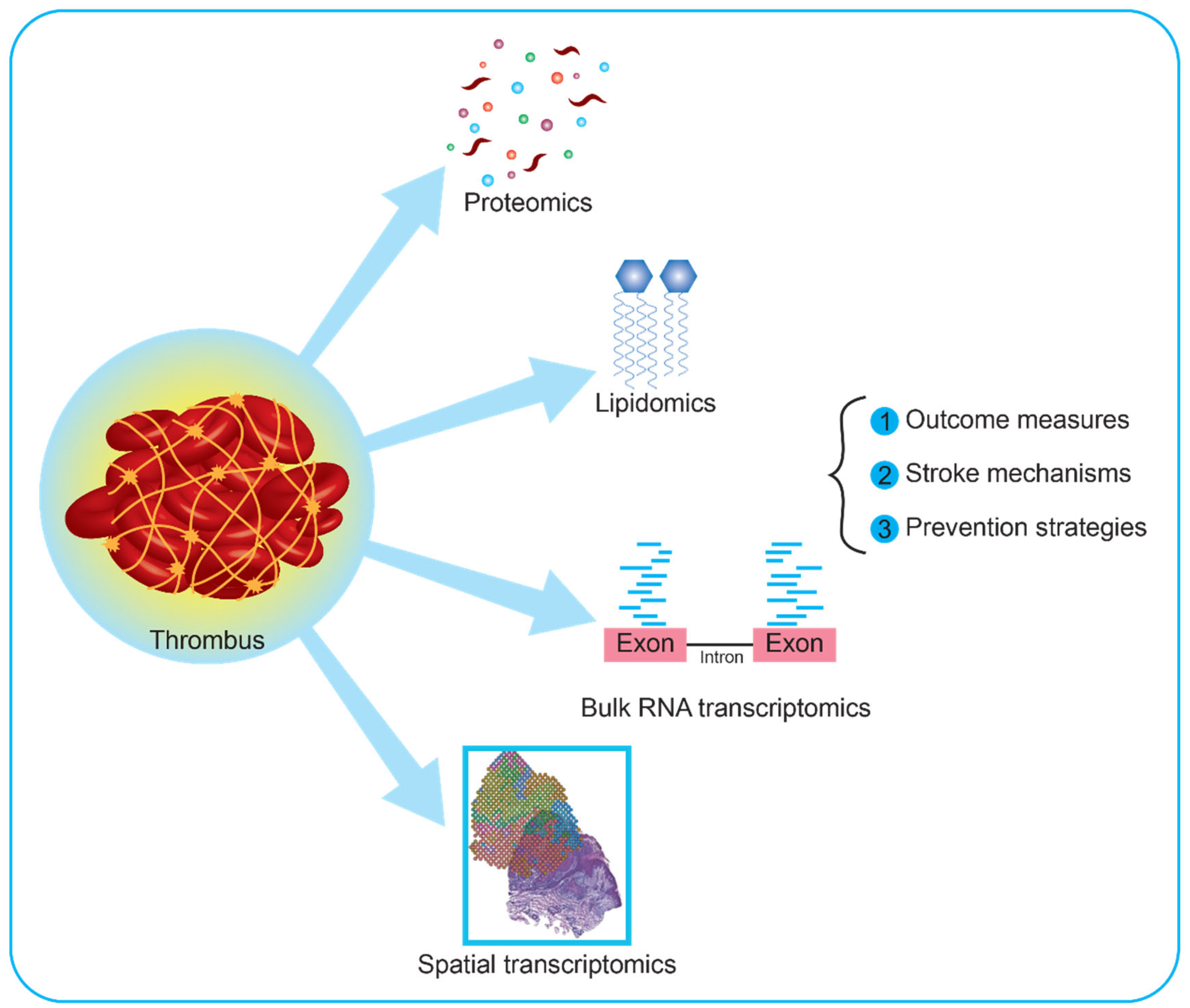
:1. Introduction
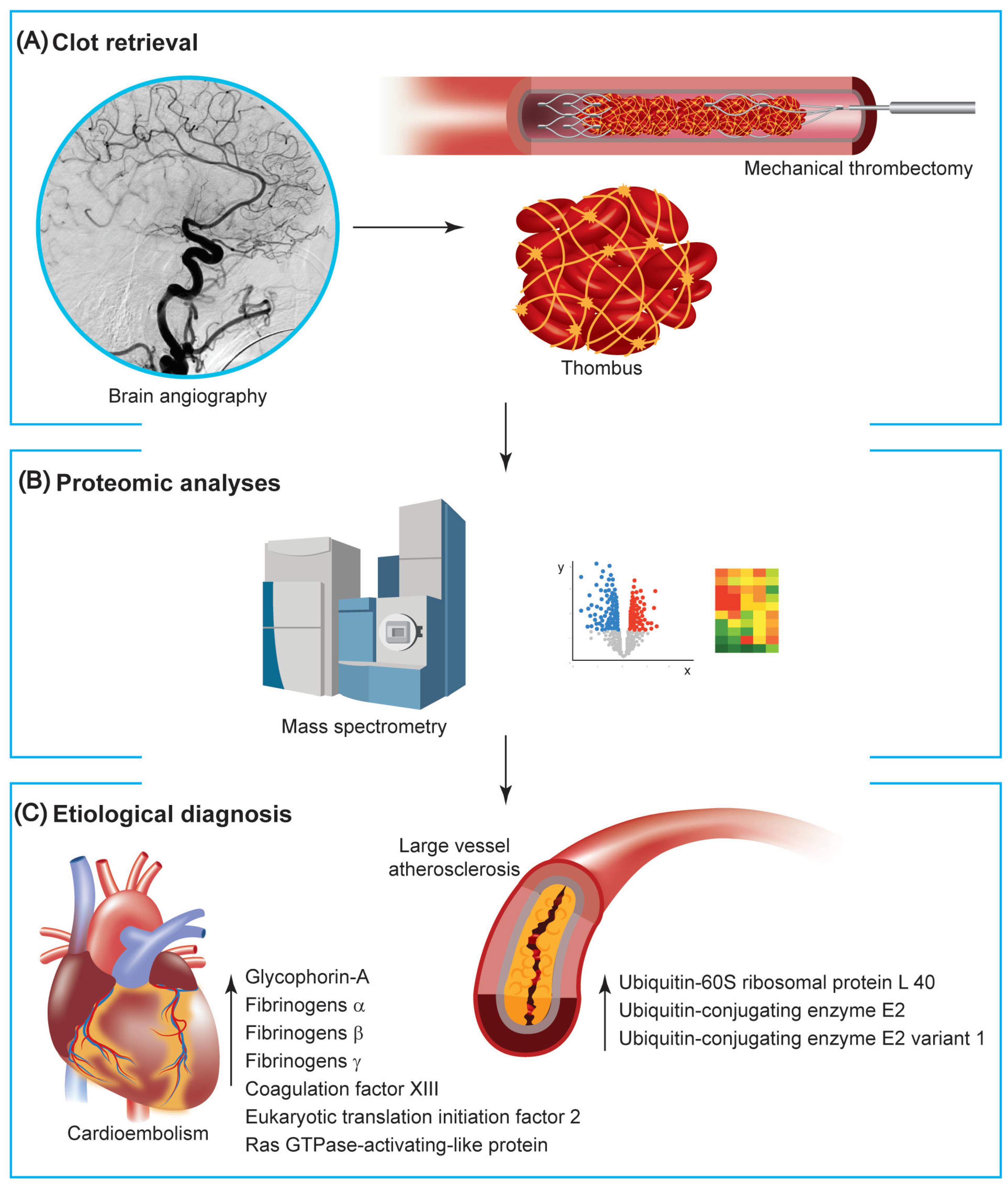
2. Anatomopathological Studies
3. Multiomic Studies
3.1. Proteomics
3.2. Metabolomics
Metabolomics and the Risk of AIS
3.3. Combined Proteomic and Metabolomics
The Integration of Multiomic Techniques
3.4. Transcriptomics
4. Limitations and Perspectives of Omic Studies in Acute Ischemic Stroke
Author Contributions
Funding
Institutional Review Board Statement
Informed Consent Statement
Data Availability Statement
Acknowledgments
Conflicts of Interest
References
- Yaghi, S.; Bernstein, R.A.; Passman, R.; Okin, P.M.; Furie, K.L. Cryptogenic Stroke. Circ. Res. 2017, 120, 527–540. [Google Scholar] [CrossRef] [PubMed]
- Navi, B.B.; Singer, S.; Merkler, A.E.; Cheng, N.T.; Stone, J.B.; Kamel, H.; Iadecola, C.; Elkind, M.S.V.; DeAngelis, L.M. Recurrent Thromboembolic Events after Ischemic Stroke in Patients with Cancer. Neurology 2014, 83, 26–33. [Google Scholar] [CrossRef] [PubMed]
- Martini, M.L.; Neifert, S.N.; Lara-Reyna, J.J.; Shuman, W.H.; Ladner, T.R.; Hardigan, T.H.; Fifi, J.T.; Mocco, J.; Yaeger, K.A. Trials in Thrombectomy for Acute Ischemic Stroke: Describing the State of Clinical Research in the Field. Clin. Neurol. Neurosurg. 2021, 200, 106360. [Google Scholar] [CrossRef]
- Staessens, S.; François, O.; Brinjikji, W.; Doyle, K.M.; Vanacker, P.; Andersson, T.; De Meyer, S.F. Studying Stroke Thrombus Composition After Thrombectomy: What Can We Learn? Stroke 2021, 52, 3718–3727. [Google Scholar] [CrossRef] [PubMed]
- Brinjikji, W.; Duffy, S.; Burrows, A.; Hacke, W.; Liebeskind, D.; Majoie, C.B.L.M.; Dippel, D.W.J.; Siddiqui, A.H.; Khatri, P.; Baxter, B.; et al. Correlation of Imaging and Histopathology of Thrombi in Acute Ischemic Stroke with Etiology and Outcome: A Systematic Review. J. Neurointerv. Surg. 2017, 9, 529–534. [Google Scholar] [CrossRef]
- De Meyer, S.F.; Andersson, T.; Baxter, B.; Bendszus, M.; Brouwer, P.; Brinjikji, W.; Campbell, B.C.; Costalat, V.; Dávalos, A.; Demchuk, A.; et al. Analyses of Thrombi in Acute Ischemic Stroke: A Consensus Statement on Current Knowledge and Future Directions. Int. J. Stroke 2017, 12, 606–614. [Google Scholar] [CrossRef] [PubMed]
- Laridan, E.; Denorme, F.; Desender, L.; François, O.; Andersson, T.; Deckmyn, H.; Vanhoorelbeke, K.; De Meyer, S.F. Neutrophil Extracellular Traps in Ischemic Stroke Thrombi. Ann. Neurol. 2017, 82, 223–232. [Google Scholar] [CrossRef]
- Staessens, S.; Denorme, F.; Francois, O.; Desender, L.; Dewaele, T.; Vanacker, P.; Deckmyn, H.; Vanhoorelbeke, K.; Andersson, T.; De Meyer, S.F. Structural Analysis of Ischemic Stroke Thrombi: Histological Indications for Therapy Resistance. Haematologica 2020, 105, 498–507. [Google Scholar] [CrossRef]
- Denorme, F.; Langhauser, F.; Desender, L.; Vandenbulcke, A.; Rottensteiner, H.; Plaimauer, B.; François, O.; Andersson, T.; Deckmyn, H.; Scheiflinger, F.; et al. ADAMTS13-Mediated Thrombolysis of t-PA-Resistant Occlusions in Ischemic Stroke in Mice. Blood 2016, 127, 2337–2345. [Google Scholar] [CrossRef]
- Nouh, A.; Mehta, T.; Hussain, M.; Song, X.; Ollenschleger, M. Clot Composition of Embolic Strokes of Undetermined Source: A Feasibility Study. BMC Neurol. 2020, 20, 383. [Google Scholar] [CrossRef]
- Ahn, S.H.; Hong, R.; Choo, I.S.; Heo, J.H.; Nam, H.S.; Kang, H.G.; Kim, H.W.; Kim, J.H. Histologic Features of Acute Thrombi Retrieved from Stroke Patients during Mechanical Reperfusion Therapy. Int. J. Stroke 2016, 11, 1036–1044. [Google Scholar] [CrossRef] [PubMed]
- Di Meglio, L.; Desilles, J.-P.; Solonomenjanahary, M.; Labreuche, J.; Ollivier, V.; Dupont, S.; Deschildre, C.; Maacha, M.B.; Consoli, A.; Lapergue, B.; et al. DNA Content in Ischemic Stroke Thrombi Can Help Identify Cardioembolic Strokes Among Strokes of Undetermined Cause. Stroke 2020, 51, 2810–2816. [Google Scholar] [CrossRef] [PubMed]
- Fitzgerald, S.; Mereuta, O.M.; Doyle, K.M.; Kallmes, D.F.; Brinjikji, W. Correlation of Imaging and Histopathology of Thrombi in Acute Ischemic Stroke with Etiology and Outcome. J. Neurosurg. Sci. 2019, 63, 292–300. [Google Scholar] [CrossRef] [PubMed]
- Duffy, S.; McCarthy, R.; Farrell, M.; Thomas, S.; Brennan, P.; Power, S.; O’Hare, A.; Morris, L.; Rainsford, E.; MacCarthy, E.; et al. Per-Pass Analysis of Thrombus Composition in Patients With Acute Ischemic Stroke Undergoing Mechanical Thrombectomy. Stroke 2019, 50, 1156–1163. [Google Scholar] [CrossRef] [PubMed]
- Sporns, P.B.; Hanning, U.; Schwindt, W.; Velasco, A.; Minnerup, J.; Zoubi, T.; Heindel, W.; Jeibmann, A.; Niederstadt, T.U. Ischemic Stroke: What Does the Histological Composition Tell Us About the Origin of the Thrombus? Stroke 2017, 48, 2206–2210. [Google Scholar] [CrossRef] [PubMed]
- Maekawa, K.; Shibata, M.; Nakajima, H.; Mizutani, A.; Kitano, Y.; Seguchi, M.; Yamasaki, M.; Kobayashi, K.; Sano, T.; Mori, G.; et al. Erythrocyte-Rich Thrombus Is Associated with Reduced Number of Maneuvers and Procedure Time in Patients with Acute Ischemic Stroke Undergoing Mechanical Thrombectomy. Cerebrovasc. Dis. Extra 2018, 8, 39–49. [Google Scholar] [CrossRef] [PubMed]
- Boeckh-Behrens, T.; Schubert, M.; Förschler, A.; Prothmann, S.; Kreiser, K.; Zimmer, C.; Riegger, J.; Bauer, J.; Neff, F.; Kehl, V.; et al. The Impact of Histological Clot Composition in Embolic Stroke. Clin. Neuroradiol. 2016, 26, 189–197. [Google Scholar] [CrossRef]
- Boeckh-Behrens, T.; Kleine, J.F.; Zimmer, C.; Neff, F.; Scheipl, F.; Pelisek, J.; Schirmer, L.; Nguyen, K.; Karatas, D.; Poppert, H. Thrombus Histology Suggests Cardioembolic Cause in Cryptogenic Stroke. Stroke 2016, 47, 1864–1871. [Google Scholar] [CrossRef]
- Douglas, A.; Fitzgerald, S.; Mereuta, O.M.; Rossi, R.; O’Leary, S.; Pandit, A.; McCarthy, R.; Gilvarry, M.; Holmegaard, L.; Abrahamsson, M.; et al. Platelet-Rich Emboli Are Associated with von Willebrand Factor Levels and Have Poorer Revascularization Outcomes. J. NeuroInterv. Surg. 2020, 12, 557–562. [Google Scholar] [CrossRef]
- Schuhmann, M.K.; Gunreben, I.; Kleinschnitz, C.; Kraft, P. Immunohistochemical Analysis of Cerebral Thrombi Retrieved by Mechanical Thrombectomy from Patients with Acute Ischemic Stroke. Int. J. Mol. Sci. 2016, 17, 298. [Google Scholar] [CrossRef]
- Suissa, L.; Guigonis, J.-M.; Graslin, F.; Robinet-Borgomano, E.; Chau, Y.; Sedat, J.; Lindenthal, S.; Pourcher, T. Combined Omic Analyzes of Cerebral Thrombi: A New Molecular Approach to Identify Cardioembolic Stroke Origin. Stroke 2021, 52, 2892–2901. [Google Scholar] [CrossRef]
- Dargazanli, C.; Zub, E.; Deverdun, J.; Decourcelle, M.; de Bock, F.; Labreuche, J.; Lefèvre, P.-H.; Gascou, G.; Derraz, I.; Riquelme Bareiro, C.; et al. Machine Learning Analysis of the Cerebrovascular Thrombi Proteome in Human Ischemic Stroke: An Exploratory Study. Front. Neurol. 2020, 11, 575376. [Google Scholar] [CrossRef] [PubMed]
- Martha, S.R.; Levy, S.H.; Federico, E.; Levitt, M.R.; Walker, M. Machine Learning Analysis of the Cerebrovascular Thrombi Lipidome in Acute Ischemic Stroke. J. Neurosci. Nurs. 2023, 55, 10–17. [Google Scholar] [CrossRef] [PubMed]
- Muñoz, R.; Santamaría, E.; Rubio, I.; Ausín, K.; Ostolaza, A.; Labarga, A.; Roldán, M.; Zandio, B.; Mayor, S.; Bermejo, R.; et al. Mass Spectrometry-Based Proteomic Profiling of Thrombotic Material Obtained by Endovascular Thrombectomy in Patients with Ischemic Stroke. Int. J. Mol. Sci. 2018, 19, 498. [Google Scholar] [CrossRef] [PubMed]
- Rao, N.M.; Capri, J.; Cohn, W.; Abdaljaleel, M.; Restrepo, L.; Gornbein, J.A.; Yong, W.H.; Liebeskind, D.S.; Whitelegge, J.P. Peptide Composition of Stroke Causing Emboli Correlate with Serum Markers of Atherosclerosis and Inflammation. Front. Neurol. 2017, 8, 427. [Google Scholar] [CrossRef] [PubMed]
- Suissa, L.; Guigonis, J.-M.; Graslin, F.; Doche, E.; Osman, O.; Chau, Y.; Sedat, J.; Lindenthal, S.; Pourcher, T. Metabolome of Cerebral Thrombi Reveals an Association between High Glycemia at Stroke Onset and Good Clinical Outcome. Metabolites 2020, 10, 483. [Google Scholar] [CrossRef] [PubMed]
- Hasin, Y.; Seldin, M.; Lusis, A. Multi-Omics Approaches to Disease. Genome Biol. 2017, 18, 83. [Google Scholar] [CrossRef] [PubMed]
- Hochrainer, K.; Yang, W. Stroke Proteomics: From Discovery to Diagnostic and Therapeutic Applications. Circ. Res. 2022, 130, 1145–1166. [Google Scholar] [CrossRef]
- Xu, R.-G.; Ariëns, R.A.S. Insights into the Composition of Stroke Thrombi: Heterogeneity and Distinct Clot Areas Impact Treatment. Haematologica 2020, 105, 257–259. [Google Scholar] [CrossRef]
- Brinjikji, W.; Nogueira, R.G.; Kvamme, P.; Layton, K.F.; Delgado Almandoz, J.E.; Hanel, R.A.; Mendes Pereira, V.; Almekhlafi, M.A.; Yoo, A.J.; Jahromi, B.S.; et al. Association between Clot Composition and Stroke Origin in Mechanical Thrombectomy Patients: Analysis of the Stroke Thromboembolism Registry of Imaging and Pathology. J. NeuroInterv. Surg. 2021, 13, 594–598. [Google Scholar] [CrossRef]
- Kim, S.K.; Yoon, W.; Kim, T.S.; Kim, H.S.; Heo, T.W.; Park, M.S. Histologic Analysis of Retrieved Clots in Acute Ischemic Stroke: Correlation with Stroke Etiology and Gradient-Echo MRI. AJNR Am. J. Neuroradiol. 2015, 36, 1756–1762. [Google Scholar] [CrossRef] [PubMed]
- Huang, J.; Killingsworth, M.C.; Bhaskar, S.M.M. Is Composition of Brain Clot Retrieved by Mechanical Thrombectomy Associated with Stroke Aetiology and Clinical Outcomes in Acute Ischemic Stroke?—A Systematic Review and Meta-Analysis. Neurol. Int. 2022, 14, 748–770. [Google Scholar] [CrossRef] [PubMed]
- Choi, M.H.; Park, G.H.; Lee, J.S.; Lee, S.E.; Lee, S.-J.; Kim, J.-H.; Hong, J.M. Erythrocyte Fraction Within Retrieved Thrombi Contributes to Thrombolytic Response in Acute Ischemic Stroke. Stroke 2018, 49, 652–659. [Google Scholar] [CrossRef]
- Shin, J.W.; Jeong, H.S.; Kwon, H.-J.; Song, K.S.; Kim, J. High Red Blood Cell Composition in Clots Is Associated with Successful Recanalization during Intra-Arterial Thrombectomy. PLoS ONE 2018, 13, e0197492. [Google Scholar] [CrossRef]
- Rossi, R.; Mereuta, O.M.; Barbachan e Silva, M.; Molina Gil, S.; Douglas, A.; Pandit, A.; Gilvarry, M.; McCarthy, R.; O’Connell, S.; Tierney, C.; et al. Potential Biomarkers of Acute Ischemic Stroke Etiology Revealed by Mass Spectrometry-Based Proteomic Characterization of Formalin-Fixed Paraffin-Embedded Blood Clots. Front. Neurol. 2022, 13, 854846. [Google Scholar] [CrossRef] [PubMed]
- Abbasi, M.; Fitzgerald, S.; Ayers-Ringler, J.; Espina, V.; Mueller, C.; Rucker, S.; Kadirvel, R.; Kallmes, D.; Brinjikji, W. Proteomic Analysis of Cardioembolic and Large Artery Atherosclerotic Clots Using Reverse Phase Protein Array Technology Reveals Key Cellular Interactions Within Clot Microenvironments. Cureus 2021, 13, e13499. [Google Scholar] [CrossRef] [PubMed]
- Tutino, V.M.; Fricano, S.; Frauens, K.; Patel, T.R.; Monteiro, A.; Rai, H.H.; Waqas, M.; Chaves, L.; Poppenberg, K.E.; Siddiqui, A.H. Isolation of RNA from Acute Ischemic Stroke Clots Retrieved by Mechanical Thrombectomy. Genes 2021, 12, 1617. [Google Scholar] [CrossRef] [PubMed]
- Tutino, V.M.; Fricano, S.; Chien, A.; Patel, T.R.; Monteiro, A.; Rai, H.H.; Dmytriw, A.A.; Chaves, L.D.; Waqas, M.; Levy, E.I.; et al. Gene Expression Profiles of Ischemic Stroke Clots Retrieved by Mechanical Thrombectomy Are Associated with Disease Etiology. J. NeuroInterv. Surg. 2022. [Google Scholar] [CrossRef] [PubMed]
- Fraser, J.F.; Collier, L.A.; Gorman, A.A.; Martha, S.R.; Salmeron, K.E.; Trout, A.L.; Edwards, D.N.; Davis, S.M.; Lukins, D.E.; Alhajeri, A.; et al. The Blood And Clot Thrombectomy Registry And Collaboration (BACTRAC) Protocol: Novel Method for Evaluating Human Stroke. J. NeuroInterv. Surg. 2019, 11, 265–270. [Google Scholar] [CrossRef]
- Maurer, L.M.; Tomasini-Johansson, B.R.; Mosher, D.F. Emerging Roles of Fibronectin in Thrombosis. Thromb. Res. 2010, 125, 287–291. [Google Scholar] [CrossRef]
- Castellanos, M.; Leira, R.; Serena, J.; Blanco, M.; Pedraza, S.; Castillo, J.; Dávalos, A. Plasma Cellular-Fibronectin Concentration Predicts Hemorrhagic Transformation after Thrombolytic Therapy in Acute Ischemic Stroke. Stroke 2004, 35, 1671–1676. [Google Scholar] [CrossRef]
- Serena, J.; Blanco, M.; Castellanos, M.; Silva, Y.; Vivancos, J.; Moro, M.A.; Leira, R.; Lizasoain, I.; Castillo, J.; Dávalos, A. The Prediction of Malignant Cerebral Infarction by Molecular Brain Barrier Disruption Markers. Stroke 2005, 36, 1921–1926. [Google Scholar] [CrossRef] [PubMed]
- Chen, Y.; Ruggeri, Z.M.; Du, X. 14-3-3 Proteins in Platelet Biology and Glycoprotein Ib-IX Signaling. Blood 2018, 131, 2436–2448. [Google Scholar] [CrossRef] [PubMed]
- Karolczak, K.; Watala, C. Blood Platelets as an Important but Underrated Circulating Source of TGFβ. Int. J. Mol. Sci. 2021, 22, 4492. [Google Scholar] [CrossRef] [PubMed]
- Maglinger, B.; Frank, J.A.; McLouth, C.J.; Trout, A.L.; Roberts, J.M.; Grupke, S.; Turchan-Cholewo, J.; Stowe, A.M.; Fraser, J.F.; Pennypacker, K.R. Proteomic Changes in Intracranial Blood during Human Ischemic Stroke. J. Neurointerv. Surg. 2021, 13, 395–399. [Google Scholar] [CrossRef] [PubMed]
- Maglinger, B.; Sands, M.; Frank, J.A.; McLouth, C.J.; Trout, A.L.; Roberts, J.M.; Grupke, S.; Turchan-Cholewo, J.; Stowe, A.M.; Fraser, J.F.; et al. Intracranial VCAM1 at Time of Mechanical Thrombectomy Predicts Ischemic Stroke Severity. J. Neuroinflamm. 2021, 18, 109. [Google Scholar] [CrossRef] [PubMed]
- Supanc, V.; Biloglav, Z.; Kes, V.B.; Demarin, V. Role of Cell Adhesion Molecules in Acute Ischemic Stroke. Ann. Saudi Med. 2011, 31, 365–370. [Google Scholar] [CrossRef]
- Zhang, R.L.; Chopp, M.; Zhang, Z.G.; Phillips, M.L.; Rosenbloom, C.L.; Cruz, R.; Manning, A. E-Selectin in Focal Cerebral Ischemia and Reperfusion in the Rat. J. Cereb. Blood Flow. Metab. 1996, 16, 1126–1136. [Google Scholar] [CrossRef]
- Zhang, R.-L.; Chopp, M.; Zaloga, C.; Zhang, Z.G.; Jiang, N.; Gautam, S.C.; Tang, W.X.; Tsang, W.; Anderson, D.C.; Manning, A.M. The Temporal Profiles of ICAM-1 Protein and MRNA Expression after Transient MCA Occlusion in the Rat. Brain Res. 1995, 682, 182–188. [Google Scholar] [CrossRef]
- Kochanek, P.M.; Hallenbeck, J.M. Polymorphonuclear Leukocytes and Monocytes/Macrophages in the Pathogenesis of Cerebral Ischemia and Stroke. Stroke 1992, 23, 1367–1379. [Google Scholar] [CrossRef]
- Rivera, R.; Garrido, N. Chapter 4.4—Metabolomics. In Oxidants, Antioxidants and Impact of the Oxidative Status in Male Reproduction; Henkel, R., Samanta, L., Agarwal, A., Eds.; Academic Press: Cambridge, MA, USA, 2019; pp. 277–285. ISBN 978-0-12-812501-4. [Google Scholar]
- Manchester, M.; Anand, A. Chapter Two—Metabolomics: Strategies to Define the Role of Metabolism in Virus Infection and Pathogenesis. In Advances in Virus Research; Kielian, M., Mettenleiter, T.C., Roossinck, M.J., Eds.; Academic Press: Cambridge, MA, USA, 2017; Volume 98, pp. 57–81. [Google Scholar]
- Liebisch, G.; Ahrends, R.; Arita, M.; Arita, M.; Bowden, J.A.; Ejsing, C.S.; Griffiths, W.J.; Holčapek, M.; Köfeler, H.; Mitchell, T.W.; et al. Lipidomics Needs More Standardization. Nat. Metab. 2019, 1, 745–747. [Google Scholar] [CrossRef]
- Wang, N.; Tall, A.R. Cholesterol in Platelet Biogenesis and Activation. Blood 2016, 127, 1949–1953. [Google Scholar] [CrossRef] [PubMed]
- Sun, D.; Tiedt, S.; Yu, B.; Jian, X.; Gottesman, R.F.; Mosley, T.H.; Boerwinkle, E.; Dichgans, M.; Fornage, M. A Prospective Study of Serum Metabolites and Risk of Ischemic Stroke. Neurology 2019, 92, e1890–e1898. [Google Scholar] [CrossRef] [PubMed]
- Lee, Y.; Khan, A.; Hong, S.; Jee, S.H.; Park, Y.H. A Metabolomic Study on High-Risk Stroke Patients Determines Low Levels of Serum Lysine Metabolites: A Retrospective Cohort Study. Mol. Biosyst. 2017, 13, 1109–1120. [Google Scholar] [CrossRef] [PubMed]
- Khan, A.; Shin, M.-S.; Jee, S.H.; Park, Y.H. Global Metabolomics Analysis of Serum from Humans at Risk of Thrombotic Stroke. Analyst 2020, 145, 1695–1705. [Google Scholar] [CrossRef]
- Vojinovic, D.; Kalaoja, M.; Trompet, S.; Fischer, K.; Shipley, M.J.; Li, S.; Havulinna, A.S.; Perola, M.; Salomaa, V.; Yang, Q.; et al. Association of Circulating Metabolites in Plasma or Serum and Risk of Stroke: Meta-Analysis From 7 Prospective Cohorts. Neurology 2021, 96, e1110–e1123. [Google Scholar] [CrossRef]
- Amarenco, P.; Bogousslavsky, J.; Caplan, L.R.; Donnan, G.A.; Wolf, M.E.; Hennerici, M.G. The ASCOD Phenotyping of Ischemic Stroke (Updated ASCO Phenotyping). CED 2013, 36, 1–5. [Google Scholar] [CrossRef]
- Suissa, L.; Bertora, D.; Kalle, R.; Bruno, C.; Romero, G.; Mahagne, M.-H. SURF (Stroke with Underlying Risk of Atrial Fibrillation): Proposals for a Definition. Clin. Neurol. Neurosurg. 2019, 182, 43–48. [Google Scholar] [CrossRef]
- Suissa, L.; Bertora, D.; Lachaud, S.; Mahagne, M.H. Score for the Targeting of Atrial Fibrillation (STAF): A New Approach to the Detection of Atrial Fibrillation in the Secondary Prevention of Ischemic Stroke. Stroke 2009, 40, 2866–2868. [Google Scholar] [CrossRef]
- Kang, D.-W.; Jeong, H.-G.; Kim, D.Y.; Yang, W.; Lee, S.-H. Prediction of Stroke Subtype and Recanalization Using Susceptibility Vessel Sign on Susceptibility-Weighted Magnetic Resonance Imaging. Stroke 2017, 48, 1554–1559. [Google Scholar] [CrossRef]
- Chen, L.Y.; Ribeiro, A.L.P.; Platonov, P.G.; Cygankiewicz, I.; Soliman, E.Z.; Gorenek, B.; Ikeda, T.; Vassilikos, V.P.; Steinberg, J.S.; Varma, N.; et al. P Wave Parameters and Indices: A Critical Appraisal of Clinical Utility, Challenges, and Future Research—A Consensus Document Endorsed by the International Society of Electrocardiology and the International Society for Holter and Noninvasive Electrocardiology. Circ. Arrhythmia Electrophysiol. 2022, 15, e010435. [Google Scholar] [CrossRef]
- Martin, D.S.; Ramirez, R.; Ossandon, I.; Escobar, E.; Akel, C.a.; Domenech, R. P Wave Duration: Atrial Fibrillation Risk Factor? J. Am. Coll. Cardiol. 2020, 75, 350. [Google Scholar] [CrossRef]
- Alexander, B.; Milden, J.; Hazim, B.; Haseeb, S.; Bayes-Genis, A.; Elosua, R.; Martínez-Sellés, M.; Yeung, C.; Hopman, W.; Bayes de Luna, A.; et al. New Electrocardiographic Score for the Prediction of Atrial Fibrillation: The MVP ECG Risk Score (Morphology-Voltage-P-Wave Duration). Ann. Noninvasive Electrocardiol. 2019, 24, e12669. [Google Scholar] [CrossRef] [PubMed]
- Li, W.; Shao, C.; Zhou, H.; Du, H.; Chen, H.; Wan, H.; He, Y. Multi-Omics Research Strategies in Ischemic Stroke: A Multidimensional Perspective. Ageing Res. Rev. 2022, 81, 101730. [Google Scholar] [CrossRef] [PubMed]
- Rappoport, N.; Shamir, R. Multi-Omic and Multi-View Clustering Algorithms: Review and Cancer Benchmark. Nucleic Acids Res. 2018, 46, 10546–10562. [Google Scholar] [CrossRef]
- Tang, F.; Barbacioru, C.; Wang, Y.; Nordman, E.; Lee, C.; Xu, N.; Wang, X.; Bodeau, J.; Tuch, B.B.; Siddiqui, A.; et al. MRNA-Seq Whole-Transcriptome Analysis of a Single Cell. Nat. Methods 2009, 6, 377–382. [Google Scholar] [CrossRef] [PubMed]
- Meneri, M.; Bonato, S.; Gagliardi, D.; Comi, G.P.; Corti, S. New Insights into Cerebral Vessel Disease Landscapes at Single-Cell Resolution: Pathogenetic and Therapeutic Perspectives. Biomedicines 2022, 10, 1693. [Google Scholar] [CrossRef]
- Rao, A.; Barkley, D.; França, G.S.; Yanai, I. Exploring Tissue Architecture Using Spatial Transcriptomics. Nature 2021, 596, 211–220. [Google Scholar] [CrossRef]
- Baek, B.H.; Kim, H.S.; Yoon, W.; Lee, Y.Y.; Baek, J.M.; Kim, E.H.; Kim, S.K. Inflammatory Mediator Expression within Retrieved Clots in Acute Ischemic Stroke. Ann. Clin. Transl. Neurol. 2018, 5, 273–279. [Google Scholar] [CrossRef]
- Soize, S.; Manceau, P.-F.; Gauberti, M.; Herbin, T.; Zuber, M.; Pierot, L.; Touzé, E. Susceptibility Vessel Sign in Relation With Time From Onset to Magnetic Resonance Imaging. Stroke 2021, 52, 1839–1842. [Google Scholar] [CrossRef]
- Laurents, D.V. AlphaFold 2 and NMR Spectroscopy: Partners to Understand Protein Structure, Dynamics and Function. Front. Mol. Biosci. 2022, 9, 906437. [Google Scholar] [CrossRef] [PubMed]
- Jumper, J.; Evans, R.; Pritzel, A.; Green, T.; Figurnov, M.; Ronneberger, O.; Tunyasuvunakool, K.; Bates, R.; Žídek, A.; Potapenko, A.; et al. Highly Accurate Protein Structure Prediction with AlphaFold. Nature 2021, 596, 583–589. [Google Scholar] [CrossRef] [PubMed]
- Zhang, Y.; Zhu, D.; Li, T.; Wang, X.; Zhao, L.; Yang, X.; Dang, M.; Li, Y.; Wu, Y.; Lu, Z.; et al. Detection of Acute Ischemic Stroke and Backtracking Stroke Onset Time via Machine Learning Analysis of Metabolomics. Biomed. Pharmacother. 2022, 155, 113641. [Google Scholar] [CrossRef] [PubMed]


| Technique | Author | Sample Size | Goal of the Study | Main Findings |
|---|---|---|---|---|
| Proteomics | Rao et al. [25] | 20 | To identify and correlate clot DEPs 1 with clinical features. | Septin-2, phosphoglycerate kinase-1, integrin α-M present in clots from patients with high LDL. |
| Munoz et al. [24] | 4 | DEPs characterization of the clot. | 342 DEPs clustered with immunological, cardiovascular, and platelet function processes. | |
| Darganzanli et al. [22] | 60 | To identify and correlate DEPs clot with AIS etiology. | 438 DEPs clustered according to metabolic pathways, cell adhesion, leukocyte activation, and migration. Clot-endothelium interaction pathway predicts etiology. Coagulation Factor XIII levels are higher in CE clots. | |
| Rossi et al. [35] | 31 | To identify and correlate DEPs clot with AIS etiology. | 14 out of 1581 DEPs involved in distinct pathways differ between LAA and CE etiologies (statistically non-significant.) | |
| Abbasi et al. [36] | 48 | Protein signatures correlate with AIS etiology. | Platelet signaling, in particular platelet-immune cell communication, prevails in CE clots. | |
| Metabolomics | Martha et al. [23] | 5 | To identify lipid clot profile. | Glycerophospholipid and fatty acids as the most represented lipids. |
| Transcriptomics | Tutino et al. [37] | 73 | To establish a protocol for RNA seq on clots. | Only 48 out of 73 clots were available for informative RNA sequencing. |
| Tutino et al. [38] | 38 | To assess clot gene expression and AIS etiology. | CE clots presented higher expression of genesinvolved in neutrophil activity, platelet function, and innate immune system activation processes. LA clots presented higher expression of genes involved in T cell-mediated processes and oxidoreductase activity. | |
| Combined (Proteomics + Metabolomics) | Suissa et al. [21] | 48 | To predict AIS etiology by multi-omic analyses. | Combined proteomic and metabolomic clot profiles have significant predictive power (100%sensitivity) of CE etiology. |
| Suissa et al. [26] | 41 | Multi-omic profile of clots and correlation with outcomes. | Multi-omic profile of clots and association with outcomes. |
Disclaimer/Publisher’s Note: The statements, opinions and data contained in all publications are solely those of the individual author(s) and contributor(s) and not of MDPI and/or the editor(s). MDPI and/or the editor(s) disclaim responsibility for any injury to people or property resulting from any ideas, methods, instructions or products referred to in the content. |
© 2023 by the authors. Licensee MDPI, Basel, Switzerland. This article is an open access article distributed under the terms and conditions of the Creative Commons Attribution (CC BY) license (https://creativecommons.org/licenses/by/4.0/).
Share and Cite
Costamagna, G.; Bonato, S.; Corti, S.; Meneri, M. Advancing Stroke Research on Cerebral Thrombi with Omic Technologies. Int. J. Mol. Sci. 2023, 24, 3419. https://doi.org/10.3390/ijms24043419
Costamagna G, Bonato S, Corti S, Meneri M. Advancing Stroke Research on Cerebral Thrombi with Omic Technologies. International Journal of Molecular Sciences. 2023; 24(4):3419. https://doi.org/10.3390/ijms24043419
Chicago/Turabian StyleCostamagna, Gianluca, Sara Bonato, Stefania Corti, and Megi Meneri. 2023. "Advancing Stroke Research on Cerebral Thrombi with Omic Technologies" International Journal of Molecular Sciences 24, no. 4: 3419. https://doi.org/10.3390/ijms24043419
APA StyleCostamagna, G., Bonato, S., Corti, S., & Meneri, M. (2023). Advancing Stroke Research on Cerebral Thrombi with Omic Technologies. International Journal of Molecular Sciences, 24(4), 3419. https://doi.org/10.3390/ijms24043419