

Prussian Blue Nanoparticle-Entrapped GelMA Gels Laden with Mesenchymal Stem Cells as Prospective Biomaterials for Pelvic Floor Tissue Repair

 and
and 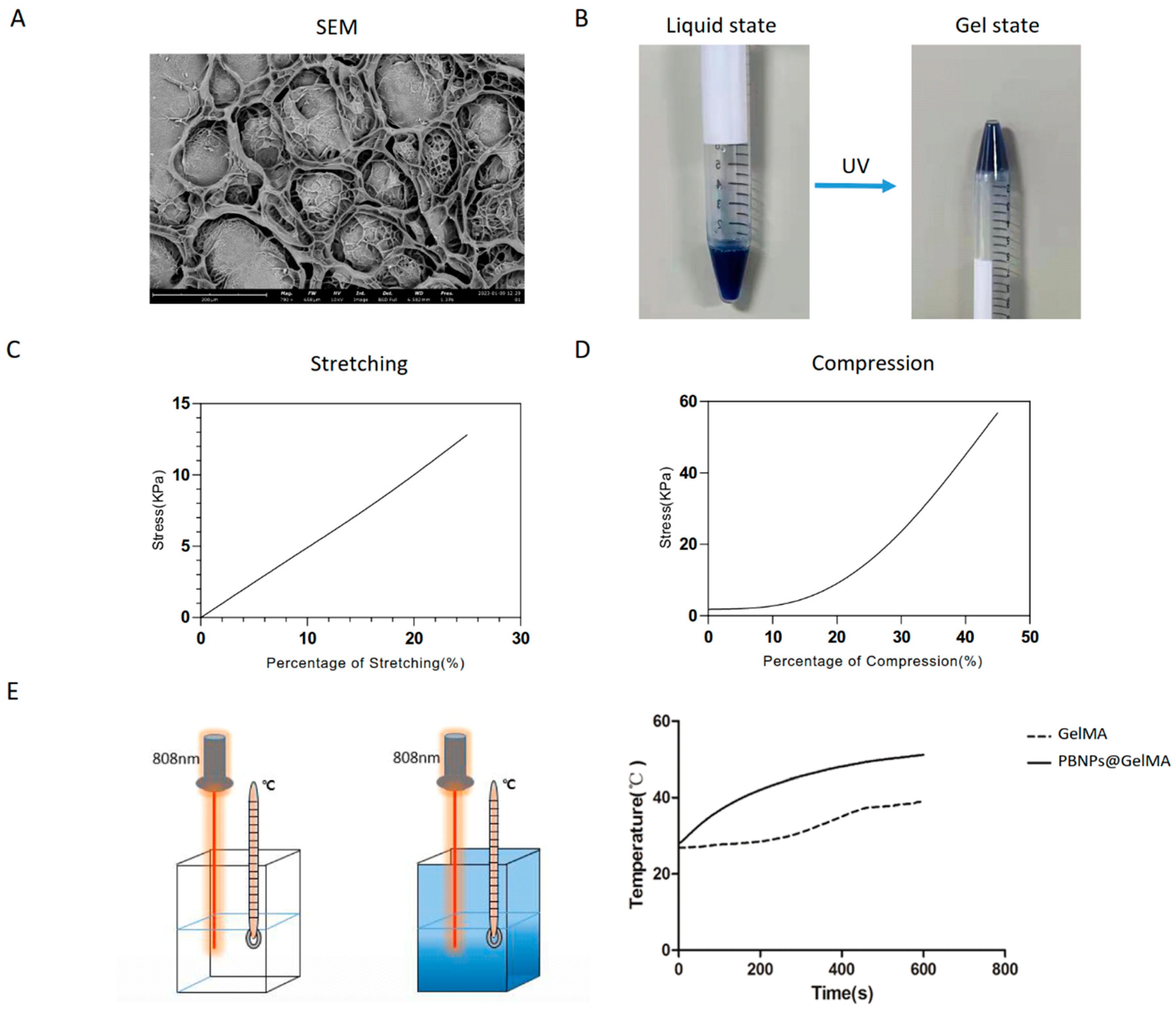
Abstract
1. Introduction
2. Results
2.1. Structure and Properties of PBNP@GelMA
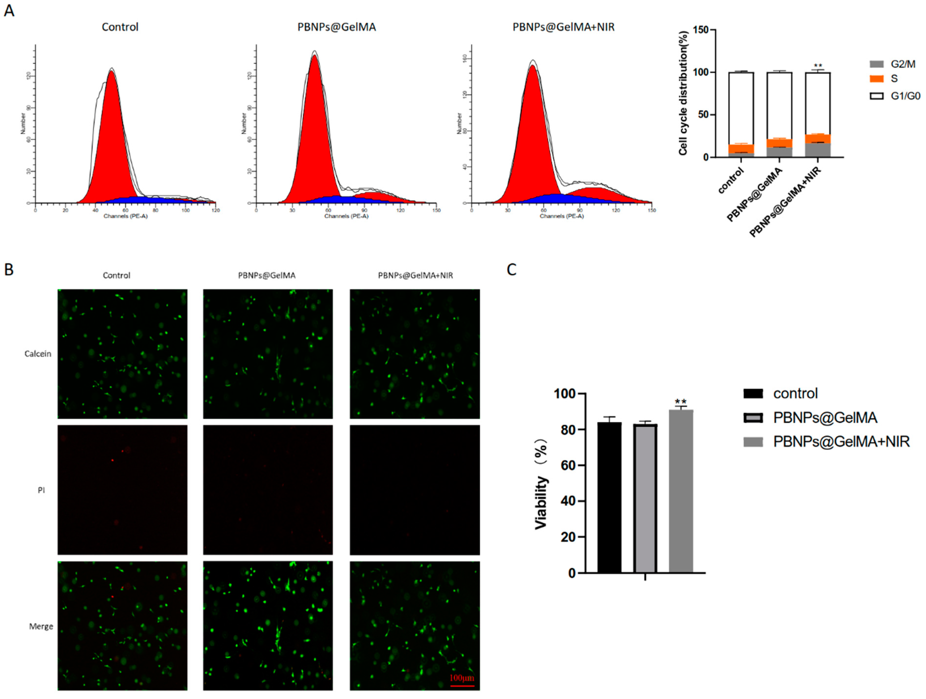
2.2. Heat Shock Treatment Induced by PBNP@GELMA Promotes the Survival of MSCs
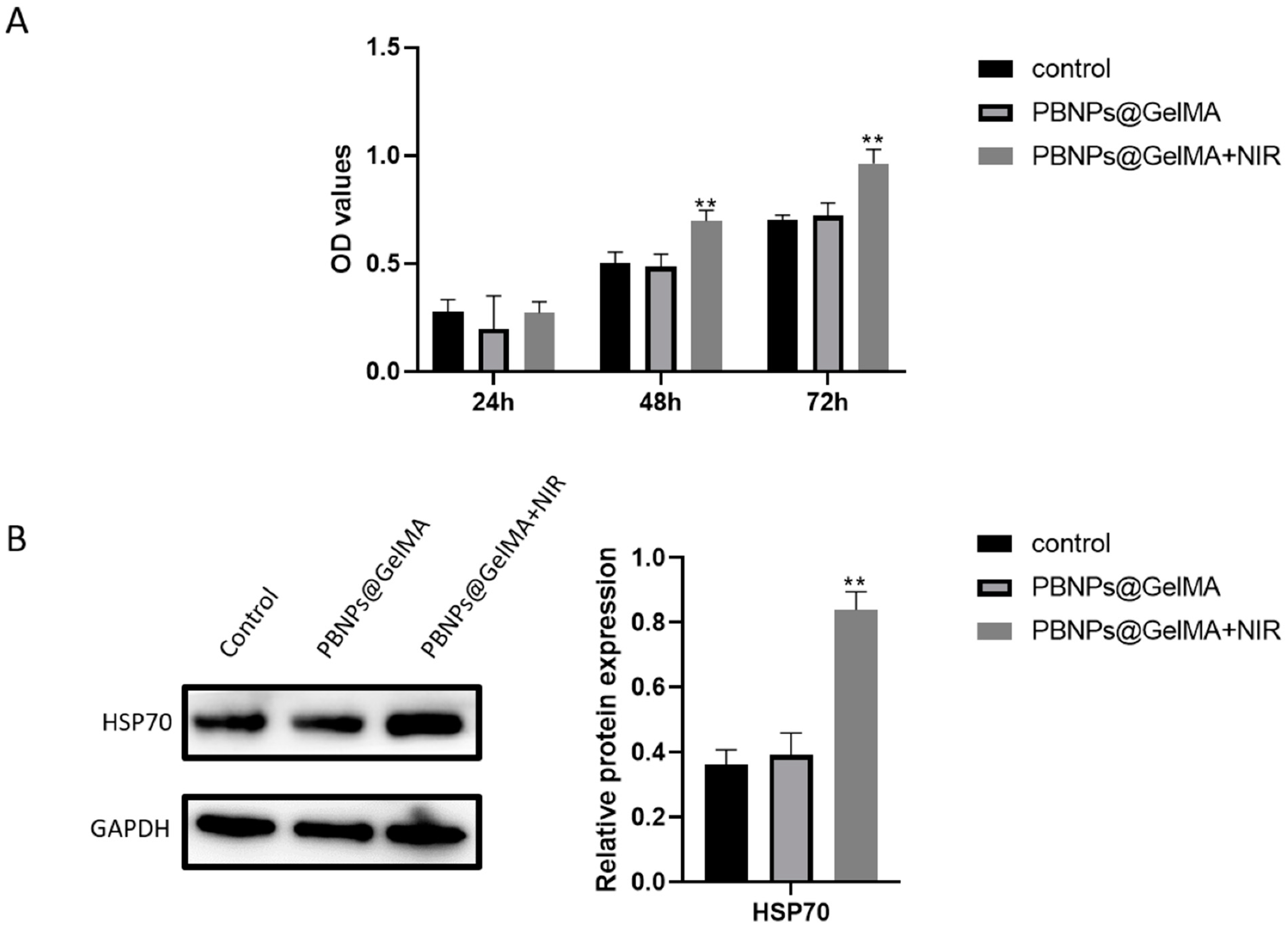
2.3. Role of HSP70 Expression in Elevated Cell Viability of MSCs Induced by Heat Shock Treatment
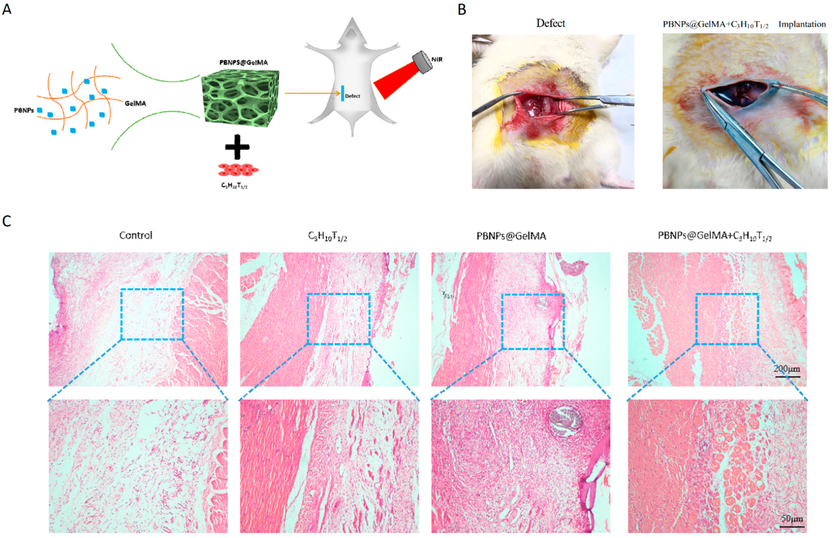
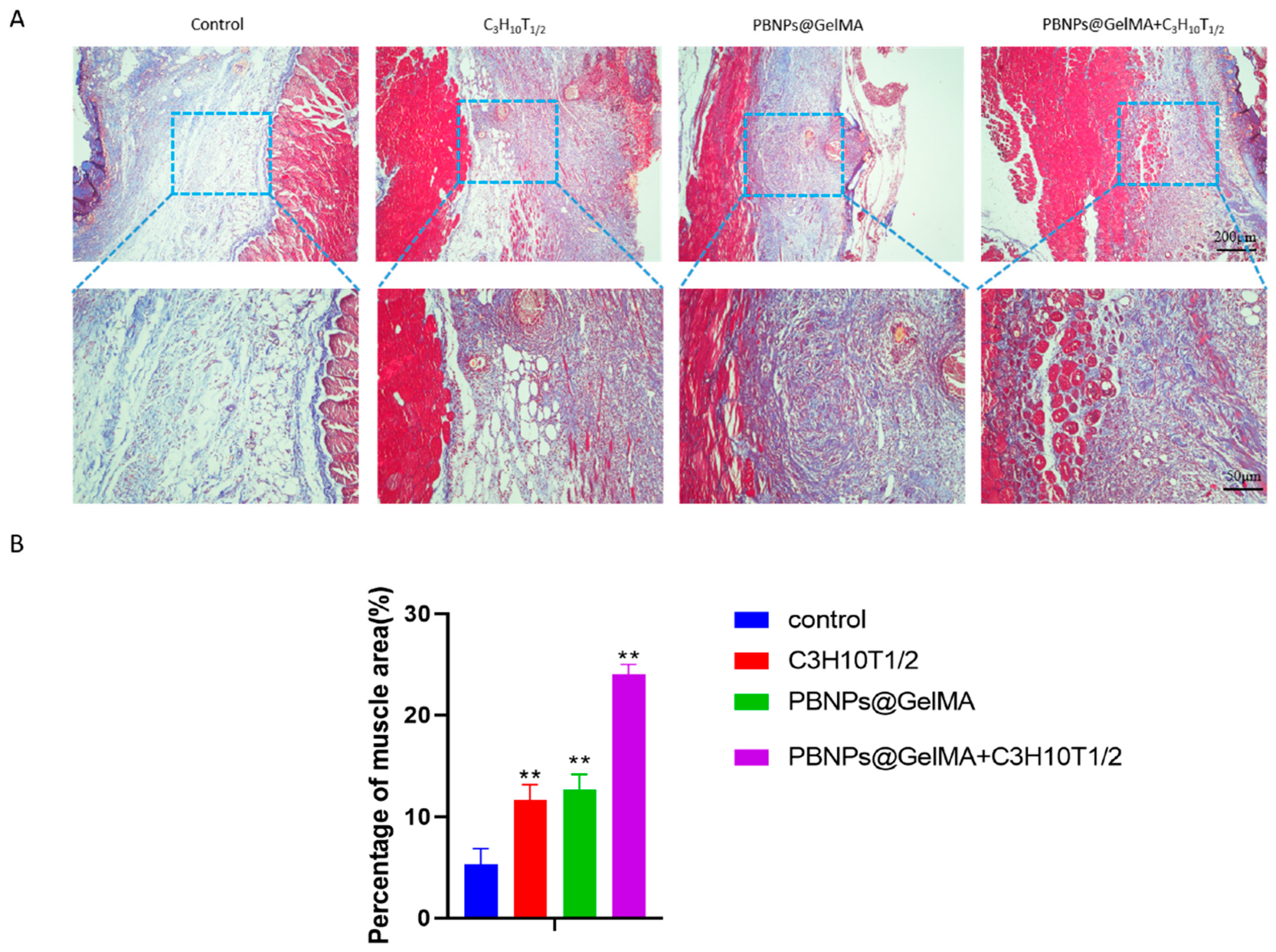
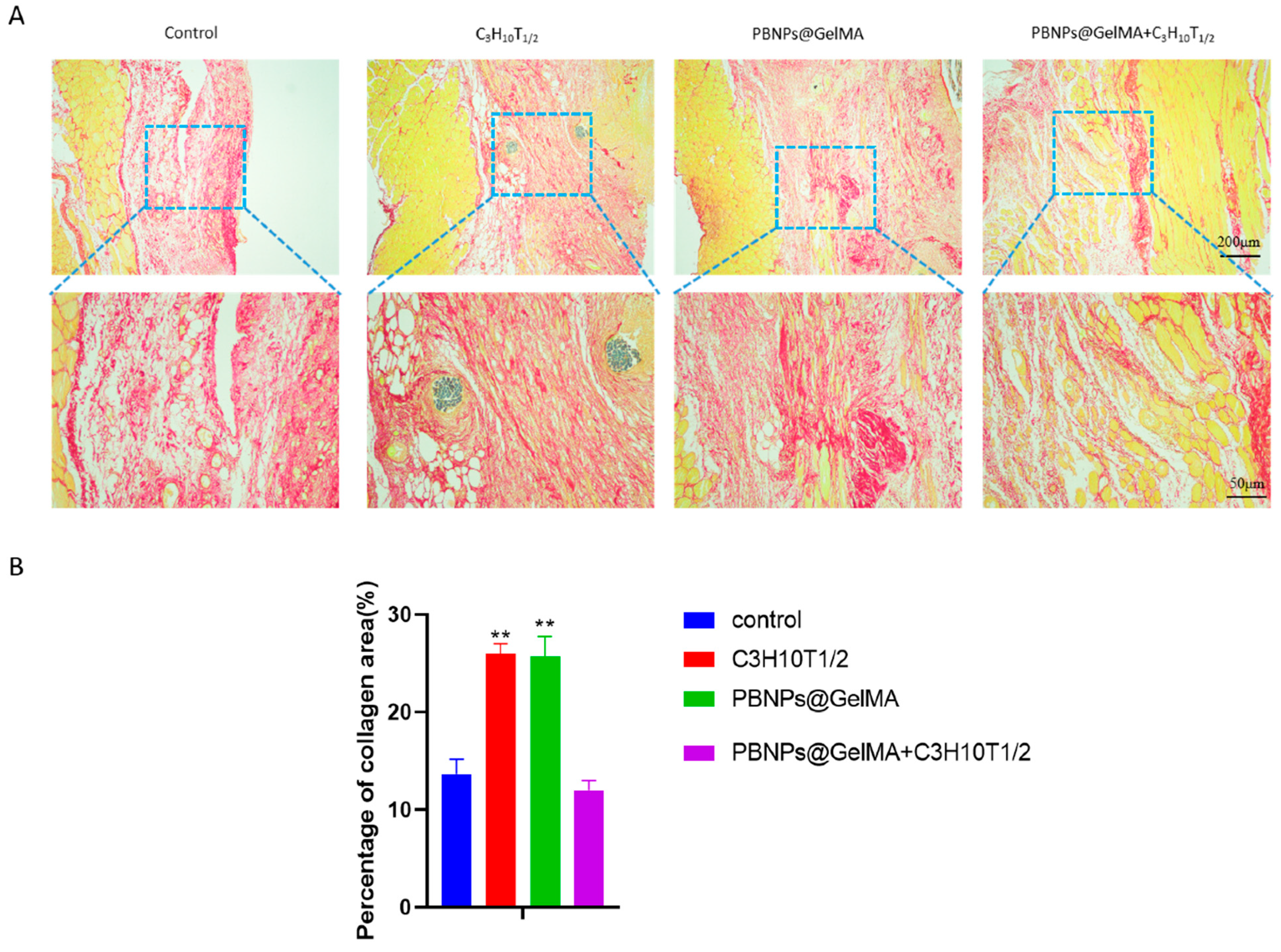
2.4. In Vivo Histochemical Assay Indicated Tissue Repair after the Implantation of Heat-Shock-Pretreated MSCs
3. Discussion
4. Materials and Methods
4.1. Fabrication of GelMA
4.2. Fabrication of PBNP@GelMA
4.3. Mechanical Testing of PBNP@GelMA
4.4. Photothermal Effect Evaluation of PBNP@GelMA
4.5. MSCs Encapsulated in PBNP@GelMA
4.6. Cell Cycle Analysis
4.7. Evaluation of Cell Viability
4.8. Western Blot
4.9. Surgical SD Rat Model
4.10. Histological Staining
4.11. Statistical Analysis
5. Conclusions
Author Contributions
Funding
Institutional Review Board Statement
Informed Consent Statement
Data Availability Statement
Conflicts of Interest
References
- Giarenis, I.; Robinson, D. Prevention and management of pelvic organ prolapse. F1000Prime Rep. 2014, 6, 77. [Google Scholar] [CrossRef] [PubMed]
- Jia, X.; Glazener, C.; Mowatt, G.; MacLennan, G.; Bain, C.; Fraser, C.; Burr, J. Efficacy and safety of using mesh or grafts in surgery for anterior and/or posterior vaginal wall prolapse: Systematic review and meta-analysis. BJOG Int. J. Obstet. Gynaecol. 2008, 115, 1350–1361. [Google Scholar] [CrossRef]
- Mangir, N.; Roman, S.; Chapple, C.R.; MacNeil, S. Complications related to use of mesh implants in surgical treatment of stress urinary incontinence and pelvic organ prolapse: Infection or inflammation? World J. Urol. 2019, 38, 73–80. [Google Scholar] [CrossRef]
- Jo, H.; Brito, S.; Kwak, B.; Park, S.; Lee, M.; Bin, B. Applications of Mesenchymal Stem Cells in Skin Regeneration and Rejuvenation. Int. J. Mol. Sci. 2021, 22, 2410. [Google Scholar] [CrossRef] [PubMed]
- Altman, A.M.; Khalek, F.; Alt, E.; Butler, C. Adipose tissue-derived stem cells enhance bioprosthetic mesh repair of ventral hernias. Plast. Reconstr. Surg. 2010, 126, 845–854. [Google Scholar] [CrossRef] [PubMed]
- Li, Q.; Wang, J.; Liu, H.; Xie, B.; Wei, L. Tissue-engineered mesh for pelvic floor reconstruction fabricated from silk fibroin scaffold with adipose-derived mesenchymal stem cells. Cell Tissue Res. 2013, 354, 471–480. [Google Scholar] [CrossRef] [PubMed]
- Wakitani, S.; Saito, T.; Caplan, A.I. Myogenic cells derived from rat bone marrow mesenchymal stem cells exposed to 5-azacytidine. Muscle Nerve 1995, 18, 1417–1426. [Google Scholar] [CrossRef]
- Nallakumarasamy, A.; Jeyaraman, M.; Maffulli, N.; Jeyaraman, N.; Suresh, V.; Ravichandran, S.; Gupta, M.; Potty, A.; El-Amin, S.F., 3rd; Khanna, M.; et al. Mesenchymal Stromal Cell-Derived Extracellular Vesicles in Wound Healing. Life 2022, 12, 1733. [Google Scholar] [CrossRef]
- O’Donnell, B.T.; Al-Ghadban, S.; Ives, C.; L’Ecuyer, M.P.; Monjure, T.; Romero-Lopez, M.; Li, Z.; Goodman, S.; Lin, H.; Tuan, R.; et al. Adipose Tissue-Derived Stem Cells Retain Their Adipocyte Differentiation Potential in Three-Dimensional Hydrogels and Bioreactors (dagger). Biomolecules 2020, 10, 1070. [Google Scholar] [CrossRef]
- Rattan, S.I.S.; Fernandes, R.A.; Demirovic, D.; Dymek, B.; Lima, C.F. Heat Stress and Hormetin-Induced Hormesis in Human Cells: Effects on Aging, Wound Healing, Angiogenesis, and Differentiation. Dose-Response 2008, 7, 90–103. [Google Scholar] [CrossRef]
- Choudhery, M.S.; Badowski, M.; Muise, A.; Harris, D.T. Effect of mild heat stress on the proliferative and differentiative ability of human mesenchymal stromal cells. Cytotherapy 2014, 17, 359–368. [Google Scholar] [CrossRef] [PubMed]
- Lu, L.; Zhang, C.; Zou, B.; Wang, Y. Hollow Prussian Blue Nanospheres for Photothermal/Chemo-Synergistic Therapy. Int. J. Nanomed. 2020, 15, 5165–5177. [Google Scholar] [CrossRef] [PubMed]
- Wen, J.; Zhao, Z.; Tong, R.; Huang, L.; Miao, Y.; Wu, J. Prussian Blue Nanoparticle-Labeled Mesenchymal Stem Cells: Evaluation of Cell Viability, Proliferation, Migration, Differentiation, Cytoskeleton, and Protein Expression In Vitro. Nanoscale Res. Lett. 2018, 13, 329. [Google Scholar] [CrossRef] [PubMed]
- Luo, R.; Lu, Y.; Liu, J.; Cheng, J.; Chen, Y. Enhancement of the efficacy of mesenchymal stem cells in the treatment of ischemic diseases. Biomed. Pharmacother. 2019, 109, 2022–2034. [Google Scholar] [CrossRef]
- Cui, J.; Wang, H.; Shi, Q.; Sun, T.; Huang, Q.; Fukuda, T. Multicellular Co-Culture in Three-Dimensional Gelatin Methacryloyl Hydrogels for Liver Tissue Engineering. Molecules 2019, 24, 1762. [Google Scholar] [CrossRef]
- Moloney, T.; Hoban, D.; Barry, F.; Howard, L.; Dowd, E. Kinetics of thermally induced heat shock protein 27 and 70 expression by bone marrow-derived mesenchymal stem cells. Protein Sci. 2012, 21, 904–909. [Google Scholar] [CrossRef]
- Chen, J.; Shi, Z.-D.; Ji, X.; Morales, J.; Zhang, J.; Kaur, N.; Wang, S. Enhanced Osteogenesis of Human Mesenchymal Stem Cells by Periodic Heat Shock in Self-Assembling Peptide Hydrogel. Tissue Eng. Part A 2013, 19, 716–728. [Google Scholar] [CrossRef]
- Chen, J.; Li, C.; Wang, S. Periodic Heat Shock Accelerated the Chondrogenic Differentiation of Human Mesenchymal Stem Cells in Pellet Culture. PLoS ONE 2014, 9, e91561. [Google Scholar] [CrossRef]
- Guo, J.; Chang, C.; Li, W. The role of secreted heat shock protein-90 (Hsp90) in wound healing—How could it shape future therapeutics? Expert Rev. Proteom. 2017, 14, 665–675. [Google Scholar] [CrossRef]
- Scieglinska, D.; Krawczyk, Z.; Sojka, D.R.; Gogler-Pigłowska, A. Heat shock proteins in the physiology and pathophysiology of epidermal keratinocytes. Cell Stress Chaperones 2019, 24, 1027–1044. [Google Scholar] [CrossRef]
- Gomez-Pastor, R.; Burchfiel, E.T.; Thiele, D.J. Regulation of heat shock transcription factors and their roles in physiology and disease. Nat. Rev. Mol. Cell Biol. 2017, 19, 4–19. [Google Scholar] [CrossRef] [PubMed]
- Oswald, J.; Boxberger, S.; Jørgensen, B.; Feldmann, S.; Ehninger, G.; Bornhäuser, M.; Werner, C. Mesenchymal Stem Cells Can Be Differentiated into Endothelial Cells In Vitro. Stem Cells 2004, 22, 377–384. [Google Scholar] [CrossRef] [PubMed]
- Nakamura, Y.; Miyaki, S.; Ishitobi, H.; Matsuyama, S.; Nakasa, T.; Kamei, N.; Akimoto, T.; Higashi, Y.; Ochi, M. Mesenchymal-stem-cell-derived exosomes accelerate skeletal muscle regeneration. FEBS Lett. 2015, 589, 1257–1265. [Google Scholar] [CrossRef]
- Sassoli, C.; Vallone, L.; Tani, A.; Chellini, F.; Nosi, D.; Zecchi-Orlandini, S. Combined use of bone marrow-derived mesenchymal stromal cells (BM-MSCs) and platelet rich plasma (PRP) stimulates proliferation and differentiation of myoblasts in vitro: New therapeutic perspectives for skeletal muscle repair/regeneration. Cell Tissue Res. 2018, 372, 549–570. [Google Scholar] [CrossRef] [PubMed]


Disclaimer/Publisher’s Note: The statements, opinions and data contained in all publications are solely those of the individual author(s) and contributor(s) and not of MDPI and/or the editor(s). MDPI and/or the editor(s) disclaim responsibility for any injury to people or property resulting from any ideas, methods, instructions or products referred to in the content. |
© 2023 by the authors. Licensee MDPI, Basel, Switzerland. This article is an open access article distributed under the terms and conditions of the Creative Commons Attribution (CC BY) license (https://creativecommons.org/licenses/by/4.0/).
Share and Cite
Wen, J.; Zhao, Z.; Fang, F.; Xiao, J.; Wang, L.; Cheng, J.; Wu, J.; Miao, Y. Prussian Blue Nanoparticle-Entrapped GelMA Gels Laden with Mesenchymal Stem Cells as Prospective Biomaterials for Pelvic Floor Tissue Repair. Int. J. Mol. Sci. 2023, 24, 2704. https://doi.org/10.3390/ijms24032704
Wen J, Zhao Z, Fang F, Xiao J, Wang L, Cheng J, Wu J, Miao Y. Prussian Blue Nanoparticle-Entrapped GelMA Gels Laden with Mesenchymal Stem Cells as Prospective Biomaterials for Pelvic Floor Tissue Repair. International Journal of Molecular Sciences. 2023; 24(3):2704. https://doi.org/10.3390/ijms24032704
Chicago/Turabian StyleWen, Jirui, Zhiwei Zhao, Fei Fang, Jingyue Xiao, Ling Wang, Juan Cheng, Jiang Wu, and Yali Miao. 2023. "Prussian Blue Nanoparticle-Entrapped GelMA Gels Laden with Mesenchymal Stem Cells as Prospective Biomaterials for Pelvic Floor Tissue Repair" International Journal of Molecular Sciences 24, no. 3: 2704. https://doi.org/10.3390/ijms24032704
APA StyleWen, J., Zhao, Z., Fang, F., Xiao, J., Wang, L., Cheng, J., Wu, J., & Miao, Y. (2023). Prussian Blue Nanoparticle-Entrapped GelMA Gels Laden with Mesenchymal Stem Cells as Prospective Biomaterials for Pelvic Floor Tissue Repair. International Journal of Molecular Sciences, 24(3), 2704. https://doi.org/10.3390/ijms24032704