Review

Zebrafish Cancer Avatars: A Translational Platform for Analyzing Tumor Heterogeneity and Predicting Patient Outcomes

by
Majd A. Al-Hamaly
1,2,
Logan T. Turner
2,3,
Angelica Rivera-Martinez
3,
Analiz Rodriguez
4 and
Jessica S. Blackburn
2,3,*
1
Pharmacology and Nutritional Sciences, University of Kentucky, Lexington, KY 40356, USA
2
Markey Cancer Center, University of Kentucky, Lexington, KY 40536, USA
3
Molecular and Cellular Biochemistry, University of Kentucky, Lexington, KY 40356, USA
4
Department of Neurosurgery, University of Arkansas for Medical Sciences, Little Rock, AR 72205, USA
*
Author to whom correspondence should be addressed.
Int. J. Mol. Sci. 2023, 24(3), 2288; https://doi.org/10.3390/ijms24032288
Submission received: 8 January 2023 / Revised: 17 January 2023 / Accepted: 19 January 2023 / Published: 24 January 2023
(This article belongs to the Special Issue Zebrafish as a Model in Human Disease)

Abstract

:
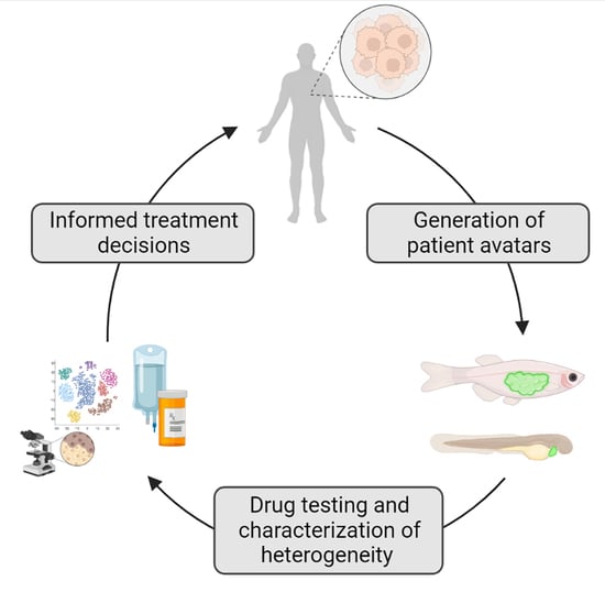
The increasing number of available anti-cancer drugs presents a challenge for oncologists, who must choose the most effective treatment for the patient. Precision cancer medicine relies on matching a drug with a tumor’s molecular profile to optimize the therapeutic benefit. However, current precision medicine approaches do not fully account for intra-tumoral heterogeneity. Different mutation profiles and cell behaviors within a single heterogeneous tumor can significantly impact therapy response and patient outcomes. Patient-derived avatar models recapitulate a patient’s tumor in an animal or dish and provide the means to functionally assess heterogeneity’s impact on drug response. Mouse xenograft and organoid avatars are well-established, but the time required to generate these models is not practical for clinical decision-making. Zebrafish are emerging as a time-efficient and cost-effective cancer avatar model. In this review, we highlight recent developments in zebrafish cancer avatar models and discuss the unique features of zebrafish that make them ideal for the interrogation of cancer heterogeneity and as part of precision cancer medicine pipelines.

Graphical Abstract

1. Introduction

The field of precision oncology focuses on the molecular characterization of a patient’s tumor, with the goal of identifying actionable and patient-specific mutations that can be exploited therapeutically [1,2,3]. Precision medicine holds great promise in the clinic, but emerging data suggest that treating a cancer patient with targeted therapy is not as simple as matching a particular drug to a tumor mutation. In the National Cancer Institute’s precision medicine trial, called the Molecular Analysis for Therapy Choice (NCI MATCH), more than 5000 patients were assigned to a treatment based on the genetics of their tumor. However, fewer than 50% of the patients had a therapeutic response, despite genomic data that matched the actionable mutation in the patient’s tumor to a specific drug [4,5,6]. These findings highlight an important limitation at the forefront of precision medicine—that is, knowing the sequence of a tumor sample is insufficient for predicting a patient’s treatment response.
One reason for the gap between tumor mutation status and treatment response is that molecular profiling of a tumor biopsy sample is unlikely to represent the breadth of that tumor’s heterogeneity. Intra-tumoral heterogeneity, or distinct genetic and phenotypic profiles between cells within a patient’s tumor, plays a significant role in treatment resistance [7,8,9]. Genetic, epigenomic and transcriptomic changes drive intra-tumoral heterogeneity and influence how individual tumor cells respond to treatment. Heterogeneity complicates the classic precision medicine approach that relies solely on DNA mutations for targeted therapy optimization, as DNA sequencing excludes the non-genetic factors that can predict the tumor’s drug response [10].
Patient-specific tumor models, or avatars, are now providing an opportunity to assess the impact of tumor heterogeneity on cancer treatments [11]. Avatars are often cancer xenograft models, where animals are transplanted with a patient’s tumor cells and serve as “stand-ins” so that therapies can be tested and refined. The ultimate goal is for these avatars to prospectively guide the patient’s treatment plan. Traditionally, mice are used to generate patient-derived xenografts (PDX) and avatars. In these models, patient tumor cells are transplanted into immunodeficient mice. The xenografted mice are then used for the pre-clinical evaluation of drug therapies [12,13]. Mouse avatars have a high predictive capacity for their matched patients in terms of treatment sensitivity [14,15,16], but the models can take months to develop before testing can begin, which is not ideal for patients with rapidly progressing cancers [17].
Xenograft of human cancer cells into zebrafish (Danio rerio) has recently emerged as a powerful technique to rapidly assess tumor phenotype and drug responses in living animals at a fraction of the cost of mice [18,19]. Zebrafish PDX (zPDX) avatar models have become a clinically relevant tool for rapid, large-scale cancer therapeutic screening and can serve as a valuable complement to more commonly used avatar models. In this review, we will focus on the benefits of zPDX in assessing intra-tumor heterogeneity and accurately predicting patient outcomes in precision medicine pipelines.

2. Tumor Heterogeneity Complicates Precision Medicine Approaches

Inter-tumoral heterogeneity, or genetic and phenotypic differences between tumors in patients, drove the need for precision medicine in oncology. As DNA sequencing technology developed, researchers and clinicians recognized that not all tumors harbored the same mutations, even between patients with the same type of cancer. Mutations within tumors have been linked to biological behaviors and drug response, and efforts to block the effect of specific mutations gave rise to targeted therapy [20,21]. For instance, the identification of a BCR–ABL translocation in most Chronic Myeloid Leukemia (CML) patients led to the development of the selective inhibitor Imatinib and improved the 5-year survival to 90% [22,23,24,25]. In more heterogeneous cancers, navigating the tumor’s molecular profile to decide on the most appropriate treatment is challenging. For example, in breast cancer, the expression levels of the established biomarkers’ estrogen receptor (ER), progesterone receptor (PR) and HER2 were primarily used for clinical decision-making [26]. Next-generation sequencing has now revealed extreme inter-tumoral heterogeneity among breast cancer patients [27], which can be driven by diverse etiological and environmental factors. Stephens et al. found 73 unique combinations of genetic lesions in cancer-related genes across 100 breast cancer patients [28,29]. These data suggest that clinicians can assign a therapy, but whether a particular tumor’s combination of mutations would make it treatment resistant is nearly impossible to predict based on sequencing data alone.
In addition to the heterogeneity between patients, intra-tumor heterogeneity exists within a single patient. Intra-tumoral heterogeneity means that the tumor is composed of a diverse population of cells, or sub-clones, with distinctive phenotypes, including altered genetics, epigenetics, metabolism, tumor behavior and drug responsiveness (Figure 1) [30]. Notably, the heterogeneity within a tumor is dynamic. The sub-clonal composition can change as the tumor develops and is exposed to naturally occurring cellular stressors, such as changes in blood supply, local hypoxic and anoxic conditions [31], nutrient deprivation and the associated metabolic dysregulation [32], the generation of reactive oxygen species [31], the body’s immune system [33], DNA damage [34] and outside stressors, such as drug treatment [35]. Sub-clones with alterations that impart growth advantages will survive at the expense of those with less fitness, and others will acquire new mutations that allow them to survive. Additionally, growth advantages that impact clone fitness may vary across tumor regions and locations, resulting in a complex tumor environment and architecture, which is further complicated by the introduction of therapeutic agents [36,37].
In an early study to examine the extent of intra-tumoral heterogeneity within a single tumor, Gerlinger et al. performed exome sequencing on nine spatially separated biopsy samples from a single renal-cell carcinoma patient. The analysis revealed remarkable heterogeneity, as about two-thirds of the somatic mutations were not detectable across all the samples and gene expression profiles from different regions of the same tumor displayed different prognosis signatures [38,39]. Similar intra-tumor heterogeneity has been reported in numerous cancers, including lung adenocarcinoma [40], meningioma [41], HER2-positive breast cancer [42], prostate cancer [43] pancreatic ductal adenocarcinoma [44], glioblastoma [45], colorectal cancer [46], liver cancer [47], lymphoma [48] and many others [49,50]. Tumor heterogeneity has been linked to poor patient prognosis and is regarded as one of the critical determinants of therapy resistance, treatment failure and decreased overall survival [7].
The data described above have important implications for precision medicine approaches, in that a single targeted agent is unlikely to be effective against a highly heterogeneous tumor. Sequencing of multiple, spatially separated biopsies may be required to reveal the degree of heterogeneity within the tumor and to assign the correct targeted drug panels to the patient. At the same time, a tumor’s drug responsiveness is often a combination of genetic and non-genetic factors that DNA sequencing may not fully encompass. For precision medicine to reach its full potential, there is a need to define the functional impact of intra-tumoral heterogeneity on drug response and the behavior of individual sub-clones. By combining tumor-specific models and drug testing with sequencing data, a patient’s treatment plan could be tailored based on the sensitivity of each sub-clone to targeted therapeutics.

3. Current Patient-Derived Avatar Models Are Not Suitable for Clinical Timelines

A patient-derived avatar model, in which a patient’s tumor is transplanted into an animal or grown as a 3D organoid culture, serves as a functional surrogate for that patient. Avatars provide an important venue to interrogate tumor behavior and drug responsiveness while sparing the patient from undesirable side effects and wasted time of ineffective therapies. Ultimately, findings from avatars can be translated back into the same patient’s treatment plan and can also inform the decision-making for a larger group of patients with tumors with matched genetic or molecular profiles [13,51,52].
Currently, mouse patient-derived xenografts (mPDX) are regarded as the most powerful model for predicting clinical outcomes. Several studies have demonstrated a high fidelity between patient samples and mPDX in histology, genomic and transcriptomic profiles [53,54]. In simple terms, a patient’s sample is implanted in immunodeficient mice, and mice are treated with drug combinations that patients would receive for that cancer type. Mouse avatars have a reasonable correlation with patient outcomes. For example, in metastatic sarcoma patients, 13 out of 16 (81%) were reported to have similar clinical responses to their avatars [55]. Patient-derived organoids (PDO) are also used as avatar models. They are self-organized, 3D cell cultures established from the patient’s biopsy and maintained in vitro. PDOs enable the examination of cell-to-cell interactions and provide insight into the effect of the tumor microenvironment on the tumor cell behavior [56]. PDO avatars can also forecast patient response to chemotherapy. For example, PDOs provided a high predictive value of 88% in metastatic gastrointestinal cancer [57].
These models have been used for several years but have significant limitations. First, for unknown reasons, not all patient samples will engraft into mice to form a mouse PDX, and some patient samples will not grow as organoids. For example, the success rate for generating mPDX of breast and renal cancers was 13% and 8.9%, respectively [19]. Similarly, for patient-derived organoids, a low success rate of 16% is reported in advanced prostate cancer [58]. The unpredictable engraftment success of these avatar models limits their utility to a random subset of individuals, with certain patient tumors unable to survive in these conditions.
A critical limiting factor of these models is that the time they take to develop is incompatible with the rapid pace with which decision-making must occur in the oncology clinic [59]. In clinical practice in the United States, the median time between diagnosis and initiation of therapy is 26 days for colon cancer to as long as 79 days for prostate cancer [60]. Timelines to develop mPDX avatars are measured in months, with the average mPDX requiring 6–8 months to develop [11]. Some mPDX models have an even longer latency. Prostate cancer mPDX may take up to 37 months to engraft and become established in mice [61]. This timeline is not feasible in particularly aggressive cancers, such as certain glioblastomas, where patients progress and can succumb to their disease within weeks [62]. Although PDOs are cultured in vitro and have shorter generation timelines, they still require 4–6 weeks to develop from patient tissue samples. Because of this extended timeline, the current cancer avatar models are generated in case the patient develops relapse or to inform treatment plans of newly diagnosed patients with tumors with similar molecular profiles. The promise of cancer avatars, which is that there will be a patient-specific tumor model to help guide clinical decisions from diagnosis onwards, has not been fully realized with the avatar models that are currently available.

4. Zebrafish Patient-Derived Xenografts (zPDX) Fill Gaps in Current Model Systems

Use of zebrafish as cancer avatars can overcome some of the limitations of the current model systems. Human cancer cells can be engrafted into zebrafish larvae less than five days old or adult zebrafish more than 30 days old [63,64]. Procedures to generate patient-derived xenografts (zPDX) in both the larvae [65] and adult zebrafish [66,67] are well-established, and each has its unique benefits. zPDX into larvae are ideal for large-scale drug screening, as hundreds of animals can be easily xenografted relatively quickly. Larval zebrafish are well-established as a pharmacological tool, as they can live in 96-well plates and readily absorb compounds from the water [68]. The immune system in zebrafish is not fully established until 21 days post-fertilization [69], and drug testing is usually completed within 96 h after xenograft. Immune rejection is typically not a concern in larval zPDX. For xenograft into adult fish, animals must first be made immune-deficient through gamma irradiation [70] or immunosuppressant treatments by dexamethasone [71]. The zebrafish immune system typically regenerates within three weeks, and the xenografted tumor will eventually be rejected. The zPDX in this adult model is, therefore, time-sensitive. More recently, Moore et al. reported a genetically modified immunodeficient zebrafish with mutations in recombination activating gene 2 (rag2), DNA-dependent protein kinase (prkdc) and janus kinase 3 (jak3), resulting in the loss of T, B and natural killer cells [72]. prkdc−/−, il2rga−/− immune deficient fish were shown to robustly engraft a wide array of human cancers at 28 days. Importantly, the phenotypes of the human xenografted cells in zebrafish resembled that of mouse PDX [64], indicating that key properties of human cells were retained whether they were transplanted in mammals or fish. Use of adult fish also permits oral gavage or intraperitoneal injection of drug treatments. Although this limits the model to more modest numbers in drug testing, the delivery method serves as an accurate and relevant mimic of patient dosing and could provide useful insight in precision medicine approaches [66].
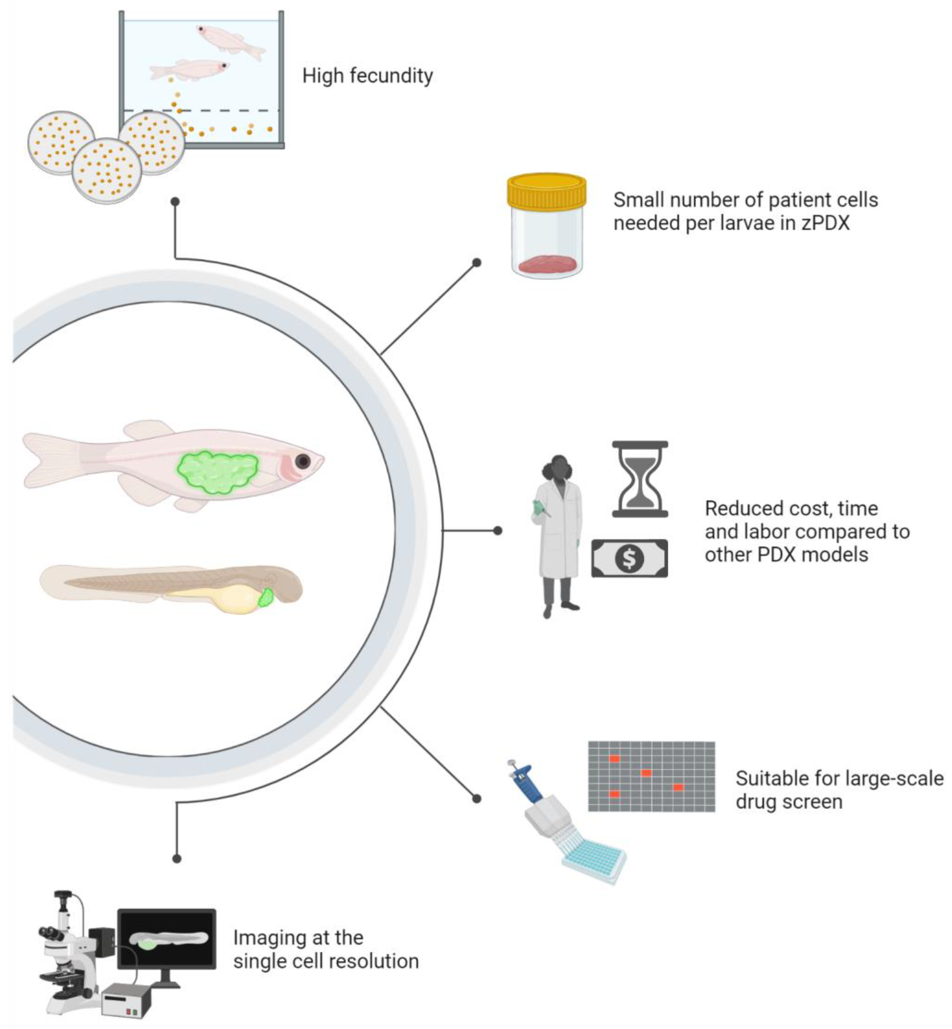
There are several benefits of zPDX (Figure 2) that make this model a valuable complement to mouse and organoid avatar systems. The most important is the time needed to generate the xenograft. Cells can be visualized immediately after transplant in a larval zPDX, and in most cases, drug treatments start 24–48 h post-transplant. Workflows are typically completed within a week [73,74]. Adult immunodeficient fish require 7–28 days for robust engraftment [64], significantly less time required than the 3–8 months typically needed for mouse PDX engraftment or 6–8 weeks for in vitro organoid formation. Zebrafish avatars can, therefore, more readily fit into clinical decision-making timelines for cancer patients.
Zebrafish are also low-cost and accessible to a wide range of laboratories. The use of mouse models requires a substantial investment in infrastructure for mouse housing and separate facilities for imaging and procedures. The cost of care and housing for four mice averages $1.05 per day, and a breeding pair will generate ~6–8 pups after 21 days of gestation. In contrast, a tank of a dozen zebrafish can be maintained for $0.25 per day [75], and each mating pair of zebrafish produces >100 eggs weekly. Facilities housing even as few as 50 animals could complete an extensive number of larval zPDX and drug testing experiments weekly in 96-well plates [76,77]. While mouse PDX will continue to play an essential role in testing small numbers of drugs in mammalian systems, their cost and labor-intensive procedures limit their applicability in large-scale screens to test the efficacy of precision medicine compounds. Zebrafish models can fill this gap.
In efforts to reduce the labor surrounding the use of zPDX and potentially increase the throughput of drug screening, steps in developing zebrafish research models have been automated (Table 1). For instance, a robotic injection system can inject up to 2000 embryos per hour with a success rate of 99% for high-throughput assays [78]. This method has been reported for injecting DNA to obtain transgenic animals. Nonetheless, there is potential for the extension of this work for large-scale xenografting in embryos. Imaging steps can also be automated with several approaches. For instance, the VAST Bio-Imager can image up to 100 larvae per hour with an 85% imaging success rate [79]. Several groups have automated image acquisition and processing for large-scale cancer cell counting [80], evaluation of tumor dissemination [80] and generation of tumor migration data [81].
Finally, zPDX provides intravital imaging approaches that are not possible in mice, which can provide new information on tumor phenotypes and drug response. Zebrafish larvae are optically transparent, and xenografted cells are typically stained with a fluorescent dye or express fluorescent proteins, allowing cells to be monitored in living animals from the moment of xenograft, through drug treatment and onwards [82]. For example, Mercatali et al. described a zPDX model of breast cancer bone metastasis in which labeled cells were observed to extravasate and colonize the caudal hematopoietic tissues (CHT), a bone marrow-like niche in zebrafish [83]. Similarly, Asokan et al. used selective plane illumination microscopy (SPIM) to record the real-time migration of cancer cells. This method was able to differentiate the dissemination patterns and speed of dissemination between breast cancer and leukemia cells [81]. Almstedt et al. integrated live fluorescent imaging and artificial intelligence-driven image analysis for a real-time evaluation of GFP-tagged glioblastoma orthotropic xenografts, examining growth, invasion and survival [84]. In adult models, Yan et al. engrafted human rhabdomyosarcoma cells into the peri-ocular muscle of immune-deficient prkdc−/−, il2rga−/− zebrafish. This site is accessible to imaging approaches to allow for the dynamic visualization of the pharmacodynamics of drug treatment at a single-cell resolution. In this experiment, before engraftment, human rhabdomyosarcoma cells were made to stably express the FUCCI4 cell-cycle fluorescent reporter. Seven days after engraftment, single and combination drug treatments were administered by oral gavage. Olaparib and temozolomide combination therapy resulted in a higher number of G2-arrested cells compared to control animals [64]. This combination is now in phase I clinical trial in pediatric rhabdomyosarcoma patients (clinicaltrials.gov identifier: NCT01858168) [85].
The unique benefits of the zebrafish, particularly the short time required to generate the zPDX and the scale at which drug screening can be completed, make them an ideal tool for cancer avatar models in clinical diagnostic pipelines. Imaging approaches, alone or in combination with drug treatment, can also provide a means to rapidly assess the sub-clonal phenotypes within a heterogenous tumor that could significantly impact therapy response. This latter attribute is currently not possible in mPDX.
Table 1. Overview of automation approaches with potential application in zPDX.
Automated StepEquipment and Software InvolvedApplicationSpeed or EfficacyAccuracyLimitationsRef.
Imaging and Image analysis Automated microscopy platform,
IncuCyte S3 and CNN for image analysis
Characterize the tumor growth in the xenografts in vivo One image every 4–6 h over 4 days77% agreement with manual analysisDifficulty of identification of early signs of death[84]
Image analysisFaster R-CNN algorithm, Inception Reset V2 feature extractor Counting of tumor cells in zPDX Not mentioned85% average precisionLarger datasets are considered for higher accuracy[86]
Image analysis SPIM for imaging, CellProfiler, CellTracker and R open source tools Real time visualization and tracking of metastatic cancer cells in zPDXNot mentionedDifferentiate the dissemination patterns between two types of cancersNot mentioned[81]
Imaging VAST BioImager System, upright microscope, LP Sampler Automate zebrafish larva handling, positioning, orientation and imaging 100 and 75 larvae/h sampling from bulk and 96-well plate 86% and 97% success rate imaging from bulk and 96-well plate Applicable for 2–7 dpf larva, high storage space for obtained images [79]
Imaging and treatment administrationAutomated pipetting workstation, automated imaging system
(ImagXpressMICRO)
Live imaging of zPDXand automated application of therapeuticsNot mentionedNot mentionedNot mentioned[87]
Imaging and treatment administrationImageXpress Micro System, JANUS automated workstationCollect High-Content images for adult and embryo zPDXNot mentionedNot mentionedNot mentioned[88]
InjectionMotorized stage with paired controller, motorized micro-manipulator, “injectman II”, z-calibration unitSemi-automate injection2000 embryos/h 99% success rate Automation is reported for injection into embryos and requires commercial injection needles [78]
Injection and screening Automated microinjection system and COPAS Automate injection into zebrafish embryos and fluorescence quantification 3000 embryos/15 min for sorting step Not mentionedNot mentioned[89]
Imaging, image pre-processing and analysis CLSM platform with movable stage, Image-Pro Software Quantification of human cancer dissemination in zPDXLess than 5 min per plate for imagingDissemination in zPDX significantly correlated with metastatic behavior in mouse modelConfocal microscopy is required[80]
CLSM: Confocal Laser Scanning Microscope, CNN: Convolutional Neural Network, COPAS: Complex Object Parametric Analysis and Sorting, dpf: days post fertilization, LP: Large Particle, R-CNN: Regions with Convolutional, Neural Network, SPIM: Selective Plane Illumination Microscopy, VAST: Vertebrate Automated Screening Technology, zPDX: Zebrafish patient-derived xenografts.

5. Zebrafish Are Clinically Relevant Cancer Avatars Models

Xenograft of human cancer cell lines into zebrafish is now a well-established technique. Researchers have found that important behavioral properties of cancer cells, such as proliferation, migration, matrix remodeling and angiogenesis, among others, can be observed in zebrafish xenografts, despite zebrafish being highly dissimilar to humans. For example, in 2006, in the first human cancer cell xenograft, melanoma cells were observed to proliferate, migrate and form masses that participated in angiogenesis [90]. Nicoli et al. similarly used human cancer cell line xenografts in zebrafish to mimic the initial stages of tumor angiogenesis. The model was able to discriminate between two endometrial cancer lines that differ for VEGF and FGF expression and angiogenic behavior. In agreement with data from nude mice, angiogenesis was observed in 75% of zebrafish larvae xenografted with the highly angiogenic line and none from the poorly angiogenic line. Treatment with anti-angiogenic compounds used in mammals diminished neovascularization in all zebrafish larvae without affecting physiological vessel development [91]. This early data proved that zebrafish xenografts can provide useful information about human cancer cell phenotypes and that anti-cancer compounds can be relevant in a zebrafish xenograft setting.
Following the success of xenograft of human cancer cell lines, patient-derived xenografts models in zebrafish have been developed and used to assess different attributes of tumor behavior. zPDX models have since been established in multiple cancer types, including gastric cancer [92], colorectal cancer [73], breast cancer [83], non-small cell lung adenocarcinoma [93], pancreatic ductal adenocarcinoma [94] and others [68,74,95]. Ai et al. highlighted the importance of an in vivo system to patient-derived cells by comparing the transcriptomic profile of glioblastoma multiforme (GBM) cells that were xenografted to zebrafish larvae brain to GBM cells grown in vitro in typical cell culture conditions. The authors used principal component analysis (PCA) to show that in vitro grown cells clustered together and away from the xenografted cells. The xenografted cells showed significant gene enrichment in categories, such as tumor metastasis, cell migration and epithelial-to-mesenchymal transition. This transcription identity suggests that the zPDX models can recapitulate the supportive tumor microenvironment that patient-derived cells need [95].
Zebrafish PDX has also been compared to mouse PDX models for use as cancer avatars. Ali et al. developed a combined mouse and zPDX testing platform by engrafting 39 preserved mPDX tissue fragments of non-small cell lung cancer into zebrafish embryos. The zPDX accurately predicted lymph node involvement with 91% sensitivity. In addition, the response of zPDX to standard treatments correlated very well with the response in mPDX and patients [93]. Fior et al. directly compared the chemosensitivity profile between colorectal mice and zebrafish PDX. In both models, chemotherapies significantly increased apoptosis and reduced proliferation. The differential sensitivities to chemotherapy in some colorectal cancer cells uncovered by zPDX models were later validated in mPDX models [73].
While the above data support a role for zPDX in the avatar setting, there are some limitations to consider. Cell-signaling between cells within the tumor microenvironment can profoundly impact xenograft behavior and drug response. Cell-to-cell communication can occur between zebrafish and human cells to impact xenograft behavior. For example, in a triple-negative breast cancer xenograft model, the zebrafish cytokine Cxcl12 was found to trigger the human chemokine receptor CXCR4 on engrafted cells, which was suggested to drive the formation of breast cancer micro-metastases in the zebrafish [96]. However, only 70% of genes are conserved between the zebrafish and the human genome [97], so some receptor/ligand interactions and signaling pathways in mammalian tissue might not be present in the zebrafish tumor microenvironment. For instance, mouse or human growth factors cannot stimulate hematopoietic precursors in zebrafish [98]. Recently, editing the zebrafish genome was employed to produce a more accurate tumor microenvironment for human xenografts. Rajan et al. described the generation of a transgenic “humanized” zebrafish that expresses human-specific cytokines, resulting in enhanced human leukemia cell migration and proliferation after xenograft transplantation [99].
Another possible issue in the generation of zebrafish cancer avatars is the difference in temperature between the xenograft and the host. Zebrafish are housed at 28.5 °C, and the physiologic temperature for human cells is 37 °C. Most larval zPDX studies house xenografted animals at 34 °C. When developing larvae are incubated at a higher temperature, they may experience changes in metabolic, immune and inflammatory responses and can develop malformations that affect the integrity of the host system [100,101]. Incubating the human tumor cells at a lower temperature than 37 °C will affect their proliferation rate [102]. This limitation can partially be resolved using the adult prkdc−/−, il2rga−/− immunodeficient fish, which are adaptable to surviving at 37 °C for more extended periods [64].
A final limitation of zebrafish in the avatar setting is that they are not ideal for orthotopic transplants. In this model, cancer cells are xenografted into an animal’s organ or tissue that matches the tumor’s histotype. Orthotopic xenografts often provide an ideal microenvironment to recapitulate tumor behavior, phenotypes and drug responses. For example, in zPDX of human glioblastoma, cells were found to proliferate and invade when transplanted into the zebrafish brain, the orthotopic site, but remain static when xenografted into the zebrafish yolk sac [103,104]. Due to the absence of some organs in zebrafish, such as the lungs, and the small size of other organs, orthotropic engraftment might be challenging for certain cancers [102]. Table 2 provides a summary on the current status, advantages and disadvantages of each model.

6. Zebrafish Bring Pharmacology Strengths into Precision Medicine Pipelines

Zebrafish have been used for many years in pharmaceutical research in large-scale, in vivo small-molecule screens [105]. Their pharmacology success is one of their significant benefits in the cancer avatar field. Because of this history, drugs are now established to have conserved effects between humans and zebrafish. For instance, research showed that human cardiotoxic drugs induced pericardial edema and circulatory disturbances in zebrafish, compared to no signs of cardiovascular toxicity for non-cardiotoxic drugs, with a 100% prediction success rate [106]. Zebrafish are a well-established model for pharmacokinetics, with many phases I and II metabolic reactions used in drug processing conserved between zebrafish and mammals [107,108,109]. Zebrafish also have direct orthologs to human cytochrome P450 (CYP) enzymes, which metabolize hepatically cleared drugs. The conservation of metabolic processes between zebrafish and humans indicates that drugs are likely processed and utilized by zebrafish and human patients in much the same way [110].
Drug screens in zebrafish can directly impact human disease, as chemical screens in zebrafish have identified several drugs across a wide range of indications that are now in different phases of clinical testing [111,112]. For example, a screen in zebrafish led to the identification of Dorsomorphin, the first bone morphogenetic protein (BMP) inhibitor. Derivatives of this compound are being clinically tested for fibrodysplasia ossificans progressiva and anemia of inflammation [113]. Pharmaceutical companies are also realizing the value of the zebrafish model and have established in-house zebrafish facilities or are collaborating with external zebrafish Contract Research Organizations (CROs) [114].
The success of zebrafish models in the drug discovery field and the translatability of their findings to human disease makes them robust and reliable candidates for drug sensitivity profiling along the precision medicine pipeline. Larval zPDX provides a whole-animal venue for evaluating drug pharmacokinetics, pharmacodynamics and toxicity. At the same time, these xenografted animals are easily manipulated in a 96-well plate [105]. The skin of zebrafish larvae has a high permeability to enable drug screening by immersion, making the drug testing process for xenografts much faster than injecting mice [115]. However, this method presents a challenge for drugs with limited water solubility. Drug treatment at temperatures lower than 37 °C might also influence the efficacy of certain chemotherapeutic agents [102]. Finally, drug screens and other tumor characterization assays are typically restricted to 7–8 days post-engraftment in larval zPDX before the immune system begins to reject the transplant [116]. The window for successful engraftment will limit the drug exposure time and might influence the drug response. This short time frame means that drugs that alter cellular proliferation or induce apoptosis are most likely to strongly impact the xenografted cells. Drugs that take a longer time to result in cell death through mechanisms involving interference with the cell cycle or epigenetic alterations, for example, may not be able to demonstrate a significant effect in this short-term model [117].
As zebrafish cancer avatars become a more established tool, further efforts will be needed to understand the pharmacokinetics and the related ADME (absorption, distribution, metabolism and excretion) factors of therapies in zPDX. This information is necessary to determine whether a targeted therapy that failed in a zPDX was ineffective because the tumor was not responsive or if the animal model itself caused an issue. For example, the drug may not be absorbed well into the fish, metabolized correctly or able to reach the cancer cells. Building these datasets for zPDX on common precision cancer medicines will allow zebrafish to become a reliable avatar model and a strong complement to mouse PDX.

7. Tank to Bedside: Zebrafish Avatars for the Prediction of Patient Outcomes

Several studies evaluated the ability of zPDX to predict patient responses (Table 3). A subset of these utilized a co-clinical trial design to implement a zebrafish avatar model [94,118,119,120]. The concept of co-clinical trials arose from the increased understating of inter-tumoral heterogeneity, or the individuality of each patient’s disease, and the need to optimize therapy at the patient level. A co-clinical trial aims to enable efficient patient treatment while minimizing the unwanted administration of ineffective therapies, which could have undesirable side effects. A parallel arm is added to the standard clinical trial design using PDX avatars to re-capitulate the patient tumor and provide a venue for testing and screening for optimal therapy [17]. Zebrafish avatars have shown promise in these types of studies. Di Franco et al. transplanted patient-derived pancreatic cancer cells into the zebrafish embryos’ yolk to screen the treatment plan chemotherapy agents. After two days of treatment, the response to treatment was evaluated by comparing the relative tumor area to the control. A statistically significant reduction in relative tumor area was reported in 6 of the 15 patient samples. Using a conversion factor to compare the chemotherapy efficacy reported in the literature as a percent partial response reported in humans, the group demonstrated that chemotherapy in zPDX reflected the efficacy of chemotherapy in clinical settings [118].
The result of this work formed the basis of the first zebrafish co-clinical trial XenoZ (clinicaltrials.gov identifier: NCT03668418), an observational, prospective clinical trial on patients with pancreatic and gastrointestinal cancers. The study will recruit 120 patients to demonstrate the zPDX model’s efficacy in predicting the optimal treatment regimen [121]. The authors first reported a conversion factor for determining the equivalent dose of chemotherapy in zebrafish; this factor was determined by combining data from zebrafish safety studies (maximum tolerated dose) and efficacy (significant decrease in tumor area). Twenty-four patients with pancreatic, colon and gastric cancers undergoing chemotherapy were enrolled in the study, and their biopsies were used to generate avatar models. Avatars were tested for chemotherapy response and evaluated using a zebrafish-adapted form of the Response Evaluation Criteria In Solid Tumors (RECIST) based on the change in tumor area in the zPDX avatar. The sensitivity of different engrafted tumor types to administered chemotherapy was consistent with that reported in the clinical context. For example, in colon cancer avatars, combination treatment (FOLFOX, FOLFIRI and FOLFOXIRI) were more effective than a single agent 5-FU [120], just as it was in patients.
zPDX avatars could also predict treatment response in pancreatic ductal adenocarcinoma (PDAC) [94]. In this study, patients were considered non-responders if their cancer reoccurred within one year and were responders if they remained disease-free. In the zPDX avatar model, responders were defined as having more than a 50% decrease in tumor size in response to treatment. Out of the 31 patients who provided samples, 16 were followed up for a median time of 19.3 months. The zPDX avatars indicated that 7 out of the 16 patients were responders, and 9 were non-responders to treatment. Of those, only 1 patient out of the 7 (14.3% error) zPDX responders reported a cancer recurrence, while 6 of 9 (66.7% accuracy) patients that the zPDX avatars identified as non-responders had a cancer recurrence. The zPDX models could, therefore, accurately identify patient response to chemotherapeutic agents.
In addition, and under the same co-clinical trial, a zPDX avatar model was developed with samples from patients with colorectal cancer and used the adaptive RECIST criteria to predict chemotherapy response when treated with 5-FU, FOLFIRI, FOLFOX, FOLFOXIRI for two days. These zPDX avatars predicted chemotherapy response with 75% accuracy [119]. A similar trial is planned, called the Evaluation of Tumor Growth and Oncological Treatment in Patients With Colorectal Liver Metastases (CRLM) Using Zebra Fish Embryo Model (CRLM-Z) (clinicaltrials.gov identifier: NCT05289076). This trial is an observational prospective cohort study set to enroll 40 patients. Tumor response to different combinations of therapies will be tested in patients and zPDX, and based on the success of the observational study, interventional studies may be planned [122]. zPDX avatars now serve as a powerful translational tool for precision medicine and may have the potential to support clinicians in making individualized and informed treatment decisions for their patients [49,117,123].
Table 3. Studies correlating zebrafish PDX outcomes to patient outcomes in different cancers.
CancerZebrafish AgeNumber of Patient SamplesOutcome AssessedTherapeutic RegimenXenograft CharacterizationCriteria for Zebrafish Response EvaluationCriteria for Human Response EvaluationConcordance 1Ref
CRC48 hpf36Chemotherapy response5-FU, FOLFOX, FOLFIRI and FOLFOXIRIEngraftment success and change in equivalent tumor volume (%∆V)Adaptive RECIST 2 CriteriaRECIST criteria75%[119]
48 hpf2Radiotherapy sensitivity25 Gy + 5-FUEngraftment success, Tumor size and Apoptosis of tumor cellsTumor size and cell deathMRI response and rectosigmoidoscopy100%[124]
48 hpf5Chemotherapy responseFOLFIRI, FOLFOX and CetumixabEngraftment success and tumor sizeTumor size, angiogenesis potential, apoptosis and metastatic potentialDisease recurrence, CEA levels and treatment response80%[73]
NSCLC48 hpf4Chemotherapy responseErlotinib and PaclitaxelEngraftment success and tumor sizeRelative tumor sizes,
dissemination
Not mentionedNot mentioned[93]
48 hpf21Chemotherapy responseOsimertinib, Pemetrexed, Docetaxel and CisplatinEngraftment success and tumor sizeTumor sizeRECIST criteria76.9%[125]
PDAC48 hpf31Chemotherapy responseGemcitabine, GEMOX, GEM/nab-P and FOLFOXIRIEngraftment success and change in equivalent tumor volume (%∆V)Adaptive WHO Criteria 3Adjuvant treatment administration, disease recurrence rate and DFS66.7% for cancer recurrence[94]
48 hpf15Chemotherapy responseGemcitabine, GEMOX, GEM/nab-P and FOLFOXIREngraftment success, Larvae survival rate, Histology and ImmunohistochemistryMean RTAPFS, overall health and failure-free survivalNot mentioned[118]
TNBC and CRC 448 hpf2Bevacizumab responseBevacizumabEngraftment successApoptosis and metastatic potentialBevacizumab treatment resistanceResistance predication 5[126]
Pancreatic CRC and GC48 hpf24Chemotherapy response5-FU, FOLFOX, FOLFIRI, FOLFOXIRI, GEM, GEMOX, GEM/nab-P, FLOT and ECFEngraftment successTumor size, Adaptive RECIST 2 criteriaNot mentionedNot mentioned[120]
GC48 hpf14Chemotherapy response and dissemination pattern5-FU, docetaxel and apatinibEngraftment success and larvae survival rateCell proliferation rateClinical and histopathological characteristicsNot mentioned 6[92]
1: between patient outcome and zebrafish PDX outcome, 2: Progressive Disease (PD): increase of ≥20% in the relative stained area at 2 dpi/2 hpi., Stable Disease (SD): decrease of <30% or increase of <20% in the relative stained area, at 2 dpi/2 hpi., Partial Response (PR): decrease of ≥30% but <90% in the relative stained area at 2 dpi/2 hpi. Complete Response (CR): decrease ≥90% in the relative stained area at 2 dpi/2 hpi. 3: Progressive Disease (PD): Increase ≥ 25% in the relative stained area at 2 dpi, Stable Disease (SD): Decrease or increase <25% in the relative stained area at 2 dpi, Minor Response (MR): Decrease ≥25% but <50% in the relative stained area at 2 dpi, Partial Response (PR): Decrease ≥50% but <90% in the relative stained area at 2 dpi, Complete Response (CR): Decrease ≥90% in the relative stained area at 2 dpi, 4: targeting VEGF signaling, 5: Bevacizumab resistance in zebrafish xenografts seem to predict resistance in matched human case studies, 6: Patient clinical information could not be tracked, Chemotherapy protocols: 5-FU: 5-Fluorouracil, ECF: 5-Fluorouracil + Cisplatin + Epirubicin, FLOT: 5-Fluorouracil + Lederfolin + Oxaliplatin + Docetaxel, FOLFOX: 5-Fluorouracil + Lederfolin + Oxaliplatin, FOLFIRI: 5-Fluorouracil + Lederfolin + Irinotecan, FOLFOXIRI: 5-Fluorouracil + Folinic acid + Oxaliplatin + Irinotecan, GEM: Gemcitabine, GEMOX: Gemcitabine + Oxaliplatin, GEM/nab-P: Gemcitabine + nab-Paclitaxel, CRC: Colorectal cancer, DFS: Disease-free survival, GC: Gastric cancer, Gy: Gray, NSCLC: Non-Small Cell Lung Cancer, PDAC: Pancreatic ductal adenocarcinoma, PFS: Progression-free survival, RECIST: Response Evaluation Criteria in Solid Tumors, RTA: Relative tumor area, TNBC: Triple-Negative Breast Cancer.

8. Zebrafish Avatars Provide New Opportunities to Assess the Impact of Intra-Tumoral Heterogeneity in Precision Medicine

Mouse, organoid and zebrafish cancer avatars have been used successfully in patient-specific drug testing. Although intra-tumoral heterogeneity has not yet been directly assessed in avatar models, heterogeneity plays a significant role in whether the avatar and the patient respond to the treatment. Genetic and non-genetic factors, such as changes in the epigenome, transcriptome and metabolome, can impact how each sub-clone within a tumor responds to therapy. Every sub-clone in a patient’s tumor must be eliminated for the patient to have a good outcome with no chance of later relapse. While genomic and molecular sequencing of the tumor can provide insight into actionable mutations and provide leads on effective precision medicine, avatars fulfill the need to functionally assess how intra-tumoral heterogeneity impacts therapy response. Ultimately, avatars can take some of the guesswork out of patient treatment. If specific therapies do not eliminate cancer in the avatar model, other drugs can be tried until all sub-clones respond. Using avatars in clinical decision-making pipelines can help predict what drug combinations might be successful in a heterogenous tumor while sparing patients the side effects and wasted time of ineffective therapies.
Mouse models are the most widely used system for pre-clinical testing of personalized medicine strategies [127,128]. However, some data suggests that current methods of generating mouse PDX may not be ideal for recapitulating patient heterogeneity in avatar models. Generating a stable mPDX for drug testing generally requires serial transplanting human tumors through three or more passages in mice [129]. mPDX generated after this degree of serial transplantation exhibits more aggressive tumor phenotypes in terms of proliferation rate, metastasis and invasion. Xenograft passage number in mice was associated with increased tumor growth and histopathological features of higher tumor grade [130]. Genomic alterations caused or selected by passaging also affected the sensitivity of tumor cells to drugs, especially at later passages [131,132].
In addition, clonal dynamics can change with serial transplantation of mPDX. Belderbos et al. examined the clonal behavior among leukemia mPDX models. Primary recipients had a diverse clonal composition, but each serial transplantation of leukemia resulted in a 30–90% reduction in the number of sub-clones [133]. Serial transplantation of breast cancer mPDX also caused changes in growth kinetics across multiple samples [134]. These findings suggest that the development of mPDX models and mouse cancer avatars may be impacted by the selective pressure on xenografted cells to adapt to the murine environment. A heterogeneous tumor in a patient is dynamic and responsive to stressors, so the fittest clones survive to drive tumor growth. However, the microenvironment in the patient that drives this selective pressure is very different from the selection that occurs during the months required for human tumor cells to be engrafted and serially passaged through mPDX. These methods may result in the potential infidelity of the model as it is related to the heterogeneity of the primary patient sample [135]. Therefore, drug screening in these avatars may not accurately represent tumor response in patients.
Larval zebrafish PDX could address this issue. While the human cancer cells would still need to survive in a foreign tumor microenvironment, cells are xenografted into larvae directly from the primary tumor after dissociation into a single-cell suspension [68,83,92,94,95,119]. Experiments are completed within 5–10 days. This short time frame gives the larval zPDX model the potential to better preserve the heterogeneity of the patient’s tumor. Xenografts in adult immunocompromised zebrafish can persist for a longer time and have been assessed up to 60 days post-transplant. Like mouse PDX, adult zPDX might be selective for specific clones capable of adapting to a zebrafish microenvironment. Overall, further investigation is needed to elucidate clonal behavior in both larval and adult zPDX, as this model becomes more commonly used as cancer avatars.
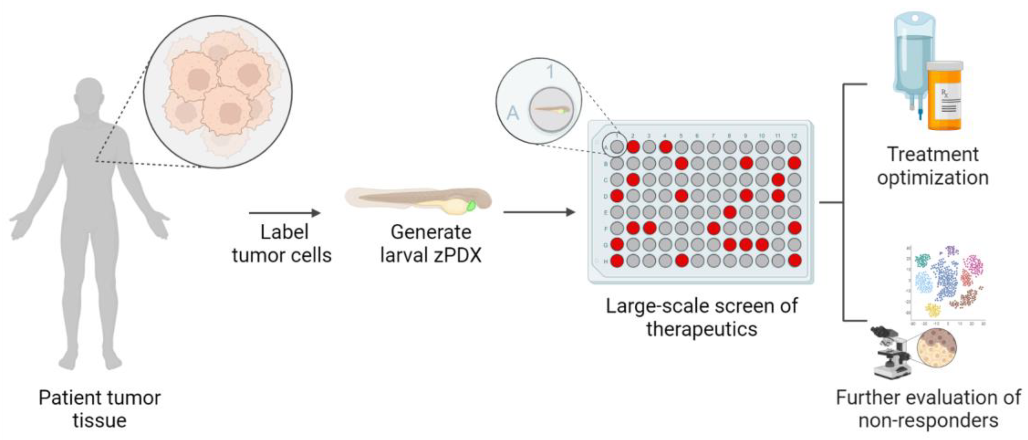
Zebrafish also provide opportunities to dissect intra-tumoral heterogeneity in ways that are not possible in mouse models. Heterogeneity within a tumor arises from the cellular subgroups with different genomic and phenotypic characteristics, which can ultimately result in therapeutic resistance and treatment failure [136]. Larval zPDX provides an opportunity to break apart heterogeneity into individual animals, in that 100–800 patient cells can be transplanted per animal, and hundreds of larvae can be transplanted with a patient’s tumor [92]. The heterogeneous tumor would now be subdivided into smaller parts, and these zPDX avatars can rapidly test therapeutic regimens and evaluate treatment responses. Some zPDX may completely respond to therapy, and cancer cells will be eliminated. Other zPDX may not respond, depending on which sub-clones are in the xenograft (Figure 3). Based on individual drug responses, combinations of therapies could be designed to be the most effective at eliminating all sub-clones within a heterogeneous tumor. Since tumor cells will be labeled before xenograft with a fluorescent dye, the resistant clones can be harvested and analyzed using sequencing approaches to uncover mechanisms of resistance that could inform second rounds of drug testing. This type of assay has the potential to provide a snapshot of the patient’s tumor landscape in days [65], compared to mouse PDX models that require months [17,137]. The large number of animals that can be involved in these studies provides statistical power, and the single-cell resolution that zebrafish larvae provide in imaging approaches may allow for the identification of previously undetectable drug-resistant sub-clones [66,73,138].
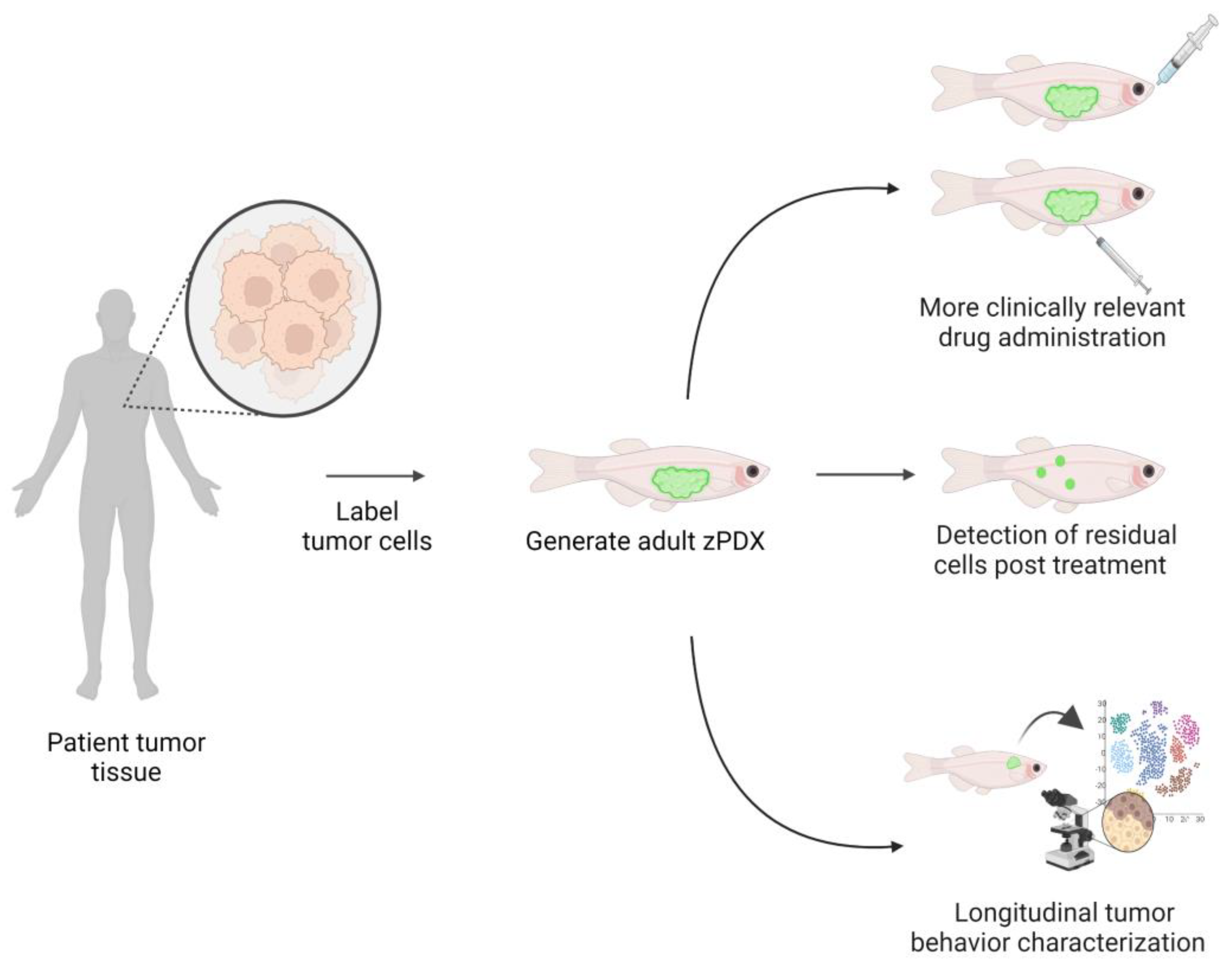
As a complement to larval zPDX, an immunodeficient Casper strain prkdc−/−, il2rga−/− zebrafish PDX model will allow more time for longitudinal evaluation of sub-clonal behavior, as engraftment is established for more than 28 days and up to 60 days. These animals require a transplant of 105–106 cells per fish, so each animal will have a heterogenous tumor that mimics the patient’s sample [64]. The optical clarity of the pigment-free Casper zebrafish would allow for the characterization of tumor phenotypes pre-treatment and during treatment in live animals. Non-responsive cells can also be quickly assessed and harvested for tumor profiling. In particular, this model could be helpful in identifying clones that drive relapse formation, potentially revealing individualized information about the relapsed tumor’s genetic and molecular profiles that would support later treatment decisions (Figure 4).

9. Conclusions

The reduced timeframe for the generation of zPDX avatars, their large-scale potential in drug screening and reduced use cost compared to mouse models are all features that make zPDX an increasingly popular and attractive model for precision cancer medicine (Figure 2). Larval zPDX is now proven to be a clinically relevant animal model that enables drug testing in 96-well plates while maintaining the complexity of a whole organism with physiological functions [105,113,115,139,140,141,142]. Adult and larval zPDX have the potential to capture intra-tumoral heterogeneity and to provide valuable information on the genetic, molecular and behavioral profiles of the sub-clones driving treatment resistance in patients (Figure 3 and Figure 4).
Nonetheless, each cancer avatar model is associated with drawbacks, and no one model can completely re-create human disease. All types of PDXs, including zPDX, should be carefully evaluated for their fitness to answer specific questions relevant to the patient’s tumor, including heterogeneity and sub-clone response to therapy administration. Ultimately, different models will play their own important roles across the precision medicine pipeline. For instance, larval zPDX is time-efficient in terms of guiding first-line treatment decisions, while adult zPDX, mouse PDX and organoid models could be reserved to investigate the post-treatment changes in tumor behavior and residual disease markers. Looking forward, some work is required to move zebrafish avatars closer to the clinical setting. Larval and adult zPDX methods should become standardized across laboratories so that results from avatar models are robust and reproducible. More comparisons between zPDX and mPDX models will show how these models both overlap and complement each other. Finally, more extensive co-clinical studies are needed that encompass different cancer types to compare zPDX avatar data to patient treatment responses. zPDX and matched clinical data will be essential in determining the predictiveness of zebrafish avatars to patient outcomes and the ultimate suitability of this promising model to precision cancer medicine pipelines.

10. Methods

Search Strategy: A comprehensive literature search was conducted using various databases such as PubMed and Google Scholar. The search was limited to articles published between 2000 and 2022. The following keywords were used in the search: “zebrafish”, “patient derived xenografts”, “intra-tumor heterogeneity”, “PDX”, “cancer avatars” and “precision medicine”.
Inclusion Criteria: Articles were included in the review if they met the following criteria: (1) They were published in peer-reviewed journals; (2) they were related to the topics of tumor heterogeneity, PDX models, the use of zebrafish in cancer research and drug screening; (3) they were published in English; and (4) they were published between 2000 and 2022.
Exclusion Criteria: Articles were excluded from the review if they did not meet the inclusion criteria.
Data Analysis: The data was analyzed by reading and summarizing the main findings of each included article and grouping them into themes or categories related to the tumor heterogeneity, existing PDX models and the application of zebrafish in biomedical research and as a cancer avatar model.

Author Contributions

Writing—original draft preparation, M.A.A.-H.; writing—review and editing, J.S.B. and A.R.; visualization, M.A.A.-H., L.T.T. and A.R.-M.; funding acquisition, J.S.B. and A.R. All authors have read and agreed to the published version of the manuscript.

Funding

This project was funded by the National Cancer Institute, R37CA227656 (J.S.B.); The ChadTough Defeat DIPG Foundation (J.S.B.); The Kentucky Pediatric Cancer Research Trust Fund (J.S.B); the National Center for Research Resources and the National Center for Advancing Translational Sciences, National Institutes of Health, UL1TR003107 to A.R. and UL1TR001998 to J.S.B. Stipend support for L.T.T. and A.R.M. was provided by the University of Kentucky’s Appalachian Career Training in Oncology (ACTION) Program, NCI R25CA221765 and the American Cancer Society Institutional Research Grant Diversity in Cancer Research Internship Supplement, IRG-19-140-31, respectively.

Institutional Review Board Statement

Not applicable.

Informed Consent Statement

Not applicable.

Data Availability Statement

Not applicable.

Conflicts of Interest

The authors declare no conflict of interest.

Abbreviations

5-FU Fluorouracil
ADME Absorption, distribution, metabolism and excretion
BMPBone morphogenetic protein
CEACarcinoembryonic antigen
CHTCaudal hematopoietic tissues
CLSMConfocal Laser Scanning Microscope
CMLChronic Myeloid Leukemia
CNNConvolutional Neural Network
COPASComplex Object Parametric Analysis and Sorting
CRComplete Response
CRCColorectal cancer
CRLMColorectal Liver Metastases
CROsContract Research Organizations
CYPHuman cytochrome P450
DFSDisease-free survival
dpfDays post fertilization
ECF5-Fluorouracil + Cisplatin + Epirubicin
EREstrogen receptor
FGF Fibroblast growth factor
FLOTFluorouracil, leucovorin, oxaliplatin and docetaxel
FOLFIRI5-Fluorouracil + Lederfolin + Irinotecan
FOLFOX5-Fluorouracil + Lederfolin + Oxaliplatin
FOLFOXIRI5-Fluorouracil + Folinic acid + Oxaliplatin + Irinotecan
FUCCI4Fluorescent, ubiquitination-based cell cycle indicator
GBMGlioblastoma multiforme
GCGastric cancer
GEMGemcitabine
GEM/nab-PGemcitabine + nab-Paclitaxel
GEMOXGemcitabine + Oxaliplatin
GFPGreen Fluorescent Protein
GyGray
jak3Janus kinase 3
LPLarge Particle
mPDXmouse patient-derived xenografts
MRMinor Response
MRIMagnetic resonance imaging
NCI MATCHNational Cancer Institute Molecular Analysis for Therapy Choice
NSCLCNon-Small Cell Lung Cancer
PCAPrincipal component analysis
PDProgressive Disease
PDACPancreatic ductal adenocarcinoma
PDOPatient-derived organoids
PDXPatient-derived xenografts
PFSProgression-free survival
PRProgesterone receptor
PRPartial Response
R-CNNRegions with Convolutional Neural Network
RECISTResponse Evaluation Criteria In Solid Tumors
RTARelative tumor area
SDStable Disease
SPIMSelective Plane Illumination Microscopy
TNBCTriple-Negative Breast Cancer
VASTVertebrate Automated Screening Technology
VEGFVascular endothelial growth factor
WHO World Health Organization
zPDX Zebrafish patient-derived xenografts

References

  1. Hayes, D.F.; Schott, A.F. Personalized medicine: Genomics trials in oncology. Trans. Am. Clin. Climatol. Assoc. 2015, 126, 133. [Google Scholar] [PubMed]
  2. Yan, L.; Zhang, W. Precision medicine becomes reality—Tumor type-agnostic therapy. Cancer Commun. 2018, 38, 6. [Google Scholar] [CrossRef] [PubMed] [Green Version]
  3. Friedman, A.A.; Letai, A.; Fisher, D.E.; Flaherty, K.T. Precision medicine for cancer with next-generation functional diagnostics. Nat. Rev. Cancer 2015, 15, 747–756. [Google Scholar] [CrossRef] [PubMed] [Green Version]
  4. Letai, A.; Bhola, P.; Welm, A.L. Functional precision oncology: Testing tumors with drugs to identify vulnerabilities and novel combinations. Cancer Cell 2021, 40, 26–35. [Google Scholar] [CrossRef]
  5. Flaherty, K.T.; Gray, R.; Chen, A.; Li, S.; Patton, D.; Hamilton, S.R.; Williams, P.M.; Mitchell, E.P.; Iafrate, A.J.; Sklar, J. The molecular analysis for therapy choice (NCI-MATCH) trial: Lessons for genomic trial design. JNCI J. Natl. Cancer Inst. 2020, 112, 1021–1029. [Google Scholar] [CrossRef] [Green Version]
  6. Flaherty, K.T.; Gray, R.J.; Chen, A.P.; Li, S.; McShane, L.M.; Patton, D.; Hamilton, S.R.; Williams, P.M.; Iafrate, A.J.; Sklar, J. Molecular landscape and actionable alterations in a genomically guided cancer clinical trial: National Cancer Institute Molecular Analysis for Therapy Choice (NCI-MATCH). J. Clin. Oncol. 2020, 38, 3883. [Google Scholar] [CrossRef]
  7. Jamal-Hanjani, M.; Quezada, S.A.; Larkin, J.; Swanton, C. Translational implications of tumor heterogeneity. Clin. Cancer Res. 2015, 21, 1258–1266. [Google Scholar] [CrossRef] [Green Version]
  8. Stanta, G.; Bonin, S. Overview on clinical relevance of intra-tumor heterogeneity. Front. Med. 2018, 5, 85. [Google Scholar] [CrossRef] [Green Version]
  9. Zellmer, V.R.; Zhang, S. Evolving concepts of tumor heterogeneity. Cell Biosci. 2014, 4, 69. [Google Scholar] [CrossRef] [Green Version]
  10. Dienstmann, R.; Vermeulen, L.; Guinney, J.; Kopetz, S.; Tejpar, S.; Tabernero, J. Consensus molecular subtypes and the evolution of precision medicine in colorectal cancer. Nat. Rev. Cancer 2017, 17, 79–92. [Google Scholar] [CrossRef]
  11. Hidalgo, M.; Amant, F.; Biankin, A.V.; Budinská, E.; Byrne, A.T.; Caldas, C.; Clarke, R.B.; de Jong, S.; Jonkers, J.; Mælandsmo, G.M. Patient-derived xenograft models: An emerging platform for translational cancer research. Cancer Discov. 2014, 4, 998–1013. [Google Scholar] [CrossRef] [Green Version]
  12. Malaney, P.; Nicosia, S.V.; Davé, V. One mouse, one patient paradigm: New avatars of personalized cancer therapy. Cancer Lett. 2014, 344, 1–12. [Google Scholar] [CrossRef] [Green Version]
  13. DeRose, Y.S.; Wang, G.; Lin, Y.-C.; Bernard, P.S.; Buys, S.S.; Ebbert, M.T.; Factor, R.; Matsen, C.; Milash, B.A.; Nelson, E. Tumor grafts derived from women with breast cancer authentically reflect tumor pathology, growth, metastasis and disease outcomes. Nat. Med. 2011, 17, 1514–1520. [Google Scholar] [CrossRef]
  14. Topp, M.D.; Hartley, L.; Cook, M.; Heong, V.; Boehm, E.; McShane, L.; Pyman, J.; McNally, O.; Ananda, S.; Harrell, M. Molecular correlates of platinum response in human high-grade serous ovarian cancer patient-derived xenografts. Mol. Oncol. 2014, 8, 656–668. [Google Scholar] [CrossRef]
  15. Davies, A.; Hidalgo, M.; Stebbing, J.; Ciznadija, D.; Katz, A.; Sidransky, D. Mouse clinical trials of pancreatic cancer: Integration of PDX models with genomics to improve patient outcomes to chemotherapeutics. Ann. Oncol. 2016, 27, vi527. [Google Scholar] [CrossRef]
  16. Na, D.; Chae, J.; Cho, S.-Y.; Kang, W.; Lee, A.; Min, S.; Kang, J.; Kim, M.J.; Choi, J.; Lee, W. Predictive biomarkers for 5-fluorouracil and oxaliplatin-based chemotherapy in gastric cancers via profiling of patient-derived xenografts. Nat. Commun. 2021, 12, 4840. [Google Scholar] [CrossRef]
  17. Clohessy, J.G.; Pandolfi, P.P. Mouse hospital and co-clinical trial project—From bench to bedside. Nat. Rev. Clin. Oncol. 2015, 12, 491–498. [Google Scholar] [CrossRef]
  18. Costa, B.; Estrada, M.F.; Mendes, R.V.; Fior, R. Zebrafish avatars towards personalized medicine—A comparative review between avatar models. Cells 2020, 9, 293. [Google Scholar] [CrossRef] [Green Version]
  19. Jung, J.; Seol, H.S.; Chang, S. The generation and application of patient-derived xenograft model for cancer research. Cancer Res. Treat. Off. J. Korean Cancer Assoc. 2018, 50, 1–10. [Google Scholar] [CrossRef] [Green Version]
  20. Fisher, R.; Pusztai, L.; Swanton, C. Cancer heterogeneity: Implications for targeted therapeutics. Br. J. Cancer 2013, 108, 479–485. [Google Scholar] [CrossRef] [Green Version]
  21. Liu, J.; Dang, H.; Wang, X.W. The significance of intertumor and intratumor heterogeneity in liver cancer. Exp. Mol. Med. 2018, 50, e416. [Google Scholar] [CrossRef] [PubMed]
  22. Saußele, S.; Krauß, M.-P.; Hehlmann, R.; Lauseker, M.; Proetel, U.; Kalmanti, L.; Hanfstein, B.; Fabarius, A.; Kraemer, D.; Berdel, W.E. Impact of comorbidities on overall survival in patients with chronic myeloid leukemia: Results of the randomized CML study IV. Blood J. Am. Soc. Hematol. 2015, 126, 42–49. [Google Scholar] [CrossRef] [PubMed] [Green Version]
  23. Larson, R.A.; Druker, B.J.; Guilhot, F.; O’Brien, S.G.; Riviere, G.J.; Krahnke, T.; Gathmann, I.; Wang, Y. Imatinib pharmacokinetics and its correlation with response and safety in chronic-phase chronic myeloid leukemia: A subanalysis of the IRIS study. Blood J. Am. Soc. Hematol. 2008, 111, 4022–4028. [Google Scholar] [CrossRef] [PubMed] [Green Version]
  24. Hughes, T.P.; Hochhaus, A.; Branford, S.; Müller, M.C.; Kaeda, J.S.; Foroni, L.; Druker, B.J.; Guilhot, F.; Larson, R.A.; O’Brien, S.G. Long-term prognostic significance of early molecular response to imatinib in newly diagnosed chronic myeloid leukemia: An analysis from the International Randomized Study of Interferon and STI571 (IRIS). Blood J. Am. Soc. Hematol. 2010, 116, 3758–3765. [Google Scholar] [CrossRef] [PubMed] [Green Version]
  25. Slamon, D.J.; Leyland-Jones, B.; Shak, S.; Fuchs, H.; Paton, V.; Bajamonde, A.; Fleming, T.; Eiermann, W.; Wolter, J.; Pegram, M. Use of chemotherapy plus a monoclonal antibody against HER2 for metastatic breast cancer that overexpresses HER2. N. Engl. J. Med. 2001, 344, 783–792. [Google Scholar] [CrossRef]
  26. Wolff, A.C.; Hammond, M.E.H.; Hicks, D.G.; Dowsett, M.; McShane, L.M.; Allison, K.H.; Allred, D.C.; Bartlett, J.M.; Bilous, M.; Fitzgibbons, P. Recommendations for human epidermal growth factor receptor 2 testing in breast cancer: American Society of Clinical Oncology/College of American Pathologists clinical practice guideline update. Arch. Pathol. Lab. Med. 2014, 138, 241–256. [Google Scholar] [CrossRef] [PubMed] [Green Version]
  27. Zardavas, D.; Irrthum, A.; Swanton, C.; Piccart, M. Clinical management of breast cancer heterogeneity. Nat. Rev. Clin. Oncol. 2015, 12, 381–394. [Google Scholar] [CrossRef]
  28. Stephens, P.J.; Tarpey, P.S.; Davies, H.; Van Loo, P.; Greenman, C.; Wedge, D.C.; Nik-Zainal, S.; Martin, S.; Varela, I.; Bignell, G.R. The landscape of cancer genes and mutational processes in breast cancer. Nature 2012, 486, 400–404. [Google Scholar] [CrossRef] [Green Version]
  29. Perou, C.M.; Sørlie, T.; Eisen, M.B.; Van De Rijn, M.; Jeffrey, S.S.; Rees, C.A.; Pollack, J.R.; Ross, D.T.; Johnsen, H.; Akslen, L.A. Molecular portraits of human breast tumours. Nature 2000, 406, 747–752. [Google Scholar] [CrossRef] [Green Version]
  30. Turajlic, S.; Sottoriva, A.; Graham, T.; Swanton, C. Resolving genetic heterogeneity in cancer. Nat. Rev. Genet. 2019, 20, 404–416. [Google Scholar] [CrossRef]
  31. Vaupel, P. Oxygen supply to malignant tumors. In Tumor Blood Circulation: Angiogenesis, Vascular Morphology and Blood Flow of Experimental and Human Tumors; CRC Press: Boca Raton, FL, USA, 2020; pp. 143–168. [Google Scholar]
  32. Lyssiotis, C.A.; Kimmelman, A.C. Metabolic interactions in the tumor microenvironment. Trends Cell Biol. 2017, 27, 863–875. [Google Scholar] [CrossRef] [Green Version]
  33. Zamarron, B.F.; Chen, W. Dual roles of immune cells and their factors in cancer development and progression. Int. J. Biol. Sci. 2011, 7, 651. [Google Scholar] [CrossRef]
  34. Bozic, I.; Antal, T.; Ohtsuki, H.; Carter, H.; Kim, D.; Chen, S.; Karchin, R.; Kinzler, K.W.; Vogelstein, B.; Nowak, M.A. Accumulation of driver and passenger mutations during tumor progression. Proc. Natl. Acad. Sci. USA 2010, 107, 18545–18550. [Google Scholar] [CrossRef] [Green Version]
  35. Vitale, I.; Shema, E.; Loi, S.; Galluzzi, L. Intratumoral heterogeneity in cancer progression and response to immunotherapy. Nat. Med. 2021, 27, 212–224. [Google Scholar] [CrossRef]
  36. Prasetyanti, P.R.; Medema, J.P. Intra-tumor heterogeneity from a cancer stem cell perspective. Mol. Cancer 2017, 16, 1–9. [Google Scholar] [CrossRef] [Green Version]
  37. Bell, C.C.; Gilan, O. Principles and mechanisms of non-genetic resistance in cancer. Br. J. Cancer 2020, 122, 465–472. [Google Scholar] [CrossRef]
  38. Longo, D.L. Tumor heterogeneity and personalized medicine. N. Engl. J. Med. 2012, 366, 956–957. [Google Scholar] [CrossRef] [Green Version]
  39. Gerlinger, M.; Rowan, A.J.; Horswell, S.; Larkin, J.; Endesfelder, D.; Gronroos, E.; Martinez, P.; Matthews, N.; Stewart, A.; Tarpey, P. Intratumor heterogeneity and branched evolution revealed by multiregion sequencing. N. Engl. J. Med. 2012, 366, 883–892. [Google Scholar] [CrossRef] [Green Version]
  40. Hua, X.; Zhao, W.; Pesatori, A.C.; Consonni, D.; Caporaso, N.E.; Zhang, T.; Zhu, B.; Wang, M.; Jones, K.; Hicks, B. Genetic and epigenetic intratumor heterogeneity impacts prognosis of lung adenocarcinoma. Nat. Commun. 2020, 11, 2459. [Google Scholar] [CrossRef]
  41. Magill, S.T.; Vasudevan, H.N.; Seo, K.; Villanueva-Meyer, J.E.; Choudhury, A.; John Liu, S.; Pekmezci, M.; Findakly, S.; Hilz, S.; Lastella, S. Multiplatform genomic profiling and magnetic resonance imaging identify mechanisms underlying intratumor heterogeneity in meningioma. Nat. Commun. 2020, 11, 1–15. [Google Scholar] [CrossRef]
  42. Rye, I.H.; Trinh, A.; Sætersdal, A.B.; Nebdal, D.; Lingjærde, O.C.; Almendro, V.; Polyak, K.; Børresen-Dale, A.L.; Helland, Å.; Markowetz, F. Intratumor heterogeneity defines treatment-resistant HER 2+ breast tumors. Mol. Oncol. 2018, 12, 1838–1855. [Google Scholar] [CrossRef] [PubMed] [Green Version]
  43. Brady, L.; Kriner, M.; Coleman, I.; Morrissey, C.; Roudier, M.; True, L.D.; Gulati, R.; Plymate, S.R.; Zhou, Z.; Birditt, B. Inter-and intra-tumor heterogeneity of metastatic prostate cancer determined by digital spatial gene expression profiling. Nat. Commun. 2021, 12, 1426. [Google Scholar] [CrossRef] [PubMed]
  44. Peng, J.; Sun, B.-F.; Chen, C.-Y.; Zhou, J.-Y.; Chen, Y.-S.; Chen, H.; Liu, L.; Huang, D.; Jiang, J.; Cui, G.-S. Single-cell RNA-seq highlights intra-tumoral heterogeneity and malignant progression in pancreatic ductal adenocarcinoma. Cell Res. 2019, 29, 725–738. [Google Scholar] [CrossRef]
  45. Jacob, F.; Salinas, R.D.; Zhang, D.Y.; Nguyen, P.T.; Schnoll, J.G.; Wong, S.Z.H.; Thokala, R.; Sheikh, S.; Saxena, D.; Prokop, S. A patient-derived glioblastoma organoid model and biobank recapitulates inter-and intra-tumoral heterogeneity. Cell 2020, 180, 188–204.e122. [Google Scholar] [CrossRef] [PubMed]
  46. Saito, T.; Niida, A.; Uchi, R.; Hirata, H.; Komatsu, H.; Sakimura, S.; Hayashi, S.; Nambara, S.; Kuroda, Y.; Ito, S. A temporal shift of the evolutionary principle shaping intratumor heterogeneity in colorectal cancer. Nat. Commun. 2018, 9, 1–11. [Google Scholar] [CrossRef] [Green Version]
  47. Ho, D.W.-H.; Tsui, Y.-M.; Sze, K.M.-F.; Chan, L.-K.; Cheung, T.-T.; Lee, E.; Sham, P.-C.; Tsui, S.K.-W.; Lee, T.K.-W.; Ng, I.O.-L. Single-cell transcriptomics reveals the landscape of intra-tumoral heterogeneity and stemness-related subpopulations in liver cancer. Cancer Lett. 2019, 459, 176–185. [Google Scholar] [CrossRef]
  48. Araf, S.; Wang, J.; Korfi, K.; Pangault, C.; Kotsiou, E.; Rio-Machin, A.; Rahim, T.; Heward, J.; Clear, A.; Iqbal, S. Genomic profiling reveals spatial intra-tumor heterogeneity in follicular lymphoma. Leukemia 2018, 32, 1261–1265. [Google Scholar] [CrossRef] [Green Version]
  49. Raynaud, F.; Mina, M.; Tavernari, D.; Ciriello, G. Pan-cancer inference of intra-tumor heterogeneity reveals associations with different forms of genomic instability. PLoS Genet. 2018, 14, e1007669. [Google Scholar] [CrossRef] [Green Version]
  50. Dentro, S.C.; Leshchiner, I.; Haase, K.; Tarabichi, M.; Wintersinger, J.; Deshwar, A.G.; Yu, K.; Rubanova, Y.; Macintyre, G.; Demeulemeester, J. Characterizing genetic intra-tumor heterogeneity across 2658 human cancer genomes. Cell 2021, 184, 2239–2254.e39. [Google Scholar] [CrossRef]
  51. Gould, S.E.; Junttila, M.R.; de Sauvage, F.J. Translational value of mouse models in oncology drug development. Nat. Med. 2015, 21, 431–439. [Google Scholar] [CrossRef]
  52. Cho, S.-Y. Patient-derived xenografts as compatible models for precision oncology. Lab. Anim. Res. 2020, 36, 14. [Google Scholar] [CrossRef]
  53. Park, H.; Kim, Y.; Sul, J.W.; Jeong, I.G.; Yi, H.J.; Ahn, J.B.; Kang, J.S.; Yun, J.; Hwang, J.J.; Kim, C.S. Synergistic anti-cancer efficacy of MEK inhibition and dual PI3K/mTOR inhibition in castration-resistant prostate cancer. Prostate 2015, 75, 1747–1759. [Google Scholar] [CrossRef]
  54. Zhang, R.; Jin, S.; Rao, W.; Song, F.; Yin, Q.; Wang, Y.; Wang, L.; Xi, Y.; Zhang, X.; Wang, M. OVA12, a novel tumor antigen, promotes cancer cell growth and inhibits 5-fluorouracil-induced apoptosis. Cancer Lett. 2015, 357, 141–151. [Google Scholar] [CrossRef]
  55. Garralda, E.; Paz, K.; López-Casas, P.P.; Jones, S.; Katz, A.; Kann, L.M.; López-Rios, F.; Sarno, F.; Al-Shahrour, F.; Vasquez, D. Integrated Next-Generation Sequencing and Avatar Mouse Models for Personalized Cancer TreatmentPersonalized Cancer Treatment using Genomics and Avatar Models. Clin. Cancer Res. 2014, 20, 2476–2484. [Google Scholar] [CrossRef] [Green Version]
  56. Öhlund, D.; Handly-Santana, A.; Biffi, G.; Elyada, E.; Almeida, A.S.; Ponz-Sarvise, M.; Corbo, V.; Oni, T.E.; Hearn, S.A.; Lee, E.J. Distinct populations of inflammatory fibroblasts and myofibroblasts in pancreatic cancer. J. Exp. Med. 2017, 214, 579–596. [Google Scholar] [CrossRef] [Green Version]
  57. Vlachogiannis, G.; Hedayat, S.; Vatsiou, A.; Jamin, Y.; Fernández-Mateos, J.; Khan, K.; Lampis, A.; Eason, K.; Huntingford, I.; Burke, R. Patient-derived organoids model treatment response of metastatic gastrointestinal cancers. Science 2018, 359, 920–926. [Google Scholar] [CrossRef] [Green Version]
  58. Servant, R.; Garioni, M.; Vlajnic, T.; Blind, M.; Pueschel, H.; Müller, D.C.; Zellweger, T.; Templeton, A.J.; Garofoli, A.; Maletti, S. Prostate cancer patient-derived organoids: Detailed outcome from a prospective cohort of 81 clinical specimens. J. Pathol. 2021, 254, 543–555. [Google Scholar] [CrossRef]
  59. Green, S.; Dam, M.S.; Svendsen, M.N. Mouse avatars of human cancers: The temporality of translation in precision oncology. Hist. Philos. Life Sci. 2021, 43, 1–22. [Google Scholar] [CrossRef]
  60. Cone, E.B.; Marchese, M.; Paciotti, M.; Nguyen, D.-D.; Nabi, J.; Cole, A.P.; Molina, G.; Molina, R.L.; Minami, C.A.; Mucci, L.A. Assessment of time-to-treatment initiation and survival in a cohort of patients with common cancers. JAMA Netw. Open 2020, 3, e2030072. [Google Scholar] [CrossRef]
  61. Lin, D.; Wyatt, A.W.; Xue, H.; Wang, Y.; Dong, X.; Haegert, A.; Wu, R.; Brahmbhatt, S.; Mo, F.; Jong, L. High Fidelity Patient-Derived Xenografts for Accelerating Prostate Cancer Discovery and Drug Development Next-Generation Models of Prostate Cancer. Cancer Res. 2014, 74, 1272–1283. [Google Scholar] [CrossRef] [Green Version]
  62. Mathew, R.K.; Rutka, J.T. Diffuse intrinsic pontine glioma: Clinical features, molecular genetics, and novel targeted therapeutics. J. Korean Neurosurg. Soc. 2018, 61, 343–351. [Google Scholar] [CrossRef] [PubMed] [Green Version]
  63. Tang, Q.; Moore, J.C.; Ignatius, M.S.; Tenente, I.M.; Hayes, M.N.; Garcia, E.G.; Torres Yordán, N.; Bourque, C.; He, S.; Blackburn, J.S. Imaging tumour cell heterogeneity following cell transplantation into optically clear immune-deficient zebrafish. Nat. Commun. 2016, 7, 10358. [Google Scholar] [CrossRef] [PubMed] [Green Version]
  64. Yan, C.; Brunson, D.C.; Tang, Q.; Do, D.; Iftimia, N.A.; Moore, J.C.; Hayes, M.N.; Welker, A.M.; Garcia, E.G.; Dubash, T.D. Visualizing engrafted human cancer and therapy responses in immunodeficient zebrafish. Cell 2019, 177, 1903–1914.e14. [Google Scholar] [CrossRef] [PubMed]
  65. Martinez-Lopez, M.; Póvoa, V.; Fior, R. Generation of zebrafish larval xenografts and tumor behavior analysis. JoVE J. Vis. Exp. 2021, 172, e62373. [Google Scholar]
  66. Yan, C.; Do, D.; Yang, Q.; Brunson, D.C.; Rawls, J.F.; Langenau, D.M. Single-cell imaging of human cancer xenografts using adult immunodeficient zebrafish. Nat. Protoc. 2020, 15, 3105–3128. [Google Scholar] [CrossRef]
  67. Saland, E.; Boutzen, H.; Castellano, R.; Pouyet, L.; Griessinger, E.; Larrue, C.; De Toni, F.; Scotland, S.; David, M.; Danet-Desnoyers, G. A robust and rapid xenograft model to assess efficacy of chemotherapeutic agents for human acute myeloid leukemia. Blood Cancer J. 2015, 5, e297. [Google Scholar] [CrossRef] [Green Version]
  68. Gamble, J.T.; Elson, D.J.; Greenwood, J.A.; Tanguay, R.L.; Kolluri, S.K. The zebrafish xenograft models for investigating cancer and cancer therapeutics. Biology 2021, 10, 252. [Google Scholar] [CrossRef]
  69. Mendonça-Gomes, J.M.; Valverde, T.M.; da Mata Martins, T.M.; Charlie-Silva, I.; Padovani, B.N.; Fénero, C.M.; da Silva, E.M.; Domingues, R.Z.; Melo-Hoyos, D.C.; Corrêa-Junior, J.D. Long-term dexamethasone treatment increases the engraftment efficiency of human breast cancer cells in adult zebrafish. Fish Shellfish. Immunol. Rep. 2021, 2, 100007. [Google Scholar] [CrossRef]
  70. Traver, D.; Winzeler, A.; Stern, H.M.; Mayhall, E.A.; Langenau, D.M.; Kutok, J.L.; Look, A.T.; Zon, L.I. Effects of lethal irradiation in zebrafish and rescue by hematopoietic cell transplantation. Blood 2004, 104, 1298–1305. [Google Scholar] [CrossRef] [Green Version]
  71. Stoletov, K.; Montel, V.; Lester, R.D.; Gonias, S.L.; Klemke, R. High-resolution imaging of the dynamic tumor cell–vascular interface in transparent zebrafish. Proc. Natl. Acad. Sci. USA 2007, 104, 17406–17411. [Google Scholar] [CrossRef] [Green Version]
  72. Moore, J.C.; Tang, Q.; Yordán, N.T.; Moore, F.E.; Garcia, E.G.; Lobbardi, R.; Ramakrishnan, A.; Marvin, D.L.; Anselmo, A.; Sadreyev, R.I. Single-cell imaging of normal and malignant cell engraftment into optically clear prkdc-null SCID zebrafish. J. Exp. Med. 2016, 213, 2575–2589. [Google Scholar] [CrossRef] [Green Version]
  73. Fior, R.; Póvoa, V.; Mendes, R.V.; Carvalho, T.; Gomes, A.; Figueiredo, N.; Ferreira, M.G. Single-cell functional and chemosensitive profiling of combinatorial colorectal therapy in zebrafish xenografts. Proc. Natl. Acad. Sci. USA 2017, 114, E8234–E8243. [Google Scholar] [CrossRef] [Green Version]
  74. Costa, B.; Estrada, M.F.; Barroso, M.T.; Fior, R. Zebrafish Patient-Derived Avatars from Digestive Cancers for Anti-cancer Therapy Screening. Curr. Protoc. 2022, 2, e415. [Google Scholar] [CrossRef]
  75. Weintraub, A. All eyes on zebrafish. Lab. Anim. 2017, 46, 323–326. [Google Scholar] [CrossRef] [Green Version]
  76. Aparicio, S.; Hidalgo, M.; Kung, A.L. Examining the utility of patient-derived xenograft mouse models. Nat. Rev. Cancer 2015, 15, 311–316. [Google Scholar] [CrossRef]
  77. Dockins, S.R.; Blackburn, J.S.; Haney, M.G.; Wilson, B. Optimization of human cancer cell xenografts into zebrafish larvae for high-throughput drug screening. Cancer Res. 2019, 79, 3697. [Google Scholar]
  78. Spaink, H.P.; Cui, C.; Wiweger, M.I.; Jansen, H.J.; Veneman, W.J.; Marín-Juez, R.; de Sonneville, J.; Ordas, A.; Torraca, V.; van der Ent, W. Robotic injection of zebrafish embryos for high-throughput screening in disease models. Methods 2013, 62, 246–254. [Google Scholar] [CrossRef]
  79. Pulak, R. Tools for automating the imaging of zebrafish larvae. Methods 2016, 96, 118–126. [Google Scholar] [CrossRef]
  80. Ghotra, V.P.; He, S.; De Bont, H.; van Der Ent, W.; Spaink, H.P.; van De Water, B.; Snaar-Jagalska, B.E.; Danen, E.H. Automated whole animal bio-imaging assay for human cancer dissemination. PLoS ONE 2012, 7, e31281. [Google Scholar] [CrossRef]
  81. Asokan, N.; Daetwyler, S.; Bernas, S.N.; Schmied, C.; Vogler, S.; Lambert, K.; Wobus, M.; Wermke, M.; Kempermann, G.; Huisken, J. Long-term in vivo imaging reveals tumor-specific dissemination and captures host tumor interaction in zebrafish xenografts. Sci. Rep. 2020, 10, 13254. [Google Scholar]
  82. Haney, M.G.; Moore, L.H.; Blackburn, J.S. Drug screening of primary patient derived tumor xenografts in zebrafish. JoVE J. Vis. Exp. 2020, e60996. [Google Scholar]
  83. Mercatali, L.; La Manna, F.; Groenewoud, A.; Casadei, R.; Recine, F.; Miserocchi, G.; Pieri, F.; Liverani, C.; Bongiovanni, A.; Spadazzi, C. Development of a patient-derived xenograft (PDX) of breast cancer bone metastasis in a zebrafish model. Int. J. Mol. Sci. 2016, 17, 1375. [Google Scholar] [CrossRef] [PubMed]
  84. Almstedt, E.; Rosén, E.; Gloger, M.; Stockgard, R.; Hekmati, N.; Koltowska, K.; Krona, C.; Nelander, S. Real-time evaluation of glioblastoma growth in patient-specific zebrafish xenografts. Neuro-Oncology 2022, 24, 726–738. [Google Scholar] [CrossRef] [PubMed]
  85. Phase I Study of Olaparib and Temozolomide for Ewings Sarcoma or Rhabdoomyosarcoma. Available online: https://clinicaltrials.gov/ct2/show/record/NCT01858168 (accessed on 15 December 2022).
  86. Albuquerque, C.; Vanneschi, L.; Henriques, R.; Castelli, M.; Póvoa, V.; Fior, R.; Papanikolaou, N. Object detection for automatic cancer cell counting in zebrafish xenografts. PLoS ONE 2021, 16, e0260609. [Google Scholar] [CrossRef]
  87. Zhang, B.; Shimada, Y.; Kuroyanagi, J.; Umemoto, N.; Nishimura, Y.; Tanaka, T. Quantitative phenotyping-based in vivo chemical screening in a zebrafish model of leukemia stem cell xenotransplantation. PLoS ONE 2014, 9, e85439. [Google Scholar] [CrossRef]
  88. Zhang, B.; Shimada, Y.; Kuroyanagi, J.; Nishimura, Y.; Umemoto, N.; Nomoto, T.; Shintou, T.; Miyazaki, T.; Tanaka, T. Zebrafish xenotransplantation model for cancer stem-like cell study and high-throughput screening of inhibitors. Tumor Biol. 2014, 35, 11861–11869. [Google Scholar] [CrossRef]
  89. Veneman, W.J.; Stockhammer, O.W.; De Boer, L.; Zaat, S.A.; Meijer, A.H.; Spaink, H.P. A zebrafish high throughput screening system used for Staphylococcus epidermidis infection marker discovery. BMC Genom. 2013, 14, 255. [Google Scholar] [CrossRef] [Green Version]
  90. Haldi, M.; Ton, C.; Seng, W.L.; McGrath, P. Human melanoma cells transplanted into zebrafish proliferate, migrate, produce melanin, form masses and stimulate angiogenesis in zebrafish. Angiogenesis 2006, 9, 139–151. [Google Scholar] [CrossRef]
  91. Nicoli, S.; Ribatti, D.; Cotelli, F.; Presta, M. Mammalian tumor xenografts induce neovascularization in zebrafish embryos. Cancer Res. 2007, 67, 2927–2931. [Google Scholar] [CrossRef] [Green Version]
  92. Wu, J.-Q.; Zhai, J.; Li, C.-Y.; Tan, A.-M.; Wei, P.; Shen, L.-Z.; He, M.-F. Patient-derived xenograft in zebrafish embryos: A new platform for translational research in gastric cancer. J. Exp. Clin. Cancer Res. 2017, 36, 160. [Google Scholar] [CrossRef] [Green Version]
  93. Ali, Z.; Vildevall, M.; Rodriguez, G.V.; Tandiono, D.; Vamvakaris, I.; Evangelou, G.; Lolas, G.; Syrigos, K.N.; Villanueva, A.; Wick, M. Zebrafish patient-derived xenograft models predict lymph node involvement and treatment outcome in non-small cell lung cancer. J. Exp. Clin. Cancer Res. 2022, 41, 58. [Google Scholar] [CrossRef]
  94. Usai, A.; Di Franco, G.; Piccardi, M.; Cateni, P.; Pollina, L.E.; Vivaldi, C.; Vasile, E.; Funel, N.; Palmeri, M.; Dente, L. Zebrafish patient-derived xenografts identify chemo-response in pancreatic ductal adenocarcinoma patients. Cancers 2021, 13, 4131. [Google Scholar] [CrossRef]
  95. Ai, X.; Ye, Z.; Xiao, C.; Zhong, J.; Lancman, J.J.; Chen, X.; Pan, X.; Yang, Y.; Zhou, L.; Wang, X. Clinically relevant orthotopic xenograft models of patient-derived glioblastoma in zebrafish. Dis. Model. Mech. 2022, 15, dmm049109. [Google Scholar] [CrossRef]
  96. Tulotta, C.; Stefanescu, C.; Beletkaia, E.; Bussmann, J.; Tarbashevich, K.; Schmidt, T.; Snaar-Jagalska, B.E. Inhibition of signaling between human CXCR4 and zebrafish ligands by the small molecule IT1t impairs the formation of triple-negative breast cancer early metastases in a zebrafish xenograft model. Dis. Model. Mech. 2016, 9, 141–153. [Google Scholar]
  97. Howe, K.; Clark, M.D.; Torroja, C.F.; Torrance, J.; Berthelot, C.; Muffato, M.; Collins, J.E.; Humphray, S.; McLaren, K.; Matthews, L. The zebrafish reference genome sequence and its relationship to the human genome. Nature 2013, 496, 498–503. [Google Scholar] [CrossRef] [Green Version]
  98. Stachura, D.L.; Reyes, J.R.; Bartunek, P.; Paw, B.H.; Zon, L.I.; Traver, D. Zebrafish kidney stromal cell lines support multilineage hematopoiesis. Blood J. Am. Soc. Hematol. 2009, 114, 279–289. [Google Scholar] [CrossRef] [Green Version]
  99. Rajan, V.; Melong, N.; Wong, W.H.; King, B.; Tong, R.S.; Mahajan, N.; Gaston, D.; Lund, T.; Rittenberg, D.; Dellaire, G. Humanized zebrafish enhance human hematopoietic stem cell survival and promote acute myeloid leukemia clonal diversity. Haematologica 2020, 105, 2391. [Google Scholar] [CrossRef] [Green Version]
  100. Pype, C.; Verbueken, E.; Saad, M.A.; Casteleyn, C.R.; Van Ginneken, C.J.; Knapen, D.; Van Cruchten, S.J. Incubation at 32.5 C and above causes malformations in the zebrafish embryo. Reprod. Toxicol. 2015, 56, 56–63. [Google Scholar] [CrossRef]
  101. Astin, J.; Keerthisinghe, P.; Du, L.; Sanderson, L.; Crosier, K.; Crosier, P.; Hall, C. Innate immune cells and bacterial infection in zebrafish. Methods Cell Biol. 2017, 138, 31–60. [Google Scholar]
  102. Cabezas-Sáinz, P.; Pensado-López, A.; Sáinz, B., Jr.; Sánchez, L. Modeling cancer using zebrafish xenografts: Drawbacks for mimicking the human microenvironment. Cells 2020, 9, 1978. [Google Scholar] [CrossRef]
  103. Lal, S.; La Du, J.; Tanguay, R.L.; Greenwood, J.A. Calpain 2 is required for the invasion of glioblastoma cells in the zebrafish brain microenvironment. J. Neurosci. Res. 2012, 90, 769–781. [Google Scholar] [CrossRef] [PubMed] [Green Version]
  104. Farin, A.; Suzuki, S.O.; Weiker, M.; Goldman, J.E.; Bruce, J.N.; Canoll, P. Transplanted glioma cells migrate and proliferate on host brain vasculature: A dynamic analysis. Glia 2006, 53, 799–808. [Google Scholar] [CrossRef] [PubMed]
  105. Rennekamp, A.J.; Peterson, R.T. 15 years of zebrafish chemical screening. Curr. Opin. Chem. Biol. 2015, 24, 58–70. [Google Scholar] [CrossRef] [PubMed] [Green Version]
  106. Zhu, J.J.; Xu, Y.Q.; He, J.H.; Yu, H.P.; Huang, C.J.; Gao, J.M.; Dong, Q.X.; Xuan, Y.X.; Li, C.Q. Human cardiotoxic drugs delivered by soaking and microinjection induce cardiovascular toxicity in zebrafish. J. Appl. Toxicol. 2014, 34, 139–148. [Google Scholar] [CrossRef] [PubMed]
  107. Sharma, A.; Saneja, A. Zebrafish as a powerful alternative model organism for pre-clinical investigation of nanomedicines. Drug Discov. Today 2022, 27, 1513–1522. [Google Scholar]
  108. de Souza Anselmo, C.; Sardela, V.F.; de Sousa, V.P.; Pereira, H.M.G. Zebrafish (Danio rerio): A valuable tool for predicting the metabolism of xenobiotics in humans? Comp. Biochem. Physiol. Part C Toxicol. Pharmacol. 2018, 212, 34–46. [Google Scholar] [CrossRef]
  109. Guarin, M.; Faelens, R.; Giusti, A.; De Croze, N.; Léonard, M.; Cabooter, D.; Annaert, P.; de Witte, P.; Ny, A. Spatiotemporal imaging and pharmacokinetics of fluorescent compounds in zebrafish eleuthero-embryos after different routes of administration. Sci. Rep. 2021, 11, 12229. [Google Scholar] [CrossRef]
  110. Goldstone, J.V.; McArthur, A.G.; Kubota, A.; Zanette, J.; Parente, T.; Jönsson, M.E.; Nelson, D.R.; Stegeman, J.J. Identification and developmental expression of the full complement of Cytochrome P450 genes in Zebrafish. BMC Genom. 2010, 11, 643. [Google Scholar] [CrossRef] [Green Version]
  111. Patton, E.E.; Zon, L.I.; Langenau, D.M. Zebrafish disease models in drug discovery: From pre-clinical modelling to clinical trials. Nat. Rev. Drug Discov. 2021, 20, 611–628. [Google Scholar] [CrossRef]
  112. Cully, M. Zebrafish earn their drug discovery stripes. Nat. Rev. Drug Discov. 2019, 18, 811–814. [Google Scholar] [CrossRef]
  113. MacRae, C.A.; Peterson, R.T. Zebrafish as tools for drug discovery. Nat. Rev. Drug Discov. 2015, 14, 721–731. [Google Scholar] [CrossRef]
  114. Fleming, A.; Alderton, W. Zebrafish in pharmaceutical industry research: Finding the best fit. Drug Discov. Today Dis. Model. 2013, 10, e43–e50. [Google Scholar] [CrossRef]
  115. Zon, L.I.; Peterson, R.T. In vivo drug discovery in the zebrafish. Nat. Rev. Drug Discov. 2005, 4, 35–44. [Google Scholar] [CrossRef]
  116. Fazio, M.; Ablain, J.; Chuan, Y.; Langenau, D.M.; Zon, L.I. Zebrafish patient avatars in cancer biology and precision cancer therapy. Nat. Rev. Cancer 2020, 20, 263–273. [Google Scholar] [CrossRef]
  117. Gatzweiler, C.; Ridinger, J.; Herter, S.; Gerloff, X.F.; ElHarouni, D.; Berker, Y.; Imle, R.; Schmitt, L.; Kreth, S.; Stainczyk, S. Functional therapeutic target validation using pediatric zebrafish xenograft models. Cancers 2022, 14, 849. [Google Scholar] [CrossRef]
  118. Di Franco, G.; Usai, A.; Funel, N.; Palmeri, M.; Montesanti, I.E.R.; Bianchini, M.; Gianardi, D.; Furbetta, N.; Guadagni, S.; Vasile, E. Use of zebrafish embryos as avatar of patients with pancreatic cancer: A new xenotransplantation model towards personalized medicine. World J. Gastroenterol. 2020, 26, 2792. [Google Scholar] [CrossRef]
  119. Di Franco, G.; Usai, A.; Piccardi, M.; Cateni, P.; Palmeri, M.; Pollina, L.E.; Gaeta, R.; Marmorino, F.; Cremolini, C.; Dente, L. Zebrafish Patient-Derived Xenograft Model to Predict Treatment Outcomes of Colorectal Cancer Patients. Biomedicines 2022, 10, 1474. [Google Scholar] [CrossRef]
  120. Usai, A.; Di Franco, G.; Colucci, P.; Pollina, L.E.; Vasile, E.; Funel, N.; Palmeri, M.; Dente, L.; Falcone, A.; Morelli, L. A model of a zebrafish avatar for co-clinical trials. Cancers 2020, 12, 677. [Google Scholar] [CrossRef] [Green Version]
  121. Sachs, N.; de Ligt, J.; Kopper, O.; Gogola, E.; Bounova, G.; Weeber, F.; Balgobind, A.V.; Wind, K.; Gracanin, A.; Begthel, H. A living biobank of breast cancer organoids captures disease heterogeneity. Cell 2018, 172, 373–386.e310. [Google Scholar] [CrossRef] [Green Version]
  122. Bose, S.; Clevers, H.; Shen, X. Promises and challenges of organoid-guided precision medicine. Med 2021, 2, 1011–1026. [Google Scholar] [CrossRef]
  123. Worst, B.C.; van Tilburg, C.M.; Balasubramanian, G.P.; Fiesel, P.; Witt, R.; Freitag, A.; Boudalil, M.; Previti, C.; Wolf, S.; Schmidt, S. Next-generation personalised medicine for high-risk paediatric cancer patients—The INFORM pilot study. Eur. J. Cancer 2016, 65, 91–101. [Google Scholar] [CrossRef] [PubMed] [Green Version]
  124. Costa, B.; Ferreira, S.; Póvoa, V.; Cardoso, M.J.; Vieira, S.; Stroom, J.; Fidalgo, P.; Rio-Tinto, R.; Figueiredo, N.; Parés, O. Developments in zebrafish avatars as radiotherapy sensitivity reporters—Towards personalized medicine. EBioMedicine 2020, 51, 102578. [Google Scholar] [CrossRef] [PubMed] [Green Version]
  125. Hua, X.; Wu, X.; Xu, K.; Zhan, P.; Liu, H.; Zhang, F.; Lv, T.; Song, Y. Zebrafish patient-derived xenografts accurately and quickly reproduce treatment outcomes in non-small cell lung cancer patients. Exp. Biol. Med. 2022, 15353702221142612. [Google Scholar] [CrossRef] [PubMed]
  126. Rebelo de Almeida, C.; Mendes, R.V.; Pezzarossa, A.; Gago, J.; Carvalho, C.; Alves, A.; Nunes, V.; Brito, M.J.; Cardoso, M.J.; Ribeiro, J. Zebrafish xenografts as a fast screening platform for bevacizumab cancer therapy. Commun. Biol. 2020, 3, 299. [Google Scholar] [CrossRef] [PubMed]
  127. Lampreht Tratar, U.; Horvat, S.; Cemazar, M. Transgenic mouse models in cancer research. Front. Oncol. 2018, 8, 268. [Google Scholar] [CrossRef] [Green Version]
  128. Consortium, E.P. Expanded encyclopaedias of DNA elements in the human and mouse genomes. Nature 2020, 583, 699–710. [Google Scholar] [CrossRef]
  129. Dobrolecki, L.E.; Airhart, S.D.; Alferez, D.G.; Aparicio, S.; Behbod, F.; Bentires-Alj, M.; Brisken, C.; Bult, C.J.; Cai, S.; Clarke, R.B. Patient-derived xenograft (PDX) models in basic and translational breast cancer research. Cancer Metastasis Rev. 2016, 35, 547–573. [Google Scholar] [CrossRef] [Green Version]
  130. Pearson, A.T.; Finkel, K.A.; Warner, K.A.; Nör, F.; Tice, D.; Martins, M.D.; Jackson, T.L.; Nör, J.E. Patient-derived xenograft (PDX) tumors increase growth rate with time. Oncotarget 2016, 7, 7993. [Google Scholar] [CrossRef] [Green Version]
  131. Perez, D.R.; Nickl, C.K.; Waller, A.; Delgado-Martin, C.; Woods, T.; Sharma, N.D.; Hermiston, M.L.; Loh, M.L.; Hunger, S.P.; Winter, S.S. High-throughput flow cytometry identifies small-molecule inhibitors for drug repurposing in T-alL. SLAS DISCOVERY Adv. Life Sci. RD 2018, 23, 732–741. [Google Scholar] [CrossRef] [Green Version]
  132. Ben-David, U.; Ha, G.; Tseng, Y.-Y.; Greenwald, N.F.; Oh, C.; Shih, J.; McFarland, J.M.; Wong, B.; Boehm, J.S.; Beroukhim, R. Patient-derived xenografts undergo mouse-specific tumor evolution. Nat. Genet. 2017, 49, 1567–1575. [Google Scholar] [CrossRef] [Green Version]
  133. Belderbos, M.E.; Koster, T.; Ausema, B.; Jacobs, S.; Sowdagar, S.; Zwart, E.; de Bont, E.; de Haan, G.; Bystrykh, L.V. Clonal selection and asymmetric distribution of human leukemia in murine xenografts revealed by cellular barcoding. Blood J. Am. Soc. Hematol. 2017, 129, 3210–3220. [Google Scholar] [CrossRef]
  134. Nguyen, L.V.; Cox, C.L.; Eirew, P.; Knapp, D.J.; Pellacani, D.; Kannan, N.; Carles, A.; Moksa, M.; Balani, S.; Shah, S. DNA barcoding reveals diverse growth kinetics of human breast tumour subclones in serially passaged xenografts. Nat. Commun. 2014, 5, 5871. [Google Scholar] [CrossRef] [Green Version]
  135. Shi, J.; Li, Y.; Jia, R.; Fan, X. The fidelity of cancer cells in PDX models: Characteristics, mechanism and clinical significance. Int. J. Cancer 2020, 146, 2078–2088. [Google Scholar] [CrossRef]
  136. Greaves, M. Evolutionary determinants of cancer. Cancer Discov. 2015, 5, 806–820. [Google Scholar] [CrossRef] [Green Version]
  137. Seidlitz, T.; Koo, B.-K.; Stange, D.E. Gastric organoids—An in vitro model system for the study of gastric development and road to personalized medicine. Cell Death Differ. 2021, 28, 68–83. [Google Scholar] [CrossRef]
  138. Blackburn, J.S.; Langenau, D.M. Zebrafish as a model to assess cancer heterogeneity, progression and relapse. Dis. Model. Mech. 2014, 7, 755–762. [Google Scholar] [CrossRef] [Green Version]
  139. Taylor, K.L.; Grant, N.J.; Temperley, N.D.; Patton, E.E. Small molecule screening in zebrafish: An in vivo approach to identifying new chemical tools and drug leads. Cell Commun. Signal. 2010, 8, 11. [Google Scholar] [CrossRef] [Green Version]
  140. Phillips, J.B.; Westerfield, M. Zebrafish models in translational research: Tipping the scales toward advancements in human health. Dis. Model. Mech. 2014, 7, 739–743. [Google Scholar] [CrossRef] [Green Version]
  141. Yoganantharjah, P.; Gibert, Y. The use of the zebrafish model to aid in drug discovery and target validation. Curr. Top. Med. Chem. 2017, 17, 2041–2055. [Google Scholar] [CrossRef]
  142. Lam, P.-Y.; Peterson, R.T. Developing zebrafish disease models for in vivo small molecule screens. Curr. Opin. Chem. Biol. 2019, 50, 37–44. [Google Scholar] [CrossRef]
Figure 1. Heterogeneity in cancer takes place at multiple levels and affects patient outcomes. Inter-tumoral heterogeneity is the genetic and non-genetic differences (represented by different colors) between the same type of tumors in different patients and contributes to how patients will respond to therapy. Inter-tumoral heterogeneity is the basis for precision medicine approaches. Intra-tumoral heterogeneity is genetic and functional differences between populations or sub-clones of a single tumor (shown by different colors), identified by next-generation sequencing approaches. Following treatment of a heterogonous tumor, some sub-clones may be inherently drug-resistant or acquire alterations to provide a survival advantage, potentially resulting in patient relapse.
Ijms 24 02288 g001
Figure 2. Unique features of the zebrafish make them powerful candidates for use as cancer patient avatars. From the top, clockwise: Zebrafish are highly fecund, with a mating pair generating hundreds of offspring per week for use in experiments. Larval zPDX can be generated with just hundreds of cells when patient material is limited. The low cost, labor and time required to generate zPDX, coupled with the available automation techniques, make zebrafish useful for drug screens to identify effective candidate treatments among many available therapeutics. Finally, the optical clarity of the zebrafish Casper stain makes them excellent for high-resolution imaging down to a single-cell level.
Ijms 24 02288 g002
Figure 3. Larval zPDX avatars can provide methods of treatment optimization and uncovering mechanisms of therapy resistance. Larval zebrafish can be xenografted with a few hundred patient tumor cells per animal, effectively breaking apart intra-tumoral heterogeneity. Larval zPDX are treated in 96-well plates to assess drug sensitivities, which could help inform treatment plans. Cells from non-responding zPDX can be isolated and further characterized via genomic, transcriptomic and histopathologic tools. These data can help optimize more effective therapies for the patient.
Ijms 24 02288 g003
Figure 4. Adult zPDX avatars provide a longitudinal analysis of heterogeneous tumors. Casper-strain prkdc−/−, il2rga−/− immune-deficient zebrafish PDX can be engrafted with up to 106 cells, mimicking the patient’s intra-tumoral heterogeneity in a single animal. Adult zPDX provides a clinically relevant route of drug administration, such as oral gavage and intraperitoneal injection. The optical clarity of the Casper zebrafish enables the detection of residual disease and analysis of relapse phenotypes. Adult zPDX can maintain engraftment for more than 28 days, which enables extensive evaluation of tumor behavior and therapy response, histological and gene expression analyses reflect the clonal composition of remining cancer cells (represented in the data plot by different colors).
Ijms 24 02288 g004
Table 2. Comparison between the different avatar models.
ModelCurrent StatusAdvantagesDisadvantages
PDOPDO are used less frequently in terms of predicting patient outcomes compared to animal models.
  • Enables the study of tumor microenvironment and cell–cell interaction
  • Medium scale drug screening due to high cost of culture
  • Engraftment at 37 °C
  • Animals are not required
  • Lacks the physiology and an integrated organ system of an in vivo model
  • Lack of a dynamic tumor microenvironment
  • Variable success rates for establishing the PDO
Mouse
PDX
Large number of models are established and well-characterized for different cancers with high predictive ability of patient response. However, applications for the first line of therapy determination are limited due to long engraftment timelines.
  • Mammal
  • Tumors generated in mPDX reflected the genomic and histologic profiles of matched patients
  • Enables a wider range of orthotropic xenografts
  • Engraftment at 37 °C
  • Weeks to months for engraftment and passaging of patient samples
  • Variable engraftment success rates
  • Passaging can affect the clonal composition of tumors
  • High cost
Zebrafish PDXLarval and adult zPDX models are established for several cancers and can predict patient treatment response. Many steps of the PDX process can be automated allowing for large-scale drug screens.
  • Rapid
  • Cost effective
  • Can be used in large-scale drug screening
  • Transparent larvae allow for tracking of tumor cells from the moment of engraftment and imaging at single cell resolution
  • Possibility to preserve the primary patient tumor clonal make-up
  • Not a mammal
  • Optimal temperature for larval zebrafish is 28.5 °C
  • Some orthotropic xenografts are not possible
PDX: Patient derived xenografts, PDO: Patient derived organoids.
Disclaimer/Publisher’s Note: The statements, opinions and data contained in all publications are solely those of the individual author(s) and contributor(s) and not of MDPI and/or the editor(s). MDPI and/or the editor(s) disclaim responsibility for any injury to people or property resulting from any ideas, methods, instructions or products referred to in the content.