Article

Inhibition Effect of Adipogenesis and Lipogenesis via Activation of AMPK in Preadipocytes Treated with Canavalia gladiata Extract

1
Department of Food Science, Sun Moon University, Natural Science 118, 70 Sunmoon-ro 221, Tangjeong-myeon, Asan-si 336-708, Chungnam, Republic of Korea
2
Center for Next-Generation Semiconductor Technology, Sun Moon University, 70 Sunmoon-ro 221, Tangjeong-myeon, Asan-si 336-708, Chungnam, Republic of Korea
3
FlexPro Biotechnology, Natural Science 128, 70 Sunmoon-ro 221, Tangjeong-myeon, Asan-si 336-708, Chungnam, Republic of Korea
*
Author to whom correspondence should be addressed.
Int. J. Mol. Sci. 2023, 24(3), 2108; https://doi.org/10.3390/ijms24032108
Submission received: 18 October 2022 / Revised: 13 January 2023 / Accepted: 16 January 2023 / Published: 20 January 2023
(This article belongs to the Section Bioactives and Nutraceuticals)

Abstract

:
The aim of this study was to investigate the effect of Canavalia gladiata extract (CGE) on the regulation of AMP-activated protein kinase (AMPK) in 3T3-L1 preadipocytes and evaluate the adipogenesis and lipogenesis mechanisms. In 3T3-L1 preadipocytes, lipid accumulation and differentiation were suppressed by 1.1, 1.3, and 1.4 times under the CGE treatment at 0.25, 0.5, and 1.0 mg/mL, respectively. The expression of the main genes involved in the inhibition of adipogenesis was evaluated at the mRNA level via a transcription-polymerase chain reaction. The extract at 1.0 mg/mL increased the mRNA expressions of AMPK and carnitine palmitoyl transferase-1 (CPT-1) by 1.9 and 1.2 times, respectively, while it decreased the expression of sterol regulatory element binding proteins-1c (SREBP-1c), peroxisome proliferator activated receptor-γ (PPAR-γ), CCAAT enhancer binding protein-α (C/EBP-α), and fatty acid synthase (FAS) by 1.1, 1.2, 1.8, and 1.5 times, respectively, indicating inhibition of the adipogenesis and lipogenesis potential of CGE. Gallic acid (4.02 mg/g) was identified as the main component of the CGE via LC-MS/MS and HPLC analysis. The results of this study suggested that CGE can be utilized as an anti-obesity food additive or medication by activating the AMPK-induced regulation and suppressing adipogenesis transcription factors.

1. Introduction

Body fat, which consists of triglycerides, sterols, and fatty acids, is essential for the maintenance of energy homeostasis and plays important roles such as supplying energy to the body, forming cell membranes, and absorbing fat-soluble vitamins [1]. In recent times, body fat hypertrophy and hyperplasia have occurred due to an imbalance in energy intake and consumption caused by the Westernized dietary habits associated with the rapid development of society and the economy, which resulted in obesity [2]. The production of excessive body fat causes metabolic syndromes, such as high blood pressure, diabetes, hyperlipidemia, and various other diseases that affect adults, including abnormalities in glucose metabolism [3]. Moreover, the critical cause of obesity is the enlargement and increase in the number of adipocytes during the differentiation of preadipocytes into adipocytes [4]. Research on the inhibition of this cell differentiation process is necessary to prevent and treat obesity.
Preadipocyte differentiation is promoted by the stimulation of hormones such as 3-isobutylmethylxanthine (IBMX), dexamethasone (DEX), and insulin, as well as by the activation of preadipocyte-related genes and related transcription factors. The regulation of the latter is thus essential to prevent obesity [5]. The AMP-activated protein kinase (AMPK) is a regulator that plays a pivotal role in obesity prevention, activated by an increase in adenosine monophosphate (AMP) when energy in the cells is exhausted and adenosine triphosphate (ATP) is depleted [6]. The activated AMPK induces catabolism to maintain energy homeostasis and mediates the synthesis and decomposition of fatty acids, thus significantly influencing the metabolism of lipid and glucose [7]. The activation of AMPK inhibits lipid accumulation by suppressing sterol regulatory element binding proteins-1c (SREBP-1c), a main regulator of gene expression during preadipocyte differentiation [8]. The inhibition of SREBP-1c suppresses the expression of peroxisome proliferator activated receptor-γ (PPAR-γ) and CCAAT/enhancer binding protein-α (C/EBP-α), eventually limiting the formation of adipocytes [9]. Moreover, this process inhibits triglyceride accumulation by deactivating the ACC necessary for ATP consumption, arresting the production of malonyl-CoA, and then activating carnitine palmitoyl transferase-I (CPT-1) to promote the β-oxidation of fatty acids [10]. Thus, inhibiting triglyceride accumulation inside adipocytes through the suppression of preadipocyte differentiation is recognized as an important obesity-prevention strategy.
Canavalia gladiata is an annual perennial plant belonging to the Apiaceae family and is mainly cultivated in Sri Lanka and East Asia [11]. It contains several bioactive components, such as canavanine, gibberellin I, and gibberellin II, which are known to have antioxidant, anti-bacterial, and anti-diabetic properties [12,13]. Several bioactive molecules from natural products exert anti-obesity activity, inhibiting the adipogenesis of preadipocytes and adipose-derived stem cells. Recently, as C. gladiata has been reported to have excellent antifungal effects, the demand for it as a food and pharmaceutical material is increasing. In addition, its industrial use is increasing as in-depth studies on various bioactive functions of C. gladiata extract (CGE), including anti-obesity, anti-inflammatory, and anti-cancer functions, are being conducted. However, studies on the inhibition of preadipocyte differentiation and on the anti-obesity mechanisms of CGE have not been widely conducted. In the present study, this extract was confirmed to inhibit triglyceride accumulation and preadipocyte differentiation through the regulation of AMPK-inducing transcription factors. Furthermore, to elucidate the adipogenesis and lipogenesis mechanisms of CGE, the effects on the mRNA expressions of AMPK, CPT-1, SREBP-1c, PPAR-γ, C/EBP-α, and FAS, which are major genes involved in preadipocyte differentiation, were evaluated. The purpose of this study was to demonstrate that the CGE treatment significantly regulated both AMPK and SREBP-1C expressions, thereby affecting the expression of CPT-1, PPAR-γ, C/EBP-α, and FAS, sub-factors in the lipogenesis and adipogenesis pathways. The results obtained provide basic data that support the use of C. gladiata as an anti-obesity functional food and pharmaceutical product.

2. Results and Discussion

2.1. Effect of CGE on Cell Viability

Cell viability analysis was performed to evaluate the impact of the CGE on preadipocyte growth and to determine the extract concentration to be applied during subsequent experiments on the expression of adipogenesis and lipogenesis genes and on the inhibition of preadipocyte differentiation (Figure 1). When preadipocytes were treated with CGE concentrations of 0.0~4.0 mg/mL, a significant reduction in cell viability was specifically observed at 2.0 mg/mL or above, confirming that the extract affected this parameter. The cell viability of preadipocytes under treatment at 1.0 and 2.0 mg/mL was 92.8% and 82.8%, respectively. The CGE did not inhibit the growth of preadipocytes at ≤1.0 mg/mL, indicating that these concentrations did not inhibit cell viability. The above percentages were 4.64 times higher than that obtained in a previous study by Jeon et al., where a treatment with Plantago asiatica L. extracts at 1.0 mg/mL resulted in a cell viability of 20.0%, proving that the CGE is less cytotoxic to preadipocytes at this concentration compared to other existing natural products [14]. Therefore, 1.0 mg/mL was set as the maximum CGE concentration that would not inhibit preadipocyte growth during the subsequent experiments on the inhibition of preadipocyte differentiation and gene expression.

2.2. Effect of CGE on Lipid Accumulation

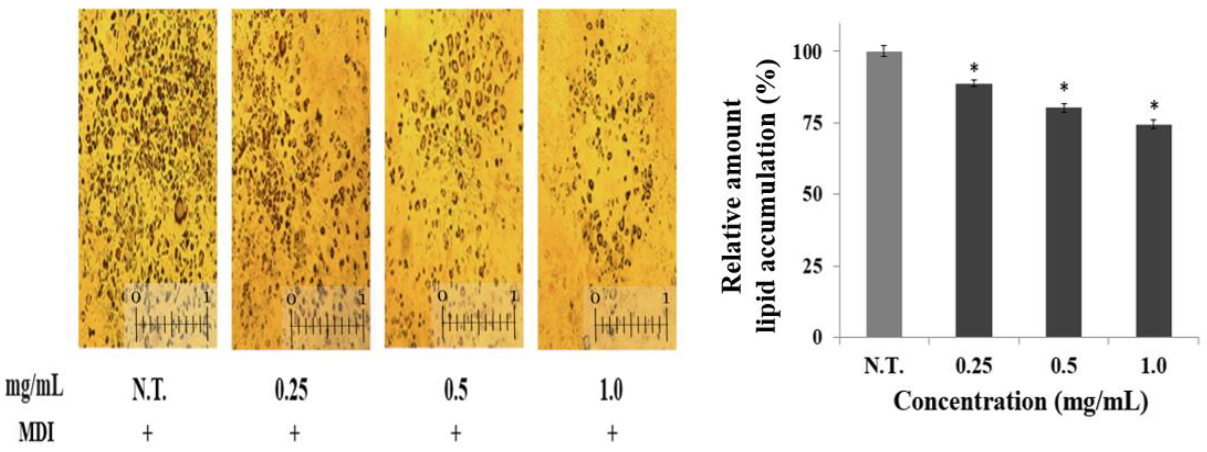
Adipocytes store excess lipids or glucose as triglycerides and play a vital role in the regulation of lipid metabolism and energy homeostasis by supplying free fatty acids and glycerol when energy is needed [15]. Therefore, preadipocyte differentiation and increased lipid accumulation are closely connected to the development of obesity [16]. Thus, in the present study, this cell differentiation process was induced in order to evaluate the effect of the CGE on adipogenesis. The CGE was applied at concentrations of 0.25, 0.5, and 1.0 mg/mL during the treatment, and the accumulation of intracellular triglycerides was then compared by staining adipocytes (Figure 2). It was shown that the accumulation of triglycerides in these cells decreased in a concentration-dependent manner. Specifically, lipid accumulation decreased by 1.4 and 1.2 times with treatment of 0.5 and 1.0 mg/mL CGE, respectively, compared to the control group. This level of triglyceride reduction was higher than the 1.2-fold reduction reported for preadipocytes treated with turmeric extract at the same concentration, demonstrating that the CGE had a superior inhibitory effect on triglyceride accumulation compared to other natural products [17]. Therefore, it was confirmed that this extract inhibits the generation of lipids in adipocytes, thereby exerting an anti-obesity effect through reduced triglyceride accumulation. To determine the mechanism underlying this process, the expression of main genes centered on AMPK and involved in differentiation was then investigated.

2.3. Effect of CGE on Expression of Adipogenesis and Lipogenesis Genes

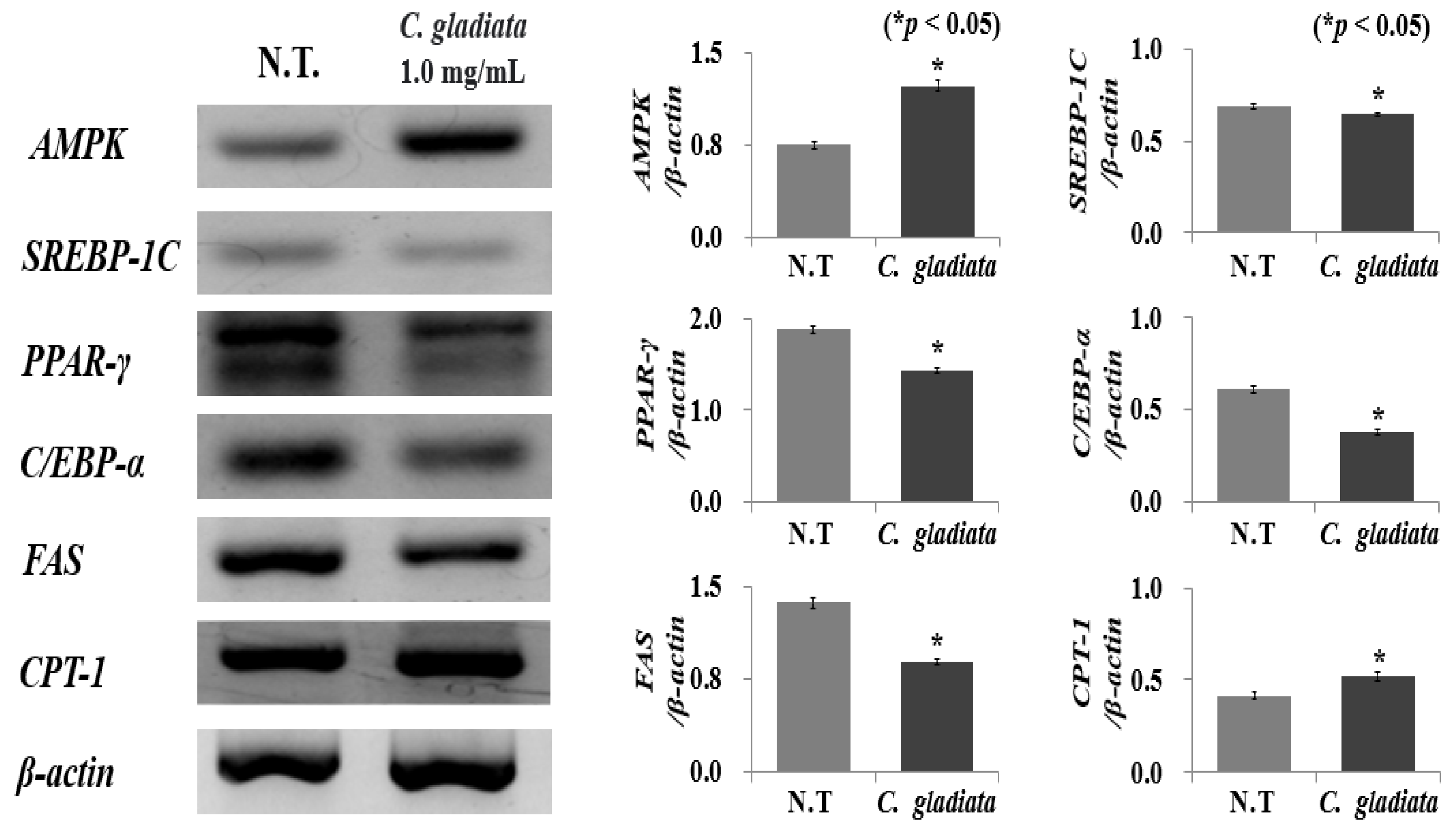
Cells use ATP and ADP as energy sources for metabolic processes and AMP generation. The increase in AMP activates AMPK based on the levels of depletion of intracellular energy, thus inducing catabolism to generate energy and allowing the maintenance of energy homeostasis [18]. In lipid metabolism, AMPK activation inhibits fatty acid synthesis and gluconeogenesis simultaneously [19]. Therefore, various anti-obesity mechanisms can activate AMPK during the treatment of this condition. To determine the inhibitory effect of AMPK activation on preadipocyte differentiation, the effect of the CGE on the expression of AMPK, CPT-1, SREBP-1c, PPAR-γ, C/EBP-α, and FAS mRNA (which are main factors in cell differentiation) was evaluated (Figure 3). The preadipocytes were treated with 1.0 mg/mL CGE, the maximum non-toxic concentration established in the previous experiment, and it was shown that the expression of AMPK and CPT-1 increased by 1.9 and 1.2 times, respectively. On the other hand, the expression of SREBP-1c, C/EBP-α, PPAR-γ, and FAS decreased by 1.1, 1.2, 1.8, and 1.5 times, respectively. In particular, the expression of AMPK showed the highest increase, by 1.9 times, which exerted an anti-obesity effect by inhibiting lipogenesis in the body through the suppression of acetyl-CoA carboxylase and malonyl-CoA, the enzymes that induce fatty acid activity [20]. In addition, AMPK activated CPT-1, an enzyme that performs beta-oxidation by inhibiting malonyl-CoA, thus increasing the permeability of fatty acids in the mitochondria. This can be expected to exert an additional inhibition of adipogenesis and lipogenesis effect through the inhibition of triglyceride generation and promotion of fat oxidation [21].
In addition, the number of triglycerides accumulated following preadipocyte cell differentiation was determined by the increase or decrease in SREBP-1c, the upregulating mechanism of adipogenesis-related genes or signal transmitters [22]. SREBP-1c, which regulates the expression of genes involved in fatty acid metabolism and lipid biosynthesis in the early stages of preadipocyte differentiation, has been shown to inhibit the expression of the PPAR-γ, C/EBP-α, and FAS sub-factors to reduce lipid accumulation [23]. The CGE suppressed PPAR-γ, C/EBP-α, and FAS expression by inhibiting SREBP-1c as well as by increasing the expression of AMPK and CPT-1, which is expected to produce a high inhibition of adipogenesis and lipogenesis effect, making this extract a potentially successful candidate substance for the treatment of obesity. In this study, the effect of CGE on AMPK regulation in 3T3-L1 preadipocytes was investigated, and the adipogenesis and adipogenesis mechanisms were identified. Additionally, gene-level-based anti-obesity mechanism evaluation has limitations in evaluating protein expression and function; further in-depth studies, specifically through the use of in vivo anti-obesity models, are required to validate this and identify the anti-obesity mechanism.

2.4. Major Substance Analysis Using LC-MS/MS

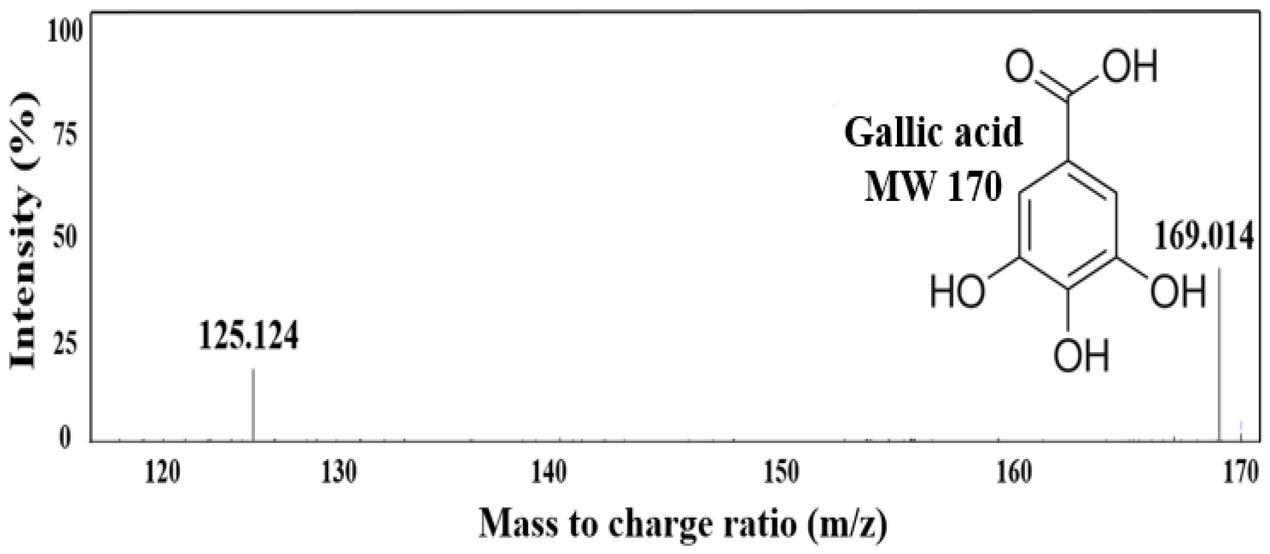
In the previous analysis of the expression of adipogenesis and lipogenesis genes, the inhibition of triglyceride accumulation was confirmed by the increased expression of AMPK and CPT-1 and decreased expression of SREBP-1c, C/EBP-α, PPAR-γ, and FAS. Therefore, to explore the main substances contained in the CGE that inhibit preadipocyte differentiation, a qualitative analysis was performed using LC-MS/MS (Figure 4). A comparison of the molecular weight distribution of CGE in negative mode revealed the presence of two peaks, m/z 169.0 and 125.1. In this mode m/z was measured with one proton removed, and the substance detected at m/z 169.0 was predicted to be gallic acid (MW 170.0). This result was consistent with that reported by Gang et al., where LC-MS/MS analysis detected gallic acid as the main substance in the separation of major polyphenols in the CGE [24]. Subsequently, additional qualitative and quantitative analyses were conducted via HPLC to confirm the identity of the primary substance of the extract.

2.5. Quantification of Major Substance Using HPLC

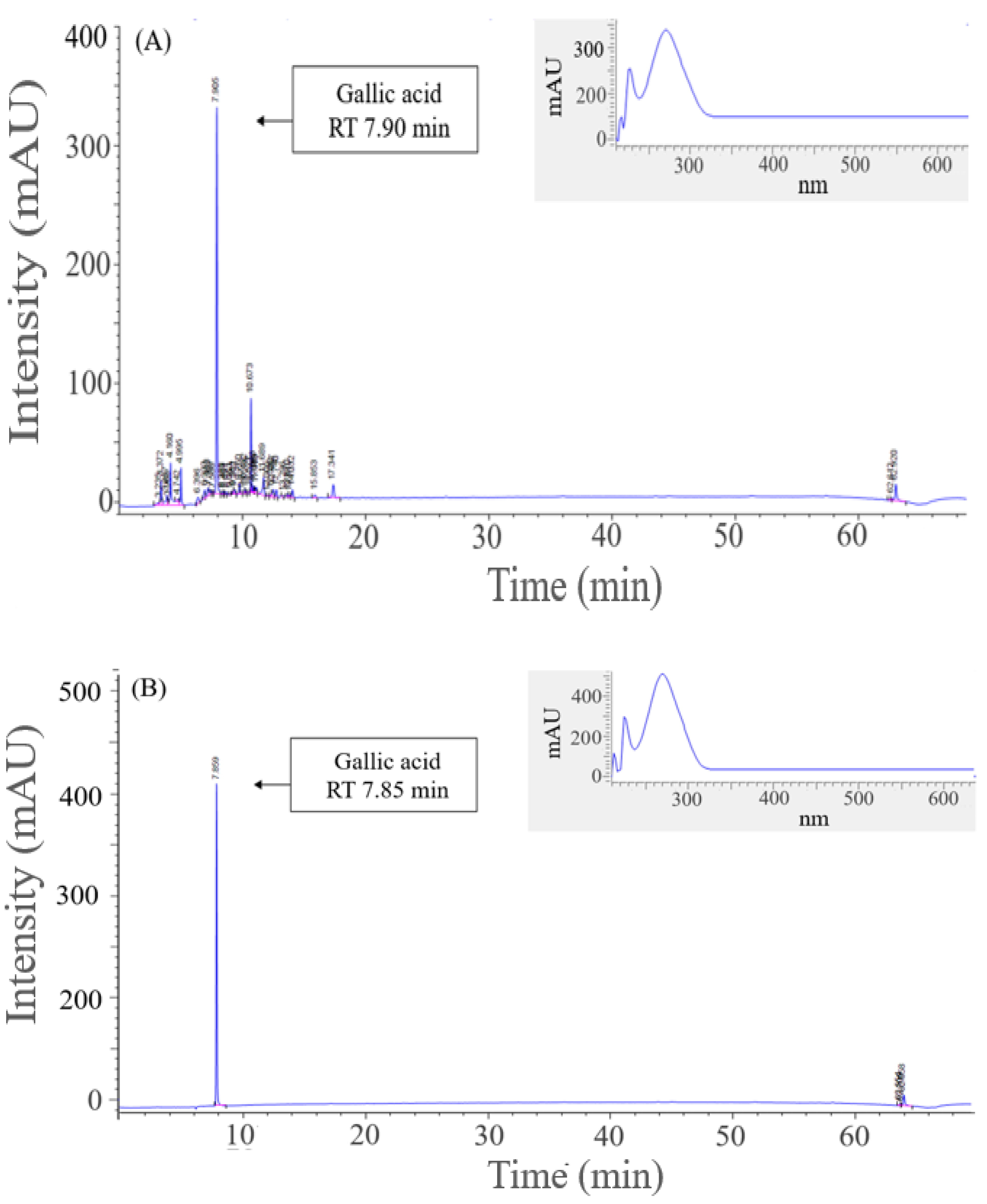
HPLC was used to perform qualitative and quantitative analyses based on retention time and DAD to validate the results of the earlier LC-MS/MS analysis. The RT of the major substances detected in the CGE was 7.9 min, which was highly consistent with that of 7.8 min found for gallic acid, the standard substance (Figure 5). The analysis of the corresponding peak with a DAD-based spectrum revealed a maximum absorption wavelength of 280 nm, which is consistent with that obtained when gallic acid was used as the standard substance in spectral form, reconfirming this compound as the main component of the CGE. The gallic acid concentration of 4.02 mg/g in the CGE was the highest among the peaks detected during HPLC analysis. Thus, this substance, which is a non-flavonoid tannin known for its anti-inflammatory, anti-cancer, liver protection, vascular disease prevention, and strong antioxidant functions, was identified as a major contributor to the inhibition of adipogenesis and lipogenesis effect of the CGE [25]. Furthermore, according to a study by Tan et al., gallic acid inhibited the expression of the main adipogenesis genes SREBP-1c and PPAR-γ by 37.2% and 51.1%, respectively, thus confirming its inhibition of adipogenesis effect through the inhibition of preadipocyte differentiation [26].
Catalase and peroxidase decompose the active oxygen generated in the body into water and oxygen molecules, thereby regulating homeostasis [27]. However, when an excessive production of active oxygen, lipid and protein denaturation, adipocyte metabolism disorders, and lowering of the body’s metabolic function are present, a homeostasis imbalance occurs, which ultimately leads to obesity. Gallic acid, the main substance of the CGE, is a typical polyphenol compound produced through the hydrolysis of tannin; it contains four hydroxyl groups and has a high active oxygen-scavenging activity, which is thought to contribute to increasing its anti-obesity effect [28]. According to previous studies by Song et al., Sasa borealis stem extracts containing gallic acid confirmed as the major substance exert anti-obesity effects on preadipocyte differentiation and lipid accumulation by regulating the expression of CEBP-α and PPAR-γ, which are early transcription factors involved in preadipocyte differentiation and attenuate hepatic steatosis in high-fat diet-induced obesity rats [29]. Overall, it was concluded that gallic acid identified as a major substance of CGE is highly effective as an anti-obesity substance, and it can also be used to remedy metabolic syndromes such as hyperlipidemia, high blood pressure, and arteriosclerosis.

3. Materials and Methods

3.1. Materials and Reagents

C. gladiata was purchased from Malgeundeul Co., Ltd. (Hongcheon, Korea) and kept frozen at −21 °C. Fetal bovine serum (FBS), new-born calf serum (NBCS), Dulbecco’s modified eagle medium (DMEM), and 0.05% trypsin-EDTA for cell culture were purchased from Thermo Fisher Sci., Inc. (Waltham, MA, USA). IBMX, DEX, insulin, and 3-(4,5-diimethylthiazole-2-yl)-2,5-diphenyl tetrazolium bromide (MTT) for preadipocyte differentiation and MTT assay were purchased from Sigma-Aldrich (St. Louis, MO, USA). Acetonitrile, acetic acid, and formic acid were purchased by high-performance liquid chromatography (HPLC) grade from Thermo Fisher Sci., Inc.

3.2. Preparation of CGE

C. gladiata was dried using a forced convection dry oven (FC 49, Lab House Co., Daejeon, Seoul, Korea) at 60 °C for 48 h and then pulverized into a particle size less than 0.42 mm using a grinder (HMF-3000S, Hanil, Buchoen, Korea). For the extraction of bioactive components from C. gladiata, 1.0 g of the dried sample was placed in a pressure-resistant Pyrex tube (PYREX-1636, Sci lab., Seoul, Korea) and mixed with 10.0 mL of 50% ethanol using a vortex mixer (VM-10, Daihan, Ltd., Wonju, Korea). Extraction was carried out at 60 °C for 30 min in the ultrasound extractor (250 W, SD-D250H, Daihan, Wonju, Korea). Then, the extract was subjected to solid–liquid separation at 10,000 rpm for 5 min using a centrifuge (LaboGene 1236R, Gyrozen Co., Daejeon, Korea).

3.3. Measurement of Preadipocyte Differentiation

Preadipocyte (3T3-L1) cells were obtained from the Korean Collection for Type Culture (KCTC, Jeongeup, Korea). 3T3-L1 preadipocyte with the inoculum density of 2.5 × 10⁴ cells/mL was cultured in DMEM supplemented with 10% NBCS, 1% penicillin, and MDI (0.5 mM IBMX, 1.0 mM DEX, and 1.0 μg/mL insulin) and cultured in a CO2 incubator (MCO-18AIC, Sanyo, Osaka, Japan) at 37 °C with 5.0% CO2. After 48 h, cells were fed with DMEM containing 10% FBS and 10 ug/mL insulin and cultured for an additional 48 h. Subsequently, the media was replaced with DMEM containing 10% FBS every 48 h.

3.4. Cell Viability Assay

The effect of CGE on the cell viability was determined using MTT assay. 3T3-L1 preadipocyte was seeded in a 96-well plate with 1 × 10⁴ cells/well of inoculum density, and different concentrations (0.0~4.0 mg/mL) of CGE were treated. After 48 h of cultivation, 0.1 mL MTT solution was added to induce formation of formazan at 37 °C and 0.1 mL DMSO was added after 4 h to dissolve formazan. Then, absorption was measured at 540 nm using a microplate reader (AMR-100, Allsheng, Seoul, Korea). In accordance with international standards (ISO 10993-5; Part 5: tests for in vitro cytotoxicity), the cytotoxic level was set below 85% cell viability [30]. The cell viability was calculated with the following equation:
Cell   viability   % = 1     Abs   ( sample ) Abs   ( control )   ×   100  
Sample: absorbance with CGE addition, control: absorbance with DMEM addition.

3.5. Lipid Accumulation Assay

Lipid accumulation of differentiated 3T3-L1 preadipocyte was measured by oil red O staining. The cells were washed with phosphate-buffered saline (PBS) and fixed with 10% formaldehyde solution for 30 min. Then, the cells were stained with oil red O solution for 60 min and washed with distilled water. The stained lipids were eluted with 99.5% isopropanol and measured using a UV–vis spectrophotometer at 497 nm by the following equation.
Lipid   accumulation   % = 1     Abs   ( sample ) Abs   ( control )   ×   100  
Sample: absorbance with CGE addition, control: absorbance with DMEM addition.

3.6. Measurement of Adipogenesis and Lipogenesis Genes Expression

RT-PCR was performed to determine the mRNA level of adipogenesis and lipogenesis genes including AMPK, CPT-1, SREBP-1c, PPAR-γ, C/EBP-α and FAS in 3T3-L1 preadipocyte. Cells were cultured in a T-25 cell culture flask (SPL Life Sci., Seoul, Korea) to a density of 3 × 10⁵ cells/mL. After collecting cells, total RNA was extracted using an AccuPrep® universal RNA extraction kit and the expression levels were measured by spectrophotometer (NanoDrop™ 2000, Thermo Fisher, Waltham, MA, USA). Total RNA was reverse transcribed into cDNA using amfiRivert cDNA synthesis platinum master mix (GenDEPOT Co., Greenville, SC, USA). Then, cDNA was amplified with each primer (Cosmo Genetech, Seoul, Korea) AMPK, CPT-1, SREBP-1c, PPAR-γ, C/EBP-α, and FAS (Table 1). The initial denaturation was performed at 95 °C for 30 s, and the denaturation temperature was 59 °C for 30 s, annealing was at 72 °C for 30 s, and extension was at 72 °C for 7 min; samples were amplified for 35 cycles. Each PCR product was electrophoresed on 1.5% agarose gel and the intensity of bands visualized by using Gel DocTM XR + system (Bio-Rad Co., Richmond, CA, USA) to compare the band intensity with the housekeeping gene (β-actin).

3.7. LC-MS/MS Analysis

To conduct a qualitative analysis for determining the main component in CGE, phenolic components in CGE were analyzed by using liquid chromatography tandem mass spectrometry (LC-MS/MS) with a ROC C18 column. The column temperature and injection volume were set at 30 °C and 10 µL. The main components in CGE were analyzed by negative ion electrospray ionization using an electrospray ionization source and mass spectra were operated in the negative and positive ion mode between m/z 100 and 800 by a triple quadrupole mass spectrometer (TSQ Quantum Ultra EMR., Thermo Fisher, Waltham, MA, USA). The mobile phase consisted of two solvents, 0.1% formic acid in distilled water (A) and 0.1% formic acid in 50.0% acetonitrile (B), and a gradient elution system for the separation of polyphenols were as follows: 0~11 min, 95.0~0.0% A; 11~14 min, 0.0~0.0% A; 14~15 min, 0.0~95.0% A; and 15~20 min, 95.0~95.0% A.

3.8. HPLC Analysis

Quantitative and qualitative analyses of polyphenol in CGE were carried out using HPLC (Agilent 1260, Agilent Tech., Santa Clara, CA, USA) equipped with an autosampler, diode array detector (DAD), and Poroshell 120 EC-C18 column (4.6 × 150 mm, Agilent Technology, Santa Clara, CA, USA). The mobile phase was composed of two solvents, 99.9% acetonitrile (A) and 1.0% acetic acid (B), and gradient elution for the separation of polyphenols were as follows: 0~5 min, 0.0~15.0% A; 5~50 min, 15~50% A; 50~60 min, 50~100% A; and 60~64 min, 100~0.0% A. Main components in CGE were identified by comparing retention time (RT) as well as the DAD spectrum (190~640 nm) with polyphenol standards. A polyphenol standard including gallic acid with purity of 99.8% was purchased from Sigma-Aldrich.

3.9. Statistical Analysis

Data were expressed as the mean ± standard deviation (SD) for all experiments, and probabilities (p) of chance difference between groups were calculated according to Student’s t-test. A statistically significant test means that the test hypothesis is false or should be rejected and the criteria were set at p < 0.05.

4. Conclusions

This study aimed to validate the inhibition of anti-adipogenesis and lipogenesis effect of the CGE by evaluating the reduction in lipids by treatment with extracts and the inhibition of preadipocyte differentiation via AMPK activation. The results confirmed that triglyceride accumulation in adipocytes was reduced by 1.4 times when the extract at a concentration 1.0 mg/mL was added to preadipocytes. The evaluation of the mechanism underlying the CGE effects on lipogenesis and adipogenesis genes revealed an increase in the expression of AMPK and CPT-1, the main genes involved in preadipocyte differentiation, whereas the expressions of SREBP-1c, PPAR-γ, C/EBP-α, and FAS were reduced. The CGE as an inhibitor of triglyceride production was proved to have a high anti-obesity effect because it reduced the expression of subsequent regulators associated with AMPK expression by affecting the process of preadipocyte differentiation. This research indicated that CGE with inhibition of lipogenesis can be used as a natural adipogenesis agent, and it is expected to effectively inhibit the expression of SREBP-1c, PPAR-γ, C/EBP-α, and FAS, which are the major genes involved in the inhibition of lipogenesis and adipogenesis effect. In addition, LC-MS/MS and HPLC analysis confirmed that gallic acid was the main substance of the CGE and that, at a concentration of 4.02 mg/g, this compound inhibited the accumulation of triglycerides and affected preadipocyte cell differentiation. Therefore, the CGE is expected to have a high value for industrial application in health foods and pharmaceutical products.

Author Contributions

The individual contributions of authors are specified as follows: writing—original draft preparation, analysis, methodology, data curation, J.W.H.; methodology, data curation, H.Y.P.; analysis, methodology, H.A.K., Y.S.H. and E.J.L.; writing—original draft preparation, funding acquisition, project administration, supervision, editing, J.W.K. All authors have read and agreed to the published version of the manuscript.

Funding

This research received no external funding.

Institutional Review Board Statement

Not applicable.

Informed Consent Statement

Not applicable.

Data Availability Statement

No new data were created or analyzed in this study. Data sharing is not applicable to this article.

Conflicts of Interest

The authors declare no conflict of interest.

Sample Availability

Samples of the compounds are not available from the authors.

References

  1. Kim, H.J.; Jang, Y.S.; Ha, J.W.; Ra, M.-J.; Jung, S.-M.; Yu, J.-N.; Kim, K.; Kim, K.H.; Um, S.H. Bioactive Phytochemicals from Salix pseudolasiogyne Twigs: Anti-Adipogenic Effect of 2′-O-Acetylsalicortin in 3T3-L1 Cells. Int. J. Mol. Sci. 2022, 23, 12006. [Google Scholar] [CrossRef]
  2. Elebeedy, D.; Ghanem, A.; Saleh, A.; Ibrahim, M.H.; Al Kamaly, O.; Abourehab, M.A.S.; Ali, M.A.; Abd El Maksoud, A.I.; El Hassab, M.A.; Eldehna, W.M. In Vivo and In Silico Investigation of the Anti-Obesity Effects of Lactiplantibacillus plantarum Combined with Chia Seeds, Green Tea, and Chitosan in Alleviating Hyperlipidemia and Inflammation. Int. J. Mol. Sci. 2022, 23, 12200. [Google Scholar] [CrossRef] [PubMed]
  3. Gam, D.H.; Hong, J.W.; Yeom, S.H.; Kim, J.W. Polyphenols in peanut shells and their antioxidant activity: Optimal extraction conditions and the evaluation of anti-obesity effects. J. Nutr. Health 2021, 54, 116–128. [Google Scholar] [CrossRef]
  4. Xu, T.; Zhao, K.; Guo, X.; Tu, J.; Zhang, D.; Sun, W.; Kong, X. Low-intensity pulsed ultrasound inhibits adipogenic differentiation via HDAC1 signalling in rat visceral preadipocytes. Adipocyte 2019, 8, 292–303. [Google Scholar] [CrossRef] [PubMed] [Green Version]
  5. Lee, J.-E.; Cho, Y.-W.; Deng, C.-X.; Ge, K. MLL3/MLL4-associated PAGR1 regulates adipogenesis by controlling induction of C/EBPβ and C/EBPδ. Mol. Cell. Biol. 2020, 40, e00209-20. [Google Scholar] [CrossRef]
  6. Fu, C.-N.; Wei, H.; Gao, W.-S.; Song, S.-S.; Yue, S.-W.; Qu, Y.-J. Obesity increases neuropathic pain via the AMPK-ERK-NOX4 pathway in rats. Aging 2021, 13, 18606. [Google Scholar] [CrossRef]
  7. Chang, Y.-H.; Chen, Y.-L.; Huang, W.-C.; Liou, C.-J. Fucoxanthin attenuates fatty acid-induced lipid accumulation in FL83B hepatocytes through regulated Sirt1/AMPK signaling pathway. Biochem. Biophys. Res. Commun. 2018, 495, 197–203. [Google Scholar] [CrossRef]
  8. Chyau, C.-C.; Chu, C.-C.; Chen, S.-Y.; Duh, P.-D. The inhibitory effects of djulis (Chenopodium formosanum) and its bioactive compounds on adipogenesis in 3T3-L1 adipocytes. Molecules 2018, 23, 1780. [Google Scholar] [CrossRef] [Green Version]
  9. Lee, H.B.; Kang, S.-S. Inhibitory Effect of Bacterial Lysates Extracted from Pediococcus acidilactici on the Differentiation of 3T3-L1 Pre-Adipocytes. Int. J. Mol. Sci. 2022, 23, 11614. [Google Scholar] [CrossRef]
  10. van Weeghel, M.; Abdurrachim, D.; Nederlof, R.; Argmann, C.A.; Houtkooper, R.H.; Hagen, J.; Nabben, M.; Denis, S.; Ciapaite, J.; Kolwicz, S.C., Jr. Increased cardiac fatty acid oxidation in a mouse model with decreased malonyl-CoA sensitivity of CPT1B. Cardiovasc. Res. 2018, 114, 1324–1334. [Google Scholar] [CrossRef]
  11. Hwang, H.-J.; Hwang, Y.J.; Kim, Y.J.; Kim, M.; Hwang, K.-A. Immature sword bean pods (Canavalia gladiata) inhibit adipogenesis in C3H10T1/2 cells and mice with high-fat diet–induced obesity. J. Chinese Med. Assoc. 2022, 85, 67–76. [Google Scholar] [CrossRef]
  12. Jung, J.-Y.; Rhee, J.-K. Roasting and Cryogenic Grinding Enhance the Antioxidant Property of Sword Beans (Canavalia gladiata). J. Microbiol. Biotechnol. 2020, 30, 1706–1719. [Google Scholar] [CrossRef]
  13. Kaur, A.; Verma, P.; Kaur, S.; Kumar, V. In vitro anti-inflammatory and antioxidant potential of leaves of Canavalia gladiata. Pharmaspire 2019, 11, 76–78. [Google Scholar]
  14. Jeon, S.Y.; Park, J.Y.; Shin, I.; Kim, S.O.; An, H.D.; Kim, M.R. Ethanol extract of Plantago asiatica L. controls intracellular fat accumulation and lipid metabolism in 3T3-L1 Adipocytes. Korea J. Herbol. 2014, 29, 77–82. [Google Scholar] [CrossRef] [Green Version]
  15. Hong, J.W.; Park, H.Y.; Park, J.H.; Kim, S.H.; Kim, H.A.; Kim, J.-W. Inhibition of Lipase Activity and Preadipocyte Differentiation in 3T3-L1 Cells Treated with Sargassum horneri Extract. Ocean Polar Res. 2022, 44, 61–67. [Google Scholar]
  16. Blanco, J.; Guardia-Escote, L.; Mulero, M.; Basaure, P.; Biosca-Brull, J.; Cabré, M.; Colomina, M.T.; Domingo, J.L.; Sánchez, D.J. Obesogenic effects of chlorpyrifos and its metabolites during the differentiation of 3T3-L1 preadipocytes. Food Chem. Toxicol. 2020, 137, 111171. [Google Scholar] [CrossRef]
  17. Kang, Y.H.; Kim, K.K.; Kim, T.W.; Yang, C.S.; Choe, M. Evaluation of the anti-obesity activity of Platycodon grandiflorum root and Curcuma longa root fermented with Aspergillus oryzae. Korean J. Food Sci. Technol. 2015, 47, 111–118. [Google Scholar] [CrossRef] [Green Version]
  18. van der Vaart, J.I.; Boon, M.R.; Houtkooper, R.H. The role of AMPK signaling in brown adipose tissue activation. Cells 2021, 10, 1122. [Google Scholar] [CrossRef]
  19. Gao, J.; Zhang, M.; Niu, R.; Gu, X.; Hao, E.; Hou, X.; Deng, J.; Bai, G. The combination of cinnamaldehyde and kaempferol ameliorates glucose and lipid metabolism disorders by enhancing lipid metabolism via AMPK activation. J. Funct. Foods 2021, 83, 104556. [Google Scholar] [CrossRef]
  20. Rai, Y.; Pathak, R.; Kumari, N.; Sah, D.K.; Pandey, S.; Kalra, N.; Soni, R.; Dwarakanath, B.S.; Bhatt, A.N. Mitochondrial biogenesis and metabolic hyperactivation limits the application of MTT assay in the estimation of radiation induced growth inhibition. Sci. Rep. 2018, 8, 1531. [Google Scholar] [CrossRef] [Green Version]
  21. Srivastava, R.A.K.; Pinkosky, S.L.; Filippov, S.; Hanselman, J.C.; Cramer, C.T.; Newton, R.S. AMP-activated protein kinase: An emerging drug target to regulate imbalances in lipid and carbohydrate metabolism to treat cardio-metabolic diseases: Thematic review series: New lipid and lipoprotein targets for the treatment of cardiometabolic diseases. J. Lipid Res. 2012, 53, 2490–2514. [Google Scholar] [CrossRef] [PubMed] [Green Version]
  22. Bai, J.; Xu, H.; Fang, J.; Zhang, C.; Song, J.; Zhang, X.; Hao, B.; Yin, B.; Xia, G. miR-15a Targets ABAT Gene to Regulate the Differentiation of Preadipocytes of Yanbian Yellow Cattle; Research Square: Durham, NC, USA, 2021. [Google Scholar]
  23. Jia, D.; Li, Z.; Gao, Y.; Feng, Y.; Li, W. A novel triazine ring compound (MD568) exerts in vivo and in vitro effects on lipid metabolism. Biomed. Pharmacother. 2018, 103, 790–799. [Google Scholar] [CrossRef] [PubMed]
  24. Gan, R.-Y.; Kong, K.-W.; Li, H.-B.; Wu, K.; Ge, Y.-Y.; Chan, C.-L.; Shi, X.-M.; Corke, H. Separation, identification, and bioactivities of the main gallotannins of red sword bean (Canavalia gladiata) coats. Front. Chem. 2018, 6, 39. [Google Scholar] [CrossRef] [PubMed] [Green Version]
  25. Wang, S.; Du, Q.; Meng, X.; Zhang, Y. Natural Polyphenols: A Potential prevention and treatment strategy for Metabolic Syndrome. Food Funct. 2022, 13, 9734–9753. [Google Scholar] [CrossRef]
  26. Tanaka, M.; Sugama, A.; Sumi, K.; Shimizu, K.; Kishimoto, Y.; Kondo, K.; Iida, K. Gallic acid regulates adipocyte hypertrophy and suppresses inflammatory gene expression induced by the paracrine interaction between adipocytes and macrophages in vitro and in vivo. Nutr. Res. 2020, 73, 58–66. [Google Scholar] [CrossRef]
  27. Marwicka, J.; Zięba, A. Antioxidants as a defence against reactive oxygen species. Aesthetic Cosmetol. Med 2021, 10, 271–276. [Google Scholar] [CrossRef]
  28. Gam, D.H.; Park, J.H.; Kim, J.H.; Beak, D.H.; Kim, J.W. Effects of Allium sativum Stem Extract on Growth and Migration in Melanoma Cells through Inhibition of VEGF, MMP-2, and MMP-9 Genes Expression. Molecules 2021, 27, 21. [Google Scholar] [CrossRef]
  29. Song, Y.; Lee, S.-J.; Jang, S.-H.; Ha, J.H.; Song, Y.M.; Ko, Y.-G.; Kim, H.-D.; Min, W.; Kang, S.N.; Cho, J.-H. Sasa borealis stem. extract attenuates hepatic steatosis in high-fat diet-induced obese rats. Nutrients 2014, 6, 2179–2195. [Google Scholar] [CrossRef]
  30. ISO 10993-5:2009; Biological Evaluation of Medical Devices—Part 5: Tests for In Vitro Cytotoxicity. ISO: Geneva, Switzerland, 2009.
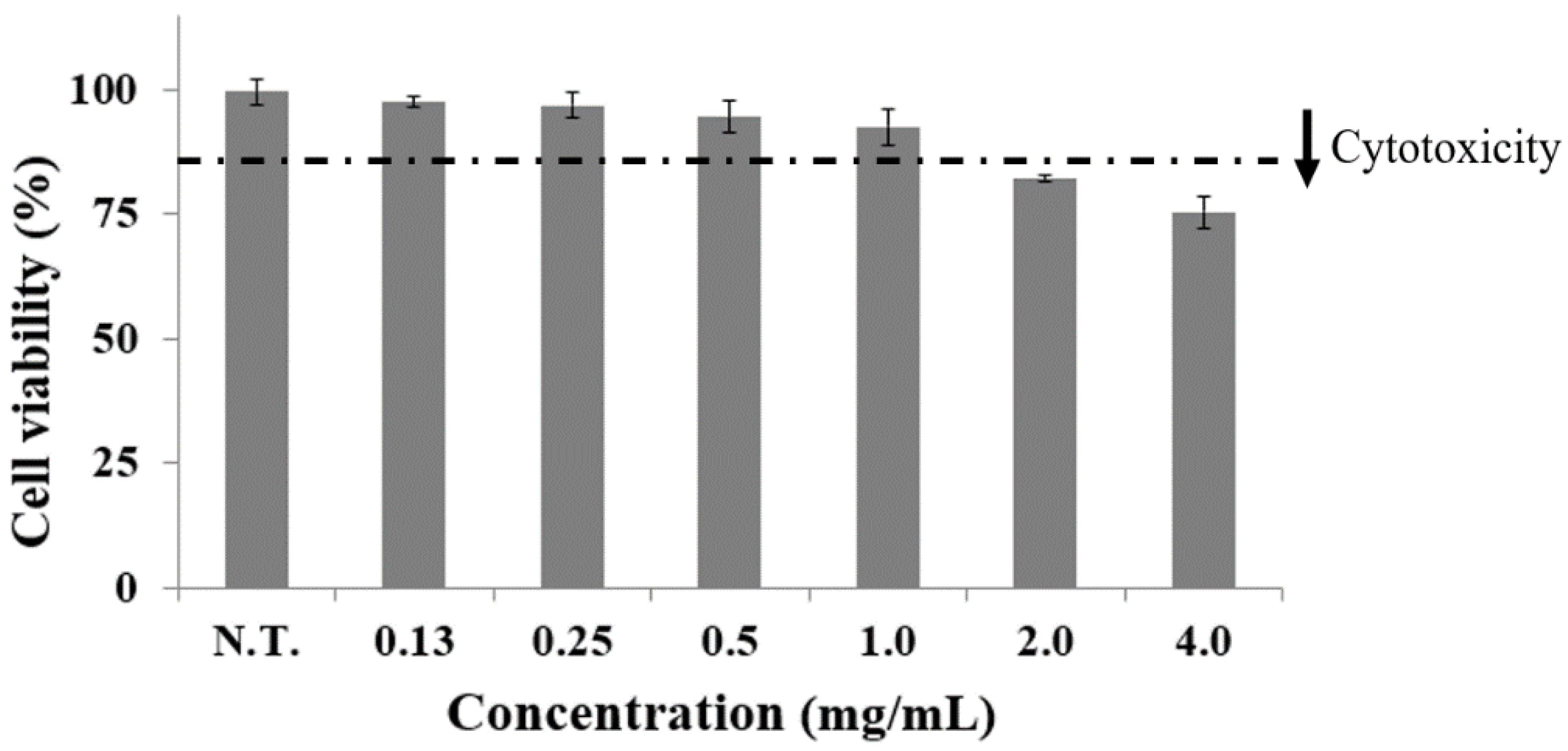
Figure 1. Effects of C. gladiata extract (0.0~4.0 mg/mL) on cell viability in preadipocytes. Bars represent the mean ± standard deviation of three independent experiments and asterisks indicate significant differences from the control.
Ijms 24 02108 g001
Figure 2. Effect of CGE on the lipid accumulation in adipocytes. Lipid droplets in adipocytes were fixed and stained with oil red O and were observed under a microscope of 64 magnification. Bar graphs represent the relative amounts of accumulated lipid based on untreated samples in adipocytes after being treated with CGE (0.0~1.0 mg/mL). Bars marked with asterisks are significantly different from the control (* p < 0.05).
Ijms 24 02108 g002
Figure 3. Effect of C. gladiata extract on AMPK, CPT-1, SREBP-1c, PPAR-γ, C/EBP-α and FAS mRNA expression levels in 3T3-L1. Values are expressed as mean ± standard deviation of three experiments. Bars marked with asterisks are significantly different from the control (* p < 0.05).
Ijms 24 02108 g003
Figure 4. Mass Spectrum of LC-MS/MS fragmentation pattern of main substance, gallic acid (MW 170), from C. gladiata extract (full scan in negative ion mode).
Ijms 24 02108 g004
Figure 5. HPLC analysis for the quantitative analysis of main substances and polyphenols in C. gladiata extract. (A) chromatogram and DAD spectrum of C. gladiata extract, (B) chromatogram and DAD spectrum of gallic acid.
Ijms 24 02108 g005
Table 1. Primer sequences used in reverse transcription-polymerase chain reaction (RT-PCR) of major genes related to adipogenesis and lipogenesis.
GenesForward (5’-3’)Reverse (5’-3’)Size (bp)
AMPK(1)CCATGCTGGAACTGATGGAGCTGAACTGTGTGACCCAGCC275
CPT-1(2)CGACATGCTCGGCCTCATAGGCCAGAAGCCCCCAAGAAAC813
SREBP-1c(3)GGTTTAGGGATGTTTGGGTTTTAAGCCCACTTCATTTCATTGGT122
PPAR-γ(4)AGCCGAGATAAAGCCAAACAACGAATCTCCTAGTCCTGGCTTGC325
C/EBP-α(5)CCCTGAAATCCCAGCACTTCGGCATGGCTGCTGTAGGGGT137
FAS(6)CGACATGCTCGGCCTCATAGGCCAGAAGCCCCCAAGAAAC416
β-actinAGCACAGAGCCTCGCCTTTCTTAATGTCACGCACGATTTCC295
(1)AMPK: AMP-activated protein kinase, (2)CPT-1: carnitine palmitoyl transferase-1, (3)SREBP-1c: sterol regulatory element binding proteins-1c, (4)PPAR-γ: peroxisome proliferator activated receptor-γ (5) C/EBP-α: CCAAT/enhancer binding protein-α, (6) FAS: fatty acid synthase.
Disclaimer/Publisher’s Note: The statements, opinions and data contained in all publications are solely those of the individual author(s) and contributor(s) and not of MDPI and/or the editor(s). MDPI and/or the editor(s) disclaim responsibility for any injury to people or property resulting from any ideas, methods, instructions or products referred to in the content.