
Effectiveness and Safety of Over-the-Counter Tooth-Whitening Agents Compared to Hydrogen Peroxide In Vitro

 , , ,
, , ,
Abstract
1. Introduction
2. Results
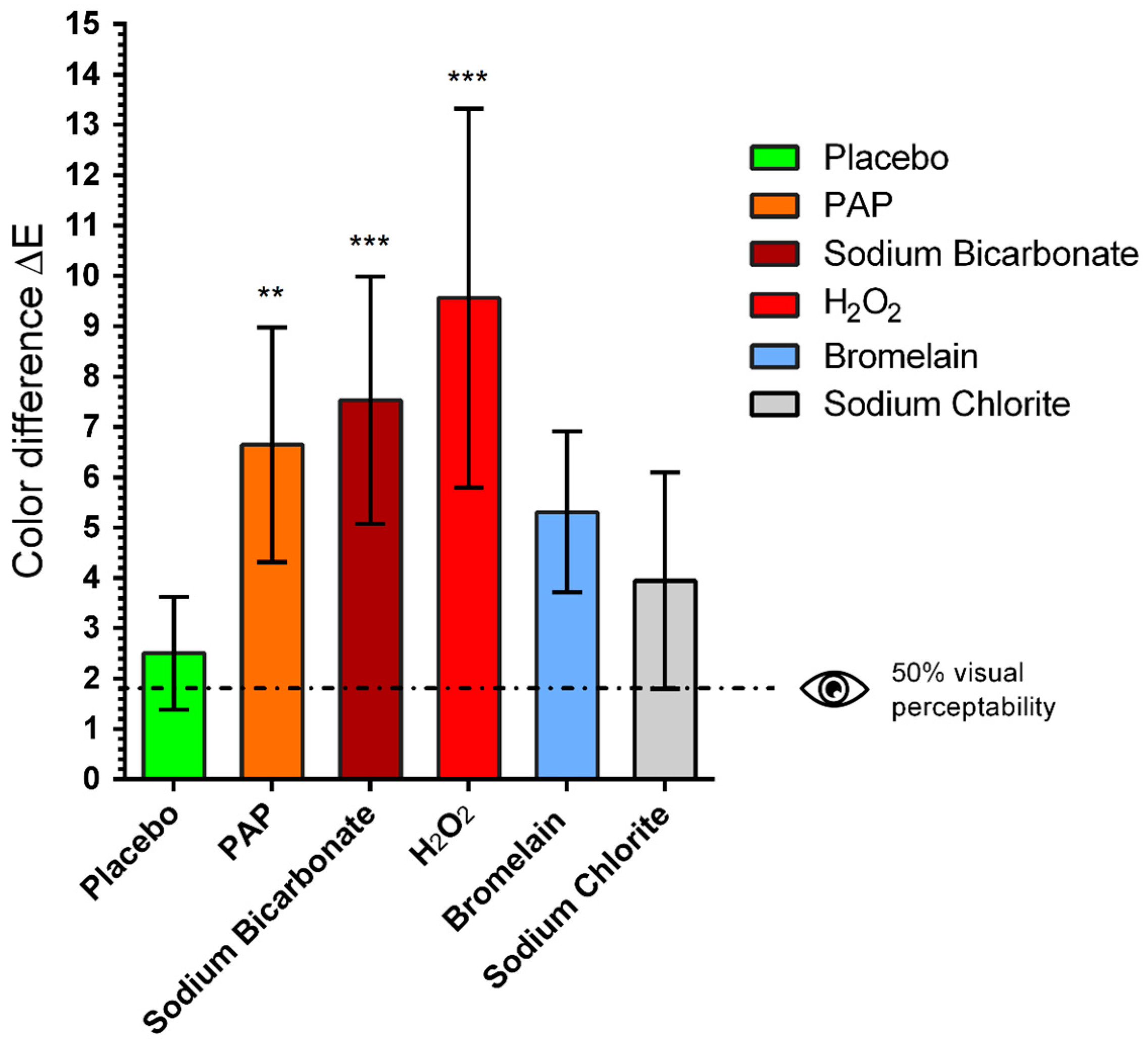
2.1. Color Changes
2.2. Microscopic Inspection by SEM
2.3. Cellular Viability
3. Discussion
4. Materials and Methods
4.1. Specimen Preparation
- staining procedure
- whitening procedure
4.2. Staining Procedure
4.3. Preparation of Bromelain Whitening Gel
4.4. Spectrophotometric Color Determination
4.5. Microscopic Inspections
4.6. Cell Isolation and Cell Culture
4.7. Cellular Viability
4.8. Statistical Analysis
5. Conclusions
Author Contributions
Funding
Institutional Review Board Statement
Informed Consent Statement
Data Availability Statement
Acknowledgments
Conflicts of Interest
References
- Rodríguez-Martínez, J.; Valiente, M.; Sánchez-Martín, M.J. Tooth whitening: From the established treatments to novel approaches to prevent side effects. J. Esthet. Restor. Dent. 2019, 31, 431–440. [Google Scholar] [CrossRef] [PubMed]
- Eachempati, P.; Kumbargere Nagraj, S.; Kiran Kumar Krishanappa, S.; Gupta, P.; Yaylali, I.E. Home-based chemically-induced whitening (bleaching) of teeth in adults. Cochrane Database Syst. Rev. 2018, 12, CD006202. [Google Scholar] [CrossRef] [PubMed]
- Naik, S.; Tredwin, C.J.; Scully, C. Hydrogen peroxide tooth-whitening (bleaching): Review of safety in relation to possible carcinogenesis. Oral. Oncol. 2006, 42, 668–674. [Google Scholar] [CrossRef] [PubMed]
- Epple, M.; Meyer, F.; Enax, J. A Critical Review of Modern Concepts for Teeth Whitening. Dent. J. 2019, 7, 79. [Google Scholar] [CrossRef]
- Joiner, A. The bleaching of teeth: A review of the literature. J. Dent. 2006, 34, 412–419. [Google Scholar] [CrossRef] [PubMed]
- He, L.B.; Shao, M.Y.; Tan, K.; Xu, X.; Li, J.Y. The effects of light on bleaching and tooth sensitivity during in-office vital bleaching: A systematic review and meta-analysis. J. Dent. 2012, 40, 644–653. [Google Scholar] [CrossRef]
- Martini, E.C.; Parreiras, S.O.; Szesz, A.L.; Coppla, F.M.; Loguercio, A.D.; Reis, A. Bleaching-induced tooth sensitivity with application of a desensitizing gel before and after in-office bleaching: A triple-blind randomized clinical trial. Clin. Oral Investig. 2020, 24, 385–394. [Google Scholar] [CrossRef]
- Moghadam, F.V.; Majidinia, S.; Chasteen, J.; Ghavamnasiri, M. The degree of color change, rebound effect and sensitivity of bleached teeth associated with at-home and power bleaching techniques: A randomized clinical trial. Eur. J. Dent. 2013, 7, 405–411. [Google Scholar] [CrossRef]
- Li, Y.; Greenwall, L. Safety issues of tooth whitening using peroxide-based materials. Br. Dent. J. 2013, 215, 29–34. [Google Scholar] [CrossRef]
- Tredwin, C.J.; Naik, S.; Lewis, N.J.; Scully, C. Hydrogen peroxide tooth-whitening (bleaching) products: Review of adverse effects and safety issues. Br. Dent. J. 2006, 200, 371–376. [Google Scholar] [CrossRef]
- Schulte, J.R.; Morrissette, D.B.; Gasior, E.J.; Czajewski, M.V. The effects of bleaching application time on the dental pulp. J. Am. Dent. Assoc. 1994, 125, 1330–1335. [Google Scholar] [CrossRef]
- Azrak, B.; Callaway, A.; Kurth, P.; Willershausen, B. Influence of Bleaching Agents on Surface Roughness of Sound or Eroded Dental Enamel Specimens. J. Esthet. Restor. Dent. 2010, 22, 391–399. [Google Scholar] [CrossRef] [PubMed]
- Abouassi, T.; Wolkewitz, M.; Hahn, P. Effect of carbamide peroxide and hydrogen peroxide on enamel surface: An in vitro study. Clin. Oral Investig. 2011, 15, 673–680. [Google Scholar] [CrossRef]
- Cadenaro, M.; Navarra, C.O.; Mazzoni, A.; Nucci, C.; Matis, B.A.; Di Lenarda, R.; Breschi, L. An In Vivo Study of the Effect of a 38 Percent Hydrogen Peroxide In-office Whitening Agent on Enamel. J. Am. Dent. Assoc. 2010, 141, 449–454. [Google Scholar] [CrossRef] [PubMed]
- Güorgan, S.; Bolay, S.; Alaçam, R. In vitro adherence of bacteria to bleached or unbleached enamel surfaces. J. Oral Rehabil. 1997, 24, 624–627. [Google Scholar] [CrossRef] [PubMed]
- Hegedüs, C.; Bistey, T.; Flóra-Nagy, E.; Keszthelyi, G.; Jenei, A. An atomic force microscopy study on the effect of bleaching agents on enamel surface. J. Dent. 1999, 27, 509–515. [Google Scholar] [CrossRef]
- Sa, Y.; Chen, D.; Liu, Y.; Wen, W.; Xu, M.; Jiang, T.; Wang, Y. Effects of two in-office bleaching agents with different pH values on enamel surface structure and color: An in situ vs. in vitro study. J. Dent. 2012, 40 (Suppl. 1), e26–e34. [Google Scholar] [CrossRef]
- Sun, L.; Liang, S.; Sa, Y.; Wang, Z.; Ma, X.; Jiang, T.; Wang, Y. Surface alteration of human tooth enamel subjected to acidic and neutral 30% hydrogen peroxide. J. Dent. 2011, 39, 686–692. [Google Scholar] [CrossRef]
- Xu, B.; Li, Q.; Wang, Y. Effects of pH values of hydrogen peroxide bleaching agents on enamel surface properties. Oper. Dent. 2011, 36, 554–562. [Google Scholar] [CrossRef]
- Attin, T.; Hannig, C.; Wiegand, A.; Attin, R. Effect of bleaching on restorative materials and restorations--a systematic review. Dent. Mater. 2004, 20, 852–861. [Google Scholar] [CrossRef] [PubMed]
- Smidt, A.; Feuerstein, O.; Topel, M. Mechanical, morphologic, and chemical effects of carbamide peroxide bleaching agents on human enamel in situ. Quintessence Int. 2011, 42, 407–412. [Google Scholar] [PubMed]
- Al-Salehi, S.K.; Wood, D.J.; Hatton, P.V. The effect of 24h non-stop hydrogen peroxide concentration on bovine enamel and dentine mineral content and microhardness. J. Dent. 2007, 35, 845–850. [Google Scholar] [CrossRef] [PubMed]
- Berger, S.B.; Cavalli, V.; Martin, A.A.; Soares, L.E.; Arruda, M.A.; Brancalion, M.L.; Giannini, M. Effects of combined use of light irradiation and 35% hydrogen peroxide for dental bleaching on human enamel mineral content. Photomed. Laser Surg. 2010, 28, 533–538. [Google Scholar] [CrossRef] [PubMed]
- Bizhang, M.; Seemann, R.; Duve, G.; Römhild, G.; Altenburger, J.M.; Jahn, K.R.; Zimmer, S. Demineralization effects of 2 bleaching procedures on enamel surfaces with and without post-treatment fluoride application. Oper. Dent. 2006, 31, 705–709. [Google Scholar] [CrossRef] [PubMed]
- Efeoglu, N.; Wood, D.J.; Efeoglu, C. Thirty-five percent carbamide peroxide application causes in vitro demineralization of enamel. Dent. Mater. 2007, 23, 900–904. [Google Scholar] [CrossRef]
- Efeoglu, N.; Wood, D.; Efeoglu, C. Microcomputerised tomography evaluation of 10% carbamide peroxide applied to enamel. J. Dent. 2005, 33, 561–567. [Google Scholar] [CrossRef]
- Pascolutti, M.; de Oliveira, D. A Radical-Free Approach to Teeth Whitening. Dent. J. 2021, 9, 148. [Google Scholar] [CrossRef]
- Zantner, C.; Beheim-Schwarzbach, N.; Neumann, K.; Kielbassa, A.M. Surface microhardness of enamel after different home bleaching procedures. Dent. Mater. 2007, 23, 243–250. [Google Scholar] [CrossRef]
- de Freitas, M.R.; de Carvalho, M.M.; Liporoni, P.C.S.; Fort, A.C.B.; Moura, R.d.M.e.; Zanatta, R.F. Effectiveness and Adverse Effects of Over-the-Counter Whitening Products on Dental Tissues. Front. Dent. Med. 2021, 2, 687507. [Google Scholar] [CrossRef]
- Kleber, C.J.; Putt, M.S.; Nelson, B.J. In vitro tooth whitening by a sodium bicarbonate/peroxide dentifrice. J. Clin. Dent. 1998, 9, 16–21. [Google Scholar]
- Barnes, C. An evidenced-based review of sodium bicarbonate as a dentifrice agent. Compend 1999, 4, 3–11. [Google Scholar]
- Kleber, C.J.; Moore, M.H.; Nelson, B.J. Laboratory assessment of tooth whitening by sodium bicarbonate dentifrices. J. Clin. Dent. 1998, 9, 72–75. [Google Scholar] [PubMed]
- Greenwall-Cohen, J.; Francois, P.; Silikas, N.; Greenwall, L.; Le Goff, S.; Attal, J.-P. The safety and efficacy of ‘over the counter’ bleaching products in the UK. Br. Dent. J. 2019, 226, 271–276. [Google Scholar] [CrossRef]
- Ribeiro, J.S.; de Oliveira da Rosa, W.L.; da Silva, A.F.; Piva, E.; Lund, R.G. Efficacy of natural, peroxide-free tooth-bleaching agents: A systematic review, meta-analysis, and technological prospecting. Phytother. Res. 2020, 34, 1060–1070. [Google Scholar] [CrossRef] [PubMed]
- Kalyana, P.; Shashidhar, A.; Meghashyam, B.; Sreevidya, K.R.; Sweta, S. Stain removal efficacy of a novel dentifrice containing papain and Bromelain extracts—An in vitro study. Int. J. Dent. Hyg. 2011, 9, 229–233. [Google Scholar] [CrossRef] [PubMed]
- Patil, P.A.; Ankola, A.V.; Hebbal, M.I.; Patil, A.C. Comparison of effectiveness of abrasive and enzymatic action of whitening toothpastes in removal of extrinsic stains—A clinical trial. Int. J. Dent. Hyg. 2015, 13, 25–29. [Google Scholar] [CrossRef]
- Münchow, E.A.; Hamann, H.J.; Carvajal, M.T.; Pinal, R.; Bottino, M.C. Stain removal effect of novel papain- and bromelain-containing gels applied to enamel. Clin. Oral Investig. 2016, 20, 2315–2320. [Google Scholar] [CrossRef]
- Ribeiro, J.S.; Barboza, A.d.S.; Cuevas-Suárez, C.E.; da Silva, A.F.; Piva, E.; Lund, R.G. Novel in-office peroxide-free tooth-whitening gels: Bleaching effectiveness, enamel surface alterations, and cell viability. Sci. Rep. 2020, 10, 10016. [Google Scholar] [CrossRef]
- Gómez-Polo, C.; Muñoz, M.P.; Lorenzo Luengo, M.C.; Vicente, P.; Galindo, P.; Martín Casado, A.M. Comparison of the CIELab and CIEDE2000 color difference formulas. J. Prosthet. Dent. 2016, 115, 65–70. [Google Scholar] [CrossRef]
- de Arajo, D.; Silva, L.; Campos, E.N.; Correia de Arajo, R. In vitro study on tooth enamel lesions related to whitening dentifrice. Indian J. Dent. Res. 2011, 22, 770–776. [Google Scholar] [CrossRef]
- Duschner, H.; Götz, H.; White, D.J.; Kozak, K.M.; Zoladz, J.R. Effects of hydrogen peroxide bleaching strips on tooth surface color, surface microhardness, surface and subsurface ultrastructure, and microchemical (Raman spectroscopic) composition. J. Clin. Dent. 2006, 17, 72–78. [Google Scholar] [PubMed]
- Joiner, A.; Thakker, G. In vitro evaluation of a novel 6% hydrogen peroxide tooth whitening product. J. Dent. 2004, 32 (Suppl. 1), 19–25. [Google Scholar] [CrossRef] [PubMed]
- Türkün, M.; Sevgican, F.; Pehlivan, Y.; Aktener, B.O. Effects of 10% Carbamide Peroxide on the Enamel Surface Morphology: A Scanning Electron Microscopy Study. J. Esthet. Restor. Dent. 2002, 14, 238–244. [Google Scholar] [CrossRef]
- van Kampen, V.; Merget, R.; Brüning, T. [Occupational allergies to bromelain]. Pneumologie 2007, 61, 159–161. [Google Scholar] [CrossRef] [PubMed]
- Gailhofer, G.; Wilders-Truschnig, M.; Smolle, J.; Ludvan, M. Asthma caused by bromelain: An occupational allergy. Clin. Allergy 1988, 18, 445–450. [Google Scholar] [CrossRef] [PubMed]
- Vejai Vekaash, C.J.; Kumar Reddy, T.V.; Venkatesh, K.V. Effect of vital bleaching with solutions containing different concentrations of hydrogen peroxide and pineapple extract as an additive on human enamel using reflectance spectrophotometer: An in vitro study. J. Conserv. Dent. 2017, 20, 337–340. [Google Scholar] [CrossRef]
- Koertge, T.E.; Brooks, C.N.; Sarbin, A.G.; Powers, D.; Gunsolley, J.C. A longitudinal comparison of tooth whitening resulting from dentifrice use. J. Clin. Dent. 1998, 9, 67–71. [Google Scholar]
- Penel, G.; Leroy, G.; Rey, C.; Bres, E. MicroRaman spectral study of the PO4 and CO3 vibrational modes in synthetic and biological apatites. Calcif. Tissue Int. 1998, 63, 475–481. [Google Scholar] [CrossRef]
- Kuramochi, E.; Iizuka, J.; Mukai, Y. Influences of bicarbonate on processes of enamel subsurface remineralization and demineralization: Assessment using micro-Raman spectroscopy and transverse microradiography. Eur. J. Oral Sci. 2016, 124, 554–558. [Google Scholar] [CrossRef]
- Sarembe, S.; Enax, J.; Morawietz, M.; Kiesow, A.; Meyer, F. In Vitro Whitening Effect of a Hydroxyapatite-Based Oral Care Gel. Eur. J. Dent. 2020, 14, 335–341. [Google Scholar] [CrossRef]
- Amaral, F.; Caldonazzo, M.; Amgartem, L.; Campos, E.; Oliveira Junior, O.; Andrade, M.; Bevilacqua, F.; Felicio, C. Effect of hydrogen peroxide in different concentrations on the degree of bleaching and susceptibility to staining. Sci. J. Dent. 2015, 2, 1–5. [Google Scholar] [CrossRef]


| Whitening Agent | ΔL | Δa | Δb | ΔE |
|---|---|---|---|---|
| Placebo | 1.4 | 0.3 | 0.0 | 2.3 |
| PAP | 4.1 | 0.9 | 4.9 | 6.6 |
| Sodium Bicarbonate | 4.6 | 1.4 | 4.5 | 7.5 |
| H2O2 | 7.0 | 1.6 | 5.7 | 9.6 |
| Bromelain | 1.6 | 0.8 | 4.7 | 5.3 |
| Sodium Chlorite | 2.1 | 0.5 | 1.6 | 3.9 |
Disclaimer/Publisher’s Note: The statements, opinions and data contained in all publications are solely those of the individual author(s) and contributor(s) and not of MDPI and/or the editor(s). MDPI and/or the editor(s) disclaim responsibility for any injury to people or property resulting from any ideas, methods, instructions or products referred to in the content. |
© 2023 by the authors. Licensee MDPI, Basel, Switzerland. This article is an open access article distributed under the terms and conditions of the Creative Commons Attribution (CC BY) license (https://creativecommons.org/licenses/by/4.0/).
Share and Cite
Müller-Heupt, L.K.; Wiesmann-Imilowski, N.; Kaya, S.; Schumann, S.; Steiger, M.; Bjelopavlovic, M.; Deschner, J.; Al-Nawas, B.; Lehmann, K.M. Effectiveness and Safety of Over-the-Counter Tooth-Whitening Agents Compared to Hydrogen Peroxide In Vitro. Int. J. Mol. Sci. 2023, 24, 1956. https://doi.org/10.3390/ijms24031956
Müller-Heupt LK, Wiesmann-Imilowski N, Kaya S, Schumann S, Steiger M, Bjelopavlovic M, Deschner J, Al-Nawas B, Lehmann KM. Effectiveness and Safety of Over-the-Counter Tooth-Whitening Agents Compared to Hydrogen Peroxide In Vitro. International Journal of Molecular Sciences. 2023; 24(3):1956. https://doi.org/10.3390/ijms24031956
Chicago/Turabian StyleMüller-Heupt, Lena Katharina, Nadine Wiesmann-Imilowski, Sebahat Kaya, Sven Schumann, Maximilian Steiger, Monika Bjelopavlovic, James Deschner, Bilal Al-Nawas, and Karl Martin Lehmann. 2023. "Effectiveness and Safety of Over-the-Counter Tooth-Whitening Agents Compared to Hydrogen Peroxide In Vitro" International Journal of Molecular Sciences 24, no. 3: 1956. https://doi.org/10.3390/ijms24031956
APA StyleMüller-Heupt, L. K., Wiesmann-Imilowski, N., Kaya, S., Schumann, S., Steiger, M., Bjelopavlovic, M., Deschner, J., Al-Nawas, B., & Lehmann, K. M. (2023). Effectiveness and Safety of Over-the-Counter Tooth-Whitening Agents Compared to Hydrogen Peroxide In Vitro. International Journal of Molecular Sciences, 24(3), 1956. https://doi.org/10.3390/ijms24031956