Article

Low Prevalence of HLA-G Antibodies in Lung Transplant Patients Detected using MAIPA-Adapted Protocol

1
Immunogenetics Laboratory, Etablissement Français du Sang, 13005 Marseille, France
2
ADES UMR 7268, Aix Marseille University, 13005 Marseille, France
3
IRCCS Ospedale Policlinico San Martino, 16100 Genova, Italy
4
Lung Transplant Department, Aix-Marseille University, APHM, 13015 Marseille, France
*
Author to whom correspondence should be addressed.
Int. J. Mol. Sci. 2023, 24(22), 16479; https://doi.org/10.3390/ijms242216479
Submission received: 30 August 2023 / Revised: 12 November 2023 / Accepted: 16 November 2023 / Published: 18 November 2023
(This article belongs to the Section Molecular Pathology, Diagnostics, and Therapeutics)

Abstract

:
Lung transplantation is often complicated by acute and/or chronic rejection leading to graft-function loss. In addition to the HLA donor-specific antibodies (HLA-DSA), a few autoantibodies are correlated with the occurrence of these complications. Recently, antibodies directed against non-classical HLA molecules, HLA-G, -E, and -F have been detected in autoimmune diseases, like systemic lupus erythematosus. Non-classical HLA molecules are crucial in the immunological acceptance of the lung graft, and some of their isoforms, like HLA-G*01:04 and -G*01:06, are associated with a negative clinical outcome. The aim of this study is to determine the frequency of detection of HLA-G antibodies in lung transplant recipients (LTRs) and their impact on the occurrence of clinical complications. After incubating the cell lines SPI-801, with and without three different HLA-G isoform expression, with sera from 90 healthy blood donors and 35 LTRs (before and after transplantation), HLA-G reactivity was revealed using reagents from commercial monoclonal antibody immobilization of platelet antigen assay (MAIPA ApDIA®). Only one serum from one blood donor had specific reactivity against the HLA-G transduced lines. Non-specific reactivity in many sera from LTRs was observed with transduced- and wild-type cell lines, which may suggest recognition of an autoantigen expressed by the SPI-801 cell line. In conclusion, this study allowed the development of a specific detection tool for non-denatured HLA-G antibodies. These antibodies seem uncommon, both in healthy subjects and in complicated LTRs. This study should be extended to patients suffering from autoimmune diseases as well as kidney and heart transplant recipients.

1. Introduction

Lung transplantation (LT) is a therapeutic option for chronic, irreversible respiratory failure. Survival after LT has steadily increased over time, but the incidence of mild and long-term morbidity and lung dysfunction remains very high due to acute or chronic rejection [1]. These clinical complications may be linked to immune dysregulation, implying humoral and cellular autoimmunity and/or alloimmunity. Recent works have suggested that autoimmune reactivity is a key element in the occurrence of chronic graft rejection [2,3,4]. Antibodies directed against Tubulin K-1, a membrane protein expressed by bronchial epithelial cells, and against collagen type V, an extracellular matrix protein normally sequestered in bronchial tissue, are strongly correlated with the occurrence of bronchiolitis obliterans syndrome (BOS) after LT [5]. This production of autoantibodies may be secondary to the exposure of cryptic antigens of the self, following tissue remodeling after LT, to the expression of neo-antigens in an inflammatory context, or to the stimulation of a cross-reaction by antigenic mimicry between pathogens and self-antigens [6]. More recently, it has been shown that the release of graft exosomes in an inflammatory context increases the risk of autoimmunity [7].
The HLA-G molecule has been suggested as a prognostic biomarker of LT outcome by two independent studies [8,9]. HLA-G has both humoral and cellular anti-inflammatory immunosuppressive properties, in particular by inhibiting NK and cytotoxic T lymphocyte (CTL)-mediated activity as well as B-cell activation via their inhibitory receptor (ILT-2, -4, and KIR2DL4) [10]. This non-classical HLA class I molecule also acts indirectly on immune control as HLA-E preferentially loads its signal peptide [11,12]. HLA-G’s anti-inflammatory potential is supported by its increased expression through anti-inflammatory cytokines, as IL-10, in a positive feedback mechanism; by its increased expression in autoimmune diseases; and by its involvement in promoting viral- or parasitic-infection escape [10].
Interestingly, HLA-G is expressed at bronchial cell membranes after interferon-β stimulation [13]. Moreover, membrane-bound HLA-G expression in lung biopsies and sHLA-G expression in bronchial alveolar fluid, but not in serum, were higher in clinically stable lung transplant recipients (LTRs) than in those developing acute rejection [8,13,14].
One hundred and seventeen HLA-G alleles are currently identified, and five protein isoforms (HLA-G*01:01, *01:03, *01:04, *01:05N, 01:06) are found with a frequency >5% in all populations [15,16]. Our team, and others, have shown that these alleles are associated with differential sHLA-G levels [15,17,18].
HLA-G alleles have also been associated with clinical outcomes: the HLA-G*01:06~UTR2 haplotype has been correlated with the pejorative evolution of cystic fibrosis, and the HLA-G*01:04~UTR3 haplotype with an increase of chronic rejection, production of HLA antibodies, and a decrease in patient survival [9].
Despite its low diversity and because of its epithelial expression, HLA-G may elicit immunization mechanisms like those observed for other HLA class I molecules.
Anti-HLA-G antibodies were recently detected in patients with systemic lupus erythematosus (SLE) in a study conducted on 69 German and 29 Mexican SLE patients and 17 German healthy individuals [19,20]. Multiplex Luminex®-based flow cytometry was used to screen sera for antibodies directed against non-classical HLA (HLA-G, -E, and -F) and β2m. Interestingly, anti-HLA-G IgG antibodies were detected in 30% of healthy subjects (6/17) and were more frequent than those directed against HLA-E and HLA-F. In addition, anti-HLA-G antibody levels were stable in five subjects over 6 months.
Our hypothesis is that HLA-G antibodies produced following LT may interfere with the HLA-G receptors and disinhibit the cellular response directed against the lung graft. Such a mechanism would participate in a persistent inflammatory response and eventually lead to respiratory failure. Also, these anti-HLA-G antibodies can actively participate in the occurrence or progression of CLAD in transplant patients.
The main goals of this study are (1) to detect HLA-G antibodies expressed in serum using an adapted MAIPA ApDia® kit and (2) to evaluate their impact on the occurrence of clinical complications in 35 LTRs.

2. Results

2.1. Population Characteristics

Ninety sera were collected from healthy blood donors (median age = 40 years (18–65); sex ratio = 0.54) with no history of transfusions, organ transplantation, or recent infections. Thirty out of forty-one women (73%) had had at least one pregnancy.
All lung transplant recipients (median age: 40 years [19–66]; sex ratio = 0.51) underwent LTx at the Marseille Lung Transplant Center. They received a first LT (8 single LTx; 26 bilateral LTx) for cystic fibrosis (49%), emphysema (20%), pulmonary fibrosis (26%), or another diagnosis (5%). One-hundred and forty-four sera were collected from 35 lung transplant recipients before and after transplant (at day 0 (D0) N = 30, at day 15 (D15) N = 25, at one month (M1) N = 28, at three months (M3) N = 32, and at twelve months (M12) N = 29). Seven LTRs had acute rejection, and seven developed chronic rejection in the first year. Seventeen produced the Donor-Specific Antibody (DSA) after LTx. One recipient experienced an acute rejection associated with DSA detection. Population characteristics are summarized in Table 1.

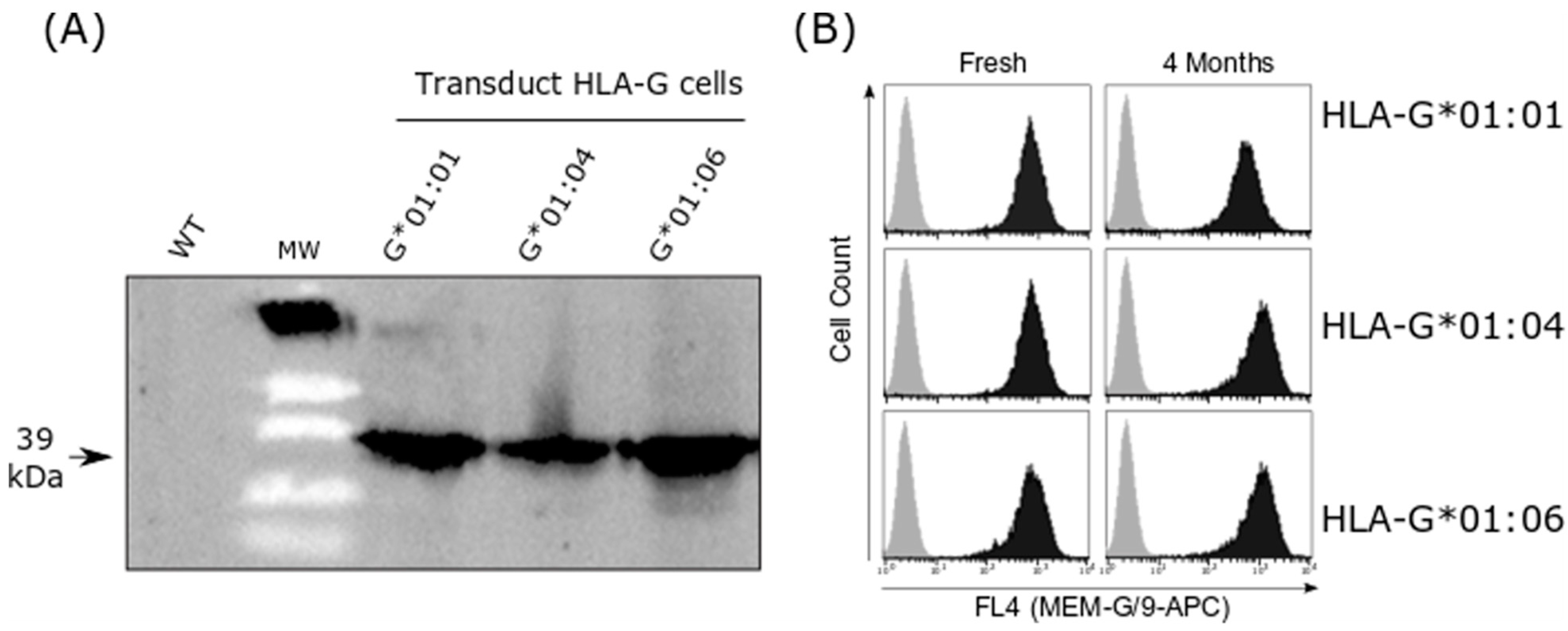
2.2. HLA-G Cell Line Transduction

The specific expression of the HLA-G isoform for each transduced cell line was assessed with Western blot, using 4H84 antibody and with flow cytometry using MEM/G9 antibody (Figure 1). Flow cytometry showed specific HLA-G membrane expression in HLA-G transduced cells, and no signal was detected for the MEM/G9 in transduced WT cells.

2.3. HLA-G Antibody Detection in Healthy Donors

Sera from 90 healthy donors were tested (Table 2). The screening pool ODs mean was 0.07 +/− 0.012 for donors, with no statistical difference between men and women (0.071 +/− 0.021 vs. 0.074 +/− 0.023; p = 0.45). The reactivity of the sera against the WT cell line was on average 0.066 +/− 0.02, corresponding to an OD ratio mean between the HLA-G pool screening and the WT cell line of 1.02. One serum was positive for HLA-G antibody detection: a 22-year-old woman with A+ blood group. A 30-year-old man, with A+ blood group, displayed a pool reactivity OD > 150 with a ratio close to one between the pool and the WT cell line (Table 2).

2.4. HLA-G Antibody Detection in Lung Transplantation Recipients

The pre-transplant sera (D0) and the post-transplant sera (from D15 to M12) from 35 LTRs were analyzed (Table 3). One LTR serum (ID = 9) displayed positive results from the screening test at D15 and M1. However, these results could not be replicated for the two sera in the screening test, and no positive result could be obtained in the identification assay. HLA-G antibody detection was negative for all LTR sera.
Sera from 11 LTRs was reactive with an OD > 150 for the HLA-G transduced line pool, but with a ratio close to one between the HLA-G transduced and wild cell lines, signifying the detection of antibodies against an unidentified cell antigen expressed by the SPI-6 cell line. These cases were reproducible, either in the screening assay, identification assay, or both. This non-specific reactivity was detected in seven sera before transplantation, in one serum at D15 after transplantation, in five sera at M1, and in two sera at M3. This non-specific reactivity was reproducible at different times in 3 LTRs (ID = 23 at D0 and M1; ID = 25 at M1 and M3; ID = 9 at D15 and M1). This latter patient was transplanted for emphysema and produced DSA before LT, which persisted until M3 with a Mean Fluorescence Intensity (MFI) varying from 13,000 to 7000. Patient ID = 23 was transplanted for bronchial dysplasia. No humoral event was detected until M12. Patient ID = 25 was transplanted for pulmonary fibrosis and produced DQ7 DSA with a stable MFI at 7000 from D15 to M12. None of these three patients displayed acute rejection in the first year. Patient ID = 23 reported chronic rejection. The blood groups of the three patients were different. The data are summarized in Table 3 and in Supplementary Table S1.

3. Discussion

Detection of HLA-G antibodies is poorly studied, although they may interfere with the immune regulation mechanisms that occur during and after organ transplant. In this study, we aimed to validate a protocol for detecting HLA-G antibodies in serum and to explore their impact on LTx outcomes. We used an adaptation assay from the MAIPA ApDIA® kit to analyze 234 sera from healthy donors and lung transplantation recipients. Surprisingly, the HLA control from MAIPA ApDIA ® kit was HLA-G reactive. This control is produced from the plasma of HLA hyperimmunized donors, the origin of which is unknown. Interestingly, it is not reactive for HLA-E suggesting a HLA-G-specific detection. However, a cross-reactivity with epitopes from classical HLA should not be excluded.
Only 1 out of 234 sera was found to be reactive against all HLA-G molecules isoforms, suggesting the presence of HLA-G autoantibodies rather than alloantibodies. HLA-G antibodies were detected in one healthy donor, a 22-year-old woman (A+ blood group), without any risk factors for HLA alloimmunization, notably pregnancy. However, HLA-G antibodies were not detected in the 30 female healthy donors who had at least one pregnancy. Like classical HLA class I antibodies, HLA-G antibodies may gradually disappear only one month after delivery in first pregnancy and increase with the number of pregnancies. However, considering the crucial role of HLA-G in pregnancy success, HLA-G antibody may be more frequent in women who underwent pregnancy complications such as pre-eclampsia or unexplained fetal loss in the second and third trimester. Sera collected from pregnant women at the onset of complications and away from childbirth should be tested. The absence of detection of anti-HLA-G antibodies in patients who have not had immunizing events suggests the absence of reactivity with non-denaturated HLA antibodies. In addition, this result is surely more physiologically compatible with the risk of pathogenicity of these antibodies on the dysregulation of tolerance of the immune system than those obtained using multiplex Luminex®-based flow cytometry.
Thus, we could not confirm previous results, from Jucaud et al., that detected HLA-G antibodies in 6 out of 17 healthy subjects using the Luminex method [19]. Of note, our cohort size was greater. The MAIPA test may be less sensitive than the Luminex method [21]. However, classical HLA class I alloantibodies’ detection from platelet lysis using the MAIPA technique (ApDIA®) is as sensitive as the Luminex technique [21]. The HLA-G-antibody calibrated standard would allow sensitivity comparison.
Luminex is a very sensitive assay for HLA antibodies’ detection, as illustrated by HLA antibodies’ identification in non-alloimmunized healthy males [22]. HLA cryptic epitopes may be exposed following antigens’ denaturation through β2m loss during the experiment process [23], as reported for HLA [24]. The main assumption concerning the detection of these HLA antibodies without an immunizing phenomenon is of cross-reactivity with epitopes of other molecules such as bacterial or animal proteins or with non-classical HLA molecules [25].
Conversely, the adapted MAIPA assay detects the complete form of HLA-G associated with β2m. However, the lack of reactivity in our adapted MAIPA assay could be due to binding competition between the MEM-G/9 antibody and those present in the sera. The MEM-G/9 monoclonal antibody was developed against the human recombinant full-length HLA-G1 protein and is today very well characterized [26]. MEM-G/9 is used in HLA-G ELISA as a capture antibody. MEM-G/9 binds cell-surface-expressed HLA-G1 monomer and dimer, HLA-G5 isoform, and also HLA-G3 isoform, suggesting that its epitope is present in the α1 domain of HLA-G [27]. So, MEM-G/9 binds to surface-expressed native HLA-G1 monomer and dimer, but not to denatured HLA-G1 [28].
MEM-G/9 only recognizes HLA-G1 isoforms associated with two microglobulins as shown by a mild acid treatment which has negative detection on the cell surface. However, these characterizations cannot rule out that MEM-G/9 may detect an epitope that may be masked by different isoforms. Moreover, no reactivity of MEM-G/9 was demonstrated on the HLA class I beads from ONE LAMBDA LABScreen and Immucor Lifecode® (Supplementary Figure S1). These data suggest that, unlike HLA-E monoclonal antibodies, 3D12 and MEM-E/02, MEM-G/9 does not cross-react with classical HLA class I molecules that are most frequent in the European population, and that its epitope may be specific to the HLA-G molecule. Furthermore, MEM-G/9 antibody did not cross-react with any of the HLA-E and HLA-F expressed on SPI-8 cell lines by FCM. However, MAIPA limits these cross-reactivities. Indeed, technically, the first deposition of serums on the transduced cells followed by washing limits the risk of cross-contamination with circulant HLA.
No HLA-G antibody was detected in sera collected at different times before and after transplantation from 35 LTRs, strongly suggesting that anti-HLA-G antibodies are not involved in the occurrence of lung transplant complications. Our hypothesis was to detect more HLA-G autoantibodies than HLA-G alloantibodies, although DSA detection was frequent in LTRs. Indeed, the occurrence of anti-HLA-G antibodies could be secondary to the increase in expression of HLA-G by the lung graft during an inflammatory syndrome. In our LTR cohort, only three patients had HLA-G phenotyping corresponding to low HLA-G expression. The UTR2, UTR5, and UTR7 haplotypes are associated with a decrease in cellular and soluble HLA-G expression [18]. This weak expression can limit the production of anti-HLA-G antibodies directed against the graft. Similarly, only six donors had the haplotype of poor prognosis for the occurrence of chronic rejection. Furthermore, the HLA-G variability is low, and HLA-G antibodies have been detected in autoimmune diseases. However, in the latter cases, it cannot be completely excluded that this detection may rely on cross-reactivity with some epitopes expressed on HLA-E molecules or another non-classical and classical HLA. Finally, since data on the donor HLA-G status were missing, the confirmation of the alloimmunization process could also not have been considered.
Twelve LTRs (and one healthy donor) had non-specific reactivity against the non-transduced HLA-G cell lines following the MEM-G9 immunocapture. These results were accurate for three patients. One possibility is that the MEM-G9 monoclonal antibody can cross-reactivate with epitopes carried by classical HLA. However, this detection was not correlated with DSA detection, and the selected line did not express any HLA molecule. Whatever the reason for this increase in non-specific reactivity, non-transfected line control is necessary.
Finally, transplantation involves extensive injury-induced inflammation, during which process cells get activated and HLA is expressed as a monomer not associated with B2-microglobulin. These monomers expressed on cells both in tissues and in circulating exosomes expose cryptic epitopes masked by B2M when they are intact. Such exposure induces immune recognition and production of antibodies not only against allele-specific HLA monomers but also epitopes shared by all HLAs [29]. Our adapted MAIPA is not capable of detecting these types of HLA antibodies, MEM-G9 binding only to non-denatured HLA-G monomers or dimers. In contrast, multiplex Luminex®-based flow cytometry can detect these antibodies; the antigens fixed on the beads can be partly denatured and have a different structure.
Notwithstanding the limits of the adapted MAIPA assay, our results supported that the production of antibodies against the various non-denatured isoforms of HLA-G is rare in non-exposed individuals (healthy donors) and in alloimmunized patients (LTRs). Such results need to be confirmed by immuno-capture performed with other HLA-G monoclonal antibodies and with other multicentric cohorts.

4. Materials and Methods

4.1. Samples

Two-hundred and thirty-four samples were analyzed from healthy donors (N = 90) and LTx patients (N = 35). LTx Patients were longitudinally followed every 6 months after LT. Inclusion criteria included at least 3 years of follow-up unless the diagnosis of CLAD was made before 3 years. Patients who died within the first 3 months post-transplantation or of a non-CLAD-related cause were excluded.
Sera from blood donors were collected after the medical interview and before blood donation. Sera from LTRs were collected serially before transplantation and then at 15 days (D15), 1 month (M1), 3 months (M3), and 12 months (M12) after transplantation. All LTRs were genotyped for classical HLA (HLA-A, -B, -C, -DRB1, -DQB1, and -DPB1) using NGS Omixon protocol (Omixon Biocomputing Ltd., Budapest, Hungary) and for HLA-G using NG-MIX (EFS, Saint-Denis, France).
Blood donations were collected in the “Etablissement Francais du Sang”, in accordance with BSL-2 practices. A medical interview was carried out prior to blood donation to exclude donors with medical contraindications. This study was carried out in accordance with the French Public Health Code (art L1221-1), approved by the institutional ethics committee and conducted in compliance with the Good Clinical Practice Guidelines, Declaration of Helsinki and Istanbul. All lung transplant recipients in this study were from a French cohort (COLT, Cohort in Lung Transplantation, l’Institut du Thorax, INSERMUMR1087/CNRS UMR 6291, CNIL 911142) and gave their written informed consent to participate in the study in accordance with the Declaration of Helsinki. These patients were sampled between 2009 and 2014, with clinical data collected in 2017.

4.2. HLA-G and HLA-G K562 Cell Line Transduction and Expression Assessment

In order to cover 81% of HLA-G haplotypes [16], cDNA coding for HLA-G*01:01 (IMGT/HLA00939), -G*01:04 (IMGT/HLA00949), and -G*01:06 (IMGT/HLA01357) were obtained from Life Technologies (France) and cloned into the pWPXL lentiviral vector (Supplementary Figures S2 and S3). The vector pWPXL-EGFP was carried out as a control. Lentiviral particles were generated using HEK 293T cells at 80% confluence (DSMZ, Brunswick), and were co-transfected with each lentiviral vector pWPXL, the vesicular stomatitis virus-G-encoding plasmid pMDG and the packaging plasmid pCMVΔR8.91 [30]. Lentiviral particles were collected from day 2 (D2) to 4 (D4) and concentrated in 10% polyethylene glycol. SPI-801 cells (5.104) derived from K562 cells (DSMZ ACC-86, Brunswick, Germany) were transduced using each lentiviral particle. This cell line was chosen because it does not express classical and non-classical HLA molecules and has already been the subject of several molecular transductions [31,32].
Non-infected (WT) and transduced SPI-801 cells were screened for HLA-G and HLA-E expression using Western blot and flow cytometry (Supplementary Figure S4). Western blot was performed with 4H84 antibody (HLA-G, R&D system #MABF2169). Flow cytometry was performed using a Myltenyi VYB cytometer with MEM-G/9 fluorescein allophycocianin–conjugated (APC) antibody (HLA-G; Thermo Fisher #MA1-19014) or the isotype-matched control-APC Ab. HLA-G-SPI-801 cells were gated on cell populations, based upon forward and side scatter, and results were expressed as % cells expressing HLA-G above the level of fluorescence detected with the isotype control mAb (Supplementary Figure S5).

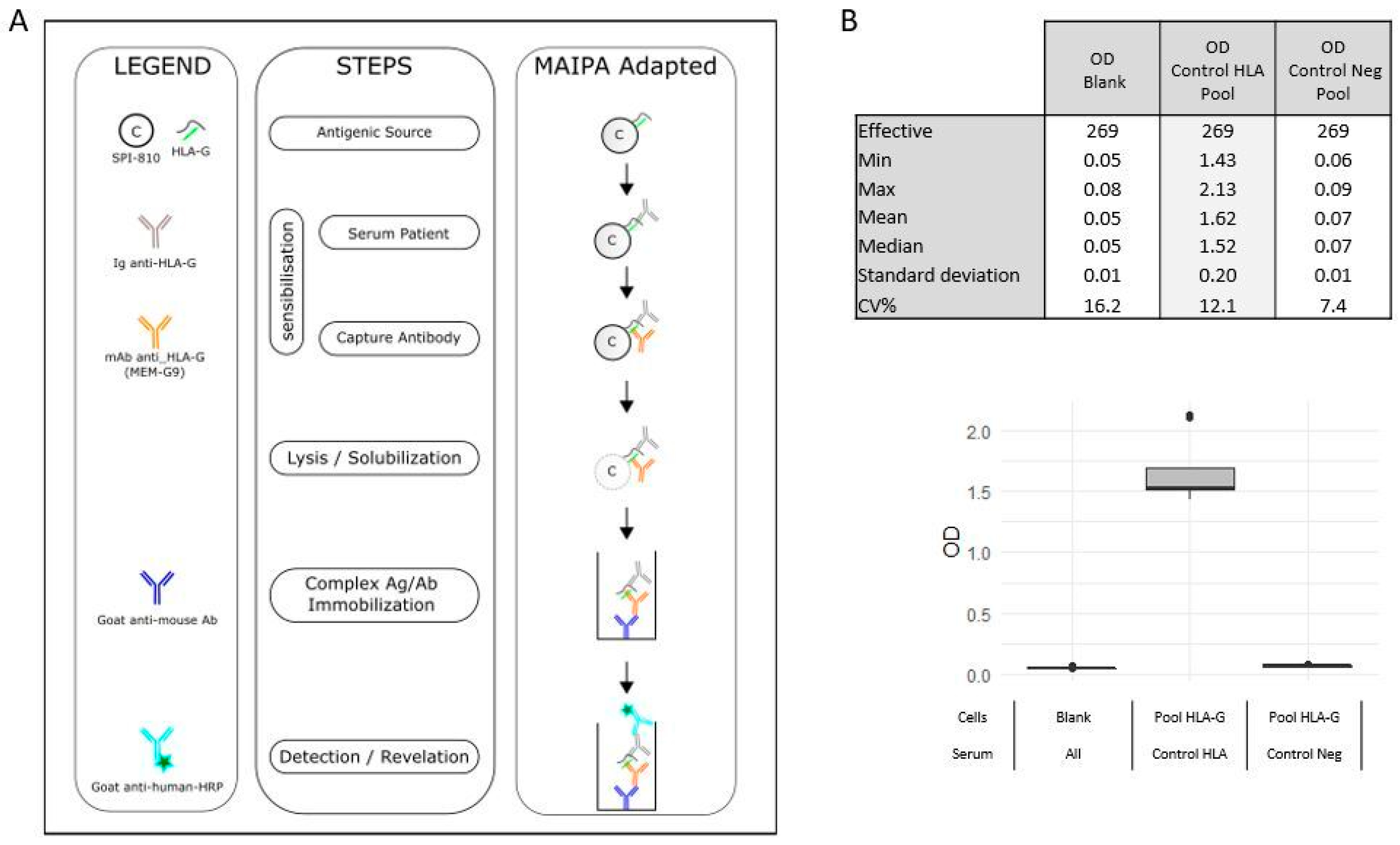
4.3. HLA-G Antibodies Detection

HLA-G antibodies were screened using an adapted MAIPA protocol, in accordance with the MAIPA ApDIA® kit. This kit allows the detection and identification of anti-platelet glycoprotein autoantibodies or anti-HPA (human platelet antigen) alloantibodies in serum using a platelet panel and platelet glycoprotein antigen immobilized using a monoclonal antibody. For the purpose of the present study, all MAIPA ApDIA® kit reagents were used, except for the platelet panel and platelet glycoprotein antigen, which were replaced by HLA-G transduced cell lines and WT.
Briefly, the adapted assay (Figure 2A) consisted of two consecutive incubations: sera with HLA-G transduced cell line and then with MEM-G/9 monoclonal antibody. Cells were lysed and solubilized complexes were immobilized on a plate coated with anti-mouse antibody. Complexes were revealed using immunoassay colorimetry.
Serum HLA-G antibodies were detected with a pool of the three HLA-G transduced and WT cells and identified with each HLA-G transduced cell and WT cell. Negative control (Contr Neg), positive control (Contr HLA), both supplied with the MAIPA ApDIA® kit, and two blank controls were included in each assay (Figure 2B).
Detection and identification protocol consisted of incubation (30 ± 5 min at 36 ± 1 °C) of transduced or WT cells (N = 1 × 10⁶), washed and adjusted in each well in PBS/1% BSA/0.33% EDTA with the positive control (Contr HLA) or negative control (Contr Neg), (50 µL dilution 1/1000) or with serum (50 µL dilution 1/5). Microplates were centrifuged (1000× g, 3 min), and each well was washed three times (ELISA Wash Buffer). MEM-G/9 monoclonal antibody was added (50 μg) and incubated for 30 ± 5 min at 36 ± 1 °C.
Cells were washed as described above and lysed (platelet lysis buffer containing triton, 130 µL, 15 min at 2–8 °C). Cell lysis supernatant (100 μL) containing the solubilized captured complex (MEM-G9/HLA-G/sera or control anti-HLA-G) were centrifuged and were incubated in a goat anti-mouse IgG-coated microplate (30 ± 5 min at 36 ± 1 °C). Plates were washed as described above, and immobilized complexes were revealed using a peroxidase reaction (substrate solution Chrom, 15 min at 36 ± 1 °C in the dark). The reaction was stopped (stop solution, 100 μL) and read with optical density (OD) using a spectrophotometer (450 nm with reference filter 650 nm).
The assay was validated when OD values were below 0.1 for the negative control (Contr Neg) and above 2 for the positive control (Contr HLA). The cut-off OD value was set at 0.15 (Contr Neg mean OD values adjusted with blank plus 3xStandard Deviation). OD values above the cut-off were considered positive. The signal-to-noise (S/N) ratio was calculated by dividing the OD value of HLA-G or HLA-E wells by that of WT cells. Serum was considered positive for HLA-G antibodies presence when the S/N ratio was >1.5.
Screening assays were performed for each serum. Identification was performed for sera with a HLA-G screening pool and OD values > 0.150, and S/N ratio > 1.5.

Supplementary Materials

The following supporting information can be downloaded at https://www.mdpi.com/article/10.3390/ijms242216479/s1.

Author Contributions

P.P. and L.H. supervised the study. J.B.B. and A.T. performed the technical analyses. F.J., C.B. and A.B. participated in the technical expertise. F.C., J.C., M.S. and C.P. assisted in the interpretation of the results. M.R.-G. and B.C. collected the clinical data from the patients. All authors have read and agreed to the published version of the manuscript.

Funding

This research received no external funding.

Institutional Review Board Statement

The study was conducted in accordance with the Declaration of Helsinki, and approved by the Ethics Committee of CPP 2009-R4 (COLT, 24 February 2009).

Informed Consent Statement

Informed consent was obtained from all subjects involved in the study.

Data Availability Statement

Data are contained within the article or Supplementary Material.

Acknowledgments

We thank Yasmina Denhadji and Ludovic Choucha, two technicians of Platelet Immunology department from EFS PACC, for their assistance and Claude Bagnis for assisting in the construction of HLA-G cell lines.

Conflicts of Interest

The authors declare no conflict of interest.

References

  1. Tabarelli, W.; Bonatti, H.; Tabarelli, D.; Eller, M.; Müller, L.; Ruttmann, E.; Lass-Flörl, C.; Larcher, C.; Geltner, C. Long term complications following 54 consecutive lung transplants. J. Thorac. Dis. 2016, 8, 1234–1244. [Google Scholar] [CrossRef] [PubMed]
  2. Moreau, A.; Varey, E.; Anegon, I.; Cuturi, M.-C. Effector Mechanisms of Rejection. Cold Spring Harb. Perspect. Med. 2013, 3, a015461. [Google Scholar] [CrossRef] [PubMed]
  3. Banan, B.; Xu, Z.; Gunasekaran, M.; Mohanakumar, T. Role of alloimmunity and autoimmunity in allograft rejection. Clin. Transpl. 2013, 39, 325–332. [Google Scholar]
  4. Bharat, A.; Saini, D.; Steward, N.; Hachem, R.; Trulock, E.P.; Patterson, G.A.; Meyers, B.F.; Mohanakumar, T. Antibodies to Self-Antigens Predispose to Primary Lung Allograft Dysfunction and Chronic Rejection. Ann. Thorac. Surg. 2010, 90, 1094–1101. [Google Scholar] [CrossRef] [PubMed]
  5. Rao, U.; Sharma, M.; Mohanakumar, T.; Ahn, C.; Gao, A.; Kaza, V. Prevalence of antibodies to lung self-antigens (Kα1 tubulin and collagen V) and donor specific antibodies to HLA in lung transplant recipients and implications for lung transplant outcomes: Single center experience. Transpl. Immunol. 2019, 54, 65–72. [Google Scholar] [CrossRef] [PubMed]
  6. Dragun, D.; Catar, R.; Philippe, A. Non-HLA antibodies in solid organ transplantation: Recent concepts and clinical relevance. Curr. Opin. Organ Transplant. 2013, 18, 430–435. [Google Scholar] [CrossRef] [PubMed]
  7. Ravichandran, R.; Bansal, S.; Rahman, M.; Sharma, M.; Liu, W.; Bharat, A.; Hachem, R.; Omar, A.; Smith, M.A.; Mohanakumar, T. The role of donor-derived exosomes in lung allograft rejection. Hum. Immunol. 2019, 80, 588–594. [Google Scholar] [CrossRef]
  8. Brugière, O.; Thabut, G.; Krawice-Radanne, I.; Rizzo, R.; Dauriat, G.; Danel, C.; Suberbielle, C.; Mal, H.; Stern, M.; Schilte, C.; et al. Role of HLA-G as a Predictive Marker of Low Risk of Chronic Rejection in Lung Transplant Recipients: A Clinical Prospective Study. Am. J. Transplant. 2015, 15, 461–471. [Google Scholar] [CrossRef]
  9. Di Cristofaro, J.; Reynaud-Gaubert, M.; Carlini, F.; Roubertoux, P.; Loundou, A.; Basire, A.; Frassati, C.; Thomas, P.; Gomez, C.; Picard, C. HLA-G*01:04∼UTR3 Recipient Correlates With Lower Survival and Higher Frequency of Chronic Rejection After Lung Transplantation: HLA-G and LTx Outcome. Am. J. Transplant. 2015, 15, 2413–2420. [Google Scholar] [CrossRef]
  10. Rizzo, R.; Bortolotti, D.; Bolzani, S.; Fainardi, E. HLA-G Molecules in Autoimmune Diseases and Infections. Front. Immunol. 2014, 5, 592. [Google Scholar] [CrossRef] [PubMed]
  11. Morandi, F.; Pistoia, V. Interactions between HLA-G and HLA-E in Physiological and Pathological Conditions. Front. Immunol. 2014, 5, 394. [Google Scholar] [CrossRef] [PubMed]
  12. Grant, E.J.; Nguyen, A.T.; Lobos, C.A.; Szeto, C.; Chatzileontiadou, D.S.M.; Gras, S. The unconventional role of HLA-E: The road less traveled. Mol. Immunol. 2020, 120, 101–112. [Google Scholar] [CrossRef]
  13. Brugière, O.; Thabut, G.; Pretolani, M.; Krawice-Radanne, I.; Dill, C.; Herbreteau, A.; Poras, I.; Moreau, P.; Colombat, M.; Danel, C.; et al. Immunohistochemical Study of HLA-G Expression in Lung Transplant Recipients. Am. J. Transplant. 2009, 9, 1427–1438. [Google Scholar] [CrossRef]
  14. White, S.R.; Floreth, T.; Liao, C.; Bhorade, S.M. Association of Soluble HLA-G with Acute Rejection Episodes and Early Development of Bronchiolitis Obliterans in Lung Transplantation. PLoS ONE 2014, 9, e103643. [Google Scholar] [CrossRef]
  15. Carlini, F.; Traore, K.; Cherouat, N.; Roubertoux, P.; Buhler, S.; Cortey, M.; Simon, S.; Doumbo, O.; Chiaroni, J.; Picard, C.; et al. HLA-G UTR Haplotype Conservation in the Malian Population: Association with Soluble HLA-G. PLoS ONE 2013, 8, e82517. [Google Scholar] [CrossRef]
  16. Castelli, E.C.; Ramalho, J.; Porto, I.O.P.; Lima, T.H.A.; Felà cio, L.P.; Sabbagh, A.; Donadi, E.A.; Mendes-Junior, C.T. Insights into HLA-G Genetics Provided by Worldwide Haplotype Diversity. Front. Immunol. 2014, 5, 476. [Google Scholar] [CrossRef]
  17. Rousseau, P.; Le Discorde, M.; Mouillot, G.; Marcou, C.; Carosella, E.D.; Moreau, P. The 14 bp Deletion-Insertion polymorphism in the 3′ UT region of the HLA-G gene influences HLA-G mRNA stability. Hum. Immunol. 2003, 64, 1005–1010. [Google Scholar] [CrossRef] [PubMed]
  18. Di Cristofaro, J.; El Moujally, D.; Agnel, A.; Mazières, S.; Cortey, M.; Basire, A.; Chiaroni, J.; Picard, C. HLA-G haplotype structure shows good conservation between different populations and good correlation with high, normal and low soluble HLA-G expression. Hum. Immunol. 2013, 74, 203–206. [Google Scholar] [CrossRef]
  19. Jucaud, V.; Ravindranath, M.H.; Terasaki, P.I.; Morales-Buenrostro, L.E.; Hiepe, F.; Rose, T.; Biesen, R. Serum antibodies to human leucocyte antigen (HLA)-E, HLA-F and HLA-G in patients with systemic lupus erythematosus (SLE) during disease flares: Clinical relevance of HLA-F autoantibodies. Clin. Exp. Immunol. 2016, 183, 326–340. [Google Scholar] [CrossRef] [PubMed]
  20. Negrini, S.; Contini, P.; Pupo, F.; Greco, M.; Murdaca, G.; Puppo, F. Expression of membrane-bound human leucocyte antigen-G in systemic sclerosis and systemic lupus erythematosus. Hum. Immunol. 2020, 81, 162–167. [Google Scholar] [CrossRef]
  21. de Freitas Dutra, V.; Costa, T.H.; Santos, L.D.; Sirianni, M.F.M.; Aravechia, M.G.; Kutner, J.M.; Bub, C.B. Platelet antibodies identification: Comparison between two laboratory tests. Hematol. Transfus. Cell Ther. 2022, 44, 365–368. [Google Scholar] [CrossRef] [PubMed]
  22. Morales-Buenrostro, L.E.; Terasaki, P.I.; Marino-Vázquez, L.A.; Lee, J.-H.; El-Awar, N.; Alberú, J. “Natural” Human Leukocyte Antigen Antibodies Found in Nonalloimmunized Healthy Males. Transplantation 2008, 86, 1111–1115. [Google Scholar] [CrossRef] [PubMed]
  23. Ravindranath, M.H.; Kaneku, H.; El-Awar, N.; Morales-Buenrostro, L.E.; Terasaki, P.I. Antibodies to HLA-E in Nonalloimmunized Males: Pattern of HLA-Ia Reactivity of Anti–HLA-E–Positive Sera. J. Immunol. 2010, 185, 1935–1948. [Google Scholar] [CrossRef]
  24. Grenzi, P.C.; de Marco, R.; Silva, R.Z.R.; Campos, É.F.; Gerbase-DeLima, M. Antibodies against denatured HLA class II molecules detected in luminex-single antigen assay. Hum. Immunol. 2013, 74, 1300–1303. [Google Scholar] [CrossRef]
  25. Ravindranath, M.H.; Pham, T.; Ozawa, M.; Terasaki, P.I. Antibodies to HLA-E may account for the non-donor-specific anti-HLA class-Ia antibodies in renal and liver transplant recipients. Int. Immunol. 2012, 24, 43–57. [Google Scholar] [CrossRef] [PubMed]
  26. Fournel, S.; Huc, X.; Aguerre-Girr, M.; Solier, C.; Legros, M.; Praud-Brethenou, C.; Moussa, M.; Chaouat, G.; Berrebi, A.; Bensussan, A.; et al. Comparative reactivity of different HLA-G monoclonal antibodies to soluble HLA-G molecules. Tissue Antigens 2000, 55, 510–518. [Google Scholar] [CrossRef]
  27. Zhao, L.; Teklemariam, T.; Hantash, B.M. Reassessment of HLA-G isoform specificity of MEM-G/9 and 4H84 monoclonal antibodies. Tissue Antigens 2012, 80, 231–238. [Google Scholar] [CrossRef] [PubMed]
  28. Furukawa, A.; Meguro, M.; Yamazaki, R.; Watanabe, H.; Takahashi, A.; Kuroki, K.; Maenaka, K. Evaluation of the Reactivity and Receptor Competition of HLA-G Isoforms toward Available Antibodies: Implications of Structural Characteristics of HLA-G Isoforms. Int. J. Mol. Sci. 2019, 20, 5947. [Google Scholar] [CrossRef]
  29. Ravindranath, M.H.; Ravindranath, N.M.; Selvan, S.R.; Hilali, F.E.; Amato-Menker, C.J.; Filippone, E.J. Cell Surface B2m-Free Human Leukocyte Antigen (HLA) Monomers and Dimers: Are They Neo-HLA Class and Proto-HLA? Biomolecules 2023, 13, 1178. [Google Scholar] [CrossRef]
  30. Naldini, L.; Blömer, U.; Gage, F.H.; Trono, D.; Verma, I.M. Efficient transfer, integration, and sustained long-term expression of the transgene in adult rat brains injected with a lentiviral vector. Proc. Natl. Acad. Sci. USA 1996, 93, 11382–11388. [Google Scholar] [CrossRef]
  31. Hayashi, T.; Amakishi, E. Detection of anti-human platelet antibodies against integrin a2ß1 using cell lines. Blood Transfus. 2014, 12, 273–280. [Google Scholar] [CrossRef]
  32. Celik, A.A.; Simper, G.S.; Huyton, T.; Blasczyk, R.; Bade-Döding, C. HLA-G mediated immune regulation is impaired by a single amino acid exchange in the alpha 2 domain. Hum. Immunol. 2018, 79, 453–462. [Google Scholar] [CrossRef] [PubMed]
Figure 1. Expression of HLA-G isoform on SPI801 cell lines. (A) Western blot analysis of HLA-G in the transduced cells in comparison with non-transduced cells line (WT). (B) Flow cytometry results showing stable expression levels of HLA-G isoforms in transduced cell lines (black) and non-transduced cell lines (gray).
Ijms 24 16479 g001
Figure 2. (A). Illustration of the different technical steps, adaptations, and compounds for MAIPA-adapted process. (B). Characteristics of MAIPA-adapted quality controls.
Ijms 24 16479 g002
Table 1. Clinical and biological characteristics of all cohort LTRs, LTRs with at least one positive serum detected on transduced and non-transduced cell lines, and healthy subjects (LTR: lung transplant recipient, DSA: donor-specific antibody).
CohortLTRsHealthy
LTRsWith Positive SerumBlood
Patient/donor
Number 351290
Age
   mean4036.539.5
   min-max19–6621–6618–69
   <40 years18753
   ≥40 years17537
Sexe
   M17541
   F18749
  PregnancyNDND30
     First pregancyNDND
     >4 pregnanciesNDND
Blood Group
   A12435
   B737
   AB228
   O14340
Pathology
Emphysema73
Cystic Fibrosis175
Fibrosis93
Bronchial dysplasia11
Histocytosis10
Type of LTx
Single LTx93
Bilateral LTx269
Mismatch HLA
052
1–421
5–8289
CMV Status
Absence mismatch258
D+ and R− without seroconversion10
D+ and R− with seroconversion41
D− and R+53
Type of rejection
Acute (DSA)72
Chronic (DSA)71
HLA antibodies
DSA after LTx268
Ab No DSA after LTx115
        DO21
        M152
        M362
        M1220
HLA-G genotyping
G*01:01,*--216
G*01:01,G*01:0643
G*01:01,G*01:0462
G*01:01,G*01:05N20
G*01:01,G*01:03 10
G*01:03,G*01:0611
Table 2. Results of adapted MAIPA on healthy subjects.
WomenMenPost-Partum
Women
Total
Effective494116105
Serum reactivity vs. Pool HLA-G cells
Negative494016103
Positive1102
Serum reactivity (screnning OD > 0.150 and S/N ratio > 1.5) vs. identification
Negative0101
Positive1001
Table 3. Results of adapted MAIPA in LTRs cohort at D0, D15, M1, M3, and M12 after LTx. Positive threshold: OD > 0.150 (OD: optical density, WT: wild type, and ND: not determined).
Collection Dates (D: Day; M: Month)Characteristic of PathologyTypage HLA-G
ODD15 M1 M3 M12
PatientsOD OD PoolODOD PoolOD OD PoolOD OD Pool ODOD PoolDiseaseAcute RejectionChronic Rejection DSA CMV StatusType of LTxHLAHLA-GHaplotypes UTR
WT CellsHLA-G CellsWT CellsHLA-G CellsWT CellsHLA-G CellsWT CellsHLA-G CellsWT CellsHLA-G CellsMismatchGenotyping
1NRNR0.0550.0660.0540.0580.0520.0730.0550.056Histiocytosis YesD150bilateral8G*01:01,*--UTR2,UTR4
20.0990.0820.0590.0710.0710.0610.0850.0580.070.06Cystic Fibrosis 0bilateral7G*01:01,*--UTR1,UTR1
30.0620.0580.0510.063NRNR0.0740.060.0590.058Fibrosis M10bilateral8G*01:01,*--UTR1,UTR1
40.1150.1360.1540.1270.1150.1060.060.0630.0840.092FibrosisM12 D153bilateral0G*01:01,*--UTR1,UTR1
50.1490.1820.0640.080.090.1080.0760.0820.0710.107Emphysema 0single3G*01:01,*--UTR1,UTR4
60.0780.077NRNR0.0580.0610.0520.0590.0510.056Cystic fibrosis 3bilateral8G*01:01,*--UTR1,UTR2
70.060.0650.0560.0580.0510.0510.0610.0670.0610.067Emphysema 2bilateral6G*01:01,G*01:06UTR2,UTR6
8NRNRNRNRNRNR0.0680.072NRNRCystic fibrosis M10bilateral6G*01:01,G*01:04UTR1,UTR3
90.0620.1050.0960.090.2620.3250.0580.0760.0570.088Emphysema D00bilateral6G*01:01,G*01:06UTR2,UTR2
100.6450.3490.0640.0570.1670.0810.0740.0770.0720.068Cystic fibrosis M30bilateral6G*01:01,G*01:04UTR3,UTR6
110.3850.3940.0650.074NRNRNRNRNRNREmphysema D150bilateral0G*01:01,G*01:06UTR2,UTR2
120.050.0630.0610.0660.0590.070.0610.060.0520.059Cystic fibrosisM1 D150single8G*01:01,G*01:05NUTR4,UTR3
130.1020.1030.0640.0620.0880.0840.070.0570.0660.072Cystic fibrosis M10single6G*01:01,*--UTR1,UTR2
140.350.357NRNR0.0570.060.0610.0590.090.068Fibrosis M10bilateral8G*01:01,G*01:04UTR3,UTR7
150.0860.0780.0540.0570.0640.060.0680.0640.0570.078Cystic fibrosis M10bilateral7G*01:01,*--UTR1,UTR2
160.060.06NRNR0.290.230.060.060.060.07Fibrosis 2single7G*01:01,*--UTR2,UTR6
170.1010.0880.0530.1020.0640.0760.0780.0850.070.07Fibrosis D150bilateral4G*01:01,G*01:03 UTR1, UTR2
18NRNRNRNRNRNR0.0570.055NRNRCystic fibrosis M10bilateral5G*01:01,*--UTR4,UTR2
190.7280.5640.1340.1040.1120.0940.0810.0730.080.074Cystic fibrosis D150bilateral8G*01:03,G*01:06UTR2,UTR5
200.1220.0810.3140.2470.0540.0590.0570.070.060.077Cystic fibrosis 0single6G*01:01,*--UTR1,UTR6
210.0780.0680.0710.058NRNR0.0510.0590.070.093FibrosisM3YesM10bilateral0G*01:01,G*01:04UTR4,UTR3
220.1380.1430.0590.0540.0590.0630.0580.070.0550.075Emphysema YesD152bilateral5G*01:01,*--UTR1,UTR2
230.1940.2090.1160.1780.1180.3010.1170.192NRNRBronchial Dysplasia YesD03bilateral7G*01:01,*--UTR4,UTR6
240.0760.070.0580.0590.1340.0550.0580.0550.0650.069Cystic fibrosis 0bilateral0G*01:01,G*01:05NUTR1,UTR2
250.110.1170.10.0720.1940.1940.4180.4660.0820.068Fibrosis 0single6G*01:01,*--UTR4,UTR7
26NRNR0.0770.081NRNRNRNR0.0560.068cystic Fibrosis D150bilateral6G*01:01,*--UTR1,UTR2
270.0590.0650.0610.0580.0550.0690.0590.0580.070.063EmphysemaM12YesM10single7G*01:01,*--UTR1,UTR2
280.1690.2080.0570.0590.1250.0740.0610.0740.060.063Cystic fibrosisM3 M13bilateral7G*01:01,*--UTR2,UTR4
29NRNRNRNRNRNR0.070.058NRNRCystic fibrosis 1bilateral7G*01:01,G*01:04UTR1,UTR3
300.0730.081NRNR0.0780.0960.0580.067NRNRCystic fibrosisM3 M12bilateral7G*01:01,*--UTR1,UTR2
310.0830.077NRNR0.0550.0620.0560.0540.0640.069Cystic fibrosis YesM10bilateral8G*01:01,*--UTR7,UTR1
320.070.1010.0670.0680.0940.0790.1390.1540.0710.061Fibrosis D03single6G*01:01,G*01:04UTR1,UTR3
330.610.3970.0570.0540.110.10.0660.0580.0880.065Cystic fibrosisM3 M10bilateral0G*01:01,G*01:06UTR2,UTR2
34NRNRNRNR0.060.066NRNR0.0530.058Emphysema M10single5G*01:01,*--UTR1,UTR7
350.0770.1NRNR0.0760.0840.0630.0760.080.131Fibrosis YesM10bilateral7G*01:01,*--UTR1, UTR1
Disclaimer/Publisher’s Note: The statements, opinions and data contained in all publications are solely those of the individual author(s) and contributor(s) and not of MDPI and/or the editor(s). MDPI and/or the editor(s) disclaim responsibility for any injury to people or property resulting from any ideas, methods, instructions or products referred to in the content.

