Review

Genetic Concordance in Primary Cutaneous Melanoma and Matched Metastasis: A Systematic Review and Meta-Analysis

by
Thamila Kerkour
1,
Catherine Zhou
1,
Loes Hollestein
1 and
Antien Mooyaart
2,*
1
Department of Dermatology, Erasmus MC Cancer Institute, 3015 GD Rotterdam, The Netherlands
2
Department of Pathology, Erasmus MC Cancer Institute, 3015 GD Rotterdam, The Netherlands
*
Author to whom correspondence should be addressed.
Int. J. Mol. Sci. 2023, 24(22), 16281; https://doi.org/10.3390/ijms242216281
Submission received: 19 October 2023 / Revised: 10 November 2023 / Accepted: 12 November 2023 / Published: 14 November 2023
(This article belongs to the Special Issue Melanoma: From Molecular Pathology to Therapeutic Approaches)

Abstract

:
Studying primary melanoma and its corresponding metastasis has twofold benefits. Firstly, to better understand tumor biology, and secondly, to determine which sample should be examined in assessing drug targets. This study systematically analyzed all the literature on primary melanoma and its matched metastasis. Following PRISMA guidelines, we searched multiple medical databases for relevant publications from January 2000 to December 2022, assessed the quality of the primary-level studies using the QUIPS tool, and summarized the concordance rate of the most reported genes using the random-effects model. Finally, we evaluated the inter-study heterogeneity using the subgroup analysis. Thirty-one studies investigated the concordance of BRAF and NRAS in 1220 and 629 patients, respectively. The pooled concordance rate was 89.4% [95% CI: 84.5; 93.5] for BRAF and 97.8% [95% CI: 95.8; 99.4] for NRAS. When high-quality studies were considered, only BRAF mutation status consistency increased. Five studies reported the concordance status of c-KIT (93%, 44 patients) and TERT promoter (64%, 53 patients). Lastly, three studies analyzed the concordance of cancer genes involved in the signaling pathways, apoptosis, and proliferation, such as CDKN2A (25%, four patients), TP53 (44%, nine patients), and PIK3CA (20%, five patients). Our study found that the concordance of known drug targets (mainly BRAF) during melanoma progression is higher than in previous meta-analyses, likely due to advances in molecular techniques. Furthermore, significant heterogeneity exists in the genes involved in the melanoma genetic makeup; although our results are based on small patient samples, more research is necessary for validation.

1. Introduction

Cutaneous melanoma (CM) is an aggressive tumor arising from the pigment-producing cells (melanocytes) located in the skin. The incidence of melanoma has steadily increased in many susceptible populations over the past five decades [1]. Patients diagnosed with localized disease undergo surgical resection with a favorable prognosis [2]. However, 40% of those patients develop metastatic disease, reducing the 5-year overall survival to less than 30% [3,4]. Unraveling the molecular biology of CM has revolutionized treatment with targeted therapy (BRAF and MEK inhibitors) and immunotherapy (anti-PD1 and anti-CTLA-4 agents) [5,6,7,8,9,10]. Patients with metastatic tumors undergo molecular testing for the BRAF V600 mutation to determine treatment options with BRAF/MEK inhibitors [7,11,12]. With the discovery of additional melanoma driver genes, strong efforts were made to develop targeted therapies for BRAF wild-type tumors [13]. Several researchers investigated potential targeted treatments for NRAS Q61 mutant melanomas. Ongoing clinical trials tested the new treatments with MEK and CD147i inhibitors in advanced melanoma after immunotherapy failure [14,15]. Moreover, further inhibitors are also available for melanoma harboring an amplified c-KIT gene [16,17]. The mutations in BRAF V600, NRAS Q61, and c-KIT occur early in melanoma development [18,19]. Consequently, if a patient develops a metastasis, the absence of these alterations in the primary tumor may be sufficient to exclude the patient from the targeted therapies. Otherwise, an invasive biopsy of the metastatic deposit needs to be performed. However, a meta-analysis conducted in 2017 showed an overall discrepancy rate of 13.4% of the BRAF status between the primary melanoma and the matched metastasis [20]. Hence, it is advised to determine the BRAF status in the metastasis deposit [21].
With advances in sequencing techniques for mutation detection, additional studies addressed the concordance of the mutation status of primary CM and matched metastasis in multiple cancer genes. However, the sensitivity of these methods for mutation detection has not yet been compared to older techniques. Therefore, collecting up-to-date information on the concordance status of BRAF and additional driver genes, along with assessing the quality of the new studies, may help to redirect the diagnosis and therapeutic decisions for CM patients. Additionally, understanding how CM tumors evolve and differ between primary and metastatic sites is important. Thus, in this study, we aimed to systematically review all the literature and undertake the meta-analysis where appropriate in order to determine the mutation concordance rate between primary CM and its matched metastatic sites.

2. Methods

We conducted this systematic review and meta-analysis in accordance with the Preferred Reporting Items for Systematic Review and Meta-Analysis (PRISMA) guidelines. The preregistered protocol is available at PROSPERO under the protocol number CRD42022327641, accessed on 15 May 2022 and available from: https://www.crd.york.ac.uk/prospero/display_record.php?ID=CRD42022327641.

2.1. Search Strategy and Eligibility Criteria

A comprehensive search strategy was employed to ensure the inclusion of relevant studies. We performed an initial search in Embase to detect the relevant keywords in the titles and the abstracts. The primary search terms used in Embase were “genetic features”, “primary cutaneous melanoma” and “metastatic melanoma”. The obtained synonymous terms from Embase were then added to enhance the search strategy. Articles published from 2000 to December 2022 were searched in the following databases: Medline, Embase, Web of Science, and the Cochrane Central Register of Controlled Trials. The search strategies are available in Supplementary Material Table S1. Two independent reviewers (C.Z. and T.K.) evaluated the titles and abstracts of the identified studies in separate EndNote libraries. Discrepancies were discussed with a third reviewer (A.M.). Inclusion criteria consisted of studies that compared the genetic patterns between primary CM and the matched metastasis within the same patient. Studies that only included uveal and mucosal melanoma were excluded, as well as conference abstracts and case studies.

2.2. Data Extraction and Quality Assessment

Full-text screening of all the relevant articles was conducted by two reviewers (C.Z. and T.K.). Data extraction was independently performed by the two reviewers (C.Z. and T.K.) using a customized data extraction Excel sheet. The following information was extracted from each study: The first author’s name and their affiliated country, the year of the publication, the technique used to determine the mutation status, the analyzed genes, the total number of patients, and the number of patients with concordant status for each gene. To assess the quality of each included study, one reviewer (T.K.) used the Quality in Prognosis Studies (QUIPS) tool [22] and discussed the outcomes with a second reviewer (A.M.) to ensure consensus. This tool consists of six domains: Study participation, study attrition, prognosis actor measurement, study confounding, statistical analysis, and reporting. For each domain, the score was assigned: High risk of bias = 0, moderate risk of bias = 0.5, and low risk of bias = 1. Studies with a total score of ≥4 were considered high quality, whereas studies with a total score of <4 were considered low quality.

2.3. Outcome of Interest

The outcome of interest in the meta-analysis is defined as the concordance rate of the mutation status between the primary cutaneous melanoma and their paired distant metastasis in each single gene. For this study, we considered meta-analysis only for the genes that were reported in more than ten studies. Therefore, we conducted the meta-analysis for BRAF V600 and NRAS Q61 mutations. We reported the available data regarding additional genes in a systematic manner.

2.4. Pathway Analysis

To determine if the mutations are associated with pathways that may be involved in the metastasis progression, we used the web-free server g:profiler [23] to identify the possible pathways associated with the mutated genes that we mutated in both primary and metastasis. We selected the top three pathways with the highest p-value. g:profiler output list details are presented in Supplementary Material Figure S1.

2.5. Statistical Analysis

Differences in the concordance of BRAF status between male and female patients were assessed using a Student’s t-test. Random-effects meta-analysis models were used to pool the concordance rate and the confidence interval (CI) across all the studies for each gene (BRAF and NRAS) because combining all the genes may be biased. Freeman–Tukey double arcsine transformation was applied to weigh the individual studies [24]. To assess the heterogeneity across the studies, we performed a chi-square test to estimate the I2 statistic (0–100%, 0% indicated no heterogeneity).
To explain potential sources of heterogeneity, pre-specified potential effect modifiers, such as the technique of detecting the mutation (molecular vs. immunohistochemistry) and the study quality (QUIPS total score), were considered. Subgroup analyses were used to calculate the concordance rates for each group. Small study effects and publication bias were assessed using funnel plots and Egger’s test (Supplementary Material Figure S2). The analyses were performed using the meta [25] and the metaphor packages [26]. All the analyses were done in R version 4.2.13.

3. Results

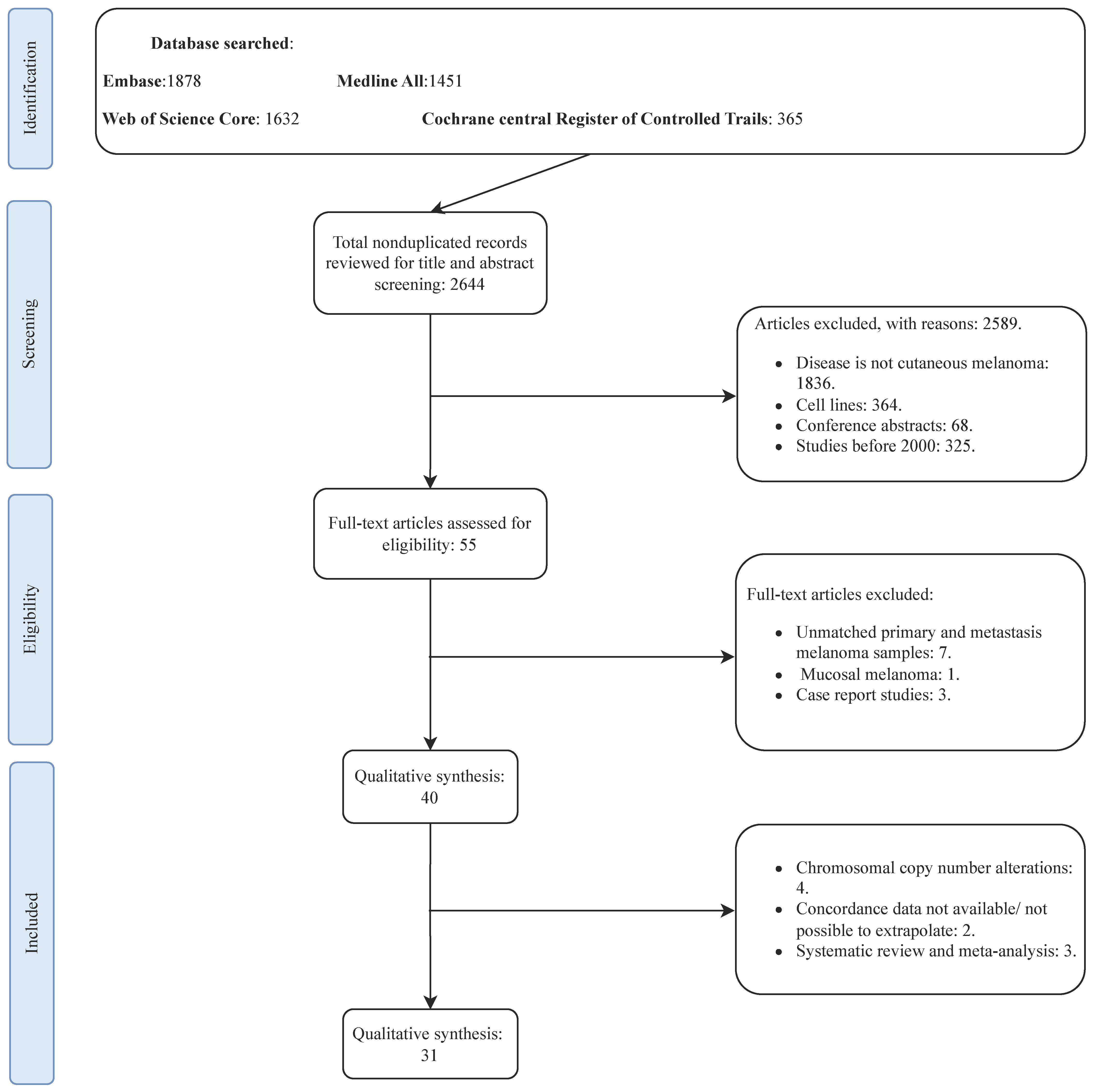
3.1. Search Results and Studies Characteristics

The PRISMA flowchart of the selection process is presented in Figure 1. A total of 2466 studies (after excluding duplicates) were identified from the selected databases. Screening and evaluation of eligibility criteria resulted in 40 studies being considered for qualitative assessment, with 31 included in the quantitative assessment. The majority of studies included in the meta-analysis (25) focused mainly on BRAF concordance status. For the studies that included mucosal or uveal melanomas, we only extrapolated the mutation data of patients with CM.

3.2. The Genetic Concordance between Primary Cutaneous Melanoma and Matched Distant Metastasis

The concordance in patients with cutaneous melanoma was reported mainly in the melanoma driver genes. Twenty-five studies [27,28,29,30,31,32,33,34,35,36,37,38,39,40,41,42,43,44,45,46,47,48,49,50,51] compared the mutation status of BRAF with the median concordance rate of 88% (range 56–100, total patients = 1220) (Table 1). BRAF was mainly reported as either muted in position V600 or wild type. NRAS status was the second most highly reported gene in 14 studies [28,30,32,38,43,45,48,49,50,51,52,53,54,55] (Table 2), with a median concordance of 97% (range 85–100, total patients = 629), and the mutation site was mainly focused on the protein position Q61. c-KIT concordance was reported in 3 studies [32,38,49] with a median concordance rate of 100% (range 88–100, total patients = 44) (Table 3). Finally, the concordance of the TERT promoter was reported in two studies [45,50], and the media concordance was 64% (range 55–68, total patients = 53) (Table 4).
Three studies [43,48,49] compared the mutational status between primary melanoma and their matched metastasis tumors in multiple cancer genes. Details of each gene are described in Table 4. Most of the reported mutated genes were reported in both the primary and metastasis tissues, except KRAS G12A and NEK E379K mutations that were found in primary tumors and CCND1 G103R in the metastasis tumor. To understand the role of these mutations, we looked at their possible association with pathways known in cancer progression. Most of the genes are involved in proliferation, cell cycle, and cellular senescence. The details of the pathways analysis are reported in Supplementary Material Figure S1.

3.3. The Genetic Concordance between Primary Cutaneous Melanoma and Matched Distant Metastasis According to Metastatic Site and Gender

To determine the impact of the metastatic site and gender on the concordance rate between primary melanoma and metastatic deposit, we extracted data on the tissue type of the metastasis (Table 5) and patient gender (Table 6) from each study. However, due to the unavailability of data for NRAS and KIT, we could only report the data for BRAF.
The lymph node (excluding sentinel lymph node) was the most commonly reported metastatic site, with eight studies encompassing 262 patients. The skin was the second most frequently reported site, with six studies comprising 84 patients. Other metastatic sites, including visceral, brain, and subcutaneous locations, were reported in limited studies, with 50, 29, and 18 patients, respectively. The concordance rates for BRAF according to gender were reported in 5 studies. Although males represented a slightly larger proportion, no statistically significant difference in the concordance rate between females and males was observed (p-value = 0.19).

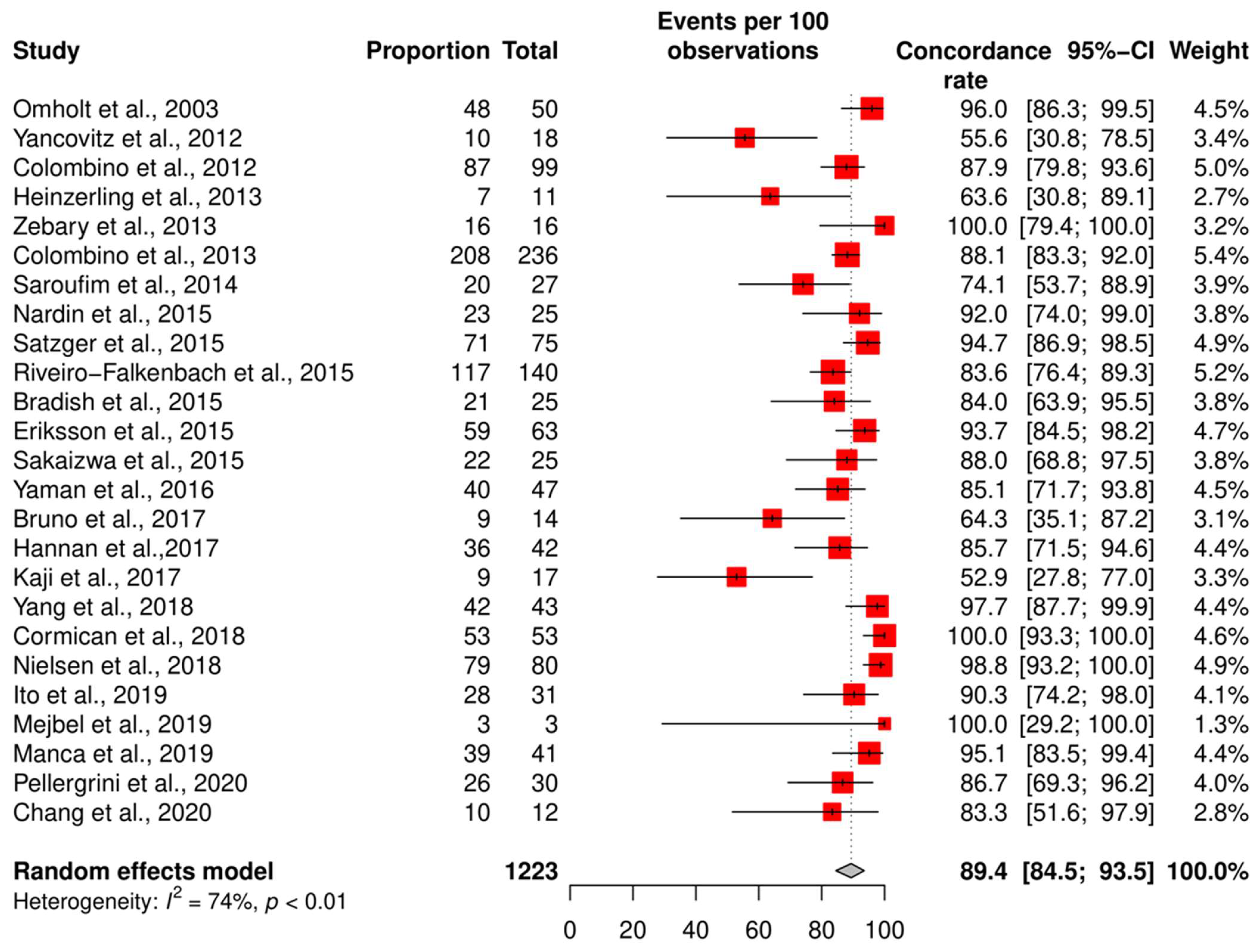
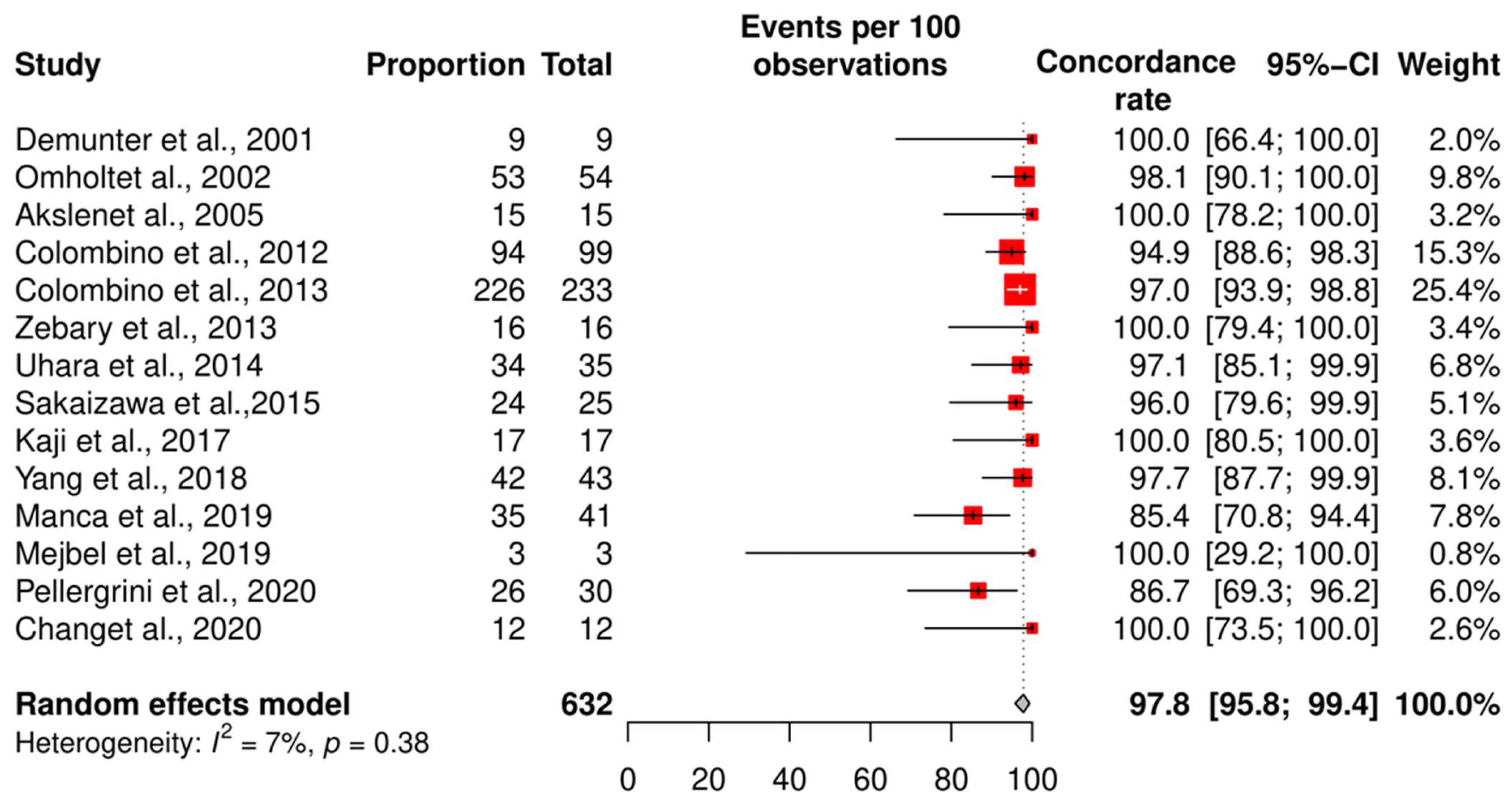
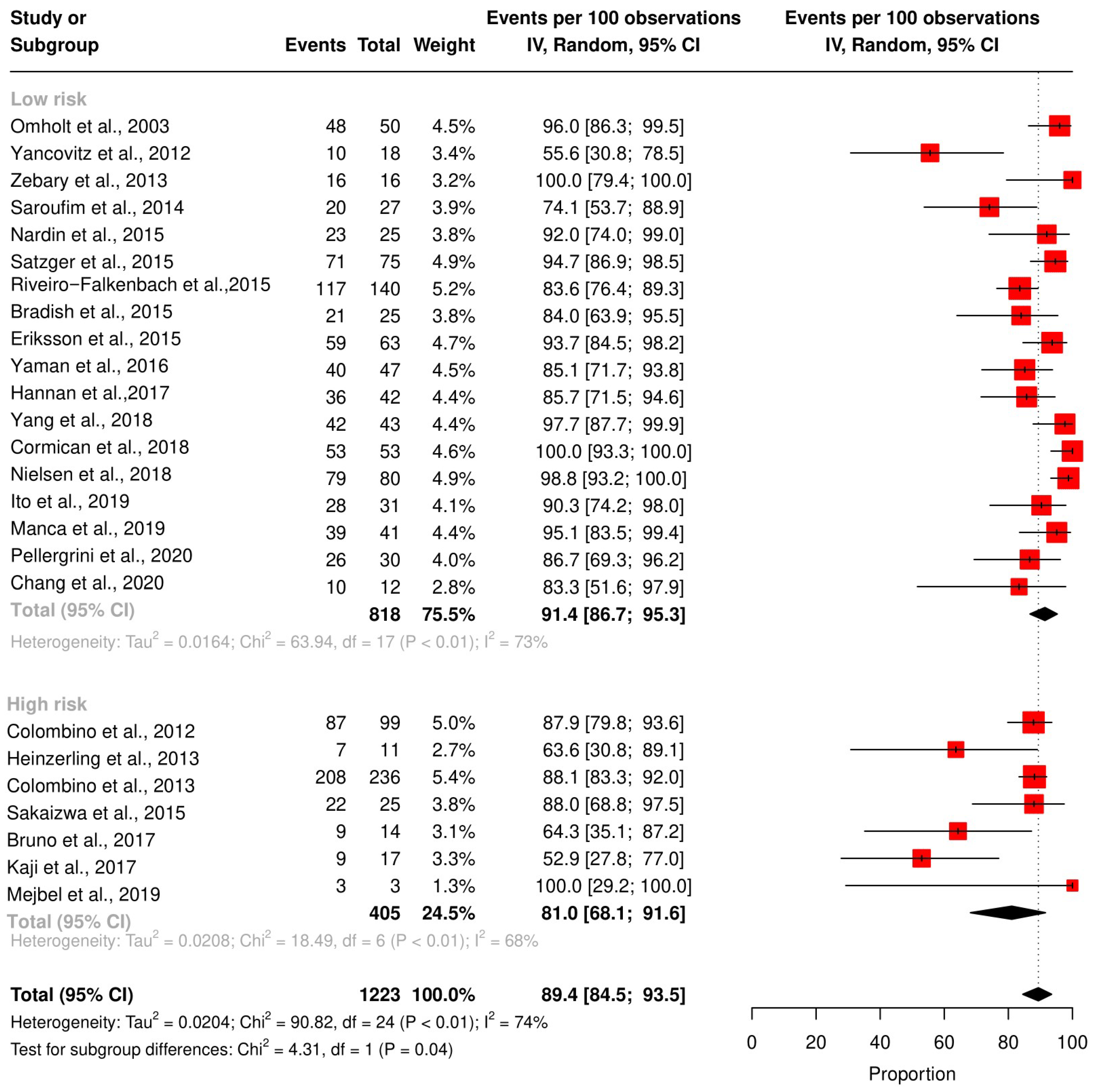
3.4. Meta-Analysis of the Concordance Rate for BRAF and NRAS

We assessed the concordance rate for BRAF in 1220 patients and for NRAS in 629 patients (Figure 2 and Figure 3). We calculated the concordance rate separately for wild-type versus mutated status for each gene. Because we focused on cutaneous melanoma in this study, we excluded uveal and mucosal melanoma patients in five studies [23,25,28,35,38].
The pooled random effects concordance rate for BRAF was 89.4% [95% CI: 84.5; 93.5] and 97.8% [95% CI: 95.8; 99.4] for NRAS. The heterogeneity between studies was high for the BRAF concordance rate (I2 = 74%, p < 0.01). However, for NRAS, the risk of bias was low (I2 = 7%, p = 0.38).

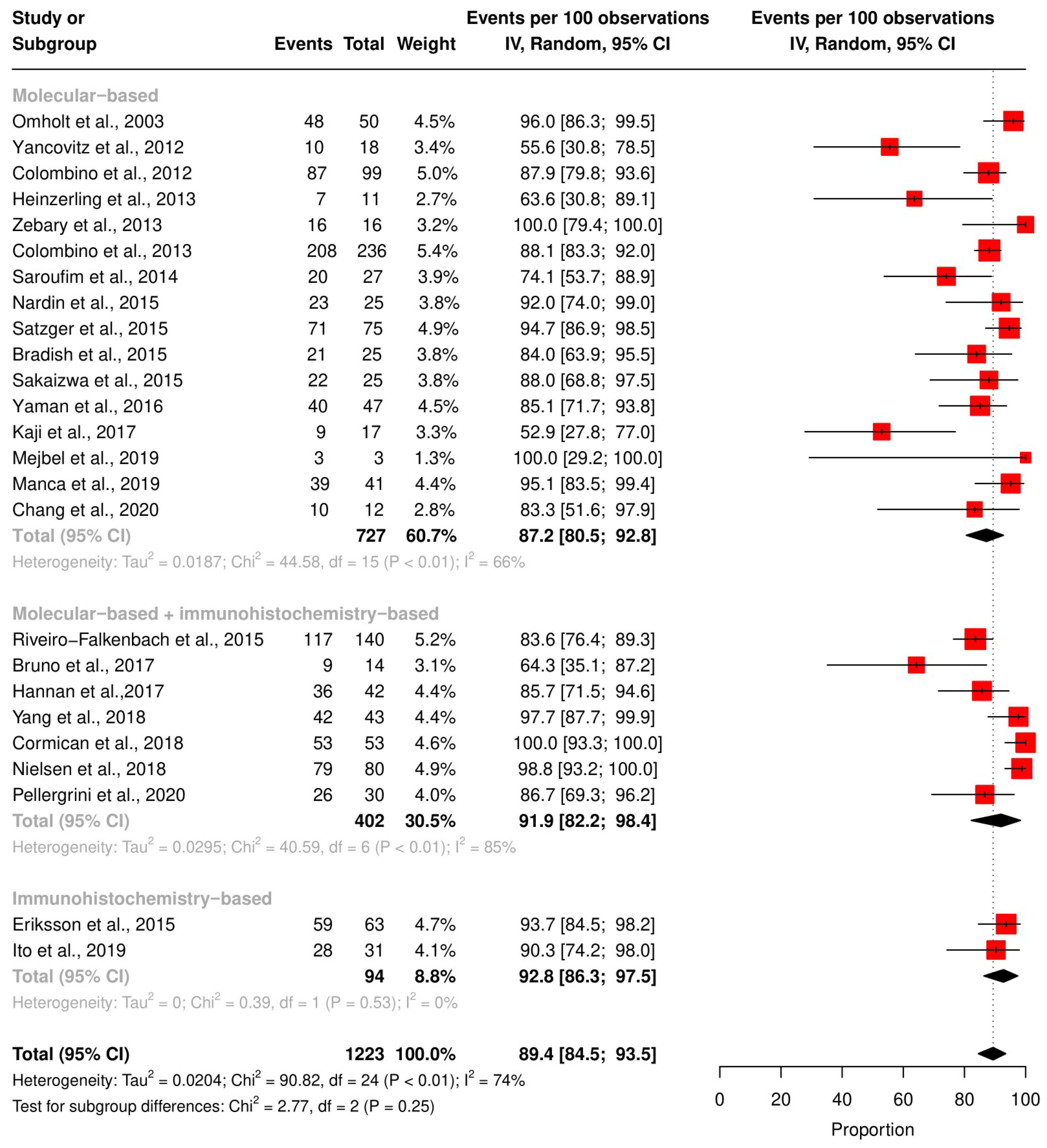
3.5. Subgroup Analysis According to the Technique and QUIPS Tool Score

The mutation status of BRAF and NRAS in the reported studies may be affected by the study design and the techniques used for mutation detection. For BRAF detection, 16 studies utilized molecular-based techniques, two utilized IHC, and seven used both. The pooled BRAF concordance rate was 86.4% [95% CI 79.6; 92.1] with a molecular-based technique, 92.8% [95% CI: 86.3; 97.5] with IHC, 81.9% [95% CI: 82.2; 98.4] with IHC, and a molecular-based technique combined. However, these differences were statistically insignificant (p-value = 0.24) (Figure 4). We did not perform subgroup analyses based on detection techniques on NRAS concordance because most of the studies used molecular-based techniques.
Other confounders, such as sample collection, analysis interpretation, and the type of statistical tests in the individual studies, may affect the mutation detections and, subsequently, the concordance rate. Therefore, we performed a subgroup analysis based on the QUIPS score for BRAF and NRAS concordance rates (Figure 5 and Figure 6). The pooled concordance rate for BRAF was 91.4% [95% CI: 86.7; 95.3] for studies with a high QUIPS score (≥4) and 81.0 [95% CI: 68.1; 91.6] for studies with a low QUIPS score (<4), (p-value = 0.03). The pooled concordance rate for NRAS was 95.7% [95% CI: 89.7; 99.5] for studies with a high QUIPS score (≥4) and 99.1% [95% CI: 97.3; 100] for studies with a low QUIPS score (<4). This difference was statistically insignificant (p-value = 0.38).

4. Discussion

In this study, we comprehensively analyzed the current knowledge on the concordance of mutated genes between primary CM and their matched metastasis. Most of the reviewed studies focused on the concordance of BRAF mutation status because it has clinical value in the treatment decision for BRAF inhibitors. The second-most studied gene was NRAS. The overall pooled concordance rates for BRAF and NRAS status were high (88.8% and 97.2%, respectively). However, the reported concordance rates in the individual studies varied widely, ranging from 56% to 100% for BRAF and 85% to 100% for NRAS. Among the factors that could explain this wide range are: Mutation detection technique, gender, metastatic site type, and study quality. We observed that all these factors were of influence on the concordance rate. First, we explored the impact of mutation detection techniques. For BRAF, the concordance rate was higher when an IHC-based technique was used (92.8%) and lower when molecular-based techniques were used (86.4%). Although we could not explore the effects of gender and metastatic sites with significant power, we did see for BRAF concordance rate the most discordant cases in the direction of female sex and skin as a metastatic site.
Only five studies [43,45,48,49,50], with study sizes ranging from 3 to 41 participants, analyzed the variability in the mutation profiles using panel cancer driver genes. The alterations in TERT, CDKN2A, TP53, RPPIK3CA, and EPHB6 were simultaneously detected in matched primary and metastasis tumors, albeit in only a small subset of patients. In contrast, other mutated genes were found exclusively in either primary or metastatic tumors. Predominantly, the majority of the genes displaying alterations were associated with critical pathways involved in tumor invasion, including those regulating proliferation, cell cycle, and apoptosis. This is in line with the current knowledge on melanoma progression [56,57,58]. In essence, investigating the sequential acquisition of mutations will be required to fully identify the key driver mutations that are responsible for the processes of invasion, adaptation, and, ultimately, melanoma metastasis dissemination [59].
CM presents cancer with a very high tumor mutational burden (TMB) [60]. This characteristic may lead to polyclonal formation within the individual primary melanoma tumor. In other words, a primary tumor can comprise a mixture of cells presenting different genetic profiles of the same gene, with the ability to metastasize to other tissue organs [61]. Two studies showed intratumoral heterogeneity within the same melanoma samples for BRAF and TERT mutation status using different micro-dissected regions of the same sample [29,50]. A meta-analysis reported similar observations when investigating EGFR and KRAS mutation status in matched primary tumors and distant metastasis of non-small cell lung cancer, which is also considered a tumor with a TMB [62]. Thus, intratumoral heterogeneity may be a common feature of high-TMB tumors. In our study, we also showed that additional mutations in genes such as KRAS and NEK10 were likely to be seen in primary melanoma. In contrast, mutations in genes such as TERT and CCND1 were likely present in metastasis. Thus, we highlight the need to investigate the clonality of more melanoma-specific genes, especially in the case of building metastasis risk prediction models that are based on the genetic profile of primary tumors, as the primary tumors may contain driver genes that are irrelevant for the recurrence of the metastasis. For example, Chang et al. have shown that within the same melanoma patients, the primary tumor had a BRAF mutant status, but the metastatic tumor had lost this mutation and acquired a new mutation in the TERT promoter [50]. As a result, a metastasis risk prediction model based on the BRAF mutation status of the primary tumor would not be accurate for this patient. Therefore, it is essential to develop metastasis risk prediction models that take into account the intratumoral heterogeneity of melanoma tumors. This could be done by sequencing multiple regions of the primary tumor or by sequencing both the primary and metastatic tumor tissues.
The accuracy of the mutation detection technique may have an effect when comparing the mutation status [63,64]. Bruno et al. tested the concordance of BRAF status between 25 paired primary and matched melanoma samples by applying both IHC and real-time PCR. Their study showed that IHC was more effective in detecting the signal of mutated BRAF V600E [41]. This finding suggested that IHC is the stable method for the BRAF testing method, especially for detecting the most common BRAF mutation, V600E. From a molecular point of view, IHC is subject to some limitations. For instance, IHC can be subjective as the pathologist’s interpretation affects the results. IHC may not detect mutations in all cases, particularly when the tumor sample is small or if the mutation is present in only a small percentage of tumor cells [65]. At the same time, molecular-based techniques such as real-time PCR are more objective and sensitive than IHC [66]. In the daily practice of BRAF testing, molecular techniques are considered the golden standard [67].
The tumors can evolve over time, and new mutations can arise in the metastases. Thus, the time to metastasis, which is defined as the time between the primary tumor and the metastasis tumor biopsy, may also affect the mutation concordance rate. In our study, we were unable to analyze the impact of time on metastasis as there were not enough eligible studies. It is essential to mention that no new studies have reported more extensive information on the timing of metastasis since the previous meta-analysis of BRAF [20]. In colorectal cancer, the concordance rate of KRAS status decreases when the time to metastasis increases [68,69]. Thus, it is recommended to perform more frequent surveillance for patients with a longer time to metastasis, as they may be at higher risk for developing metastases with a different KRAS mutation status [68].
The metastatic site plays a crucial role in changing the mutation status between matched primary and metastasis melanoma tumors. Firstly, the microenvironment of the metastasis site can exert a unique selection pressure on the melanoma cancer site. In other words, the metastasis tissue site can offer distinct biochemical, immune, and structural conditions that differ from the skin site. This means the melanoma cells can adapt and acquire new mutations to thrive in the specific microenvironment of the metastasis site. For example, the melanoma cells that metastasize to the brain may develop mutations that evade the immune system, as the brain is an immune-privileged site [70]. Melanoma that metastases to the lung may develop mutations that allow it to resist the effects of chemotherapy, as the lung is a vascularized organ [71].
The overall study quality had no significant effect on the concordance rates of NRAS mutation status. Yielding that the reliability of NRAS mutation status determination was fairly consistent across studies, regardless of their quality ratings. However, we observed higher concordance rates for BRAF mutation status in studies rated as high quality compared to studies rated as low quality. These findings suggest that beyond mutation detection techniques, other factors such as patient selection and time and type of fixation, as well as improper tumor sampling, may affect the concordance rate of the mutation profile [72]. From a biological standpoint, it is worth noting that both BRAF and NRAS mutations are present in nevi, suggesting that such mutations might represent early events that could already be present in all tumor cells. This is also supported by the results seen in immunohistochemistry (IHC) [29,73]. It appears that, based on the available data, discrepancies in the current literature may be attributed to sample management and mutation detection techniques rather than being solely due to tumor heterogeneity. Refining and standardizing sample collection and analysis procedures are crucial to enhancing the accuracy and consistency of mutation profiling in melanoma cancer research. Ultimately, this leads to more reliable insights into this complex disease.
Our study has several limitations; firstly, the original studies that we included in our meta-analysis for BRAF and NRAS status were relatively small, which limited the power of our study to detect statistically significant results. Secondly, most of the included studies used different criteria to select patients, mainly for the metastasis tumor (lymph node or distant metastasis). Lastly, we did not have access to clinical data for all of the patients in the original studies, which prevented us from analyzing the effect of clinical variables, such as the tumor thickness and the presence of the ulceration, on the concordance rate. Despite these limitations, our study presents the first meta-analysis that pooled the NRAS concordance rate in matched primary and metastasis of cutaneous melanoma. This is an important finding, as NRAS mutations are responsible for a significant proportion of melanomas, but no well-established NRAS-targeted therapies are yet known. In addition to the updated data about the pooled BRAF concordance rate from the previous meta-analysis.
Our results consolidate the evidence of the mutation concordance between primary melanoma and matched metastasis. We demonstrated a high concordance for BRAF and NRAS mutation status. We observed that the BRAF concordance rate is higher than reported in the last systematic review. This finding may suggest that advances have been made in molecular techniques and IHC techniques. Nevertheless, further investigations are required to comprehend the intricacies of melanoma, including its genetic heterogeneity and the complex interaction of multiple genes driving the metastatic progression. Expanding the scope of our study is essential to attaining a comprehensive understanding of tumor heterogeneity and the genetic changes that take place throughout the metastatic evolution of melanoma. This should include not only a wider range of genes but also the integration of more clinical information, as these multi-faceted forms of research will aid in uncovering the complexities of the genetic landscape of melanoma.

5. Conclusions

The complete explanation of the genomic background behind melanoma pathogenesis remains elusive. This study presents a summary of current understanding regarding the clonal relatedness of mutated genes in primary cutaneous melanoma and matched tumors. We draw attention to the knowledge gap regarding the genetic heterogeneity of melanoma-specific genes, which could serve as a valuable tool in identifying suitable patients for trials testing possible new targeted therapies. Furthermore, we found that CM presents a significant concordance in BRAF status between primary and metastatic tumors than in previous meta-analyses, probably due to technical advances, although a minority of patients showed inconsistencies. Therefore, we propose that molecular testing in metastatic tissue is a more appropriate method for determining BRAF status to tailor the treatment decision, although it is still reasonable to use the primary tumor in cases of difficulty. Further research should investigate the factors causing these discrepancies and the feasibility of using other molecular markers with BRAF status to better stratify patients for melanoma treatment.

Supplementary Materials

The following supporting information can be downloaded at: https://www.mdpi.com/article/10.3390/ijms242216281/s1.

Author Contributions

Conceptualization, T.K., L.H. and A.M.; methodology, T.K., L.H. and A.M.; software, T.K. and C.Z.; validation, T.K. and C.Z.; formal analysis, T.K.; resources, T.K. and C.Z.; writing—original draft preparation, T.K.; writing—review and editing, L.H., T.K., A.M. and C.Z.; visualization, T.K.; supervision, A.M. and L.H. All authors have read and agreed to the published version of the manuscript.

Funding

This research received no external funding.

Institutional Review Board Statement

Not applicable.

Informed Consent Statement

Not applicable.

Data Availability Statement

All data relevant to this study are included in the manuscript.

Acknowledgments

The authors thank W.M. Bramer of the Erasmus MC Medical Library for developing the search strategy.

Conflicts of Interest

The authors declare no conflict of interest.

References

  1. Whiteman, D.C.; Green, A.C.; Olsen, C.M. The growing burden of invasive melanoma: Projections of incidence rates and numbers of new cases in six susceptible populations through 2031. J. Investig. Dermatol. 2016, 136, 1161–1171. [Google Scholar] [CrossRef] [PubMed]
  2. Garbe, C.; Keim, U.; Amaral, T.; Berking, C.; Eigentler, T.K.; Flatz, L.; Gesierich, A.; Leiter, U.; Stadler, R.; Sunderkötter, C. Prognosis of patients with primary melanoma stage I and II according to American Joint Committee on Cancer Version 8 Validated in two independent cohorts: Implications for adjuvant treatment. J. Clin. Oncol. 2022, 40, 3741. [Google Scholar] [CrossRef] [PubMed]
  3. Ertekin, S.S.; Podlipnik, S.; Riquelme-Mc Loughlin, C.; Barreiro-Capurro, A.; Arance, A.; Carrera, C.C.; Malvehy, J.; Puig, S. Initial stage of cutaneous primary melanoma plays a key role in the pattern and timing of disease recurrence. Acta Derm. Venereol. 2021, 101, adv00502. [Google Scholar] [CrossRef] [PubMed]
  4. Zhou, C.; Louwman, M.; Wakkee, M.; Van der Veldt, A.; Grünhagen, D.; Verhoef, C.; Mooyaart, A.; Nijsten, T.; Hollestein, L. Primary melanoma characteristics of metastatic disease: A nationwide cancer registry study. Cancers 2021, 13, 4431. [Google Scholar] [CrossRef]
  5. Bedikian, A.Y.; DeConti, R.C.; Conry, R.; Agarwala, S.; Papadopoulos, N.; Kim, K.B.; Ernstoff, M. Phase 3 study of docosahexaenoic acid–paclitaxel versus dacarbazine in patients with metastatic malignant melanoma. Ann. Oncol. 2011, 22, 787–793. [Google Scholar] [CrossRef] [PubMed]
  6. Garbe, C.; Amaral, T.; Peris, K.; Hauschild, A.; Arenberger, P.; Bastholt, L.; Bataille, V.; Del Marmol, V.; Dréno, B.; Fargnoli, M.C. European consensus-based interdisciplinary guideline for melanoma. Part 2: Treatment–Update 2019. Eur. J. Cancer 2020, 126, 159–177. [Google Scholar] [CrossRef] [PubMed]
  7. Hodi, F.S.; O’Day, S.J.; McDermott, D.F.; Weber, R.W.; Sosman, J.A.; Haanen, J.B.; Gonzalez, R.; Robert, C.; Schadendorf, D.; Hassel, J.C. Improved survival with ipilimumab in patients with metastatic melanoma. N. Engl. J. Med. 2010, 363, 711–723. [Google Scholar] [CrossRef]
  8. Larkin, J.; Minor, D.; D’Angelo, S.; Neyns, B.; Smylie, M.; Miller Jr, W.H.; Gutzmer, R.; Linette, G.; Chmielowski, B.; Lao, C.D. Overall survival in patients with advanced melanoma who received nivolumab versus investigator’s choice chemotherapy in CheckMate 037: A randomized, controlled, open-label phase III trial. J. Clin. Oncol. 2018, 36, 383. [Google Scholar] [CrossRef]
  9. Patel, P.M.; Suciu, S.; Mortier, L.; Kruit, W.H.; Robert, C.; Schadendorf, D.; Trefzer, U.; Punt, C.J.A.; Dummer, R.; Davidson, N. Extended schedule, escalated dose temozolomide versus dacarbazine in stage IV melanoma: Final results of a randomised phase III study (EORTC 18032). Eur. J. Cancer 2011, 47, 1476–1483. [Google Scholar] [CrossRef]
  10. Sibaud, V. Dermatologic reactions to immune checkpoint inhibitors: Skin toxicities and immunotherapy. Am. J. Clin. Dermatol. 2018, 19, 345–361. [Google Scholar] [CrossRef]
  11. Robert, C.; Thomas, L.; Bondarenko, I.; O’Day, S.; Weber, J.; Garbe, C.; Lebbe, C.; Baurain, J.-F.; Testori, A.; Grob, J.-J. Ipilimumab plus dacarbazine for previously untreated metastatic melanoma. N. Engl. J. Med. 2011, 364, 2517–2526. [Google Scholar] [CrossRef] [PubMed]
  12. Sosman, J.A.; Kim, K.B.; Schuchter, L.; Gonzalez, R.; Pavlick, A.C.; Weber, J.S.; McArthur, G.A.; Hutson, T.E.; Moschos, S.J.; Flaherty, K.T. Survival in BRAF V600–mutant advanced melanoma treated with vemurafenib. N. Engl. J. Med. 2012, 366, 707–714. [Google Scholar] [CrossRef]
  13. Ko, J.M.; Fisher, D.E. A new era: Melanoma genetics and therapeutics. J. Pathol. 2011, 223, 242–251. [Google Scholar] [CrossRef] [PubMed]
  14. Dummer, R.; Schadendorf, D.; Ascierto, P.A.; Arance, A.; Dutriaux, C.; Di Giacomo, A.M.; Rutkowski, P.; Del Vecchio, M.; Gutzmer, R.; Mandala, M. Binimetinib versus dacarbazine in patients with advanced NRAS-mutant melanoma (NEMO): A multicentre, open-label, randomised, phase 3 trial. Lancet Oncol. 2017, 18, 435–445. [Google Scholar] [CrossRef] [PubMed]
  15. Landras, A.; Reger de Moura, C.; Villoutreix, B.O.; Battistella, M.; Sadoux, A.; Dumaz, N.; Menashi, S.; Fernández-Recio, J.; Lebbé, C.; Mourah, S. Novel treatment strategy for NRAS-mutated melanoma through a selective inhibitor of CD147/VEGFR-2 interaction. Oncogene 2022, 41, 2254–2264. [Google Scholar] [CrossRef]
  16. Guo, J.; Si, L.; Kong, Y.; Flaherty, K.T.; Xu, X.; Zhu, Y.; Corless, C.L.; Li, L.; Li, H.; Sheng, X. Phase II, open-label, single-arm trial of imatinib mesylate in patients with metastatic melanoma harboring c-Kit mutation or amplification. J. Clin. Oncol. 2011, 29, 2904–2909. [Google Scholar] [CrossRef]
  17. Hodi, F.S.; Corless, C.L.; Giobbie-Hurder, A.; Fletcher, J.A.; Zhu, M.; Marino-Enriquez, A.; Friedlander, P.; Gonzalez, R.; Weber, J.S.; Gajewski, T.F. Imatinib for melanomas harboring mutationally activated or amplified KIT arising on mucosal, acral, and chronically sun-damaged skin. J. Clin. Oncol. 2013, 31, 3182. [Google Scholar] [CrossRef]
  18. Mehnert, J.M.; Kluger, H.M. Driver mutations in melanoma: Lessons learned from bench-to-bedside studies. Curr. Oncol. Rep. 2012, 14, 449–457. [Google Scholar] [CrossRef]
  19. Pham, D.D.M.; Guhan, S.; Tsao, H. KIT and melanoma: Biological insights and clinical implications. Yonsei Med. J. 2020, 61, 562. [Google Scholar] [CrossRef]
  20. Valachis, A.; Ullenhag, G.J. Discrepancy in BRAF status among patients with metastatic malignant melanoma: A meta-analysis. Eur. J. Cancer 2017, 81, 106–115. [Google Scholar] [CrossRef]
  21. en de Nederlandse, N.G.N. Richtlijn Melanoom: Modulaire revisie 2016. Ned. Tijdschr. Oncol. 2016, 13, 218–220. [Google Scholar]
  22. Hayden, J.A.; van der Windt, D.A.; Cartwright, J.L.; Côté, P.; Bombardier, C. Assessing bias in studies of prognostic factors. Ann. Intern. Med. 2013, 158, 280–286. [Google Scholar] [CrossRef]
  23. Kolberg, L.; Raudvere, U.; Kuzmin, I.; Adler, P.; Vilo, J.; Peterson, H. g: Profiler—Interoperable web service for functional enrichment analysis and gene identifier mapping (2023 update). Nucleic Acids Res. 2023, 51, gkad347. [Google Scholar] [CrossRef] [PubMed]
  24. Chen, Y.; Chen, D.; Wang, Y.; Han, Y. Using Freeman-Tukey Double Arcsine Transformation in Meta-analysis of Single Proportions. Aesthetic Plast. Surg. 2023, 47, 83–84. [Google Scholar] [CrossRef] [PubMed]
  25. Schwarzer, G. meta: An R package for meta-analysis. R News 2007, 7, 40–45. [Google Scholar]
  26. Viechtbauer, W. Conducting meta-analyses in R with the metafor package. J. Stat. Softw. 2010, 36, 1–48. [Google Scholar] [CrossRef]
  27. Omholt, K.; Platz, A.; Kanter, L.; Ringborg, U.; Hansson, J. NRAS and BRAF Mutations Arise Early during Melanoma Pathogenesis and Are Preserved throughout Tumor Progression. Clin. Cancer Res. 2003, 9, 6483–6488. [Google Scholar]
  28. Colombino, M.; Capone, M.; Lissia, A.; Cossu, A.; Rubino, C.; De Giorgi, V.; Massi, D.; Fonsatti, E.; Staibano, S.; Nappi, O.; et al. BRAF/NRAS mutation frequencies among primary tumors and metastases in patients with melanoma. J. Clin. Oncol. 2012, 30, 2522–2529. [Google Scholar] [CrossRef]
  29. Yancovitz, M.; Litterman, A.; Yoon, J.; Ng, E.; Shapiro, R.L.; Berman, R.S.; Pavlick, A.C.; Darvishian, F.; Christos, P.; Mazumdar, M.; et al. Intra- and inter-tumor heterogeneity of BRAFV600Emutations in primary and metastatic melanoma. PLoS ONE 2012, 7, e29336. [Google Scholar] [CrossRef]
  30. Colombino, M.; Lissia, A.; Capone, M.; De Giorgi, V.; Massi, D.; Stanganelli, I.; Fonsatti, E.; Maio, M.; Botti, G.; Caracò, C.; et al. Heterogeneous distribution of BRAF/NRAS mutations among Italian patients with advanced melanoma. J. Transl. Med. 2013, 11, 202. [Google Scholar] [CrossRef]
  31. Heinzerling, L.; Baiter, M.; Kühnapfel, S.; Schuler, G.; Keikavoussi, P.; Agaimy, A.; Kiesewetter, F.; Hartmann, A.; Schneider-Stock, R. Mutation landscape in melanoma patients clinical implications of heterogeneity of BRAF mutations. Br. J. Cancer 2013, 109, 2833–2841. [Google Scholar] [CrossRef] [PubMed]
  32. Zebary, A.; Omholt, K.; Vassilaki, I.; Höiom, V.; Lindén, D.; Viberg, L.; Kanter-Lewensohn, L.; Johansson, C.H.; Hansson, J. KIT, NRAS, BRAF and PTEN mutations in a sample of Swedish patients with acral lentiginous melanoma. J. Dermatol. Sci. 2013, 72, 284–289. [Google Scholar] [CrossRef] [PubMed]
  33. Saroufim, M.; Habib, R.H.; Gerges, R.; Saab, J.; Loya, A.; Amr, S.S.; Sheikh, S.; Satti, M.; Oberkanins, C.; Khalifeh, I. Comparing BRAF mutation status in matched primary and metastatic cutaneous melanomas: Implications on optimized targeted therapy. Exp. Mol. Pathol. 2014, 97, 315–320. [Google Scholar] [CrossRef] [PubMed]
  34. Bradish, J.R.; Richey, J.D.; Post, K.M.; Meehan, K.; Sen, J.D.; Malek, A.J.; Katona, T.M.; Warren, S.; Logan, T.F.; Fecher, L.A.; et al. Discordancy in BRAF mutations among primary and metastatic melanoma lesions: Clinical implications for targeted therapy. Mod. Pathol. 2015, 28, 480–486. [Google Scholar] [CrossRef]
  35. Eriksson, H.; Zebary, A.; Vassilaki, I.; Omholt, K.; Ghaderi, M.; Hansson, J. BRAFV600E protein expression in primary cutaneous malignant melanomas and paired metastases. JAMA Dermatol. 2015, 151, 410–416. [Google Scholar] [CrossRef] [PubMed]
  36. Nardin, C.; Puzenat, E.; Prétet, J.L.; Algros, M.P.; Doussot, A.; Puyraveau, M.; Mougin, C.; Aubin, F. BRAF mutation screening in melanoma: Is sentinel lymph node reliable? Melanoma Res. 2015, 25, 328–334. [Google Scholar] [CrossRef] [PubMed]
  37. Riveiro-Falkenbach, E.; Villanueva, C.A.; Garrido, M.C.; Ruano, Y.; García-Martín, R.M.; Godoy, E.; Ortiz-Romero, P.L.; Ríos-Martín, J.J.; Santos-Briz, A.; Rodríguez-Peralto, J.L. Intra- and inter-tumoral homogeneity of BRAF V600E mutations in melanoma tumors. J. Investig. Dermatol. 2015, 135, 3078–3085. [Google Scholar] [CrossRef]
  38. Sakaizawa, K.; Ashida, A.; Uchiyama, A.; Ito, T.; Fujisawa, Y.; Ogata, D.; Matsushita, S.; Fujii, K.; Fukushima, S.; Shibayama, Y.; et al. Clinical characteristics associated with BRAF, NRAS and KIT mutations in Japanese melanoma patients. J. Dermatol. Sci. 2015, 80, 33–37. [Google Scholar] [CrossRef]
  39. Satzger, I.; Marks, L.; Kerick, M.; Klages, S.; Berking, C.; Herbst, R.; Völker, B.; Schacht, V.; Timmermann, B.; Gutzmer, R. Allele frequencies of BRAFV600 mutations in primary melanomas and matched metastases and their relevance for BRAF inhibitor therapy in metastatic melanoma. Oncotarget 2015, 6, 37895–37905. [Google Scholar] [CrossRef]
  40. Yaman, B.; Kandiloglu, G.; Akalin, T. BRAF-V600 Mutation Heterogeneity in Primary and Metastatic Melanoma: A Study with Pyrosequencing and Immunohistochemistry. Am. J. Dermatopathol. 2016, 38, 113–120. [Google Scholar] [CrossRef]
  41. Bruno, W.; Martinuzzi, C.; Andreotti, V.; Pastorino, L.; Spagnolo, F.; Dalmasso, B.; Cabiddu, F.; Gualco, M.; Ballestrero, A.; Bianchi-Scarrà, G.; et al. Heterogeneity and frequency of BRAF mutations in primary melanoma: Comparison between molecular methods and immunohistochemistry. Oncotarget 2017, 8, 8069–8082. [Google Scholar] [CrossRef] [PubMed]
  42. Hannan, E.J.; O’Leary, D.P.; Macnally, S.P.; Kay, E.W.; Farrell, M.A.; Morris, P.G.; Power, C.P.; Hill, A.D.K. The significance of BRAF V600E mutation status discordance between primary cutaneous melanoma and brain metastases. Medicine 2017, 96, e8404. [Google Scholar] [CrossRef] [PubMed]
  43. Kaji, T.; Yamasaki, O.; Takata, M.; Otsuka, M.; Hamada, T.; Morizane, S.; Asagoe, K.; Yanai, H.; Hirai, Y.; Umemura, H.; et al. Comparative study on driver mutations in primary and metastatic melanomas at a single Japanese institute: A clue for intra- and inter-tumor heterogeneity. J. Dermatol. Sci. 2017, 85, 51–57. [Google Scholar] [CrossRef] [PubMed]
  44. Nielsen, L.B.; Dabrosin, N.; Sloth, K.; Bønnelykke–Behrndtz, M.L.; Steiniche, T.; Lade-Keller, J. Concordance in BRAF V600E status over time in malignant melanoma and corresponding metastases. Histopathology 2018, 72, 814–825. [Google Scholar] [CrossRef] [PubMed]
  45. Yang, S.; Leone, D.A.; Biswas, A.; Deng, A.; Jukic, D.; Singh, R.; Sundram, U.; Mahalingam, M. Concordance of somatic mutation profiles (BRAF, NRAS, and TERT) and tumoral PD-L1 in matched primary cutaneous and metastatic melanoma samples. Hum. Pathol. 2018, 82, 206–214. [Google Scholar] [CrossRef]
  46. Cormican, D.; Kennedy, C.; Murphy, S.; Werner, R.; Power, D.G.; Heffron, C.C.B.B. High concordance of BRAF mutational status in matched primary and metastatic melanoma. J. Cutan. Pathol. 2019, 46, 117–122. [Google Scholar] [CrossRef]
  47. Ito, T.; Kaku-Ito, Y.; Murata, M.; Ichiki, T.; Kuma, Y.; Tanaka, Y.; Ide, T.; Ohno, F.; Wada-Ohno, M.; Yamada, Y.; et al. Intra-and inter-tumor braf heterogeneity in acral melanoma: An immunohistochemical analysis. Int. J. Mol. Sci. 2019, 20, 6191. [Google Scholar] [CrossRef]
  48. Manca, A.; Paliogiannis, P.; Colombino, M.; Casula, M.; Lissia, A.; Botti, G.; Caracò, C.; Ascierto, P.A.; Sini, M.C.; Palomba, G.; et al. Mutational concordance between primary and metastatic melanoma: A next-generation sequencing approach. J. Transl. Med. 2019, 17, 289. [Google Scholar] [CrossRef]
  49. Mejbel, H.A.; Arudra, S.K.C.; Pradhan, D.; Torres-Cabala, C.A.; Nagarajan, P.; Tetzlaff, M.T.; Curry, J.L.; Ivan, D.; Duose, D.Y.; Luthra, R.; et al. Immunohistochemical and molecular features of melanomas exhibiting intratumor and intertumor histomorphologic heterogeneity. Cancers 2019, 11, 1714. [Google Scholar] [CrossRef]
  50. Chang, G.A.; Wiggins, J.M.; Corless, B.C.; Syeda, M.M.; Tadepalli, J.S.; Blake, S.; Fleming, N.; Darvishian, F.; Pavlick, A.; Berman, R.; et al. TERT, BRAF, and NRAS Mutational Heterogeneity between Paired Primary and Metastatic Melanoma Tumors. J. Investig. Dermatol. 2020, 140, 1609–1618. [Google Scholar] [CrossRef]
  51. Pellegrini, C.; Cardelli, L.; De Padova, M.; Di Nardo, L.; Ciciarell, V.; Rocco, T.; Cipolloni, G.; Clementi, M.; Cortellini, A.; Ventura, A.; et al. Intra-patient heterogeneity of BRAF and NRAS molecular alterations in primary melanoma and metastases. Acta Derm. Venereol. 2020, 100, adv00040. [Google Scholar] [CrossRef] [PubMed]
  52. Demunter, A.; Stas, M.; Degreef, H.; De Wolf-Peeters, C.; Van den Oord, J.J. Analysis of N- and K-ras mutations in the distinctive tumor progression phases of melanoma. J. Investig. Dermatol. 2001, 117, 1483–1489. [Google Scholar] [CrossRef] [PubMed]
  53. Omholt, K.; Karsberg, S.; Platz, A.; Kanter, L.; Ringborg, U.; Hansson, J. Screening of N-ras codon 61 mutations in paired primary and metastatic cutaneous melanomas: Mutations occur early and persist throughout tumor progression. Clin. Cancer Res. 2002, 8, 3468–3474. [Google Scholar] [PubMed]
  54. Akslen, L.A.; Angelini, S.; Straume, O.; Bachmann, I.M.; Molven, A.; Hemminki, K.; Kumar, R. BRAF and NRAS mutations are frequent in nodular melanoma but are not associated with tumor cell proliferation or patient survival. J. Investig. Dermatol. 2005, 125, 312–317. [Google Scholar] [CrossRef] [PubMed]
  55. Uhara, H.; Ashida, A.; Koga, H.; Ogawa, E.; Uchiyama, A.; Uchiyama, R.; Hayashi, K.; Kiniwa, Y.; Okuyama, R. NRAS mutations in primary and metastatic melanomas of Japanese patients. Int. J. Clin. Oncol. 2014, 19, 544–548. [Google Scholar] [CrossRef]
  56. Davis, E.J.; Johnson, D.B.; Sosman, J.A.; Chandra, S. Melanoma: What do all the mutations mean? Cancer 2018, 124, 3490–3499. [Google Scholar] [CrossRef]
  57. Zhang, T.; Dutton-Regester, K.; Brown, K.M.; Hayward, N.K. The genomic landscape of cutaneous melanoma. Pigment. Cell Melanoma Res. 2016, 29, 266–283. [Google Scholar] [CrossRef]
  58. Vergara, I.A.; Mintoff, C.P.; Sandhu, S.; McIntosh, L.; Young, R.J.; Wong, S.Q.; Colebatch, A.; Cameron, D.L.; Kwon, J.L.; Wolfe, R. Evolution of late-stage metastatic melanoma is dominated by aneuploidy and whole genome doubling. Nat. Commun. 2021, 12, 1434. [Google Scholar] [CrossRef]
  59. Cherepakhin, O.S.; Argenyi, Z.B.; Moshiri, A.S. Genomic and transcriptomic underpinnings of melanoma genesis, progression, and metastasis. Cancers 2021, 14, 123. [Google Scholar] [CrossRef]
  60. Castle, J.C.; Uduman, M.; Pabla, S.; Stein, R.B.; Buell, J.S. Mutation-derived neoantigens for cancer immunotherapy. Front. Immunol. 2019, 10, 1856. [Google Scholar] [CrossRef]
  61. Tímár, J.; Vizkeleti, L.; Doma, V.; Barbai, T.; Rásó, E. Genetic progression of malignant melanoma. Cancer Metastasis Rev. 2016, 35, 93–107. [Google Scholar] [CrossRef] [PubMed]
  62. Wang, S.; Wang, Z. Meta-analysis of epidermal growth factor receptor and KRAS gene status between primary and corresponding metastatic tumours of non-small cell lung cancer. Clin. Oncol. 2015, 27, 30–39. [Google Scholar] [CrossRef] [PubMed]
  63. Pinzani, P.; Santucci, C.; Mancini, I.; Simi, L.; Salvianti, F.; Pratesi, N.; Massi, D.; De Giorgi, V.; Pazzagli, M.; Orlando, C. BRAFV600E detection in melanoma is highly improved by COLD-PCR. Clin. Chim. Acta 2011, 412, 901–905. [Google Scholar] [CrossRef]
  64. Barbano, R.; Pasculli, B.; Coco, M.; Fontana, A.; Copetti, M.; Rendina, M.; Valori, V.M.; Graziano, P.; Maiello, E.; Fazio, V.M. Competitive allele-specific TaqMan PCR (Cast-PCR) is a sensitive, specific and fast method for BRAF V600 mutation detection in Melanoma patients. Sci. Rep. 2015, 5, 18592. [Google Scholar] [CrossRef] [PubMed]
  65. Colomba, E.; Hélias-Rodzewicz, Z.; Von Deimling, A.; Marin, C.; Terrones, N.; Pechaud, D.; Surel, S.; Côté, J.-F.; Peschaud, F.; Capper, D. Detection of BRAF p. V600E mutations in melanomas: Comparison of four methods argues for sequential use of immunohistochemistry and pyrosequencing. J. Mol. Diagn. 2013, 15, 94–100. [Google Scholar] [CrossRef]
  66. Yakout, N.M.; Abdallah, D.M.; Abdelmonsif, D.A.; Kholosy, H.M.; Talaat, I.M.; Elsakka, O. BRAFV600E mutational status assessment in cutaneous melanocytic neoplasms in a group of the Egyptian population. Cancer Cell Int. 2023, 23, 17. [Google Scholar] [CrossRef] [PubMed]
  67. Thiel, A.; Moza, M.; Kytölä, S.; Orpana, A.; Jahkola, T.; Hernberg, M.; Virolainen, S.; Ristimäki, A. Prospective immunohistochemical analysis of BRAF V600E mutation in melanoma. Hum. Pathol. 2015, 46, 169–175. [Google Scholar] [CrossRef]
  68. Ardito, F.; Razionale, F.; Salvatore, L.; Cenci, T.; Vellone, M.; Basso, M.; Panettieri, E.; Calegari, M.A.; Tortora, G.; Martini, M. Discordance of KRAS mutational status between primary tumors and liver metastases in colorectal cancer: Impact on long-term survival following radical resection. Cancers 2021, 13, 2148. [Google Scholar] [CrossRef]
  69. Bhullar, D.S.; Barriuso, J.; Mullamitha, S.; Saunders, M.P.; O’Dwyer, S.T.; Aziz, O. Biomarker concordance between primary colorectal cancer and its metastases. eBioMedicine 2019, 40, 363–374. [Google Scholar] [CrossRef]
  70. Abate-Daga, D.; Ramello, M.C.; Smalley, I.; Forsyth, P.A.; Smalley, K.S.M. The biology and therapeutic management of melanoma brain metastases. Biochem. Pharmacol. 2018, 153, 35–45. [Google Scholar] [CrossRef]
  71. Röckmann, H.; Schadendorf, D. Drug resistance in human melanoma: Mechanisms and therapeutic opportunities. Oncol. Res. Treat. 2003, 26, 581–587. [Google Scholar] [CrossRef] [PubMed]
  72. Mao, C.; Wu, X.-Y.; Yang, Z.-Y.; Threapleton, D.E.; Yuan, J.-Q.; Yu, Y.-Y.; Tang, J.-L. Concordant analysis of KRAS, BRAF, PIK3CA mutations and PTEN expression between primary colorectal cancer and matched metastases. Sci. Rep. 2015, 5, 8065. [Google Scholar] [CrossRef] [PubMed]
  73. Lin, J.; Takata, M.; Murata, H.; Goto, Y.; Kido, K.; Ferrone, S.; Saida, T. Polyclonality of BRAF mutations in acquired melanocytic nevi. JNCI J. Natl. Cancer Inst. 2009, 101, 1423–1427. [Google Scholar] [CrossRef] [PubMed]
Figure 1. PRISMA flow diagram of the literature search.
Ijms 24 16281 g001
Figure 2. Results of the BRAF meta-analysis. Twenty-five studies were included to pool the concordance rate of BRAF status [27,28,29,30,31,32,33,34,35,36,37,38,39,40,41,42,43,44,45,46,47,48,49,50,51], red square: point estimate of each study, and grey diamond: summary estimate of the total studies.
Ijms 24 16281 g002
Figure 3. Results of the meta-analysis NRAS. Fourteen studies were included to pool the concordance rate of NRAS status [28,30,32,38,43,45,48,49,50,51,52,53,54,55], red square: point estimate of each study, and grey diamond: summary estimate of the total studies.
Ijms 24 16281 g003
Figure 4. Subgroup analysis for BRAF concordance rate based on mutation detection methods. Studies with molecular-based technique only [27,28,29,30,31,32,33,34,36,38,39,40,43,48,49,50], studies with molecular-based + immunohistochemistry-based technique [37,41,42,44,45,46,51], studies with immunohistochemistry-based technique only [35,47], red square: point estimate of each study, and black rhomb: summary estimate of each subgroup.
Ijms 24 16281 g004
Figure 5. Subgroup analysis of the BRAF concordance rate based on QUIPS score. Low-risk bias studies were [27,29,32,33,34,35,36,37,39,40,42,44,45,46,47,48,50,51], high-risk bias studies were [28,30,31,38,41,43,49], red square: point estimate of each study, and black rhomb: summary estimate of each subgroup.
Ijms 24 16281 g005
Figure 6. Subgroup analysis of the NRAS concordance rate based on QUIPS score. High-risk bias studies were [28,30,38,43,49,52,54,55], low-risk bias studies were [32,45,48,50,51,53], red square: point estimate of each study, and black rhomb: summary estimate of each subgroup.
Ijms 24 16281 g006
Table 1. Characteristics of the studies included in the meta-analysis for BRAF concordance rate.
StudyCountryTechniqueTotal
Cohort (n)
Patients with Concordant Status (n)Risk of Bias Score (Study Quality)
Omholt et al., 2003 [27]SwedenPCR50485 (high)
Yancovitz et al., 2012 [29]USAMS-PCR18104 (high)
Colombino et al., 2012 [28]ItalyADS99872.5 (low)
Heinzerling et al., 2013 [31]GermanyPCR1171 (low)
Zebary et al., 2013 [32]SwedenPCR16164 (high)
Colombino et al., 2013 [30]ItalyADS2362083 (low)
Saroufim et al., 2014 [33]LebanonPCR27204.5 (high)
Nardin et al., 2015 [36]FrancePyrosequencing25236 (high)
Satzger et al., 2015 [39]GermanyUltra-deep NGS75716 (high)
Riveiro-Falkenbach et al., 2015 [37]SpainCobas + IHC1401175 (high)
Bradish et al., 2015 [34]USAPCR25215 (high)
Eriksson et al., 2015 [35]SwedenIHC63594 (high)
Sakaizwa et al., 2015 [38]JapanDS25223 (low)
Yaman et al., 2016 [40]TurkeyPyrosequencing + IHC47405 (high)
Bruno et al., 2017 [41]ItalyPNA, IHC, capillary seq1492 (low)
Hannan et al.,2017 [42]IrelandPCR and IHC42365.5 (high)
Kaji et al., 2017 [43]JapanMassARRAY1793.5 (low)
Yang et al., 2018 [45]USAIHC and direct + Sanger sequencing43426 (high)
Cormican et al., 2018 [46]IrelandPCR and IHC53535.5 (high)
Nielsen et al., 2018 [44]DenmarkCobas test and IHC80795.5 (high)
Manca et al., 2019 [48]ItalyTargeted NGS41395.5 (high)
Ito et al., 2019 [47]JapanIHC31284 (high)
Mejbel et al., 2019 [49]USANGS332.5 (high)
Pellergrini et al., 2020 [51]ItalyPCR and IHC30265.5 (high)
Chang et al., 2020 [50]USASNaPshot assays, Sanger sequencing, MS PCR12104 (high)
ADS: automated direct sequencing, DS: direct sequencing, IHC: immunohistochemistry, MS-PCR: methylation specific PCR, NGS: next-generation sequencing.
Table 2. Characteristics of the studies included in the meta-analysis for NRAS concordance rate.
StudyCountryTechniqueTotal Cohort (n)Patients with Concordant Status (n)Risk of Bias Score
Demunter et al., 2001 [52]BelgiumDOP-PCR991
Omholtet al., 2002 [53]SwedenPCR and SSCP54535
Akslenet al., 2005 [54]GermanySSCP15153
Colombino et al., 2012 [28]ItalyADS99942.5
Colombino et al., 2013 [30]ItalyADS2332263
Zebary et al., 2013 [32]SwedenPCR16164
Uhara et al., 2014 [55]JapanPCR35342.5
Sakaizawa et al., 2015 [38]JapanDS25243
Kaji et al., 2017 [43]JapanSequenom MelaCarta MassARRAY17173.5
Yang et al., 2018 [45]USADirect and Sanger sequencing + IHC 43426
Manca et al., 2019 [48]ItalyTargeted NGS41355.5
Mejbel et al., 2019 [49]USANGS332.5
Pellergrini et al., 2020 [51]ItalyPCR and IHC30265.5
Chang et al., 2020 [50]USASNaPshot assays, Sanger sequencing, MS PCR12124
ADS: automated direct sequencing, DS: direct sequencing, IHC: immunohistochemistry, DOP-PCR: degenerate oligonucleotide-primed polymerase chain reaction, MS PCR: methylation-specific polymerase chain reaction, SSCP: single-strand conformational polymorphism.
Table 3. Characteristics of the studies reporting the c-KIT status.
Study NameCountryTechniquePopulation
Cohort (n)
Patients with Concordant Status (n)Risk of Bias Score
Zebary et al., 2013 [32]SwedenSequencing16164
Sakaizawa et al., 2015 [38]JapanDS25223
Mejbel et al., 2019 [49]USAPCR332.5
DS: direct sequencing, PCR: polymerase chain reaction.
Table 4. Reported additional genes for the concordance between the primary melanoma and matched metastasis.
StudyTotal
Patient
GeneMutationN Mutated PrimaryN Mutated MetastasisN Mutated SamplesN
Concordant Patients
Chang et al., 2020 [50]11TERTpromoter (146 C > T)411 *156
Yang et al., 2018 [45]41TERTpromoterN/AN/AN/A28
Kaji et al., 2017 [43]17CDK4R24C1230
17KRASG12A1010
17NEK10E379K1010
17EPHB6G404S2131
Manca et al., 2019 [48]41TP53V216M0220
41TP53R158C0110
41MAP2K1Q46Tter0110
41MAP2K1Q110Ter0110
41PTENG1270110
41PTENQ110ter0110
41CCND1G103R0110
41CDKN2AG23S1010
41CDKN2AR131H1010
41PIK3CAT1031I1010
41PIK3CAG1049S1010
41TP53E286K1230
41TP53R196L1010
41MAP2K1Q383ter1010
41MAP2K1Q243Ter1010
41MAP2K1Q354ter1120
41RB1Q354ter1120
41PTENG165R1010
41CDKN2AA40V1121
41PIK3CAV344M1121
41TP53R196Ter1121
41TP53P278L1121
41TP53P278S1121
41PIK3CAV344A2020
Mejbel et al., 2019 [49]3RAC1P29S0110
3CTNNB1S37F1010
3HNFA1A269T1010
3TP53H179Y11 *21
* (patients with multiple metastases).
Table 5. Concordance studies by metastatic site.
StudyLymph Node
Metastasis
Brain MetastasisVisceral
Metastasis
Subcutaneous
Metastasis
Skin
Metastasis/
Other Type
Concordance Rate % (Concordant Cases/Total Cases)
Heinzerling et al., 2013 [31]----100 (7/7)
Zebary et al., 2013 [32]100 (15/15)---100 (1/1)
Colombino et al., 2013 [30]90 (109/120)92 (22/24)93 (37/40)-77 (40/52)
Saroufim et al., 2014 [33]89 (16/18)--67 (4/6)50 (2/4)
Nardin et al., 2015 [36]100 (14/14)-80 (4/5)92 (11/12)
Bradish et al., 2015 [34]-50 (2/4)--92 (11/12)
Yaman et al., 2016 [40]83 (34/41)----
Kaji et al., 2017 [43]53 (9/17)----
Manca et al., 2019 [48]94 (17/18)-100 (3/3)--
Pellergrini et al., 2020 [51]89 (17/19)100 (1/1)50 (1/2)-88 (7/8)
Total samples in all studies26229501884
Table 6. BRAF concordance rate by gender in the reported studies.
StudyRisk of
Bias Score
Total
Cohort (n)
Total
Females (n)
Total Males (n)Concordant
Female Patients (n)
Concordant Male
Patients (n)
Heinzerling et al., 2013 [31]1117544
Saroufim et al., 2014 [33]4.527719515
Bradish et al., 2015 [34]5251311119
Yaman et al., 2016 [40]54718291426
Kaji et al., 2017 [43]3.5179835
Total 12754723759
Concordance rate %
(p-value = 0.19)
68.581.9
Disclaimer/Publisher’s Note: The statements, opinions and data contained in all publications are solely those of the individual author(s) and contributor(s) and not of MDPI and/or the editor(s). MDPI and/or the editor(s) disclaim responsibility for any injury to people or property resulting from any ideas, methods, instructions or products referred to in the content.

