Communication

Cost-Efficient Detection of NTRK1/2/3 Gene Fusions: Single-Center Analysis of 8075 Tumor Samples

by
Aleksandr A. Romanko
1,
Rimma S. Mulkidjan
1,
Vladislav I. Tiurin
1,
Evgeniya S. Saitova
1,
Elena V. Preobrazhenskaya
1,2,
Elena A. Krivosheyeva
1,
Natalia V. Mitiushkina
1,
Anna D. Shestakova
1,
Evgeniya V. Belogubova
1,
Alexandr O. Ivantsov
1,
Aglaya G. Iyevleva
1,2 and
Evgeny N. Imyanitov
1,2,*
1
Department of Tumor Growth Biology, N.N. Petrov Institute of Oncology, 197758 St.-Petersburg, Russia
2
Department of Medical Genetics, St.-Petersburg Pediatric Medical University, 194100 St.-Petersburg, Russia
*
Author to whom correspondence should be addressed.
Int. J. Mol. Sci. 2023, 24(18), 14203; https://doi.org/10.3390/ijms241814203
Submission received: 27 July 2023 / Revised: 26 August 2023 / Accepted: 14 September 2023 / Published: 17 September 2023
(This article belongs to the Special Issue Novel Targeted Therapies in Cancer)

Abstract

:
The majority of NTRK1, NTRK2, and NTRK3 rearrangements result in increased expression of the kinase portion of the involved gene due to its fusion to an actively transcribed gene partner. Consequently, the analysis of 5′/3′-end expression imbalances is potentially capable of detecting the entire spectrum of NTRK gene fusions. Archival tumor specimens obtained from 8075 patients were subjected to manual dissection of tumor cells, DNA/RNA isolation, and cDNA synthesis. The 5′/3′-end expression imbalances in NTRK genes were analyzed by real-time PCR. Further identification of gene rearrangements was performed by variant-specific PCR for 44 common NTRK fusions, and, whenever necessary, by RNA-based next-generation sequencing (NGS). cDNA of sufficient quality was obtained in 7424/8075 (91.9%) tumors. NTRK rearrangements were detected in 7/6436 (0.1%) lung carcinomas, 11/137 (8.0%) pediatric tumors, and 13/851 (1.5%) adult non-lung malignancies. The highest incidence of NTRK translocations was observed in pediatric sarcomas (7/39, 17.9%). Increased frequency of NTRK fusions was seen in microsatellite-unstable colorectal tumors (6/48, 12.5%), salivary gland carcinomas (5/93, 5.4%), and sarcomas (7/143, 4.9%). None of the 1293 lung carcinomas with driver alterations in EGFR/ALK/ROS1/RET/MET oncogenes had NTRK 5′/3′-end expression imbalances. Variant-specific PCR was performed for 744 tumors with a normal 5′/3′-end expression ratio: there were no rearrangements in 172 EGFR/ALK/ROS1/RET/MET-negative lung cancers and 125 pediatric tumors, while NTRK3 fusions were detected in 2/447 (0.5%) non-lung adult malignancies. In conclusion, this study describes a diagnostic pipeline that can be used as a cost-efficient alternative to conventional methods of NTRK1–3 analysis.

1. Introduction

The neurotrophic tyrosine receptor kinase (NTRK) genes can be activated by gene rearrangements and play a driving role in pathogenesis of some human tumors. Two drugs, entrectinib and larotrectinib, have been recently approved for the treatment of NTRK-associated tumors, therefore the detection of these fusions is of high medical value [1,2]. Clinical detection of NTRK translocations is somewhat more complicated as compared to other well-known gene rearrangements, e.g., ALK, ROS1, and RET fusions. The NTRK family includes three closely related genes (NTRK1, NTRK2, and NTRK3) that encode for TrkA, TrkB, and TrkC kinases, respectively. Although some tumor types demonstrate preferences towards activation of one of these receptors, comprehensive analysis of all mentioned NTRKs is usually required for proper patient management. NTRK rearrangements are highly characteristic for rare tumor varieties, e.g., infantile fibrosarcomas and breast or salivary secretory adenocarcinomas, occur at moderate frequency in pediatric malignancies, but are exceptionally rare in common cancer types [3,4,5,6]. Overall, the rare occurrence of NTRK translocations complicates their routine detection and raises questions about the cost efficiency of relevant laboratory procedures [7]. While the analysis of ALK, ROS1, and RET rearrangements is generally limited to several tumor types, NTRK1, NTRK2, and NTRK3 are currently positioned as “agnostic” targets, calling, in theory, for systematic testing of the wide spectrum of tumors [6].
All difficulties related to the clinical detection of gene translocations are relevant for the NTRK status assessment. There is a multiplicity of gene partners and breakpoints involved in the emergence of NTRK1, NTRK2, and NTRK3 fusions. NTRK rearrangements often but not always result in the overexpression of the kinase portion of the involved gene. However, a high level of NTRK expression may occur without gene translocation. Pan-NTRK immunohistochemical (IHC) analysis is positioned as an acceptable screening tool; nevertheless, many studies questioned its suitability due to a high number of false-positive results, insufficient sensitivity, and the need to account for the histological origin of the tumor [8]. FISH requires the analysis of three slides using NTRK1, NTRK2, and NTRK3 probes, respectively. IHC and FISH are considered non-expensive in the Western world, although the cost of diagnostic kits is significant, and the analysis of the obtained images is relatively time-consuming. PCR assays are criticized because they consider only the most common NTRK fusion variants. Conventional DNA-based next generation sequencing (NGS) may not detect the entire spectrum of NTRK translocations due to difficulties in the analysis of intronic regions. RNA-based NGS is regarded as the best tool for NTRK testing; however, many tissue specimens fail to pass quality control, and not all diagnostic panels are capable of detecting rare and new translocation variants. NGS is expensive, poorly available in some countries, and the turn-around time for this method is usually estimated in weeks [3,5,9,10,11,12,13,14].
We have developed a fast pipeline for the detection of tyrosine kinase gene translocations, which combines low cost with a sufficient level of comprehension. Its key component is a laboratory developed test for the 5′/3′-end unbalanced expression [15]. The majority of NTRK fusions result in increased expression of the kinase portion of the gene when it is merged to an actively transcribed gene partner. Consequently, while the amount of neighboring exonic sequences is the same in the normal gene transcript, translocation is manifested by increased production of a kinase-domain-specific RNA message as compared to the upstream portion of the gene [16,17]. The test for the 5′/3′-end unbalanced expression, in theory, requires only one PCR reaction per gene, and it is potentially capable of detecting all translocation variants. Here we evaluated the performance of the 5′/3′-end unbalanced expression test, followed by or combined with variant-specific PCR, for the detection of conventional NTRK fusions. We demonstrate that this approach is potentially efficient both for pre-screening and validation of translocations, as it allows for significant reduction of the number of samples undergoing RNA-based NGS.

2. Results

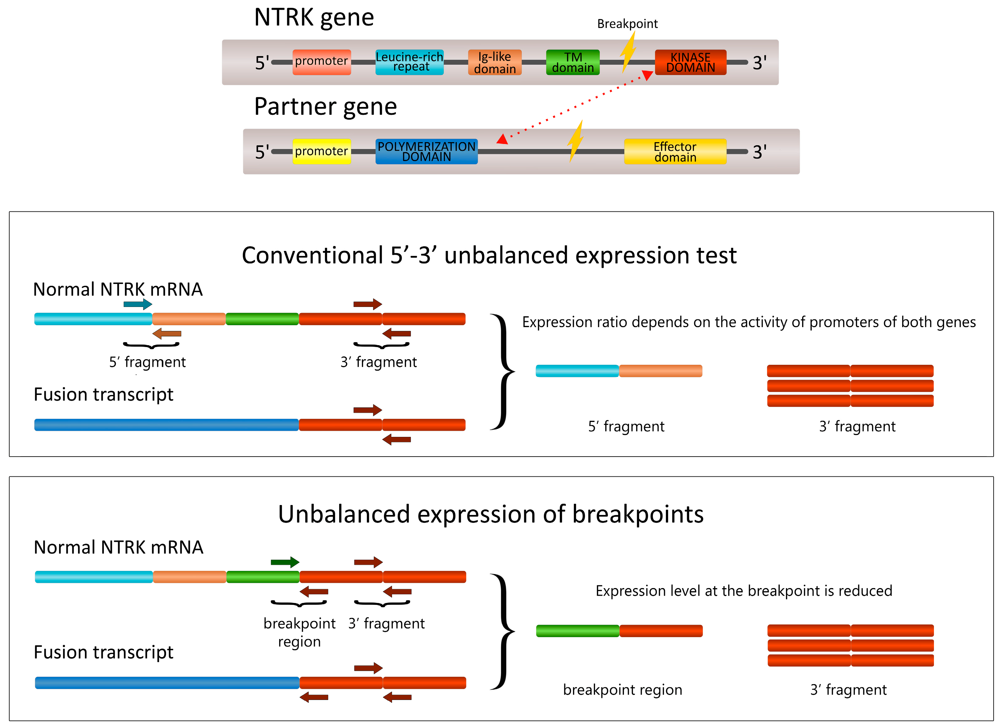
2.1. The Development of the 5′/3′-End NTRK Expression Test

The NTRK1 assay involved primers located at the junctions of exons 3–4 (5′-end) and exons 14–15 (3′-end). NTRK2 assays included primers corresponding to the borders of exons 11–12 (5′-end) and 15–16 (3′-end). However, when we tried a similar design for the detection of NTRK3 rearrangements and utilized primers for exons 7–8 (5′-end) and 15–16 (3′-end), we observed an unacceptably high frequency of false-positive findings, probably due to alternative splicing of this gene. In order to overcome this limitation, we placed the 5′-end primers on the regions of the most common breakpoints, i.e., the border between exons 13 and 14, and exons 14 and 15, while the 3′-end primers were located in exons 16 and 17. This design allowed us to observe the depletion of the expression of exon 13–14 or 14–15 RNA sequences in case of the presence of NTRK3 translocation (Figure 1).

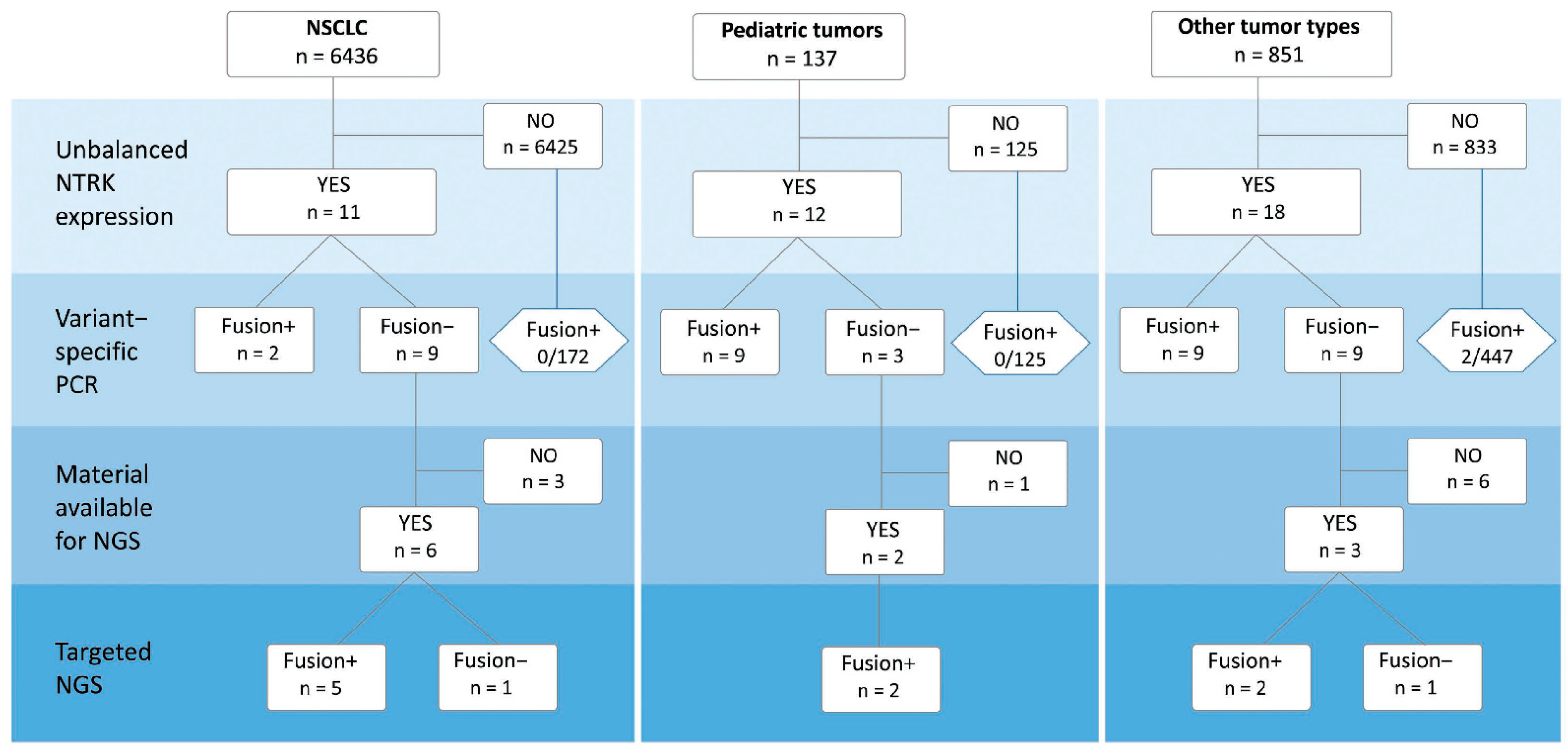
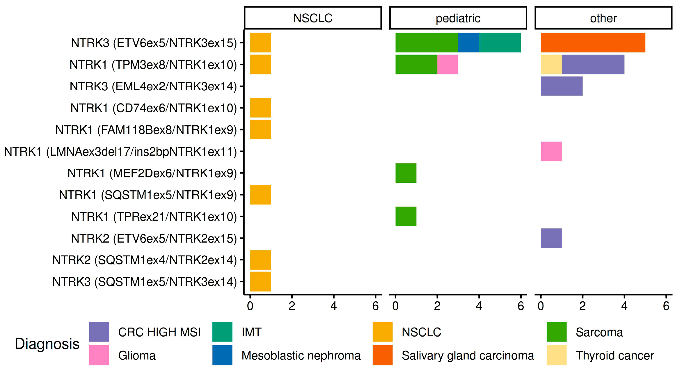
2.2. Detection of NTRK1/2/3 Rearrangements

The study initially included 8075 tissue specimens; 651 (8.1%) of these samples failed to pass the cDNA quality control. The analysis of 6436 non-small-cell lung carcinomas (NSCLCs) revealed 11 (0.2%) instances of 5′/3′-end NTRK unbalanced expression. Strikingly, none of the 1293 NSCLC cases with known activating events in EGFR, ALK, ROS1, RET, or MET oncogenes demonstrated evidence of NTRK activation. The analysis of 137 pediatric and 851 non-NSCLC malignancies revealed unbalanced expression in 12/137 (8.8%) and 18/851 (2.1%) patients, respectively. In total, 41 tumors had 5′/3′-end expression imbalances. These tumors were tested for the 44 most common NTRK translocations by variant-specific PCR; rearrangements were detected in 20/41 (48.8%) cases. In 11 out of the remaining 21 samples, tissue samples for additional RNA extraction necessary for targeted NGS analysis were available. NGS confirmed the presence of the NTRK rearrangement in nine of these tumors (Figure 2).
We further evaluated to what extent the 5′/3′-end NTRK unbalanced expression is prone to false-negative results. Variant-specific PCR for the 44 most common NTRK translocations did not reveal instances of the rearrangement in 172 EGFR/ALK/ROS1/RET/MET-negative NSCLCs or 125 pediatric tumors, which showed no imbalance upon 5′/3′-end expression tests. However, variant-specific analysis of 447 tumors of other categories (colorectal carcinomas, gliomas, salivary gland carcinomas, sarcomas, thyroid tumors, etc.) identified two (0.5%) instances of NTRK3 translocation (EML4::NTRK3 (E2;N14) in microsatellite-unstable colorectal carcinoma and ETV6::NTRK3 (E5;N15) in salivary gland carcinoma).
The performance of the 5′/3′-end NTRK unbalanced expression assay depends on the chosen threshold. We utilized deltaCt > 3 as a cut-off, given that this approach produced satisfactory results for the testing of other rearranged kinases [16,17,18]. We evaluated the reliability of this threshold by comparing samples with known NTRK rearrangements against tumors with presumably normal NTRK status (Figure S1). From our tumor bank, we were able to obtain 39 samples with NTRK1 or NTRK3 fusions in which the presence of alteration was confirmed by variant-specific PCR or NGS. However, we could not perform the validation study for NTRK2, as only two samples with NTRK2 fusions were available. The control group was composed of 50 NSCLCs, which carried alterations in EGFR, ALK, RET, ROS1, and MET oncogenes and, therefore, were highly unlikely to have NTRK rearrangements. For NTRK1, 10/10 (100%) NTRK1 fusion-containing samples were correctly identified by the unbalanced NTRK1 expression test (Figure S1A). The test evaluating the depletion of the NTRK3 ex13–14 junction detected only 2/7 (28.6%) tumors with NTRK3 fusions involving this breakpoint (Figure S1B). The test for NTRK3 ex14–15 depletion identified 15/22 (68.2%) cases with translocations involving the breakpoint at exon 15 (Figure S1C). As the chosen threshold (deltaCt > 3) performed poorly for both NTRK3 assays, we attempted to evaluate whether the modification of the cut-off will improve the testing procedure. We performed PCR analysis for 11 individual NTRK3 translocation variants in 225 cases showing either deltaCt > 0.9 for the NTRK3 ex13–14 depletion assay or deltaCt > 1.5 for the NTRK3 ex14–15 depletion assay; however, none of these samples with low-level NTRK3 expression imbalances carried NTRK3 translocations.
While performing the analysis of distribution of NTRK rearrangements in different cancer types, we considered only tumor varieties with identified translocation variants. Overall, 31 tumors with NTRK fusions were revealed. There were 14 NTRK1 fusions and 15 NTRK3 translocations, while only two tumors showed evidence of NTRK2 activation. NTRK rearrangements were detected in 7/6436 (0.1%) NSCLCs, 11/137 (8.0%) pediatric tumors, and 13/851 (1.5%) adult carcinomas other than NSCLC. The frequencies and spectrum of identified rearrangements are described in detail in Table 1 and Table 2, and in Figure 3. A relatively high incidence of NTRK fusions was seen in microsatellite unstable colorectal carcinomas (6/48, 12.5%), sarcomas (7/143, 4.9%), and salivary gland carcinomas (5/93, 5.4%). The highest frequency of NTRK translocations was documented in pediatric sarcomas (7/39, 17.9%). In particular, fusions were detected in 2/3 infantile fibrosarcomas, 1/5 malignant peripheral nerve sheath tumors, 1/2 dermatofibrosarcomas protuberans, 1/1 hemangioendothelioma, and 2/8 sarcomas not otherwise specified. Instances of NTRK rearrangements were also observed in gliomas, thyroid carcinomas, inflammatory myofibroblastic tumors, and mesoblastic nephromas; however, the number of cases was insufficient for the evaluation of the frequency of this event.

3. Discussion

This report presents the results of the NTRK analysis in a large collection of human tumors. It shows that the test for unbalanced 5′/3′-end expression is a promising prescreening tool for this rare category of gene rearrangements. Importantly, 91.9% of the included tumors passed the RNA quality control for this test. These data are comparable with the estimates obtained in the NGS RNA studies [19,20,21,22,23]. The most common cause of RNA isolation failure is poor processing of morphological samples, which is still an issue in many hospitals [24]. It is highly likely that RNA/cDNA samples, which are not suitable for reliable PCR-based testing, will also not be compatible with RNA-based NGS analysis. This study considered biological samples arriving from a few dozen different cancer centers, therefore some variations in the tissue handling could have inflated the failure rate.
Most of the studies aimed at detecting gene rearrangements employ IHC, FISH, or NGS, while the comparison of the amount of transcripts of 5′- and 3′-portions of the genes is a less common technique [25,26,27,28,29]. An example of its successful implementation is the fully automated commercial Idylla GeneFusion assay (Biocartis, Mechelen, Belgium) [30,31]. The utilization of the 5′-3′ NTRK imbalance assay is complicated by the requirement for robust in-house validation using a sufficient number of positive and negative controls. Furthermore, while most of the published protocols rely on the analysis of the relative overexpression of the kinase portion of the gene [30,32,33], we found here that this methodology is not suitable for the analysis of NTRK3 gene status. As an alternative, we suggested a novel approach: we assumed, that the junction of exons, which is affected by a breakpoint, will not be present in the rearranged transcript, and, therefore, we searched for the depletion of the expression of exon 13–14 or exon 14–15 NTRK3-specific sequences. This methodology is potentially applicable to translocations, which are characterized by relatively narrow clustering of breakpoints, and which do not necessarily result in an easily detectable overexpression of a portion of the gene. Although being elegant, the test for depletion of expression of exon boundaries is potentially error-prone: it may be compromised by the presence of normal RNA message originating from the remaining gene allele or non-malignant cells contaminating the tumor sample. Not surprisingly, all instances of tumors that were positive by variant-specific PCR but negative by the test for unbalanced 5′/3′-end expression, contained NTRK3 rearrangements. We attempted to decrease the cut-off for deltaCt in order to reduce the number of false-negative results obtained upon the quantitation of the 5′- and 3′-specific NTRK3 transcripts, however, our experiments revealed that the modification of the threshold does not improve the performance of the assay (Supplementary Figure S1). While establishing a PCR-based prescreening procedure, it is, therefore, advisable to supplement 5′/3′-end expression measurement by the multiplexed variant-specific analysis of NTRK3 translocations. Furthermore, our protocol has not been rigorously validated for the detection of NTRK2 fusions due to the low number of “positive” samples, which is another limitation of this study.
There were two tumors, which demonstrated clear-cut 5′/3′-end NTRK2 (NSCLC) and NTRK3 (adult sarcoma) expression imbalances, but turned out to be NTRK rearrangement-negative by the TruSight RNA Fusion NGS panel. It is unclear if these tumors utilize alternative splicing for one of the NTRK genes, or if these observations are related to the NGS failures in identifying NTRK fusions. It is noteworthy that NGS testing revealed a potentially actionable EWSR1::CCDC80 fusion in the above-described sarcoma, thus making the existence of NTRK3 translocation unlikely.
Our data strongly indicate that the occurrence of NTRK fusions in NSCLCs is low, even for the tumors enriched by exclusion of other actionable mutations (7/5143, 0.14%). NSCLC is also characterized by a high diversity of NTRK fusion types: none of the seven identified translocations occurred more than once. Obviously, the detection of NTRK rearrangements in NSCLCs is challenging due to the above factors, therefore the extent of this testing may need to be adjusted to the available resources. While defining the priorities, it is highly advisable to provide comprehensive NTRK testing to pediatric cancer patients, especially children suffering from sarcomas. Among the adult non-NSCLC malignancies, our study has confirmed a substantial frequency of NTRK fusions in microsatellite-unstable colorectal carcinomas (12.5%). Overall, our data on the occurrence and spectrum of NTRK translocations are in good agreement with previously published studies [34,35,36,37,38].
This investigation did not involve thorough validation of NTRK-rearranged and NTRK-wild-type samples by RNA sequencing, which is a limitation of the study. While the confirmation of NTRK fusions detected by the combination of the 5′/3′-end expression imbalance assay and variant-specific PCR seems unnecessary, it is unclear what is the risk of missing some rare NTRK rearrangements by the described above pipeline. The frequency of NTRK fusions is low, therefore, it is challenging to ensure that any given technology does not produce false-negative results. This validation would require RNA-based NGS analysis involving huge tumor collections, and, therefore, excessive costs. It is noteworthy that the frequencies of NTRK rearrangements presented in this dataset match the previously reported NTRK prevalence estimates, thus supporting the suitability of the 5′-3′ NTRK imbalance assay [25,28,35,36,37,39,40]. Furthermore, we have encouraging results from the NSCLC study, which focused on patients with a very high probability of the presence of actionable gene fusion in the tumor tissue, i.e., young-onset female non-smokers. RNA-based NGS was performed for 87 tumors, which lacked activating mutations in EGFR, KRAS, NRAS, BRAF, MET, and HER2 oncogenes and were negative for ALK, ROS1, RET, or NTRK1/2/3 translocations by the 5′/3′-end expression test and variant-specific PCR. Strikingly, no instances of rearrangements in the above genes have been revealed by RNA sequencing [41]. While data on NSCLC appear to be conclusive, we did not specifically address the performance of the NTRK unbalanced expression assay separately in other tumor types. There are significant differences in the patterns of NTRK expression and spectrum of NTRK rearrangements across various categories of malignancies [3,4,5,6,8,9,10], which may influence the performance of this test. This is particularly relevant to tumor entities, which have a high baseline expression level of NTRK kinases [26], as the presence of the background normal transcripts is likely to compromise the performance of the 5′/3′-end expression assay. The actual rate of false-negative results produced by our methodology remains to be determined in further investigations.
PCR-based fusion detection and NGS require distinct processing of the samples. Our PCR testing procedure involved simultaneous DNA and RNA isolation followed by the cDNA synthesis using the entire volume of the sample. In order to perform NGS, we subjected samples with 5′/3′-end expression imbalances to a new round of RNA isolation, with only 11 out of 21 samples available for this procedure. This low success rate is attributed to the nature of our sample collection, where most of the tumors were not initially intended to undergo comprehensive NTRK testing and therefore were returned to the primary hospitals right after the completion of other diagnostic procedures. This disadvantage can be resolved by dividing the DNA/RNA preparations into two parts, with one sample undergoing PCR analysis and the remaining one subjected to NGS when necessary.
Many laboratories currently utilize pan-TRK IHC as a screening tool for the detection of NTRK rearrangements [9]. Several investigations evaluated the performance of IHC testing in the real-world setting. Hondelink et al. [8] evaluated the sensitivity of the IHC by analyzing the tumors with NGS-detected NTRK translocations. These authors considered their own dataset (24 tumors) and all relevant published studies (200 tumors in total) and revealed that IHC failed to detect fusions in 40/224 (18%) of NTRK-rearranged samples (NTRK1: 6%; NTRK2: 14%; NTRK3: 27%) [8]. These data correspond well to our experience, as we observed satisfactory performance of the 5′-/3′-end expression imbalance screening test for the NTRK1 but not for the NTRK3 (Supplementary Figure S1). IHC often produces false-positive results. Overbeck et al. [25] reported the results of the pan-TRK staining for 973 NSCLCs. IHC failures were observed for 75/973 (8%) tumors. TRK expression was detected in 133/898 (15%) NSCLCs; 120 of these carcinomas were available for NGS analysis, with only two instances of NTRK fusions eventually confirmed. Zito Marino et al. [42] revealed pan-TRK IHC positivity in 16 out of 83 triple-negative breast malignancies; however, none of these samples carried translocation upon NGS. Vingiani et al. [43] analyzed the collection composed of several tumor types. The presence of NTRK fusions was confirmed only in 11/30 (37%) specimens identified as candidates by IHC. It is desirable to compare the performance of IHC with our NTRK testing procedure in future studies.
Cost considerations are highly important for the comparison of various diagnostic techniques, as they significantly impact the overall treatment budget. This is particularly relevant for rare actionable genetic alterations, given that only a small number of analyzed patients are eventually eligible for targeted therapy [7]. Pricing for laboratory reagents and molecular tests may vary by an order of magnitude between different countries; nevertheless, some rough comparison is possible. The cost of RNA-based NGS per sample is generally above 300–400 EURO or USD [23,44]. IHC is often regarded as a relatively cheap method, although its budget usually exceeds 150 EURO/USD [44]. The price for a single PCR reaction may be around 1.5 EURO/USD or even lower [45]. The NTRK testing pipeline presented in this study includes 1 assay for the cDNA quality check, 4 tests for the expression imbalance, and, whenever feasible, 2–8 multiplexed variant-specific PCRs; therefore, the overall expenses for reagents fall within 8–20 EURO/USD. This budget may double in the case of NSCLC analysis, because lung carcinomas have to be additionally tested for EGFR, BRAF, KRAS, MET, and HER2 mutations as well as ALK, ROS1, and RET translocations [17,18,46,47]. Therefore, the described diagnostic approach may be financially advantageous as compared to NGS, even assuming that a small subset of samples still require additional testing by alternative technologies. Within this study, only 21/7424 (0.3%) samples had to undergo further analysis after PCR, and this estimate may become approximately twice as high when considering ALK, ROS1, and RET translocations in addition to NTRK1, NTRK2, and NTRK3 rearrangements.
In summary, our report describes a pipeline for the detection of NTRK1, NTRK2, and NTRK3 gene fusions. As a screening step, this procedure relies on four PCR tests for 5′/3′-end unbalanced expression (one for NTRK1, one for NTRK2, and two for NTRK3), followed by PCR-driven identification of common translocations. It is advisable to supplement the screening by two multiplexed variant-specific PCRs for ETV6::NTRK3 and EML4::NTRK3 rearrangements, respectively. RNA-based NGS is to be applied only to tumors, which demonstrate NTRK 5′/3′-end expression imbalances but are negative upon variant-specific PCR. The described approach may be considered to be a cost-efficient alternative to conventional methods of NTRK analysis and, therefore, deserves to be evaluated for interlaboratory reproducibility and validity against other methods. For NSCLC, this procedure may be combined with similarly designed PCR-based methods of detecting ALK, ROS1, RET, and MET alterations [17,18,47], thus reducing the total cost of molecular testing.

4. Materials and Methods

The study included three categories of patients receiving molecular genetic testing in the N.N. Petrov Institute of Oncology between the years of 2019–2022. In particular, we analyzed 6980 consecutive non-small cell lung cancer (NSCLC) cases forwarded for regular testing of actionable genetic events. In addition, NTRK testing was applied to 158 pediatric cancer patients; there were 46 sarcomas, 33 inflammatory myofibroblastic tumors, 28 gliomas or neuroepithelial tumors, 4 salivary gland carcinomas, 4 mesoblastic nephromas, 2 thyroid carcinomas, and 41 tumors of other types. The third group was composed of diverse tumor types, which were subjected to testing either due to a known elevated occurrence of NTRK translocations or due to the preference of the treating physician (n = 937). It included colorectal carcinomas (n = 133), sarcomas (n = 118), brain tumors (n = 104), salivary gland carcinomas (n = 102), breast tumors (n = 54), thyroid carcinomas (n = 30), and other malignancies. Details on sarcoma and salivary gland carcinoma histology are presented in Table S1.
Formalin-fixed tissue specimens were subjected to manual dissection of tumor cells followed by DNA/RNA isolation and cDNA synthesis, as described previously [17]. Briefly, two–three 10 μm thick tissue sections were lysed for 5–6 h in 200 μL of 10 mM Tris–HCl (pH 8.0), 0.1 mM EDTA (pH 8.0), 2% SDS, and 500 μg/mL proteinase K at 65 °C. The organic extraction was performed with 200 μL Trizol and 90 μL chloroform–isoamyl alcohol mix (24:1). The samples were centrifuged, and the supernatant was incubated with 1 μL of glycogen (20 mg/mL) and 300 μL of isopropanol overnight at −20 °C. Nucleic acids were pelleted by centrifugation, rinsed with 70% ethanol and dissolved in 10 μL of sterile water. cDNA was obtained by the addition of 5× reverse transcriptase reaction buffer, 200 units of RevertAid Reverse Transcriptase (Thermo Fisher Scientific, Waltham, MA, USA), 20 units of RiboCare RNase Inhibitor (Evrogen, Moscow, Russia), dNTP mix (20 nM each), and random hexamers (0.25 μmol). Primer annealing was achieved by incubating the mix at 70 °C, 65 °C, and 60 °C for 5 min, then the reaction was cooled at 0 °C for 2 min. After addition of the enzyme, cDNA synthesis was performed at 20 °C for 5 min and 38 °C for 30 min; the reaction was terminated by heating at 95 °C for 5 min. Quality control relied on PCR amplification of SDHA gene fragments; samples producing PCR product after 35 cycles (cycle threshold, Ct) were considered unsuitable for further analysis. Primers and probes for the tests for 5′/3′-end unbalanced expression are given in Table S2; deltaCt > 3 was taken as threshold for the expression imbalance [16,17,18]. Primers and probes for variant-specific PCR for the 44 most common translocations (involving TPM3ex7–10 and NTRK1ex9,10,12; BCANex12 and NTRK1ex10; LMNAex3,4,8,10,11 and NTRK1ex10–12; IRF2BP2ex1 and NTRK1ex8,10; BCRex1 and NTRK2ex17; SQSTM1ex5–6 and NTRK2ex16; ETV6ex4–6 and NTRK3ex13–15; EML4ex2 and NTRK3ex13,14), and the corresponding transcript names, are given in Tables S3 and S4. PCR reactions for unbalanced expression of each NTRK gene were performed separately. Variant-specific PCR tests were multiplexed in eight reactions according to the involved NTRK gene and gene-partner: LMNA::NTRK1, BCAN::NTRK1, IRF2BP2::NTRK1, TPM3::NTRK1, BCR::NTRK2, SQSTM1::NTRK2, ETV6::NTRK3, and EML4::NTRK3. PCR reactions contained 1 μL of cDNA, 1× GeneAmp PCR Buffer I (Applied Biosystems, Waltham, MA, USA), 250 mkM of each dNTP, 200 nM of each primer and probe, 2.5 mM MgCl2, and 1 U of TaqM polymerase (AlkorBio, Saint-Petersburg, Russia) in a total volume of 20 μL. PCR started from enzyme activation (95 °C, 10 min.) and included 38 cycles (95 °C for 15 s followed by 58 °C for 1 min.).
NGS analysis was performed for the samples with 5′/3′-end unbalanced expression in which the type of translocation could not be identified by variant-specific PCR. Tissue samples were subjected to RNA extraction with the PureLink FFPE RNA Isolation kit (Invitrogen, Carlsbad, CA, USA). The analysis was performed with the TruSight RNA Fusion Panel using NextSeq 500 instrument (Illumina, San Diego, CA, USA).

Supplementary Materials

The following supporting information can be downloaded at: https://www.mdpi.com/article/10.3390/ijms241814203/s1.

Author Contributions

Conceptualization, A.A.R., A.G.I. and E.N.I.; methodology, A.A.R., V.I.T., R.S.M. and N.V.M.; investigation, A.A.R., R.S.M., V.I.T., E.S.S., E.V.P., E.A.K., A.D.S., E.V.B. and A.O.I.; analysis and interpretation of data, A.A.R. and V.I.T.; writing—original draft preparation, A.A.R., A.G.I. and E.N.I.; writing—review and editing, E.N.I.; visualization, A.A.R.; supervision, E.N.I. All authors have read and agreed to the published version of the manuscript.

Funding

This research has been supported by the Russian Science Foundation (grant number 17-75-30027-P). Funding sources did not influence how the study was conducted, or the description of its results.

Institutional Review Board Statement

The study was conducted in accordance with the Declaration of Helsinki and approved by the local Ethical Committee of N.N. Petrov Institute of Oncology (Approval Code: 20. Approval Date: 23 November 2017).

Informed Consent Statement

Informed consent was obtained from all subjects involved in the study.

Data Availability Statement

The data that support the findings of this study are available from the corresponding author upon reasonable request.

Acknowledgments

We are cordially thankful to Priscilla S. Amankwah for critical reading and editing of this manuscript.

Conflicts of Interest

The authors declare no conflict of interest.

References

  1. Doebele, R.C.; Drilon, A.; Paz-Ares, L.; Siena, S.; Shaw, A.T.; Farago, A.F.; Blakely, C.M.; Seto, T.; Cho, B.C.; Tosi, D.; et al. Entrectinib in patients with advanced or metastatic NTRK fusion-positive solid tumours: Integrated analysis of three phase 1-2 trials. Lancet Oncol. 2020, 21, 271–282. [Google Scholar] [CrossRef] [PubMed]
  2. Hong, D.S.; DuBois, S.G.; Kummar, S.; Farago, A.F.; Albert, C.M.; Rohrberg, K.S. van Tilburg, C.M.; Nagasubramanian, R.; Berlin, J.D.; Federman, N.; et al. Larotrectinib in patients with TRK fusion-positive solid tumours: A pooled analysis of three phase 1/2 clinical trials. Lancet Oncol. 2020, 21, 531–540. [Google Scholar] [CrossRef]
  3. Solomon, J.P.; Hechtman, J.F. Detection of NTRK Fusions: Merits and Limitations of Current Diagnostic Platforms. Cancer Res. 2019, 79, 3163–3168. [Google Scholar] [CrossRef]
  4. Weiss, L.M.; Funari, V.A. NTRK fusions and Trk proteins: What are they and how to test for them. Hum. Pathol. 2021, 112, 59–69. [Google Scholar] [CrossRef] [PubMed]
  5. Hechtman, J.F. NTRK insights: Best practices for pathologists. Mod. Pathol. 2022, 35, 298–305. [Google Scholar] [CrossRef]
  6. Marchetti, A.; Ferro, B.; Pasciuto, M.P.; Zampacorta, C.; Buttitta, F.; D’Angelo, E. NTRK gene fusions in solid tumors: Agnostic relevance, prevalence and diagnostic strategies. Pathologica 2022, 114, 199–216. [Google Scholar] [CrossRef] [PubMed]
  7. Huygens, S.; Vellekoop, H.; Versteegh, M.; Santi, I.; Szilberhorn, L.; Zelei, T.; Nagy, B.; Tsiachristas, A.; Koleva-Kolarova, R.; Wordsworth, S.; et al. Cost-Effectiveness Analysis of Treating Patients With NTRK-Positive Cancer With the Histology-Independent Therapy Entrectinib. Value Health 2023, 26, 193–203. [Google Scholar] [CrossRef]
  8. Hondelink, L.M.; Schrader, A.M.R.; Asri Aghmuni, G.; Solleveld-Westerink, N.; Cleton-Jansen, A.M.; van Egmond, D.; Boot, A.; Ouahoud, S.; Khalifa, M.N.; Wai Lam, S.; et al. The sensitivity of pan-TRK immunohistochemistry in solid tumours: A meta-analysis. Eur. J. Cancer 2022, 173, 229–237. [Google Scholar] [CrossRef]
  9. Marchiò, C.; Scaltriti, M.; Ladanyi, M.; Iafrate, A.J.; Bibeau, F.; Dietel, M. ESMO recommendations on the standard methods to detect NTRK fusions in daily practice and clinical research. Ann. Oncol. 2019, 30, 1417–1427. [Google Scholar] [CrossRef]
  10. Solomon, J.P.; Benayed, R.; Hechtman, J.F.; Ladanyi, M. Identifying patients with NTRK fusion cancer. Ann. Oncol. 2019, 30 (Suppl. S8), viii16–viii22. [Google Scholar] [CrossRef]
  11. Park, H.J.; Baek, I.; Cheang, G.; Solomon, J.P.; Song, W. Comparison of RNA-Based Next-Generation Sequencing Assays for the Detection of NTRK Gene Fusions. J. Mol. Diagn. 2021, 23, 1443–1451. [Google Scholar] [CrossRef]
  12. Bormann Chung, C.; Lee, J.; Barritault, M.; Bringuier, P.P.; Xu, Z.; Huang, W.Y.; Beharry, A.; Castillo, J.; Christiansen, J.; Lin, J.C.; et al. Evaluating Targeted Next-Generation Sequencing Assays and Reference Materials for NTRK Fusion Detection. J. Mol. Diagn. 2022, 24, 18–32. [Google Scholar] [CrossRef]
  13. Koopman, B.; Kuijpers, C.C.H.J.; Groen, H.J.M.; Timens, W.; Schuuring, E.; Willems, S.M.; van Kempen, L.C. Detection of NTRK Fusions and TRK Expression and Performance of pan-TRK Immunohistochemistry in Routine Diagnostics: Results from a Nationwide Community-Based Cohort. Diagnostics 2022, 12, 668. [Google Scholar] [CrossRef]
  14. Stockley, T.L.; Lo, B.; Box, A.; Corredor, A.G.; DeCoteau, J.; Desmeules, P.; Feilotter, H.; Grafodatskaya, D.; Greer, W.; Hawkins, C.; et al. CANTRK: A Canadian Ring Study to Optimize Detection of NTRK Gene Fusions by Next-Generation RNA Sequencing. J. Mol. Diagn. 2023, 25, 68–174. [Google Scholar] [CrossRef]
  15. Wang, R.; Pan, Y.; Li, C.; Hu, H.; Zhang, Y.; Li, H.; Luo, X.; Zhang, J.; Fang, Z.; Li, Y.; et al. The use of quantitative real-time reverse transcriptase PCR for 5′ and 3′ portions of ALK transcripts to detect ALK rearrangements in lung cancers. Clin. Cancer Res. 2012, 18, 4725–4732. [Google Scholar] [CrossRef]
  16. Preobrazhenskaya, E.V.; Iyevleva, A.G.; Suleymanova, A.M.; Tiurin, V.I.; Mitiushkina, N.V.; Bizin, I.V.; Ivanstov, A.O.; Gorustovich, O.A.; Shelekhova, K.V.; Kachanov, D.Y.; et al. Gene rearrangements in consecutive series of pediatric inflammatory myofibroblastic tumors. Pediatr. Blood Cancer 2020, 67, e28220. [Google Scholar] [CrossRef] [PubMed]
  17. Mitiushkina, N.V.; Romanko, A.A.; Preobrazhenskaya, E.V.; Tiurin, V.I.; Ermachenkova, T.I.; Martianov, A.S.; Mulkidjan, R.S.; Sokolova, T.N.; Kholmatov, M.M.; Bizin, I.V.; et al. Comprehensive evaluation of the test for 5′-/3′-end mRNA unbalanced expression as a screening tool for ALK and ROS1 fusions in lung cancer. Cancer Med. 2022, 11, 3226–3237. [Google Scholar] [CrossRef]
  18. Tiurin, V.I.; Preobrazhenskaya, E.V.; Mitiushkina, N.V.; Romanko, A.A.; Anuskina, A.A.; Mulkidjan, R.S.; Saitova, E.S.; Krivosheyeva, E.A.; Kharitonova, E.D.; Shevyakov, M.P.; et al. Rapid and Cost-Efficient Detection of RET Rearrangements in a Large Consecutive Series of Lung Carcinomas. Int. J. Mol. Sci. 2023, 24, 10530. [Google Scholar] [CrossRef] [PubMed]
  19. Newton, Y.; Sedgewick, A.J.; Cisneros, L.; Golovato, J.; Johnson, M.; Szeto, C.W.; Rabizadeh, S.; Sanborn, J.Z.; Benz, S.C.; Vaske, C. Large scale, robust, and accurate whole transcriptome profiling from clinical formalin-fixed paraffin-embedded samples. Sci. Rep. 2020, 10, 17597. [Google Scholar] [CrossRef] [PubMed]
  20. Shrestha, M.; Blay, S.; Liang, S.; Swanson, D.; Lerner-Ellis, J.; Dickson, B.; Wong, A.; Charames, G.S. Improving RNA Fusion Call Confidence and Reliability in Molecular Diagnostic Testing. J. Mol. Diagn. 2023, 25, 320–330. [Google Scholar] [CrossRef]
  21. Ramani, N.S.; Chen, H.; Broaddus, R.R.; Lazar, A.J.; Luthra, R.; Medeiros, L.J.; Patel, K.P.; Rashid, A.; Routbort, M.J.; Stewart, J.; et al. Utilization of cytology smears improves success rates of RNA-based next-generation sequencing gene fusion assays for clinically relevant predictive biomarkers. Cancer Cytopathol. 2021, 129, 374–382. [Google Scholar] [CrossRef] [PubMed]
  22. Benayed, R.; Offin, M.; Mullaney, K.; Sukhadia, P.; Rios, K.; Desmeules, P.; Ptashkin, R.; Won, H.; Chang, J.; Halpenny, D.; et al. High Yield of RNA Sequencing for Targetable Kinase Fusions in Lung Adenocarcinomas with No Mitogenic Driver Alteration Detected by DNA Sequencing and Low Tumor Mutation Burden. Clin. Cancer Res. 2019, 25, 4712–4722. [Google Scholar] [CrossRef]
  23. Cohen, D.; Hondelink, L.M.; Solleveld-Westerink, N.; Uljee, S.M.; Ruano, D.; Cleton-Jansen, A.M.; von der Thüsen, J.H.; Ramai, S.R.S.; Postmus, P.E.; Graadt van Roggen, J.F.; et al. Optimizing Mutation and Fusion Detection in NSCLC by Sequential DNA and RNA Sequencing. J. Thorac. Oncol. 2020, 15, 1000–1014. [Google Scholar] [CrossRef] [PubMed]
  24. Chung, J.Y.; Braunschweig, T.; Williams, R.; Guerrero, N.; Hoffmann, K.M.; Kwon, M.; Song, Y.K.; Libutti, S.K.; Hewitt, S.M. Factors in tissue handling and processing that impact RNA obtained from formalin-fixed, paraffin-embedded tissue. J. Histochem. Cytochem. 2008, 56, 1033–1042. [Google Scholar] [CrossRef] [PubMed]
  25. Overbeck, T.R.; Reiffert, A.; Schmitz, K.; Rittmeyer, A.; Körber, W.; Hugo, S.; Schnalke, J.; Lukat, L.; Hugo, T.; Hinterthaner, M.; et al. NTRK Gene Fusions in Non-Small-Cell Lung Cancer: Real-World Screening Data of 1068 Unselected Patients. Cancers 2023, 15, 2966. [Google Scholar] [CrossRef]
  26. Solomon, J.P.; Linkov, I.; Rosado, A.; Mullaney, K.; Rosen, E.Y.; Frosina, D.; Jungbluth, A.A.; Zehir, A.; Benayed, R.; Drilon, A.; et al. NTRK fusion detection across multiple assays and 33,997 cases: Diagnostic implications and pitfalls. Mod. Pathol. 2020, 33, 38–46. [Google Scholar] [CrossRef] [PubMed]
  27. Haberecker, M.; Töpfer, A.; Melega, F.; Moch, H.; Pauli, C. A systematic comparison of pan-Trk immunohistochemistry assays among multiple cancer types. Histopathology 2023, 82, 1003–1012. [Google Scholar] [CrossRef]
  28. Schraa, S.J.; Stelloo, E.; Laclé, M.M.; Swennenhuis, J.F.; Brosens, L.A.A.; Fijneman, R.J.A.; Feitsma, H.; Koopman, M.; de Leng, W.W.; Vink, G.R.; et al. Comparison of NTRK fusion detection methods in microsatellite-instability-high metastatic colorectal cancer. Virchows Arch. 2023, 482, 983–992. [Google Scholar] [CrossRef] [PubMed]
  29. Hofman, V.; Heeke, S.; Bontoux, C.; Chalabreysse, L.; Barritault, M.; Bringuier, P.P.; Fenouil, T.; Benzerdjeb, N.; Begueret, H.; Merlio, J.P.; et al. Ultrafast Gene Fusion Assessment for Nonsquamous NSCLC. JTO Clin. Res. Rep. 2022, 4, 100457. [Google Scholar] [CrossRef]
  30. Sorber, L.; Van Dorst, B.; Bellon, E.; Zwaenepoel, K.; Lambin, S.; De Winne, K.; Lardon, F.; Pauwels, P.; Siozopoulou, V. NTRK Gene Fusion Detection in a Pan-Cancer Setting Using the Idylla GeneFusion Assay. J. Mol. Diagn. 2022, 24, 750–759. [Google Scholar] [CrossRef] [PubMed]
  31. Guillard, M.; Caumont, C.; Marcorelles, P.; Merlio, J.P.; Cappellen, D.; Uguen, A. Performances of the Idylla GeneFusion Assay: Contribution to a rapid diagnosis of targetable gene fusions in tumour samples. J. Clin. Pathol. 2023, jcp-2023-208798. [Google Scholar] [CrossRef]
  32. Lira, M.E.; Choi, Y.L.; Lim, S.M.; Deng, S.; Huang, D.; Ozeck, M.; Han, J.; Jeong, J.Y.; Shim, H.S.; Cho, B.C.; et al. A single-tube multiplexed assay for detecting ALK, ROS1, and RET fusions in lung cancer. J. Mol. Diagn. 2014, 16, 229–243. [Google Scholar] [CrossRef]
  33. Heydt, C.; Wölwer, C.B.; Velazquez Camacho, O.; Wagener-Ryczek, S.; Pappesch, R.; Siemanowski, J.; Rehker, J.; Haller, F.; Agaimy, A.; Worm, K.; et al. Detection of gene fusions using targeted next-generation sequencing: A comparative evaluation. BMC Med. Genom. 2021, 14, 62. [Google Scholar] [CrossRef]
  34. O’Haire, S.; Franchini, F.; Kang, Y.J.; Steinberg, J.; Canfell, K.; Desai, J.; Fox, S.; IJzerman, M. Systematic review of NTRK 1/2/3 fusion prevalence pan-cancer and across solid tumours. Sci. Rep. 2023, 13, 4116. [Google Scholar] [CrossRef] [PubMed]
  35. Forsythe, A.; Zhang, W.; Phillip Strauss, U.; Fellous, M.; Korei, M.; Keating, K. A systematic review and meta-analysis of neurotrophic tyrosine receptor kinase gene fusion frequencies in solid tumors. Ther. Adv. Med. Oncol. 2020, 12, 1758835920975613. [Google Scholar] [CrossRef] [PubMed]
  36. Sato, K.; Kawazu, M.; Yamamoto, Y.; Ueno, T.; Kojima, S.; Nagae, G.; Abe, H.; Soda, M.; Oga, T.; Kohsaka, S.; et al. Fusion Kinases Identified by Genomic Analyses of Sporadic Microsatellite Instability-High Colorectal Cancers. Clin. Cancer Res. 2019, 25, 378–389. [Google Scholar] [CrossRef]
  37. Ukkola, I.; Nummela, P.; Kero, M.; Tammio, H.; Tuominen, J.; Kairisto, V.; Kallajoki, M.; Haglund, C.; Peltomäki, P.; Kytölä, S.; et al. Gene fusions and oncogenic mutations in MLH1 deficient and BRAFV600E wild-type colorectal cancers. Virchows Arch. 2022, 480, 807–817. [Google Scholar] [CrossRef]
  38. Wang, J.; Li, R.; Li, J.; Yi, Y.; Liu, X.; Chen, J.; Zhang, H.; Lu, J.; Li, C.; Wu, H.; et al. Comprehensive analysis of oncogenic fusions in mismatch repair deficient colorectal carcinomas by sequential DNA and RNA next generation sequencing. J. Transl. Med. 2021, 19, 433. [Google Scholar] [CrossRef] [PubMed]
  39. Zhao, R.; Yao, F.; Xiang, C.; Zhao, J.; Shang, Z.; Guo, L.; Ding, W.; Ma, S.; Yu, A.; Shao, J.; et al. Identification of NTRK gene fusions in lung adenocarcinomas in the Chinese population. J. Pathol. Clin. Res. 2021, 7, 375–384. [Google Scholar] [CrossRef]
  40. Liu, F.; Wei, Y.; Zhang, H.; Jiang, J.; Zhang, P.; Chu, Q. NTRK Fusion in Non-Small Cell Lung Cancer: Diagnosis, Therapy, and TRK Inhibitor Resistance. Front. Oncol. 2022, 12, 864666. [Google Scholar] [CrossRef]
  41. Preobrazhenskaya, E.; Zagrebin, F.; Mulkidzhan, R.; Krivosheeva, E.; Saitova, E.; Raskin, G.; Bizin, I.; Imyanitov, E. Novel kinase-activating fusions in lung adenocarcinomas. In Proceedings of the 35st European Congress of Pathology, Dublin, Ireland, 9–13 September 2023. [Google Scholar]
  42. Zito Marino, F.; Buono, S.; Montella, M.; Giannatiempo, R.; Messina, F.; Casaretta, G.; Arpino, G.; Vita, G.; Fiorentino, F.; Insabato, L.; et al. NTRK gene aberrations in triple-negative breast cancer: Detection challenges using IHC, FISH, RT-PCR, and NGS. J. Pathol. Clin. Res. 2023, 9, 367–377. [Google Scholar] [CrossRef]
  43. Vingiani, A.; Lorenzini, D.; Conca, E.; Volpi, C.C.; Trupia, D.V.; Gloghini, A.; Perrone, F.; Tamborini, E.; Dagrada, G.P.; Agnelli, L.; et al. Pan-TRK immunohistochemistry as screening tool for NTRK fusions: A diagnostic workflow for the identification of positive patients in clinical practice. Cancer Biomark. 2023, in press. [Google Scholar] [CrossRef]
  44. Beresford, L.; Murphy, P.; Dias, S.; Claxton, L.; Walton, M.; Metcalf, R.; Schlecht, H.; Ottensmeier, C.; Pereira, M.; Hodgson, R. Appraising the Costs of Genomic Testing for Histology-Independent Technologies: An Illustrative Example for NTRK Fusions. Value Health 2022, 25, 1133–1140. [Google Scholar] [CrossRef] [PubMed]
  45. Schlatter, R.P.; Matte, U.; Polanczyk, C.A.; Koehler-Santos, P.; Ashton-Prolla, P. Costs of genetic testing: Supporting Brazilian Public Policies for the incorporating of molecular diagnostic technologies. Genet. Mol. Biol. 2015, 38, 332–337. [Google Scholar] [CrossRef]
  46. Imyanitov, E.N.; Iyevleva, A.G.; Levchenko, E.V. Molecular testing and targeted therapy for non-small cell lung cancer: Current status and perspectives. Crit. Rev. Oncol. Hematol. 2021, 157, 103194. [Google Scholar] [CrossRef] [PubMed]
  47. Mitiushkina, N.V.; Kholmatov, M.M.; Tiurin, V.I.; Romanko, A.A.; Yatsuk, O.S.; Sokolova, T.N.; Ivantsov, A.O.; Kuligina, E.S.; Stepanov, I.A.; Belyaev, A.M.; et al. Comparative analysis of expression of mutant and wild-type alleles is essential for reliable PCR-based detection of MET exon 14 skipping. Biochimie 2019, 165, 267–274. [Google Scholar] [CrossRef] [PubMed]
Figure 1. The design of the tests for NTRK unbalanced 5′/3′-end expression. The conventional test relies on the overexpression of the kinase portion of the gene upon fusion (top); consequently, the 3′-end-specific fragment is overrepresented in the rearranged transcript as compared to the fragment corresponding to the beginning of the gene. The novel version of the test relies on the depletion of expression of the portion of the gene, which is disrupted by the breakpoint and, therefore, absent in the rearranged NTRK transcript (bottom). Colors represent various portions of the genes. Arrows represent PCR primers.
Ijms 24 14203 g001
Figure 2. Flow chart of the study.
Ijms 24 14203 g002
Figure 3. Distribution and spectrum of the identified NTRK fusions in different tumor types.
Ijms 24 14203 g003
Table 1. Frequency of NTRK fusions in different tumor types.
Tumor TypeNumber of Cases with Confirmed NTRK Fusion
NSCLC 7/6436 1 (0.1%)
Sarcoma 7/143 2 (4.9%)
Glioma, neuroepithelial tumor 2/124 (1.6%)
Salivary gland carcinoma 5/93 3 (5.4%)
MSI-high CRC 6/48 (12.5%)
IMT 2/34 (5.9%)
Thyroid cancer 1/26 (3.9%)
Mesoblastic nephroma 1/4 (25.0%)
Other 0/516 (0.0%)
1 All fusions were identified in EGFR/ALK/ROS1/RET/MET-negative cases (n = 5134). 2 The studied sarcoma cohort included 3 infantile fibrosarcomas, 5 malignant peripheral nerve sheath tumors, and 2 dermatofibrosarcomas protuberans. 3 The studied salivary gland tumor cohort included 2 cases of secretory carcinoma.
Table 2. Clinical data of cases with identified NTRK fusions.
IDTumor TypeAgeGenderFusion Type
NTRK1
22240 CRC, MSI-high 60f TPM3::NTRK1 (T8;N10)
31589 CRC, MSI-high 64f TPM3::NTRK1 (T8;N10)
21693 CRC, MSI-high 85m TPM3::NTRK1 (T8;N10)
28114 Glioblastoma 62f LMNA::NTRK1 (L3del17;ins2N11)
27845 Oligodendroglioma 3f TPM3::NTRK1 (T8;N10)
15197 NSCLC 47f CD74::NTRK1 (C6;N10)
10580 NSCLC 64m FAM118B::NTRK1 (F8;N9)
12285 NSCLC 64f SQSTM1::NTRK1 (S5;N9)
14281 NSCLC 60f TPM3::NTRK1 (T8;N10)
14131 Dermatofibrosarcoma protuberans 6m TPM3::NTRK1 (T8;N10)
30476 Sarcoma, NOS 11f TPR::NTRK1 (T21;N10)
16009 Infantile fibrosarcoma 2m TPM3::NTRK1 (T8;N10)
13669 Malignant peripheral nerve sheath tumor 0f MEF2D::NTRK1 (M6;N9)
353067 Papillary thyroid cancer 19f TPM3::NTRK1 (T8;N10)
NTRK2
29665 CRC, MSI-high 74m ETV6::NTRK2 (E5;N15)
13098 NSCLC 79m SQSTM1::NTRK2 (S4;N14)
NTRK3
13666 Congenital mesoblastic nephroma 0m ETV6::NTRK3 (E5;N15)
24631 CRC, MSI-high 68f EML4::NTRK3 (E2;N14)
33968 CRC, MSI-high 70f EML4::NTRK3 (E2;N14)
21856 IMT 3m ETV6::NTRK3 (E5;N15)
12193 IMT17m ETV6::NTRK3 (E5;N15)
11479 NSCLC 58m ETV6::NTRK3 (E5;N15)
22443 NSCLC 44m SQSTM1::NTRK3 (S5;N14)
32662 Salivary gland adenocarcinoma, NOS 55f ETV6::NTRK3 (E5;N15)
33039 Salivary gland adenocarcinoma, NOS 32f ETV6::NTRK3 (E5;N15)
787 Salivary duct carcinoma 65f ETV6::NTRK3 (E5;N15)
798 Salivary duct carcinoma 49f ETV6::NTRK3 (E5;N15)
24388 Salivary gland secretory carcinoma 37f ETV6::NTRK3 (E5;N15)
12471 Soft tissue sarcoma 3m ETV6::NTRK3 (E5;N15)
16011 Hemangioendothelioma 4f ETV6::NTRK3 (E5;N15)
16396 Infantile fibrosarcoma 1mETV6::NTRK3 (E5;N15)
1 NOS—not otherwise specified.
Disclaimer/Publisher’s Note: The statements, opinions and data contained in all publications are solely those of the individual author(s) and contributor(s) and not of MDPI and/or the editor(s). MDPI and/or the editor(s) disclaim responsibility for any injury to people or property resulting from any ideas, methods, instructions or products referred to in the content.

