Review

Meiotic Cell Cycle Progression in Mouse Oocytes: Role of Cyclins

1
Department of Biological Science, Inje University, Gimhae 50834, Republic of Korea
2
Department of Research Center, Dongnam Institute of Radiological and Medical Sciences, Busan 46033, Republic of Korea
3
Institute for Digital Antiaging Healthcare, Inje University, Gimhae 50834, Republic of Korea
4
Department of Medical Biotechnology, Inje University, Gimhae 50834, Republic of Korea
5
Institute of Basic Science, Inje University, Gimhae 50834, Republic of Korea
*
Author to whom correspondence should be addressed.
These authors contributed equally to this work.
Int. J. Mol. Sci. 2023, 24(17), 13659; https://doi.org/10.3390/ijms241713659
Submission received: 14 August 2023 / Revised: 31 August 2023 / Accepted: 1 September 2023 / Published: 4 September 2023
(This article belongs to the Special Issue Ovary and Testis: Molecular Biological Insights)

Abstract

:
All eukaryotic cells, including oocytes, utilize an engine called cyclin-dependent kinase (Cdk) to drive the cell cycle. Cdks are activated by a co-factor called cyclin, which regulates their activity. The key Cdk–cyclin complex that regulates the oocyte cell cycle is known as Cdk1–cyclin B1. Recent studies have elucidated the roles of other cyclins, such as B2, B3, A2, and O, in oocyte cell cycle regulation. This review aims to discuss the recently discovered roles of various cyclins in mouse oocyte cell cycle regulation in accordance with the sequential progression of the cell cycle. In addition, this review addresses the translation and degradation of cyclins to modulate the activity of Cdks. Overall, the literature indicates that each cyclin performs unique and redundant functions at various stages of the cell cycle, while their expression and degradation are tightly regulated. Taken together, this review provides new insights into the regulatory role and function of cyclins in oocyte cell cycle progression.

1. Introduction

The statement “Omnis cellula-e-cellula” coined by Rudolf Virchow means “all cells come from cells”, and this concept forms the basis of the cell cycle. Based on this concept, the phrase “Omne vivum ex ovo” coined by William Harvey signifies the importance of the oocyte cell as the origin of life. In elucidating the key regulators of the cell cycle, including that of oocytes, cell division cycle (Cdc) genes involved in cell cycle progression were discovered in yeast during the 1960s. Subsequently, treating frog (Rana pipiens) oocytes with progesterone was found to induce cell cycle progression (oocyte maturation), and the injection of the cytoplasm of mature oocytes into immature oocytes promoted cell cycle progression without progesterone stimulation. The substance in the cytoplasm of the matured oocytes that promoted the cell cycle progression was named maturation-promoting factor (MPF) [1]. In 1983, Hunt’s group identified a protein stored as maternal mRNA in sea urchin (Arbacia punctulate) eggs that undergoes synthesis and degradation in a cell-cycle-dependent manner and named it cyclin [2]. Studies have revealed that MPF is a complex of cyclin and cyclin-dependent kinase 1 (Cdk1), also known as p34/cdc2, activated in a cyclin-dependent manner [3,4]. Subsequent experiments further identified several types of Cdks and cyclins that regulate various cell cycle points.
The cell cycle in multicellular eukaryotes is primarily regulated by Cdk1 and Cdk2, and animal cells have additional Cdks, such as Cdk4 and Cdk6, for controlling cell cycle entry in response to extracellular growth factors [5,6]. The human genome contains at least 20 Cdk genes. These Cdks function as kinases by generating specific Cdk–cyclin complexes at particular stages of the cell cycle. These complexes phosphorylate specific substrates to control cell cycle progression [7] or are involved in the regulation of transcription and other cellular processes [8].
Cyclins, which possess a well-conserved cyclin box domain (CBD), do not have enzymatic activity themselves, but provide a substrate-binding site for Cdks and direct Cdks to specific cellular locations. Various cyclins have been discovered, including 9–15 from fungi, 14 from flies and echinoderms, and more than 30 from human cells [6,9]. During cell cycle regulation, D-type cyclins regulate the transition from the G0 to G1 phase by binding to Cdk4 or Cdk6 [10,11], while E-type cyclins form complexes with Cdk2 and function during the late G1 and early S phases. Cyclin A2 binds to Cdk2, promoting both G1/S and S/G2 transitions and DNA replication, while regulating the initiation of mitosis by binding to Cdk1 during the G2/M transition [12]. The Cdk1–cyclin B complex, known as MPF, controls the progression of the M phase [13,14].
The meiotic process, a specialized cell division process (reduction division) where the chromosomes undergo two rounds of division after a single round of DNA replication to generate gametes with half the chromosome number, is also regulated by Cdk and cyclin complexes, similar to that of other somatic cells (Figure 1). Among gametes, oocytes are relatively large, have a relatively long cell cycle length, and show distinct cell cycle arrest and progression stages. In many organisms, oocytes are mostly in a state of cell cycle arrest and can only exit this arrest state by specific hormonal signals [15,16]. This fact suggests that oocytes require distinct and specific mechanisms, different from those of somatic cells, for the regulation of Cdk activity.
Mice lacking Cdk3, Cdk4, or Cdk6 are viable and undergo a normal oocyte maturation, indicating that these Cdks are not essential for oocyte maturation [17,18,19]. Cdk1 and Cdk2 expression has been observed in mouse oocytes, and oocyte-specific deletion of Cdk1 and Cdk2 showed that only the loss of Cdk1 prevents the resumption of meiosis in oocytes, leading to female infertility [20,21]. Therefore, Cdk1 is considered a key Cdk that regulates the resumption of meiosis in mouse oocytes [20,21,22].
Until recently, cyclin B1 was considered to be the primary cyclin component of the MPF complex, which regulates the meiotic cell cycle in oocytes. However, recent studies have reported the expression of various cyclins, including cyclins B2, B3, A1, A2, E, and O, in oocytes of various species, such as Xenopus, Danio, mice, and humans [23,24,25]. The specific roles of these cyclins in oocyte maturation have not been extensively detailed, and they were previously thought to have minor functions compared to cyclin B1 [26,27,28,29]. For example, cyclin B2 was presumed to be redundant with cyclin B1. However, recent biochemical and cell biological approaches and studies based on knockout mice have revealed that these cyclins play distinct and unique roles in oocyte maturation (Figure 1). For instance, recent research on the role of cyclin B3 in oocytes has led to increased interest in the roles of other cyclins besides cyclin B1 [27,28,29,30,31,32,33,34,35,36,37,38,39,40,41]. This review focuses on the roles of various cyclins in the regulation of the mouse oocyte cell cycle, with a particular emphasis on recent discoveries; discusses the roles of different cyclins in oocyte cell cycle progression; and examines the regulation of cyclin expression by translation and degradation in accordance with the sequential progression of the oocyte cell cycle.

1.1. Overview of Cyclins

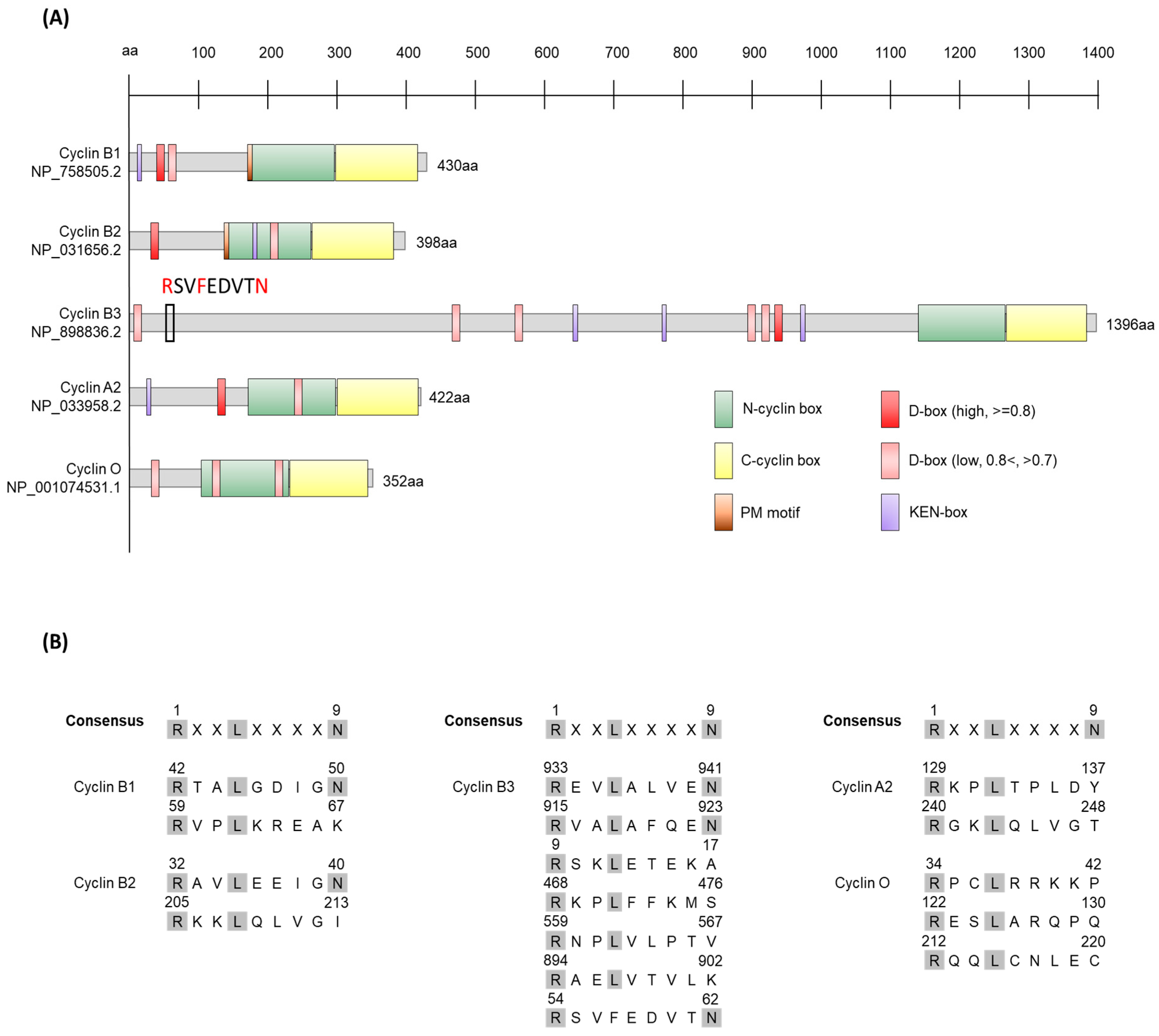
More than 30 cyclins have been discovered in mice. Among them, the functions of cyclins A2, B1, B2, B3, and O in mouse oocytes have been studied (Figure 2) [27,28,29,30,31,32,33,34,35,36,37,38,39,40]. The fundamental features of these cyclins are discussed below.

1.1.1. Cyclin B1

Synthesis of cyclin B1 begins during the S phase, and it exhibits activity during the G2/M transition after forming a complex with Cdk1. It is rapidly degraded during mitosis. In somatic cells, cyclin B1 is predominantly present in the cytosol but translocates into the nucleus when mitosis begins. It contributes to nuclear envelope breakdown, chromosome condensation, and mitotic spindle formation [42,43]. Deletion of Ccnb1 (the gene encoding cyclin B1) results in embryonic lethality in the uterus, indicating its importance in mitotic progression. Furthermore, tissue-specific knockout studies have revealed its significance in the generation of oocytes and sperms [27,29,44]. Cyclin B1 has been traditionally considered to regulate all aspects of oocyte maturation. However, recent findings from literature searches demonstrate that cyclin B1 functions only at specific stages of oocyte maturation.

1.1.2. Cyclin B2

Cyclin B2 is a subtype of cyclin B that is conserved in numerous vertebrate species [45]. In human tissue culture cells, cyclin B2 expression decreases during the G1 phase and increases during the S and G2 phases, reaching its peak during mitosis [46]. In Xenopus laevis, cyclin B2 is essential for cell cycle control during the G2/M transition and is steadily accumulated during the G2 phase before being rapidly degraded during mitosis [47,48]. Cyclin B2 in human culture cells is predominantly distributed in the Golgi apparatus and is evenly dispersed throughout the cell, unlike cyclin B1, which undergoes nuclear redistribution during prophase [43]. Cyclin B2, like cyclin B1, forms a complex with Cdk1 to activate it [43]. Knockout mice lacking Ccnb2 (the gene encoding cyclin B2) are viable and develop normally [27,29].

1.1.3. Cyclin B3

Cyclin B3, initially identified in chickens, is approximately 33% similar to cyclin B2 and approximately 30% similar to A-type cyclins [23,49]. In humans, cyclin B3 mRNA is expressed in all cell cycle stages, including the G0 phase, and is localized within the nucleus similar to A-type cyclins [49,50,51]. In Drosophila and C. elegans, cyclin B3 is found in mitotic and maternal germ cells. It regulates the S phase and promotes the metaphase-to-anaphase transition by alleviating the spindle assembly checkpoint (SAC) during mitosis [52,53,54,55,56]. In Drosophila, cyclin B3 is dispensable for mitosis but is necessary for female fertility [57]. In humans, cyclin B3 is most abundantly expressed in the testis and is found in all tissues [51]. Mice carrying a genetic knockout of Ccnb3 (the gene encoding cyclin B3) show viability, indicating that cyclin B3 is dispensable for mitosis. For meiosis, males exhibit fertility but female mice are sterile [31,32,58,59].

1.1.4. Cyclin A

Two forms of cyclin A have been identified in Xenopus [60], mice [61], and humans [62]. Cyclin A1 is considered to be the embryonic form and its expression in the germ cell lineage and in leukemia and brain cells is highly restricted. It is particularly highly expressed in the testes of mice and humans [61,62,63]. Moreover, it forms complexes with both Cdk2 and Cdk1 [64], and is involved in DNA double-strand break repair and ciliogenesis through its interaction with Cdk2 [65,66]. Male Ccna1 (the gene encoding cyclin A1) knockout mice exhibit a block in spermatogenesis before the first meiotic division, resulting in sterility, while females are unaffected [34].
Cyclin A2 is the somatic form of cyclin A and is expressed in various tissues after embryonic development. Its expression begins during the S phase of the cell cycle and accumulates until prometaphase [61,67]. Cyclin A2 is important for DNA replication and mitosis, activating Cdk2 during G1/S transition and Cdk1 during mitosis [68,69]. It regulates entry into cell division and ensures faithful chromosome segregation by controlling kinetochore microtubules [70]. Cyclin A2 is rapidly degraded, independent of the SAC, from prometaphase onwards, prior to the degradation of cyclin B by the anaphase-promoting complex/cyclosome (APC/C) complex [68,71,72,73,74]. Deletion of Ccna2 (the gene encoding cyclin A2) leads to embryonic lethality [75].

1.1.5. Cyclin O

Cyclin O (also known as UNG2 or UDG2) is a cyclin-like DNA glycosylase involved in the removal of uracil misinserted or cytosine deaminated in DNA [76]. In human B cells, cyclin O binds to replicating chromatin in G1/S phase, specifically interacting with replication protein A, and is degraded in the G2 phase [77]. In mouse lymphoid cells, cyclin O associates with Cdk2 and activates it, leading to DNA damage-induced apoptosis [78]. In mice with genetic deletion of Ccno (the gene encoding cyclin O) multiciliated cells exhibit abnormalities in centriole amplification, resulting in a reduction in the number of multiple motile cilia [40].

2. Germinal Vesicle (GV) Arrest

Numerous mammalian females, including those of humans and mice, produce and store oocytes in the ovary during prenatal embryonic development. These oocytes undergo a meiotic process before birth and remain in prophase I (diplotene) of the cell cycle., The oocytes in small follicles (less than ~75 um in diameter) at this stage are meiotically incompetent and the cell cycle arrest is primarily due to insufficient levels of cell cycle promoting proteins, including Cdk1 [79]. Therefore, the meiotic arrest in this stage is not achieved through cAMP signaling or inhibition of Cdk1. As the follicles grow, typically reaching the primary follicle stage, the oocytes become meiotically competent, a stage that is characterized by large nuclei and is referred to as the germinal vesicle (GV) stage (Figure 1). The GV oocytes in the ovaries need to maintain this arrested state for an extended period of time, ranging from several months in mice to several decades in humans, before undergoing maturation and fertilization [80]. To ensure the completion of meiosis at the appropriate time, precise cell cycle regulation is required in the oocyte.
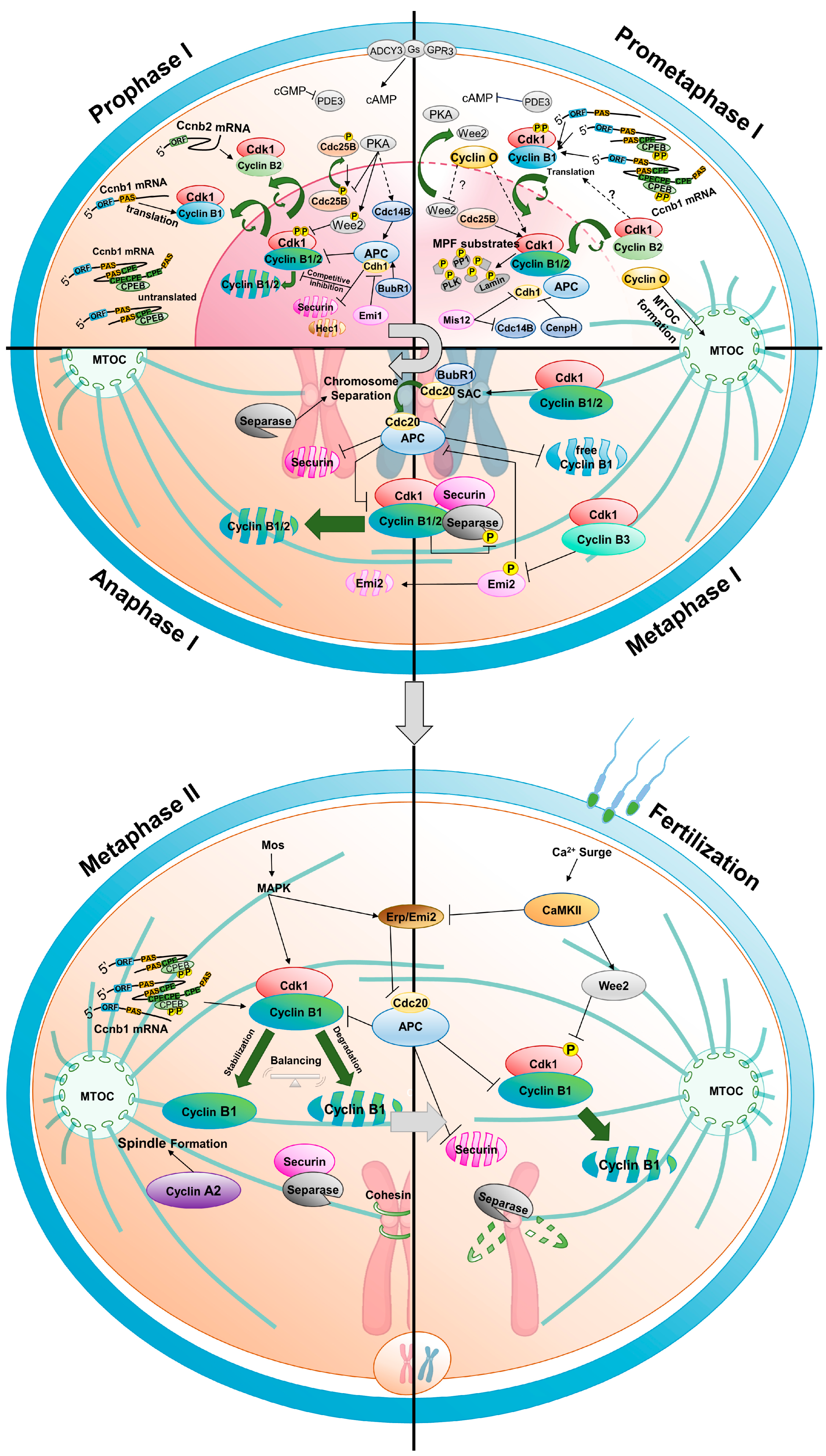
The prophase I arrest of the oocyte cell cycle is primarily regulated by high levels of cAMP produced by oocyte-specific adenylyl cyclase type 3 [81,82]. The protein kinase A (PKA) activated by cAMP plays a crucial role in maintaining the cell cycle arrest in oocytes [83,84,85,86,87]. However, it is not directly involved in regulating the key cell cycle engine, MPF, to arrest the oocyte cell cycle. In GV oocytes, PKA indirectly regulates MPF activity through Wee2 (Wee1B) kinase and Cdc25B. PKA promotes the activity of Wee2 kinase, which primarily phosphorylates and inhibits the Cdk1 component of MPF in the nucleus (Figure 3). When Wee2 is knocked down or its protein level is reduced by morpholino, GV oocytes are released from the cell cycle arrest [88,89,90,91]. Additionally, PKA inactivates cdc25 phosphatase, which promotes Cdk1 activity [92,93].

2.1. GV Arrest by Translational Regulation

MPF activity is regulated not only by protein modifications but also cyclin expression. Considering that transcription does not occur in fully grown oocytes [94], the translation of accumulated maternal mRNA is the sole gene expression mechanism underlying the stimulation of oocyte maturation in most species [95,96]. The translation of each mRNA within the oocyte is precisely and timely regulated for their role during the oocyte development [97].
Translation control in GV oocytes is little known in mouse oocytes. However, there are relatively many studies using Xenopus oocyte. The regulation occurs at the translational initiation stage. In Xenopus GV oocytes, elF4E binding to the 5’ cap structure of transcripts and various RNA binding proteins, (such as CPEB1, cleavage and polyadenylation specificity factor (CPSF), embryonic poly(A) binding protein (ePABP)), adenylase (Gld2), deadenylase (PARN), and scaffold proteins (symplekin, maskin) in the 3′ untranslated region (3′ UTR), form a complex that maintains a short poly(A) tail, thereby inhibiting translation [98,99]. This mechanism suppresses the translation of several key cell cycle regulators, including cyclin B1 and Mos, resulting in low MPF activity. Whether this complex functions similarly in translation regulation of mammalian oocytes including mouse oocytes is currently unclear.
Cyclin concentration in the GV state varies among species. For example, in Xenopus GV oocytes, one of the most extensively studied oocytes, the stockpile of cyclins is relatively low compared to the amount of Cdk1, resulting in insufficient generation of the Cdk1–cyclin B complex. Therefore, de novo protein synthesis is required for germinal vesicle breakdown (GVBD) to occur [100]. In contrast, mouse GV oocytes have a relatively high abundance of cyclins, and the Cdk1–cyclin B complex is preassembled [101,102]. In this case, the preassembled Cdk1–cyclin B complex is phosphorylated and inhibited by Wee2, preventing meiotic cell cycle re-entry of the oocyte (Figure 3). Recent studies have shown, using the RiboTag immunoprecipitation method that in mouse GV-arrested oocytes, that the Ccnb2 transcript is more abundantly associated with ribosomes than the Ccnb1 transcript. Consequently, cyclin B2, whose concentration exceeds that of cyclin B1, maintains the preassembled Cdk1–cyclin B complex [39]. Additionally, the presence of Ccnb3 mRNA associated with ribosome complexes in GV oocytes suggests isotype translation [26].
Mouse GV oocytes express the cyclin A1 protein, but the mRNA levels are low. The mRNA of cyclin A2 is expressed in all tissues, including oocytes, but the protein is not detected in GV oocyte [35,37,61,71]. Cyclin O is highly expressed in oocytes [33]. However, research on the expression regulation of these cyclins in GV oocytes is very rare.

2.2. GV Arrest by Degradation

When the cyclin concentration in the GV oocyte is excessively high, the oocyte undergoes an unwanted cell cycle without a cell cycle resumption signal. Conversely, extremely low cyclin concentration complicates the rapid resumption of the cell cycle even when the related signal emerges. Therefore, GV stage oocytes maintain a delicate balance between cyclin synthesis and degradation to ensure an appropriate level of cyclins for prompt Cdk1 activation and cell cycle resumption [103]. In mouse prophase I arrested oocytes, cyclin B1 is predominantly present in the cytoplasm, with some cyclin B1 localized in the nucleus. The APC/C activated by Cdh1 promotes the degradation of cyclin B1, thereby maintaining low Cdk1 activity [104,105,106]. Cdc14B attenuates the inhibition of Cdh1 phosphorylation to regulate its activity, and BubR1 contributes to the GV arrest by maintaining high levels of Cdh1, leading to reduced Cdk1 activity [107,108].
Conversely, securin acts as a competitive substrate of cyclin B1 for APC/CCdh1 degradation, and Emi suppresses APC/CCdh1. Therefore, both help maintain an appropriate level of cyclin B1 [109,110]. Exogenous Emi1 or the inhibition of Emi destruction in GV oocytes leads to an accumulation of cyclin B1 that is sufficient to promote GVBD [111]. Similarly, the degradation of cyclin B2 is finely tuned by Hec1 (also known as Ndc80), a kinetochore protein and subunit of the Ndc80 complex, which acts as a co-competing substrate for APC/CCdh1, thus regulating the amount of cyclin B2 in GV oocytes [112].

3. From Resumption of Oocyte Maturation to Metaphase I

The progression of the cell cycle from the prophase I arrested oocyte to metaphase II egg is as follows: (1) resumption of the cell cycle, accompanied by GVBD, chromosome condensation, and spindle formation (Figure 3 prometaphase I); (2) complete formation of the spindle and alignment of chromosomes in metaphase I (Figure 3 metaphase I); (3) entry into anaphase I and separation of homologous chromosomes, followed by extrusion of the first polar body (Figure 3 anaphase I); (4) the transition from meiosis I to meiosis II without an intervening S-phase; and (5) the metaphase II state, where the cell cycle is arrested again (Figure 3 metaphase II).
Gonadotrophic hormones stimulate the periodic ovulation of progressively growing oocytes within the ovary. Changes in the signaling pathways of various cells within the follicle, induced by luteinizing hormone, ultimately activate phosphodiesterase 3A (PDE3A) in the oocyte, leading to the degradation of cAMP [113,114,115,116,117,118]. Consequently, PKA is inactivated, allowing Wee2 to move out of the nucleus. Cdk1–cyclin B1 and Cdc25B translocate into the nucleus, leading to the breakdown of the GV membrane and resumption of the oocyte cell cycle [91,119,120]. The nuclear translocation of exogenous cyclin B1 promotes GVBD, suggesting the importance of the nuclear translocation of cyclin B1 for GVBD [121].
Cyclin B2 is located around the germinal vesicle in Xenopus oocytes, but is primarily present in the Golgi during mitosis [122]. In mouse oocytes, cyclin B2 translocates into the nucleus before GVBD, and cyclin B2 moves into the nucleus to compensate for the role of cyclin B1 in cyclin B1 knockout oocytes, indicating that it also regulates nuclear MPF activity [29]. Cyclin O is also present in the GV and localizes to the spindle region after GVBD [33]. However, the timing, sequence, and rate of translocation for each cyclin, and their specific substrates, have not been studied.
In addition to the regulation of MPF activity by Wee2-Cdc25B, kinases such as c-Mos and Polo-like kinase, as well as phosphatases, including PP1 and MASTL-Arpp19 (ENSA)-PP2A, regulate MPF activity through positive and negative feedback mechanisms. The regulation of MPF by these kinases and phosphatases has been extensively reviewed [123,124].

3.1. Role of Cyclins from Resumption of Oocyte Maturation to Metaphase I

The prometaphase I of oocyte meiosis is relatively prolonged compared to that of somatic cells. The slow progression during prometaphase allows for chromosome condensation, congression, kinetochore−microtubule attachment, and proper alignment of chromosomes at the metaphase plate [125]. This is crucial for accurate separation of homologous chromosomes and prevention of aneuploidy. The main driving force for GVBD and prometaphase progression is the increasing activity of Cdk1, which reaches its peak at metaphase I. Continuous synthesis and accumulation of cyclin B1 have been believed to be essential for increasing Cdk1 activity and meiosis progression [103,126]. However, recent studies have shown that Ccnb1−/− oocytes undergo normal GVBD, complete meiosis I, and extrude the first polar body. Furthermore, no abnormalities in chromosome alignment and spindle formation are observed during meiosis I [27,29]. These findings suggest that cyclin B1 may not be required for the progression of meiosis I.
However, the upregulation of cyclin B2 by an unknown mechanism in Ccnb1−/− oocytes results in complete MPF activation [29], which is also observed in mitosis [127]. Therefore, whether cyclin B1 is truly dispensable for GVBD and metaphase I processes or whether its significance is counteracted by compensation through cyclin B2 has not been determined. Recent studies suggest that cyclins forming complexes with Cdk1 are more stable than free cyclins [128]; therefore, there is a possibility that Cdk1 preferentially associates with cyclin B2 in the absence of cyclin B1, leading to increased stability and elevated levels of cyclin B2.
Ccnb2 knockout mice are viable, but oocytes lacking cyclin B2 are unable to efficiently activate Cdk1, resulting in significant delays in GVBD or metaphase arrest owing to SAC activation. Consequently, these mice exhibit impaired oocyte maturation, early ovarian insufficiency, and compromised fertility (Figure 1) [27,29,39,112,129]. In vitro studies have shown that overexpression of cyclin B2 induces GVBD more efficiently than that of cyclin B1 in the presence of dbcAMP, an oocyte maturation inhibitor [103]. These findings demonstrate the necessity of cyclin B2 for sufficient MPF activation which is required for re−entry and progression of meiotic divisions. However, the fact that oocytes remain arrested in the GV state when both cyclin B1 and B2 are absent indicates that one of the two cyclins is essential for GVBD [29].
The onset of GVBD at normal timing in Ccnb3−/− mice demonstrates that cyclinB3 is not required for the early stages of oocyte maturation [31]. Overexpression of cyclin A1 induces GVBD without cyclin B1 and B2 in the presence of the GVBD inhibitor milrinone [36,130]. Cyclin A2 is present in GV oocytes, and microinjection of stable cyclin A2 mutants lacking the destruction box induces spontaneous GVBD, while microinjection of cyclin A2 antibodies slows down the rate of GVBD [37]. Both endogenous cyclin A1 and A2 can bind to Cdk1, but their knockout does not affect GVBD, suggesting that they may not function during this stage in vivo [34,74].
Cyclin O in oocytes seemingly regulates GVBD through different mechanisms compared to other cyclins. Knockdown of Ccno using short interfering RNA microinjection prevents the dephosphorylation of the Cdk1 inhibitory phosphorylation site, Tyr15, leading to the oocytes remaining in the GV state. This GV arrest can be rescued by the overexpression of cyclin B1 but not Cdc25B, suggesting that it is not directly related to the Cdc25B activity. Whether the exact role of cyclin O in GVBD is inactivation of Wee2 has not been determined. In Ccno-knockdown oocytes, the formation of microtubule-organizing centers is disrupted in the perinuclear region [33]. This suggests that the primary role of Ccno in oocytes is not the regulation of MPF but rather the regulation of early spindle formation.

3.2. Translational Regulation of Cyclins

The continuous synthesis and accumulation of various proteins, including cyclin B1, are the main driving force for oocyte maturation. The mechanism regulating protein translation in mouse oocyte meiotic cells remains largely unexplored. However, the process has been relatively well-studied due to the requirement for mRNA translation during re-entry into meiosis in Xenopus oocytes [131,132]. Therefore, results gained from Xenopus studies could potentially offer clues regarding protein translation in mouse oocytes. The length of the poly(A) tail of mRNA is crucial for the regulation of translation, and relatively longer poly(A) tails (approximately 80–500 residues) are generally associated with increased translation. In Xenopus GV oocytes, the poly(A) tail of mRNAs encoding proteins, such as Mos and cyclin, is relatively short: approximately 20 nucleotides [133]. Upon stimulation by progesterone to initiate oocyte maturation, the poly(A) tails of mRNAs lengthen to approximately 100 nucleotides, leading to increased translation [131].
The regulation of mRNA poly(A) tail length involves two main cis-acting sequences: the hexanucleotide polyadenylation signal (AAUAAA) and the cytoplasmic polyadenylation element (CPE, UUUUUAU) [134]. The hexanucleotide polyadenylation signal is a highly conserved sequence located 10–30 nucleotides upstream of the mRNA’s cleavage/polyadenylation site [135]. It is identified by a complex of three or four proteins, including CPSF, which catalyzes the cleavage and polyadenylation of pre-mRNA [136]. The CPE site binds to the RNA-binding protein CPEB, a highly conserved protein that regulates the elongation of mRNA’s poly(A) tail. CPEB generally activates the translation of target mRNAs but can also act as a translation repressor depending on its phosphorylation state [137,138]. In Xenopus GV oocytes, CPEB binds to proteins, such as maskin, which inhibits the translation of CPE-containing mRNAs, and PARN, which possesses 3′-exoribonuclease activity, to maintain a short polyadenylated tail [139]. When translation is activated upon oocyte maturation, CPEB is phosphorylated by Eg2/Aurora A kinase [140], PARN is released from the inhibitory complex, and mRNA is polyadenylated by cytoplasmic poly(A) polymerase GLD-2 [141]. The PABP forms a complex with eIF4G of the eIF4F cap-binding structure at the 5’ end of mRNA, thereby promoting mRNA looping and enhancing translation [133,142,143,144,145]. CPE-containing mRNAs are not polyadenylated simultaneously with the initiation of oocyte maturation, but rather undergo translation at specific time points. For example, mRNA encoding proteins like Mos are polyadenylated early during prophase I, while mRNAs encoding cyclin B1 are polyadenylated late after GVBD. Early polyadenylation is induced by CPEB phosphorylation mediated by aurora A, while late polyadenylation requires Mos synthesis and cdk1 kinase to phosphorylate CPEB, with most CPEBs being degraded [146].
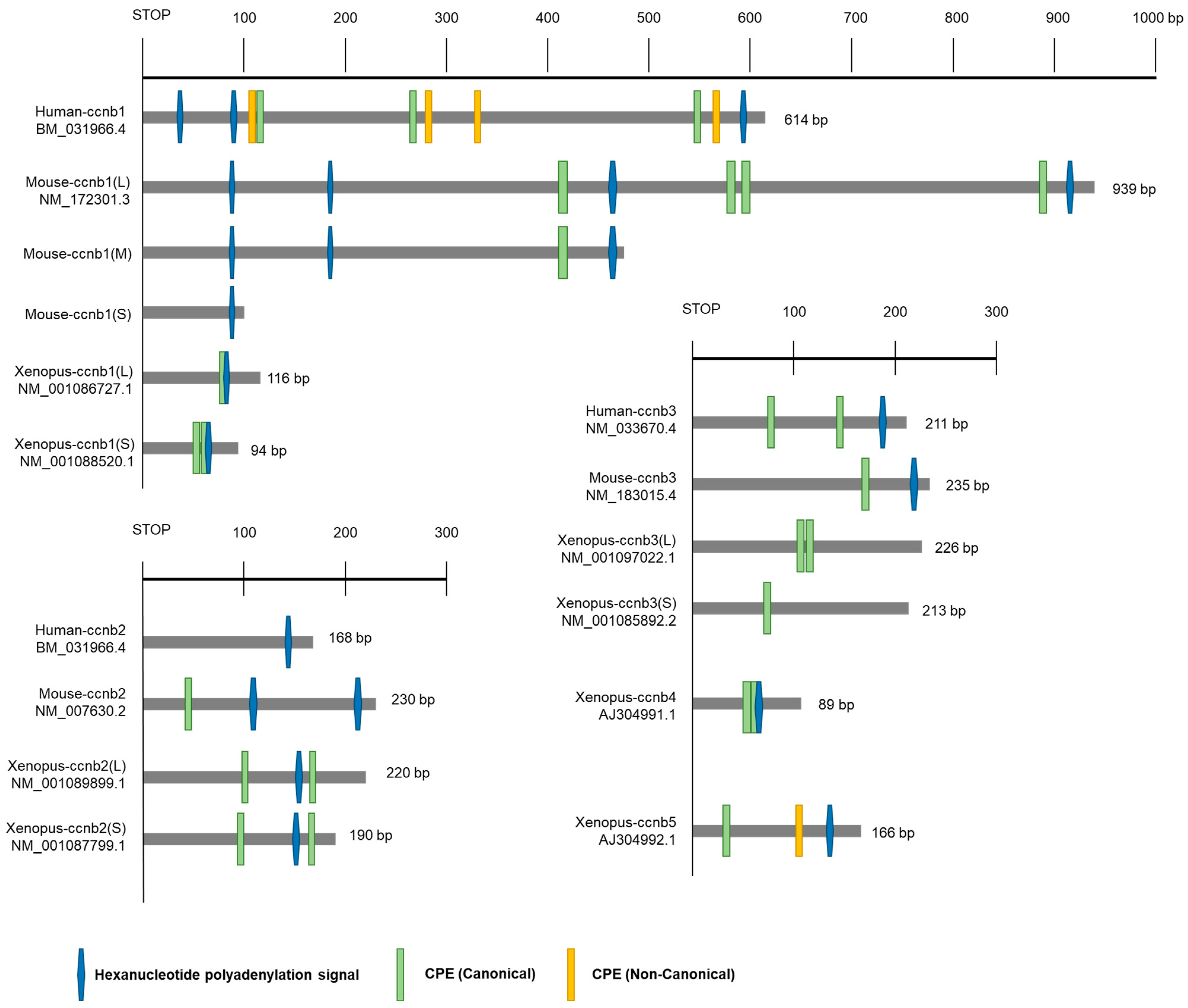
While the mechanisms regulating mRNA polyadenylation and translation during oocyte development in other vertebrates, including mice, have not been extensively studied, it is presumed that similar regulations occur. For example, in zebrafish and mice, polyadenylation of cyclin B1 mRNA occurs during oocyte maturation, and the hexanucleotide and CPE sequences are essential for mRNA translation [95]. However, the position and sequence of CPE in cyclin B1 mRNA 3′ UTR are not well conserved across species (Figure 4). This is likely because the factors and mechanisms that regulate the translation of maternal mRNAs are conserved, but the levels of cyclin proteins required for early oocyte maturation may differ among species.
The recent discovery of various isoforms of cyclin B in oocytes has increased interest in understanding their specific expression regulation and unique functions during oocyte maturation. In Xenopus oocytes, five types of cyclin B isotypes (B1–B5) have been identified, each with different expression patterns regulated during oocyte maturation. In GV oocytes, all five cyclin mRNAs have short poly(A) tails, and, upon progesterone stimulation, cytoplasmic polyadenylation occurs for cyclin B1, B2, B4, and B5, but not for B3 mRNA. Synthesis of cyclin B2 and B5 is required for the progression of early meiosis I, while new synthesis of B1 and B4 is needed for the progression of meiosis II [146,147].
There is a clear difference between the protein translation patterns of cyclin B1 mRNA and cyclin B2 mRNAs in mouse oocytes. In GV stage mouse oocytes, cyclin B1 expression is kept low through the regulation of CPEB and mRNA granule formation [148,149], while the expression level of cyclin B2 remains relatively constant during oocyte maturation [26]. Mouse oocytes can enter meiosis without de novo protein synthesis, likely due to the relatively high concentration of cyclin B2 [150]. The difference in the expression of cyclin B1 and B2 can be attributed to the distinct structures of their 3′ UTRs and the number and location of CPEs (Figure 4). When RNA immunoprecipitation using CPEB antibodies is performed to confirm CPEB binding to mRNAs, cyclin B1 transcripts are significantly coprecipitated, while cyclin B2 transcripts show minimal binding. Knockdown of CPEB1 using morpholinos had little effect on the expression of the cyclin B2 3′ UTR reporter, but significantly reduced the expression of the reporter containing cyclin B1 3′ UTR. Consequently, the translation of cyclin B1 mRNA is regulated by CPEB1, while cyclin B2 acts independently of CPEB1. Additionally, the regulation of cyclin B1 expression by CPEB is Cdk1-associated activity-dependent [26]. According to recent research, the presence or absence of cyclin B2 affects the translation of cyclin B1 mRNA [39]. This suggests that the Cdk1-associated activity involved in regulating CPEB could be the Cdk1–cyclin B2 complex, but further studies are needed to confirm this.
Cyclin B1 mRNA expression during oocyte maturation is also regulated by a newly identified mechanism. Cyclin B1 mRNA utilizes three alternative hexanucleotide poly(A) sites to generate three transcripts with varying lengths of the 3′ UTR in the oocyte (Figure 4). The transcript with a short 3′ UTR is expressed from the GV stage, while the intermediate and long 3′ UTR transcripts are suppressed in the GV stage, and their expression begins after GVBD (Figure 3) [151]. Recent studies have shown that cyclin B2 contains two alternative poly(A) sites, but their expression does not exhibit differences [39]. Whether the short form of the mouse cyclin B2 transcript is actually expressed in the oocyte has not been elucidated. Furthermore, the regulation of cyclin B1 and B2 expression is influenced by the 3′ UTR, 5′ UTR, and open reading frame [152]. Using these regulatory elements, the oocyte is able to control the precise timing and amount of cyclin B1 and B2 production.
The mRNA levels of cyclin B3 increase approximately 3–4 times during the metaphase compared to levels in the GV phase [30]. However, the amount of cyclin B3 mRNA associated with polysomes is very high in the GV state and gradually decreases with oocyte maturation [26]. Therefore, measurement of the precise amount and regulation of cyclin B3 expression during oocyte maturation is required for the examination of changes in protein levels.

3.3. Regulation of Cyclin Degradation

During the GV phase, the levels of cyclin B are regulated to a low state by APC/CCdh1. However, during the GVBD stage, the degradation of cyclin B1 by APC/CCdh1 is inhibited, leading to its increased accumulation. Recent research has revealed that the kinetochore complex component, Mis12, regulates cyclin B1 degradation by controlling Cdh1 or Cdc14B, thereby inducing the accumulation of cyclin B1 (Figure 3. prometaphase I). Depletion of Mis12 prevents the accumulation of cyclin B1, resulting in the inhibition of GVBD [153]. Additionally, CenpH, a component of the inner kinetochore protein, also modulates APC/CCdh1 to enhance the accumulation of cyclin B1 at the GVBD stage. Therefore, depletion of CenpH by morpholino injection resulted in attenuation of MPF activation [110].
Once the spindle has been formed and microtubules attach to kinetochores, homologous chromosomes are aligned accurately at the metaphase plate. Only when the homologous chromosomes are properly positioned and aligned, can they be evenly divided into two cells. Until then, the cell cycle is arrested by the SAC triggered by improper attachment of kinetochores to microtubules. SAC inhibits APC/CCdc20, resulting in the stabilization and accumulation of securin and the Cdk1–cyclin B1 complex. These components bind to and inhibit separase by phosphorylation [106,154,155,156,157,158,159].
Recent research has shown that cyclin B2 is also involved in these processes. In mouse oocytes, Cdk1–cyclin B2 binds to separase, and cyclin B2 degradation is necessary for separase activation during the metaphase I-anaphase I transition. Furthermore, in cyclin B1-null oocytes, the presence of stable cyclin B2 does not prevent chromosome separation if non-phosphorylatable separase is present, indicating that Cdk1–cyclin B2 phosphorylates separase to inhibit anaphase I progression (Figure 3. metaphase I) [129,160]. Oocyte-specific deletion of cyclin B2 leads to delayed spindle assembly and increased errors in chromosome segregation during the metaphase I- anaphase I transition, highlighting the important role of cyclin B2 in metaphase I of mouse oocytes [39].

4. Metaphase I-Anaphase I Transition

Metaphase I-anaphase I transition during oocyte maturation is primarily regulated by protein degradation. When the chromosomes are properly aligned and all microtubules are attached to the kinetochores, the SAC is inactivated, and APC/CCdc20 promotes the polyubiquitination of securin and cyclin B1 through the ubiquitin-proteasome system for their degradation [158,161,162,163,164]. Overexpression or stable expression of cyclin B1 arrests oocytes at metaphase I [158], indicating that the degradation of cyclin B1 is essential for the metaphase I-anaphase I transition and the extrusion of the first polar body [162,165,166]. Recent studies have shown that cyclin B2 is also degraded after metaphase I; if cyclin B2 is not degraded, homologous chromosome separation does not occur, and the oocyte remains arrested at metaphase I.
Indeed, the degradation of cyclins relies on the presence of a destruction box, also known as the D box, and KEN box within the cyclin protein (Figure 2). B-type cyclins typically contain a conserved D box motif RXALGXIXN, where the arginine at position 1 and the leucine at position 4 are highly conserved. Mutations in the D box lead to the stabilization of cyclins and a decrease or lack of ubiquitination [167]. However, recent research has identified a novel degradation site that is involved in regulating the timing of spindle alignment. In the early prometaphase stage of oocytes, there are two groups of cyclin B1. One group is a pool of free cyclin B1, and the other is cyclin B1 bound to Cdk1. During progression to metaphase I, most of the free cyclin B1 is degraded through the newly identified degradation site (Figure 2A, Prometaphase motif, PM motif), while the Cdk1-bound cyclin B1 remains protected from degradation until SAC is satisfied and APC/CCdc20 is fully activated. Only then does degradation of Cdk1-bound cyclin B1 begins, leading to a rapid decrease in Cdk1 activity and the initiation of anaphase I [128]. This mechanism allows oocytes extra time for the precise alignment of chromosomes. This motif was also found in the cyclin B2, indicating that cyclin B2 is possibly regulated by similar mechanism.
The role of cyclin B3 in the metaphase I-anaphase I transition has been elucidated by recent studies. Oocytes lacking cyclin B3 maintain the metaphase I spindle and arrest at metaphase I, resulting in female mouse infertility [31,32]. This suggests that cyclin B3 plays an important role in the metaphase I-anaphase I transition. Upon metaphase arrest by cyclin B3 removal, cyclin B1 remains stable, and Cdk1 activity is maintained in a high state, even when SAC is inactive. Therefore, cyclin B3 appears to regulate the degradation of cyclin B1 and securin by modulating APC/C activation [30]. The inability of cyclin B1 expression to compensate for the lack of cyclin B3 suggests that B3 plays a unique role at this stage.
The mechanism by which cyclin B3 promotes anaphase I onset was unclear until the recent revelation of the role played by Cdk1–cyclin B3 in metaphase. Cdk1–cyclin B3 phosphorylates the conserved site of Emi2 during meiosis I, leading to Emi2 phosphorylation by various kinases, including polo-like kinase 1, and degradation (Figure 3 metaphase I and anaphase I). Therefore, in the absence of cyclin B3 in mouse and Xenopus oocytes, the Emi2-dependent cell cycle arrest occurs prematurely at metaphase I, resulting in cell cycle arrest. Cyclin B3 is present at a sufficiently high level in early meiosis I and is degraded upon completion of meiosis I, preventing its re-accumulation [38]. Furthermore, in female fruit fly germ cells, the absence or mutation of cyclin B3 leads to an inability to complete meiosis I and infertility, suggesting the conservation of cyclin B3′s role across species [54,57,168].
The mechanisms underlying the sparing of cyclin B3 until completion of meiosis I and the selective degradation of only cyclin B1 and B2 during the metaphase I-anaphase I transition, and the mechanism underlying the distinction of the cyclins by APC/CCdc20 can potentially be explained by the differences in the D boxes of each cyclin. Cyclin B1 and B2 have well-conserved D boxes, whereas cyclin B3 has a quasi-D box which contains phenylalanine instead of leucine at the conserved position 4 of the D box motif (Figure 2). This difference may lead to differences in affinity and specificity for APC/C, making cyclin B3 more resistant to degradation during the metaphase I-anaphase I transition than B1 and B2. Further experiments and research are needed to validate these possibilities and investigate the mechanisms underlying the degradation of cyclin B3 after anaphase I.
Cyclin A2 and cyclin O also possess putative D-box motifs (Figure 2). Cyclin A2 undergoes two waves of degradation during oocyte maturation, with most of cyclin A2 being degraded during prometaphase I. Overexpression of wild-type cyclin A2 inhibits PB extrusion in meiosis I, while constitutive cyclin A2 activity in meiosis I leads to precocious sister separation, suggesting that cyclin A2 degradation during prometaphase I is essential for oocyte meiotic progression [37]. The degradation of cyclin O in the mouse oocyte has not been reported.

5. Anaphase I-Metaphase II Transition

Anaphase I to metaphase II transition is relatively rapid, and therefore, research on the regulation of this stage is limited. Following the first polar body extrusion, Cdk1–cyclin B1 activity increases, preventing the onset of interphase or S-phase and facilitating entry into the second meiotic division (meiosis II). Ccnb1−/− oocytes fail to progress to meiosis II after the first polar body extrusion and instead undergo chromosome decondensation and enter interphase. However, this defect can be rescued via the exogenous injection of mRNA encoding cyclin B2. This suggests that sufficient MPF activation, regardless of cyclin B1 or B2, is crucial for entry into meiosis II [27,29].
Given that oocyte-specific Ccnb1-null female mice are infertile due to MPF reactivation failure in meiosis II, the reason for the inability of endogenous cyclin B2 to compensate for the absence of cyclin B1 in Ccnb1−/− mice is not fully understood. It may be because translation regulation mechanisms involving factors like CPEB may selectively enhance the translation of cyclin B1 in wild-type mice, while suppressing that of cyclin B2. Alternatively, the degradation of cyclin B2 and a decrease in the degradation of cyclin B1, or a combination of both factors may be the underlying reason. However, these possibilities and the regulation of cyclin B2 protein level in this step have not been extensively studied.
In Drosophila oocytes, cyclin B3 plays a role in inhibiting the entry into the S phase between the first and second meiotic divisions by cooperating with cyclin A [54]. In contrast, in Xenopus, the XeWee1A protein (ortholog of mouse Wee2) is absent, resulting in the maintenance of high Cdk1 activity and the inhibition of re-entry into the S phase between the first and second M phases [169,170]. In mice, Wee2 is present at this stage; however, the mechanism by which Wee2 activity is reduced is unknown [88,90,171].
For proper spindle formation during metaphase II, cyclin A2 is necessary [74]. Cyclin A2, which decreases during prometaphase I, is re-expressed and maintained until arrest of oocytes at metaphase II. Conditional deletion of cyclin A2 does not have a specific impact on the first meiotic division but leads to impaired metaphase II spindles and increased merotelic attachments. Therefore, cyclin A2 plays a role in spindle assembly during metaphase II and provides flexibility in kinetochore-microtubule attachments, allowing for error correction and prevention of merotelic attachments and lagging chromosomes [37]. However, the detailed mechanisms involved in this process have not been elucidated.

6. Arrest at Metaphase II and Fertilization

Prior to undergoing fertilization, oocytes remain arrested in the metaphase II phase owing to cytostatic factor (CSF) activity [1,172,173]. CSF activity is maintained by the mitogen-activated protein kinase pathway activated by Mos, which regulates MPF activity [174]. During the metaphase II phase, Cdk1 activity is maintained at a high level, and the balance between synthesis and degradation of cyclin B1 is crucial for maintaining a threshold (Figure 3 metaphase II) [175,176,177]. Synthesis of cyclin B1 has been observed during metaphase II arrest; at the same time, cyclin B1 is degraded by APC/CCdc20 activity. If excessive degradation of cyclin B1 leads to a decrease in Cdk1 activity, another regulatory pathway called the Mos-MAPK pathway is activated, thereby inhibiting APC/C activity through components, such as Emi/Erp (Emi2, XErp1 in Xenopus), and resulting in the stabilization of cyclin B1 [36,178,179]. Both MPF and CSF activities are lost upon fertilization or parthenogenetic activation [180].
In Xenopus laevis oocytes, cyclin E1 and E2 form a cyclin E–Cdk2 complex, which acts as a CSF by inhibiting APC/C, thus contributing to metaphase II arrest [181]. Additionally, cyclin E is essential for the development of Drosophila melanogaster and C. elegans, but it is not required for normal mouse embryonic development [182,183,184]. In mammals, fertilization by sperm triggers a Ca2+ spike, leading to the activation of the oocyte. The main downstream target of Ca2+ oscillation is calmodulin-dependent protein kinase II (CaMKII, CaMK2A) [185,186,187]. CaMKII phosphorylates Wee2, leading to the inactivation of MPF and initiating resumption of meiosis and exit from metaphase II [90,188,189]. CaMKII also inactivates Emi2, which activates APC/C. Activated APC/C then targets the cohesin, which is holding sister chromatids together, and cyclin B1 for degradation [190,191], leading to sister chromatid segregation and anaphase II entry. The oocyte exits meiosis II, and two pronuclei, one from the oocyte and another from the sperm, are formed. These pronuclei fuse to form a diploid zygote with a complete set of chromosomes [192]. The expression of stable cyclin B2 prevents sister chromatid separation due to insufficient separase activity, leading to a failure in pronucleus formation. However, when a Cdk1-resistant phosphorylation site mutant separase (PM-separase) is injected, sister chromatid separation occurs. Therefore, the cyclin B/CDK1 complex contributes to inhibition of separase activity during the second meiotic division in oocytes [160].
In C. elegans, the absence of cyclin B3 hinders the segregation of sister chromatids during anaphase II. This phenotype is partially rescued when SAC is simultaneously knocked down, suggesting that the function of cyclin B3 is SAC-dependent [56]. Additionally, in C. elegans, cyclin B3 plays a crucial role in zygotic genome activation during the maternal-to-zygotic transition [193]. However, there is no research on the role of cyclin B3 in mouse metaphase II oocytes. Cyclin A2 also appears to be involved in sister chromatid separation in meiosis II, as interfering with its function inhibits this process [37]. Various cyclins are involved in meiosis II phase and the activation of oocytes by fertilization, but research on the roles and regulation of cyclins during these stages is relatively limited.

7. Discussion

Precise regulation of oocyte maturation is crucial because oocytes are relatively large and have a long cell cycle. The discovery of various types of cyclins expressed in oocytes, following the initial identification of cyclin B1 as a key regulator of oocyte maturation, indicates that the process of oocyte maturation is considerably more complex and finely regulated than previously understood. The reason why oocytes utilize such diverse cyclins is still veiled. Particularly, for cyclin B1 and B2, although they differ in the timing and quantity of synthesis, they can compensate for each other in most processes and have similar levels of degradation. However, the reasons for the presence of both cyclins in oocytes are unknown. Each cyclin likely phosphorylates specific substrates during oocyte maturation. Therefore, research aimed at identifying these cyclin-specific substrates is essential and may yield interesting results regarding the regulation of oocyte cell cycle progression. Furthermore, a clear understanding of how the expression and degradation of each cyclin is regulated in time and space is necessary. It is also important to elucidate the differences in Cdk1 activation induced by each cyclin.
Detailed studies on the relative localization of each cyclin in oocytes are required. Most studies on the localization of cyclins in oocytes have been based on the overexpression of cyclins fused with GFP, which does not accurately measure the temporal changes in the localization of endogenous cyclins. For example, it is necessary to determine whether there is a time difference in the nuclear translocation of cyclin B1 and B2 during the GVBD stage and, if so, what their respective roles are.
In oocytes, Cdk1 primarily functions as a cell cycle-regulating kinase, and all the cyclins, including cyclin B1, B2, B3, and A2, associate with this kinase and exhibit activity in oocytes. In this context, examining cyclin competition to bind to Cdk1 would be intriguing. Determining the cyclin with the highest affinity for Cdk1 at each oocyte cell cycle stage and answering other related questions would be a fascinating area of research. In addition, the reasons why cyclin B2 increases when cyclin B1 is deleted, and whether the absence of one cyclin results in the other cyclins compensating for its roles in all substrates, are unknown. Additionally, further research to identify the possible existence of currently unknown cyclins expressed in oocytes and their functions is needed.

Author Contributions

M.K.K., C.G.L. and S.J.H. designed the manuscript and collected and analyzed the data. H.M.K., M.K.K., S.Y.S., J.H.J., M.J.K., E.K.S., C.G.L. and S.J.H. wrote the original drafts and reviewed the manuscript. All authors have read and agreed to the published version of the manuscript.

Funding

This research was supported by Basic Science Research Program through the National Research Foundation of Korea (NRF) funded by the Ministry of Education (2020R1I1A3070491) and the Dongnam Institute of Radiological and Medical Science (DIRAMS) grant (50491-2023) funded by Ministry of Science and ICT (MSIT), Republic of Korea. S.Y. Seong, J.H. Jo, and M.J. Kim, were supported by the step 4 BK21 program, funded by the Ministry of Education (MOE, Korea) and the NRF of Korea.

Institutional Review Board Statement

Not applicable.

Informed Consent Statement

Not applicable.

Data Availability Statement

No new data were created in this study. All the data reported in this review were found in original articles cited in the text.

Conflicts of Interest

The authors declare no conflict of interest.

Abbreviations

CDCCell division cycle
MPFMaturation-promoting factor
CDKCyclin-dependent kinase
CBDCyclin box domain
SACSpindle assembly checkpoint
APC/CAnaphase-promoting complex/cyclosome
GVGerminal vesicle
PKAProtein kinase A
GVBDGerminal vesicle breakdown
PDE3APhosphodiesterase 3A
CPSFCleavage and polyadenylation specificity factor
ePABPEmbryonic poly(A) binding protein
CPECytoplasmic polyadenylation element
3′ UTR3′ untranslated region
5′ UTR5′ untranslated region
CaMKII, CaMK2ACalmodulin-dependent protein kinase II

References

  1. Masui, Y.; Markert, C.L. Cytoplasmic control of nuclear behavior during meiotic maturation of frog oocytes. J. Exp. Zool. 1971, 177, 129–145. [Google Scholar] [CrossRef]
  2. Evans, T.; Rosenthal, E.T.; Youngblom, J.; Distel, D.; Hunt, T. Cyclin: A protein specified by maternal mRNA in sea urchin eggs that is destroyed at each cleavage division. Cell 1983, 33, 389–396. [Google Scholar] [CrossRef]
  3. Draetta, G.; Luca, F.; Westendorf, J.; Brizuela, L.; Ruderman, J.; Beach, D. Cdc2 protein kinase is complexed with both cyclin A and B: Evidence for proteolytic inactivation of MPF. Cell 1989, 56, 829–838. [Google Scholar] [CrossRef]
  4. Booher, R.N.; Alfa, C.E.; Hyams, J.S.; Beach, D.H. The fission yeast cdc2/cdc13/suc1 protein kinase: Regulation of catalytic activity and nuclear localization. Cell 1989, 58, 485–497. [Google Scholar] [CrossRef] [PubMed]
  5. Nigg, E.A. Cyclin-dependent protein kinases: Key regulators of the eukaryotic cell cycle. Bioessays 1995, 17, 471–480. [Google Scholar] [CrossRef] [PubMed]
  6. Cao, L.; Chen, F.; Yang, X.; Xu, W.; Xie, J.; Yu, L. Phylogenetic analysis of CDK and cyclin proteins in premetazoan lineages. BMC Evol. Biol. 2014, 14, 10. [Google Scholar] [CrossRef]
  7. Kaldis, P.; Russo, A.A.; Chou, H.S.; Pavletich, N.P.; Solomon, M.J. Human and yeast cdk-activating kinases (CAKs) display distinct substrate specificities. Mol. Biol. Cell 1998, 9, 2545–2560. [Google Scholar] [CrossRef]
  8. Malumbres, M. Cyclin-dependent kinases. Genome Biol. 2014, 15, 122. [Google Scholar] [CrossRef]
  9. Gunbin, K.V.; Suslov, V.V.; Turnaev, I.I.; Afonnikov, D.A.; Kolchanov, N.A. Molecular evolution of cyclin proteins in animals and fungi. BMC Evol. Biol. 2011, 11, 224. [Google Scholar] [CrossRef]
  10. Meyerson, M.; Harlow, E. Identification of G1 kinase activity for cdk6, a novel cyclin D partner. Mol. Cell. Biol. 1994, 14, 2077–2086. [Google Scholar] [CrossRef]
  11. Sherr, C.J. D-type cyclins. Trends Biochem. Sci. 1995, 20, 187–190. [Google Scholar] [CrossRef]
  12. Obeyesekere, M.N.; Tucker, S.L.; Zimmerman, S.O. A model for regulation of the cell cycle incorporating cyclin A, cyclin B and their complexes. Cell Prolif. 1994, 27, 105–113. [Google Scholar] [CrossRef]
  13. Fung, T.K.; Poon, R.Y. A roller coaster ride with the mitotic cyclins. Semin. Cell Dev. Biol. 2005, 16, 335–342. [Google Scholar] [CrossRef]
  14. de Vant’ery, C.; Gavin, A.C.; Vassalli, J.D.; Schorderet-Slatkine, S. An accumulation of p34cdc2 at the end of mouse oocyte growth correlates with the acquisition of meiotic competence. Dev. Biol. 1996, 174, 335–344. [Google Scholar] [CrossRef]
  15. Capalbo, A.; Hoffmann, E.R.; Cimadomo, D.; Ubaldi, F.M.; Rienzi, L. Human female meiosis revised: New insights into the mechanisms of chromosome segregation and aneuploidies from advanced genomics and time-lapse imaging. Hum. Reprod. Update 2017, 23, 706–722. [Google Scholar] [CrossRef] [PubMed]
  16. Das, D.; Arur, S. Regulation of oocyte maturation: Role of conserved ERK signaling. Mol. Reprod. Dev. 2022, 89, 353–374. [Google Scholar] [CrossRef] [PubMed]
  17. Ye, X.; Zhu, C.; Harper, J.W. A premature-termination mutation in the Mus musculus cyclin-dependent kinase 3 gene. Proc. Natl. Acad. Sci. USA 2001, 98, 1682–1686. [Google Scholar] [CrossRef] [PubMed]
  18. Tsutsui, T.; Hesabi, B.; Moons, D.S.; Pandolfi, P.P.; Hansel, K.S.; Koff, A.; Kiyokawa, H. Targeted disruption of CDK4 delays cell cycle entry with enhanced p27(Kip1) activity. Mol. Cell. Biol. 1999, 19, 7011–7019. [Google Scholar] [CrossRef]
  19. Malumbres, M.; Sotillo, R.; Santamaria, D.; Galan, J.; Cerezo, A.; Ortega, S.; Dubus, P.; Barbacid, M. Mammalian cells cycle without the D-type cyclin-dependent kinases Cdk4 and Cdk6. Cell 2004, 118, 493–504. [Google Scholar] [CrossRef]
  20. Adhikari, D.; Zheng, W.; Shen, Y.; Gorre, N.; Ning, Y.; Halet, G.; Kaldis, P.; Liu, K. Cdk1, but not Cdk2, is the sole Cdk that is essential and sufficient to drive resumption of meiosis in mouse oocytes. Hum. Mol. Genet. 2012, 21, 2476–2484. [Google Scholar] [CrossRef]
  21. Santamaría, D.; Barrière, C.; Cerqueira, A.; Hunt, S.; Tardy, C.; Newton, K.; Cáceres, J.F.; Dubus, P.; Malumbres, M.; Barbacid, M. Cdk1 is sufficient to drive the mammalian cell cycle. Nature 2007, 448, 811–815. [Google Scholar] [CrossRef]
  22. Diril, M.K.; Ratnacaram, C.K.; Padmakumar, V.C.; Du, T.; Wasser, M.; Coppola, V.; Tessarollo, L.; Kaldis, P. Cyclin-dependent kinase 1 (Cdk1) is essential for cell division and suppression of DNA re-replication but not for liver regeneration. Proc. Natl. Acad. Sci. USA 2012, 109, 3826–3831. [Google Scholar] [CrossRef] [PubMed]
  23. Nieduszynski, C.A.; Murray, J.; Carrington, M. Whole-genome analysis of animal A- and B-type cyclins. Genome Biol. 2002, 3, research0070.1. [Google Scholar] [CrossRef] [PubMed]
  24. Pines, J.; Hunter, T. Isolation of a human cyclin cDNA: Evidence for cyclin mRNA and protein regulation in the cell cycle and for interaction with p34cdc2. Cell 1989, 58, 833–846. [Google Scholar] [CrossRef] [PubMed]
  25. Chapman, D.L.; Wolgemuth, D.J. Identification of a mouse B-type cyclin which exhibits developmentally regulated expression in the germ line. Mol. Reprod. Dev. 1992, 33, 259–269. [Google Scholar] [CrossRef]
  26. Han, S.J.; Martins, J.P.S.; Yang, Y.; Kang, M.K.; Daldello, E.M.; Conti, M. The Translation of Cyclin B1 and B2 is Differentially Regulated during Mouse Oocyte Reentry into the Meiotic Cell Cycle. Sci. Rep. 2017, 7, 14077. [Google Scholar] [CrossRef] [PubMed]
  27. Brandeis, M.; Rosewell, I.; Carrington, M.; Crompton, T.; Jacobs, M.A.; Kirk, J.; Gannon, J.; Hunt, T. Cyclin B2-null mice develop normally and are fertile whereas cyclin B1-null mice die in utero. Proc. Natl. Acad. Sci. USA 1998, 95, 4344–4349. [Google Scholar] [CrossRef]
  28. Fuchimoto, D.; Mizukoshi, A.; Schultz, R.M.; Sakai, S.; Aoki, F. Posttranscriptional regulation of cyclin A1 and cyclin A2 during mouse oocyte meiotic maturation and preimplantation development. Biol. Reprod. 2001, 65, 986–993. [Google Scholar] [CrossRef]
  29. Li, J.; Tang, J.X.; Cheng, J.M.; Hu, B.; Wang, Y.Q.; Aalia, B.; Li, X.Y.; Jin, C.; Wang, X.X.; Deng, S.L.; et al. Cyclin B2 can compensate for Cyclin B1 in oocyte meiosis I. J. Cell Biol. 2018, 217, 3901–3911. [Google Scholar] [CrossRef]
  30. Zhang, T.; Qi, S.T.; Huang, L.; Ma, X.S.; Ouyang, Y.C.; Hou, Y.; Shen, W.; Schatten, H.; Sun, Q.Y. Cyclin B3 controls anaphase onset independent of spindle assembly checkpoint in meiotic oocytes. Cell Cycle 2015, 14, 2648–2654. [Google Scholar] [CrossRef]
  31. Karasu, M.E.; Bouftas, N.; Keeney, S.; Wassmann, K. Cyclin B3 promotes anaphase I onset in oocyte meiosis. J. Cell Biol. 2019, 218, 1265–1281. [Google Scholar] [CrossRef]
  32. Li, Y.; Wang, L.; Zhang, L.; He, Z.; Feng, G.; Sun, H.; Wang, J.; Li, Z.; Liu, C.; Han, J.; et al. Cyclin B3 is required for metaphase to anaphase transition in oocyte meiosis I. J. Cell Biol. 2019, 218, 1553–1563. [Google Scholar] [CrossRef]
  33. Ma, J.Y.; Ou-Yang, Y.C.; Luo, Y.B.; Wang, Z.B.; Hou, Y.; Han, Z.M.; Liu, Z.; Schatten, H.; Sun, Q.Y. Cyclin O regulates germinal vesicle breakdown in mouse oocytes. Biol. Reprod. 2013, 88, 110. [Google Scholar] [CrossRef]
  34. Liu, D.; Matzuk, M.M.; Sung, W.K.; Guo, Q.; Wang, P.; Wolgemuth, D.J. Cyclin A1 is required for meiosis in the male mouse. Nat. Genet. 1998, 20, 377–380. [Google Scholar] [CrossRef]
  35. Wei, H.; Li, Y.; Zhao, C.; Jiang, X.; Chen, H.; Lang, M.F.; Sun, J. Cyclin A1 is expressed in mouse ovary. Int. J. Med. Sci. 2014, 11, 754–757. [Google Scholar] [CrossRef]
  36. Radonova, L.; Pauerova, T.; Jansova, D.; Danadova, J.; Skultety, M.; Kubelka, M.; Anger, M. Cyclin A1 in Oocytes Prevents Chromosome Segregation And Anaphase Entry. Sci. Rep. 2020, 10, 7455. [Google Scholar] [CrossRef] [PubMed]
  37. Touati, S.A.; Cladière, D.; Lister, L.M.; Leontiou, I.; Chambon, J.P.; Rattani, A.; Böttger, F.; Stemmann, O.; Nasmyth, K.; Herbert, M.; et al. Cyclin A2 is required for sister chromatid segregation, but not separase control, in mouse oocyte meiosis. Cell Rep. 2012, 2, 1077–1087. [Google Scholar] [CrossRef] [PubMed]
  38. Bouftas, N.; Schneider, L.; Halder, M.; Demmig, R.; Baack, M.; Cladiere, D.; Walter, M.; Al Abdallah, H.; Kleinhempel, C.; Messaritaki, R.; et al. Cyclin B3 implements timely vertebrate oocyte arrest for fertilization. Dev. Cell 2022, 57, 2305–2320.e6. [Google Scholar] [CrossRef] [PubMed]
  39. Daldello, E.M.; Luong, X.G.; Yang, C.R.; Kuhn, J.; Conti, M. Cyclin B2 is required for progression through meiosis in mouse oocytes. Development 2019, 146, dev172734. [Google Scholar] [CrossRef]
  40. Funk, M.C.; Bera, A.N.; Menchen, T.; Kuales, G.; Thriene, K.; Lienkamp, S.S.; Dengjel, J.; Omran, H.; Frank, M.; Arnold, S.J. Cyclin O (Ccno) functions during deuterosome-mediated centriole amplification of multiciliated cells. EMBO J. 2015, 34, 1078–1089. [Google Scholar] [CrossRef]
  41. Mehmet, E.K.; Nora, B.; Scott, K.; Katja, W. Cyclin B3 promotes APC/C activation and anaphase I onset in oocyte meiosis. bioRxiv 2018. [Google Scholar] [CrossRef]
  42. Pines, J.; Hunter, T. Human cyclins A and B1 are differentially located in the cell and undergo cell cycle-dependent nuclear transport. J. Cell Biol. 1991, 115, 1–17. [Google Scholar] [CrossRef] [PubMed]
  43. Jackman, M.; Firth, M.; Pines, J. Human cyclins B1 and B2 are localized to strikingly different structures: B1 to microtubules, B2 primarily to the Golgi apparatus. EMBO J. 1995, 14, 1646–1654. [Google Scholar] [CrossRef] [PubMed]
  44. Tang, J.X.; Li, J.; Cheng, J.M.; Hu, B.; Sun, T.C.; Li, X.Y.; Batool, A.; Wang, Z.P.; Wang, X.X.; Deng, S.L.; et al. Requirement for CCNB1 in mouse spermatogenesis. Cell Death Dis. 2017, 8, e3142. [Google Scholar] [CrossRef] [PubMed]
  45. Fang, F.; Newport, J.W. Evidence that the G1-S and G2-M transitions are controlled by different cdc2 proteins in higher eukaryotes. Cell 1991, 66, 731–742. [Google Scholar] [CrossRef]
  46. Lew, D.J.; Dulic, V.; Reed, S.I. Isolation of three novel human cyclins by rescue of G1 cyclin (Cln) function in yeast. Cell 1991, 66, 1197–1206. [Google Scholar] [CrossRef]
  47. Minshull, J.; Blow, J.J.; Hunt, T. Translation of cyclin mRNA is necessary for extracts of activated xenopus eggs to enter mitosis. Cell 1989, 56, 947–956. [Google Scholar] [CrossRef]
  48. Gautier, J.; Minshull, J.; Lohka, M.; Glotzer, M.; Hunt, T.; Maller, J.L. Cyclin is a component of maturation-promoting factor from Xenopus. Cell 1990, 60, 487–494. [Google Scholar] [CrossRef]
  49. Gallant, P.; Nigg, E.A. Identification of a novel vertebrate cyclin: Cyclin B3 shares properties with both A- and B-type cyclins. EMBO J. 1994, 13, 595–605. [Google Scholar] [CrossRef]
  50. Tschop, K.; Muller, G.A.; Grosche, J.; Engeland, K. Human cyclin B3. mRNA expression during the cell cycle and identification of three novel nonclassical nuclear localization signals. FEBS J. 2006, 273, 1681–1695. [Google Scholar] [CrossRef]
  51. Lozano, J.C.; Perret, E.; Schatt, P.; Arnould, C.; Peaucellier, G.; Picard, A. Molecular cloning, gene localization, and structure of human cyclin B3. Biochem. Biophys. Res. Commun. 2002, 291, 406–413. [Google Scholar] [CrossRef] [PubMed]
  52. Kreutzer, M.A.; Richards, J.P.; De Silva-Udawatta, M.N.; Temenak, J.J.; Knoblich, J.A.; Lehner, C.F.; Bennett, K.L. Caenorhabditis elegans cyclin A- and B-type genes: A cyclin A multigene family, an ancestral cyclin B3 and differential germline expression. J. Cell Sci. 1995, 108 Pt 6, 2415–2424. [Google Scholar] [CrossRef] [PubMed]
  53. Yuan, K.; O’Farrell, P.H. Cyclin B3 is a mitotic cyclin that promotes the metaphase-anaphase transition. Curr. Biol. 2015, 25, 811–816. [Google Scholar] [CrossRef]
  54. Bourouh, M.; Dhaliwal, R.; Rana, K.; Sinha, S.; Guo, Z.; Swan, A. Distinct and Overlapping Requirements for Cyclins A, B, and B3 in Drosophila Female Meiosis. G3 Genes Genomes Genet. 2016, 6, 3711–3724. [Google Scholar] [CrossRef]
  55. Michael, W.M. Cyclin CYB-3 controls both S-phase and mitosis and is asymmetrically distributed in the early C. elegans embryo. Development 2016, 143, 3119–3127. [Google Scholar] [CrossRef]
  56. Deyter, G.M.; Furuta, T.; Kurasawa, Y.; Schumacher, J.M. Caenorhabditis elegans cyclin B3 is required for multiple mitotic processes including alleviation of a spindle checkpoint-dependent block in anaphase chromosome segregation. PLoS Genet. 2010, 6, e1001218. [Google Scholar] [CrossRef] [PubMed]
  57. Jacobs, H.W.; Knoblich, J.A.; Lehner, C.F. Drosophila Cyclin B3 is required for female fertility and is dispensable for mitosis like Cyclin B. Genes Dev. 1998, 12, 3741–3751. [Google Scholar] [CrossRef] [PubMed]
  58. Bouftas, N.; Wassmann, K. Cycling through mammalian meiosis: B-type cyclins in oocytes. Cell Cycle 2019, 18, 1537–1548. [Google Scholar] [CrossRef]
  59. Karasu, M.E.; Keeney, S. Cyclin B3 is dispensable for mouse spermatogenesis. Chromosoma 2019, 128, 473–487. [Google Scholar] [CrossRef]
  60. Howe, J.A.; Howell, M.; Hunt, T.; Newport, J.W. Identification of a developmental timer regulating the stability of embryonic cyclin A and a new somatic A-type cyclin at gastrulation. Genes Dev. 1995, 9, 1164–1176. [Google Scholar] [CrossRef]
  61. Sweeney, C.; Murphy, M.; Kubelka, M.; Ravnik, S.E.; Hawkins, C.F.; Wolgemuth, D.J.; Carrington, M. A distinct cyclin A is expressed in germ cells in the mouse. Development 1996, 122, 53–64. [Google Scholar] [CrossRef] [PubMed]
  62. Yang, R.; Morosetti, R.; Koeffler, H.P. Characterization of a second human cyclin A that is highly expressed in testis and in several leukemic cell lines. Cancer Res. 1997, 57, 913–920. [Google Scholar] [PubMed]
  63. Krämer, A.; Hochhaus, A.; Saussele, S.; Reichert, A.; Willer, A.; Hehlmann, R. Cyclin A1 is predominantly expressed in hematological malignancies with myeloid differentiation. Leukemia 1998, 12, 893–898. [Google Scholar] [CrossRef]
  64. Joshi, A.R.; Jobanputra, V.; Lele, K.M.; Wolgemuth, D.J. Distinct properties of cyclin-dependent kinase complexes containing cyclin A1 and cyclin A2. Biochem. Biophys. Res. Commun. 2009, 378, 595–599. [Google Scholar] [CrossRef] [PubMed]
  65. Muller-Tidow, C.; Ji, P.; Diederichs, S.; Potratz, J.; Baumer, N.; Kohler, G.; Cauvet, T.; Choudary, C.; van der Meer, T.; Chan, W.Y.; et al. The cyclin A1-CDK2 complex regulates DNA double-strand break repair. Mol. Cell. Biol. 2004, 24, 8917–8928. [Google Scholar] [CrossRef] [PubMed]
  66. Vladar, E.K.; Stratton, M.B.; Saal, M.L.; Salazar-De Simone, G.; Wang, X.; Wolgemuth, D.; Stearns, T.; Axelrod, J.D. Cyclin-dependent kinase control of motile ciliogenesis. eLife 2018, 7, e36375. [Google Scholar] [CrossRef]
  67. Ravnik, S.E.; Wolgemuth, D.J. The developmentally restricted pattern of expression in the male germ line of a murine cyclin A, cyclin A2, suggests roles in both mitotic and meiotic cell cycles. Dev. Biol. 1996, 173, 69–78. [Google Scholar] [CrossRef]
  68. Girard, F.; Strausfeld, U.; Fernandez, A.; Lamb, N.J. Cyclin A is required for the onset of DNA replication in mammalian fibroblasts. Cell 1991, 67, 1169–1179. [Google Scholar] [CrossRef]
  69. Pagano, M.; Pepperkok, R.; Verde, F.; Ansorge, W.; Draetta, G. Cyclin A is required at two points in the human cell cycle. EMBO J. 1992, 11, 961–971. [Google Scholar] [CrossRef]
  70. Kabeche, L.; Compton, D.A. Cyclin A regulates kinetochore microtubules to promote faithful chromosome segregation. Nature 2013, 502, 110–113. [Google Scholar] [CrossRef]
  71. den Elzen, N.; Pines, J. Cyclin A is destroyed in prometaphase and can delay chromosome alignment and anaphase. J. Cell Biol. 2001, 153, 121–136. [Google Scholar] [CrossRef]
  72. Erlandsson, F.; Linnman, C.; Ekholm, S.; Bengtsson, E.; Zetterberg, A. A detailed analysis of cyclin A accumulation at the G(1)/S border in normal and transformed cells. Exp. Cell Res. 2000, 259, 86–95. [Google Scholar] [CrossRef]
  73. Wolthuis, R.; Clay-Farrace, L.; van Zon, W.; Yekezare, M.; Koop, L.; Ogink, J.; Medema, R.; Pines, J. Cdc20 and Cks direct the spindle checkpoint-independent destruction of cyclin A. Mol. Cell 2008, 30, 290–302. [Google Scholar] [CrossRef]
  74. Zhang, Q.H.; Yuen, W.S.; Adhikari, D.; Flegg, J.A.; FitzHarris, G.; Conti, M.; Sicinski, P.; Nabti, I.; Marangos, P.; Carroll, J. Cyclin A2 modulates kinetochore-microtubule attachment in meiosis II. J. Cell Biol. 2017, 216, 3133–3143. [Google Scholar] [CrossRef] [PubMed]
  75. Murphy, M.; Stinnakre, M.G.; Senamaud-Beaufort, C.; Winston, N.J.; Sweeney, C.; Kubelka, M.; Carrington, M.; Bréchot, C.; Sobczak-Thépot, J. Delayed early embryonic lethality following disruption of the murine cyclin A2 gene. Nat. Genet. 1997, 15, 83–86. [Google Scholar] [CrossRef]
  76. Muller, S.J.; Caradonna, S. Cell cycle regulation of a human cyclin-like gene encoding uracil-DNA glycosylase. J. Biol. Chem. 1993, 268, 1310–1319. [Google Scholar] [CrossRef] [PubMed]
  77. Hagen, L.; Kavli, B.; Sousa, M.M.; Torseth, K.; Liabakk, N.B.; Sundheim, O.; Pena-Diaz, J.; Otterlei, M.; Hørning, O.; Jensen, O.N.; et al. Cell cycle-specific UNG2 phosphorylations regulate protein turnover, activity and association with RPA. EMBO J. 2008, 27, 51–61. [Google Scholar] [CrossRef] [PubMed]
  78. Roig, M.B.; Roset, R.; Ortet, L.; Balsiger, N.A.; Anfosso, A.; Cabellos, L.; Garrido, M.; Alameda, F.; Brady, H.J.; Gil-Gómez, G. Identification of a novel cyclin required for the intrinsic apoptosis pathway in lymphoid cells. Cell Death Differ. 2009, 16, 230–243. [Google Scholar] [CrossRef] [PubMed]
  79. Firmani, L.D.; Uliasz, T.F.; Mehlmann, L.M. The switch from cAMP-independent to cAMP-dependent arrest of meiotic prophase is associated with coordinated GPR3 and CDK1 expression in mouse oocytes. Dev. Biol. 2018, 434, 196–205. [Google Scholar] [CrossRef] [PubMed]
  80. Saitou, M.; Barton, S.C.; Surani, M.A. A molecular programme for the specification of germ cell fate in mice. Nature 2002, 418, 293–300. [Google Scholar] [CrossRef]
  81. Horner, K.; Livera, G.; Hinckley, M.; Trinh, K.; Storm, D.; Conti, M. Rodent oocytes express an active adenylyl cyclase required for meiotic arrest. Dev. Biol. 2003, 258, 385–396. [Google Scholar] [CrossRef] [PubMed]
  82. Mehlmann, L.M. Oocyte-specific expression of Gpr3 is required for the maintenance of meiotic arrest in mouse oocytes. Dev. Biol. 2005, 288, 397–404. [Google Scholar] [CrossRef] [PubMed]
  83. Bornslaeger, E.A.; Mattei, P.; Schultz, R.M. Involvement of cAMP-dependent protein kinase and protein phosphorylation in regulation of mouse oocyte maturation. Dev. Biol. 1986, 114, 453–462. [Google Scholar] [CrossRef] [PubMed]
  84. Maller, J.L.; Krebs, E.G. Progesterone-stimulated meiotic cell division in Xenopus oocytes. Induction by regulatory subunit and inhibition by catalytic subunit of adenosine 3′:5’-monophosphate-dependent protein kinase. J. Biol. Chem. 1977, 252, 1712–1718. [Google Scholar] [CrossRef] [PubMed]
  85. Conti, M.; Andersen, C.B.; Richard, F.; Mehats, C.; Chun, S.Y.; Horner, K.; Jin, C.; Tsafriri, A. Role of cyclic nucleotide signaling in oocyte maturation. Mol. Cell. Endocrinol. 2002, 187, 153–159. [Google Scholar] [CrossRef]
  86. Kalinowski, R.R.; Berlot, C.H.; Jones, T.L.; Ross, L.F.; Jaffe, L.A.; Mehlmann, L.M. Maintenance of meiotic prophase arrest in vertebrate oocytes by a Gs protein-mediated pathway. Dev. Biol. 2004, 267, 1–13. [Google Scholar] [CrossRef]
  87. Schultz, R.M.; Montgomery, R.R.; Ward-Bailey, P.F.; Eppig, J.J. Regulation of oocyte maturation in the mouse: Possible roles of intercellular communication, cAMP, and testosterone. Dev. Biol. 1983, 95, 294–304. [Google Scholar] [CrossRef]
  88. Han, S.J.; Chen, R.; Paronetto, M.P.; Conti, M. Wee1B is an oocyte-specific kinase involved in the control of meiotic arrest in the mouse. Curr. Biol. 2005, 15, 1670–1676. [Google Scholar] [CrossRef]
  89. Han, S.J.; Conti, M. New pathways from PKA to the Cdc2/cyclin B complex in oocytes: Wee1B as a potential PKA substrate. Cell Cycle 2006, 5, 227–231. [Google Scholar] [CrossRef]
  90. Oh, J.S.; Susor, A.; Conti, M. Protein tyrosine kinase Wee1B is essential for metaphase II exit in mouse oocytes. Science 2011, 332, 462–465. [Google Scholar] [CrossRef]
  91. Oh, J.S.; Han, S.J.; Conti, M. Wee1B, Myt1, and Cdc25 function in distinct compartments of the mouse oocyte to control meiotic resumption. J. Cell Biol. 2010, 188, 199–207. [Google Scholar] [CrossRef] [PubMed]
  92. Xiao, J.; Liu, C.; Hou, J.; Cui, C.; Wu, D.; Fan, H.; Sun, X.; Meng, J.; Yang, F.; Wang, E.; et al. Ser149 is another potential PKA phosphorylation target of Cdc25B in G2/M transition of fertilized mouse eggs. J. Biol. Chem. 2011, 286, 10356–10366. [Google Scholar] [CrossRef] [PubMed]
  93. Zhang, Y.; Zhang, Z.; Xu, X.Y.; Li, X.S.; Yu, M.; Yu, A.M.; Zong, Z.H.; Yu, B.Z. Protein kinase A modulates Cdc25B activity during meiotic resumption of mouse oocytes. Dev. Dyn. 2008, 237, 3777–3786. [Google Scholar] [CrossRef] [PubMed]
  94. De La Fuente, R.; Eppig, J.J. Transcriptional activity of the mouse oocyte genome: Companion granulosa cells modulate transcription and chromatin remodeling. Dev. Biol. 2001, 229, 224–236. [Google Scholar] [CrossRef]
  95. Zhang, Y.; Sheets, M.D. Analyses of zebrafish and Xenopus oocyte maturation reveal conserved and diverged features of translational regulation of maternal cyclin B1 mRNA. BMC Dev. Biol. 2009, 9, 7. [Google Scholar] [CrossRef]
  96. Kuge, H.; Inoue, A. Maturation of Xenopus laevis oocyte by progesterone requires poly(A) tail elongation of mRNA. Exp. Cell Res. 1992, 202, 52–58. [Google Scholar] [CrossRef]
  97. Chen, J.; Melton, C.; Suh, N.; Oh, J.S.; Horner, K.; Xie, F.; Sette, C.; Blelloch, R.; Conti, M. Genome-wide analysis of translation reveals a critical role for deleted in azoospermia-like (Dazl) at the oocyte-to-zygote transition. Genes Dev. 2011, 25, 755–766. [Google Scholar] [CrossRef]
  98. Fernández-Miranda, G.; Méndez, R. The CPEB-family of proteins, translational control in senescence and cancer. Ageing Res. Rev. 2012, 11, 460–472. [Google Scholar] [CrossRef]
  99. Ivshina, M.; Lasko, P.; Richter, J.D. Cytoplasmic polyadenylation element binding proteins in development, health, and disease. Annu. Rev. Cell Dev. Biol. 2014, 30, 393–415. [Google Scholar] [CrossRef]
  100. Richter, J.D.; Wasserman, W.J.; Smith, L.D. The mechanism for increased protein synthesis during Xenopus oocyte maturation. Dev. Biol. 1982, 89, 159–167. [Google Scholar] [CrossRef]
  101. Kanatsu-Shinohara, M.; Schultz, R.M.; Kopf, G.S. Acquisition of meiotic competence in mouse oocytes: Absolute amounts of p34(cdc2), cyclin B1, cdc25C, and wee1 in meiotically incompetent and competent oocytes. Biol. Reprod. 2000, 63, 1610–1616. [Google Scholar] [CrossRef]
  102. Arooz, T.; Yam, C.H.; Siu, W.Y.; Lau, A.; Li, K.K.; Poon, R.Y. On the concentrations of cyclins and cyclin-dependent kinases in extracts of cultured human cells. Biochemistry 2000, 39, 9494–9501. [Google Scholar] [CrossRef]
  103. Ledan, E.; Polanski, Z.; Terret, M.E.; Maro, B. Meiotic maturation of the mouse oocyte requires an equilibrium between cyclin B synthesis and degradation. Dev. Biol. 2001, 232, 400–413. [Google Scholar] [CrossRef] [PubMed]
  104. Reis, A.; Chang, H.Y.; Levasseur, M.; Jones, K.T. APCcdh1 activity in mouse oocytes prevents entry into the first meiotic division. Nat. Cell Biol. 2006, 8, 539–540. [Google Scholar] [CrossRef] [PubMed]
  105. Holt, J.E.; Weaver, J.; Jones, K.T. Spatial regulation of APCCdh1-induced cyclin B1 degradation maintains G2 arrest in mouse oocytes. Development 2010, 137, 1297–1304. [Google Scholar] [CrossRef]
  106. Reis, A.; Madgwick, S.; Chang, H.Y.; Nabti, I.; Levasseur, M.; Jones, K.T. Prometaphase APCcdh1 activity prevents non-disjunction in mammalian oocytes. Nat. Cell Biol. 2007, 9, 1192–1198. [Google Scholar] [CrossRef] [PubMed]
  107. Schindler, K.; Schultz, R.M. CDC14B acts through FZR1 (CDH1) to prevent meiotic maturation of mouse oocytes. Biol. Reprod. 2009, 80, 795–803. [Google Scholar] [CrossRef]
  108. Homer, H.; Gui, L.; Carroll, J. A spindle assembly checkpoint protein functions in prophase I arrest and prometaphase progression. Science 2009, 326, 991–994. [Google Scholar] [CrossRef]
  109. Marangos, P.; Carroll, J. Securin regulates entry into M-phase by modulating the stability of cyclin B. Nat. Cell Biol. 2008, 10, 445–451. [Google Scholar] [CrossRef]
  110. Zhang, T.; Zhou, Y.; Li, L.; Wang, Z.B.; Shen, W.; Schatten, H.; Sun, Q.Y. CenpH regulates meiotic G2/M transition by modulating the APC/CCdh1-cyclin B1 pathway in oocytes. Development 2017, 144, 305–312. [Google Scholar] [CrossRef]
  111. Marangos, P.; Verschuren, E.W.; Chen, R.; Jackson, P.K.; Carroll, J. Prophase I arrest and progression to metaphase I in mouse oocytes are controlled by Emi1-dependent regulation of APC(Cdh1). J. Cell Biol. 2007, 176, 65–75. [Google Scholar] [CrossRef] [PubMed]
  112. Gui, L.; Homer, H. Hec1-dependent cyclin B2 stabilization regulates the G2-M transition and early prometaphase in mouse oocytes. Dev. Cell 2013, 25, 43–54. [Google Scholar] [CrossRef] [PubMed]
  113. Norris, R.P.; Ratzan, W.J.; Freudzon, M.; Mehlmann, L.M.; Krall, J.; Movsesian, M.A.; Wang, H.; Ke, H.; Nikolaev, V.O.; Jaffe, L.A. Cyclic GMP from the surrounding somatic cells regulates cyclic AMP and meiosis in the mouse oocyte. Development 2009, 136, 1869–1878. [Google Scholar] [CrossRef] [PubMed]
  114. Norris, R.P.; Freudzon, M.; Mehlmann, L.M.; Cowan, A.E.; Simon, A.M.; Paul, D.L.; Lampe, P.D.; Jaffe, L.A. Luteinizing hormone causes MAP kinase-dependent phosphorylation and closure of connexin 43 gap junctions in mouse ovarian follicles: One of two paths to meiotic resumption. Development 2008, 135, 3229–3238. [Google Scholar] [CrossRef]
  115. Sela-Abramovich, S.; Edry, I.; Galiani, D.; Nevo, N.; Dekel, N. Disruption of gap junctional communication within the ovarian follicle induces oocyte maturation. Endocrinology 2006, 147, 2280–2286. [Google Scholar] [CrossRef]
  116. Sela-Abramovich, S.; Chorev, E.; Galiani, D.; Dekel, N. Mitogen-activated protein kinase mediates luteinizing hormone-induced breakdown of communication and oocyte maturation in rat ovarian follicles. Endocrinology 2005, 146, 1236–1244. [Google Scholar] [CrossRef]
  117. Su, Y.Q.; Denegre, J.M.; Wigglesworth, K.; Pendola, F.L.; O’Brien, M.J.; Eppig, J.J. Oocyte-dependent activation of mitogen-activated protein kinase (ERK1/2) in cumulus cells is required for the maturation of the mouse oocyte-cumulus cell complex. Dev. Biol. 2003, 263, 126–138. [Google Scholar] [CrossRef]
  118. Su, Y.Q.; Wigglesworth, K.; Pendola, F.L.; O’Brien, M.J.; Eppig, J.J. Mitogen-activated protein kinase activity in cumulus cells is essential for gonadotropin-induced oocyte meiotic resumption and cumulus expansion in the mouse. Endocrinology 2002, 143, 2221–2232. [Google Scholar] [CrossRef]
  119. Conti, M. Signaling networks in somatic cells and oocytes activated during ovulation. Ann. Endocrinol. 2010, 71, 189–190. [Google Scholar] [CrossRef]
  120. Huo, L.J.; Yu, L.Z.; Liang, C.G.; Fan, H.Y.; Chen, D.Y.; Sun, Q.Y. Cell-cycle-dependent subcellular localization of cyclin B1, phosphorylated cyclin B1 and p34cdc2 during oocyte meiotic maturation and fertilization in mouse. Zygote 2005, 13, 45–53. [Google Scholar] [CrossRef]
  121. Marangos, P.; Carroll, J. The dynamics of cyclin B1 distribution during meiosis I in mouse oocytes. Reproduction 2004, 128, 153–162. [Google Scholar] [CrossRef]
  122. Yoshitome, S.; Furuno, N.; Prigent, C.; Hashimoto, E. The subcellular localization of cyclin B2 is required for bipolar spindle formation during Xenopus oocyte maturation. Biochem. Biophys. Res. Commun. 2012, 422, 770–775. [Google Scholar] [CrossRef] [PubMed]
  123. Kishimoto, T. Entry into mitosis: A solution to the decades-long enigma of MPF. Chromosoma 2015, 124, 417–428. [Google Scholar] [CrossRef] [PubMed]
  124. Vigneron, S.; Robert, P.; Hached, K.; Sundermann, L.; Charrasse, S.; Labbé, J.C.; Castro, A.; Lorca, T. The master Greatwall kinase, a critical regulator of mitosis and meiosis. Int. J. Dev. Biol. 2016, 60, 245–254. [Google Scholar] [CrossRef]
  125. Mihajlovic, A.I.; FitzHarris, G. Segregating Chromosomes in the Mammalian Oocyte. Curr. Biol. 2018, 28, R895–R907. [Google Scholar] [CrossRef] [PubMed]
  126. Polanski, Z.; Ledan, E.; Brunet, S.; Louvet, S.; Verlhac, M.H.; Kubiak, J.Z.; Maro, B. Cyclin synthesis controls the progression of meiotic maturation in mouse oocytes. Development 1998, 125, 4989–4997. [Google Scholar] [CrossRef]
  127. Bellanger, S.; de Gramont, A.; Sobczak-Thepot, J. Cyclin B2 suppresses mitotic failure and DNA re-replication in human somatic cells knocked down for both cyclins B1 and B2. Oncogene 2007, 26, 7175–7184. [Google Scholar] [CrossRef]
  128. Levasseur, M.D.; Thomas, C.; Davies, O.R.; Higgins, J.M.G.; Madgwick, S. Aneuploidy in Oocytes Is Prevented by Sustained CDK1 Activity through Degron Masking in Cyclin B1. Dev. Cell 2019, 48, 672–684.e5. [Google Scholar] [CrossRef]
  129. Li, J.; Ouyang, Y.C.; Zhang, C.H.; Qian, W.P.; Sun, Q.Y. The cyclin B2/CDK1 complex inhibits separase activity in mouse oocyte meiosis I. Development 2019, 146, dev182519. [Google Scholar] [CrossRef]
  130. Li, J.; Dong, F.; Ouyang, Y.C.; Sun, Q.Y.; Qian, W.P. Overexpression of cyclin A1 promotes meiotic resumption but induces premature chromosome separation in mouse oocyte. J. Cell. Physiol. 2020, 235, 7136–7145. [Google Scholar] [CrossRef]
  131. Groisman, I.; Huang, Y.-S.; Mendez, R.; Cao, Q.; Theurkauf, W.; Richter, J.D. CPEB, Maskin, and Cyclin B1 mRNA at the Mitotic Apparatus: Implications for Local Translational Control of Cell Division. Cell 2000, 103, 435–447. [Google Scholar] [CrossRef] [PubMed]
  132. Richter, J.D. CPEB: A life in translation. Trends Biochem. Sci. 2007, 32, 279–285. [Google Scholar] [CrossRef] [PubMed]
  133. de Moor, C.H.; Meijer, H.; Lissenden, S. Mechanisms of translational control by the 3′ UTR in development and differentiation. Semin. Cell Dev. Biol. 2005, 16, 49–58. [Google Scholar] [CrossRef] [PubMed]
  134. Sun, Y.; Zhang, Y.; Hamilton, K.; Manley, J.L.; Shi, Y.; Walz, T.; Tong, L. Molecular basis for the recognition of the human AAUAAA polyadenylation signal. Proc. Natl. Acad. Sci. USA 2018, 115, E1419–E1428. [Google Scholar] [CrossRef]
  135. Colgan, D.F.; Manley, J.L. Mechanism and regulation of mRNA polyadenylation. Genes Dev. 1997, 11, 2755–2766. [Google Scholar] [CrossRef]
  136. Schönemann, L.; Kühn, U.; Martin, G.; Schäfer, P.; Gruber, A.R.; Keller, W.; Zavolan, M.; Wahle, E. Reconstitution of CPSF active in polyadenylation: Recognition of the polyadenylation signal by WDR33. Genes Dev. 2014, 28, 2381–2393. [Google Scholar] [CrossRef]
  137. Tay, J.; Hodgman, R.; Richter, J.D. The control of cyclin B1 mRNA translation during mouse oocyte maturation. Dev. Biol. 2000, 221, 1–9. [Google Scholar] [CrossRef]
  138. Hake, L.E.; Richter, J.D. CPEB is a specificity factor that mediates cytoplasmic polyadenylation during Xenopus oocyte maturation. Cell 1994, 79, 617–627. [Google Scholar] [CrossRef]
  139. Dejene, E.A.; Li, Y.; Showkatian, Z.; Ling, H.; Seto, E. Regulation of poly(a)-specific ribonuclease activity by reversible lysine acetylation. J. Biol. Chem. 2020, 295, 10255–10270. [Google Scholar] [CrossRef]
  140. Mendez, R.; Hake, L.E.; Andresson, T.; Littlepage, L.E.; Ruderman, J.V.; Richter, J.D. Phosphorylation of CPE binding factor by Eg2 regulates translation of c-mos mRNA. Nature 2000, 404, 302–307. [Google Scholar] [CrossRef]
  141. Radford, H.E.; Meijer, H.A.; de Moor, C.H. Translational control by cytoplasmic polyadenylation in Xenopus oocytes. Biochim. Biophys. Acta 2008, 1779, 217–229. [Google Scholar] [CrossRef] [PubMed]
  142. Tarn, W.-Y.; Lai, M.-C. Translational control of cyclins. Cell Div. 2011, 6, 5. [Google Scholar] [CrossRef] [PubMed]
  143. Cao, Q.; Richter, J.D. Dissolution of the maskin-eIF4E complex by cytoplasmic polyadenylation and poly(A)-binding protein controls cyclin B1 mRNA translation and oocyte maturation. EMBO J. 2002, 21, 3852–3862. [Google Scholar] [CrossRef]
  144. Frolova, L.; Le Goff, X.; Zhouravleva, G.; Davydova, E.; Philippe, M.; Kisselev, L. Eukaryotic polypeptide chain release factor eRF3 is an eRF1- and ribosome-dependent guanosine triphosphatase. RNA 1996, 2, 334–341. [Google Scholar]
  145. Mendez, R.; Murthy, K.G.; Ryan, K.; Manley, J.L.; Richter, J.D. Phosphorylation of CPEB by Eg2 mediates the recruitment of CPSF into an active cytoplasmic polyadenylation complex. Mol. Cell 2000, 6, 1253–1259. [Google Scholar] [CrossRef]
  146. Piqué, M.; López, J.M.; Foissac, S.; Guigó, R.; Méndez, R. A Combinatorial Code for CPE-Mediated Translational Control. Cell 2008, 132, 434–448. [Google Scholar] [CrossRef]
  147. Hochegger, H.; Klotzbücher, A.; Kirk, J.; Howell, M.; le Guellec, K.; Fletcher, K.; Duncan, T.; Sohail, M.; Hunt, T. New B-type cyclin synthesis is required between meiosis I and II during Xenopus oocyte maturation. Development 2001, 128, 3795–3807. [Google Scholar] [CrossRef]
  148. Kotani, T.; Yasuda, K.; Ota, R.; Yamashita, M. Cyclin B1 mRNA translation is temporally controlled through formation and disassembly of RNA granules. J. Cell Biol. 2013, 202, 1041–1055. [Google Scholar] [CrossRef] [PubMed]
  149. Takahashi, N.; Franciosi, F.; Daldello, E.M.; Luong, X.G.; Althoff, P.; Wang, X.; Conti, M. CPEB1-dependent disruption of the mRNA translation program in oocytes during maternal aging. Nat. Commun. 2023, 14, 416. [Google Scholar] [CrossRef]
  150. Fulka, J., Jr.; Motlík, J.; Fulka, J.; Jílek, F. Effect of cycloheximide on nuclear maturation of pig and mouse oocytes. J. Reprod. Fertil. 1986, 77, 281–285. [Google Scholar] [CrossRef]
  151. Yang, Y.; Yang, C.R.; Han, S.J.; Daldello, E.M.; Cho, A.; Martins, J.P.S.; Xia, G.; Conti, M. Maternal mRNAs with distinct 3′ UTRs define the temporal pattern of Ccnb1 synthesis during mouse oocyte meiotic maturation. Genes Dev. 2017, 31, 1302–1307. [Google Scholar] [CrossRef] [PubMed]
  152. Kim, B.; Kim, H.M.; Kang, M.K.; Sohn, D.H.; Han, S.J. 5’-UTR and ORF elements, as well as the 3′-UTR regulate the translation of Cyclin. Biochem. Biophys. Res. Commun. 2020, 527, 968–973. [Google Scholar] [CrossRef] [PubMed]
  153. Bai, G.Y.; Choe, M.H.; Kim, J.S.; Oh, J.S. Mis12 controls cyclin B1 stabilization via Cdc14B-mediated APC/C(Cdh1) regulation during meiotic G2/M transition in mouse oocytes. Development 2020, 147, dev185322. [Google Scholar] [CrossRef] [PubMed]
  154. Hellmuth, S.; Pöhlmann, C.; Brown, A.; Böttger, F.; Sprinzl, M.; Stemmann, O. Positive and negative regulation of vertebrate separase by Cdk1-cyclin B1 may explain why securin is dispensable. J. Biol. Chem. 2015, 290, 8002–8010. [Google Scholar] [CrossRef] [PubMed]
  155. Lee, J.Y.; Orr-Weaver, T.L. The molecular basis of sister-chromatid cohesion. Annu. Rev. Cell Dev. Biol. 2001, 17, 753–777. [Google Scholar] [CrossRef]
  156. Stemmann, O.; Zou, H.; Gerber, S.A.; Gygi, S.P.; Kirschner, M.W. Dual inhibition of sister chromatid separation at metaphase. Cell 2001, 107, 715–726. [Google Scholar] [CrossRef]
  157. Yu, J.; Raia, P.; Ghent, C.M.; Raisch, T.; Sadian, Y.; Cavadini, S.; Sabale, P.M.; Barford, D.; Raunser, S.; Morgan, D.O.; et al. Structural basis of human separase regulation by securin and CDK1-cyclin B1. Nature 2021, 596, 138–142. [Google Scholar] [CrossRef]
  158. Herbert, M.; Levasseur, M.; Homer, H.; Yallop, K.; Murdoch, A.; McDougall, A. Homologue disjunction in mouse oocytes requires proteolysis of securin and cyclin B1. Nat. Cell Biol. 2003, 5, 1023–1025. [Google Scholar] [CrossRef]
  159. McGuinness, B.E.; Anger, M.; Kouznetsova, A.; Gil-Bernabé, A.M.; Helmhart, W.; Kudo, N.R.; Wuensche, A.; Taylor, S.; Hoog, C.; Novak, B.; et al. Regulation of APC/C activity in oocytes by a Bub1-dependent spindle assembly checkpoint. Curr. Biol. 2009, 19, 369–380. [Google Scholar] [CrossRef]
  160. Li, J.; Zhang, H.Y.; Wang, F.; Sun, Q.Y.; Qian, W.P. The Cyclin B2/CDK1 Complex Conservatively Inhibits Separase Activity in Oocyte Meiosis II. Front. Cell Dev. Biol. 2021, 9, 648053. [Google Scholar] [CrossRef]
  161. Homer, H. The APC/C in female mammalian meiosis I. Reproduction 2013, 146, R61–R71. [Google Scholar] [CrossRef] [PubMed]
  162. Gorbsky, G.J. The spindle checkpoint and chromosome segregation in meiosis. FEBS J. 2015, 282, 2471–2487. [Google Scholar] [CrossRef] [PubMed]
  163. Yang, C.H.; Kuo, W.T.; Chuang, Y.T.; Chen, C.Y.; Lin, C.C. Cyclin B1 destruction box-mediated protein instability: The enhanced sensitivity of fluorescent-protein-based reporter gene system. BioMed Res. Int. 2013, 2013, 732307. [Google Scholar] [CrossRef] [PubMed]
  164. Jin, F.; Hamada, M.; Malureanu, L.; Jeganathan, K.B.; Zhou, W.; Morbeck, D.E.; van Deursen, J.M. Cdc20 is critical for meiosis I and fertility of female mice. PLoS Genet. 2010, 6, e1001147. [Google Scholar] [CrossRef] [PubMed]
  165. Sanders, J.R.; Jones, K.T. Regulation of the meiotic divisions of mammalian oocytes and eggs. Biochem. Soc. Trans. 2018, 46, 797–806. [Google Scholar] [CrossRef]
  166. Chen, L.; Ouyang, Y.C.; Gu, L.J.; Guo, J.N.; Han, Z.M.; Wang, Z.B.; Hou, Y.; Schatten, H.; Sun, Q.Y. Septin 9 controls CCNB1 stabilization via APC/C(CDC20) during meiotic metaphase I/anaphase I transition in mouse oocytes. Cell Prolif. 2023, 56, e13359. [Google Scholar] [CrossRef]
  167. Yamano, H.; Tsurumi, C.; Gannon, J.; Hunt, T. The role of the destruction box and its neighbouring lysine residues in cyclin B for anaphase ubiquitin-dependent proteolysis in fission yeast: Defining the D-box receptor. EMBO J. 1998, 17, 5670–5678. [Google Scholar] [CrossRef]
  168. Sigrist, S.; Jacobs, H.; Stratmann, R.; Lehner, C.F. Exit from mitosis is regulated by Drosophila fizzy and the sequential destruction of cyclins A, B and B3. EMBO J. 1995, 14, 4827–4838. [Google Scholar] [CrossRef]
  169. Nakajo, N.; Yoshitome, S.; Iwashita, J.; Iida, M.; Uto, K.; Ueno, S.; Okamoto, K.; Sagata, N. Absence of Wee1 ensures the meiotic cell cycle in Xenopus oocytes. Genes Dev. 2000, 14, 328–338. [Google Scholar] [CrossRef]
  170. Okamoto, K.; Nakajo, N.; Sagata, N. The existence of two distinct Wee1 isoforms in Xenopus: Implications for the developmental regulation of the cell cycle. EMBO J. 2002, 21, 2472–2484. [Google Scholar] [CrossRef]
  171. Nebreda, A.R.; Ferby, I. Regulation of the meiotic cell cycle in oocytes. Curr. Opin. Cell Biol. 2000, 12, 666–675. [Google Scholar] [CrossRef] [PubMed]
  172. Verlhac, M.H.; Lefebvre, C.; Kubiak, J.Z.; Umbhauer, M.; Rassinier, P.; Colledge, W.; Maro, B. Mos activates MAP kinase in mouse oocytes through two opposite pathways. EMBO J. 2000, 19, 6065–6074. [Google Scholar] [CrossRef] [PubMed]
  173. Verlhac, M.H.; Kubiak, J.Z.; Clarke, H.J.; Maro, B. Microtubule and chromatin behavior follow MAP kinase activity but not MPF activity during meiosis in mouse oocytes. Development 1994, 120, 1017–1025. [Google Scholar] [CrossRef]
  174. Sagata, N.; Watanabe, N.; Vande Woude, G.F.; Ikawa, Y. The c-mos proto-oncogene product is a cytostatic factor responsible for meiotic arrest in vertebrate eggs. Nature 1989, 342, 512–518. [Google Scholar] [CrossRef] [PubMed]
  175. Kubiak, J.Z.; Weber, M.; de Pennart, H.; Winston, N.J.; Maro, B. The metaphase II arrest in mouse oocytes is controlled through microtubule-dependent destruction of cyclin B in the presence of CSF. EMBO J. 1993, 12, 3773–3778. [Google Scholar] [CrossRef]
  176. Tunquist, B.J.; Maller, J.L. Under arrest: Cytostatic factor (CSF)-mediated metaphase arrest in vertebrate eggs. Genes Dev. 2003, 17, 683–710. [Google Scholar] [CrossRef]
  177. Yamamoto, T.M.; Iwabuchi, M.; Ohsumi, K.; Kishimoto, T. APC/C-Cdc20-mediated degradation of cyclin B participates in CSF arrest in unfertilized Xenopus eggs. Dev. Biol. 2005, 279, 345–355. [Google Scholar] [CrossRef]
  178. Liu, J.; Grimison, B.; Lewellyn, A.L.; Maller, J.L. The anaphase-promoting complex/cyclosome inhibitor Emi2 is essential for meiotic but not mitotic cell cycles. J. Biol. Chem. 2006, 281, 34736–34741. [Google Scholar] [CrossRef]
  179. Tung, J.J.; Hansen, D.V.; Ban, K.H.; Loktev, A.V.; Summers, M.K.; Adler, J.R., 3rd; Jackson, P.K. A role for the anaphase-promoting complex inhibitor Emi2/XErp1, a homolog of early mitotic inhibitor 1, in cytostatic factor arrest of Xenopus eggs. Proc. Natl. Acad. Sci. USA 2005, 102, 4318–4323. [Google Scholar] [CrossRef]
  180. Zernicka-Goetz, M.; Ciemerych, M.A.; Kubiak, J.Z.; Tarkowski, A.K.; Maro, B. Cytostatic factor inactivation is induced by a calcium-dependent mechanism present until the second cell cycle in fertilized but not in parthenogenetically activated mouse eggs. J. Cell Sci. 1995, 108 Pt 2, 469–474. [Google Scholar] [CrossRef]
  181. Tunquist, B.J.; Schwab, M.S.; Chen, L.G.; Maller, J.L. The spindle checkpoint kinase bub1 and cyclin e/cdk2 both contribute to the establishment of meiotic metaphase arrest by cytostatic factor. Curr. Biol. 2002, 12, 1027–1033. [Google Scholar] [CrossRef]
  182. Geng, Y.; Yu, Q.; Sicinska, E.; Das, M.; Schneider, J.E.; Bhattacharya, S.; Rideout, W.M.; Bronson, R.T.; Gardner, H.; Sicinski, P. Cyclin E ablation in the mouse. Cell 2003, 114, 431–443. [Google Scholar] [CrossRef] [PubMed]
  183. Knoblich, J.A.; Sauer, K.; Jones, L.; Richardson, H.; Saint, R.; Lehner, C.F. Cyclin E controls S phase progression and its down-regulation during Drosophila embryogenesis is required for the arrest of cell proliferation. Cell 1994, 77, 107–120. [Google Scholar] [CrossRef]
  184. Fay, D.S.; Han, M. Mutations in cye-1, a Caenorhabditis elegans cyclin E homolog, reveal coordination between cell-cycle control and vulval development. Development 2000, 127, 4049–4060. [Google Scholar] [CrossRef] [PubMed]
  185. Jones, K.T. Mammalian egg activation: From Ca2+ spiking to cell cycle progression. Reproduction 2005, 130, 813–823. [Google Scholar] [CrossRef] [PubMed]
  186. Perry, A.C.; Verlhac, M.H. Second meiotic arrest and exit in frogs and mice. EMBO Rep. 2008, 9, 246–251. [Google Scholar] [CrossRef]
  187. Markoulaki, S.; Matson, S.; Ducibella, T. Fertilization stimulates long-lasting oscillations of CaMKII activity in mouse eggs. Dev. Biol. 2004, 272, 15–25. [Google Scholar] [CrossRef]
  188. Sanders, J.R.; Swann, K. Molecular triggers of egg activation at fertilization in mammals. Reproduction 2016, 152, R41–R50. [Google Scholar] [CrossRef]
  189. Yang, X.; Shu, L.; Cai, L.; Sun, X.; Cui, Y.; Liu, J. Homozygous missense mutation Arg207Cys in the WEE2 gene causes female infertility and fertilization failure. J. Assist. Reprod. Genet. 2019, 36, 965–971. [Google Scholar] [CrossRef]
  190. Hansen, D.V.; Tung, J.J.; Jackson, P.K. CaMKII and polo-like kinase 1 sequentially phosphorylate the cytostatic factor Emi2/XErp1 to trigger its destruction and meiotic exit. Proc. Natl. Acad. Sci. USA 2006, 103, 608–613. [Google Scholar] [CrossRef]
  191. Nixon, V.L.; Levasseur, M.; McDougall, A.; Jones, K.T. Ca(2+) oscillations promote APC/C-dependent cyclin B1 degradation during metaphase arrest and completion of meiosis in fertilizing mouse eggs. Curr. Biol. 2002, 12, 746–750. [Google Scholar] [CrossRef] [PubMed]
  192. Clift, D.; Schuh, M. Restarting life: Fertilization and the transition from meiosis to mitosis. Nat. Rev. Mol. Cell Biol. 2013, 14, 549–562. [Google Scholar] [CrossRef] [PubMed]
  193. Treen, N.; Heist, T.; Wang, W.; Levine, M. Depletion of Maternal Cyclin B3 Contributes to Zygotic Genome Activation in the Ciona Embryo. Curr. Biol. 2018, 28, 1150–1156.e4. [Google Scholar] [CrossRef] [PubMed]
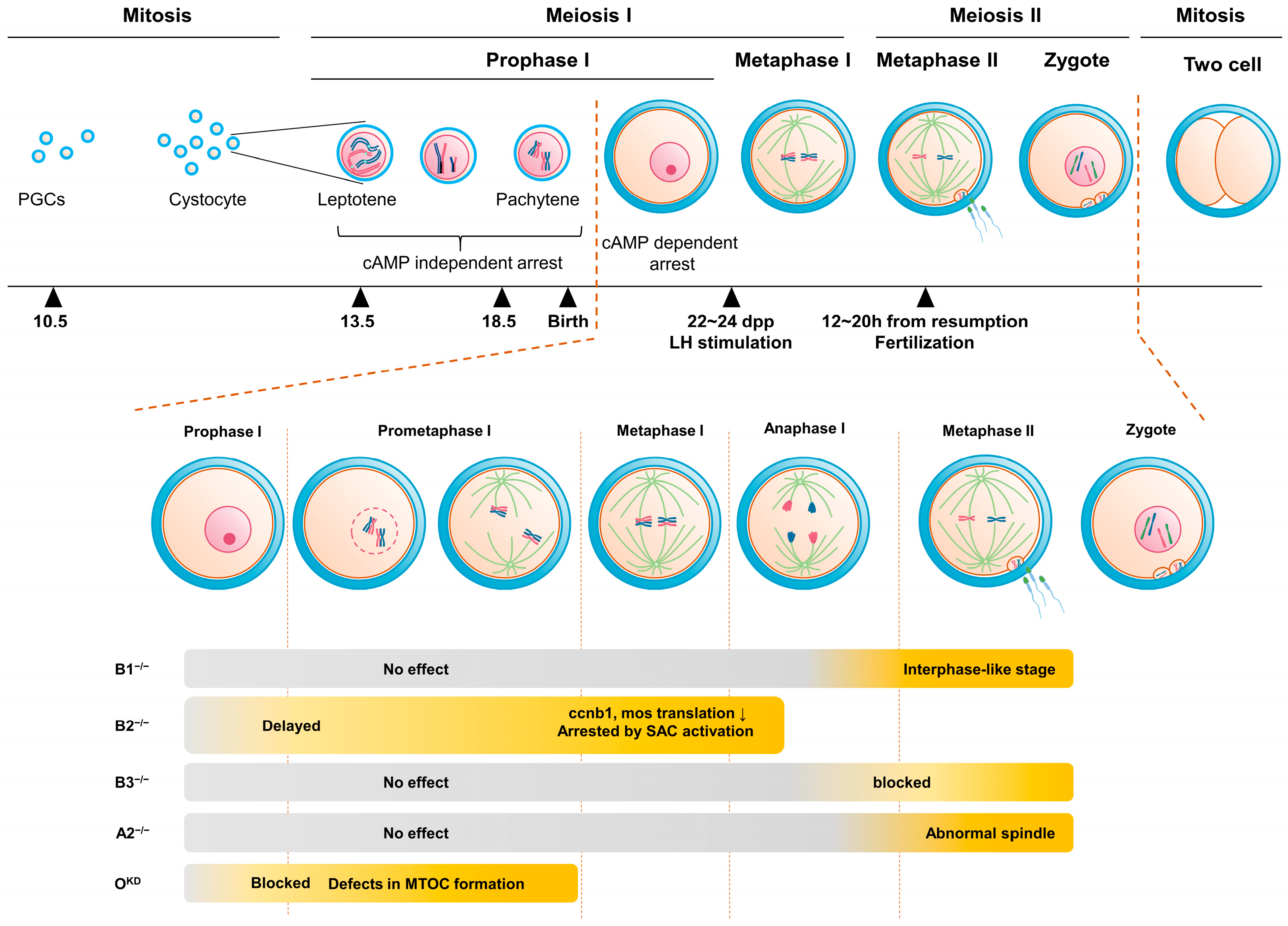
Figure 1. Oocyte maturation and impact of cyclin deletion. Mouse oocyte remains arrested in prophase I of the meiotic cell cycle until ovulation. At this stage, a distinctive large nucleus known as the germinal vesicle (GV) is observed in the oocyte, and upon resumption of the cell cycle triggered by luteinizing hormone the nuclear envelope disassembles in a process called germinal vesicle breakdown (GVBD). Subsequently, the oocyte proceeds through the first meiotic division, releasing the first polar body, and then progresses to metaphase II of the second meiotic division, and the oocyte is arrested again until fertilization. Upon fertilization, the second meiotic division is completed, leading to the release of the second polar body, followed by zygote formation and mitotic cell cycle. This process is primarily governed by the Cdk1–cyclin complex, commonly known as MPF. Recently, various cyclins have been identified in mouse oocytes. Deletion of each cyclin at specific stages of oocyte maturation results in defects, underscoring their unique roles in the process.
Ijms 24 13659 g001
Figure 2. Structural features of cyclins found in oocytes. (A) Mouse cyclin proteins commonly possess a cyclin-box domain, a characteristic structural motif. Additionally, sequences containing the D box and KEN box motifs, which facilitate protein degradation, are also present. These motifs are crucial for regulating protein levels during the cell cycle. The D box and KEN box motifs were predicted at http://slim.icr.ac.uk/apc/index.php (accessed on 10 August 2023), with a similarity score threshold of 0.8. Recently, a new degradation site (PM motif) has been identified in the cyclin B1 and B2. This motif is involved in regulating the timing of spindle alignment by allowing free cyclin B to be degraded earlier during progression to metaphase I. The cyclin accession numbers utilized for the analysis are indicated on the left. (B) The comparison of D-boxes found in various cyclins. The D box has a highly conserved (especially arginine and leucine at 1 and 4 position) RXALGXIXN motif. Cyclin B3 has a quasi-D box, which contains phenylalanine instead of leucine at the conserved position 4 of the D box.
Ijms 24 13659 g002
Figure 3. Roles of cyclins in the oocyte maturation process. (Prophase I) Maturation-promoting factor (MPF), composed of Cdk1 kinase and cyclin B, governs cell cycle arrest and resumption. Maternal mRNA is translated selectively. Cyclin B2 expression persists during prophase I arrest, while only the shortest cyclin B1 transcript translates, regulated by cytoplasmic polyadenylation element (CPE) in the 3′ UTR via CPEB. MPF remains inactive due to active Wee2, which is phosphorylated by cAMP-PKA. BubRI, Emi1, Hec1, and securin control APCCdh1 activity and APCcdh1-mediated cyclin degradation maintains levels, ensuring controlled progression without undesired resumption or arrest (Prometaphase I). Cell cycle resumes by inactivating PKA and Wee2. Activated Cdc25B triggers GVBD with mainly cyclin B2-based MPF. Cyclin B1 transcripts, the intermediate and longest, translate, elevating cyclin B1 protein levels. APCCdh1 activity is modulated by Mis12 and CenpH. Cyclin O’s function in Cdk1 inhibitory phosphate removal and MTOC formation is unclear. (Metaphase I and Anaphase I) MPF and SAC inhibit APCcdc20 and separase until proper spindle alignment. Residual APCcdc20 degrades free cyclin B1, extending metaphase I. SAC inhibition activates APCcdc20, degrading cyclin B1 and B2, and lowering MPF activity. Cyclin B3 phosphorylates Emi2, promoting anaphase I (Metaphase II and Fertilization). After meiosis I, S phase-free cell division ensues. Anaphase I-metaphase II transition increases Cdk1-cyclin B1 activity. Cyclin B1 absence leads to interphase re-entry, unique to this stage. Metaphase II oocytes arrest via cytostatic factor (CSF), regulated by MPF and mitogen-activated protein kinase pathway. Fertilization activates APC/C and Wee2 via CaMKII, reducing MPF and enabling zygote’s mitotic cell cycle entry.
Ijms 24 13659 g003
Figure 4. Structural motifs in the 3′ untranslated regions (UTRs) of cyclin B transcripts in mouse, human, and Xenopus. The 3′ UTR architecture of Ccnb gene orthologs varies across species, including mice, humans, and Xenopus. Horizontal bars represent sections of the 3′ UTR from each stop codon, with lengths indicated in base pairs (bp). Distinct hexanucleotide motifs within the same transcript generate mRNAs with different 3′ UTR lengths. Currently known examples include mouse ccnb1 with three transcripts. Transcripts exhibit varying lengths of polyadenylation at different time points based on the structure of the 3′ UTR, thereby regulating translation. The structural motifs include hexanucleotide polyadenylation signal, canonical cytoplasmic element (CPE) which has well-conserved sequence, and non-canonical cytoplasmic element (CPE-NC) which has some modification.
Ijms 24 13659 g004
Disclaimer/Publisher’s Note: The statements, opinions and data contained in all publications are solely those of the individual author(s) and contributor(s) and not of MDPI and/or the editor(s). MDPI and/or the editor(s) disclaim responsibility for any injury to people or property resulting from any ideas, methods, instructions or products referred to in the content.