Article

Impact of Somatic DNA Repair Mutations on the Clinical Outcomes of Bone Metastases from Castration-Resistant Prostate Cancer

1
Department of Medical Oncology, Istituto Romagnolo per lo Studio dei Tumori “Dino Amadori”—IRST S.r.l., 47014 Meldola, Italy
2
Unit of Biostatistics and Clinical Trials, Department of Medical Oncology, Istituto Romagnolo per lo Studio dei Tumori “Dino Amadori”—IRST S.r.l., 47014 Meldola, Italy
3
Biosciences Laboratory, Istituto Romagnolo per lo Studio dei Tumori “Dino Amadori”—IRST S.r.l., 47014 Meldola, Italy
4
Medical Oncology Department, Campus Bio-Medico University of Rome, 00128 Rome, Italy
5
Unit of Medical Oncology and Biomolecular Therapy, Department of Medical and Surgical Sciences, Policlinico Riuniti, University of Foggia, 71122 Foggia, Italy
6
Medical Oncology Department, “La Sapienza” University of Rome, 00185 Rome, Italy
*
Author to whom correspondence should be addressed.
These authors contributed equally to the work.
Int. J. Mol. Sci. 2023, 24(15), 12436; https://doi.org/10.3390/ijms241512436
Submission received: 7 July 2023 / Revised: 1 August 2023 / Accepted: 1 August 2023 / Published: 4 August 2023
(This article belongs to the Special Issue Aberrations of DNA Repair Pathways in Prostate Cancer 2.0)

Abstract

:
Up to 80% of castration-resistant prostate cancer (CRPC) patients develop bone metastases during the natural history of disease and about 25% harbor mutations in DNA damage repair (DDR) genes. This retrospective observational study evaluated the prevalence of DDR alterations in CRPC patients and their effect on the clinical outcomes associated with bone metastases. The mutational status of CRPC patients was analyzed per FoundationOne® analysis in tissue biopsy or, when it was not possible, in liquid biopsy performed at the onset of metastatic CRPC (mCRPC). The impact of DDR gene mutations on bone-related efficacy endpoints was evaluated at the time of mCRPC diagnoses. In total, 121 mCRPC patients with bone metastases were included: 38 patients had mutations in at least one DDR gene, the remaining 83 ones had a non-mutated DDR status. DDR mutated status was associated with bone metastases volume (p = 0.006), but did not affect SRE (skeletal-related events) incidence and time to SRE onset. Liquid and tissue biopsies were both available for 61 patients with no statistically significant difference in terms of incidence and type of molecular DDR alterations. Mutated DDR status was associated with higher bone metastasic volume, although a not detrimental effect on the other bone-related efficacy endpoints was observed.

1. Introduction

In prostate cancer (PCa), comprehensive genomic profiling (CGP) is attractive due to the diversity of emerging treatment options. In particular, defects in Breast Related Cancer Antigens (BRCA) 1 and BRCA2 genes result in impairments of DNA damage repair (DDR) deficiency [1]. DDR-deficient cells are sensitive to the inhibition of poly (ADP-ribose) polymerase (PARP), which results in irreversible DNA damage, cell cycle arrest and cell death. Recently, PARP-inhibitors have been approved for treatment of metastatic castration resistant prostate cancer (mCRPC) patients with pathogenic mutation of BRCA1/2 [2,3,4,5,6]. Beyond BRCA1/2, deleterious alterations in other genes have been associated with DDR deficiency: ATM and CHEK2 (sensors of DNA damage), CDK12 (positive regulator of BRCA genes), and PALB2 and FANCA (which interact with BRCA1 and/or BRCA2 during DNA repair). These defects cause single-strand annealing, non-homologous end joining and, consequently, genomic instability [7,8,9,10,11]. In PCa, the CGP has been previously characterized using tissue biopsies in order to identify mechanisms of resistance to ARSI (androgen receptor selective inhibitors, such as enzalutamide and abiraterone) [1,12,13]. Tissue testing remains the gold standard for CGP, however, the main site of metastases in PCa is bone, which is challenging to sample, analyze and is associated with high-failure rates of DNA sequencing [1,12]. On the other hand, performing CGP on primary tumor samples can be unsatisfying because of often old archival samples and/or lacking or poor material for analysis [1,12]. GCP of plasma cell-free circulating tumor DNA (ctDNA) offers a compelling and minimally invasive complement to tissue testing. Liquid biopsy may overcome the technical difficulties and high-failure rates associated with bone metastasis biopsy with the added value of reflecting the heterogeneity of the metastatic disease [14,15].
The molecular stratification of mCRPC patients remains an unmet clinical need. BRCA2 mutations have been associated with more aggressive disease and poor clinical outcomes [16,17], but the prognostic implications of other DDR genes are less well established as well as their impact on bone metastases outcomes. PCa bone metastases affect quality of life (QoL) due to the risk of bone pain and the development of “skeletal-related events” (SREs) [18], which is negatively correlated with survival [19]. Pathological bone fractures, hypercalcemia, spinal cord compression, bone surgery and bone radiation therapy are the five events defined as SREs by the Food and Drug Administration (FDA) [20].
This retrospective observational study evaluated the prevalence on liquid and tissue samples of DDR alterations of mCRPC patients at baseline of the first line treatment and their effect on the clinical outcomes of bone metastases.

2. Results

2.1. Patients: Sample Disposition and Genomic Profile

From January 2021 to September 2022, 150 mCRPC patients were enrolled in the prospective observational study. Plasma samples were obtained from 105 patients before starting first line treatment for mCRPC, whilst tissue samples from the primary tumor were available for 104 patients. Liquid and tissue biopsy were both available for 61 patients. The baseline characteristics of all patients enrolled in the study are described in Table 1. The distribution of molecular alterations found in tissue and liquid biopsy is resumed in Table 2 and graphically described in Figure 1. A total number of 54 mutations in DDR-related genes were found in liquid biopsy, compared to 34 mutations in tissue biopsy of primary tumor. A total of 6 BRCA1/2 mutated patients have also experienced other mutations in other DDR-related genes. We included in group A all patients with at least one or more pathogenic mutations in DDR-related genes. No statistically significant difference was found in terms incidence of molecular DDR alterations between tissue and liquid biopsy. In patients with both tissue and liquid samples available, the level of agreement between tissue and liquid biopsy is represented in Table 3. Among the 150 patients enrolled, 11 did not experience mCRPC, remaining hormone-sensitive during the observation period. Among the 139 mCRPC patients enrolled, 18 did not develop bone metastases and, consequently, they have been excluded from the final analyses. We therefore included 121 mCRPC patients with bone metastases, divided into group A (“BRCA/DDR mutated”, 38 patients, 31.4%) and group B (not mutated, 83 patients, 68.6%) according to their molecular status. Among the 121 mCRPC patients with bone metastases divided into group A and group B, 45 patients have both liquid and solid samples. Every patient in group B has no mutation in both liquid and solid biopsy.

2.2. Clinical Outcomes

Clinical characteristics of these patients are resumed in Table 4. There were no statistically significant differences between Group A and Group B patients in terms of clinical characteristics: age at diagnosis, Gleason score of the primary PCa, stage at diagnosis, type of presentation of mHSPC (high risk and/or high volume according to CHARTEED and LATITUDE criteria [21,22], and type of first-line treatment for mCRPC received.
Then, we evaluated the impact of DDR gene mutations on bone-related efficacy endpoints at the time of mCRPC diagnosis, by dividing patients in two aforementioned molecular groups.
We investigated differences between the two groups in terms of time from bone metastases onset to death, skeletal metastatic tumor burden (sites and number of lesions), skeletal-related events (SREs) incidence, and time to first on-study SRE. The use of antiresorptive agents (bisphosphonates or denosumab) was similar between the two groups.
Concerning bone sites, we divided the patients in those with lesions localized in the axial skeleton only and those with at least one extra-axial lesion; we did not find any difference according to this parameter between group A and B.
SRE were similar in both groups, in terms of incidence but also of median onset time from the diagnosis of bone metastases. Bone pain did not differ between the two groups. (Table 5)
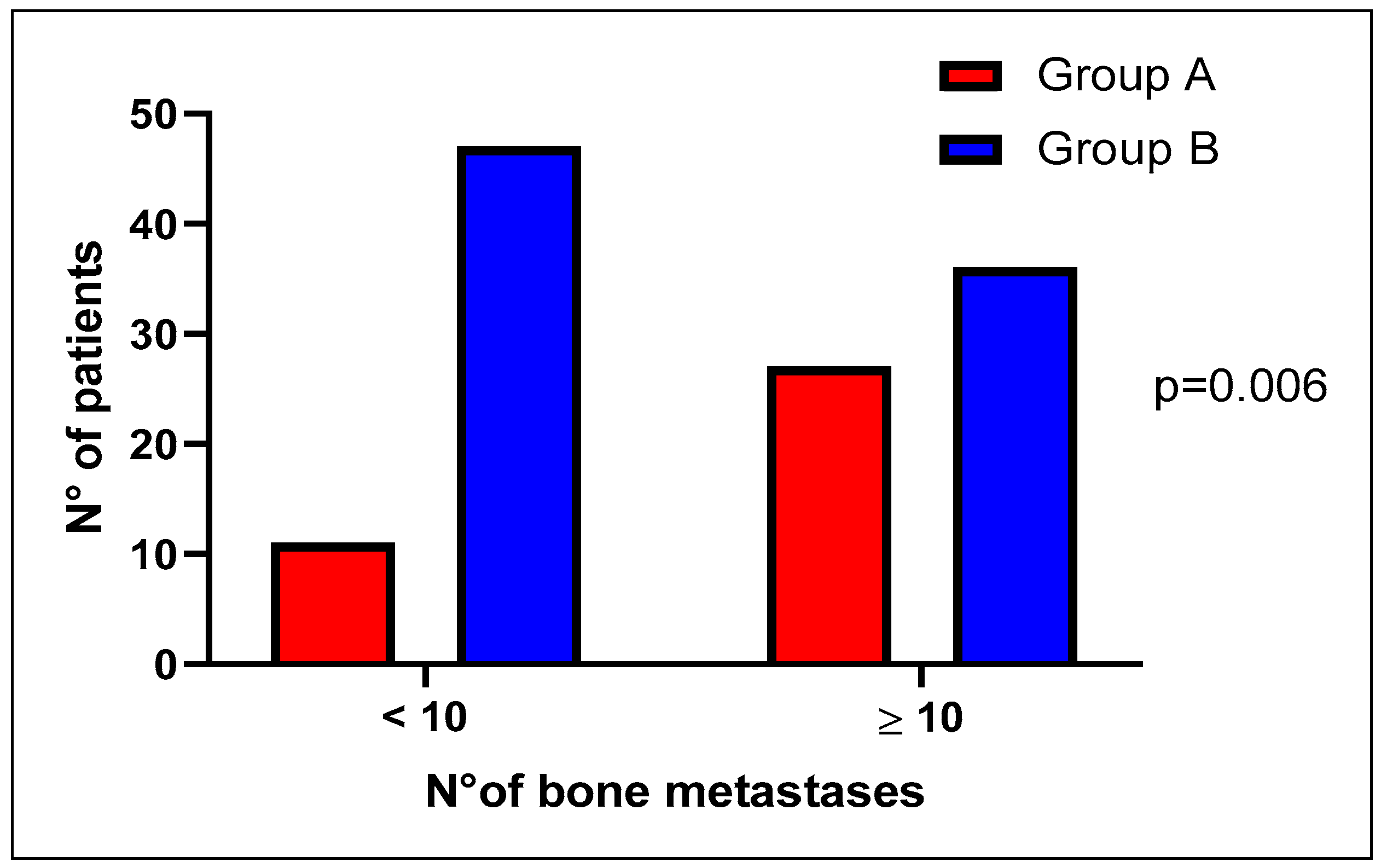
For the number of bone lesions, we adopted two different threshold values, 4 and 10 lesions; interestingly, we found a higher bone metastatic burden in group A than group B, being statistically significant only for the value of 10 lesions as the threshold (p = 0.006) (Figure 2).
We also evaluated the median time from bone metastases onset to death, without finding any difference (HR 0.90 CI 0.46–1.78, p = 0.763) (Figure 3).

3. Discussion

DDR genes are involved in the mechanisms of genetic instability, the repair of DNA aberrations during cell cycle, and the detection and repair of DNA damage, leading to apoptosis of dangerous mutated cells [23]. In this study, we propose testing all PCa patients for sDRR mutations at diagnosis of mCRPC. Somatic determination of molecular alterations on liquid biopsy and/or on the primary site on historic paraffin preparations (if available) was performed.
The incidence of pathogenic variants of somatic mutations in DDR genes among men with mCRPC varied between 11% and 33% [24], significantly higher than in non-metastatic PCa. BRCA2 mutations were more frequent when compared to other DDR genes (13%), followed by an ATM incidence of 7.3% [25]. The incidence of sDDR mutations in our study is in line with literature data; 25.3% of all patients enrolled presented at least a mutation in DDR-related genes and BRCA2 was the more frequent one, followed by ATM.
BRCA alterations have been associated with short metastatic-free survival, short cancer-specific survival (CSS) and are predictive of a response to PARP inhibitors and to platinum salts [6,26].
In PCa, Castro E. et al. demonstrated that BRCA1-2 mutations were more frequently associated with a Gleason score of ≥8, T3/T4 stage, nodal involvement and metastases at diagnosis [27]. The impact of DDR gene alterations and other biomarkers on the clinical outcome of radiometabolic agents in mCRPC is under investigation [28,29], but there are no data about the association between DDR gene alterations and other clinical-biological features rising during the clinical history of mCRPC as PSA/tumor flare, neuroendocrine differentiation, androgen receptor amplification and others [30,31,32,33].
Furthermore, there are no literature data regarding the impact of sDDR genes alterations on bone outcomes of mCRPC patients. In PCa, bone represents the most preferential target site of metastases with an incidence of nearly 75%; autopsy data indicate that the incidence of metastatic bone lesions is 65–75% in PCa patients [34,35].
Bone metastases led to the disruption of normal bone homeostasis as a result of complex interactions between tumor cells, bone marrow cells, and resident bone cells [36].
Prostate cancer cells are attracted to skeletal tissue by chemotactic cytokines, which normally regulate the migration of Hematopoietic Stem Cells (HSCs) into the hematopoietic stem cell niche: in fact, osteoblastic-induced stromal-derivedfactor-1 (SDF-1 or CXCL12) binds the CXCR4 receptor expressed both by HSCs and PCa cells. The competitive binding of SDF-1/CXCR4 of HSCs and PCa cells leads to the formation of the “onco-niche”, in which PCa cells can migrate and then may stay in a quiescent state that can last over years, or can be activated. In the latter case, they can damage physiological bone remodeling by interfering with normal osteoclastic and osteoblastic activity processes through secretion of paracrine factors, such as transforming growth factor β1 (TGF β1), parathroid-hormone-related peptide (PTHrP) and interleukin 6 (IL-6). The result is an aberrant activation of the RANK/RANK ligand (RANKL) pathway and, consequently, abnormal stimulation of bone resorption [37,38].
In PCa bone metastasis, the first step of enhanced osteolysis is followed by strong osteoblastic stimulation, resulting in an excessive abnormal bone apposition. During the natural history of PCa, the development of SREs negatively correlates with survival: pathologic fractures and metastatic spinal cord compressions are associated with a significantly increased risk of death [38,39,40].
This study investigated differences between the DDR gene alterations-carriers and non-carriers in mCRCP patients with bone metastases in terms of time from bone metastases onset to death, skeletal metastatic tumor burden (sites and number of lesions), skeletal-related events (SREs) incidence, and time to first on-study SRE. We hypothesized that the DNA-repair defects in mCRPC may be associated with poor prognoses in terms of bone related outcomes as an indirect consequence of the acquired survival advantages of these tumor cells.
This study does not demonstrate any difference in bone-related outcomes in sDDR genes mutations carriers compared to non-carriers: incidence of SREs, median onset time of the first SRE from the diagnosis of bone metastases as well as bone pain did not differ between the two groups. However, sDDR mutated status showed an association with higher bone tumor burden in mCRPC: these patients had a superior count of bone metastases at the diagnoses of mCRPC compared to patients with normal sDDR status. It should be noticed that the two groups of patients were balanced in terms of use of antiresorptive agents bisphosphonates or denosumab) and first-line treatment performed for mCRPC [41].
In this study, patients were defined as sDDR-gene mutation carriers if one or more mutation in DDR-related genes was detected in the solid and/or liquid biopsy. It is well- known that genomic alterations can be acquired during the progression of the disease as a consequence of the selective pressure of treatments and biological molecular changes occurring during disease progression itself [42]; consequently, a biopsy of a metastatic site represents the ideal approach to identify molecular alterations. The PROfound trial [6], which evaluated 2792 biopsies of mCRPC, showed that DDR genes alterations were present in 28% of all samples, with a similar incidence considering the primary tumor (27%) or metastatic sites (32%). However, somatic determination on a metastatic site, in particular, bone, may be associated with various biases, as well as possible side effects [12]; the PROfound study, for example, pointed out that 30% of biopsy samples may not be of sufficient quality for gene sequencing [42].
The analysis of free circulating tumor DNA (ctDNA) is a promising approach as it may overcome the difficulties that are associated with obtaining tissue; however, there are no solid data that currently allow the reliable use of this test. In our study, the incidence of mutations in DDR-related genes in liquid biopsy (performed at the diagnosis of mCRPC) is numerically higher as compared to solid biopsy (performed on tissue samples of primary tumor); therefore, there was a substantial agreement between solid and liquid biopsy for DDR gene alterations, with different values according to the gene analyzed. For example, mutations of BRCA1/2, PALB 2, FANCA showed an almost perfect agreement; on the other hand, ATM showed a moderate agreement between solid and liquid biopsy. According to these data, liquid biopsy could be a valid tool also in PCa with prevalent bone involvement, in which solid biopsy of the metastatic sites is difficult to perform and also in the case of older primary tissues, which are unlikely to be adequate for molecular analysis [43,44]. It is difficult to establish if the higher number of genomic alterations in liquid biopsy is due to a time- and treatment-dependent increase or to a selection of clones harboring those mutations rather than resulting from diagnostic issues such as assay sensitivity; further study is needed to answer to this question.
The retrospective nature of the data analyzed is a limitation of this study. Another limitation is that patients did not perform a solid biopsy of the metastatic site at the time of diagnosis of mCRPC, which may be more comparable to liquid biopsy, reflecting the selective pressure of treatment received from the diagnosis of PCa to the diagnosis of mCRPC. Standard imaging was performed to diagnose mCRPC (CT scan and/or bone scan), however, more sensitive imaging tools as choline PET scan and, more recently, PSMA PET scan, could have an impact on the number of metastatic bone lesions [45,46].
These results should be considered preliminary and further work is needed to determine the relevance of these findings.

4. Materials and Methods

4.1. Study Design and Aims

The primary aim was to assess the impact of somatic DDR (sDDR) mutations on clinical course of bone metastases in mCRPC patients.
The impact of sDDR mutations on clinical outcomes of bone metastases is defined as: bone metastatic burden, defined as the number and sites (axial only vs. non-axial) of bone metastases at time of mCRPC diagnosis with standard imaging (CT scan and/or bone scan); bone metastases-specific survival, defined as the time from bone metastases onset to death for any cause; prevalence and type of Skeletal-related-events (SREs); time to first on-study SRE, defined as the time from bone metastases onset to first SRE; Bone pain, defined as necessity of opioid use for bone pain at the diagnosis of bone metastases in mCRPC.
As defined by FDA, SREs have been considered: Pathologic bone fractures, hypercalcemia, spinal cord compression, surgery to bone, and radiotherapy to bone.
The analysis of the impact of sDDR mutations have been performed according to 2 groups: patients with mutation in at least one of these gene ATM/BRCA1/BRCA2/RAD51/PALB2/FANCA/ATM/CDK12/CHECK2 (Group A, all patients “positive” for genomic defects in DDR genes); no DDR carriers (Group B, all patients negative for genomic defects in DDR genes).
The secondary and point of the study is to evaluate the concordance between liquid and tissue biopsy in terms of presence or absence and type of molecular DDR alterations observed.
Patients were retrospectively included for the analysis at the “Department of Medical Oncology, IRCCS Istituto Romagnolo per lo Studio dei Tumori (IRST) Dino Amadori” (Meldola, Italy) from January 2021 to September 2022 and prospectively observed until the data cut-off time on 31 December 2022.

4.2. Patients

Patients enrolled were included in the biological observational prospective study IRSTB073 “Biomarker study: the next generation of prostate cancer biomarkers” (Identifier Code: L3P1380). Local Ethical Committee (“IRST Ethical Committee”) approved the IRST B073 single center prospective study. All patients provided written informed consent.
Main inclusion criteria were: histological or cytological confirmed diagnosis of prostate cancer or unequivocal increased of PSA; Patients must have metastatic and/or inoperable disease; Life expectancy of greater than 3 months; ECOG performance status < 2; Age ≥ 18 years; no previous line of treatment for mCRPC; sample tissue of the primary PCa tumor available for NGS analyses and/or blood sample baseline first-line treatment for mCRPC.

4.3. Study Procedures and Somatic Variants Analyses

Next generation sequencing using FoundationOne DX1 (Foundation Medicine®, Cambridge, MA, USA) was performed on DNA from tumor-biopsy samples obtained at diagnoses of PCa and/or on circulating cell-free DNA from 10-mL blood sample obtained at diagnoses of mCRPC, if available.
Through genomic testing of plasma and/or tumor tissue (archival, if available), patients were screened for the presence of a deleterious somatic alteration in BRCA1, BRCA2, ATM, CDK12, CHEK2, FANCA, PALB2, RAD51.
We considered DDR to be mutated in all patients with at least one pathogenic mutation (according to the classification of American College of Medical Genetics and Genomics, ACMG) [13], in one of the DDR genes evaluated (BRCA1/2, ATM, FANCA, CHEK2, RAD51, PALB2, and CDK12) in solid and/or liquid biopsy. Patients performing NGS analyses on solid sample and liquid biopsy were included in group B if no mutation was detected in both liquid and solid NGS analysis.
If a mutation in one or more DDR-related genes occurred in liquid biopsy and was not detected in solid biopsy (or vice versa), the patient was included in group A.

4.4. Statistical Analyses

The description of the cases was carried out through the use of descriptive statistics such as absolute frequencies and percentage frequencies for variables measured on a nominal or ordinal scale, medians, and intervals of variation for variables measured on a continuous scale.
Comparisons of median values of markers within different clinical features were obtained using the nonparametric Wilcoxon test of medians.
Time to skeletal event (SRE) was calculated as the time between the date of bone metastases onset and the date of the first SRE onset for patients who had at least one skeletal event and the difference between the date of bone metastases onset and last follow-up date for patients who did not have any SRE. Events are represented by patients who had at least one SRE.
The curves of the time-dependent variables were determined with the Kaplan–Meier limit product method and the relative comparisons were made according to the log-rank test.
All p-values were obtained considering two-tailed tests and statistical analyzes were performed with SAS statistical software, version 9.4.
For each biomarker, concordance was defined as either positive or negative in both tumor and metastasis and discordance was defined as positivity at one site and negativity at the other or vice versa. For each receptor, the discordance rate (DR) was calculated as the proportion of discordant cases with respect to the total number of patients. The two-sided exact binomial 95% confidence interval (95% CI) was estimated.
The relation between the value and the level of agreement was first reported by Landis and Kock [47], with values indicating agreement as follows: 0.00–0.20, slight agreement; 0.21–0.40, fair agreement; 0.41–0.60, moderate agreement; 0.61–0.80, substantial agreement, and 0.81–1.00, almost perfect agreement (perfect agreement = 1.00).

5. Conclusions

Mutated DDR status showed an association with higher bone tumor burden in mCRPC. Nevertheless, DDR mutated patients showed neither a higher incidence of SRE or shorter time to the first SRE. In prostate cancer liquid biopsy could be a valid tool for DDR mutational status assessment.

Author Contributions

Conceptualization, M.C.C., F.P., V.C. and U.D.G.; data curation, M.C.C., E.F.G., E.S., C.C., A.V., P.U., S.B., N.B. and U.D.G.; formal analysis, E.S., A.V. and P.U.; investigation, E.F.G., S.B. and N.B.; methodology, E.F.G. and E.S.; supervision, F.P., V.C., D.S. and U.D.G.; validation, E.S., A.V. and P.U.; writing—original draft, M.C.C. and E.F.G.; writing—review and editing, C.C., S.B., N.B., G.R., F.P. and U.D.G. All authors have read and agreed to the published version of the manuscript.

Funding

This research received no external funding.

Institutional Review Board Statement

The study was conducted in accordance with the Declaration of Helsinki, and approved by the Local Ethical Committee (“IRST Ethical Committee”) (REC 2192/2013).

Informed Consent Statement

Informed consent was obtained from all subjects involved in the study.

Data Availability Statement

Data is unavailable due to privacy and ethical restrictions.

Acknowledgments

This work was partly supported thanks to the contribution of Ricerca Corrente by the Italian Ministry of Health within the research line “Precision, gender and ethnicity-based medicine and geroscience: genetic-molecular mechanisms in the development, characterization and treatment of tumor”. Partly results of this study have well presented at ASCO GU meeting 2023.

Conflicts of Interest

U.D.G: Consultant: Janssen, AstellasPharma, Sanofi, Bayer, Pfizer, Bristol-Myers Squibb, Novartis, Ipsen, Merck; Institutional funding: Roche, Sanofi, AstraZeneca. EFG: travel accommodation from Janssen-Cilag; M.C.C. travel accommodation from IPSEN; V.C. has served as a consultant/advisory board member for Janssen, Astellas, Merck, AstraZeneca, Amgen and Bayer and has received speaker honoraria or travel support from Astellas, Janssen, Ipsen, Bayer, Bristol and Sanofi; D.S. Onoraria per adv board con janssen MSD Merck Daiichi Sankyo Novartis Astellas roche SERVIER EISAI Gilead Incyte; F.P. advisory board per Lilly, Gilead, Novartis, AstraZeneca.

References

  1. Abida, W.; Armenia, J.; Gopalan, A.; Brennan, R.; Walsh, M.; Barron, D.; Danila, D.; Rathkopf, D.; Morris, M.; Slovin, S.; et al. Prospective Genomic Profiling of Prostate Cancer Across Disease States Reveals Germline and Somatic Alterations That May Affect Clinical Decision Making. JCO Precis. Oncol. 2017, 1, 1–16. [Google Scholar] [CrossRef] [PubMed]
  2. Mateo, J.; Carreira, S.; Sandhu, S.; Miranda, S.; Mossop, H.; Perez-Lopez, R.; Nava Rodrigues, D.; Robinson, D.; Omlin, A.; Tunariu, N.; et al. DNA-Repair Defects and Olaparib in Metastatic Prostate Cancer. N. Engl. J. Med. 2015, 373, 1697–1708. [Google Scholar] [CrossRef] [PubMed]
  3. MJonsson, P.; Bandlamudi, C.; Cheng, M.L.; Srinivasan, P.; Chavan, S.S.; Friedman, N.D.; Rosen, E.Y.; Richards, A.L.; Bouvier, N.; Selcuklu, S.D.; et al. Tumour lineage shapes BRCA-mediated phenotypes. Nature 2019, 571, 576–579. [Google Scholar] [CrossRef] [PubMed]
  4. Mateo, J.; Porta, N.; Bianchini, D.; McGovern, U.; Elliott, T.; Jones, R.; Syndikus, I.; Ralph, C.; Jain, S.; Varughese, M.; et al. Olaparib in patients with metastatic castration-resistant prostate cancer with DNA repair gene aberrations (TOPARP-B): A multicentre, open-label, randomised, phase 2 trial. Lancet Oncol. 2019, 21, 162–174. [Google Scholar] [CrossRef] [PubMed]
  5. Abida, W.; Patnaik, A.; Campbell, D.; Shapiro, J.; Bryce, A.H.; McDermott, R.; Sautois, B.; Vogelzang, N.J.; Bambury, R.M.; Voog, E.; et al. Rucaparib in men with metastatic castration-resistant prostate cancer harboring aBRCA1orBRCA2gene alteration. J. Clin. Oncol. 2020, 38, 3763–3772. [Google Scholar] [CrossRef] [PubMed]
  6. Hussain, M.; Mateo, J.; Fizazi, K.; Saad, F.; Shore, N.; Sandhu, S.; Chi, K.N.; Sartor, O.; Agarwal, N.; Olmos, D.; et al. Survival with Olaparib in Metastatic Castration-Resistant Prostate Cancer. N. Engl. J. Med. 2020, 383, 2345–2357. [Google Scholar] [CrossRef] [PubMed]
  7. Zhang, F.; Fan, Q.; Ren, K.; Andreassen, P.R. PALB2 functionally connects the breast cancer susceptibility proteins BRCA1 and BRCA2. Mol. Cancer. Res. 2009, 7, 1110–1118. [Google Scholar] [CrossRef] [Green Version]
  8. Folias, A.; Matkovic, M.; Bruun, D.; Reid, S.; Hejna, J.; Grompe, M.; D’Andrea, A.; Moses, R. BRCA1 interacts directly with the Fanconi anemia protein FANCA. Hum. Mol. Genet. 2002, 11, 2591–2597. [Google Scholar] [CrossRef] [Green Version]
  9. Zannini, L.; Delia, D.; Buscemi, G. CHK2 kinase in the DNA damage response and beyond. J. Mol. Cell Biol. 2014, 6, 442–457. [Google Scholar] [CrossRef] [Green Version]
  10. Weigelt, B.; Bi, R.; Kumar, R.; Blecua, P.; Mandelker, D.L.; Geyer, F.C.; Pareja, F.; James, P.A.; Couch, F.J.; Eccles, D.M.; et al. The Landscape of Somatic Genetic Alterations in Breast Cancers from ATM Germline Mutation Carriers. Gynecol. Oncol. 2018, 110, 1030–1034. [Google Scholar] [CrossRef] [Green Version]
  11. Wu, Y.M.; Cieslik, M.; Lonigro, R.J.; Vats, P.; Reimers, M.A.; Cao, X.; Ning, Y.; Wang, L.; Kunju, L.P.; De Sarkar, N.; et al. Inactivation of CDK12 delineates a distinct immunogenic class of advanced prostate cancer. Cell 2018, 173, 1770–1782. [Google Scholar] [CrossRef]
  12. Armenia, J.; Wankowicz, S.A.M.; Liu, D.; Gao, J.; Kundra, R.; Reznik, E.; Chatila, W.K.; Chakravarty, D.; Han, G.C.; Coleman, I.; et al. The long tail of oncogenic drivers in prostate cancer. Nat. Genet. 2018, 50, 645–651. [Google Scholar] [CrossRef] [PubMed]
  13. Richards, S.; Aziz, N.; David Liu, D.; Gao, J.; Kundra, R.; Reznik, E.; Chatila, W.K.; Chakravarty, D.; Han, G.C.; Coleman, I.; et al. Standards and guidelines for the interpretation of sequence variants: A joint consensus recommendation of the American College of Medical Genetics and Genomics and the Association for Molecular Pathology. Genet. Med. 2015, 17, 405–424. [Google Scholar] [CrossRef] [Green Version]
  14. Annala, M.; Vandekerkhove, G.; Khalaf, D.; Taavitsainen, S.; Beja, K.; Warner, E.W.; Sunderland, K.; Kollmannsberger, C.; Eigl, B.J.; Finch, D.; et al. Circulating tumor DNA genomics correlate with resistance to abiraterone and enzalutamide in prostate cancer. Cancer Discov. 2018, 8, 444–457. [Google Scholar] [CrossRef] [Green Version]
  15. Torga, G.; Pienta, K.J. Patient-Paired Sample Congruence Between 2 Commercial Liquid Biopsy Tests. JAMA Oncol. 2018, 4, 868–870. [Google Scholar] [CrossRef] [PubMed]
  16. Hatano, K.; Nonomura, N. Genomic Profiling of Prostate Cancer: An Updated Review. World J. Men’s Health 2022, 40, 368–379. [Google Scholar] [CrossRef]
  17. Castro, E.; Romero-Laorden, N.; del Pozo, A.; Lozano, R.; Medina, A.; Puente, J.; Piulats, J.M.; Lorente, D.; Saez, M.I.; Morales-Barrera, R.; et al. PROREPAIR-B: A Prospective Cohort Study of the Impact of Germline DNA Repair Mutations on the Outcomes of Patients With Metastatic Castration-Resistant Prostate Cancer. J. Clin. Oncol. 2019, 37, 490–503. [Google Scholar] [CrossRef]
  18. Weinfurt, K.P.; Li, Y.; Castel, L.D.; Saad, F.; Timbie, J.W.; Glendenning, G.A.; Schulman, K.A. The significance of skeletal-related events for the health-related quality of life of patients with metastatic prostate cancer. Ann. Oncol. 2005, 16, 579–584. [Google Scholar] [CrossRef] [PubMed]
  19. De Puy, V.; Anstrom, K.J.; Castel, L.D.; Schulman, K.A.; Weinfurt, K.P.; Saad, F. Effects of skeletal morbidities on longitudinal patient-reported outcomes and survival in patients with metastatic prostate cancer. Support Care Cancer 2007, 15, 869–876. [Google Scholar] [CrossRef]
  20. U.S. Department of Health and Human Services, Food and Drug Administration; Center for Drug Evaluation and Research (CDER); Center for Biologics Evaluation and Research (CBER). Clinical Trials Endpoints for the Approval of Cancer Drugs and Biologics; U.S. Department of Health and Human Services, Food and Drug Administration: Washington, DC, USA, 2007.
  21. Kyriakopoulos, C.E.; Chen, Y.-H.; Carducci, M.A.; Liu, G.; Jarrard, D.F.; Hahn, N.M.; Shevrin, D.H.; Dreicer, R.; Hussain, M.; Eisenberger, M.; et al. Chemohormonal Therapy in Metastatic Hormone-Sensitive Prostate Cancer: Long-Term Survival Analysis of the Randomized Phase III E3805 CHAARTED Trial. J. Clin. Oncol. 2018, 36, 1080–1087. [Google Scholar] [CrossRef] [PubMed] [Green Version]
  22. Fizazi, K.; Tran, N.; Fein, L.; Matsubara, N.; Rodriguez-Antolin, A.; Alekseev, B.Y.; Özgüroğlu, M.; Ye, D.; Feyerabend, S.; Protheroe, A.; et al. Abiraterone acetate plus prednisone in patients with newly diagnosed high-risk metastatic castration-sensitive prostate cancer (LATITUDE): Final overall survival analysis of a randomised, double-blind, phase 3 trial. Lancet Oncol. 2019, 20, 686–700. [Google Scholar] [CrossRef] [PubMed]
  23. Pritchard, C.C.; Mateo, J.; Walsh, M.F.; De Sarkar, N.; Abida, W.; Beltran, H.; Garofalo, A.; Gulati, R.; Carreira, S.; Eeles, R.; et al. Inherited DNA-Repair Gene Mutations in Men with Metastatic Prostate Cancer. N. Engl. J. Med. 2016, 375, 443–453. [Google Scholar] [CrossRef] [PubMed]
  24. ICGC/TCGA Pan-Cancer Analysis of Whole Genomes Consortium. Pan-Cancer Analysis of Whole Genomes Consortium. Pan-cancer analysis of whole genomes. Nature 2020, 578, 82–93. [Google Scholar] [CrossRef] [Green Version]
  25. Eeles, R.; Goh, C.; Castro, E.; Bancroft, E.; Guy, M.; Al Olama, A.A.; Easton, D.; Kote-Jarai, Z. The genetic epidemiology of prostate cancer and its clinical implications. Nat. Rev. Urol. 2013, 11, 18–31. [Google Scholar] [CrossRef] [PubMed]
  26. Schmid, S.; Omlin, A.; Higano, C.; Sweeney, C.; Chanza, N.M.; Mehra, N.; Kuppen, M.C.P.; Beltran, H.; Conteduca, V.; De Almeida, D.V.P.; et al. Activity of Platinum-Based Chemotherapy in Patients With Advanced Prostate Cancer With and Without DNA Repair Gene Aberrations. JAMA Netw. Open 2020, 3, e2021692. [Google Scholar] [CrossRef] [PubMed]
  27. Castro, E.; Goh, C.; Olmos, D.; Saunders, E.; Leongamornlert, D.; Tymrakiewicz, M.; Mahmud, N.; Dadaev, T.; Govindasami, K. Germline BRCA mutations are associated with higher risk of nodal involvement, distant metastasis, and poor survival outcomes in prostate cancer. J. Clin. Oncol. 2013, 31, 1748–1757. [Google Scholar] [CrossRef] [Green Version]
  28. De Giorgi, U.; Sansovini, M.; Severi, S.; Nicolini, S.; Monti, M.; Gurioli, G.; Foca, F.; Casadei, C.; Conteduca, V.; Celli, M.; et al. Circulating androgen receptor gene amplification and resistance to177Lu-PSMA-617 in metastatic castration-resistant prostate cancer: Results of a Phase 2 trial. Br. J. Cancer 2021, 125, 1226–1232. [Google Scholar] [CrossRef]
  29. Castro, E.; Lozano Mejorada, R.; Medina, A.; De Giorgi, U.; Romero Laorden, N.; Conteduca, V.; De Velasco, G.; Alameda, D.; Sanz, A.; Puente, J.; et al. Prospective analysis of the impact of germline mutations in homologous recombination (HR) genes on the response to radium-223 for metastatic castration resistant prostate cancer (mCRPC). Ann. Oncol. 2021, 32 (Suppl. S2), S626–S677. [Google Scholar] [CrossRef]
  30. Burgio, S.L.; Conteduca, V.; Rudnas, B.; Carrozza, F.; Campadelli, E.; Bianchi, E.; Fabbri, P.; Montanari, M.; Carretta, E.; Menna, C.; et al. PSA Flare With Abiraterone in Patients With Metastatic Castration-Resistant Prostate Cancer. Clin. Genitourin. Cancer 2015, 13, 39–43. [Google Scholar] [CrossRef]
  31. Burgio, S.L.; Conteduca, V.; Menna, C.; Carretta, E.; Rossi, L.; Bianchi, E.; Kopf, B.; Fabbri, F.; Amadori, D.; De Giorgi, U. Chromogranin A predicts outcome in prostate cancer patients treated with abiraterone. Endocr. Relat. Cancer 2014, 21, 487–493. [Google Scholar] [CrossRef] [Green Version]
  32. Martignano, F.; Gurioli, G.; Salvi, S.; Calistri, D.; Costantini, M.; Gunelli, R.; De Giorgi, U.; Foca, F.; Casadio, V. GSTP1 Methylation and Protein Expression in Prostate Cancer: Diagnostic Implications. Dis. Markers 2016, 2016, 4358292. [Google Scholar] [CrossRef] [PubMed] [Green Version]
  33. Conteduca, V.; Jayaram, A.; Romero-Laorden, N.; Wetterskog, D.; Salvi, S.; Gurioli, G.; Scarpi, E.; Castro, E.; Marin-Aguilera, M.; Lolli, C.; et al. Plasma Androgen Receptor and Docetaxel for Metastatic Castration-resistant Prostate Cancer. Eur. Urol. 2018, 75, 368–373. [Google Scholar] [CrossRef] [PubMed] [Green Version]
  34. Costa, L.; Badia, X.; Chow, E.; Lipton, A.; Wardley, A. Impact of skeletal complications on patients’ quality of life, mobility, and functional independence. Support Care Cancer 2008, 16, 879–889. [Google Scholar] [CrossRef]
  35. Coleman, R. Metastatic bone disease: Clinical features, pathophysiology and treatment strategies. Cancer Treat. Rev. 2001, 27, 165–176. [Google Scholar] [CrossRef]
  36. Guise, T. Examining the metastatic niche: Targeting the microenvironment Semin. Oncology 2010, 37, S2–S14. [Google Scholar] [CrossRef] [PubMed]
  37. Cursano, M.; Iuliani, M.; Casadei, C.; Stellato, M.; Tonini, G.; Paganelli, G.; Santini, D.; De Giorgi, U. Combination radium-223 therapies in patients with bone metastases from castration-resistant prostate cancer: A review. Crit. Rev. Oncol. 2020, 146, 102864. [Google Scholar] [CrossRef]
  38. Saad, F.; Lipton, A.; Cook, R.; Chen, Y.M.; Smith, M.; Coleman, R. Pathologic fractures correlate with reduced survival in patients with malignant bone disease. Cancer 2007, 110, 1860–1867. [Google Scholar] [CrossRef]
  39. Aass, N.; Fosså, S.D. Pre- and post-treatment daily life function in patients with hormone resistant prostate carcinoma treated with radiotherapy for spinal cord compression. Radiother. Oncol. 2005, 74, 259–265. [Google Scholar] [CrossRef]
  40. Rades, D.; Stalpers, L.J.; Veninga, T.; Rudat, V.; Schulte, R.; Hoskin, P.J. Evaluation of Functional Outcome and Local Control After Radiotherapy for Metastatic Spinal Cord Compression in Patients With Prostate Cancer. J. Urol. 2006, 175, 552–556. [Google Scholar] [CrossRef]
  41. Miles, B.; Tadi, P. Genetics, Somatic Mutation; Europe PMC: London, UK, 2023. [Google Scholar]
  42. Galletti, G.; Leach, B.I.; Sibilla, C.; Fizazi, K.; Saad, F.; Shore, N.; Sandhu, S.; Mateo, J.; Olmos, D.; Mehra, N.; et al. Mechanisms of resistance to systemic therapy in metastatic castration-resistant prostate cancer. Cancer Treat. Rev. 2017, 57, 16–27. [Google Scholar] [CrossRef]
  43. Hussain, M.; Corcoran, C.; Sibilla, C.; Fizazi, K.; Saad, F.; Shore, N.; Sandhu, S.; Mateo, J.; Olmos, D.; Mehra, N.; et al. Tumor Genomic Testing for >4000 Men with Metastatic Castration-resistant Prostate Cancer in the Phase III Trial PROfound (Olaparib). Clin. Cancer Res. 2022, 28, 1518–1530. [Google Scholar] [CrossRef] [PubMed]
  44. Salvi, S.; Casadio, V.; Conteduca, V.; Lolli, C.; Gurioli, G.; Martignano, F.; Schepisi, G.; Testoni, S.; Scarpi, E.; Amadori, D.; et al. Circulating AR copy number and outcome to enzalutamide in docetaxel-treated metastatic castration-resistant prostate cancer. Oncotarget 2016, 7, 37839–37845. [Google Scholar] [CrossRef] [PubMed] [Green Version]
  45. De Giorgi, U.; Caroli, P.; Scarpi, E.; Conteduca, V.; Burgio, S.L.; Menna, C.; Moretti, A.; Galassi, R.; Rossi, L.; Amadori, D.; et al. 18F-Fluorocholine PET/CT for early response assessment in patients with metastatic castration-resistant prostate cancer treated with enzalutamide. Eur. J. Nucl. Med. Mol. Imaging 2015, 42, 1276–1283. [Google Scholar] [CrossRef] [PubMed]
  46. Caroli, P.; Sandler, I.; Matteucci, F.; De Giorgi, U.; Uccelli, L.; Celli, M.; Foca, F.; Barone, D.; Romeo, A.; Sarnelli, A.; et al. 68Ga-PSMA PET/CT in patients with recurrent prostate cancer after radical treatment: Prospective results in 314 patients. Eur. J. Nucl. Med. 2018, 45, 2035–2044. [Google Scholar] [CrossRef]
  47. Landis, J.R.; Kock, G.G. The measurement of observer agreement for categorical data. Biometrics 1977, 33, 159–174. [Google Scholar] [CrossRef] [PubMed]
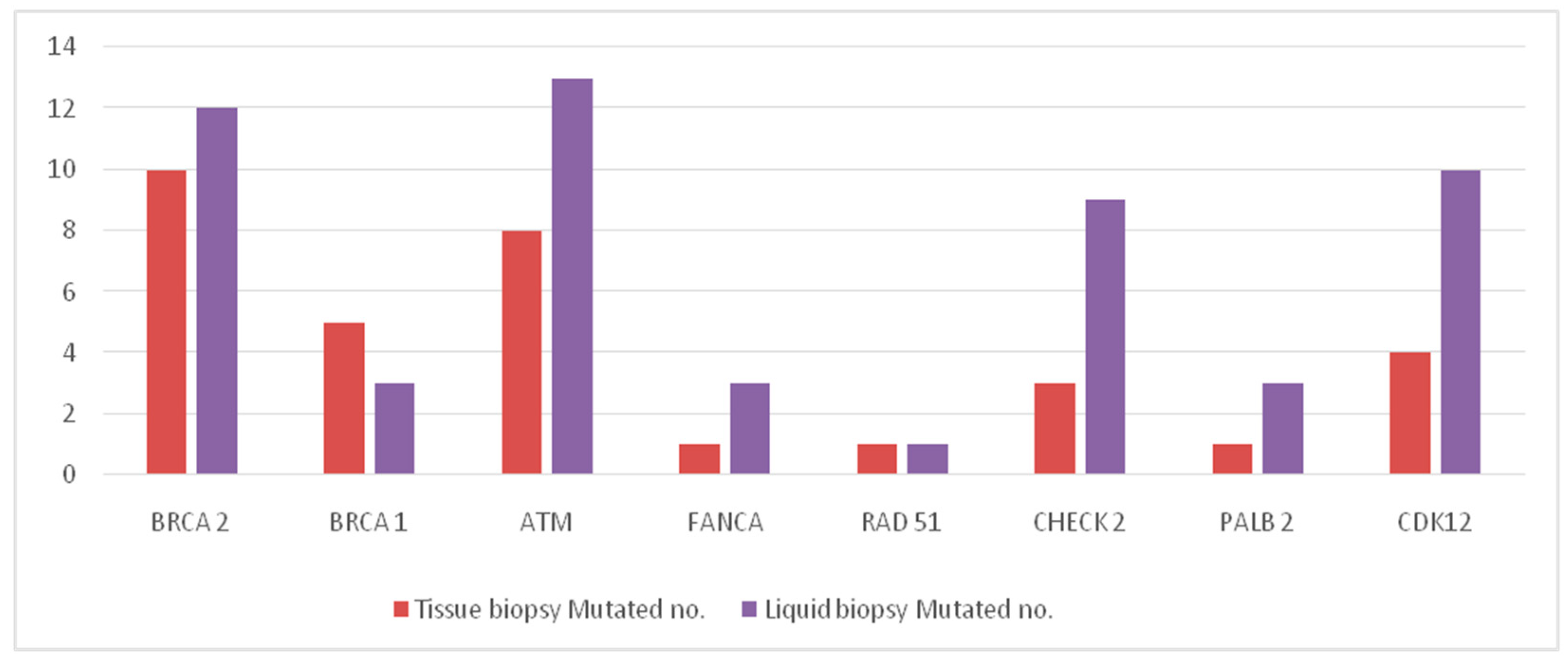
Figure 1. Distribution of molecular alterations found in solid and liquid biopsies.
Ijms 24 12436 g001
Figure 2. Number of bone metastases in the two molecular groups (DDR mutated, group A versus DDR wild type, Group B), adopting the threshold of 10 lesions.
Ijms 24 12436 g002
Figure 3. Time from bone metastases onset to death according to molecular status (DDR mutated, group A versus DDR wild type, Group B).
Ijms 24 12436 g003
Table 1. Baseline patients’ characteristics.
VariableNumber (%) Total = 150
Age at diagnosis, years Median value (range):65.5 (42–86)
Gleason score
<847 (31.3%)
≥892 (61.4%)
Unknown11 (7.3%)
Stage at diagnosis
Localized PCa73 (48.8%)
mHSPC64 (51.2%)
Type of mHSPC
High risk and/or high volume59 (39.3%)
No high risk/volume66 (44%)
No mHSPC25 (16.7%)
Sites of metastases in mCRPC
Bone only53 (35.4%)
Bone and visceral11 (7.3%)
Bone and nodes55 (36.6%)
Bone, visceral and nodes2 (1.4%)
Visceral or nodes only18 (12%)
No mCRPC11 (7.3%)
Treatment for mHSPC
LhRH analogue69 (46%)
LhRH analogue + docetaxel31 (20.7%)
LhRH analogue + ARSI25 (16.6%)
No mHSPC25 (16.7%)
Tissue biopsy
No46 (30.6%)
Yes104 (69.3%)
Liquid biopsy
No45 (30%)
Yes105 (70%)
First line treatment for mCRPC
Chemotherapy13 (8.7%)
ARSI126 (91.3%)
Abbreviations: PCa: prostate cancer; mHSPC: Metastatic hormone sensitive prostate cancer; mCRPC: Metastatic castration resistant prostate cancer; LhRH: Luteinizing hormone-releasing hormone; ARSI: androgen receptor selective inhibitor.
Table 2. Distribution of molecular alterations found in tissue and liquid biopsy.
GeneTissue Biopsy (n = 104)Liquid Biopsy (n = 105)
Wild Type No.Mutated No.Wild Type No.Mutated No.
BRCA 294109312
BRCA 19951023
ATM9689213
FANCA10311023
RAD 5110311041
CHECK 21003969
PALB 210311023
CDK1210049510
Total mutations-33-54
Table 3. Concordance analysis between solid and liquid biopsy.
Liquid Biopsy
Tissue BiopsyWTMutatedTotalK Value (95% CI)
N (%)N (%)N (%)
BRCA2
WT52 (98.1)1 (1.9)53 (100)
Mutated08 (100)8 (100)0.94 (0.80 to 1.00)
Total52961
DR (95% CI)1.64% (0–4.83)
ATM
WT51 (96.2)2 (3.8)53
Mutated1 (12.5)7 (87.5)80.80 (0.57 to 1.00)
Total52961
DR (95% CI)4.92% (0–10.34)
FANCA
WT60 (100)060 (100)
Mutated01 (100)1 (100)1.00 (1.00 to 1.00)
Total60161
DR (95% CI)0%
RAD 51
WT60 (100)060 (100)
Mutated1 (100)01 (100)-
Total60061
DR (95% CI)1.64% (0–4.83)
BRCA1
WT56 (100)056 (100)
Mutated2 (40.0)3 (60.0)5 (100)0.73 (0.38 to 1.00)
Total58361
DR (95% CI)3.28% (0–7.97)
CHECK 2
WT56 (96.7)2 (3.3)58 (100)
Mutated03 (100)3 (100)0.73 (0.38 to 1.00)
Total56561
DR (95% CI)3.28% (0–7.97)
PALB2
WT60 (100)060 (100)
Mutated01 (100)1 (100)-
Total60161
DR (95% CI)0%
CDK12
WT56 (98.3)1 (1.7)57 (100)
Mutated1 (25.0)3 (75.0)4 (100)-
Total57461
DR (95% CI)3.28% (0–7.97)
Abbreviations: WT: wild type; DR (95% CI): Discordance Rate (95% Confidence Interval).
Table 4. Clinical characteristics of mCRPC patients with bone metastasis.
VariableTotal
(No. 121)
Group A DDR Mutated
(No. 38)
Group B
DDR Normal
(No. 83)
p Value
No. (%)No. (%)No. (%)
Age at diagnosis (years)
Median value (range)65 (42–85)65 (52–85)65 (42–82)0.773
Gleason score
<841 (33.9%)14 (36.8%)27 (32.5%)0.642
≥880 (66.1%)24 (63.2%)56 (67.5%)
Stage at diagnosis
LocalizedPCa59 (48.8%)19 (50%)40 (48.2%)0.853
mHSPC62 (51.2%)19 (50%)43 (51.8%)
mHSPC type
High risk and/or volume59 (48.8%)17 (44.7%)42 (50.6%)0.549
No high risk and/or volume62 (51.2%)21 (55.3%)41 (49.4%)
Sites of metastases (mCRPC)
Bone only5316 (42.1%)37 (44.6%)0.772
Bone and visceral114 (10.5%)7 (8.4%)
Bone and nodes5518 (47.4%)37 (44.6%)
Bone, visceral and nodes202 (2.4%)
Type of first line treatment mCRPC
Chemotherapy134 (10.5%)9 (10.8%)0.958
ARSI10834 (89.5%)74 (89.2%)
Denosumab or bisphosphonates
Yes3810 (26.3%)28 (33.7%)0.414
No8328 (73.7%)55 (66.3%)
Abbreviations: mCRPC: Metastatic castration resistant prostate cancer;ARSI: androgen receptor selective inhibitors.
Table 5. Variables in bone metastases positive mCRPC cohort.
VariableGroup A (=38)Group B (=83)p Value
Age at mCRPC diagnosis (range)71 (53–86)69 (44–85)0.196
Bone sites
  -
axial only
10 (26.3%)33 (39.8%)
  -
extra-axial
28 (73.7%)50 (60.2%)0.152
Number of bone metastases
  -
<4
7 (18.4%)29 (34.9%)
  -
≥4
31 (81.6%)54 (65.1%)0.065
Number of bone metastases
  -
<10
11 (28.9%)47 (56.6%)
  -
≥10
27 (71.1%)36 (43.4%)0.006
Incidence of SREs16/38 (42.1%)38/83 (45.7%)0.706
Median time to SRE onset (mo.)48 (13–not reached)21 (11–not reached)0.312
Median time from bone
metastases onset to death (mo.)
Not reached57.6 (44.6–not reached)0.763
Bone pain
  -
No
17 (46%)45 (57.7%)0.238
  -
Yes
20 (54%)33 (42.3%)
  -
Unknown/missing
15
Abbreviations: mCRPC: Metastatic castration resistant prostate cancer; SREs: Skeletal-related events.
Disclaimer/Publisher’s Note: The statements, opinions and data contained in all publications are solely those of the individual author(s) and contributor(s) and not of MDPI and/or the editor(s). MDPI and/or the editor(s) disclaim responsibility for any injury to people or property resulting from any ideas, methods, instructions or products referred to in the content.